research article open access endogenous cholinergic … · ronal activity will hinder and confound...
TRANSCRIPT

Hammond et al. BMC Neuroscience 2013, 14:38http://www.biomedcentral.com/1471-2202/14/38
RESEARCH ARTICLE Open Access
Endogenous cholinergic tone modulatesspontaneous network level neuronal activity inprimary cortical cultures grown onmulti-electrode arraysMark W Hammond1, Dimitris Xydas2, Julia H Downes2, Giovanna Bucci1, Victor Becerra2, Kevin Warwick2,Andrew Constanti3, Slawomir J Nasuto2 and Benjamin J Whalley1*
Abstract
Background: Cortical cultures grown long-term on multi-electrode arrays (MEAs) are frequently and extensivelyused as models of cortical networks in studies of neuronal firing activity, neuropharmacology, toxicology andmechanisms underlying synaptic plasticity. However, in contrast to the predominantly asynchronous neuronal firingactivity exhibited by intact cortex, electrophysiological activity of mature cortical cultures is dominated byspontaneous epileptiform-like global burst events which hinders their effective use in network-level studies,particularly for neurally-controlled animat (‘artificial animal’) applications. Thus, the identification of culture featuresthat can be exploited to produce neuronal activity more representative of that seen in vivo could increase theutility and relevance of studies that employ these preparations. Acetylcholine has a recognised neuromodulatoryrole affecting excitability, rhythmicity, plasticity and information flow in vivo although its endogenous production bycortical cultures and subsequent functional influence upon neuronal excitability remains unknown.
Results: Consequently, using MEA electrophysiological recording supported by immunohistochemical and RT-qPCRmethods, we demonstrate for the first time, the presence of intrinsic cholinergic neurons and significant,endogenous cholinergic tone in cortical cultures with a characterisation of the muscarinic and nicotiniccomponents that underlie modulation of spontaneous neuronal activity. We found that tonic muscarinic AChreceptor (mAChR) activation affects global excitability and burst event regularity in a culture age-dependentmanner whilst, in contrast, tonic nicotinic ACh receptor (nAChR) activation can modulate burst duration and theproportion of spikes occurring within bursts in a spatio-temporal fashion.
Conclusions: We suggest that the presence of significant endogenous cholinergic tone in cortical cultures and thecomparability of its modulatory effects to those seen in intact brain tissues support emerging, exploitablecommonalities between in vivo and in vitro preparations. We conclude that experimental manipulation ofendogenous cholinergic tone could offer a novel opportunity to improve the use of cortical cultures for studies ofnetwork-level mechanisms in a manner that remains largely consistent with its functional role.
Keywords: Acetylcholine, Endogenous cholinergic tone, Cortical culture, Multi-electrode array
* Correspondence: [email protected] of Chemistry, Food and Nutritional Sciences and Pharmacy,University of Reading, Whiteknights, Reading, Berkshire RG6 6AP, UKFull list of author information is available at the end of the article
© 2013 Hammond et al.; licensee BioMed Central Ltd. This is an Open Access article distributed under the terms of theCreative Commons Attribution License (http://creativecommons.org/licenses/by/2.0), which permits unrestricted use,distribution, and reproduction in any medium, provided the original work is properly cited.

Hammond et al. BMC Neuroscience 2013, 14:38 Page 2 of 17http://www.biomedcentral.com/1471-2202/14/38
BackgroundDespite lacking a genetically defined, layered or ‘cell nu-cleus’ topology, primary neuronal cultures share manyfeatures with the tissues from which they are obtained,including cell phenotypes, receptor and ion channel com-plements, intrinsic electrical membrane properties, syn-aptic development and plasticity [1-4]. As such, cultureshave been widely used as models of in vivo networks innumerous study types, including neuronal firing dynamicsduring development [5], neuropharmacology [6], neuro-toxicity [7,8], disease states such as Alzheimer’s [9] and,most recently, the study of information flow and synapticplasticity within networks [10-15]. However, the dominantmode of spontaneous neuronal activity exhibited by suchcultures, including cortical cultures, is recurrent, highfrequency, synchronised activity (termed ‘global bursts’[16,17]) which, outside of hypersynchrony diseases suchas epilepsy [18], early periods of synaptic development[19] and slow wave sleep [20], is rarely seen in vivo.In addition to being unrepresentative, the presence of
spontaneous global bursting interferes with experimentalaims, confounds findings and so hinders the translationof results obtained from cultures to in vivo conditions. Forexample, conflicting results have been obtained regardingexperimentally-induced synaptic plasticity in cortical cul-tures at the network level possibly due to overriding syn-aptic changes induced by global burst activity [14,15].Furthermore, hypersynchronous activity can hinder effec-tive signal processing of extracellular electrophysiologicalrecordings [21,22] and provides a poorly suited driver forclosed-loop neural animat (artificial animal) paradigms inwhich a culture is embodied using, for example, sensorsand actuators of a mobile robot [23-25]. This latter pointis of particular relevance as animat use has received con-siderable support as a platform for investigations of net-work level processing within a behavioural context withouta priori knowledge of underlying cellular and/or molecularmechanisms [23,26-30]. Moreover, unrepresentative neu-ronal activity will hinder and confound the use of em-bodied cultured networks in the design of model neuralsystems and effective brain-computer interfaces for dis-abled human patients [31].Global burst activity in cultures shares common fea-
tures with neuronal activity exhibited during epilepticseizures in vivo and both states typically arise via excita-tory and inhibitory synaptic imbalance [32]. We postu-lated that such activity could arise and/or be modulatedby the release of ‘tonic’ endogenous neurotransmitter(s)within the culture environment (e.g. glutamate [33,34] oracetylcholine (ACh) [35]). In this regard, central musca-rinic acetylcholine receptor (mAChR) activation causesin vivo seizure activity [36] and in vitro epileptiform activityin acute brain slices [37,38]; neuronal firing can be eitherincreased or ablated by pharmacological manipulation of
muscarinic ACh receptor (mAChR)-mediated postsynapticincreases in excitability and presynaptic inhibition of neu-rotransmitter release respectively [39,40]. In addition tomodulating seizure-related excitability, mAChRs mediate abroad functional role in vivo, modulating plasticity, infor-mation flow, network communication and plateau potentialgeneration [41] together with central pre- and postsynapticnicotinic acetylcholine receptors (nAChRs) also influencingneuronal firing and transmitter release to modulate higherorder functions in learning and memory [42].Since all culture-based studies rely upon the tenet that
some common features are shared with in vivo tissues,a means to modulate physiologically unrepresentativeburst activity could support the growing need for betterphysiological and functional comparability. Thus, identi-fication and manipulation of a postulated endogenouscholinergic system in cortical cultures represents anattractive target by which to achieve these ends. Here,using immunohistochemical, RT-qPCR and electrophys-iological methods we show the presence of intrinsic cho-linergic neurons and significant endogenous cholinergic‘tone’ in cortical cultures grown on multi-electrode ar-rays (MEAs) and that tonic neuronal activation of bothmAChRs and nAChRs affects both global excitabilityand burst event regularity in a culture age-dependentmanner.
MethodsCell cultureTimed pregnant Wistar-Kyoto dams (Charles River,Margate, Kent, UK) were sacrificed in accordance withthe UK Animals (Scientific Procedures) Act 1986 byoverdose with inhaled isoflurane (Merial Animal Health,Harlow, Essex, UK) at embryonic day 18. Embryos werethen rapidly removed, decapitated and decerebrated on ice.Cortical sections from each foetus were removed underaseptic conditions and finely chopped in cooled phosphate-buffered saline (PBS; Lonza, Slough, Berkshire, UK). Thetissue was then enzymatically dissociated in 0.04% trypsinEDTA (Invitrogen, Paisley, Renfrewshire, UK) at 37°C (NAPCO 6500 TC Incubator, Thomson Scientific, London,UK) before quenching with horse serum (Invitrogen) after20 minutes. The clear phase of the resulting suspensionwas then pipetted off and made up to 10 ml with warmsterile PBS plus 20 mM glucose solution followed by 6-9trituration passes and centrifugation at 800 rpm for4 minutes at room temperature. The resulting cell pelletwas resuspended in 12 ml of control media that consistedof Eagles minimum essential medium base (92.8% EMEM;Invitrogen) supplemented with gentamycin (0.1%; Invitro-gen), 1 M glucose (1.5%; Sigma Aldrich, Poole, Dorset,UK), HrS (5%; Invitrogen) and L-glutamine (0.5%; Invitro-gen). Viable cell density was determined by visual inspec-tion on a haemocytometer by 0.4% trypan blue (Invitrogen)

Hammond et al. BMC Neuroscience 2013, 14:38 Page 3 of 17http://www.biomedcentral.com/1471-2202/14/38
staining. Cultures were maintained by bi-weekly 50% mediaexchange and, after 7 days in vitro (DIV), media L-glutamine content was reduced to 0.25%.
Seeding and restrictionMulti Electrode Arrays (see below) were soaked in anaqueous solution of 1% Terg-a-zyme (Alconox via Cole-Palmer, London, UK; ~20 minutes) before washing in70% ethanol (Thermo-Fisher, Epsom, Surrey, UK), rins-ing in ultrapure water and air drying before applicationof a sellotape inverse template equal in diameter to theMEA total electrode area (~1 mm2). MEAs were thenautoclaved and coated by addition of 50 μl 0.1 mg/mlpoly-d-lysine (Sigma Aldrich) on the electrode area for20 minutes before rinsing in PBS (Invitrogen) and over-night sterilisation under ultraviolet light. Finally, MEAswere incubated with 1 ml of EMEM plus 10% foetal calfserum (Sigma Aldrich) for a minimum of 2 hours. Thismedia was removed immediately prior to seeding and re-placed with 1 ml of a 500,000 cells/ml suspension. Seededcultures were allowed to settle for 30 minutes before tem-plate removal and addition of Potter rings [43]. When notunder recording conditions (see below), cultures weremaintained at 37°C, 5% CO2 in a humidified incubator.
Developmental classificationCultures were classified as either immature (DIV14-25)or mature (DIV37-61) on the basis of previously pub-lished developmental classifications (~DIV30; [44]). Forbrevity in text, the letters ‘m’ and ‘i’ are used to denotemature and immature respectively. The minority of cul-tures that failed to exhibit a mean global burst incidenceof >0.25 Hz within 5 minutes of commencing controlrecordings were considered atypical and excluded fromthe present study.
ImmunohistochemistryCultures on cover slips were prepared and maintainedin an identical manner to those seeded onto MEAs (seeabove) and using tissue from the same source embryoson three separate occasions. Cultures were fixed infreshly made 4% paraformaldehyde (pH 7.4, SigmaAldrich) immediately prior to immunohistochemical stain-ing. Cultures were washed in PBS three times between eachof the following steps and all immunohistochemical reac-tions were conducted at room temperature. Non-specificbinding was prevented by blocking with 10% normal goatserum (Invitrogen) in PBS and cells were permeabalisedwith 0.02% Triton TX-100 (Sigma Aldrich) for 5 minutes.Primary antibodies directed to ß-tubulin (1:500; Invitro-gen; G7121), tyrosine kinase A (TrkA; 1:10000; ReichardtLab, University of California; [45]), α-bungarotoxin-488(100 nm; Invitrogen; B-14322), M1 (1:100 Millipore; AB5164) and M2 (1:200; Abcam; BA2805) were added for
1 hour before addition of the secondary antibodies, RedAlexa Fluor 568 (1:500; Invitrogen; A-11031) and GreenAlexa Fluor 488 (1:500; Invitrogen; A-21206) for 30 minutes.Cell nuclei were stained using 10 mg/ml Hoechst blue33342 (Invitrogen; H3570) for 5 seconds prior to culturesbeing mounted on standard glass slides (Thermo Fisher)using Vectashield (Vector Laboratories, Peterborough,Cambridgeshire, UK) and followed by storage at 4°C. Neg-ative controls were produced by omission of primaryor secondary antibodies. Positive controls were obtainedfrom perfusion fixed medial septal slice tissue (50 μm cor-onal sections, +0.2 mm from Bregma) obtained from P > 40Wistar-Kyoto rats and in which the primary antibody(1:500) was incubated at room temperature overnight.Fluorescence images were acquired using a Zeiss AxioImager.A1 and an AxioCam MRm coupled with Axiovisionsoftware (Carl Zeiss MicroImaging, Welwyn Garden City,Hertfordshire, UK). Confocal fluorescence images were ac-quired using the Leica confocal laser unit (Leica, MiltonKeynes, Buckinghamshire, UK) coupled to a Leica DMIRE2 microscope equipped with a Leica X63, 1.4 NA oil-immersion objective lens.Ribonucleic acid (RNA) was extracted from cultures
(grown, as described above, in T75 flasks) and septal sec-tions (frozen on dry ice during dissection and subsequentlyhomogenised) by addition of RNAbee (AMS Biotechnol-ogy Ltd, Abingdon, Oxfordshire, UK) for 5 minutes beforeaddition of 200 μl chloroform (Sigma Aldrich) and centri-fugation at 15,000 rpm (Biofuge 15R, Heraeus, NewportPagnell, Buckinghamshire, UK). The clear phase was thenremoved, added to 500 μl isopropanol (Sigma Aldrich) andcentrifuged again at 15,000 rpm. Finally, the pellet wasdislodged, centrifuged in 1 ml of 75% ethanol at 8000 rpmfor 4 minutes and air dried before resuspension in RNAresuspension buffer (2 M lithium chloride, 10 mM so-dium acetate) at 65°C and stored at -80°C. Complementarydeoxyribonucleic acid (DNA) was produced by reversetranscription of 2 μg of RNA in a total reaction volumeof 40 μl containing: 8 μl MgCl2 (Promega, Southampton,Hampshire, UK), 4 μl 10x buffer (Promega), 4 μl deoxy-nucleoside triphosphates (dNTPS; GE Healthcare, LittleChalfont, Buckinghamshire, UK), 1 μl oligo-dT primer(Promega), and 0.5 μl reverse transcriptase (Promega)made up to volume with RNase and Dnase free water(Sigma Aldrich). Reactions were conducted at 42°C for 60 -minutes followed by 95°C for 5 minutes. Specific oligo-nucleotide primers were designed to amplify 143 basesof TrkA (forward: TGATGCTGGCTTGTGCTTGCGCC,reverse CACATAGAGCTCCGTCAGGTTCCCGGC; ac-cession number NM_021589) and 147 bases of cholineacetyltransferase (forward: TGGTGTACAGCAGCGCTGGTTCGG, reverse: GCTCCTCCGGAAAAGAACAC CTCCCCC; accession number XM-001061520). Reversetranscriptase quantitative polymerase chain reactions

Hammond et al. BMC Neuroscience 2013, 14:38 Page 4 of 17http://www.biomedcentral.com/1471-2202/14/38
(RT-qPCR) was carried out on a reaction volume of 14 μlthat comprised 5 μl 1:50 cDNA diluted in TE buffer (Invi-trogen), 1 μl forward primer, 1 μl reverse primer and 7 μlQuantiTect SYBR Green (Qiagen, Crawley, Sussex, UK)in a qPCR thermal cycler (Applied Biosystems, Paisley,Renfrewshire, UK) for 40 cycles. Products were confirmedvia both melt curve analysis and visualisation by electro-phoresis on a 3% agarose gel (Bioline, London, UK) with1:10000 ethidium bromide (Sigma Aldrich). Β-actin primers(forward: ATCGTGGGCCGCCCTAGGCAC, reverse: TGGCCTTAGGGTTCAGAGGGGC; accession number NM031144) were used as a positive control alongside a ‘no sam-ple’ negative control.
Electrophysiological recordingAll recordings were undertaken within a humidified 37°C,5% CO2 incubator (NAPCO 6500 TC Incubator, ThomsonScientific) using MEAs sealed with Potter rings ([43];Scientifica, Brambleside, Sussex, UK). All electrical hard-ware was sealed in a custom box, open to the humidifiedenvironment only at the Potter ring membrane interface.Unit and multi-unit spontaneous neuronal spike firingevents were electrophysiologically recorded via ‘8 × 8’MEAs of 59 planar electrodes (30 μm diameter; 200 μminter-electrode spacing; Multi Channel Systems, Reut-lingen, Germany) housed in a MEA1060BC headstage(Multi Channel Systems). The stability of the recordingenvironment was periodically assessed by examining themean array-wide firing rate (bin size: 5 mins) over a 72 hourrecording period in addition to continuous monitoring ofincubator humidity and temperature. Movement-inducedchanges in activity [17] were mitigated against by observinga 10 minute waiting period following MEA insertion in theheadstage or drug application, but before beginning dataacquisition. Signals were amplified (1100× gain), band-passfiltered (10-3200 Hz) and recorded as raw data streamsand spike cut-outs (Limada threshold: 5.5) in parallel at25 kHz using MEABench [44]. Following a given experi-ment, drug was washed off by immediately replacing 100%of the media followed by least two 50% media changes dur-ing the following seven days. A minimum waiting periodof seven days between successive experiments employinga given culture was established by comparison of thearray-wide spike rate in control conditions prior to theapplication of drug with the same measure derived froma recording made under the same conditions seven dayslater; no significant difference was found between thesepairs of recordings (n = 5; P > 0.5).Cholinergic agents used in this study were oxotremorine
methiodide (OXO-M), atropine (sulfate monohydrate), andmecamylamine (nicotine hydrogen tartrate; MEC), each ofwhich were obtained from Sigma Aldrich (UK). SincemAChR activation can cause neuronal depolarizationand the appearance of slow post-burst afterdepolarizing
potentials in cortical neurons (sADPs: [46]) and the samephenomenon can also be induced by cortical metabotropicglutamate receptor (mGluR) activation [46], the group I/IImGluR antagonist (S)-α-methyl-4-carboxyphenylglycine(MCPG: 1 mM) was applied in the absence and presence ofthe mAChR agonist, OXO-M (10 μM) to prevent possiblemGluR-mediated postsynaptic events contributing to ac-tivity changes in states of high mAChR activation [47]. Nosignificant difference between any measured parameterrelative to control was found in the presence of MCPGalone or between OXO-M responses obtained in the pres-ence and absence of MCPG. Where multiple antagonistswere sequentially applied, the effect of application sequencewas examined, but no statistically significant differencesbetween any measures were found.
Signal post-processing and analysisAll signal processing was performed using in-houseMATLAB (v2007b; The Mathworks) tools. Global burstevent identification was performed using the MATLAB-based SIMMUX [44] algorithm, adapted to provide reli-able burst identification under the conditions employedin the present study, particularly OXO-M-treated stateswhere high levels of tonic firing were evident. The cri-teria used for global burst identification were ≥4 spikeswithin 100 ms on ≥4 individual channels which over-lapped within in a 250 ms window (see [48] for a reviewof burst detection methods). Positive-going spikes of≤50 μV amplitude were taken as representative of therectification phase of a previous negative-going spikeand so were not considered as events in any recordings[49]. The definitions of analysis measures used to quantifychanges to spike firing features are shown in Table 1. Withthe exception of MEC in immature cultures (n = 5), six(MEA) replicates for the different drug states in matureand immature cultures were used.Representative traces were produced from raw data
files using custom MATLAB functions. Due to inherentculture-to-culture variability in basal neuronal activity,measures of drug-induced effects were normalised bycalculating the difference between a measure in a drug-treated state from the control state on the same cultureand expressed as a percentage of the control value (i.e.percentage change from starting control values). Histo-grams for given measures (Table 1) were constructed ina similar manner by applying the same process to derivevalues for each time bin. This approach, as opposed torepresentations solely by means, was employed to pro-duce visually clear plots. All data are presented as mean ±SEM. Differences between control and multiple drugstates for measures listed in Table 1 were assessed using aWilcoxon signed rank test and the family-wise error ratecontrolled by taking P ≤ 0.05/4. A two-way ANOVA wasemployed to establish differences in burst profile between

Table 1 Formal definitions of the measures employed by the current study and their abbreviations used inaccompanying figures
Measure name Abbreviated label used in figures Formal definition
Network spike rate Not applicable Total number of spike events occurring on all channels within a4 ms bin.
Network spike profile Not applicableNetwork spike rate vs time during global burst events. T0 isdefined by the time at which the bin containing the maximumrate occurred and is based on the Simmux algorithm [17].
Spikes per channel during global bursts C-spikes Number of spike events per channel during a global burst event.
Spikes on all channels during global bursts G-spikesThe sum of spike events occurring on all channels during aglobal burst event.
Active channels during global bursts C-activeNumber of channels showing spiking activity during a globalburst event when single channel burst criteria are met; see Methods.
Burst duration (channel) C-durationTime between first and last spike events on a channel during aglobal burst event.
Burst duration (global) G-durationTime between first and last spike events on all participatingchannels during a global burst event.
In-burst ISI ISI-burstMean interspike interval derived from all events occurring on allchannels during a global burst event.
Overall ISI ISI-allMean interspike interval derived from all events occurring on allchannels during a given recording.
Proportion of spikes occurring in bursts SIB‘Spikes in bursts’: spike events occurring within global burst eventsas a proportion of the total number of spike events occurringduring a recording.
Inter-burst interval IBI‘Inter-burst interval’: time between the last spike of apreceding burst event and the first spike of the next burst event.
Hammond et al. BMC Neuroscience 2013, 14:38 Page 5 of 17http://www.biomedcentral.com/1471-2202/14/38
control and drug-treated states and significant differencesaccepted at P ≤ 0.05. A Student’s t-test was used to assessdrug effects on non-normalised means describing spikefiring measures.
Preparation of drug stocksAll drugs were dissolved in sterile filtered dH2O, dividedinto 100 μl aliquots and stored at -20°C until use. Drugstocks were thawed before application by pipette (50 μlvolume) directly to the MEA recording chamber toachieve the desired final bath concentration. Applicationof vehicle (50 μl dH2O; n = 6; data not shown) producedno significant changes to any of the activity measuresemployed. In all cases following drug application, anequilibration period of 10 minutes was observed prior toelectrophysiological recording.
ResultsDevelopmental differences in spontaneous neuronal activityPrimary cortical cultures display complex spontaneousspike firing patterns with considerable developmentalvariation [17] that is subserved by a variety of neuronalcell types including principal glutamatergic neurons,GABAergic interneurons and glial types [14,17]. Conse-quently, and prior to undertaking investigations of cho-linergic responsiveness, we developmentally characterisedspontaneous neuronal activity exhibited by these cultures(Tables 1 & 2).
Both immature (‘i’; Figure 1Ai) and mature (‘m’;Figure 1Bi) cultures spontaneously exhibited variableduration bursts, containing ~50% of spiking events ob-served and separated by variable inter-burst intervalsduring which sparse tonic firing was evident (Figures 1Aiiand 1Bii). Bursts typically consisted of an initial largeamplitude compound spike followed by a variable du-ration train of mixed amplitude events. Immature cul-tures exhibited spontaneous activity on a significantlylarger number of channels, generated significantly morespikes during significantly longer global bursts consistentwith a higher level of overall network excitability(Table 2). No other significant differences between matureand immature cultures were found, although it is notablethat the considerable variability observed between cultures(‘Range’ in Table 2) would hinder such comparisons. Thus,culture responses to pharmacological treatment reportedhereafter are expressed as percentage changes fromstarting control values. However, these data confirm thatspontaneous neuronal activity exhibited by the culturesemployed in the present study is comparable to that previ-ously reported [16,17,50].
mAChR agonist-induced changes in spontaneous activitySpontaneous neuronal firing by mature (DIV 40-57) pri-mary cortical cultures on MEAs has been reported todesynchronize in response to application of the non-hydrolysable mAChR agonist, carbachol [13,51]. In order

Table 2 Electrophysiological measures of spiking and bursting activity from mature and immature cortical cultures
AgeSpikes per channelduring global bursts
Spikes on all channelsduring global burst
Active channelsduring global bursts
Burst duration(channel; ms)
Burst duration(global; ms)
Range Mean ± SEM Range Mean ± SEM Range Mean ± SEM Range Mean ± SEM Range Mean ± SEM
i 9-23 13 ± 1* 70-592 264 ± 43 7-38 19 ± 2* 20-80 50 ± 5 80-190 140 ± 8*
m 6-20 11 ± 1 35-374 140 ± 22 5-20 12 ± 1 20-50 40 ± 3 40-160 100 ± 1
In-burst ISI (ms) Overall ISI (ms) Proportion of spikes in bursts (%) Inter-burst interval (s)
Range Mean ± SEM Range Mean ± SEM Range Mean ± SEM Range Mean ± SEM
i 2-9 4.6 ± 0.5 13-61 33 ± 4 28-93 56 ± 6.5 1.9-30.1 7.3 ± 2.0
m 2-6 4 ± 0.3 22-79 44 ± 4 26-91 58 ± 4.8 0.9-27.2 5.1 ± 1.5
Data are presented as minima and maxima ranges and means ± SEM. ‘m’ and ‘i’ represent mature and immature cultures respectively (see Methods). * indicatessignificant differences (P ≤ 0.05) between mature and immature cultures for a given measure.
Hammond et al. BMC Neuroscience 2013, 14:38 Page 6 of 17http://www.biomedcentral.com/1471-2202/14/38
to confirm comparable responsiveness in the culturesemployed here, to further elucidate the contribution ofmAChR-mediated modulation to spontaneous neuronalactivity and assess possible developmental changes in cho-linergic responsiveness, the effects of the non-hydrolysablemAChR agonist, OXO-M were first assessed (Figure 2).In both immature (Figure 2Ai-vii) and mature (Figure 2-
Bi-vii) cultures, OXO-M (10 μM) caused changes in spon-taneous spike firing characterised by a transition to highlyorganised and reproducible global bursts, continuoustonic firing with transient increases in firing frequency ora mixture of these two activity types. Analysis of the cul-ture ages at which these three activity types manifested re-vealed that OXO-M caused tonic firing in mature culturesbut burst firing in immature cultures (Figure 3A). WhilstOXO-M elicited classical bursting activity separated by
Figure 1 Representative spontaneous neuronal activity exhibited by (under control conditions. (i) Array-wide neuronal activity. (ii) An extendedsingle channel. (iii) An expanded single channel trace obtained from the pand (v) right star in iii. Superimposed black sections within grey bars represSIMMUX algorithm. Note that neural activity was generally more evident ac
periods of quiescence during which little or no tonic activ-ity was evident (Figure 2Aiv) from immature cultures, it isnotable that the predominantly asynchronous tonic firingit caused in mature cultures also included transient, mul-tiple channel, firing frequency increases at intervals con-sistent with burst event incidence seen under controlconditions (Figure 2Bi). Thus, for the comparative analysisundertaken here, such increases in mature cultures arecategorised as burst events.Interestingly, quantification of the effects of OXO-M
upon the neuronal firing measures employed revealedchanges in immature and mature cultures that wereconsistent in their direction but not extent. Here, in ma-ture cultures, OXO-M significantly increased ISI (overalland in-burst), reduced the proportion of spiking activityoccurring in bursts and the number of involved channels
A) immature and (B) mature cortical cultures recorded via MEAperiod of spontaneous neuronal activity (160 s) recorded from a
eriod indicated by a star in ii. (iv) Expanded burst events from the leftent bursts identified within the raw traces shown by the adaptedross the array channels in the immature culture.

Figure 2 Representative spontaneous neuronal activity exhibited by (A) immature and (B) mature cortical cultures recorded via MEA incontrol and following application of the mAChR agonist, OXO-M (10 μM). (i) 100 s of spontaneous neuronal activity occurring on a singlechannel, where the left and right asterisks indicate periods later expanded to show a long (ii) and short burst (iii) respectively. (iv) ~50 s ofactivity following application of OXO-M, showing occurrence of bursting and (iv.a) asynchronous mixed tonic and bursting activity types.(v) Expanded period indicated by asterisk in iv. (vi) Array-wide neuronal activity in control conditions and (vii) following addition of OXO-M. Notethe appearance of burst events on several channels in OXO-M, in both mature and immature cultures.
Hammond et al. BMC Neuroscience 2013, 14:38 Page 7 of 17http://www.biomedcentral.com/1471-2202/14/38
despite concomitant increases in channel and globalburst durations (Figure 4).Notably, whilst OXO-M affected the activity mea-
sures obtained from immature cultures in the same di-rection (Figure 4), only strong trends to differ fromcontrol (P ≤ 0.1) were found which suggest an attenuatedmAChR-mediated response in immature (c.f. mature)cultures. Such similarities in the direction of OXO-M-induced changes could initially appear contradictorywhen set beside the gross developmental differences
Figure 3 Developmental differences in the effect of mAChR activation ovia MEA. (A) The relationship between culture developmental stage (DIV) andbottom= 25th percentile, top = 75th percentile, error bars bottom= 5th perceB) Network spike profile (see Table 1) for immature (grey) and mature (black)control following mAChR activation. t0 = time bin at which maximum firing ra
between bursting (immature) and tonic firing (mature)activities observed (Figure 3A). However, the underlyingreasons for this can be most clearly visualised via net-work spike profiles [16,17,51] (Figure 3B; see Methods)since OXO-M significantly (P ≤ 0.05) increased meannetwork spike rate during non-bursting periods in mature,but not immature cultures. This specific developmentaldifference which occurred outside of burst events not-withstanding, OXO-M-induced changes in activity duringbursts did not differ from control for either immature or
n spontaneous neuronal activity recorded from cortical culturesfiring type following mAChR activation by 10 μM OXO-M. Outer boxntile, top = 95th percentile, central line =median, central square =mean.cultures presented as normalised change to network spiking rate vste was detected.
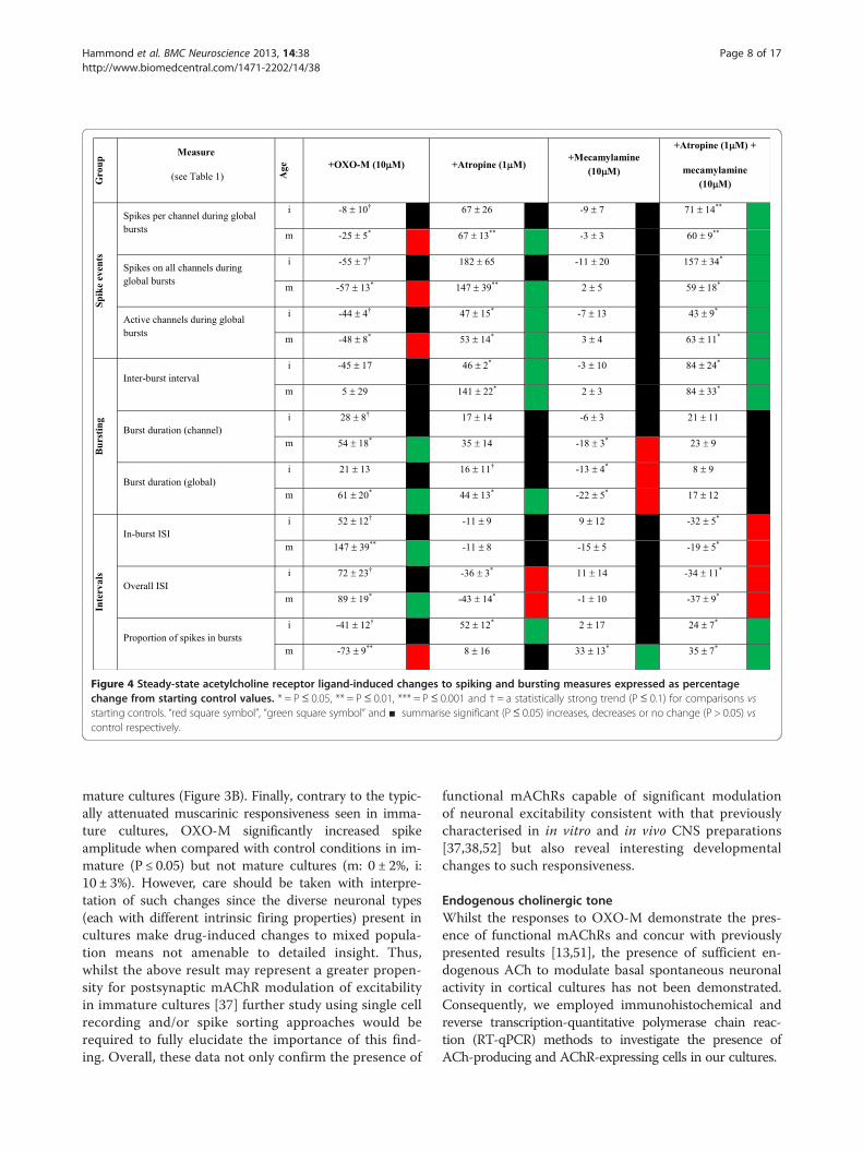
Figure 4 Steady-state acetylcholine receptor ligand-induced changes to spiking and bursting measures expressed as percentagechange from starting control values. * = P≤ 0.05, ** = P≤ 0.01, *** = P≤ 0.001 and † = a statistically strong trend (P≤ 0.1) for comparisons vsstarting controls. “red square symbol”, “green square symbol” and ■ summarise significant (P≤ 0.05) increases, decreases or no change (P > 0.05) vscontrol respectively.
Hammond et al. BMC Neuroscience 2013, 14:38 Page 8 of 17http://www.biomedcentral.com/1471-2202/14/38
mature cultures (Figure 3B). Finally, contrary to the typic-ally attenuated muscarinic responsiveness seen in imma-ture cultures, OXO-M significantly increased spikeamplitude when compared with control conditions in im-mature (P ≤ 0.05) but not mature cultures (m: 0 ± 2%, i:10 ± 3%). However, care should be taken with interpre-tation of such changes since the diverse neuronal types(each with different intrinsic firing properties) present incultures make drug-induced changes to mixed popula-tion means not amenable to detailed insight. Thus,whilst the above result may represent a greater propen-sity for postsynaptic mAChR modulation of excitabilityin immature cultures [37] further study using single cellrecording and/or spike sorting approaches would berequired to fully elucidate the importance of this find-ing. Overall, these data not only confirm the presence of
functional mAChRs capable of significant modulationof neuronal excitability consistent with that previouslycharacterised in in vitro and in vivo CNS preparations[37,38,52] but also reveal interesting developmentalchanges to such responsiveness.
Endogenous cholinergic toneWhilst the responses to OXO-M demonstrate the pres-ence of functional mAChRs and concur with previouslypresented results [13,51], the presence of sufficient en-dogenous ACh to modulate basal spontaneous neuronalactivity in cortical cultures has not been demonstrated.Consequently, we employed immunohistochemical andreverse transcription-quantitative polymerase chain reac-tion (RT-qPCR) methods to investigate the presence ofACh-producing and AChR-expressing cells in our cultures.

Hammond et al. BMC Neuroscience 2013, 14:38 Page 9 of 17http://www.biomedcentral.com/1471-2202/14/38
Morphologically diverse cells from both immature andmature cultures showed robust immunoreactivity for theTrkA antibody (Figure 5A) on the cell surface andwithin the cytosol (Figure 5Ai; ~95% of TrkA-expressingcells co-express choline acetyltransferase (ChAT) [45]).Cultures also expressed M1 and M2 mAChRs as well asα7-containing nAChRs (Figure 5B-F) where strong M1and M2 mAChR expression was found on somata sur-faces, apical dendrites and throughout the dendritic treefor a number of morphologically distinct cell types(Figure 5B-E). In contrast, α7 containing nAChRs werestrongly expressed at somata surfaces and the initialapical dendrite for a number of cellular morphologies(Figure 5Fi-iv) but showed only limited punctate expres-sion in dendritic trees and within the neuropil (Figure 5Eiv;arrows). RNA extracted from cultures contained bothTrkA and ChAT mRNA (Figure 5G) although no signifi-cant differences in expression levels were found betweenimmature and mature cultures (TrkA RT-PCR cycle timesm: 11.1 ± 1.1; i: 9.4 ± 0.5, ChAT RT-PCR cycle times m:12.3 ± 0.6; i: 12.4 ± 0.2; P > 0.05 for m vs i for TrkA andChAT). These results support the presence of both ACh-producing cells and differentially-expressed muscarinic
A
100 µm
D E
30 µm 30 µm
F.i
iii
20 µm
20 µm
B
Figure 5 Immunohistochemical and RT PCR studies confirm the preseα7-containing nAChRs and m2 mAChRs. Fluorescence image of TrkA-imß-tubulin (red) and DNA nuclear stain Hoechst 33342 (blue) in mature (A) athe cytosol (upper arrow) and on the surface of a different cell located at aCo-localisation of m2 mAChR (red) and m1 mAChRs (green) showing relati(arrows) and general expression throughout the neuropil in a mature cultu(green) demonstrating the high level of α7-containing nAChR expression bthroughout the dendritic tree (Fiv arrows) and labelled with beta tubulin incorresponding bands at 143 (TrkA) and 147 (ChAT) and 244 (ß-actin) in bot
and nicotinic AChRs within the neuronal population com-prising these cortical cultures.
mAChR-mediated effects of endogenous ACh uponspontaneous spike firing activityGiven that our immunohistochemical and RT-qPCR re-sults support the presence of intrinsic ACh-producingneurons in the cultures, we next investigated the effectsof the mAChR-selective antagonist atropine alone (1 μM)upon spike firing measures, to pharmacologically establishwhether endogenously-released ACh concentrations hadattained functionally relevant levels that could affect spon-taneous neuronal activity.In both immature (Figure 6A) and mature (Figure 6B)
cultures, atropine produced a clear change from the previ-ously described heterogenous (mixed tonic and bursting)spontaneous activity in control conditions to regularlyspaced, spontaneous burst events of uniform duration anda concurrent reduction of asynchronous tonic activity.Interestingly and like OXO-M, atropine significantly in-creased spike amplitude when compared with controlconditions in immature (P ≤ 0.05) but not mature cultures(m: 3 ± 4%, i: 50 ± 21%). For the previously noted reasons
TrK
A
-Actin
ChA
T
TrK
A
-Actin
ChA
T
(bp)
200
300
100
20 µm
Gii
iv
20 µm
20 µm
100 µm
C
Matureculture
Septal
nce of TrkA in mature and immature cortical cultures alongsidemunoreactivity (green; see arrows) co-stained with neuron-specificnd immature (B) cultures. (C) Confocal image of TrKA localised to bothlower level in the Z-stack (lower arrow) in a mature culture. (D&E)vely higher levels of m1 mAChR expression along the apical dendritere. (F i-iv) Confocal images of α7-containing nAChR positive cellsoth on the soma and apical dendrite but limited punctate expression(ii) in a mature culture. (G) Electrophoresis of RT PCR products showsh mature cultures and rat medial septum tissue positive control.
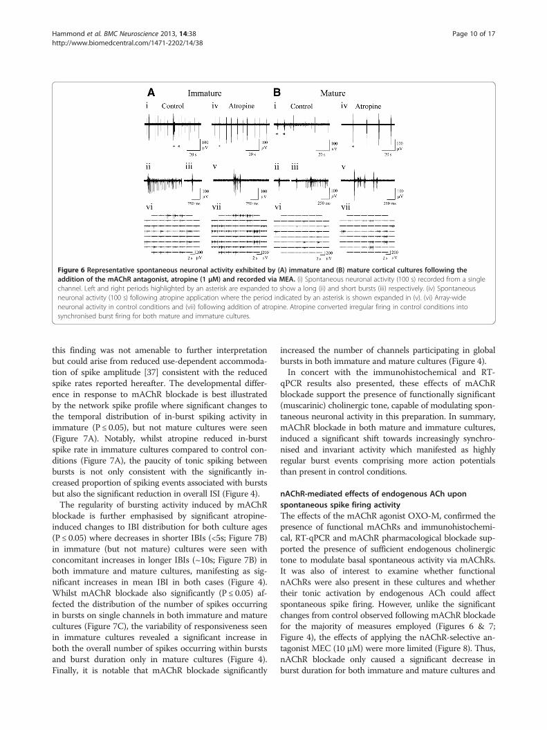
Figure 6 Representative spontaneous neuronal activity exhibited by (A) immature and (B) mature cortical cultures following theaddition of the mAChR antagonist, atropine (1 μM) and recorded via MEA. (i) Spontaneous neuronal activity (100 s) recorded from a singlechannel. Left and right periods highlighted by an asterisk are expanded to show a long (ii) and short bursts (iii) respectively. (iv) Spontaneousneuronal activity (100 s) following atropine application where the period indicated by an asterisk is shown expanded in (v). (vi) Array-wideneuronal activity in control conditions and (vii) following addition of atropine. Atropine converted irregular firing in control conditions intosynchronised burst firing for both mature and immature cultures.
Hammond et al. BMC Neuroscience 2013, 14:38 Page 10 of 17http://www.biomedcentral.com/1471-2202/14/38
this finding was not amenable to further interpretationbut could arise from reduced use-dependent accommoda-tion of spike amplitude [37] consistent with the reducedspike rates reported hereafter. The developmental differ-ence in response to mAChR blockade is best illustratedby the network spike profile where significant changes tothe temporal distribution of in-burst spiking activity inimmature (P ≤ 0.05), but not mature cultures were seen(Figure 7A). Notably, whilst atropine reduced in-burstspike rate in immature cultures compared to control con-ditions (Figure 7A), the paucity of tonic spiking betweenbursts is not only consistent with the significantly in-creased proportion of spiking events associated with burstsbut also the significant reduction in overall ISI (Figure 4).The regularity of bursting activity induced by mAChR
blockade is further emphasised by significant atropine-induced changes to IBI distribution for both culture ages(P ≤ 0.05) where decreases in shorter IBIs (<5s; Figure 7B)in immature (but not mature) cultures were seen withconcomitant increases in longer IBIs (~10s; Figure 7B) inboth immature and mature cultures, manifesting as sig-nificant increases in mean IBI in both cases (Figure 4).Whilst mAChR blockade also significantly (P ≤ 0.05) af-fected the distribution of the number of spikes occurringin bursts on single channels in both immature and maturecultures (Figure 7C), the variability of responsiveness seenin immature cultures revealed a significant increase inboth the overall number of spikes occurring within burstsand burst duration only in mature cultures (Figure 4).Finally, it is notable that mAChR blockade significantly
increased the number of channels participating in globalbursts in both immature and mature cultures (Figure 4).In concert with the immunohistochemical and RT-
qPCR results also presented, these effects of mAChRblockade support the presence of functionally significant(muscarinic) cholinergic tone, capable of modulating spon-taneous neuronal activity in this preparation. In summary,mAChR blockade in both mature and immature cultures,induced a significant shift towards increasingly synchro-nised and invariant activity which manifested as highlyregular burst events comprising more action potentialsthan present in control conditions.
nAChR-mediated effects of endogenous ACh uponspontaneous spike firing activityThe effects of the mAChR agonist OXO-M, confirmed thepresence of functional mAChRs and immunohistochemi-cal, RT-qPCR and mAChR pharmacological blockade sup-ported the presence of sufficient endogenous cholinergictone to modulate basal spontaneous activity via mAChRs.It was also of interest to examine whether functionalnAChRs were also present in these cultures and whethertheir tonic activation by endogenous ACh could affectspontaneous spike firing. However, unlike the significantchanges from control observed following mAChR blockadefor the majority of measures employed (Figures 6 & 7;Figure 4), the effects of applying the nAChR-selective an-tagonist MEC (10 μM) were more limited (Figure 8). Thus,nAChR blockade only caused a significant decrease inburst duration for both immature and mature cultures and

Figure 7 mAChR blockade by atropine (1 μM) modulates spontaneous neuronal activity in cortical cultures recorded via MEA.(A) Network spike profile (see Table 1) for immature (grey) and mature (black) cultures presented as normalised change from control followingmAChR blockade. (B) The effect of atropine upon the distribution of interburst intervals in immature (grey) and mature (black) cortical cultures.(C) The effect of atropine upon the number of spikes occurring on individual channels during bursting.
Hammond et al. BMC Neuroscience 2013, 14:38 Page 11 of 17http://www.biomedcentral.com/1471-2202/14/38
an increase in the proportion of spikes occurring in burstsfor mature cultures only (Figure 4). Unlike, OXO-M andatropine, MEC had no significant effect upon spike ampli-tude when compared with control conditions in either im-mature or mature cultures (m: 3 ± 2%, i: 1 ± 1%; P > 0.05 vscontrol in each case).
Spontaneous neuronal activity in the absence ofcholinergic modulationFinally, to assess total ‘tonic’ cholinergic influences onspontaneous firing behaviour, all cultures were also treatedwith a combination of atropine (1 μM) plus MEC (10 μM)in order to block activation of both mAChRs and nAChRs(Figure 9). The results obtained were very consistent in di-rection and extent with those seen following the blockadeof mAChRs alone (Figure 4; Figures 6 & 7) which is per-haps unsurprising given the limited effects caused bynAChR blockade alone (Figure 4 & Figure 8). The onlynotable differences in significant effects upon burst
measures following mAChR plus nAChR blockade thatwere not seen following mAChR blockade alone were anincreased proportion of spikes occurring within bursts inimmature cultures and a decrease in in-burst ISI for bothimmature and mature cultures, although the direction ofboth changes were consistent with those seen followingmAChR blockade alone (Figure 4).The network spike profile obtained following mAChR
plus nAChR blockade (Figure 10A) again revealed changesin both immature and mature cultures that were compa-rable to those seen mAChR blockade alone (c.f. Figure 7)although, notably, unlike the latter condition, significanteffects (P ≤ 0.05) upon the temporal distribution of spikefiring were only seen in immature cultures, most likelydue to the reduced variability of in-burst spike firing(Figure 7A vs Figure 10A).Consistent with the dominance of muscarinic effects
upon spike firing measures, mAChR plus nAChR block-ade significantly increased spike amplitude in immature

Figure 8 Representative spontaneous neuronal activity exhibited by (A) immature and (B) mature cortical cultures followingapplication of the nAChR antagonist, mecamylamine (10 μM; MEC). (i) Spontaneous neuronal activity (100 s) on a single channel where leftand right asterisks indicate typical short and long bursts that are expanded in (ii) and (iii) respectively. (iv) Spontaneous activity (100 s) followingmecamylamine application where the period indicated by an asterisk is shown expanded in (v). (vi) Array-wide neuronal activity in controlconditions and (vii) following mecamylamine application. Note the limited effect of nAChR blockade (c.f. mAChR blockade; Figures 6 & 7) suchthat only burst duration was significantly affected by mecamylamine (Figure 4).
Hammond et al. BMC Neuroscience 2013, 14:38 Page 12 of 17http://www.biomedcentral.com/1471-2202/14/38
(P ≤ 0.05) but not mature cultures to an extent compa-rable to that seen following mAChR blockade alone (m:3 ± 4%, i: 49 ± 21%).Overall, these findings suggest a dominance of mAChR-
mediated influences across the cellular population as awhole with nAChR-mediated modulation being less pre-valent and possibly circuit specific.
Basal ACh levels affect agonist responsivenessInterestingly, the consequences of applied mAChR agonisttreatment upon some burst measures appeared to be re-lated to the starting (control) amplitude of the measurefor a given trial (Figure 10B) such that two notable exam-ples of negative correlation were apparent in mature cul-tures. Cultures exhibiting lower starting values for a givenmeasure typically exhibited the most substantial changesfollowing mAChR activation, whilst those cultures withhigher starting values exhibited little change followingmAChR agonist application. This is suggestive of temporaland inter-culture variation in endogenous ACh-mediatedactivation of mAChRs that is not only consistent with in-tact tissue and/or in vivo ACh level fluctuations but alsocould be experimentally exploited. In addition, this rela-tionship was found exclusively in mature cultures andtherefore adds support to the previously described attenu-ated immature mAChR response.
DiscussionThis study has demonstrated, for the first time, the pres-ence of significant, culture age-dependent, endogenous
cholinergic tone in cortical cultures of embryonic originthat orchestrates a neuromodulatory role largely consis-tent with that reported in intact cortical networksin vitro [37,38,40,52-54] and in vivo [55]. This findingadds credence to the previously identified conservationof underlying cellular and network mechanisms in suchpreparations [1,56] despite the absence of behaviourallyrelevant, environmental and genetic regulation. In sum-mary, endogenous ACh activating mAChRs broadlymodulated excitability and consequently determinedspatiotemporal variability of activity whilst endogenousACh effects upon nAChRs manifested as a more re-stricted, subtle, but significant modulation of excitability.
mAChR-mediated responsivenessWhilst the spontaneous activity types exhibited by bothimmature and mature cultures in control conditions wereboth largely similar and consistent with previous reports[16,17,50], responsiveness developmentally diverged uponpharmacological modulation of cholinergic receptors.Agonist-induced mAChR activation with OXO-M, al-
tered control firing activity characterised primarily by glo-bal burst events interspersed with low levels of tonicactivity, to either persistent asynchronous firing coupledwith transient global firing frequency increases in maturecultures, or predominantly global bursting activity in im-mature cultures; these changes were consistent with ef-fects of the mAChR agonist, carbachol, on cortical/MEAcultures reported by Tateno et al. [13,51] and so import-antly, support commonalities between these preparations.
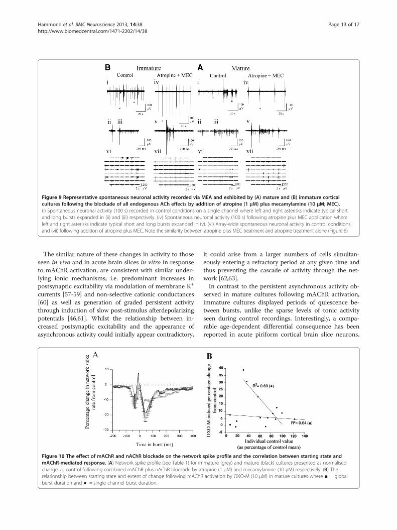
Figure 9 Representative spontaneous neuronal activity recorded via MEA and exhibited by (A) mature and (B) immature corticalcultures following the blockade of all endogenous ACh effects by addition of atropine (1 μM) plus mecamylamine (10 μM; MEC).(i) Spontaneous neuronal activity (100 s) recorded in control conditions on a single channel where left and right asterisks indicate typical shortand long bursts expanded in (ii) and (iii) respectively. (iv) Spontaneous neuronal activity (100 s) following atropine plus MEC application whereleft and right asterisks indicate typical short and long bursts expanded in (v). (vi) Array-wide spontaneous neuronal activity in control conditionsand (vii) following addition of atropine plus MEC. Note the similarity between atropine plus MEC treatment and atropine treatment alone (Figure 6).
Hammond et al. BMC Neuroscience 2013, 14:38 Page 13 of 17http://www.biomedcentral.com/1471-2202/14/38
The similar nature of these changes in activity to thoseseen in vivo and in acute brain slices in vitro in responseto mAChR activation, are consistent with similar under-lying ionic mechanisms; i.e. predominant increases inpostsynaptic excitability via modulation of membrane K+
currents [57-59] and non-selective cationic conductances[60] as well as generation of graded persistent activitythrough induction of slow post-stimulus afterdepolarizingpotentials [46,61]. Whilst the relationship between in-creased postsynaptic excitability and the appearance ofasynchronous activity could initially appear contradictory,
Figure 10 The effect of mAChR and nAChR blockade on the networkmAChR-mediated response. (A) Network spike profile (see Table 1) for imchange vs. control following combined mAChR plus nAChR blockade by atrelationship between starting state and extent of change following mAChRburst duration and ● = single channel burst duration.
it could arise from a larger numbers of cells simultan-eously entering a refractory period at any given time andthus preventing the cascade of activity through the net-work [62,63].In contrast to the persistent asynchronous activity ob-
served in mature cultures following mAChR activation,immature cultures displayed periods of quiescence be-tween bursts, unlike the sparse levels of tonic activityseen during control recordings. Interestingly, a compa-rable age-dependent differential consequence has beenreported in acute piriform cortical brain slice neurons,
0 -5
20 40 60 80 100 120 140
0
5
10
15
20
25
30
35
40
R2= 0.64
B
R2= 0.69 (
0 -5
20 40 60 80 100 120 140
0
5
10
15
20
25
30
35
40
R2= 0.64
B
R2= 0.69 (
spike profile and the correlation between starting state andmature (grey) and mature (black) cultures presented as normalisedropine (1 μM) and mecamylamine (10 μM) respectively. (B) Theactivation by OXO-M (10 μM) in mature cultures where ■ = global

Hammond et al. BMC Neuroscience 2013, 14:38 Page 14 of 17http://www.biomedcentral.com/1471-2202/14/38
where mAChR activation by OXO-M caused increasesin spontaneous tonic firing activity in adult (>40 dayspost-natal) slices but elicited spontaneous epileptiformactivity in immature (14-28 day post-natal) slices, char-acterised by clearly defined periods of high frequencybursting separated by periods of quiescence [37,40]. Al-though we detected both M1 and M2 mAChRs in ourcultures, it is not possible to assign pharmacological spe-cificity to the effects on firing in mature vs immatureneurons described here as this requires further investiga-tion using subtype selective mAChR antagonists.
mAChR-mediated influence of endogenous ACh uponspontaneous spike firing activityIt is most likely that the presence of significant endogenousACh release in cortical cultures has not previously beenconsidered due to the exclusion of tissue containing cholin-ergic cell bodies (e.g.medial septum) during the preparationof cortical/forebrain tissue for culture. However, severalstudies support mechanisms from which physiologically sig-nificant levels of neuromodulatory transmitters can arise incortical cultures which include environmentally-dependentprogenitor differentiation into neuromodulatory pheno-types [64,65] and the presence of intrinsic cholinergic cellsand synapses within rat cerebral cortex itself [66]. Here, weconfirmed the presence of ChAT and TrkA mRNA and thedistribution of TrkA- expressing cells [45] along with thepresence of M1 and M2 mAChRs and α7 nAChRs.Blockade of the effects of mAChR-mediated endogen-
ous cholinergic tone by atropine significantly increasedexcitability within global bursts and their regularity,suggesting that high tonic ACh levels could underliethe observed variability within the temporal structuresof both burst and tonic spontaneous neuronal activity(Figures 3 vs 6). We thus propose that the regularity ofspontaneous neuronal activity exhibited by cultures inthe absence of mAChR-mediated contributions could beattributed to comparable mechanisms as those in vivo,although tempered by the highly connected nature ofcultures. In addition, increased synchronous neuronalactivation following mAChR blockade could arise fromthe removal of tonic mAChR-mediated presynaptic in-hibition of excitatory neurotransmitter release [40,67].Accordingly, such presynaptic disinhibition would resultin the entraining of the majority of units during a globalburst and so concomitantly synchronise the refractoryperiods of involved units which could underlie the re-duced variability in burst timing [55,68]. It is notablethat a previous investigation of mAChR agonist effectsupon cortical cultures [13,51] reported that atropine(10 μM) application in the continued presence of themAChR agonist muscarine (20 μM) inconsistently abol-ished spontaneous activity, although the application ofatropine alone did not. This is in contrast to the results
presented here where the effects of mAChR agonist plusantagonist and the application of antagonist alone didnot significantly differ. It is uncertain whether this differ-ence between studies is a result of differences betweenthe muscarinic agonists used or the high concentrationof atropine they applied, exerting non-specific effects(10 μM; [69]). Alternatively, the variability in responsive-ness reported by Tateno et al. [13,51] could suggest apotentially high degree of variability in presence or levelof endogenous cholinergic ‘tone’ under the conditionsemployed.
nAChR-mediated responsivenessThe significant decreases observed in both channel andglobal burst durations, coupled with a concomitant re-duction in the number of spikes occurring outside burstevents in the presence of the nAChR antagonist MEC,suggests a predominantly excitatory role for nAChRsin these preparations. The more limited influence ofnAChRs, in comparison to the more extensive mAChRnetwork influence, suggests that the effects of nAChRactivation may be restricted to a subset of neural circuitsas found in vivo [70]. However, the combination of net-work mapping using high spatial resolution recordingtechniques such as calcium imaging, or high resolutionMEAs, would be required to confirm this.Finally, the distribution of α7-containing nAChRs
appeared relatively limited throughout the dendritic treerelative to the ultrastructural location of nAChRs in vivo[71]. However, as nAChRs are preferentially expressed onneurons processing afferent sensory information in vivo[72,73], the differences seen in culture may be a productof the lack of afferent sensory input [74] and would makea comparison of nAChR levels and distribution followingartificially introduced re-afferentation [75] of considerableinterest. It is also worth noting that acetylcholinesterase(AChE) is expressed in primary cortical cultures [76,77]; acomparison of the effects of atropine and MEC describedabove, with those of an AChE inhibitor such as neostig-mine would also be worthwhile (c.f. [38]).
Implications for the cortical cell culture model and theiruse in animat paradigmsAs previously described, cultures exhibiting mixed (tonicand bursting) activity would be more representative of ac-tivity observed in intact cortex and therefore provide amore suitable substrate not only for conventional studies(see Background) but especially as a driver for neuralanimat paradigms. The results presented here suggest thatthe cholinergic system is an attractive target for such animprovement in addition to providing opportunities to in-fluence mAChR- and/or nAChR-mediated effects uponsynaptic plasticity [78-81]; a core feature in such studiesthat has thus far been unreliable in this preparation type

Hammond et al. BMC Neuroscience 2013, 14:38 Page 15 of 17http://www.biomedcentral.com/1471-2202/14/38
[14]. Moreover, if endogenous ACh levels are indeed tem-porally variable (Figure 9B) as our results suggest, suchintrinsic variation also presents an attractive feature for ex-ploitation by animat implementations since periods of ‘at-tention’ (during training stimulation) and ‘consolidation’(after training stimulation) are necessary and so could beconducted during periods of ‘high’ and ‘low’ cholinergictone respectively.
ConclusionsIn conclusion, we propose that the presence of an intactcholinergic system which exerts tonic effects upon neur-onal activity in this preparation make it well suited toexploitation in animat and related paradigms by offeringa physiologically relevant target for controlling neuronalexcitability, modifying information flow, increasing physio-logical relevance and improving experimental reproducibil-ity [56,82]. In summary, the expression of endogenouscholinergic tone and age-dependent developmental changesin cholinergic responsiveness in cortical cultures supportthe use of such cultures as physiologically useful models forthe study of neural-robotic interfaces and other more con-ventional applications, and should not only be consideredbut actively exploited and further explored.
AbbreviationsACh: Acetylcholine; AChE: Acetylcholinesterase; ChAT: Cholineacetyltransferase; CNS: Central nervous system; DIV: Days in vitro;DNA: Deoxyribonucleic acid; dNTPS: Deoxynucleoside triphosphates;EMEM: Eagle’s minimum essential medium; i: immature; LTP: Long termpotentiation; m: Mature; mAChR: Muscarinic acetycholine receptor;mGluR: Metabotropic glutamate receptor; MCPG: (S)-α-methyl-4-carboxyphenylglycine; MEA: Multi-electrode array; MEC: Mecamylamine(nicotine hydrogen tartrate salt); nAChR: Nicotinic acetylcholine receptor;OXO-M: Oxotremorine methiodide; PBS: Phosphate buffered saline;RNA: Ribonucleic acid; RT-qPCR: Reverse transcription quantitativepolymerase chain reaction; sADP: Slow after-depolarizing potential;TrkA: Tyrosine kinase A.See also Table 1 for the list of abbreviations used to denote electrophysiologicalmeasures employed and their definitions.
Competing interestsThe authors declare they have no competing interests.
Authors’ contributionsAll experiments were performed in the laboratory of Dr B. J. Whalley,University of Reading, UK. Experimental conception, design and analysis wereperformed by Dr M. Hammond and Dr B. J. Whalley. Dr M. Hammond, Dr B.J. Whalley and Dr A. Constanti prepared the manuscript.Immunocytochemical staining and imaging were conducted by Dr M.Hammond and Dr G. Bucci. The manuscript was appraised, revised andapproved by Prof. K. Warwick, Dr S. J. Nasuto, Dr V. Becerra, Dr G. Bucci, Dr D.Xydas and Dr J. Downes. All authors read and approved the final manuscript.
AcknowledgementsThis work was funded by the Engineering and Physical Sciences ResearchCouncil (EP/D080134/1). The authors thank Prof. P. Knight, Dr M. A. N. Rattrayand Dr A. Spilsbury for invaluable assistance with immunocytochemical andRT-qPCR aspects of the work and Dr G. J. Stephens for valuable contributionsto the interpretation of our findings. The tyrosine kinase primary antibodywas the kind gift of Prof. L. Reichardt, University of California.
Author details1School of Chemistry, Food and Nutritional Sciences and Pharmacy,University of Reading, Whiteknights, Reading, Berkshire RG6 6AP, UK. 2Schoolof Systems Engineering, University of Reading, Whiteknights, Reading,Berkshire RG6 6AP, UK. 3UCL School of Pharmacy, 29-39 Brunswick Square,London WC1N 1AX, UK.
Received: 18 June 2012 Accepted: 8 March 2013Published: 26 March 2013
References1. Stichel CC, Müller HW: Dissociated cell culture of rat cerebral cortical
neurons in serum-free, conditioned media: GABA-immunopositiveneurons. Developmental Brain Research 1991, 64:145–154.
2. Hirano T, Kasano K: Spatial distribution of excitatory and inhibitorysynapses on a Purkinje cell in a rat cerebellar culture. J Neurophysiol1993, 70:1316.
3. Lin YC, Huang ZH, Jan I: Development of excitatory synapses in culturedneurons dissociated from the cortices of rat embryos and rat pups atbirth. J Neurosci Res 2002, 67:484–493.
4. Valor LM, Charlesworth P, Humphreys L, Anderson CNG, Grant SGN:Network activity-independent coordinated gene expression program forsynapse assembly. PNAS 2007, 104:4658–4663.
5. Chiappalone M, Bove M, Vato A, Tedesco M, Martinoia S: Dissociatedcortical networks show spontaneously correlated activity patterns duringin vitro development. Brain Res 2006, 1093(1):41–53.
6. Xiang G, Pan L, Huang L, Yu Z, Song X, Cheng J, Xing W, Zhou Y:Microelectrode array-based system for neuropharmacologicalapplications with cortical neurons cultured in vitro. Biosens Bioelectron2007, 22(11):2478–2484.
7. Choi DW, Koh JY, Peter S: Pharmacology of glutamate neurotoxicity incortical cell culture: attenuation by NMDA antagonists. J Neurosci1988, 8:185.
8. Hogberg HT, Sobanski T, Novellino A, Whelan M, Weiss DG, Bal-Price AK:Application of micro-electrode arrays (MEAs) as an emerging technologyfor developmental neurotoxicity: evaluation of domoic acid-inducedeffects in primary cultures of rat cortical neurons. Neurotoxicology 2011,32(1):158–168.
9. Kim MJ, Oh SJ, Park SH, Kang HJ, Won MH, Kang TC, Park JB, Kim JI, Kim J,Lee JY: Neuronal loss in primary long-term cortical culture involvesneurodegeneration-like cell death via calpain and p35 processing, butnot developmental apoptosis or aging. Exp Mol Med 2007, 39:14.
10. Jimbo Y, Tateno T, Robinson H: Simultaneous induction ofpathway-specific potentiation and depression in networks ofcortical neurons. Biophys J 1999, 76:670–678.
11. Marom S, Shahaf G: Development, learning and memory in large randomnetworks of cortical neurons: lessons beyond anatomy. Q Rev Biophys2002, 35:63–87.
12. Eytan D, Brenner N, Marom S: Selective adaptation in networks of corticalneurons. J Neurosci 2003, 23(28):9349–9356.
13. Tateno T, Jimbo Y, Robinson HPC: Spatio-temporal cholinergic modulationin cultured networks of rat cortical neurons: spontaneous activity.Neuroscience 2005, 134:425–437.
14. Wagenaar DA, Pine J, Potter SM: Searching for plasticity in dissociatedcortical cultures on multi-electrode arrays. J Negat Results Biomed 2006, 5:16.
15. Chiappalone M, Massobrio P, Martinoia S: Network plasticity in corticalassemblies. Eur J Neurosci 2008, 28(1):221–237.
16. Van Pelt J, Wolters PS, Corner MA, Rutten WL, Ramakers GJ: Long-termcharacterization of firing dynamics of spontaneous bursts in culturedneural networks. IEEE Trans Biomed Eng 2004, 51:2051–2062.
17. Wagenaar DA, Pine J, Potter SM: An extremely rich repertoire of burstingpatterns during the development of cortical cultures. BMC Neurosci 2006, 7:11.
18. Prince DA: Neurophysiology of epilepsy. Annu Rev Neurosci 1978, 1:395–415.19. Ben-Ari Y: Developing networks play a similar melody. Trends Neurosci
2001, 24:353–360.20. Verzeano M: Activity of cerebral neurons in the transition from
wakefulness to sleep. Science 1956, 124:366–367.21. Quiroga Q, Nadasdy Z, Ben-Shaul Y: Unsupervised spike detection and
sorting with wavelets and superparamagnetic clustering. Neural Comput2004, 16(8):1661–1687.

Hammond et al. BMC Neuroscience 2013, 14:38 Page 16 of 17http://www.biomedcentral.com/1471-2202/14/38
22. Gullo F, Maffezzoli A, Dossi E, Wanke E: Short-latency cross-andautocorrelation identify clusters of interacting cortical neurons recordedfrom multi-electrode array. J Neurosci Methods 2009, 181:186–198.
23. DeMarse TB: The neurally controlled animat: biological brains acting withsimulated bodies. Auton Robots 2001, 11:305–310.
24. Bakkum DJ, Shkolnik AC, Ben-Ary G, Gamblen P, DeMarse TB, Potter SM:Removing Some 'A' from AI: Embodied Cultured Networks. In proceedingof: Ad-Hoc, Mobile, and Wireless Networks, Second International Conference.Montreal, Canada: ADHOC-NOW 2003; 2003. Proceedings.
25. Chao ZC, Bakkum DJ, Potter SM: Shaping embodied neural networks foradaptive goal-directed behavior. PLoS Comput Biol 2008, 4(3):e1000042.
26. Martinoia S, Sanguineti V, Cozzi L, Berdondini L, Van Pelt J, Tomas J, LeMasson G, Davide F: Towards an embodied in vitro electrophysiology: theNeuroBIT project. Neurocomputing 2004, 58:1065–1072.
27. Cozzi L, D’Angelo P, Chiappalone M, Ide A, Novellino A, Martinoia S,Sanguineti V: Coding and decoding of information in a bi-directionalneural interface. Neurocomputing 2005, 65:783–792.
28. Hayashi I, Taguchi T, Kudoh SN, Takatsuki J, Ikeda J: Learning and memory inliving neuronal networks connected to moving robot International Symposiumon Advanced Intelligent Systems. Korea: Sokcho; 2007:79–81.
29. Xydas D, Norcott DJ, Warwick K, Whalley BJ, Nasuto SJ, Becerra VM,Hammond MW, Downes J, Marshall S: Architecture for neuronal cellcontrol of a mobile robot. Springer Tracts in Advanced Robotics2008, 44:23–31.
30. Pizzi R, Rossetti D, Cino G, Marino D: A cultured human neural networkoperates a robotic actuator. Biosystems 2009, 95:137–144.
31. Bakkum DJ, Chao ZC, Gamblen P, Ben-Ary G, Shkolnik AG, DeMarse TB,Potter SM: Embodying cultured networks with a robotic drawing arm.Conf Proc IEEE Eng Med Biol Soc 2007, 2007:2996–2999.
32. Furshpan E, Potter D: Seizure-like activity and cellular damage in rathippocampal neurons in cell culture. Neuron 1989, 3:199–207.
33. Herrero MT, Oset-Gasque MJ, Cañadas S, Vicente S, González MP: Effect ofvarious depolarizing agents on endogenous amino acid neurotransmitterrelease in rat cortical neurons in culture. Neurochem Int 1998,32(3):257–264.
34. Mangan PS, Kapur J: Factors underlying bursting behavior in a network ofcultured hippocampal neurons exposed to zero magnesium.J Neurophysiol 2004, 91(2):946–957.
35. Corner MA, Baker RE, van Pelt J: Homeostatically regulated spontaneousneuronal discharges protect developing cerebral cortex networks frombecoming hyperactive following prolonged blockade of excitatorysynaptic receptors. Brain Res 2006, 1106(1):40–45.
36. Jones NA, Glyn SE, Akiyama S, Hill TD, Hill AJ, Weston SE, Burnett MD,Yamasaki Y, Stephens GJ, Whalley BJ, Williams CM: Cannabidiol exertsanti-convulsant effects in animal models of temporal lobe and partialseizures. Seizure 2012, 21(5):344–352.
37. Whalley BJ, Postlethwaite M, Constanti A: Further characterization ofmuscarinic agonist-induced epileptiform bursting activity in immaturerat piriform cortex, in vitro. Neuroscience 2005, 134:549–566.
38. Aitchison E, Weston SE, Constanti A, Whalley BJ: Anticholinesterase-inducedepileptiform activity in immature rat piriform cortex slices, in vitro. NeurosciLett 2010, 473(3):252–256.
39. Turski L, Ikonomidou C, Turski WA, Bortolotto ZA, Cavalheiro EA: Review:cholinergic mechanisms and epileptogenesis. The seizures induced bypilocarpine: a novel experimental model of intractable epilepsy. Synapse1989, 3:154–171.
40. Whalley BJ, Constanti A: Developmental changes in presynapticmuscarinic modulation of excitatory and inhibitory neurotransmission inrat piriform cortex in vitro: Relevance to epileptiform burstingsusceptibility. Neuroscience 2006, 140:939–956.
41. Hasselmo ME: Neuromodulation and cortical function: modeling thephysiological basis of behavior. Behav Brain Res 1995, 67:1–27.
42. Barik J, Wonnacott S: Molecular and cellular mechanisms of action ofnicotine in the CNS. Handb Exp Pharmacol 2009, 192:173–207.
43. Potter SM, DeMarse TB: A new approach to neural cell culture forlong-term studies. J Neurosci Methods 2001, 110:17–24.
44. Wagenaar DA, DeMarse TB, Potter SM: MeaBench: a toolset formulti-electrode data acquisition and on-line analysis NeuralEngineering, 2005. 2nd International IEEE EMBS 2005:518–521.
45. Sobreviela T, Clary DO, Reichardt LF, Brandabur MM, Kordower JH, Mufson EJ:TrkA-immunoreactive profiles in the central nervous system: colocalization
with neurons containing p75 nerve growth factor receptor, cholineacetyltransferase, and serotonin. J Comp Neurol 1994, 350(4):587–611.
46. Constanti A, Bagetta G, Libri V: Persistent muscarinic excitation in guinea-pigolfactory cortex neurons: involvement of a slow post-stimulusafterdepolarizing current. Neuroscience 1993, 56(4):887–904.
47. Libri V, Constanti A, Zibetti M, Postlethwaite M: Metabotropic glutamatereceptor subtypes mediating slow inward tail current (IADP) inductionand inhibition of synaptic transmission in olfactory cortical neurones.Br J Pharmacol 1997, 120(6):1083–1095.
48. Chiappalone M, Novellino A, Vajda I, Vato A, Martinoia S, van Pelt J: Burstdetection algorithms for the analysis of spatio-temporal patterns incortical networks of neurons. Neurocomputing 2005, 65–66:653–662.
49. Xydas D, Warwick K, Nasuto SJ, Downes J, Spencer M, Hammond M, BecerraV, Whalley BJ: Revealing ensemble state transition patterns in multi-electrode neuronal recordings using hidden Markov models. IEEE TransNeural Syst Rehabil Eng 2011, 19(4):345–355.
50. Tateno T, Jimbo Y: Activity-dependent enhancement in the reliability ofcorrelated spike timings in cultured cortical neurons. Biol Cybern 1999,80:45–55.
51. Tateno T, Jimbo Y, Robinson HPC: Spatio-temporal cholinergic modulationin cultured networks of rat cortical neurons: evoked activity. Neuroscience2005, 134:439–448.
52. McCormick DA, Prince DA: Mechanisms of action of acetylcholine in theguinea-pig cerebral cortex in vitro. J Physiol 1986, 375:169–194.
53. Barkai E, Bergman RE, Horwitz G, Hasselmo ME: Modulation of associativememory function in a biophysical simulation of rat piriform cortex.J Neurophysiol 1994, 72:659–677.
54. Klink R, Alonso A: Muscarinic modulation of the oscillatory and repetitivefiring properties of entorhinal cortex layer II Neurons. J Neurophysiol 1997,77:1813–1828.
55. McCormick DA, Contreras D: On the cellular and network bases ofepileptic seizures. Ann Rev Physiol 2001, 63:815–846.
56. Ayali A, Fuchs E, Zilberstein Y, Robinson A, Shefi O, Hulata E, Baruchi I,Ben-Jacob E: Contextual regularity and complexity of neuronal activity:from stand-alone cultures to task-performing animals. Complexity 2004,9:25–32.
57. Constanti A, Sim JA: Calcium-dependent potassium conductance inguinea-pig olfactory cortex neurones in vitro. J Physiol 1987, 387:173–194.
58. Madison DV, Lancaster B, Nicoll RA: Voltage clamp analysis of cholinergicaction in the hippocampus. J Neurosci 1987, 7:733–741.
59. Womble MD, Moises HC: Muscarinic inhibition of M-current and apotassium leak conductance in neurones of the rat basolateralamygdala. J Physiol 1992, 457:93–114.
60. Haj-Dahmane S, Andrade R: Muscarinic activation of a voltage-dependentcation nonselective current in rat association cortex. J Neurosci 1996,16(12):3848–3861.
61. Major G, Tank D: Persistent neural activity: prevalence and mechanisms.Curr Opin Neurobiol 2004, 14:675–684.
62. Latham PE, Richmond BJ, Nelson PG, Nirenberg S: Intrinsic dynamics inneuronal networks. I. Theory. J Neurophysiol 2000, 83:808–827.
63. Latham PE, Richmond BJ, Nirenberg S, Nelson PG: Intrinsic dynamics inneuronal networks. II. Experiment. J Neurophysiol 2000, 2000(83):828–835.
64. Zhou J, Bradford HF, Stern GM: The stimulatory effect of brain-derivedneurotrophic factor on dopaminergic phenotype expression ofembryonic rat cortical neurons in vitro. Dev Brain Res 1994, 81:318–324.
65. Markakis EA, Palmer TD, Randolph-Moore L, Rakic P, Gage FH: Novelneuronal phenotypes from neural progenitor cells. J Neurosci 2004,24(12):2886–2897.
66. Houser C, Crawford G, Salvaterra P, Vaughn J: Immunocytochemicallocalization of choline acetyltransferase in rat cerebral cortex: a study ofcholinergic neurons and synapses. J Comparative Neurology 1985, 234:17–34.
67. Sheridan RD, Sutor B: Presynaptic M1 muscarinic cholinoceptors mediateinhibition of excitatory synaptic transmission in the hippocampusin vitro. Neurosci Lett 1990, 108:273–278.
68. de Sevilla DF, Garduno J, Galvan E, Buno W: Calcium-activatedafterhyperpolarizations regulate synchronization and timing ofepileptiform bursts in hippocampal CA3 pyramidal neurons.J Neurophysiol 2006, 96:3028–3041.
69. Behling RW, Yamane T, Navon G, Sammon MJ, Jelinski L: Measuring relativeacetylcholine receptor agonist binding by selective proton nuclearmagnetic resonance relaxation experiments. Biophys J 1988, 53:947–954.

Hammond et al. BMC Neuroscience 2013, 14:38 Page 17 of 17http://www.biomedcentral.com/1471-2202/14/38
70. Kimura F: Cholinergic modulation of cortical function: a hypothetical rolein shifting the dynamics in cortical network. Neurosci Res 2000, 38:19–26.
71. Fabian-Fine R, Skehel P, Errington ML, Davies HA, Sher E, Stewart MG, FineA: Ultrastructural distribution of the {alpha}7 nicotinic acetylcholinereceptor subunit in rat hippocampus. J Neurosci 2001, 21:7993–8003.
72. Broide R, Robertson R, Leslie F: Regulation of alpha α7 nicotinicacetylcholine receptors in the developing rat somatosensory cortex bythalamocortical afferents. J Neurosci 1996, 16:2956.
73. Khan IM, Wennerholm M, Singletary E, Polston K, Zhang L, Deerinck T, YakshTL, Taylor P: Ablation of primary afferent terminals reduces nicotinicreceptor expression and the nociceptive responses to nicotinic agonistsin the spinal cord. J Neurocytology 2004, 33:543–556.
74. Aramakis VB, Metherate R: Nicotine selectively enhances NMDAreceptor-mediated synaptic transmission during postnataldevelopment in sensory neocortex. J Neurosci 1998, 18:8485–8495.
75. Brewer GJ, Boehler MD, Ide AN, Wheeler BC: Chronic electrical stimulationof cultured hippocampal networks increases spontaneous spike rates.J Neurosci Methods 2009, 184:104–109.
76. Sawyer TW, Weiss MT: Parallel development of acetylcholinesterasein vivo and in primary neuron surface culture. Brain Res Dev Brain Res1993, 71(1):147–149.
77. Fodero LR, Mok SS, Losic D, Martin LL, Aguilar MI, Barrow CJ, Livett BG,Small DH: Alpha7-nicotinic acetylcholine receptors mediate an Abeta(1-42)-induced increase in the level of acetylcholinesterase in primarycortical neurones. J Neurochem 2004, 88(5):1186–1193.
78. Calabresi P, Centonze D, Gubellini P, Bernardi G: Activation of M1-likemuscarinic receptors is required for the induction of corticostriatal LTP.Neuropharmacology 1999, 38:323–326.
79. Shinoe T, Matsui M, Taketo M, Manabe T: Modulation of synaptic plasticityby physiological activation of M1 muscarinic acetylcholine receptors inthe mouse hippocampus. J Neurosci 2005, 25:11194–11200.
80. Luo L, Chen WH, Wang M, Zhu DM, She JQ, Ruan DY: Modulation oflong-term potentiation by individual subtypes of muscarinicacetylcholine receptor in the rat dentate gyrus. Hippocampus 2008,18:989–995.
81. Rosato-Siri M, Cattaneo A, Cherubini E: Nicotine-induced enhancement ofsynaptic plasticity at CA3 and CA1 synapses requires GABAergicinterneurons in adult anti-NGF mice. J Physiol 2006, 576:361–377.
82. Fuchs E, Ayali A, Robinson A, Hulata E, Ben-Jacob E: Coemergence ofregularity and complexity during neural network development.Dev Neurobiol 2007, 67:1802–1814.
doi:10.1186/1471-2202-14-38Cite this article as: Hammond et al.: Endogenous cholinergic tonemodulates spontaneous network level neuronal activity in primarycortical cultures grown on multi-electrode arrays. BMC Neuroscience 201314:38.
Submit your next manuscript to BioMed Centraland take full advantage of:
• Convenient online submission
• Thorough peer review
• No space constraints or color figure charges
• Immediate publication on acceptance
• Inclusion in PubMed, CAS, Scopus and Google Scholar
• Research which is freely available for redistribution
Submit your manuscript at www.biomedcentral.com/submit