research article sequence similarity-driven proteomics in
TRANSCRIPT

RESEARCH ARTICLE
Sequence similarity-driven proteomics in organisms
with unknown genomes by LC-MS/MS and automated
de novo sequencing
Patrice Waridel1*, Ari Frank2, Henrik Thomas1, Vineeth Surendranath1, Shamil Sunyaev3,Pavel Pevzner2 and Andrej Shevchenko1
1 Max Planck Institute of Molecular Cell Biology and Genetics, Dresden, Germany2 Department of Computer Science and Engineering, University of California, San Diego, CA, USA3 Birgham & Women’s Hospital and Harvard Medical School, Boston, MA, USA
LC-MS/MS analysis on a linear ion trap LTQ mass spectrometer, combined with data processing,stringent, and sequence-similarity database searching tools, was employed in a layered mannerto identify proteins in organisms with unsequenced genomes. Highly specific stringent searches(MASCOT) were applied as a first layer screen to identify either known (i.e. present in a database)proteins, or unknown proteins sharing identical peptides with related database sequences. Oncethe confidently matched spectra were removed, the remainder was filtered against a non-annotated library of background spectra that cleaned up the dataset from spectra of commonprotein and chemical contaminants. The rectified spectral dataset was further subjected to rapidbatch de novo interpretation by PepNovo software, followed by the MS BLASTsequence-similaritysearch that used multiple redundant and partially accurate candidate peptide sequences. Impor-tantly, a single dataset was acquired at the uncompromised sensitivity with no need of manualselection of MS/MS spectra for subsequent de novo interpretation. This approach enabled acompletely automated identification of novel proteins that were, otherwise, missed by conven-tional database searches.
Received: January 2, 2007Revised: March 19, 2007
Accepted: April 2, 2007
Keywords:
de novo sequencing / LC-MS/MS / MS BLAST / organisms with unknown genomes /sequence-similarity searches
2318 Proteomics 2007, 7, 2318–2329
1 Introduction
In bottom-up proteomics, proteins are digested in-solutionor in-gel with proteolytic enzymes and recovered peptidesfragmented in nano-ESI MS/MS, -LC-MS/MS, or -LC-MALDI experiments (reviewed in ref. [1–5]). Uninterpreted
tandem mass spectra are subsequently searched againstprotein, or translated DNA, databases using dedicated soft-ware (reviewed in ref. [6–8]). Regardless of the employed al-gorithm, the software identifies peptide sequences (and,hence, corresponding database entries [9]) by comparingpeaks in MS/MS spectra with m/z (or m/z and intensities) ofpeptide fragments, which have been precomputed fromdatabase sequences, assuming some enzyme cleavage speci-ficity and peptide fragmentation models [10–13]. While thisis an efficient approach towards the characterization ofknown proteins (i.e., proteins, whose sequences are accu-
Correspondence: Dr. Andrej Shevchenko, Max Planck Institute ofMolecular Cell Biology and Genetics, Pfotenhauerstrasse 108,01307 Dresden, GermanyE-mail: [email protected]: 149-351-210-2000
Abbreviations: IT, ion trap; mgf, MASCOT generic format; MS
BLAST, MS driven BLAST; XIC, extracted ion current* Current address: Protein Analysis Facility, University of Lau-
sanne, 1015 Lausanne, Switzerland
DOI 10.1002/pmic.200700003
© 2007 WILEY-VCH Verlag GmbH & Co. KGaA, Weinheim www.proteomics-journal.com

Proteomics 2007, 7, 2318–2329 Technology 2319
rately represented in a database resource), the identificationof proteins unrepresented in a database (further termed asunknown proteins) remains a challenging problem(reviewed in ref. [14, 15]). Conventional database searchescould, in principle, recognize peptides that are identical bothin the unknown protein and known homologous protein(s)from closely related species [16]. This, however, is not effi-cient for identifying proteins that either originate from spe-cies phylogenetically distant from corresponding referenceorganisms, or belong to poorly conserved protein families, orwere collected from species with an undefined genetic back-ground and manifest strong sequence polymorphism (seeref. [17–21] for representative case studies).
Sequence-similarity search is a powerful tool for theidentification of proteins from organisms with unsequencedgenomes [22–27]. Full peptide sequences, or short sequencestretches (also termed sequence tags) [28], could be deduceddirectly from MS/MS spectra with no recourse to databaseresources (reviewed in ref. [29]). Multiple peptide sequences(or sequence tags) are then searched against a database in anerror-tolerant fashion and, in this way, even proteins havingonly marginal sequence similarity to reference databaseentries could be identified [23, 26, 27].
Dedicated sequence-similarity search engines, such asCIDentify [30], an MS tailored version of gapped BLAST [25],an MS driven BLAST (MS BLAST) [22], FASTS [24], MS-Homology [31], OpenSea [32], among others, have been suc-cessfully applied in various proteomics studies. They, how-ever, most efficiently handle queries composed of a relativelysmall number (typically, 5–50) of peptide sequences typicallydeduced from the spectra of “hand-picked” precursors,which is nowhere close to the total number of informativeMS/MS spectra produced by LC-MS/MS under data-depend-ent acquisition control [33, 34]. Thus, the entire approachusually encompasses a selection of relatively abundant pep-tides that often originate from most conserved and well-characterized protein families and, therefore, are readilyidentifiable by conventional means.
Furthermore, usually only a small fraction of acquiredspectra matches target proteins, while the rest of the spectrais attributed to chemical [35] or peptide [36, 37] backgrounds.Computational methods have been developed to recognizebackground spectra by comparison with a reference libraryor with spectra from a blank LC-MS/MS run [38, 39]. Whiledue to their high specificity, conventional database searcheslargely tolerate peptide background, it strongly impairssequence-similarity identifications [40]. Hence, rapid data-dependent acquisition of MS/MS spectra has dramaticallyincreased the success rate of conventional protein identifica-tion, since it has become possible to sequence many morepeptides in a single LC-MS/MS run. However, because ofexactly the same reason, it undermined the performance ofsequence-similarity searches.
Here, we present an approach for the seamless integra-tion of automated de novo sequencing and LC-MS/MS into aprotein characterization pipeline. A single dataset of tandem
mass spectra was acquired at the uncompromised sensitivity,and then a combination of database searching and spectraprocessing software tools was employed for its in-depthanalysis. In a few case studies based on LC-MS/MS analyses,each producing ca. 6000 of low resolution linear ion trap (IT)tandem mass spectra, we demonstrated how conventional(stringent) searches by MASCOT [41], de novo interpretationof spectra by PepNovo [42], and sequence-similarity searchesby MS BLAST [16, 22], bundled by simple data handlingscripts, enabled the comprehensive interpretation of datasetsacquired from digests of unknown proteins.
2 Materials and methods
2.1 Chemicals
Cleland’s reagent (DTT) was obtained from Merck (Darm-stadt, Germany) and other chemicals from Sigma-Aldrich(Munich, Germany). Modified porcine trypsin (Trypsin Goldgrade) was purchased from Promega (Mannheim, Ger-many). HPLC solvents (Lichrosolv grade), formic acid andTFA were purchased from Merck.
2.2 Protein preparations
Protein samples were obtained in on-going characterizationof the proteome of Dunaliella salina, a halotolerant unicellulargreen alga [20] (Katz, A., Waridel, P., Shevchenko, A., Pick, U.,Mol. Cell. Proteomics 2007, in press). Protein spots, visualizedby CBB R250 staining, were excised from Blue Native gels [43]and in-gel digested with trypsin as described previously [44].
2.3 Analysis by LC-MS/MS
Dried peptide extracts were redissolved in 15–25 mL of 0.05%v/v TFA, depending on the staining abundance of the proteinspots, and 4 mL was loaded using a FAMOS autosampler on anano-LC-MS/MS Ultimate system (Dionex, Amstersdam,The Netherlands) interfaced on-line to a linear IT LTQ massspectrometer (Thermo Fisher Scientific, San Jose, CA). Themobile phase was composed of 95:5 H2O:ACN v/v with 0.1%formic acid (solvent A) and 20:80 H2O:ACN v/v with 0.1%formic acid (solvent B). Peptides were first loaded onto atrapping microcolumn C18 PepMAP100 (1 mm6300 mmid, 5 mm, LC Packings) in 0.05% TFA at a flow rate of 20 mL/min. After 4 min, they were back-flush eluted and separatedon a nanocolumn C18 PepMAP100 (15 cm675 mm id,3 mm, LC Packings) at a flow rate of 200 nL/min in themobile phase gradient: from 5 to 20% of solvent B in 20 min,20–50% B in 16 min, 50–100% B in 5 min, 100% B during10 min, and back to 5% B in 5 min; % B refers to the solventB content in an A 1 B mixture.
Peptides were infused into the mass spectrometer via adynamic nanospray probe (Thermo Fisher Scientific) andanalyzed in positive mode. Uncoated needles Silicatip,
© 2007 WILEY-VCH Verlag GmbH & Co. KGaA, Weinheim www.proteomics-journal.com

2320 P. Waridel et al. Proteomics 2007, 7, 2318–2329
20 mm id, 10 mm tip id (New Objective, Woburn, MA) wereused with a spray voltage of 1.8 kV, and the capillary transfertemperature was set to 2007C. In a typical data dependentacquisition cycle controlled by Xcalibur 1.4 software (ThermoFisher Scientific), the four most abundant precursor ionsdetected in the full MS survey scan (m/z range of 350–1500)were isolated within a 4.0 amu window and fragmented. MS/MS fragmentation was triggered by a minimum signalthreshold of 500 counts and carried out at the normalizedcollision energy of 35%. Spectra were acquired under auto-matic gain control (AGC) in one microscan for survey spectraand in three microscans for MS/MS spectra with a maximalion injection time of 100 ms per each microscan. M/z of thefragmented precursors were then dynamically excluded foranother 60 s. No precompiled exclusion lists were applied.
From raw files, MS/MS spectra were exported as dta (textformat) files using BioWorks 3.1 software (Thermo FisherScientific) under the following settings: peptide mass range,500–3500; minimal total ion intensity threshold, 1000;minimal number of fragment ions, 15; precursor mass tol-erance, 1.4 amu; group scan, 1; minimum group count, 1.
We note that BioWorks 3.1 software named each dta fileaccording to the original name of the raw file, the scannumber and the assumed charge of the precursor ion. If thelow mass resolution in the normal scan mode did not allowdetermination of the precursor charge state, then redundantdta files were created that only differed by the assumed pre-cursor charge.
2.4 Database searches
Dta files were merged into a single MASCOT generic format(mgf) file and searched against a MSDB database (updatedMay 15, 2005, containing 2 011 572 entries), or a D. salinaEST database (downloaded from NCBI, updated March 16,2006, containing 3998 entries) by MASCOT v. 2.1 software(Matrix Science, London, UK) installed on a local 2 CPUserver. Tolerance for precursor and fragment masses was 2.0and 0.5 Da, respectively; instrument profile: ESI-Trap; fixedmodification: carbamidomethyl (cysteine); variable mod-ification: oxidation (methionine).
2.5 Filtering MS/MS spectra
Prior to batch de novo sequencing of individual spectra, mgffiles were filtered to remove spectra originating from com-mon peptide and nonpeptide backgrounds. Briefly, theapplied filtering routine comprised three main components:a similarity measure between MS/MS spectra, a search algo-rithm and a statistical framework to identify significantmatches between the compared spectra. The similaritymeasure was a normalized Pearson correlation [45] of theintensities corresponding to fragment ions with matchingm/z. Given the precompiled background library (see below)and the assumed similarity measure, the statistical frame-work was based on a well studied “extreme value problem”
[45]. Accordingly, a measure of the statistical confidence of amatch between a sample spectrum and the correspondingspectrum from the background library, was a double expo-nential function fit to data obtained in a simulation experi-ment by way of considering the distribution of similarityscores for ca 2000 nonbackground dta files obtained fromhigh quality spectra of Saccharomyces cerevisiae peptides. Thefiltering software was implemented in the Python program-ming language (Ubuntu 5.10 Linux Operating System,Python 2.3) and is available from the authors upon request.
During the filtering process, all spectra from singlycharged precursors and “void” spectra (typically containingless than ten fragment ions, whose relative intensities wereabove 3% of the base peak intensity) were removed. Theremaining spectra were screened against a backgroundlibrary containing, in total, 13 000 MS/MS spectra that werepooled together from several LC-MS/MS analyses of in-geldigests of blank gel pieces. Additionally, more than 800spectra of common trypsin and keratin peptides, identifiedby MASCOT or MS BLAST searches, were handpicked fromvarious LC-MS/MS runs and added to the library. To limit thelibrary redundancy, each new spectrum was first screenedagainst the current library and added only if no matchingspectrum was recognized. For each sample spectrum, thefiltering software at first identified, in the background library,the spectra that were acquired from precursors with thesame (within 2.5 amu tolerance) mass. Then, each pair ofspectra was independently examined by comparing both m/z(within 0.5 amu tolerance) and intensities of their fragmentions. The quality of each match was scored and the scorescompared to the threshold expected for randomly matchingspectra. Spectra having statistically significant similarities tocorresponding library spectra were removed, along with theirredundant variants that assumed alternative charge states ofthe same precursor. Note that the described procedure doesnot rely on any assumed identity of spectra, preliminaryknowledge of their origin, or database sequence resources.
2.6 De novo sequencing and sequence-similarity
searches
A basic version of de novo sequencing software PepNovo [42]was modified to produce several, maximally complete, albeitredundant, degenerate, and partially inaccurate sequencecandidates per each interpreted spectrum. An MS BLASTcompatible version of PepNovo is available on the UCSDComputational MS Research Group server at http://peptide.ucsd.edu. PepNovo estimated the de novo interpretation con-fidence by assigning a sequence quality score, which reflec-ted the expected number of correct amino acids in the topsequence candidate. All sequences, whose scores exceeded auser-defined threshold (typically, the value of 5.0) [46], weremerged into a single query string and submitted to an MSBLAST (MS driven BLAST) search against nr database(nrdb95) at the web-accessible server (http://genetics.bwh.harvard.edu/msblast/).
© 2007 WILEY-VCH Verlag GmbH & Co. KGaA, Weinheim www.proteomics-journal.com
Proteomics 2007, 7, 2318–2329 Technology 2321
2.7 Confidence of database searching hits
In MASCOT searches against an MSDB database, proteinhits were considered confident if database entries werematched with at least two peptides and a total peptide ionsscore of 100, while all individual peptides with scores below20 were disregarded. Because of the small size of the D. sal-ina EST database, we accepted as confident hits identified byat least one peptide whose ions score was above 40. Thethreshold ions scores suggested by MASCOT for confidentsingle peptide identifications in MSDB and EST databases,were 53 and 31 (p,0.05), respectively. Nonetheless, all sin-gle-peptide hits (regardless of their peptide ion scores, even ifthey exceeded the threshold of 53), and hits matched by a fewlower scoring peptides were considered borderline and in-dependently validated as described in ref. [46]. In sequence-similarity searches, the statistical significance of hits wasevaluated according to MS BLAST scoring scheme [16].However, only HSPs with a score of 62 or above were con-sidered. Borderline MASCOT hits independently validatedby PepNovo/MS BLAST [46] were also accepted.
3 Results and discussion
3.1 The workflow
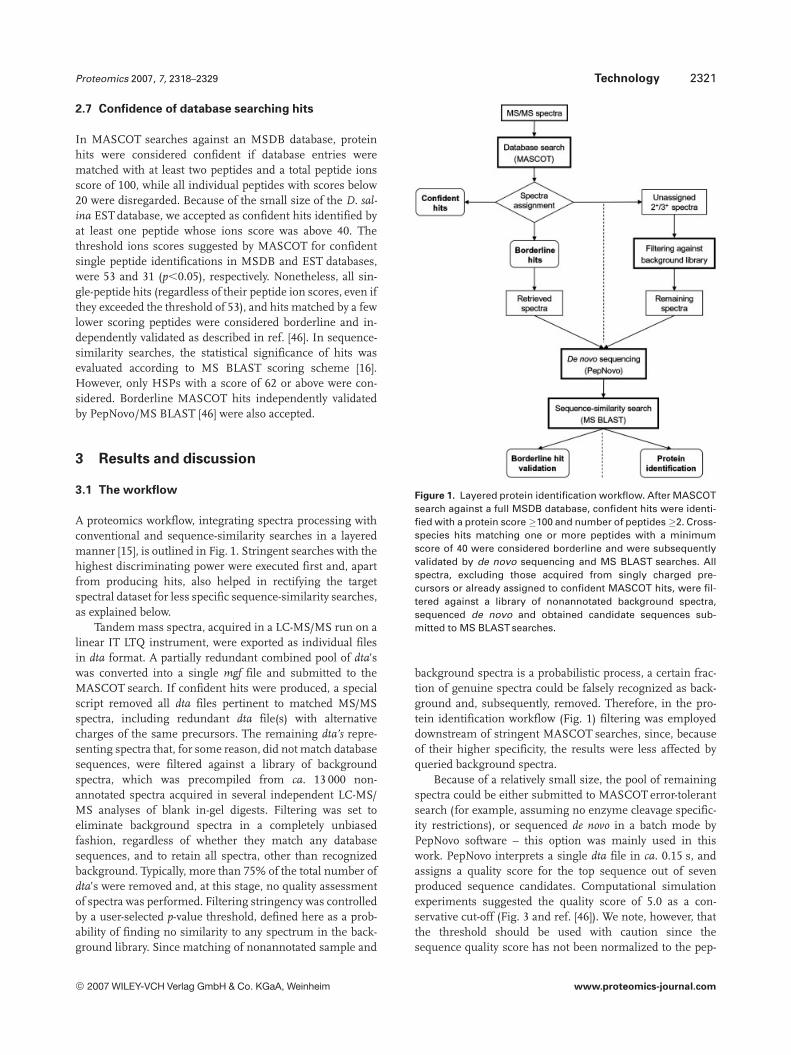
A proteomics workflow, integrating spectra processing withconventional and sequence-similarity searches in a layeredmanner [15], is outlined in Fig. 1. Stringent searches with thehighest discriminating power were executed first and, apartfrom producing hits, also helped in rectifying the targetspectral dataset for less specific sequence-similarity searches,as explained below.
Tandem mass spectra, acquired in a LC-MS/MS run on alinear IT LTQ instrument, were exported as individual filesin dta format. A partially redundant combined pool of dta’swas converted into a single mgf file and submitted to theMASCOT search. If confident hits were produced, a specialscript removed all dta files pertinent to matched MS/MSspectra, including redundant dta file(s) with alternativecharges of the same precursors. The remaining dta’s repre-senting spectra that, for some reason, did not match databasesequences, were filtered against a library of backgroundspectra, which was precompiled from ca. 13 000 non-annotated spectra acquired in several independent LC-MS/MS analyses of blank in-gel digests. Filtering was set toeliminate background spectra in a completely unbiasedfashion, regardless of whether they match any databasesequences, and to retain all spectra, other than recognizedbackground. Typically, more than 75% of the total number ofdta’s were removed and, at this stage, no quality assessmentof spectra was performed. Filtering stringency was controlledby a user-selected p-value threshold, defined here as a prob-ability of finding no similarity to any spectrum in the back-ground library. Since matching of nonannotated sample and
Figure 1. Layered protein identification workflow. After MASCOTsearch against a full MSDB database, confident hits were identi-fied with a protein score �100 and number of peptides �2. Cross-species hits matching one or more peptides with a minimumscore of 40 were considered borderline and were subsequentlyvalidated by de novo sequencing and MS BLAST searches. Allspectra, excluding those acquired from singly charged pre-cursors or already assigned to confident MASCOT hits, were fil-tered against a library of nonannotated background spectra,sequenced de novo and obtained candidate sequences sub-mitted to MS BLAST searches.
background spectra is a probabilistic process, a certain frac-tion of genuine spectra could be falsely recognized as back-ground and, subsequently, removed. Therefore, in the pro-tein identification workflow (Fig. 1) filtering was employeddownstream of stringent MASCOT searches, since, becauseof their higher specificity, the results were less affected byqueried background spectra.
Because of a relatively small size, the pool of remainingspectra could be either submitted to MASCOT error-tolerantsearch (for example, assuming no enzyme cleavage specific-ity restrictions), or sequenced de novo in a batch mode byPepNovo software – this option was mainly used in thiswork. PepNovo interprets a single dta file in ca. 0.15 s, andassigns a quality score for the top sequence out of sevenproduced sequence candidates. Computational simulationexperiments suggested the quality score of 5.0 as a con-servative cut-off (Fig. 3 and ref. [46]). We note, however, thatthe threshold should be used with caution since thesequence quality score has not been normalized to the pep-
© 2007 WILEY-VCH Verlag GmbH & Co. KGaA, Weinheim www.proteomics-journal.com

2322 P. Waridel et al. Proteomics 2007, 7, 2318–2329
tide length and, aiming at a priori high scores would dis-criminate short, yet accurately sequenced peptides. Insuffi-cient quality of MS/MS spectra is not the only reason for lowscoring interpretations. PepNovo was optimized for se-quencing doubly charged precursor ions and is less efficientin sequencing triply charged precursors. Therefore, PepNovosequence quality scores could not substitute spectra qualityestimates developed for conventional stringent searches [47,48] since they are biased towards interpretable (rather thanabundant) patterns of fragment ions. All sequence candi-dates selected from all interpreted MS/MS spectra weremerged into a single query string and submitted for MSBLAST sequence-similarity search. Typically, these searcheseither revealed new proteins, or detected new peptides fromproteins already identified by MASCOT.
In parallel, PepNovo and MS BLAST were also used forvalidating borderline hits produced by MASCOT searches asdescribed previously [46, 49]. Briefly, if de novo sequencing bythe MS BLASTsearch independently hit the same peptide, aswas previously found by the MASCOT search, it was con-sidered as positively identified.
3.2 Seamless integration of de novo sequencing with
MS BLAST searches
An efficient and fast de novo sequencing program tailored forinterpreting low-resolution MS/MS spectra is a key element ofthe proposed workflow. PepNovo software was particularlysuccessful in interpreting ITspectra and scored favorably whencompared to other de novo sequencing programs [42, 50].Importantly, PepNovo interprets a single MS/MS spectrum inca. 0.15 s and, in batch mode, operates with spectra in mgf for-mat, which simplifies the processing of LC-MS/MS data. Inthis work, we used a new version of PepNovo, which was tai-lored for interpreting MS/MS spectra obtained on linear ITinstruments and whose output format conforms to MS BLASTconventions [16, 40] (Fig. 2).
Next, we asked if the parallel consideration of multiple,partially redundant candidate sequences produced by PepNovo interpretation of the same MS/MS spectrum increasedthe success rate of MS BLAST identifications, compared tononerror-tolerant searches with only the top candidatesequence – a frequently used method of cross-species proteinidentification. To this end, a simulation dataset was compiledfrom 71 high quality MS/MS spectra. Each of them unequi-vocally hit a single peptide sequence of, on average, 14 aminoacid residues upon a MASCOT database search with the ionsscore above the value of 63. In each spectrum, peaks withrelative intensities below 1% of the base peak intensity weredeclared noise and their absolute intensity left unchanged,whereas the absolute intensity of other fragment peaks wasgradually reduced [46]. All unmodified and computationallymanipulated spectra were sequenced de novo. Up to sevencandidate sequences, along with the corresponding PepNovoquality score, were registered for each interpreted spectrum.The entire range of quality scores was divided into five bins
(Fig. 3) and spectra, interpreted with the correspondingscores, were picked for each score bin. The full dataset usedfor the simulation comprised 71 unmodified spectra andtheir 131 computationally altered “clones”. Note that we used“spoiled clones” of initially high quality spectra, rather thansome native low scoring spectra since, for each sequencedspectrum, we should precisely know the “source” peptidesequence to judge if its de novo interpretation was correct.Within each score bin, we determined fractions of inter-preted spectra, in which (i) the top candidate sequence wascomplete and correct; (ii) one of the listed candidate sequen-ces was complete and correct; and (iii) candidate sequence(s)contained a noninterrupted stretch of at least eight accurateamino acid residues (here termed as a tag). Finally, we com-bined all candidate sequences produced from a given spec-trum into MS BLAST query string, searched it against acomprehensive database and checked if the correct “source”peptide sequence was confidently hit. The distribution offractions of de novo interpreted spectra were plotted sepa-rately for each quality score bin (Fig. 3).
Figure 3 suggests that considering more candidatesequences and their error-tolerant matching to databaseentries improved the success rate of protein identification,although, regardless of the used method of sequence match-ing, it was below 20% for low scoring candidate sequences.Interestingly, even highly scored top sequences were seldomfully accurate. Since quality scores were not normalized tothe full peptide length (see the discussion above), higherscores were expected for larger peptides, although theirspectra are usually complex and (in case of IT) are affected bylow m/z cut-off. Therefore, their full interpretation is seldomcompletely accurate and it is not surprising that a smallerfraction of these spectra also produced fully accurate tags. Atthe same time, a few miscalled amino acid residues within alargely accurate long peptide sequence did not affect theidentification performance of MS BLAST.
We note that sequences with high quality scores might alsobe obtained from low abundant peptide precursors or fromspectra with abundant chemical noise. The TIC and TIC frac-tion reported by PepNovo for each interpreted spectrum couldbe used, with some caution, either for preselecting the mostrepresentative precursors or for postsearch validation ofassignments based on sequence alignments of the borderlineconfidence. Note that reported TIC does not directly reflect theprecursor abundance, since the spectrum might not be taken atthe apex of the chromatographic peak. There is, nevertheless,some correlation between spectrum TIC and precursor inten-sity, since, because of statistical sampling, a more abundantprecursors would more likely produce spectra with higher TIC.
3.3 Unbiased filtering of background MS/MS spectra
is essential for sequence-similarity identifications
Removing background spectra prior to batch de novo se-quencing is important for the successful identification of
© 2007 WILEY-VCH Verlag GmbH & Co. KGaA, Weinheim www.proteomics-journal.com

Proteomics 2007, 7, 2318–2329 Technology 2323
Figure 2. An example of the output of batch de novo interpretation of MS/MS spectra by PepNovo. For each interpreted spectrum, Pep-Novo reported: intact mass of the fragmented peptide (column 1); name of the dta file, including the scan number and assumed charge(column 2); TIC of the MS/MS spectrum (sum of fragment absolute intensities) (column 3); TIC fraction covered by expected fragments ofthe top candidate sequence (column 4); sequence quality score, representing the expected number of correct amino acids in the top (first)candidate sequence (column 5); candidate sequences (typically, limited to seven) (column 6). Note that F and M (oxidized) residues, as wellas Q and K, are isobaric and are not distinguishable in low-resolution MS/MS spectra. In these instances, optional sequences were includedinto the query. All candidate sequences obtained by interpreting all submitted MS/MS spectra (column 6) with the quality score (column 5)that typically exceeded the value of 5.0 were merged into a single MS BLAST query string and searched against the nr database. MS BLASTweb interface disregards all numbers and nonconventional symbols and, therefore, the entire output (or only the selected sequences) canbe directly pasted into the query window.
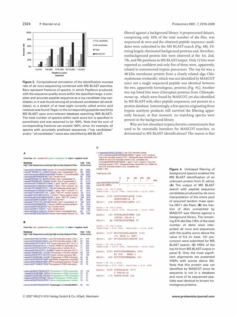
proteins by sequence-similarity searches at the high sensi-tivity. The characterization of a Coomassie stained spot withan apparent molecular weight of 55 kDa, which containedmembrane proteins from D. salina, is presented here as arepresentative example. The spot was excised from a 2-DBlue Native gel, in-gel digested with trypsin and the recov-ered peptides sequenced by LC-MS/MS. First, all MS/MSspectra were interpreted de novo and the entire pool of can-didate sequences was submitted to an MS BLAST search(Fig. 4A). The search produced a multitude of formally con-fident alignments with typical background proteins, mainlykeratins, trypsins, and serine proteases from a variety ofspecies. In the output, the three top nonbackground proteinswere only listed at 137th, 190th, and 233rd positions.
Enlarging the query with sequences, apparently unre-lated to target proteins, does not directly affect the scores ofreported HSPs. This, however, increases the significancethreshold scores [16] and, hence, indirectly impairs the con-fidence of hits with only a few aligned HSPs. Note, that ker-atin sequences are rich in low complexity regions that arealso common in many proteins in a database. Therefore,sequence-similarity searches with keratin sequences pro-duce a large number of formally confident hits that are, atfirst glance, apparently unrelated to keratins.
The same pool of spectra was then processed accordingto the workflow shown in Fig. 1. Upon the MASCOT search,spectra from matching peptides (including peptides fromkeratins and trypsin) were removed and the remainder was
© 2007 WILEY-VCH Verlag GmbH & Co. KGaA, Weinheim www.proteomics-journal.com
2324 P. Waridel et al. Proteomics 2007, 7, 2318–2329
Figure 3. Computational simulation of the identification successrate of de novo sequencing combined with MS BLAST searches.Bars represent fractions of spectra, in which PepNovo produced,with the sequence quality score within the specified range, a com-plete and accurate peptide sequence as a top candidate (top can-didate); or it was found among all produced candidates (all candi-dates); or a stretch of at least eight correctly called amino acidresidues was found (Tags); or thecorresponding peptide washit byMS BLAST upon error-tolerant database searching (MS BLAST).The total number of spectra within each score bin is specified inparenthesis and was assumed to be 100%. Note that the sum ofcorresponding fractions can exceed 100%, since, for example, allspectra with accurately predicted sequences (“top candidates”and/or “all candidates”) were also identified by MS BLAST.
filtered against a background library. A preprocessed dataset,comprising only 16% of the total number of dta files, wassequenced de novo and the obtained peptide sequence candi-dates were submitted to the MS BLAST search (Fig. 4B). Fil-tering largely eliminated background proteins and, therefore,nonbackground protein hits were observed at the 1st, 2nd,7th, and 9th positions in MS BLASToutput. Only 12 hits werereported as confident and only five of them were, apparently,related to nonremoved trypsin precursors. The top hit was a48 kDa membrane protein from a closely related alga Chla-mydomonas reinhardtii, which was not identified by MASCOTsince not a single sequenced peptide was identical betweenthe two, apparently homologous, proteins (Fig. 4C). Anothertwo top listed hits were chloroplast proteins from Chlamydo-monas sp., which were found by MASCOT, yet also identifiedby MS BLAST with other peptide sequences, not present in aprotein database. Interestingly, a few spectra originating fromtrypsin autolysis products still survived the filtering appar-ently because, at that moment, no matching spectra werepresent in the background library.
Why are low abundant trypsin/keratin contaminants thatused to be essentially harmless for MASCOT searches, sodetrimental to MS BLAST identifications? The reason is that
Figure 4. Unbiased filtering ofbackground spectra enabled theMS BLAST identification of anunknown protein from D. salina.(A) The output of MS BLASTsearch with peptide sequencecandidates produced by de novointerpretation of the entire poolof acquired tandem mass spec-tra (5511 dta files); (B) the frac-tion of dta’s unmatched byMASCOT was filtered against abackground library. The remain-ing 874 dta files (16% of the totalnumber of dta’s) were inter-preted de novo and sequenceswith the quality score above thevalue of 5.0 (in total, 131 pre-cursors) were submitted for MSBLAST search; (C) HSPs of thetop hit from MS BLASToutput inpanel B. Only the most signifi-cant alignments are presented(HSPs with scores above 55).Note that this protein was notidentified by MASCOT since itssequence is not in a databaseand none of its sequenced pep-tides was identical to known ho-mologous proteins.
© 2007 WILEY-VCH Verlag GmbH & Co. KGaA, Weinheim www.proteomics-journal.com

Proteomics 2007, 7, 2318–2329 Technology 2325
de novo interpretation of MS/MS spectra produces several (inthis work we considered up to seven) partially redundantsequence candidates, essentially “cloning” the samesequence into multiple copies. MS BLAST engine treatsthem equally and tries to match them to different sequencesegments of the same protein, or different proteins in adatabase. Since keratins are rich in low complexity sequencestretches, similarity searches with their multiple, partiallydifferent, variant sequences trigger an avalanche of hits, lar-gely based on similarly looking HSPs. This, however, doesnot happen in MASCOT searches because a much higherstringency of the match is required and any deviation fromthe expected fragment patterns heavily penalized.
As shown in Fig. 4B, stringent (MASCOT) searches wereunable to completely remove peptide spectra matchingbackground proteins. In contrast, the identification-inde-pendent filtering, that is based on the rapid comparison ofuninterpreted fragment ion patterns between the examinedand library spectra, recognized and removed backgroundspectra, regardless of whether they matched anything in adatabase. The representative examples included, but werenot limited to, orifice fragmentation products, products ofunspecific enzyme cleavage or polymorphic sequences.Continuous updating the library by adding newly recognizedpeptide spectra of trypsin, keratins, and other common (or,on user’s request, experiment-specific) contaminants even-tually should eliminate background spectra (almost) com-pletely from any probed query. We note, however, that thenumber of falsely retained background spectra also dependson the user-defined filtering stringency. Under stringent fil-tering settings, less background spectra would be retained,albeit with a higher chance of losing bona fide spectra fromtarget proteins due to their random similarity to background.
3.4 Validation of MASCOT cross-species
identifications with borderline statistical
confidence
Conventional proteomics methodologies are capable ofcross-species identification of unknown proteins by match-ing identical peptides in known homologous proteins. How-ever, such peptides are relatively rare and their identificationtypically relies on matching only a few peptide sequences,often with borderline statistical confidence. Here, wedemonstrate how de novo sequencing and MS BLASTsearches provided independent validation of borderlinecross-species MASCOT hits.
In the above sample of D. salina proteins, the MASCOTsearch identified a plausible homologue of the ATP synthasefrom another alga, Bigelowiella natans. However, this identi-fication relied upon a single exactly matching peptide(Fig. 5A) and, in line with current proteomics guidelines [51],it should be considered as borderline. To validate this hit, thedta file corresponding to the matched spectrum was theninterpreted de novo (Fig. 5B) and partially redundant candi-date sequences submitted to the MS BLAST search (Fig. 5C),
which produced a statistically confident hit to the over-lapping sequence stretch in a related database entry. We notethat peptide sequences of the MASCOT hit and de novo can-didates differed at their N-termini and, currently, it is notpossible to judge which peptide sequence was correct, sincethe full sequence of D. salina protein remains unknown.This, however, did not affect the confidence of MS BLAST hitassignment, which relies upon an independent scoringscheme that only considers the local similarity of sequencestretches aligned within the HSP.
3.5 The characterization of a complex mixture of
unknown proteins by a layered proteomics
workflow
The workflow (Fig. 1) was applied to the complete analysis ofa 55 kDa spot excised from a 2-D Blue Native gel separatingD. salina membrane proteins (Table 1). Out of the ten pro-teins identified in this sample, five were identified by MAS-COTsearches. Two were known D. salina proteins (proteins 1and 2 in Table 1) and another three were identified by statis-tically confident cross-species matches to multiple identicalpeptide sequences in proteins from related algal species(proteins 3, 4, and 5). Three more identifications were statis-tically borderline (proteins 6, 7, 8, see also Fig. 5) and weresubsequently validated by a combination of PepNovo se-quencing and MS BLAST searches, as described above.When all dta’s corresponding to matching peptides wereremoved and the rest was submitted to de novo sequencingfollowed by the MS BLAST search, we identified two moreproteins (proteins 9 and 10), which were missed by theMASCOT search. Importantly, the examination of the corre-sponding extracted ion current (XIC) traces in LC-MS/MSprofiles suggested that, in fact, protein 9 could be the majorcomponent of the mixture. Additionally, MS BLAST searchesalso revealed several new peptides from proteins alreadyidentified by MASCOT (proteins 3 and 4) thus improving thesequence coverage and confidence of cross-species identifi-cations.
This analysis is representative of a series of 32 samplesfrom D. salina, isolated by Blue Native gel electrophoresis(Table 2) and suggested the high relevance of the multi-layerdatamining strategy, compared to previously reported iden-tifications based on de novo sequencing by nano-ESI MS/MS[20]. Stringent (MASCOT) and sequence-similarity searchesproved to be complementary identification tools and the lat-ter increased the number of confident hits by more than15%, including several major protein components in ana-lyzed samples.
Out of a total of 75 proteins identified in 32 spots, 55proteins were unique (Table 2). Eight proteins (15%) wereidentified by matching to known D. salina sequences. Con-fident cross-species identifications by MASCOTaccounted foralmost a half of all identified proteins (25 hits, 45%), matchingsequences from algae (13 proteins), plants (3 proteins),
© 2007 WILEY-VCH Verlag GmbH & Co. KGaA, Weinheim www.proteomics-journal.com

2326 P. Waridel et al. Proteomics 2007, 7, 2318–2329
Figure 5. De novo sequencingand an MS BLAST search vali-dated a borderline cross-specieshit produced by the MASCOTsearch. (A) A MASCOT searchproduced a candidate hit withone matched peptide to the pro-tein from the related organism(B) The dta file corresponding tothe spectrum in panel A wasretrieved by a script and its denovo interpretation producedtwo candidate sequences withthe quality score of 11.1; (C)
They were merged into an MSBLAST query and the search hitthe same peptide from the relat-ed alga B. natans. According tothe MS BLAST scoring scheme,the hit was confident albeit theN-terminal piece of thesequence, for some reason,mismatched. Note that the N-terminal stretch of amino acidresidues in all three variant se-quences is isobaric and that thetrue sequence of the fragmentedpeptide is currently unknown.
bacteria (8 proteins) and fungi (1 protein). Sequence-simi-larity searches with MS BLAST added a substantial numberof novel identifications (6 hits, 11%) by matching corre-spondent protein homologues from algae (3 proteins), plants(1 protein) and bacteria (2 proteins). Importantly, the exam-ination of peptide signal intensities in XIC traces showedthat two of them were, most likely, the major components ofthe samples. Finally, MASCOT searches against an ESTdatabase containing about 4000 D. salina sequences morethan doubled the number of intra-species identifications (al-though relatively short EST sequences do not provide thefunctional annotation of hits directly). Additionally, validat-
ing MASCOT identifications by MS BLAST rescued one,otherwise a false negative, borderline hit and unequivocallyrejected another 11 borderline hits.
These results demonstrate the efficiency of the workflowthat combines stringent and sequence-similarity searches forthe proteomic characterization of organisms with unse-quenced genomes. The relative contribution of com-plementary database interrogation strategies depends on thespecies of origin, particularly on the number of sequencesavailable in protein or EST databases, and the phylogeneticdistance between the studied species and reference organ-ism(s), completely or partially covered by genomic sequenc-
© 2007 WILEY-VCH Verlag GmbH & Co. KGaA, Weinheim www.proteomics-journal.com

Proteomics 2007, 7, 2318–2329 Technology 2327
Table 1. Proteins identified in a single spot on a 2-D Blue Native gel separating D. salina membrane proteins
Hitnumber
Protein Species MW(kDa)
MASCOTscore(peptides)
MSBLASTscore(HSPs,score.55)
HighestXIC ofmatchedpeptides
Comments
1 High affinity nitrate transporter D. salina 59 450 (10) 3E105 Known D. salinaproteins2 Transferrin-like protein Ttf-1 D. salina 140 137 (4) 1E105
3 Photosystem II reaction centersCP47 apoprotein
C. reinhardtii 56 297 (6) 94(1) 6E105 Cross-speciesMASCOT hits
4 Photosystem II chlorophylla-binding protein psbC
Chlamydomonaseugametos
50 215 (5) 120(1) 5E105
5 P700 chlorophyll a-apoprotein A2 Chlamydomonasmoewusii
82 149 (3) 7E104
6 Putative plastidic ATP/ADP-transporter (fragment)
Protothecawickerhamii
24 72 (1) 80(1) 3E105 Borderline hitsvalidated byMS BLAST7 ATP synthase gamma subunit B. natans 42 68 (1) 64(1) 2E105
8 Probable multispanningmembrane protein
Arabidopsisthaliana
74 44 (1) 67 (1) 8E104
9a Low-CO2 inducible protein LCICa C. reinhardtii 48 – 578(5) 2E106 New proteinsidentified byMS BLAST
10 UDP-N-acetylglucosamine-N-acetylmuramyl-(penta-peptide)pyrophosphoryl-undecaprenol N-acetyl-glucosamine transferase
Geobactersulfur-reducens
40 – 75(1) 9E104
a) The most abundant protein (as judged by corresponding XIC traces) is shown in bold.
Table 2. Overview of protein identifications by MASCOT andPepNovo–MS BLAST in the preparations of D. salinamembrane proteins separated into 32 individual spotsby Blue Native gel electrophoresis
Search Species Database IdentificationsHits (%)
MASCOT D. salina MSDB 8 15EST 15 27
Other MSDB 25 45
MASCOT/MSBLASTa
MSDB/nrdb95
1 2
MS BLAST nrdb95 6 11
Total 55 100
a) Borderline hits from MASCOT searches validated by de novosequencing and MS BLAST.
ing [16]. It is important, however, that complementary data-base searches operate with the same dataset of MS/MSspectra and no adjustment of the data acquisition routine isrequired.
4 Concluding remarks
We demonstrated that a combination of a fast and accurate denovo sequencing software and MS BLAST searches enabledsequence similarity-driven proteomic interpretation of largeLC-MS/MS datasets acquired on a rapid scanning, low massresolution linear IT instrument. A layered database miningworkflow improves substantially the characterization of pro-teomes of organisms with unsequenced genomes. Yet, wehave reason to believe that it might also have importantimplications for proteomics in fully sequenced organisms, asit validates borderline hits produced by conventional data-base searches and has the potential for unbiased screeningfor PTMs, sequence polymorphism and unrecognized splic-ing variants.
It is important that, regardless of the availability of data-base sequences, a single LC-MS/MS dataset is alwaysacquired and, subsequently, used for conventional (strin-gent) and sequence-similarity searches. There is no need forchemical derivatization or isotopic labeling of analyzed pep-tides, or for repetitive LC-MS/MS analysis under specificsettings, which, for example, would target the data acquisi-tion at the most abundant ions or employ zoom scans. Onceacquired, the complete spectral dataset can be post-processed
© 2007 WILEY-VCH Verlag GmbH & Co. KGaA, Weinheim www.proteomics-journal.com
2328 P. Waridel et al. Proteomics 2007, 7, 2318–2329
according to the user’s needs. More interpretation or dataprocessing layers could be included, such as, for example,searches against species-restricted databases, or searcheswith an alternative set of variable PTMs, or screening againsta spectra library produced in specific control experiments.Whenever the search produces confident hits, the corre-sponding dta’s should be subtracted, thus compacting thequery down to essential and informative spectra that do notmatch anything in a database in a stringent manner and,hence, require error-tolerant interpretation. The entire dataprocessing routine could be automated and integrated intoany proteomics pipeline adopted in the laboratory.
In the rapidly evolving field of proteomics, it is importantthat data interpretation pipelines maintain a modularorganization that utilizes a common data format and allowsunrestricted and independent operations with individualMS/MS spectra, while program elements could be added orreplaced, according to specific user demands.
The authors are grateful to Dr. Bianca Habermann andmembers of Shevchenko laboratory for useful discussions andexperimental support, and to Dr. Uri Pick and Dr. Adriana Katz(Weizmann Institute, Israel) for fruitful collaboration on D. sal-ina proteomics; Dr. Ivan Adzhubey (Brigham and Women’sHospital) for expert set up of the MS BLAST server. We areindebted to Ms. Judith Nicholls for a critical reading of themanuscript. The work was supported by grants PTJ-BIO/0313130 from BMBF to A.S. and 1R01GM070986-01A1 fromNIH NIGMS to S.S. and A.S.
5 References
[1] Aebersold, R., Mann, M., Nature 2003, 422, 198–207.
[2] Yates, J. R. III, Gilchrist, A., Howell, K. E., Bergeron, J. J., Nat.Rev. Mol. Cell. Biol. 2005, 6, 702–714.
[3] Elias, J. E., Haas, W., Faherty, B. K., Gygi, S. P., Nat. Methods2005, 2, 667–675.
[4] Domon, B., Aebersold, R., Science 2006, 312, 212–217.
[5] Delahunty, C., Yates, J. R. III, Methods 2005, 35, 248–255.
[6] Sadygov, R. G., Cociorva, D., Yates, J. R. III, Nat. Methods2004, 1, 195–202.
[7] Fenyo, D., Curr. Opin. Biotechnol. 2000, 11, 391–395.
[8] Shadforth, I., Crowther, D., Bessant, C., Proteomics 2005, 5,4082–4095.
[9] Rappsilber, J., Mann, M., Trends Biochem. Sci. 2002, 27, 74–78.
[10] Tabb, D. L., Smith, L. L., Breci, L. A., Wysocki, V. H. et al.,Anal. Chem. 2003, 75, 1155–1163.
[11] Zhang, Z., Anal. Chem. 2004, 76, 3908–3922.
[12] Elias, J. E., Gibbons, F. D., King, O. D., Roth, F. P. et al., Nat.Biotechnol. 2004, 22, 214–219.
[13] Gibbons, F. D., Elias, J. E., Gygi, S. P., Roth, F. P., J. Am. Soc.Mass Spectrom. 2004, 15, 910–912.
[14] Liska, A. J., Shevchenko, A., Proteomics 2003, 3, 19–28.
[15] Liska, A. J., Shevchenko, A., Trends Anal. Chem. 2003, 22,291–298.
[16] Habermann, B., Oegema, J., Sunyaev, S., Shevchenko, A.,Mol. Cell. Proteomics 2004, 3, 238–249.
[17] Guercio, R. A., Shevchenko, A., Lopez-Lozano, J. L., Paba, J.et al., Proteome Sci. 2006, 4, 11.
[18] Fullekrug, J., Shevchenko, A., Simons, K., BMC Biochem.2006, 7, 8.
[19] Shevchenko, A., Leal de Sousa, M. M., Waridel, P., Bitten-court, S. T. et al., J. Proteome Res. 2005, 4, 862–869.
[20] Liska, A. J., Shevchenko, A., Pick, U., Katz, A., Plant Physiol.2004, 136, 2806–2817.
[21] Liska, A. J., Popov, A. V., Sunyaev, S., Coughlin, P. et al.,Proteomics 2004, 4, 2707–2721.
[22] Shevchenko, A., Sunyaev, S., Loboda, A., Shevchenko, A. etal., Anal. Chem. 2001, 73, 1917–1926.
[23] Sunyaev, S., Liska, A. J., Golod, A., Shevchenko, A., Anal.Chem. 2003, 75, 1307–1315.
[24] Mackey, A. J., Haystead, T. A. J., Pearson, W. R., Mol. Cell.Proteomics 2002, 1, 139–147.
[25] Huang, L., Jacob, R. J., Pegg, S. C., Baldwin, M. A. et al., J.Biol. Chem. 2001, 17, 28327–28339.
[26] Frank, A., Tanner, S., Bafna, V., Pevzner, P., J. Proteome Res.2005, 4, 1287–1295.
[27] Tabb, D. L., Saraf, A., Yates, J. R. III, Anal. Chem. 2003, 75,6415–6421.
[28] Mann, M., Wilm, M., Anal. Chem. 1994, 66, 4390–4399.
[29] Standing, K. G., Curr. Opin. Struct. Biol. 2003, 13, 595–601.
[30] Taylor, J. A., Johnson, R. S., Rapid Commun. Mass. Spec-trom. 1997, 11, 1067–1075.
[31] Chalkley, R. J., Baker, P. R., Huang, L., Hansen, K. C. et al.,Mol. Cell. Proteomics 2005, 4, 1194–1204.
[32] Searle, B. C., Dasari, S., Turner, M., Reddy, A. P. et al., Anal.Chem. 2004, 76, 2220–2230.
[33] Chalkley, R. J., Baker, P. R., Hansen, K. C., Medzihradszky, K.F. et al., Mol. Cell. Proteomics 2005, 4, 1189–1193.
[34] Mayya, V., Rezaul, K., Cong, Y. S., Han, D., Mol. Cell. Prote-omics 2005, 4, 214–223.
[35] Schlosser, A., Volkmer-Engert, R., J. Mass Spectrom. 2003,38, 523–525.
[36] Parker, K. C., Garrels, J. I., Hines, W., Butler, E. M. et al.,Electrophoresis 1998, 19, 1920–1932.
[37] Shevchenko, A., Chernushevich, I., Wilm, M., Mann, M.,Mol. Biotechnol. 2002, 20, 107–118.
[38] Yates, J. R. III, Morgan, S. F., Gatlin, C. L., Griffin, P. R. et al.,Anal. Chem. 1998, 70, 3557–3565.
[39] Gentzel, M., Kocher, T., Ponnusamy, S., Wilm, M., Proteom-ics 2003, 3, 1597–1610.
[40] Shevchenko, A., Sunyaev, S., Liska, A., Bork, P. et al., Meth-ods Mol. Biol. 2003, 211, 221–234.
[41] Perkins, D. N., Pappin, D. J., Creasy, D. M., Cottrell, J. S.,Electrophoresis 1999, 20, 3551–3567.
[42] Frank, A., Pevzner, P., Anal. Chem. 2005, 77, 964–973.
[43] Schagger, H., Methods Cell. Biol. 2001, 65, 231–244.
© 2007 WILEY-VCH Verlag GmbH & Co. KGaA, Weinheim www.proteomics-journal.com

Proteomics 2007, 7, 2318–2329 Technology 2329
[44] Shevchenko, A., Wilm, M., Vorm, O., Mann, M., Anal. Chem.1996, 68, 850–858.
[45] Feller, W., An Introduction to Probability Theory and ItsApplications, John Wiley & Sons, Inc., New York, London,Sydney 1966.
[46] Wielsch, N., Thomas, H., Surendranath, V., Waridel, P. et al.,J. Proteome Res. 2006, 5, 2448–2456.
[47] Savitski, M. M., Nielsen, M. L., Zubarev, R. A., Mol. Cell.Proteomics 2005, 4, 1180–1188.
[48] Bern, M., Goldberg, D., McDonald, W. H., Yates, J. R. III,Bioinformatics 2004, 20, 149–154.
[49] Taylor, J. A., Johnson, R. S., Anal. Chem. 2001, 73, 2594–2604.
[50] Pevtsov, S., Fedulova, I., Mirzaei, H., Buck, C. et al., J. Pro-teome Res. 2006, 5, 3018–3028.
[51] Carr, S., Aebersold, R., Baldwin, M., Burlingame, A. et al.,Mol. Cell. Proteomics 2004, 3, 531–533.
© 2007 WILEY-VCH Verlag GmbH & Co. KGaA, Weinheim www.proteomics-journal.com