research article structural analysis of respirasomes in
TRANSCRIPT

Hindawi Publishing CorporationISRN BiophysicsVolume 2013, Article ID 295718, 14 pageshttp://dx.doi.org/10.1155/2013/295718
Research ArticleStructural Analysis of Respirasomes inElectron Transfer Pathway of Acidithiobacillus ferrooxidans:A Computer-Aided Molecular Designing Study
Mahesh Chandra Patra, Sukanta Kumar Pradhan,Surya Narayan Rath, and Jitendra Maharana
BIF-Centre, Department of Bioinformatics, Orissa University of Agriculture and Technology, Bhubaneswar, Odisha 751 003, India
Correspondence should be addressed to Mahesh Chandra Patra; [email protected]
Received 18 June 2013; Accepted 17 July 2013
Academic Editors: O. Flomenbom, S. Taneva, and N. Weisleder
Copyright © 2013 Mahesh Chandra Patra et al. This is an open access article distributed under the Creative Commons AttributionLicense, which permits unrestricted use, distribution, and reproduction in any medium, provided the original work is properlycited.
Acidithiobacillus ferrooxidans obtains its metabolic energy by reducing extracellular ferrous iron with either downhill or uphillelectron transfer pathway. The downhill electron transfer pathway has been substantially explored in recent years to underpin themechanism of iron respiration but, there exists a wide gap in our present understanding on how these proteins are organized as asupercomplex and what sort of atomic level interactions governs their stability in the iron respiratory chain. In the present study,we aimed at unraveling the structural basis of supermolecular association of respirasomes using protein threading, protein-proteindocking, and molecular dynamics (MD) simulation protocols. Our results revealed that Phe312 of outer membrane cytochrome cplays a crucial role in diffusing electrons from heme C group to Asp73 of rusticyanin. In line with the previous experimental results,His143 of rusticyaninwas found to have a stable interactionwithGlu121 of periplasmic cytochrome c4. Cytochrome c4 interacts withsubunit B of cytochrome c oxidase through Lys146 and Thr148 of the conserved hydrophobic/aromatic motif 145-WKWTFSY-151to attain stability during simulation. Phe468 of cytochrome c oxidase was found indispensable for stabilizing heme aa3 during MDsimulation. Taken together, we conclude that the molecular interactions of charged and hydrophobic amino acids present on thesurface of each respirasome form a hypothetical electron wire in the iron respiratory supercomplex of A. ferrooxidans.
1. Introduction
Acidithiobacillus ferrooxidans is an extensively studied Gram-negative acidophilic chemolithoautotrophic bacteriumadored for its remarkable ability to oxidize insoluble ferrousiron (Fe2+) to soluble ferric iron (Fe3+). This obligate prok-aryote derives its metabolic energy by oxidizing reducedsulfur compounds or ores containing Fe2+ under acidiccondition using intracellular O
2as oxidant [1]. The electrons
released during oxidation of Fe2+ are transported across thecytoplasmic membrane either through a thermodynamicallyfavorable “downhill” pathway for the reduction of O
2to
H2O or pushed against the redox potential gradient along an
“uphill” pathway to reduce NAD(P)+ to NAD(P)(H) [2–4].Unlike many other anaerobic respiratory chains, the
bioenergetic metabolism of this organism involves severalproteins with highest redox potential and is encoded by
rus and petI operons for the downhill and uphill pathways,respectively [4, 5]. The rus operon constitutes a set of eightgenes encoding an outer membrane (OM) cytochrome c(Cyc2), a periplasmic blue copper (Cu) rusticyanin (RcY),a periplasmic cytochrome c4 (Cyc1), a putative OM proteinof unknown function (ORF1), and an inner membrane (IM)aa3 Cox [5–8]. It has been demonstrated that expressionof the rus operon at transcriptional and posttranscriptionallevel is specifically triggered by the presence of Fe2+ at theextracellular environment. Further, biochemical studies haveestablished that Fe2+ is oxidized by high molecular Cyc2,present at theOM, and electrons are transferred downward toO2through periplasmic RcY and Cyc1, finally reaching inner
membrane aa3 Cox [6, 9–12] for energy transduction.Recently, it has been proposed that the proteins required
for downhill electron transfer are organized as a super-complex spanning OM, periplasm, and IM [13]. Amongst

2 ISRN Biophysics
the four respirasomes of downhill pathway, crystal structuresof periplasmic RcY [14, 15] and Cyc1 [16] have been deter-mined, but experimental information on the structure of OMCyc2 and IM Cox is yet to be elucidated. This limitationis hindering postulation of a valid model for the specificbinding surfaces of respirasomes. However, a working modelof binding interaction between RcY and Cyc1 has beenproposed by Mukhopadhyay et al., who showed that watermolecules are implicated in transferring electrons from RcYto Cyc1 [17–19]. It is intriguing to note that, despite morethan 40 years of research, the structural features of Cyc2 andits interactions with RcY are poorly understood. In addition,the interaction between Cox and Cyc1 is still elusive. Inthis scenario, theoretical models for the unsolved structuresof respirasomes would be highly beneficial to establish acrude hypothesis on the probable electron channel in A.ferrooxidans.
In the present context, we employed rigid-body protein-protein docking and explicit membrane water simulationprotocols to construct abstract models of the respirasomes(Cyc2, RcY, Cyc1, and Cox) for identifying the domainslikely to persuade transferring of electrons. In order toachieve this, first we constructed the three-dimensional(3D) model of Cyc2 using threading algorithms followed bydocking with the crystal structure of RcY. Second, the X-ray structure of Cyc1 was docked with RcY that is boundto Cyc2. Third, the docked homology models of CoxA andCoxB subunits were tied up against Cyc1 of the Cyc2-RcY-Cyc1 complex. Subsequently, the whole respirasome complexwas subjected to molecular dynamics (MD) simulation forobtaining completely relaxed molecular conformations. Theresults thus obtained provided us a convincing picture ofpopular electron-wire model postulated to occur withindownhill electron transfer pathway [6, 16].
2. Materials and Methods
2.1. ProteinThreading of Cyc2. Theprimary sequence of Cyc2protein (accession number: O33823) was obtained from theUniProtKB database. Template search using BLAST and PSI-BLAST web servers [20] against Protein Data Bank (PDB)revealed that Cyc2 sequence had extremely low (10% to 14%)identity with all the solved structures available in PDB. At thislow sequence identity, structure prediction using standardhomologymodeling protocol is prone to numerous structuralerrors. Therefore, the 3D model of Cyc2 was built withcommonly and successfully employed fold recognition orthreading approaches [21].The target sequence was subjectedto several fold recognition servers such as PHYRE [22],FUGUE [23], SAMT02 [24], pGenTHREADER [25] and twometaservers such as Genesilico [26] and Pcons [27]. The besthit of each server was compared against the secondary struc-ture of the target protein predicted by PSIPREDv3.0 [28].Our analysis revealed that the second top scored template(PDB ID: 2VSE [29]) suggested by pGenTHREADER hadcomparable secondary structure as that of Cyc2. The target-template pair-wise sequence alignment was performed usingDiscovery Studio v3.1 software (DS 3.1; Accelrys Software
Inc.). The alignment was carefully edited to comply with thesequence alignment proposed by pGenTHREADER. The N-terminal residues of Cyc2 corresponding to the standard sig-nal sequence were truncated before constructing the model.Ten models of Cyc2 protein were generated using DS 3.1, andthe best model was selected according to minimum discreteoptimized potential energy (DOPE) score.The resultingCyc23D model was optimized and refined by energy minimiza-tion with steepest descent integrator using GROMOS96-43a1 force field of GROMACS software [30]. Stereochemicalevaluation of the model was done in Auto Dep Input Tool(ADIT) validation server (http://validate.rcsb.org/), SAVeSserver (http://nihserver.mbi.ucla.edu/SAVES/), and Z-scorewas calculated by ProSA-web [31].
2.2. Docking of heme C with Cyc2. In A. ferrooxidans, hemeC group is attached to the CXXCH signature motif presentat the N terminus of Cyc2 protein [32]. The heme C groupwas obtained from NCBI’s PubChem database [33], usingthe PubChem ID: 444125. The docking of heme C with Cyc2was performed by PatchDock server [34] using a clusteringRMSD value of 1.5 A, which is recommended for clusteringprotein-small molecule docking solutions. Top ten dockingsolutions were downloaded from the server, and the singlebest solution was selected according to highest geometricshape complementarity score [35]. The interaction of hemeC with its binding residues and correctness of docking wereascertained by visual inspection using graphical interface ofDS 3.1.
2.3. Docking of Cyc2 with RcY. The X-ray structure ofRcY was obtained from PDB using PDB ID: 2CAK [14].The surface amino acids of Cyc2 and RcY were analyzedfor charged residues that might form potential interactinginterfaces. It was reported that the Cu atom of RcY interactswith heme A of Cyc1 to transfer the electron [16]. Hence,it is reasonable to postulate that opposite surface residuesof the Cu binding site must interact with Cyc2. Therefore,residues near the heme C group of Cyc2 and residues oppositeto Cu binding site of RcY were chosen as probable bindinginterface. The structure of RcY was corrected for errors likenonstandard names, alternate conformations, missing N andC termini, and incomplete atom sets in amino acids. Missinghydrogen atoms were added using DS 3.1 before subjectingto docking analysis. The docking of Cyc2 with RcY wasperformedwith PatchDock. In order to get relevant solutions,potential binding site residues of Cyc2 andRcYwere includedin a text file and subjected to PatchDock for docking searchstage. Residues surrounding the surface region near hemeC were considered as receptor binding site, while residuesopposite to Cu site of RcY were specified as ligand bindingsite. The final clustering of docked complexes was done byspecifying a clustering RMSD value of 4 A, which is recom-mended for protein-protein docking. PatchDock implementsdifferent sets of parameters optimized for different types ofcomplexes. We specified the “default complex” type, which isrecommended for general purpose protein-protein docking

ISRN Biophysics 3
experiments. The best docked conformations were rankedbased on geometric shape complementarity score.
2.4. Docking of Cyc1 with Cyc2-RcY Complex. The crystalstructure of Cyc1 was downloaded from PDB using the PDBID: 1H1O [16]. The structure preparation of Cyc1 was similarto RcY as described in the previous section. All hydrogenatoms were added before performing docking experimentusing PatchDock. In this docking experiment, the Cyc2-RcY complex was taken as receptor, and the Cyc1 structurewas considered ligand. Cyc2 and RcY were represented bychains A and B, respectively. The electrons are transferredfrom the Cu center of RcY to the heme A group of Cyc1through the interaction of charged residues on RcY and Cyc1[16]. Therefore, the oppositely charged surfaces of both theproteins were supplied as binding sites to PatchDock server.Other docking parameters are same as those used for dockingof Cyc2 and RcY.
2.5. Modeling and Docking of CoxA and CoxB. The 3Dmodel for CoxA and CoxB subunits was constructed usingrespective atomic coordinates of the crystal structure ofParacoccus denitrificans (PDB ID: 1AR1 [36]). Sequencealignment and model building were performed with DS3.1 software. Accuracy of the built models was confirmedby ADIT, SAVeS, and ProSA-web servers. Final docking ofCoxAB dimer with Cyc1 of Cyc2-RcY-Cyc1 complex wascarried out in PatchDock using same parameters as describedfor protein-protein docking above. The complete molecularassociation of the respirasomes (Cyc2-RcY-Cyc1-CoxAB)wasthen energy minimized and optimized by MD simulation inexplicit membrane aqueous environment before continuingfurther interaction studies.
2.6. MD Simulation. The proteins responsible for electrontransfer in A. ferrooxidans are arranged in the form of anelectron wire spanning OM, periplasm, and IM or cytoplas-mic membrane [13]. Therefore, structural refinement of thedocked complex was performed by placing the proteins in atwo-membrane compartment system in such a manner thatCyc2 was partially integrated in OM, while CoxAB dimerwas embedded in IM. For membrane construction, pree-quilibrated palmitoyl-oleoyl-phosphoethanolamine (POPE)bilayer was obtained from the D. Peter Tieleman website[37]. InflateGRO methodology [38] was implemented forthe insertion of the docked respirasome complex withinthe membrane bilayer. MD simulation was carried out witha hybrid force field containing Gromos96-53a6 [39] andBerger-lipid [40] parameters. Water layers were added tothe simulation system using a van der Walls radius of0.375 A for carbon atoms of lipid acyl chains, thus avoidingwater molecules within hydrophobic core of the bilayers.A physiological ionic strength (0.15M) of counter ions wasadded to attain electroneutrality. The resultant system wasenergy minimized in GROMACS 4.5 simulation package for10,000 steps constraining backbone atoms of the proteinswith steepest descent integrator until convergence point atmaximum force of 1000 kJmol−1 nm−1 was observed.Thiswas
followed by 5,000 steps of conjugate gradient minimizationwithout backbone constraints to obtain strain-free molecularconformations. The energy minimized structures were thendivided into separate index groups for calculating energyvalues and other dynamic properties for individual proteinsafter MD simulation. Position restraints dynamics was per-formed by constraining backbone atoms of the proteins firstin NVT ensemble for 100 ps followed by NPT ensemble for1 ns. Finally, production run of 5 ns was performed withoutbackbone constraints. The simulations were performed at300K using a time step of 0.002 ps. A distance cutoff of 12 Awith switching at 10 A was used for van der Waals interac-tions.The particle mesh Ewald (PME)method was applied totreat electrostatic interactions. The trajectory data was savedat every 2,500 steps (corresponding to 5 ps), generating 1,000structures over 5 ns of simulation for analysis. Trajectorydata was analyzed using VMD 1.8.7 [41] and gnuplot 4.2(http://www.gnuplot.vt.edu/) programs. Molecular graphicsand visualization were prepared using DS 3.1 and VMDsoftware.
3. Results and Discussion
3.1. Construction of Cyc2 3D Model. In the downhill ironrespiratory chain of A. ferrooxidans, electrons are transferredalong a series of membrane-located and periplasmic solubleproteins which are arranged in the form of a supercomplexthrough direct protein-protein interactions spanning bothOM and IM [13]. Cyc2 protein is the first electron carrier inthe Fe2+ respiratory pathway [6] and is one of themost diversekinds of protein molecules having extremely low sequenceidentity with the homologous proteins of known structures.Our sequence analysis of Cyc2 in PDB using BlastP toolrevealed that all the resultant hits have ∼14% sequence identi-ties, which is far below theminimum identity of 30% requiredfor homology modeling [42]. Therefore, the task of templateselection and target-template alignment was carried out byemploying fold recognition or threading method. Previously,Schoonman et al. have suggested that the threading methodsare able to predict more accurate models than comparativemodeling in cases of low sequence identity (<30%) [43].Thus,based on the recent studies of successful implementationof threading approach [44–49], we carried out templatesearch against the fold databases using several threadingalgorithms including PHYRE, FUGUE, pGenTHREADER,SAMT02, Genesilico, and Pcons metaservers. Amongst allthe results of threading algorithms, only the mosquitoci-dal holotoxin from Bacillus sphaericus (2VSE) proposed bypGenTHREADER had similar secondary structure as thatof Cyc2 with a 𝑃 value of 0.0006, SolvE score of −4.2, andnet score of 48.983 amid a high confidence prediction range.Pairwise sequence alignment of 2VSE and Cyc2 is shown inFigure 1(a). Accuracy of the constructed model was validatedby stereochemical analysis (dihedral angles; Phi/Psi) usingRamachandran plot [50], which indicated that 86.9% ofresidues fall in the most favored region, 12.2% of residues inthe additional allowed region, 0.9% of residues in the gener-ously allowed region, andno residues in the disallowed region

4 ISRN Biophysics
2VSE
Cyc2
Cyc2
Cyc2
Cyc2
Cyc2
2VSE
2VSE
2VSE
2VSE
(a)
2VSECyc2
N-terminal
C-terminal
(b)
Figure 1: (a) Sequence alignment between Cyc2 (target) and 2VSE (template). Deep blue, light blue, and cyan colors refer to identicalresidues, conserved and semiconserved substitutions, respectively. (b) Schematic representation of modeled Cyc2 (yellow) superimposedto the template 2VSE (red) with a backbone RMSD of 0.9 A.
(Figure S1 of the Supplementary Material available onlineat http://dx.doi.org/10.1155/2013/295718). The resulting Z-score of Cyc2 was found to be −7.25 (Figure S2), whichis comparable to that of the known X-ray structures ofsimilar size available in PDB. For additional verification ofthe compatibility of the generated model with its primarysequence, Verify 3D was implemented, which revealed that91.84% of the residues had an average 3D-1D score >0.2,indicating a good quality model. Secondary structure pre-diction of the modeled Cyc2 protein revealed predominantpresence of beta sheets and random coils with few alpha-helical segments (Figure S3). Further, the modeled proteinwas structurally analyzed by superimposing on the template
using C𝛼-atoms. Although the sequence identity betweenCyc2 and 2VSE was only 14%, the low RMSD value of 0.9Aindicated that the quality of the generatedmodel is reasonablygood having entire beta sheets and coil regions with similarspatial orientations (Figure 1(b)). With this well-validatedgeometry, the modeled Cyc2 structure was considered forfurther analysis. Hydropathy plot analysis suggests that Cyc2contains no putative transmembrane (TM) segments (FigureS4). Hence, it is plausible that Cyc2 is partially integrated inthe OM and the heme C containing portions; that is, CXXCHsignature motif at the N terminus is likely to accept electronsreleased during the oxidation of Fe2+. The OM localization ofCyc2 finds support from some earlier study showing presence
ISRN Biophysics 5
of cytochrome c in the OM region of some neutrophilicmicroorganisms deriving metabolic energy from insolublemetals and minerals [51–54].
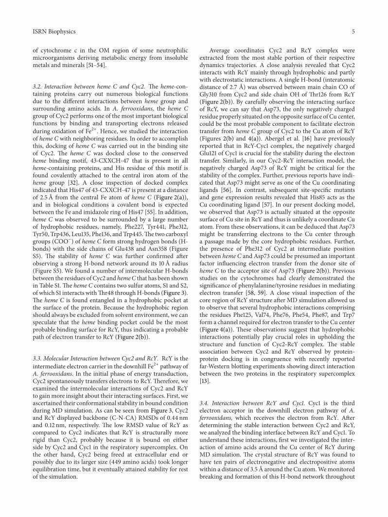
3.2. Interaction between heme C and Cyc2. The heme-con-taining proteins carry out numerous biological functionsdue to the different interactions between heme group andsurrounding amino acids. In A. ferrooxidans, the heme Cgroup of Cyc2 performs one of the most important biologicalfunctions by binding and transporting electrons releasedduring oxidation of Fe2+. Hence, we studied the interactionof heme C with neighboring residues. In order to accomplishthis, docking of heme C was carried out in the binding siteof Cyc2. The heme C was docked close to the conservedheme binding motif, 43-CXXCH-47 that is present in allheme-containing proteins, and His residue of this motif isfound covalently attached to the central iron atom of theheme group [32]. A close inspection of docked complexindicated that His47 of 43-CXXCH-47 is present at a distanceof 2.5 A from the central Fe atom of heme C (Figure 2(a)),and in biological conditions a covalent bond is expectedbetween the Fe and imidazole ring of His47 [55]. In addition,heme C was observed to be surrounded by a large numberof hydrophobic residues, namely, Phe227, Tyr441, Phe312,Tyr50, Trp436, Leu135, Phe136, and Trp445.The two carboxylgroups (COO−) of heme C form strong hydrogen bonds (H-bonds) with the side chains of Glu438 and Asn358 (FigureS5). The stability of heme C was further confirmed afterobserving a strong H-bond network around its 10 A radius(Figure S5). We found a number of intermolecular H-bondsbetween the residues of Cyc2 and hemeC that has been shownin Table S1. The heme C contains two sulfur atoms, S1 and S2,of which S1 interacts withThr48 throughH-bonds (Figure 3).The heme C is found entangled in a hydrophobic pocket atthe surface of the protein. Because the hydrophobic regionshould always be excluded from solvent environment, we canspeculate that the heme binding pocket could be the mostprobable binding surface for RcY, thus indicating a probablepath of electron transfer to RcY (Figure 2(b)).
3.3. Molecular Interaction between Cyc2 and RcY. RcY is theintermediate electron carrier in the downhill Fe2+ pathway ofA. ferrooxidans. In the initial phase of energy transduction,Cyc2 spontaneously transfers electrons to RcY. Therefore, weexamined the intermolecular interactions of Cyc2 and RcYto gainmore insight about their interacting surfaces. First, weascertained their conformational stability in bound conditionduring MD simulation. As can be seen from Figure 3, Cyc2and RcY displayed backbone (C-N-CA) RMSDs of 0.44 nmand 0.12 nm, respectively. The low RMSD value of RcY ascompared to Cyc2 indicates that RcY is structurally morerigid than Cyc2, probably because it is bound on eitherside by Cyc2 and Cyc1 in the respiratory supercomplex. Onthe other hand, Cyc2 being freed at extracellular end orpossibly due to its larger size (449 amino acids) took longerequilibration time, but it eventually attained stability for restof the simulation.
Average coordinates Cyc2 and RcY complex wereextracted from the most stable portion of their respectivedynamics trajectories. A close analysis revealed that Cyc2interacts with RcY mainly through hydrophobic and partlywith electrostatic interactions. A single H-bond (interatomicdistance of 2.7 A) was observed between main chain CO ofGly310 from Cyc2 and side chain OH of Thr126 from RcY(Figure 2(b)). By carefully observing the interacting surfaceof RcY, we can say that Asp73, the only negatively chargedresidue properly situated on the opposite surface of Cu center,could be the most probable component to facilitate electrontransfer from heme C group of Cyc2 to the Cu atom of RcY(Figures 2(b) and 4(a)). Abergel et al. [16] have previouslyreported that in RcY-Cyc1 complex, the negatively chargedGlu121 of Cyc1 is crucial for the stability during the electrontransfer. Similarly, in our Cyc2-RcY interaction model, thenegatively charged Asp73 of RcY might be critical for thestability of the complex. Further, previous reports have indi-cated that Asp73 might serve as one of the Cu coordinatingligands [56]. In contrast, subsequent site-specific mutantsand gene expression results revealed that His85 acts as theCu coordinating ligand [57]. In our present docking model,we observed that Asp73 is actually situated at the oppositesurface of Cu site in RcY and thus is unlikely a coordinate Cuatom. From these observations, it can be deduced that Asp73might be transferring electrons to the Cu center througha passage made by the core hydrophobic residues. Further,the presence of Phe312 of Cyc2 at intermediate positionbetween heme C and Asp73 could be presumed an importantfactor influencing electron transfer from the donor site ofheme C to the acceptor site of Asp73 (Figure 2(b)). Previousstudies on the cytochromes had clearly demonstrated thesignificance of phenylalanine/tyrosine residues in mediatingelectron transfer [58, 59]. A close visual inspection of thecore region of RcY structure after MD simulation allowed usto observe that several hydrophobic interactions comprisingthe residues Phe125, Val74, Phe76, Phe54, Phe87, and Trp7form a channel required for electron transfer to the Cu center(Figure 4(a)). These observations suggest that hydrophobicinteractions potentially play crucial roles in upholding thestructure and function of Cyc2-RcY complex. The stableassociation between Cyc2 and RcY observed by protein-protein docking is in congruence with recently reportedfar-Western blotting experiments showing direct interactionbetween the two proteins in the respiratory supercomplex[13].
3.4. Interaction between RcY and Cyc1. Cyc1 is the thirdelectron acceptor in the downhill electron pathway of A.ferrooxidans, which receives the electron from RcY. Afterdetermining the stable interaction between Cyc2 and RcY,we analyzed the binding interface between RcY and Cyc1. Tounderstand these interactions, first we investigated the inter-action of amino acids around the Cu center of RcY duringMD simulation. The crystal structure of RcY was found tohave ten pairs of electronegative and electropositive atomswithin a distance of 3.5 A around the Cu atom.Wemonitoredbreaking and formation of this H-bond network throughout

6 ISRN Biophysics
Tyr50
Phe312
Trp455
Tyr441
Trp436
Phe227
heme C S1 S2
Leu135
His47
Cyc2
(a)
heme C
Gly310
Phe312
His128
Ala70
His39
Thr37 Lys36
Asp73
Cyc2
RcY
(b)
Figure 2: (a) Crucial amino acids ofCyc2 taking part in hydrophobic interactionswith hemeC.The active site hydrophobic residues stabilizingheme C have been represented as stick models. (b)The graphical representation of favorable electron pathway from heme C group of Cyc2 toAsp73 of RcY.
Table 1: Calculation of H-bonds between the residues around 3.5 A of Cu atom within RcY protein during MD simulation.
RcY (residues) Donor Acceptor RcY (residues) Distance (A) Average duration (ns)Ser53 OG O Pro52 2.6 3Asn80 N O Val45 2.7 2.9Asn80 ND2 NE2 His85 2.9 3.9His85 N OG Ser112 2.7 4Ser86 N SG Cys138 3.1 4.9Ser112 N O Gly84 2.8 2.9Ser112 OG O His85 2.5 4.1Ile40 N O Cys138 3.0 1.6His143 N O Ile40 2.9 4.5Met148 N O Pro52 3.1 4.3
the 5 ns MD trajectories. As can be seen from Table 1, sevenH-bonds persisted for longer than 3 ns, and interestingly, allCu coordinating ligands except Cyc138 continued to formH-bonds for longer than 4 ns. Similar to the calculated RMSDof RcY, the crystal structure of Cyc1 showed a low RMSDprofile (0.19 nm) during MD simulation (Figure 3). SinceCyc1 is bound on one side by RcY and on the other sideby Cox, it remained reasonably stable. Average coordinateswere obtained from the stable dynamics trajectories, and H-bonds were analyzed. The side chain amino group (NH
2) of
Lys166 from Cyc1 forms two H-bonds with CO groups ofGly48 and Phe49 from RcY. The NH
2group of Gln163 from
Cyc1 interacts with CO group of Phe49 from RcY. CO groupof Gln115 from Cyc1 interacts with NH
2group of Lys60 and
OH of Ser53 from RcY. Main chain CO of Thr112 from Cyc1interacts with main chain NH group of Gly147 from RcY.Main chain CO of Ala145 from RcY forms two H-bonds withmain chain NH of Thr112 and main chain NH of Val111 fromCyc1 (Figure 4(b) andTable S2). Our docking result identified
a strong H-bond between His143 of RcY and Glu121 of Cyc1,which is in agreement with the X-ray crystallography studyof Abergel et al. [16] (Figure 4(c)). Cyc1 contains two typesof heme groups: heme A and heme B. The former interactswith the Cu atom of RcY, and the latter receives electronform heme A and transfers it to the Cox. Analysis of theheme groups within the Cyc1 structure revealed that carboxylgroups of heme A and heme B interact directly with eachother indicating a possible path of electron transfer to thenext recipient protein (Figure 4(d)). The heme A and hemeB exhibit high redox potentials of +365mV and +480mV,respectively [60], which was found to be largely attributedto the environment of these two molecules. The CO groupsof both the hemes were found to be involved in numerousH-bond interactions with surrounding amino acids of Cyc1(Table S3). Apart fromH-bonds, the two hemes are stabilizedby several nonbond interactions comprising residues Cys16,Cys19, His20, Pro33, Leu35, and Met64 for heme A andCys119, Cys122, His123, Pro134, Leu136, Leu147, and Met161

ISRN Biophysics 7
0.1
0.2
0.3
0.4
0.5
RMSD
(nm
)
0 1 2 3 4 5Time (ns)
Cyc1CoxAB RcY
Cyc2
Figure 3: Comparative RMSD graphs for Cyc2 (magenta), RcY(blue), Cyc1 (green), and CoxAB (red). The 𝑥-axis representssimulation time (in ps), and the 𝑦-axis indicates RMSD value (innm).
for heme B. A closer look on the number of H-bonds andthe residues that participate in H-bonds revealed that hemeA is stabilized by five H-bonds and six direct nonbondinteractions, whereas for heme B seven H-bonds and sevennonbond interactionwere observed. Several past studies havesuggested that RcY is present at the intermediate positionbetween Cyc2 and Cyc1 in A. ferrooxidans, as is shownin Figure 5. However, this model is probably specific toA. ferrooxidans, because of the fact that some acidophilicbacteria involved in Fe2+ oxidation reportedly do not possessthe gene encoding RcY and that OM c type cytochrome isalways located next to periplasmic c4 type cytochrome [61].In such a situation, theOMcytochrome c and the periplasmiccytochrome c could be directly interacting in vivo whiletransferring electrons for energy metabolism [62].
3.5. Construction of CoxA and CoxB 3D Models. Cox isthe terminal electron acceptor in the electron pathway ofA. ferrooxidans. The diheme Cyc1 transfers electrons fromOM Cyc2 to Cox’s catalytic subunit for the reduction ofO2to form H
2O [5, 6]. Cox is comprised of four subunits,
namely, CoxA, CoxB, CoxC, and CoxD. CoxB is implicatedin electron transfer from Cyc1 to CoxA subunit, where O
2
is converted into a molecule of H2O, whereas CoxC and
CoxD mainly have a structural role [5, 36, 63]. Therefore, weconstructed homology models for CoxA and CoxB subunitsbased on the respective subunits of Paracoccus denitrificans.BLAST search revealed that the target protein had 34%identity, 51% positives, and 73% query coverage with thetemplate. The structure evaluation of the modeled proteinusing Ramachandran plot showed that 89.6% of residues fallwithin the most favored region and no residue was found inthe disallowed region of the plot indicating conformationalaccuracy (Figure S6). The calculated Z-score of the modeledprotein was −5.2, which falls within the Z-score range ofX-ray determined structures of similar size (Figure S7).
Structure evaluation using Verify 3D showed that 80.09% ofthe residues had an average 3D–1D score >0.2. Normally, ascore >80% is a sign of satisfactory quality of the predictedmodel. The cofactors in the core region of the twelve TMdomains, namely, one low spin heme a, one high spin heme a3,and one Cu atom, as reported in the experimental structureof the template, were obtained to the same coordinate spacein the target model using structure alignment in DS 3.1.Similarly, homology model for CoxB was obtained fromCoxB of Paracoccus denitrificans. The structure evaluationprograms including Ramachandran plot indicated that 88.9%of residues were in most favored region with 0.9% residues indisallowed region. Verify 3D analysis revealed that 80.2% ofthe residues had averaged 3D-1D score >0.2, and ProSA-webcalculation showed that the modeled structure had a Z-scorethat is within the range characteristic of native proteins ofsimilar size. CoxB consists of a total of 254 amino acids with acalculated molecular weight of 28, 240 dalton [5].The first 51amino acids of this protein correspond to the standard signalsequence, and the mature protein is comprised of two Nterminal TM domains and a C terminal periplasmic domainwhich are believed to transfer electrons to CoxA subunit[36, 63].
3.6. Interaction between Cyc1 and CoxAB. Our study revealedthat the N terminal TM helices of CoxB interact with 8thand 9th helices of CoxA subunitmainly through hydrophobicinteractions involving side chains of both the subunits. Theperiplasmic domain of CoxB interacts with the periplasmicloops of CoxA through numerous H-bonds. The H-bondinteraction between CoxB and CoxA subunits is shown inTable S4. As Cyc1 transfers electrons to the catalytic site ofCoxA by interacting with the Cu center of CoxB [5], wecarried out a detail analysis of the interaction between Cyc1and CoxB. During MD simulation, the coordinates of CoxAand CoxB were grouped into a single index so that dynamicproperties and energy profiles could be collectively obtainedfor comprehensive analysis. Stability of the CoxAB dimer wasascertained by C𝛼 RMSD from the starting structure, whichwas calculated to be 0.31 nm. Here, it may be noted that likeCyc2, CoxAB is freed from the cytoplasmic end and hencerequired longer simulation time to reach equilibration. Theoverall compactness of the individual proteins with respect totheir center ofmasses in the complexwas analyzed by plottingradius of gyration (Rg) values during MD simulation. Inter-estingly, we did not find an appreciable difference betweenthe Rg values for Cyc2 and CoxAB (Figure 6). Altogether, asexhibited in Figure 6, Cyc2 and CoxAB structures showed Rgvalues of ∼2.4 nm. In contrast, both RcY and Cyc1 structuresshowed Rg values of ∼1.5 nm. Since Cyc2 and CoxAB arecomparative models, structural artifacts are inevitable tooccur in the backbone or side chain conformations, andtherefore, it might have resulted in higher Rg values duringequilibration simulation. On the other hand, since RcY-Cyc1complex is packed against both Cyc2 and CoxAB in thebacterial periplasm, it experienced greater compactness asindicated by lower Rg values.

8 ISRN Biophysics
Asp73
Phe125
Val74
Trp7Phe76
Phe54
His85
Cu
His143
RcY
(a)
RcYCyc1
Ala145Val111
Gly147
Lys143
Lys60
Ser53
Asn45
Gly48
Gln163
Lys166
Glu121
(b)
RcYCyc1
heme A
heme B
His143
Glu121
His85
Met148
Lys138Cu
(c)
Cyc1
heme B
heme ATyr143Gln139
Gln147
Arg156
Asn158
Gln46Arg55Gln38
(d)
Figure 4: (a) The spatial distribution of core hydrophobic residues (stick) that facilitate electron transfer to RcY protein. (b) Illustration ofH-bonds (green dashed lines) formed between the residues of RcY-Cyc1 interacting complex. (c)The figure describes intermolecular contactsbetween the Cu center of RcY and the heme groups of Cyc1 through the His143-Glu121 interaction. (d)The figure depicts that the CO groupsof heme A and heme B play crucial roles in their stability and electron transfer toward CoxB as revealed by strong intermolecular H-bondinteractions (∼2.5 A).
Post-MD analysis for the average coordinates of Cyc1-CoxAB revealed that Cyc1 forms a tight complex with theperiplasmic domain of CoxB subunit involving numerous H-bonds and electrostatic interactions. The H-bond networkbetween these two proteins is as follows: the C terminalCO of Phe254 and OH of Thr157 from CoxB form H-bondswith NH
2of Gln172 and epsilon nitrogen of His152 from
Cyc1, respectively. The main chain NH of Gly155 from CoxBinteracts with the side chain CO of Asn153 from Cyc1. OneZn2+ atom was found at the junction between CoxB andCyc1 coordinated by His152 at a distance of 2.0 A. The Zn2+atom was found to be surrounded by Val159, Thr157, Gly155of CoxB, and His152 of Cyc1 with a distance of 3.9, 2.1, 1.9,
and 2.0 A, respectively. The side chain NH2of Lys146 from
CoxB interacts with the main chain CO of Gly142 fromCyc1 through H-bonds with a bond distance of 2.7 A. Theside chain NH of Gln58 from Cyc1 forms a comparativelyloose H-bond with the main chain CO of Asn228 fromCoxB at a distance of 3.1 A (Figure 7(a) and Table S5). Apartfrom these H-bonds, the interaction between Cyc1 and CoxBstructures was found to be stabilized by several hydrophobicresidues comprising Val170, Tyr63, Tyr29, and Ile62 of Cyc1and Val159, Trp145, and Val179 of CoxB.
A closer look into the binding interface of Cyc1 and CoxBrevealed that CoxB possessed a stretch of seven residues,mostly hydrophobic in nature (145-WKWTFSY-151), which

ISRN Biophysics 9
Outer membrane
Inner membrane
Periplasmic space
Cyc2
RcY
Cyc1
CoxA CoxB
(a)
Cu
Cu
Cu Cua b
heme C
heme A
heme B
heme a
heme a3
WKWTFSY
CoxB
CoxA
RcY
Cyc2
Cyc1
Phe312Asp73
His143
Glu121
Phe468
Outer membrane
Inner membrane
Periplasmic space
H2O2H++ 1/2 O2
H+
Fe3+Fe2+
e−
+
+
+
+
∗
∗
∗
∗
∗
+
(b)
Figure 5: (a)The figure shows the membrane solvated model for docked respirasomes (Cyc2: red, RcY: blue, Cyc1: golden, CoxB: purple, andCoxA: yellow).The figure was prepared using VMD software. (b) Graphical representation of electron-wire (green line) spanning Cyc2, RcY,Cyc1, and CoxAB subunits.
1.2
1.4
1.6
2.2
2.6
1.8
2
2.4
0 1 2 3 4 5Time (ns)
RCYCyc2 Cyc1
CoxAB
Rg (n
m)
Figure 6: (a) Comparison of Rg values of each respirasome duringMD simulation. The plot shows two groups of Rg values, one forCyc2 and CoxAB, that is, ∼2.4 A, and the other for RcY and Cyc1,that is, ∼1.5 A. The figure indicates that RcY and Cyc1 proteins werehighly stable (lowRg value) in comparison toCyc2 andCoxAB (highRg value) during MD simulation.
forms boundary line between surfaces of the two proteins.Lys146 and Thr148 of this motif were found to be respon-sible for stabilizing Cyc1-CoxB complex by forming twostrong H-bonds with Gly142 and Gly155 of Cyc1, respectively(Figure 7(a)).These observations agree well the experimentaldata suggesting that the binuclear Cu center of CoxB subunitaccepts electrons from heme B of Cyc1 channeled through theWKWTFSY hydrophobic motif [5, 36, 63]. The CO group ofheme Bmight be the electron donor in this case. The two Cuatoms at CoxB periplasmic domain are separated from eachother by a distance of 2.6 A, and a disulfide bridge is formedbetween Cyc222 and Cyc226. CuA is coordinated by His181,Cyc226, and Met233, whereas CuB is coordinated by Cyc222andHis230 (Figure 7(b)). From this Cu site, electron is passedto the CoxA catalytic site where CO of heme a, situated ata distance of ∼7 A from the Cu center of CoxB, acts as theelectron acceptor.
3.7. Stereochemistry of heme aa3 in the Catalytic Site of CoxA.CoxA ofA. ferrooxidans contains heme a at two different sites,each with a different function.The heme a acts as an electroncarrier and transfers electrons to heme a3, whereas the latterbinds molecular O
2and reduces it into H
2O with the help

10 ISRN Biophysics
Phe254
CoxB Tyr151
Ser150
Phe149Trp147
Lys146Asn153
Thr148Gly155
Thr157
His152
heme A
heme B
Gln58Asn228
Trp145
CuA
CuB
(a)
Cys226His230
His181
Cys222
CuA
CuB
(b)
Phe332
His159
His469Phe468
His467
heme a3
heme a
His382
His383His333
Cu
(c)
Figure 7: (a)The figure represents the interaction between Cyc1 and CoxB.The hydrophobic motif 145-WKWTFSY-151 essential for electrontransfer is highlighted in green color. H-bonds are shown as dashed lines, heme groups of Cyc1 are represented as stick models, and CuAand CuB are shown as space fill models in red color. (b) The schematic representation of binuclear Cu center of CoxB showing the CuAcoordinated by Cys222, Cys226, and His230 and CuB coordinated by Met223, His181, Cys222, and Cys226. The Cu site of CoxB plays animportant role in further electron transfer to the catalytic site of CoxA. (c) The figure illustrates the interaction between heme a and heme a3within the catalytic site of CoxA.
of an electron received from extracellular environment(Figure 7(c)). While Fe of heme a is hexacoordinated, fourfrom the porphyrin ring of heme a and the remainingtwo from His159 and His469 of CoxA, Fe of heme a3 iscoordinated by five different nitrogen atoms, four from thenitrogen atoms of the porphyrin ring and one from His467of CoxA, thus leaving the sixth site available to bind O
2
[64]. The heme a3, in turn, is involved in reduction of O2
to form H2O with the help of a Cu cofactor. The Cu atom
is coordinated by His333, His382, and His383 (Figure 7(b)).One of the Cu coordinating histidine amino acids might playelectrophilic role required for the catalysis. The details ofstabilizing H-bonds between heme groups and amino acids
of CoxA are presented in Table S6. In addition to this, itwas proposed that the conserved Phe468 found in betweenHis467 and His469 of CoxA might be important for thetransfer of electrons from heme a to heme a3 [65, 66], andour simulation results support this hypothesis. During the5 ns MD simulation, two heme groups were found to bequite stable, probably due to the hydrophobic effect put forthby Phe468 as indicated by the RMSD graph (Figure 8). Theresults indicated that heme a experienced less fluctuationduring MD simulation because it is connected to His159and His469 from both sides, whereas heme a3 experiencedcomparatively larger fluctuation due to the absence of O
2
at one end. The stability of heme aa3 binding region was

ISRN Biophysics 11
0 1 2 3 4 5Time (ns)
0
RMSD
(nm
)
0.020.04
0.20.180.160.140.12
0.10.080.06
hemeheme a3
Figure 8: Comparative RMSD graphs for heme a (green) and hemea3 (red).The 𝑥 axis represents simulation time (in ps) and the 𝑦 axisindicates RMSD value (in nm). Although the RMSD value of hemea (0.15 nm) was found to be greater than that of heme a3 (0.9 nm), itmaintains stability throughout the simulation.
0.1
0.2
0.3
0.4
0.5
RMSF
(nm
)
0 100 200 300Residue
400
0.6
0.0
heme binding region
600500
heme binding region
Figure 9: The figure shows fluctuation of CoxA residues duringMD simulation. The fluctuation of heme binding region (residues461–471), specifically, the residues His467, Phe468, and His469, wasfound to be very low indicating their structural stability.
ascertained by observing root mean square fluctuation ofbackbone residues during MD simulation (Figure 9). Theresidues that connect heme a to heme a3, that is, His467,Phe468, and His469, were found to be completely stableduring simulation. Our modeling and simulation resultssuggest that the residues of Cox involved in stabilizing hemeaa3 are found at the upper side of cytoplasmic membranenear the periplasm. From this observation, we can postulatethat O
2reduction takes place at a pH nearly equivalent to
periplasmic pH of 3.0 [67, 68].
4. Conclusions
The interdependence among the docked structures was firstobserved when OM and Phe312. At the interacting surface,
Asp73 of RcY was considered to be involved in directelectron transfer between two complexes, which might befacilitated by physical interactions of Phe312 and Asp73.A detailed analysis revealed that the hydrophobic residuesenclosed within core region of RcY form a passage forelectrons to reach the Cu center. Subsequent docking ofCyc1 with Cyc2-RcY complex indicated that His143 of RcYassociates with Glu121 of Cyc1, suggesting a probable channelof electron transfer between the two proteins. The heme Aand heme B of Cyc1 play crucial roles in transferring electronreceived from RcY to the binuclear Cu center of CoxBthrough the aromatic/hydrophobic sequence motif, that is,145-WKWTFSY-151. Phe468 was found to stabilize heme aand heme a3 byhydrophobic interaction. Our results providea comprehensive insight on the structural organization ofthe respiratory supercomplex and on the probable path ofelectrons within downhill pathway of A. ferrooxidans. Alto-gether, this computational model can be used to understandthe popular electron wire concept of metalloproteins.
Conflict of Interests
Theauthors declare that no competing interests exist with anycommercial bodies.
Acknowledgments
The authors are thankful to Department of Biotechnology,Government of India, for providing computational facility tocarry out the present research. The authors also declare thatvalid license has been acquired for the commercial softwareused in this study.
References
[1] L. R. Croal, J. A. Gralnick, D. Malasarn, and D. K. Newman,“The genetics of geochemistry,” Annual Review of Genetics, vol.38, pp. 175–202, 2004.
[2] T. Rohwerder, T. Gehrke, K. Kinzler, andW. Sand, “Bioleachingreview part A: progress in bioleaching: fundamentals andmechanisms of bacterial metal sulfide oxidation,” AppliedMicrobiology and Biotechnology, vol. 63, no. 3, pp. 239–248,2003.
[3] A. Elbehti, G. Brasseur, and D. Lemesle-Meunier, “First evi-dence for existence of an uphill electron transfer through the𝑏𝑐
1and NADH-Q oxidoreductase complexes of the acidophilic
obligate chemolithotrophic ferrous ion-oxidizing bacteriumThiobacillus ferrooxidans,” Journal of Bacteriology, vol. 182, no.12, pp. 3602–3606, 2000.
[4] G. Brasseur, G. Levican, V. Bonnefoy, D. Holmes, E. Jedlicki,and D. Lemesle-Meunier, “Apparent redundancy of electrontransfer pathways via 𝑏𝑐
1complexes and terminal oxidases
in the extremophilic chemolithoautotrophic Acidithiobacillusferrooxidans,” Biochimica et Biophysica Acta, vol. 1656, no. 2-3,pp. 114–126, 2004.
[5] C. Appia-Ayme, N. Guiliani, J. Ratouchniak, and V. Bon-nefoy, “Characterization of an operon encoding two c-typecytochromes, an aa3-type cytochrome oxidase, and rusticyanininThiobacillus ferrooxidansATCC 33020,”Applied and Environ-mental Microbiology, vol. 65, no. 11, pp. 4781–4787, 1999.

12 ISRN Biophysics
[6] A. Yarzabal, G. Brasseur, J. Ratouchniak et al., “The high-molecular-weight cytochrome c Cyc2 of Acidithiobacillus fer-rooxidans is an outer membrane protein,” Journal of Bacteriol-ogy, vol. 184, no. 1, pp. 313–317, 2002.
[7] G. Malarte, G. Leroy, E. Lojou, C. Abergel, M. Bruschi, andM. T. Giudici-Orticoni, “Insight into molecular stability andphysiological properties of the diheme cytochrome CYC
41
from the acidophilic bacterium Acidithiobacillus ferrooxidans,”Biochemistry, vol. 44, no. 17, pp. 6471–6481, 2005.
[8] A. Bengrine, N. Guiliani, C. Appia-Ayme et al., “Sequence andexpression of the rusticyanin structural gene fromThiobacillusferrooxidansATCC33020 strain,” Biochimica et Biophysica Acta,vol. 1443, no. 1-2, pp. 99–112, 1998.
[9] P. Bruscella, C. Appia-Ayme, G. Levican et al., “Differentialexpression of two 𝑏𝑐
1complexes in the strict acidophilic
chemolithoautotrophic bacterium Acidithiobacillus ferrooxi-dans suggests a model for their respective roles in iron or sulfuroxidation,”Microbiology, vol. 153, no. 1, pp. 102–110, 2007.
[10] W. J. Ingledew, “Thiobacillus ferrooxidans. The bioenergetics ofan acidophilic chemolithotroph,” Biochimica et Biophysica Acta,vol. 683, no. 2, pp. 89–117, 1982.
[11] D. Holmes and V. Bonnefoy, “Genetic and bioinformaticinsights into iron and sulfur oxidationmechanisms of bioleach-ing organisms,” inBiomining, D. E. Rawlings, E. Douglas, andD.B. Johnson, Eds., pp. 281–307, Springer, Berlin, Germany, 2007.
[12] A. Yarzabal, C. Appia-Ayme, J. Ratouchniak, and V. Bonnefoy,“Regulation of the expression of the Acidithiobacillus ferroox-idans rus operon encoding two cytochromes c, a cytochromeoxidase and rusticyanin,”Microbiology, vol. 150, no. 7, pp. 2113–2123, 2004.
[13] C. Castelle, M. Guiral, G. Malarte et al., “A new iron-oxidizing/O
2-reducing supercomplex spanning both inner
and outer membranes, isolated from the extreme acidophileAcidithiobacillus ferrooxidans,” Journal of Biological Chemistry,vol. 283, no. 38, pp. 25803–25811, 2008.
[14] M. L. Barrett, I. Harvey, M. Sundararajan et al., “Atomicresolution crystal structures, EXAFS, and quantum chemicalstudies of rusticyanin and its two mutants provide insight intoits unusual properties,” Biochemistry, vol. 45, no. 9, pp. 2927–2939, 2006.
[15] R. L. Walter, S. E. Ealick, A. M. Friedman, R. C. Blake II,P. Proctor, and M. Shoham, “Multiple wavelength anomalousdiffraction (MAD) crystal structure of rusticyanin: a highlyoxidizing cupredoxin with extreme acid stability,” Journal ofMolecular Biology, vol. 263, no. 5, pp. 730–751, 1996.
[16] C. Abergel, W. Nitschke, G. Malarte, M. Bruschi, J.-M. Claverie,and M.-T. Giudici-Orticoni, “The structure of Acidithiobacil-lus ferrooxidans 𝑐
4-cytochrome: a model for complex-induced
electron transfer tuning,” Structure, vol. 11, no. 5, pp. 547–555,2003.
[17] B. P. Mukhopadhyay, B. Ghosh, H. R. Bairagya, T. K. Nandi,B. Chakrabarti, and A. K. Bera, “Molecular modeling ofthe ternary complex of rusticyanin-cytochrome 𝑐
4-cytochrome
oxidase: an insight to possible H-bond mediated recognitionand electron transfer reaction in T. ferrooxidans,” Journal ofBiomolecular Structure and Dynamics, vol. 25, no. 5, pp. 543–551, 2008.
[18] B. P. Mukhopadhyay, B. Ghosh, H. R. Bairagya, A. K. Bera,T. K. Nandi, and S. B. Das, “Modeling study of rusticyanin-cytochrome 𝑐
4complex: an insight to possible H-bond medi-
ated recognition and electron—transfer process,” Journal ofBiomolecular Structure andDynamics, vol. 25, no. 2, pp. 157–164,2007.
[19] B. P. Mukhopadhyay, B. Ghosh, H. R. Bairagya, A. K. Bera,and R. K. Roy, “Conserved water molecular dynamics of thedifferent X-ray structures of rusticyanin: an unique aquationpotentiality of the ligand bonded Cu++ center,” Journal ofBiomolecular Structure and Dynamics, vol. 24, no. 4, pp. 369–377, 2007.
[20] S. F. Altschul,W. Gish,W.Miller, E.W.Myers, and D. J. Lipman,“Basic local alignment search tool,” Journal ofMolecular Biology,vol. 215, no. 3, pp. 403–410, 1990.
[21] J. U. Bowie, R. Luthy, and D. Eisenberg, “A method to identifyprotein sequences that fold into a known three-dimensionalstructure,” Science, vol. 253, no. 5016, pp. 164–170, 1991.
[22] L. A. Kelley andM. J. E. Sternberg, “Protein structure predictionon the Web: a case study using the Phyre server,” NatureProtocols, vol. 4, no. 3, pp. 363–371, 2009.
[23] J. Shi, T. L. Blundell, and K. Mizuguchi, “FUGUE: sequence-structure homology recognition using environment-specificsubstitution tables and structure-dependent gap penalties,”Journal of Molecular Biology, vol. 310, no. 1, pp. 243–257, 2001.
[24] K. Karplus, R. Karchin, J. Draper et al., “Combining local-structure, fold-recognition, and new fold methods for proteinstructure prediction,” Proteins, vol. 53, supplement 6, pp. 491–496, 2003.
[25] A. Lobley, M. I. Sadowski, and D. T. Jones, “pGenTHREADERand pDomTHREADER: new methods for improved proteinfold recognition and superfamily discrimination,” Bioinformat-ics, vol. 25, no. 14, pp. 1761–1767, 2009.
[26] M. A. Kurowski and J. M. Bujnicki, “GeneSilico protein struc-ture prediction meta-server,”Nucleic Acids Research, vol. 31, no.13, pp. 3305–3307, 2003.
[27] J. Lundstrom, L. Rychlewski, J. Bujnicki, and A. Elofsson,“Pcons: a neural-network—based consensus predictor thatimproves fold recognition,” Protein Science, vol. 10, no. 11, pp.2354–2362, 2001.
[28] D. T. Jones, “Protein secondary structure prediction basedon position-specific scoring matrices,” Journal of MolecularBiology, vol. 292, no. 2, pp. 195–202, 1999.
[29] N. Treiber, D. J. Reinert, I. Carpusca, K. Aktories, and G.E. Schulz, “Structure and mode of action of a mosquitocidalholotoxin,” Journal of Molecular Biology, vol. 381, no. 1, pp. 150–159, 2008.
[30] D. van der Spoel, E. Lindahl, B. Hess, G. Groenhof, A. E. Mark,and H. J. C. Berendsen, “GROMACS: fast, flexible, and free,”Journal of Computational Chemistry, vol. 26, no. 16, pp. 1701–1718, 2005.
[31] M. Wiederstein and M. J. Sippl, “ProSA-web: interactive webservice for the recognition of errors in three-dimensional struc-tures of proteins,”Nucleic Acids Research, vol. 35, supplement 2,pp. W407–W410, 2007.
[32] T. E. Meyer, “Evolution and classification of c-typecytochromes,” in Cytochrome C: A Multidisciplinary Approach,R. A. Scott and A. G. Mauk, Eds., University Science Books,Sausalito, Calif, USA, 1995.
[33] E. E. Bolton, Y. Wang, P. A. Thiessen, and S. H. Bryant, “Pub-Chem: integrated platform of small molecules and biologicalactivities,” Annual Reports in Computational Chemistry, vol. 4,pp. 217–241, 2008.
[34] D. Schneidman-Duhovny, Y. Inbar, R. Nussinov, and H. J.Wolfson, “PatchDock and SymmDock: servers for rigid andsymmetric docking,”Nucleic Acids Research, vol. 33, supplement2, pp. W363–W367, 2005.

ISRN Biophysics 13
[35] D.Duhovny, R.Nussinov, andH. J.Wolfson, “Efficient unbounddocking of rigid molecules,” in Algorithms in Bioinformat-ics: Second International Workshop, WABI 2002, Rome, Italy,September 17–21, 2002, Proceedings, R. Guigo and D. Gusfeld,Eds., vol. 2452 of Lecture Notes in Computer Science, pp. 185–200, Springer, 2002.
[36] S. Iwata, C. Ostermeier, B. Ludwig, and H. Michel, “Structureat 2.8 A resolution of cytochrome c oxidase from Paracoccusdenitrificans,” Nature, vol. 376, pp. 660–669, 2002.
[37] C. Neale, W. F. D. Bennett, D. P. Tieleman, and R. Pomes, “Sta-tistical convergence of equilibrium properties in simulationsof molecular solutes embedded in lipid bilayers,” Journal ofChemical Theory and Computation, vol. 7, no. 12, pp. 4175–4188,2011.
[38] C. Kandt, W. L. Ash, and D. P. Tieleman, “Setting up and run-ning molecular dynamics simulations of membrane proteins,”Methods, vol. 41, no. 4, pp. 475–488, 2007.
[39] J. F. Nagle, “Area/lipid of bilayers from NMR,” BiophysicalJournal, vol. 64, no. 5, pp. 1476–1481, 1993.
[40] O. Berger, O. Edholm, and F. Jahnig, “Molecular dynamicssimulations of a fluid bilayer of dipalmitoylphosphatidylcholineat full hydration, constant pressure, and constant temperature,”Biophysical Journal, vol. 72, no. 5, pp. 2002–2013, 1997.
[41] W. Humphrey, A. Dalke, and K. Schulten, “VMD: visualmolecular dynamics,” Journal of Molecular Graphics, vol. 14, no.1, pp. 33–38, 1996.
[42] D. Baker and A. Sali, “Protein structure prediction and struc-tural genomics,” Science, vol. 294, no. 5540, pp. 93–96, 2001.
[43] M. J. L. Schoonman, R. M. A. Knegtel, and P. D. J. Grootenhuis,“Practical evaluation of comparative modelling and threadingmethods,”Computers and Chemistry, vol. 22, no. 5, pp. 369–375,1998.
[44] K. L. Tkaczuk, “Trm13p, the tRNA:Xm4 modification enzymefrom Saccharomyces cerevisiae is a member of the Rossmann-foldMTase superfamily: prediction of structure and active site,”Journal of Molecular Modeling, vol. 16, no. 3, pp. 599–606, 2010.
[45] K. A. Majorek and J. M. Bujnicki, “Modeling of Escherichia coliEndonuclease V structure in complex with DNA,” Journal ofMolecular Modeling, vol. 15, no. 2, pp. 173–182, 2009.
[46] A. N. Ramli, N.M.Mahadi,M. S. Shamsir et al., “Structural pre-diction of a novel chitinase from the psychrophilic Glaciozymaantarctica PI12 and an analysis of its structural properties andfunction,” Journal of Computer Aided Molecular Design, vol. 26,no. 8, pp. 947–996, 2012.
[47] P. Anand, S. Sankaran, S. Mukherjee et al., “Structural annota-tion of Mycobacterium tuberculosis proteome,” PLoS ONE, vol.6, no. 10, Article ID e27044, 2011.
[48] M. Ferrer, A. Ghazi, A. Beloqui et al., “Functional metage-nomics unveils a multifunctional glycosyl hydrolase from thefamily 43 catalysing the breakdown of plant polymers in the calfrumen,” PLoS ONE, vol. 7, no. 6, Article ID e38134, 2012.
[49] F. Ruckert, G. Dawelbait, C. Winter et al., “Examination ofapoptosis signaling in pancreatic cancer by computationalsignal transduction analysis,” PLoS ONE, vol. 5, no. 8, ArticleID e12243, 2010.
[50] G. N. Ramachandran, C. Ramakrishnan, and V. Sasisckharan,“Stereochemistry of polypeptide chain configurations,” Journalof Molecular Biology, vol. 7, pp. 95–99, 1963.
[51] F. van Ommen Kloeke, R. D. Bryant, and E. J. Laishley,“Localization of cytochromes in the outer membrane of Desul-fovibrio vulgaris (hildenborough) and their role in anaerobicbiocorrosion,” Anaerobe, vol. 1, no. 6, pp. 351–358, 1995.
[52] S. Gaspard, F. Vazquez, and C. Holliger, “Localization and solu-bilization of the iron(III) reductase ofGeobacter sulfurreducens,”Applied and Environmental Microbiology, vol. 64, no. 9, pp.3188–3194, 1998.
[53] C. R. Myers and J. M. Myers, “Localization of cytochromesto the outer membrane of anaerobically grown Shewanellaputrefaciens MR-1,” Journal of Bacteriology, vol. 174, no. 11, pp.3429–3438, 1992.
[54] J. M. Myers and C. R. Myers, “Role for outer membranecytochromesOmcAandOmcBof Shewanella putrefaciensMR-1in reduction ofmanganese dioxide,”Applied and EnvironmentalMicrobiology, vol. 67, no. 1, pp. 260–269, 2001.
[55] W. J. Ingledew, J. C. Cox, and P. J. Halling, “A proposedmechanism for energy conservation during Fe2+ oxidation byThiobacillus ferro-oxidans: chemiosmotic coupling to net H+influx,” FEMS Microbiology Letters, vol. 2, no. 4, pp. 193–197,1977.
[56] M. Ronk, J. E. Shively, E. A. Shute, and R. C. Blake II, “Aminoacid sequence of the blue copper protein rusticyanin fromThiobacillus ferrooxidans,” Biochemistry, vol. 30, no. 39, pp.9435–9442, 1991.
[57] D. R. Casimiro, A. Toy-Palmer, R. C. Blake II, and H. J. Dyson,“Gene synthesis, high-level expression, and mutagenesis ofThiobacillus ferrooxidans rusticyanin: His 85 is a ligand to theblue copper center,” Biochemistry, vol. 34, no. 20, pp. 6640–6648, 1995.
[58] N. Liang,G. J. Pielak, A.G.Mauk,M. Smith, andB.M.Hoffman,“Yeast cytochrome c with phenylalanine or tyrosine at position87 transfers electrons to (zinc cytochrome c peroxidase)+ ata rate ten thousand times that of the serine-87 or glycine-87variants,” Proceedings of the National Academy of Sciences of theUnited States of America, vol. 84, no. 5, pp. 1249–1252, 1987.
[59] C. S. Miles, N. Rouviere-Fourmy, F. Lederer et al., “Tyr-143facilitates interdomain electron transfer in flavocytochrome 𝑏
2,”
Biochemical Journal, vol. 285, part 1, pp. 187–192, 1992.[60] M.-T. Giudici-Orticoni, G. Leroy, W. Nitschke, and M. Bruschi,
“Characterization of a newdihemic 𝑐4-type cytochrome isolated
fromThiobacillus ferrooxidans,” Biochemistry, vol. 39, no. 24, pp.7205–7211, 2000.
[61] C. Jeans, S. W. Singer, C. S. Chan et al., “Cytochrome 572 isa conspicuous membrane protein with iron oxidation activitypurified directly from a natural acidophilic microbial commu-nity,”The ISME Journal, vol. 2, no. 5, pp. 542–550, 2008.
[62] W. J. Ingledew and J. G. Cobley, “A potentiometric and kineticstudy on the respiratory chain of ferrous-iron-grownThiobacil-lus ferrooxidans,” Biochimica et Biophysica Acta, vol. 590, no. 2,pp. 141–158, 1980.
[63] P. Lappalainen, N. J.Watmough, C. Greenwood, andM. Saraste,“Electron transfer between cytochrome c and the isolatedCuA domain: identification of substrate-binding residues incytochrome c oxidase,” Biochemistry, vol. 34, no. 17, pp. 5824–5830, 1995.
[64] S. Yoshikawa, K. Shinzawa-Itoh, R. Nakashima et al., “Redox-coupled crystal structural changes in bovine heart cytochromec oxidase,” Science, vol. 280, no. 5370, pp. 1723–1729, 1998.
[65] C. Ostermeier, S. Iwata, andH.Michel, “Cytochrome c oxidase,”Current Opinion in Structural Biology, vol. 6, no. 4, pp. 460–466,1996.
[66] M. Saraste, J. Castresana, D. Higgins, M. Lbben, and M.Wilmanns, “Evolution of cytochrome oxidase,” in Origin andEvolution of Biological Energy Conversion, H. Baltscheffsky, Ed.,pp. 255–289, Wiley-VCH, New York, NY, USA, 1994.

14 ISRN Biophysics
[67] M. Kai, T. Yano, Y. Fukumori, and T. Yamanaka, “Cytochromeoxidase of an acidophilic iron-oxidizing bacterium,Thiobacillusferrooxidans, functions at pH 3.5,” Biochemical and BiophysicalResearch Communications, vol. 160, no. 2, pp. 839–843, 1989.
[68] M. Kai, T. Yano, H. Tamegai, Y. Fukumori, and T. Yamanaka,“Thiobacillus ferrooxidans cytochrome c oxidase: purification,andmolecular and enzymatic features,” Journal of Biochemistry,vol. 112, no. 6, pp. 816–821, 1992.

Submit your manuscripts athttp://www.hindawi.com
Hindawi Publishing Corporationhttp://www.hindawi.com Volume 2014
Anatomy Research International
PeptidesInternational Journal of
Hindawi Publishing Corporationhttp://www.hindawi.com Volume 2014
Hindawi Publishing Corporation http://www.hindawi.com
International Journal of
Volume 2014
Zoology
Hindawi Publishing Corporationhttp://www.hindawi.com Volume 2014
Molecular Biology International
GenomicsInternational Journal of
Hindawi Publishing Corporationhttp://www.hindawi.com Volume 2014
The Scientific World JournalHindawi Publishing Corporation http://www.hindawi.com Volume 2014
Hindawi Publishing Corporationhttp://www.hindawi.com Volume 2014
BioinformaticsAdvances in
Marine BiologyJournal of
Hindawi Publishing Corporationhttp://www.hindawi.com Volume 2014
Hindawi Publishing Corporationhttp://www.hindawi.com Volume 2014
Signal TransductionJournal of
Hindawi Publishing Corporationhttp://www.hindawi.com Volume 2014
BioMed Research International
Evolutionary BiologyInternational Journal of
Hindawi Publishing Corporationhttp://www.hindawi.com Volume 2014
Hindawi Publishing Corporationhttp://www.hindawi.com Volume 2014
Biochemistry Research International
ArchaeaHindawi Publishing Corporationhttp://www.hindawi.com Volume 2014
Hindawi Publishing Corporationhttp://www.hindawi.com Volume 2014
Genetics Research International
Hindawi Publishing Corporationhttp://www.hindawi.com Volume 2014
Advances in
Virolog y
Hindawi Publishing Corporationhttp://www.hindawi.com
Nucleic AcidsJournal of
Volume 2014
Stem CellsInternational
Hindawi Publishing Corporationhttp://www.hindawi.com Volume 2014
Hindawi Publishing Corporationhttp://www.hindawi.com Volume 2014
Enzyme Research
Hindawi Publishing Corporationhttp://www.hindawi.com Volume 2014
International Journal of
Microbiology