research article the physician tendency in stereotactic...
TRANSCRIPT
Henry Kodrat et al
82
eJKI
AbstractStereotactic radiosurgery (SRS) is one of the treatment modalities for benign intra-cranial tumor, especially
for the tumor located next to the critical neural structure. The prescribed dose for radiosurgery depends on the maximal tumor diameter and surrounding normal tissue tolerance dose. This cross sectional study was conducted to evaluate the physician’s tendency in radiosurgery dose prescription. We observed treatment planning data of 32 patients with benign intra-cranial tumor, which had been treated with SRS at Dr. Cipto Mangunkusumo National Hospital in 2009-2010. The peripheral dose, organ at risk (OAR) dose limitiation and maximum tumor diameter were recorded. We compared our SRS dose with dose limitation, which allowed safer dosing based on maximal tumor diameter perspective and the nearest OAR dose constraint. From maximal tumor diameter perspective, we prescribed mean±SD radiosurgery doses, which were 11.63±2.21Gy, 10.21±1.29Gy and 9.88±1.07Gy for the tumor size ≤2cm, 2.01-3cm and 3,01-4cm respectively. Our radiosurgery dose was the lowest than dose limitation based on the nearest OAR perspective, followed by maximal tumor diameter perspective. It was concluded that radiosurgery dose had the tendency to be influenced by surrounding healthy tissue tolerance rather than maximal tumor diameter.Keywords: stereotactic, radiosurgery, benign tumor, dose.
Kecenderungan Dokter dalam Menentukan Dosis Stereotactic Radiosurgery untuk Tumor Jinak Intrakranial di
RSUP Nasional dr. Cipto Mangunkusumo, Jakarta
AbstrakStereotactic radiosurgery (SRS) merupakan salah satu modalitas pengobatan tumor jinak intra-kranial
terutama untuk tumor yang berdekatan dengan struktur saraf penting. Penentuan dosis pada radiosurgery tergantung pada diameter tumor maksimal dan dosis toleransi jaringan sehat sekitarnya. Penelitian ini dilakukan untuk mengevaluasi kecenderungan dokter dalam menentukan dosis radiosurgery. Penelitian cross sectional ini mengevaluasi data perencanaan radiasi dari 32 pasien dengan tumor jinak intra-kranial yang telah dilakukan prosedur SRS di RS dr.Cipto Mangunkusumo pada tahun 2009-2010. Dilakukan pencatatan dosis perifer tumor, dosis toleransi jaringan sehat dan diameter tumor maksimal. Dosis SRS dibandingkan dengan batasan dosis aman yang diperbolehkan berdasarkan aspek diameter tumor maksimal dan dosis toleransi jaringan sehat terdekat. Dari aspek diameter tumor maksimal, diberikan dosis radiosurgery rata-rata±SD sebesar 11,63±2,21Gy, 10,21±1,29Gy dan 9,88±1,07Gy untuk ukuran tumor ≤2cm, 2,01-3cm dan 3,01-4cm. Dosis radiosurgery yang kami berikan lebih rendah dibandingkan dengan dosis tolerasi jaringan sehat sekitarnya diikuti dengan batasan dosis berdasarkan aspek diameter tumor maksimal. Disimpulkan bahwa penentuan dosis radiosurgery lebih dipengaruhi oleh dosis toleransi jaringan sehat sekitarnya daripada diameter tumor maksimal.Kata kunci: stereotaktik, radiosurgery, tumor jinak, dosis.
RESEARCH ARTICLE
The Physician Tendency in Stereotactic Radiosurgery Dose Prescription in Benign Intracranial Tumor at
dr. Cipto Mangunkusumo National Hospital, Jakarta
Henry Kodrat,1* Soehartati Gondhowiardjo,1 Renindra Ananda Aman2
1Department of Radiotherapy, 2Department of Neurosurgery FM Universitas Indonesia-
dr. Cipto Mangunkusumo National Hospital
*Correspondence: [email protected] 13th June 2016; Accepted 7th August 2016

The Physician Tendency
83
Vol. 4, No. 2, Agustus 2016
IntroductionStereotactic is derived from the Greek word
“stereos” which means solid and “taxis” which means arrangement or order. Stereotactic radiosurgery (SRS) implies the delivery of a single large dose of focused radiation, which can kill tumor cell and /or obliterate blood vessels with the application of a three-dimensional coordinate system to navigate the localization of small targets inside the skull cavity. The use of high-dose radiation in radiosurgery requires three-dimensional mapping technique to guide radiosurgery procedure because it will produce precise target localization while minimizing radiation dose to surrounding normal tissue. The efficacy of stereotactic radiosurgery allows high probability of local control comparable to surgery.1-3
The definitive treatment of benign intra-cranial tumors is surgery. In many cases, a benign tumor is located in areas where there are many vital structures surrounding it. Therefore, the surgery is difficult to do because surgery can cause high morbidity and mortality. Because of the large risk of complications, radiotherapy becomes an alternative treatment. In a benign tumor, radiotherapy is less responsive because it is classified as a radio-resistant tumor. The objective of radiosurgery in benign tumor is no progression in tumor size. Tumor treated with radiosurgery needs latent period to shrink.2,4,5
In parallel with the development of radiobiology, knowledge of the benign tumor’s radio-sensitivity to fractionation begins to be known. From several studies, a benign tumor has a α/β ratio approximately 3Gray (Gy). With a low α/β ratio, re-oxygenation plays a less important role, which causes fractionation less important. So in small low α/β ratio tumor, it is more reasonable to give a single fraction or hypofractionation. Radiosurgery process does not require much time. The whole process usually requires 4-5 hours.2,6,7
This study was conducted to evaluate factor that influences the physician’s tendency in radiosurgery dose determination in radiosurgery procedure at Dr. Cipto Mangunkusumo Hospital, Jakarta.
MethodsThis cross sectional study analyzed 32
treatment planning data from the medical record for radiosurgery of benign tumor, which had been undergone radiosurgery procedure in Department of Radiotherapy Dr. Cipto Mangunkusumo National Hospital, Jakarta, from January 2009 until December 2010. SRS was delivered using Elekta® Synergy S linear accelerator (LINAC) and radiosurgery dose calculation was performed in Precise PLAN® treatment planning system.
The data collected were gender, age, maximal tumor diameter, tumor volume, and tumor dose. We collected information of the dose from treatment planning data in the medical record in the form of dose volume histogram (DVH). Tumor dose is determined by dose at the edge (peripheral) of tumor. The organ at risk (OAR) dose is determined by the maximum dose (Dmax) stated in the DVH. For benign tumor, we classified into group of the tumor. The group consisted of pituitary adenoma, skull base meningioma, convexity meningioma, craniopharyngioma, acoustic neuroma and pineal tumor.
From these tumor groups, we evaluated factor that influences the radiosurgery dose determination based on maximal tumor diameter and OAR tolerance dose. OAR dose was determined based on the lowest OAR tolerance dose based on the critical structure located next to the tumor.
All statistical analysis was performed using the Statistical Package for Social Sciences (SPSS version 17.0, Chicago, USA).
ResultsCharacteristic data are shown in Table 1. Most
of the patients were female and acoustic neuroma was the most found benign tumor in this study.
Henry Kodrat et al
84
eJKI
Table 1. Characteristics of Benign Intra-cranial Tumors Patients, Underwent Radiosurgery at Dr. Cipto Mangunkusumo National Hospital, 2009-2010
Characteristic n (%)
Gender Male Female
Diagnostic Acoustic neuroma Pituitary adenoma Skull base meningioma Convexity meningioma Pineal tumor Craniopharyngioma
Maximal tumor diameter group ≤2cm 2.01–3cm 3.01–4cm >4cm
Age (years old) Median Range
Maximal tumor diameter (cm) Median Range
Tumor volume (cc) Median Range
Dose (Gy) Mean Standard deviation
7 (21.9%)25 (78.1%)
12 (37.5%)8 (25%)
5 (15.6%)3 (9.4%)3 (9.4%)1 (3.1%)
4 (12.5%)14 (43.7%)12 (37.5%)
2 (6.2%)
417–63
2.80.7–5.2
6.00.1–35.2
10.21.3
Table 2. Comparison of Dose (Gy), from Maximal Tumor Diameter Perspective Compared to Dose Constraint Allowed by RTOG 90-05 Protocol
Maximal Tumor Diameter Mean Dose (SD) RTOG 90-05
≤2cm 11.63 (±2.21)Gy 24Gy2.01–3cm 10.21 (±1.29)Gy 18Gy3.01–4cm 9.88 (±1.07)Gy 15Gy
>3cm 10Gy N/A
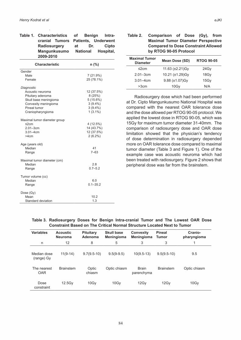
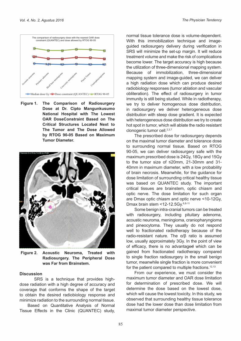
Radiosurgery dose which had been performed at Dr. Cipto Mangunkusumo National Hospital was compared with the nearest OAR tolerance dose and the dose allowed per RTOG 90-05 protocol. We applied the lowest dose in RTOG 90-05, which was 15Gy for maximum tumor diameter 31-40mm. The comparison of radiosurgery dose and OAR dose limitation showed that the physician’s tendency of dose determination in radiosurgery depended more on OAR tolerance dose compared to maximal tumor diameter (Table 3 and Figure 1). One of the example case was acoustic neuroma which had been treated with radiosurgery. Figure 2 shows that peripheral dose was far from the brainstem.
Table 3. Radiosurgery Doses for Benign Intra-cranial Tumor and The Lowest OAR Dose Constraint Based on The Critical Normal Structure Located Next to Tumor
Variables Acoustic Neuroma
Pituitary Adenoma
Skull base Meningioma
Convexity Meningioma
Pineal Tumor
Cranio-pharyngioma
n 12 8 5 3 3 1
Median dose (range) Gy
11(9-14) 9.7(9.5-10) 9.5(9-9.5) 10(9.5-13) 9.5(9.5-10) 9.5
The nearest OAR
Brainstem Optic chiasm
Optic chiasm Brain parenchyma
Brainstem Optic chiasm
Dose constraint
12.5Gy 10Gy 10Gy 12Gy 12Gy 10Gy
The Physician Tendency
85
Vol. 4, No. 2, Agustus 2016
Figure 1. The Comparison of Radiosurgery Dose at Dr. Cipto Mangunkusumo National Hospital with The Lowest OAR DoseConstraint Based on The Critical Structures Located Next to The Tumor and The Dose Allowed by RTOG 90-05 Based on Maximum Tumor Diameter.
Figure 2. Acoustic Neuroma, Treated with Radiosurgery. The Peripheral Dose was Far from Brainstem.
Discussion SRS is a technique that provides high-
dose radiation with a high degree of accuracy and coverage that conforms the shape of the target to obtain the desired radiobiology response and minimize radiation to the surrounding normal tissue.
Based on Quantitative Analysis of Normal Tissue Effects in the Clinic (QUANTEC) study,
4
RTOG 90-05 protocol. We applied the lowest dose in RTOG 90-05, which was 15Gy for maximum tumor diameter 31-40mm. The comparison of radiosurgery dose and OAR dose limitation showed that the physician’s tendency of dose determination in radiosurgery dependedmoreon OAR tolerance dose compared to maximal tumor diameter (Table3 and Figure 1). One of the example case was acoustic neuroma which had been treated with radiosurgery.Figure 2 showsthat peripheral dose was far from the brainstem.
Table 3. Radiosurgery Doses for Benign Intra-cranial Tumor andThe Lowest OAR Dose Constraint Based on The Critical Normal Structure Located Next to Tumor
Variables Acoustic Neuroma
Pituitary Adenoma
Skull base Meningioma
Convexity Meningioma
Pineal Tumor
Cranio-pharyngioma
n 12 8 5 3 3 1
Median dose (range) Gy
11(9-14) 9.7(9.5-10) 9.5(9-9.5) 10(9.5-13) 9.5(9.5-10) 9.5
The nearest OAR
Brainstem Optic chiasm Optic chiasm Brain parenchyma
Brainstem Optic chiasm
Dose constraint
12.5Gy 10Gy 10Gy 12Gy 12Gy 10Gy
Figure 1. The Comparison of Radiosurgery DoseatDr. CiptoMangunkusumo National HospitalwithThe Lowest OAR DoseConstraint Based on The Critical StructuresLocated Next to The Tumor and The Dose Allowed by RTOG 90-05 Based on Maximum Tumor Diameter.
11 9.75 9.5 10 9.5 9.5
12.510 10 12 12 10
1515 15 15 15 15
The comparison of radiosurgery dose with the nearest OAR dose constraint (QUANTEC) and dose allowed by RTOG 90-05
Median dose Gy Dose constraint (QUANTEC) RTOG 90-05
normal tissue tolerance dose is volume-dependent. With this immobilization technique and image-guided radiosurgery delivery during verification in SRS will minimize the set-up margin. It will reduce treatment volume and make the risk of complications become lower. The target accuracy is high because the utilization of three-dimensional mapping system. Because of immobilization, three-dimensional mapping system and image-guided, we can deliver a high radiation dose which can produce desired radiobiology responses (tumor ablation and vascular obliteration). The effect of radiosurgery in tumor immunity is still being studied. While in radiotherapy, we try to deliver homogenous dose distribution, in radiosurgery we deliver heterogeneous dose distribution with steep dose gradient. It is expected with heterogeneous dose distribution we try to create hot spot in tumor, which will ablate the radio resistant clonogenic tumor cell.2,3,7
The prescribed dose for radiosurgery depends on the maximal tumor diameter and tolerance dose to surrounding normal tissue. Based on RTOG 90-05, we can deliver radiosurgery safe with the maximum prescribed dose is 24Gy, 18Gy and 15Gy to the tumor size of ≤20mm, 21-30mm and 31-40mm in maximum diameter, with a low probability of brain necrosis. Meanwhile, for the guidance for dose limitation of surrounding critical healthy tissue was based on QUANTEC study. The important critical tissues are brainstem, optic chiasm and optic nerve. The dose limitation for such organ are Dmax optic chiasm and optic nerve <10-12Gy, Dmax brain stem <12-12.5Gy.6,8-11
Some benign intra-cranial tumors can be treated with radiosurgery, including pituitary adenoma, acoustic neuroma, meningioma, craniopharyngioma and pineocytoma. They usually do not respond well to fractionated radiotherapy because of the radio-resistant nature. The α/β ratio is assumed low, usually approximately 3Gy. In the point of view of efficacy, there is no advantaged which can be gained from fractionated radiotherapy compared to single fraction radiosurgery in the small benign tumor, meanwhile single fraction is more convenient for the patient compared to multiple fractions.12,13
From our experience, we must consider the maximum tumor diameter and OAR dose limitation for determination of prescribed dose. We will determine the dose based on the lowest dose, which will cause the lowest toxicity. In this study, we observed that surrounding healthy tissue tolerance dose had the lower dose than dose limitation from maximal tumor diameter perspective.

Henry Kodrat et al
86
eJKI
We delivered radiosurgery dose below many other studies because in many of our case, the tumor was located next to critical normal tissue. As an example, skull base meningioma was attached to the brainstem. The radiosurgery dose for meningioma is usually 14-16Gy, but if the meningioma located attached to the optic chiasm, we can only prescribe the dose not more than optic chiasm tolerance dose, which is 10Gy based on QUANTEC study. In our experience, we only delivered 9.5Gy to tumor peripheral. So if we compare the dose between skull base meningioma and convexity meningioma, we can deliver the higher dose in convexity meningioma compared with skull base meningioma. If we review the result of RTOG 90-05, we can only deliver that dose in non-eloquent area of the brain, in which the tumor location is far from critical OAR.14-22
ConclusionsThe dose determination in radiosurgery
depended on the maximal tumor diameter and OAR tolerance dose, but the physician’s tendency in dose prescription was more in OAR tolerance dose compared with maximal tumor diameter perspective.
Conflicts of interestThe author declares no conflict of interest in
preparing this article.
References1. Flickinger JC, Niranjan A. Stereotactic radiosurgery
and radiotherapy. In: Halperin EC, Perez CA, Brady LW (ed). Principle and Practice of Radiation Oncology. 5th ed. Philadephia: Lippincott Williams & Wilkins; 2008.p.378-88.
2. Brown JM, Carlson DJ, Brenner DJ. The tumor radiobiology of SRS and SBRT: Are more than 5Rs involved? Int J Radiat Oncol Biol Phys. 2014;88(2):254-62.
3. De Salles AAF, Gorgulhu AA, Pereira JLB, McLaughlin N. Intracranial stereotactic radiosurgery concepts and techniques. Neurosurg Clin N Am. 2013;24:491-8.
4. Johnson J, Barani IJ. Radiotherapy for malignants tumors of the skull base. Neurosurg Clin N Am. 2013;24:125-35.
5. Karpinos M, The BS, Zeck O, Carpenter LS, Phan C et al. Treatment of acoustic neuroma: Stereotactic Radiosurgery vs. microsurgery. Int J Radiat Oncol Biol Phys. 2002;54:1410-21.
6. Milano MT, Usuki KY, Walter KA, Clark D, Schell MC. Stereotactic radiosurgery and hypofractionated stereotactic radiotherapy: Normal tissue dose constraints of the central nervous system. Cancer Treat Rev. 2011;37(7):567-78.
7. Roberge D, Menard C, Bauman G, Chan A, Mulroy L et al. Radiosurgery scope of practice in Canada: a report of the canadianassociation of radiation oncology (CARO) radiosurgery advisory committee. Radiother Oncol. 2010;95:123-8.
8. Shaw E, Scott C, Souhami L, Dinapoli R, Kline R, et al. Single dose radiosurgical treatment of recurrent previously irradiated primary brain tumors and brain metastases: final report of RTOG protocol 90-05. Int J Radiat Oncol Biol Phys. 2000;47:291-8.
9. Lawrence YR, Li XA, Naqa IE, Hahn CA, Marks LB et al. Radiation dose-volume effects in the brain. Int J Radiat Oncol Biol Phys. 2010;76:S20-7.
10. Mayo C, Martel MK, Marks LB, Flickinger J, Nam J, et al. Radiation dose-volume effects in the optic nerves and chiasm. Int J Radiat Oncol Biol Phys. 2010;76:S28-35.
11. Mayo C, Yorke E, Merchant TE. Radiation associated brainstem injury. Int J Radiat Oncol Biol Phys. 2010;76:S36-41.
12. Kopp C, Fauser C, Muller A, Astner ST, Jacob V, Lumenta C, et al. Stereotactic fractionated radiotherapy and linac radiosurgery in the treatment of vestibular schwanomma – report about both stereotactic methods from a single institution. Int J Radiat Oncol Biol Phys. 2011;80:1485-91.
13. McGregor JM, Sarkar A. Stereotactic radiosurgery and stereotactic radiotherapy in the treatment of skull base meningiomas. Otolaryngol Clin N Am. 2009;42:677-88.
14. Murphy ES, Suh JH. Radiotherapy for vestibular schwanommas: A critical review. Int J Radiat Oncol Biol Phys. 2011;79:985-97.
15. Flickinger JC, Kondziolka D, Niranjan A, MaitzA, Voynov G, et al. Acoustic neuroma radiosurgery with marginal tumor doses of 12 to 13 Gy. Int J Radiat Oncol Biol Phys. 2004;60:225-30.
16. Pollock BE, Stafford SL, Utter A, Giannini C, Schreiner SA. Stereotactic radiosurgery provides equivalent tumor control to simpson grade 1 resection for patients with small- to medium-size meningiomas. Int J Radiat Oncol Biol Phys. 2003;55:1000-5.
17. Kondziolka D, Lunsford LD, Flickinger JC. Stereotactic radiosurgery for meningiomas. Prog Neurol Surg.1998;14:60-77.
18. Takanashi M, Fukuoka S, Hojyo A, Sasaki T, Nakagawara J, et al. Gamma knife radiosurgery for skull base meningioma. Prog Neurol Surg. 2009;22:96-111.
19. Snead FE, Amdur RJ, Morris CG, Mendenhall WM. Long–term outcomes of radiotherapy for pituitary adenomas. Int J Radiat Oncol Biol Phys. 2008;71:994-8.
20. Pollock BE. Radiosurgery for pituitary adenoma. Prog Neurol Surg. 2007;20:164-71.
21. Kobayashi T. Long-term results of stereotactic gamma knife radiosurgery for pituitary adenoma. Prog Neurol Surg. 2009;22:77-95.
22. Niranjan A, Kano M, Mathieu D, Kondziolka D, Flickinger JC, Lunsford LD. Radiosurgery for craniopharyngioma. Int J Radiat Oncol Biol Phys. 2010;78(1):64-71.