research open access development of a simple procedure … · research open access development of a...
TRANSCRIPT
Lebouvier et al. Stem Cell Research & Therapy (2015) 6:68 DOI 10.1186/s13287-015-0036-y
RESEARCH Open Access
Development of a simple procedure for thetreatment of femoral head osteonecrosis withintra-osseous injection of bone marrowmesenchymal stromal cells: study of theirbiodistribution in the early time points afterinjectionAngélique Lebouvier1,2,3, Alexandre Poignard1,3,4, Madeleine Cavet5, Jérôme Amiaud6, Julie Leotot1,2,3,Philippe Hernigou1,3,4, Alain Rahmouni5, Philippe Bierling2,3, Pierre Layrolle6, Hélène Rouard1,2,3,7†
and Nathalie Chevallier1,2,3*†
Abstract
Introduction: Osteonecrosis of the femoral head (ONFH) is a degenerative disease progressing to a femoral head(FH) collapse. Injection of osteoprogenitor cells like bone marrow mesenchymal stromal cells (BMSCs) into the FHappears to be a good therapeutic treatment. However, safety and efficacy of BMSCs to treat bone defect are themain preclinical data required for clinical application. Efficacy and the lack of risk of cell transformation afteramplification of BMSCs have been extensively described. The main objectives of this study were to develop asimple and usable procedure for clinicians and control its feasibility by evaluating the biodistribution of BMSCs afterinjection into the FH in a large animal model. The impact of this approach was evaluated on one natural pig ONFH.
Methods: BMSCs were directly injected in the pig FH, and then the biodistribution of grafted cells was detected byquantitative real-time polymerase chain reaction, cytometry, or a combination of classic histology analysis and in situhybridization (ISH). BMSC efficacy on bone regeneration was evaluated by magnetic resonance imaging (MRI) and histology.
Results: After 30-minute and 24-hour follow-up, grafted cells were detected at the injection site and no BMSCs were de-tected in filter organs or body fluids. The combination of classic histology analysis and ISH showed a good homogeneity ofcell distribution in FH. Local delivery of BMSCs onto a bone scaffold associated with bone formation in vivo confirmedthe preferential tropism of BMSCs to the bone tissue as well as their efficacy to form bone. Treatment of a natural pigONFH by autologous BMSCs indicated a beginning of bone healing as early as 2 weeks with a complete healing after9 weeks. At this stage, MRI and histological analysis were similar to those of a normal FH.
Conclusions: Intra-osseous injection of BMSCs in FH seems to be a good strategy for ONFH treatment as the safetyconcerning the biodistribution of BMSCs is ensured. Moreover, the efficacy of BMSCs in natural ONFH seems to indicatethat this is a promising approach. Altogether, these results constitute the preclinical data necessary for the setup of aclinical application with expanded BMSCs in the context of advanced therapy medicinal products.
* Correspondence: [email protected]†Equal contributors1Université Paris-Est, Faculté de médecine, Laboratoire de “Bioingénierie cellulaire,tissulaire et sanguine”, EA3952, 5 rue Gustave Eiffel, 94000 Créteil, France2Etablissement Français du Sang d’Ile-de-France, Unité d’Ingénierie et deThérapie Cellulaire, 5 rue Gustave Eiffel, 94017 Créteil cedex, FranceFull list of author information is available at the end of the article
© 2015 Lebouvier et al.; licensee BioMed CentCommons Attribution License (http://creativecreproduction in any medium, provided the orDedication waiver (http://creativecommons.orunless otherwise stated.
ral. This is an Open Access article distributed under the terms of the Creativeommons.org/licenses/by/4.0), which permits unrestricted use, distribution, andiginal work is properly credited. The Creative Commons Public Domaing/publicdomain/zero/1.0/) applies to the data made available in this article,
Lebouvier et al. Stem Cell Research & Therapy (2015) 6:68 Page 2 of 14
IntroductionOsteonecrosis of femoral head (ONFH) is a progressivedegenerative disease due mainly to the loss or comprom-ise of blood flow to the femoral head (FH) and boneprogenitor deficiency. If the necrotic bone lesion is nottreated early, it may progress to a collapse of FH and re-quire a total hip replacement [1]. This painful disordercommonly occurs in a young population (mean age of36 years) [2]. To avoid arthroplasty, many conservativeprocedures are used in the early pre-collapse stage ofONFH, including core decompression associated (ornot) with autologous bone marrow (BM) grafting [3,4].However, even if positive results are obtained, the treat-ment of the ONFH continues to be a challenging prob-lem for orthopedic surgeons. Bone tissue engineering,using mesenchymal stromal cells (MSCs), provides apromising approach [5]. Indeed, MSCs used in variousanimal models of bone repair were described to havesignificant osteogenic potential [6-8], and promising casereports have been published [9,10].MSCs have the potential to migrate and the capacity
to be mobilized to sites of injury. However, it has beenshown that injected MSCs via intra-artery and intraven-ous (IV) portals lead to their detection in the lungswithin 15 minutes and then in the liver, kidneys, andspleen, indicating a large spectre of cell dissemination[11-14]. Several articles seem to indicate that if the cellsare injected in the site of injury, they stay preferentiallyand with a better viability to this site compared with anIV injection [13-15]. As bone is the physiological envir-onment of BM-MSCs (BMSCs), we hypothesized that alocal delivery of BMSCs into the FH during surgerywould facilitate their location and participation in tissueregeneration.For clinical applications of this advanced therapy me-
dicinal product (ATMP), preclinical data on BMSCsafety concerning their innocuity and their biodistribu-tion after their injection are required [16,17]. It has beenpreviously shown that there is no risk of BMSC trans-formation after their amplification and graft in vivo[18]. Currently, it is necessary to demonstrate the hom-ing pattern of the injected cells to avoid inappropriatedifferentiation in other organs or the development ofcancer cells [18-21]. Cells spreading can be impactedby the route of administration [14]. Therefore, to assessthe biodistribution, it is essential to administer the cellsby the exact portal that will be used in the clinic. Pre-clinical recommendations require to perform the testwith human BMSCs (hBMSCs) on a large animalmodel of the disease [17]. The pig is considered atranslational model in biomedical research because ofanatomical, physiological, and biochemical similaritiesto humans and has been commonly used to obtain pre-clinical data [22,23].
The goals of our work were to develop a new thera-peutic treatment of ONFH by injecting BMSCs directlyinto the FH without using any BMSC carrier and tomake the orthopedic surgery procedure easy and suitablefor clinicians. One essential piece of preclinical data re-quired for this procedure is the analysis of their biodis-tribution in a large animal model. To this end, human orpig BMSCs were directly injected in the pig FH as isdone in the clinic for the injection of the concentratedBM with a trocar of 4 mm in diameter. To checkwhether grafted BMSCs are confined to the target siteand not found in proximal tissues or filter organs suchas lungs and kidneys or body fluids (blood and BM), weused different highly sensitive techniques like cell cy-tometry analysis and an innovative approach usingspecies-specific human primers for quantitative real-time polymerase chain reaction (qPCR). In parallel, tolocalize the human cells, we conducted classic histologyanalysis associated with in situ hybridization (ISH) ofthe human Alu sequence. To confirm the preferentialtropism of BMSCs to bone, a local delivery of BMSCsonto a bone scaffold in a mouse model was performedin vivo and BMSC efficacy was also evaluated on onenatural pig ONFH.
MethodsAnimalsEthical approval for all animal experimentation was ob-tained from the local ethics committee (ComEth Afssa/ENVA/UPEC, Maisons-Alfort, France) (#12-036) in ac-cordance with the European Guidelines for Animal Care(Directive 2010/63/EU).
PigsFive female pigs (hybrid of Landrace and large whitepigs) with a weight of 35 to 50 kg and age of 3 to6 months were used (Lebeau Christian, Gambais,France). Pigs were managed in accordance with the in-structions of the ethics committee.
MiceTwo severe combined immunodeficiency (SCID) mice(males, 7 weeks old) purchased from Charles RiverLaboratories (Chatillon, France) were used for the ectopicimplantation procedure. The mice were anesthetized withisoflurane (Abbott, Rungis, France) and were eutha-nized with an overdose of pentobarbital (Centravet,Maisons-Alfort, France).
BiomaterialsScaffolds of Tutoplast Process Bone (Tutogen Medical,Metz, France) were derived from human cancellousbone. The Tutoplast process consisted of a delipidiza-tion, an osmotic cell destruction treatment, hydrogene
Lebouvier et al. Stem Cell Research & Therapy (2015) 6:68 Page 3 of 14
peroxide treatment, and washing cycles for removal ofthe non-collagen proteins followed by a solvant dehy-drated step and finally a γ-irradiation procedure. Frag-ments of 2 to 4 mm were cut manually and were storedat room temperature (RT) under sterile condition. Bonescaffolds of equivalent size, volume, and weight (8.0 ±1.0 mg) were used in this study to ensure a comparablesurface area for in vivo analyses.
Bone marrow mesenchymal stromal cell culturesPig BMSCs were isolated from BM (5 to 10 mL) of pighumerus (pBMSCs). Human MSCs were isolated fromBM (3 to 5 mL) collected from the iliac crest (hBMSCs)of patients undergoing standard BM transplantation pro-cedures (AP-HP Hôpital Henri Mondor, Créteil, France),after having received their informed consent in accord-ance with the Declaration of Helsinki. The project wasapproved by the Ethical Committee of Ile de France(section 4 #DC-2009-1049). pBMSCs and hBMSCs werecultured in alpha-modified Eagle’s medium (αMEM)(PAA, Les Mureaux, France) supplemented with 10% offoetal calf serum (FCS) (Stem Cell Technologies, Grenoble,France) and 0.5% ciprofloxacine (Bayer Pharma, Puteaux,France). The hBMSCs used in this study were positive forCD90, CD105, and CD73 and negative for CD34 andCD45 and were able to differentiate into osteogenic, adi-pogenic, and chondrogenic lineages (data not shown) aspreviously described [7,24,25].
Functional characterizationTo characterize pBMSCs, their capacity to differentiateinto mesenchymal lineages was assessed. For osteogenicdifferentiation, at 50% confluence the growth mediumwas replaced by αMEM-10% FCS supplemented with50 μM L-ascorbic acid-2-phosphate (AA), 10 mM βGly-cerophosphate (βGly), 0.1 μM dexamethasone (Dex)(Sigma, Saint Quentin Fallavier, France), and 100 ng/mLrhBMP2 (recombinant human bone morphogenetic pro-tein 2, Inductos; Laboratoire Wyeth Pharmaceuticals,Philadelphia, PA, USA). On day 10, the monolayers werefixed in 70% ethanol (Cooper, Melun, France) for 1 hourat 4°C and stained for 15 minutes with Alizarin Red S(Sigma) at RT.For adipogenic differentiation, at 80% confluence the
medium was replaced by a high-glucose medium (Invi-trogen, which is part of Life Technologies, Villebon surYvette, France) supplemented with 10% FCS, 0.1 mMDex, 0.2 mM indomethacin, 0.01 mg/mL insulin, and0.5 mM IBMX (Sigma). On day 10, the monolayers werefixed by using 4% paraformaldehyde (VWR, FontenaySous Bois, France) for 5 minutes at RT and stained for15 minutes with 0.3% Oil Red O (Sigma)/60% isopropa-nol (VWR). Chondrogenic differentiation was performedin pellet culture by using Stempro, a Chondrogenesis
Differentiation Kit (Life Technologies), as described bythe manufacturer. On day 21, pellets were fixed in 4%formaldehyde (Sigma) and embedded in paraffin. Sec-tions (3 μm) were stained with Alcian Blue 8GX (Sigma)as described by the manufacturer and counterstainedwith hematoxylin (Sigma).
Surgical procedure in pigsPigs were managed in accordance with the instructionsof the ethics committee. Access to FH was done in ac-cordance with the previously described protocol by a per-cutaneous approach of the hip [26]. Pigs received aninjection of 140 × 106 autologous pBMSCs (n = 1) orhBMSCs (n = 2) in 7 mL of 5% human serum albumin(Albunorm; Octapharma, Boulogne-Billancourt, France),and one pig served as a negative control. To push all thecells inside the FH and to allow the cells to migrate cor-rectly to the necrotic site, a volume of air (2 to 5 mL)was injected and then the trocar was let 5 minutes be-fore being removed.Blood was collected before (T0) and after injection
with a kinetic from 1 minute to 24 hours. Liver, kidneys,spleen, and lungs were collected at either 30 minutes or24 hours after injection. BM was collected before (T0)and 24 hours after injection. Injected FH and adjacenttissues (that is, capsule, periarticular muscles, gluteusmaximus muscle, and round ligament) were analysed30 minutes after injection. Non-injected pig served as anegative control. Organs were dissected into severalpieces and ground on a cell strainer with a suitable pis-ton and used for cytometry and molecular biology.
Cell labeling with DiOC18Before injection, pBMSCs (20 × 106 cells/mL) were incu-bated in 10 μg/mL of DiOC18 (3, 3′-dioctadecyloxacarbo-cyanine perchlorate) solution (Molecular Probes, part ofLife Technologies) containing 3% FCS for 20 minutes at37°C [27]. Finally, pBMSCs were washed with 1X Hanks’balanced salt solution (PAA) three times to get rid of dyeremnant and re-suspended in 5% human serum albumin(Albunorm; Octapharma).
Flow cytometryGround organs and body fluids of pigs that received anhBMSC injection were stained for BMSC marker CD73-APC (Becton, Dickinson and Company, Franklin Lakes,NJ, USA) for 15 minutes. Then the different samples ofpigs that received an injection of DiOC18
+ pBMSCs (n = 1)or hBMSCs (n = 2) were examined by using FACSCanto™II (Becton, Dickinson and Company). The data wereanalysed by using BD FACS DIVA™ software (Becton,Dickinson and Company). The efficacy of labeling waschecked before injection with positive expression ofstained cells defined as fluorescence greater than 95% of
Lebouvier et al. Stem Cell Research & Therapy (2015) 6:68 Page 4 of 14
that of the corresponding control pBMSCs not labeledDiOC18 or hBMSCs not labeled CD73.
DNA purification and quantitative real-time polymerasechain reactionTwo specimens were sacrificed 30 minutes and 24 hoursafter hBMSC injection. Samples were immediately placedin DNA lysis buffer (Qiagen, Courtaboeuf, France) aftercollection. Total DNA was isolated by using a QIAmpDNA Mini Kit for blood, BM, organs, and tissue samplesand using a QIAmp DNA Investigator Kit for FH samplesas described by the manufacturer (Qiagen). FH sampleswere previously pulverized to a fine powder by using aceramic ball of 6.35 mm and a Fast Prep System (MPBiomedical, Santa Ana, CA, USA). The human genomicDNA (gDNA) obtained was quantified by using humanTaqMan Copy Number Reference Assay, RNase P (Ap-plied Biosystems, part of Life Technologies, Courta-boeuf, France) with a 7500HT Fast Real-Time PCRSystem (Applied Biosystems). hBMSC standard rangewas realized with decreasing concentrations of hBMSCgDNA diluted in pig gDNA (5 ng/μL). The straightequation (y = −3.526 × 38.163; R2 = 0.9983) of standardcurve had permitted us to obtain the number of cellscorresponding to detected cycle threshold (Ct).
In situ hybridization of human Alu sequencesAfter euthanasia, half of FH was removed 30 minutesafter hBMSC injection, fixed for 48 hours, and decalci-fied in 4.13% EDTA solution (pH 7.4) (Sigma). After de-hydration, clearing, paraffin-embedding, and cuttingsteps, ISH was performed on the FH sections as previ-ously described by Redwine and Armstrong [28]. Lockednucleic acid-based probes were ordered from Exiqon,Inc. (Woburn, MA, USA), and the sequence used was/5DigN/TCTCGATCTTCCTGACCTCATGA/3Dig_N/.Sections were deparaffinized, rehydrated, washed, andtreated with 3% hydrogen peroxide for 15 minutes.After washing, sections were treated in 0.1 M triethanola-mine pH 8.0 and 0.25% acetic acid for 20 minutes at RTand pre-hybridized for 1 hour at 56°C in buffer containing4X SSC (sodium saline citrate) (VWR), 50% deionizedformamide, 1X Denhardt’s solution, 5% Dextrane Sulfate,and 100 μg/mL Salmon Sperm DNA. Hybridization bufferwas replaced by fresh buffer containing 70 nM of Aluprobe and was denatured for 5 minutes at 95°C.Hybridization was carried out for 2 hours at 56°C in awet chamber. Slides were washed twice for 5 minutes in2X SSC and twice for 5 minutes in 0.5X SSC at 56°Ceach. Signals were detected by using anti-DIG horseradishperoxidase-conjugated Fab fragments (Roche, BoulogneBillancourt, France) and diaminobenzidine (Dako,Carpinteria, CA, USA) as substrate. Sections werecounterstained with Gill-2 hematoxylin (Thermo Shandon
Ltd., Runcorn, UK). Two negative controls were producedto compare and ensure consistent interpretation: negativecontrols of pig FH injected with human cells in omittingAlu probe and sections of FH injected with physiologicalsaline only exposed to Alu probe. Slides were observed byusing a DMRXA microscope (Leica, Nussloch, Germany).
Magnetic resonance imagingMagnetic resonance imaging (MRI) was performed on a1.5-T MRI device (Siemens Avanto, Erangen, Germany).Pigs were in the supine position, and their hind legswere tied in extension. T1-weighted and T2 with fat sat-uration (T2 FS)-weighted sequences were obtained inthe coronal planes.
Ectopic implantation procedure in immunodeficient miceSix subcutaneous dorsal pockets (0.5-cm incisions) wereprepared in each of the SCID mice. In each pocket, onescaffold was implanted and 300,000 pBMSCs (P2) wereinjected onto the scaffold in the pocket. The skin wasclosed with 5-0 sutures (Ethicon, San Lorenzo, PuertoRico, USA). Cell-free scaffolds were implanted undersimilar conditions and served as controls. pBMSCs fromporcine BM of three independent pigs were tested in du-plicate (n = 6 scaffolds).
HistologyPig femoral headPig FHs were fixed with 4% formaldehyde solution(VWR), decalcified in 6.8% nitric acid (VWR) for2 weeks, and rinsed abundantly in tap water before em-bedding in paraffin. Sections (3 μm) were stained withMasson’s Tri-chrome (hematoxylin: nuclear staining;acid fuchsin/xylidine ponceau: cytoplasmic staining; lightgreen SF yellowish: collagen staining; all from VWR).Images were visualized by standard light microscopy andcaptured by using a UC30 Digital Color Camera andCellSens Entry software (Olympus, Rungis, France).
Mice scaffoldsAfter 7 weeks, scaffolds were excised from mice and im-mediately fixed in 70% ethanol, decalcified for 3 hours in6.8% nitric acid (VWR), and rinsed in tap water beforeembedding in paraffin. Sections (3 to 5 μm) were stainedwith Masson’s Tri-chrome. Fifteen sections of each sam-ple were analyzed (five at the beginning, five in the mid-dle, and five at the end).
ResultsHaving previously described that only 7 mL can beinjected in the FH (data not shown) [29] and publishedthat a concentration of 20 × 106 BMSCs/mL is efficientto induce bone formation [7,8,24], we decided for the fu-ture clinical protocol to inject 140 × 106 BMSCs in the
Lebouvier et al. Stem Cell Research & Therapy (2015) 6:68 Page 5 of 14
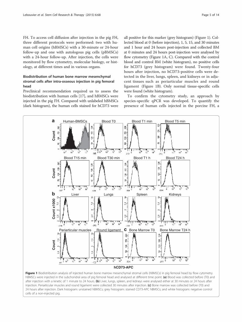
FH. To access cell diffusion after injection in the pig FH,three different protocols were performed: two with hu-man cell origins (hBMSCs) with a 30-minute or 24-hourfollow-up and one with autologous pig cells (pBMSCs)with a 24-hour follow-up. After injection, the cells weremonitored by flow cytometry, molecular biology, or hist-ology, at different times and in various organs.
Biodistribution of human bone marrow mesenchymalstromal cells after intra-osseous injection in pig femoralheadPreclinical recommendation required us to assess thebiodistribution with human cells [17], and hBMSCs wereinjected in the pig FH. Compared with unlabeled hBMSCs(dark histogram), the human cells stained for hCD73 were
Blood T0Human-BMSCs
Co
un
t
Blood T30 minBlood T15 min
Co
un
t
LungsLiver
Co
un
t x1
000
Round ligamentPeriarticular muscles
Co
un
t
hCD73
a
b
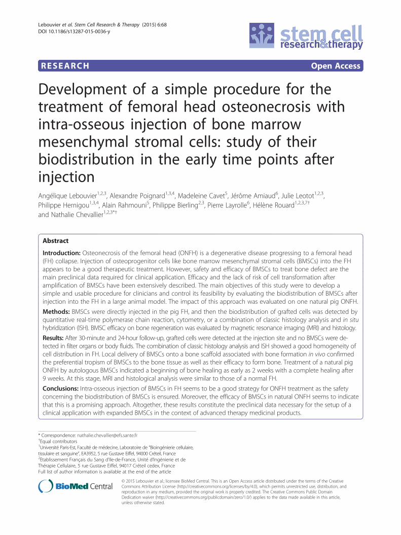
Figure 1 Biodistribution analysis of injected human bone marrow mesenchBMSCs were injected in the subchondral area of pig femoral head and anafter injection with a kinetic of 1 minute to 24 hours. (b) Liver, lungs, spleeinjection. Periarticular muscles and round ligament were collected 30 minu24 hours after injection. Dark histogram: unstained hBMSCs; grey histogramcells of a non-injected pig.
all positive for this marker (grey histogram) (Figure 1). Col-lected blood at 0 (before injection), 1, 5, 15, and 30 minutesand 1 hour and 24 hours post-injection and collected BMat 0 minutes and 24 hours post-injection were analysed byflow cytometry (Figure 1A, C). Compared with the controlblood and control BM (white histogram), no positive cellsfor hCD73 (grey histogram) were found. Twenty-fourhours after injection, no hCD73-positive cells were de-tected in the liver, lungs, spleen, and kidneys or in adja-cent tissues such as periarticular muscles and roundligament (Figure 1B). Only normal tissue-specific cellswere found (white histogram).To confirm the cytometry study, an approach by
species-specific qPCR was developed. To quantify thepresence of human cells injected in the porcine FH, a
Blood T1 min Blood T5 min
Blood T1 h Blood T24 h
Spleen Kidneys
Bone Marrow T0 Bone Marrow T24 h
-APC
c
hymal stromal cells (hBMSCs) in pig femoral head by flow cytometry.alysed at different time point. (a) Blood was collected before (T0) andn, and kidneys were analysed either at 30 minutes or 24 hours aftertes after injection. (c) Bone marrow was collected before (T0) and: stained CD73-APC hBMSCs; and white histogram: negative control
0
500
1000
1500
2000
2500
3000
3500
26 27 28 29 30 31 32 33 34
Average Ct
Nu
mb
er o
f ce
lls
y = -3,526x + 38,163
R2 = 0,9983
20232629323538
1,0 1,5 2,0 2,5 3,0 3,5 4,0
LOG [number of cells]
Ct
26
28
30
32
34
1639
820
3279
410205 102 51 1326
Ct
n = 24
b
c
a
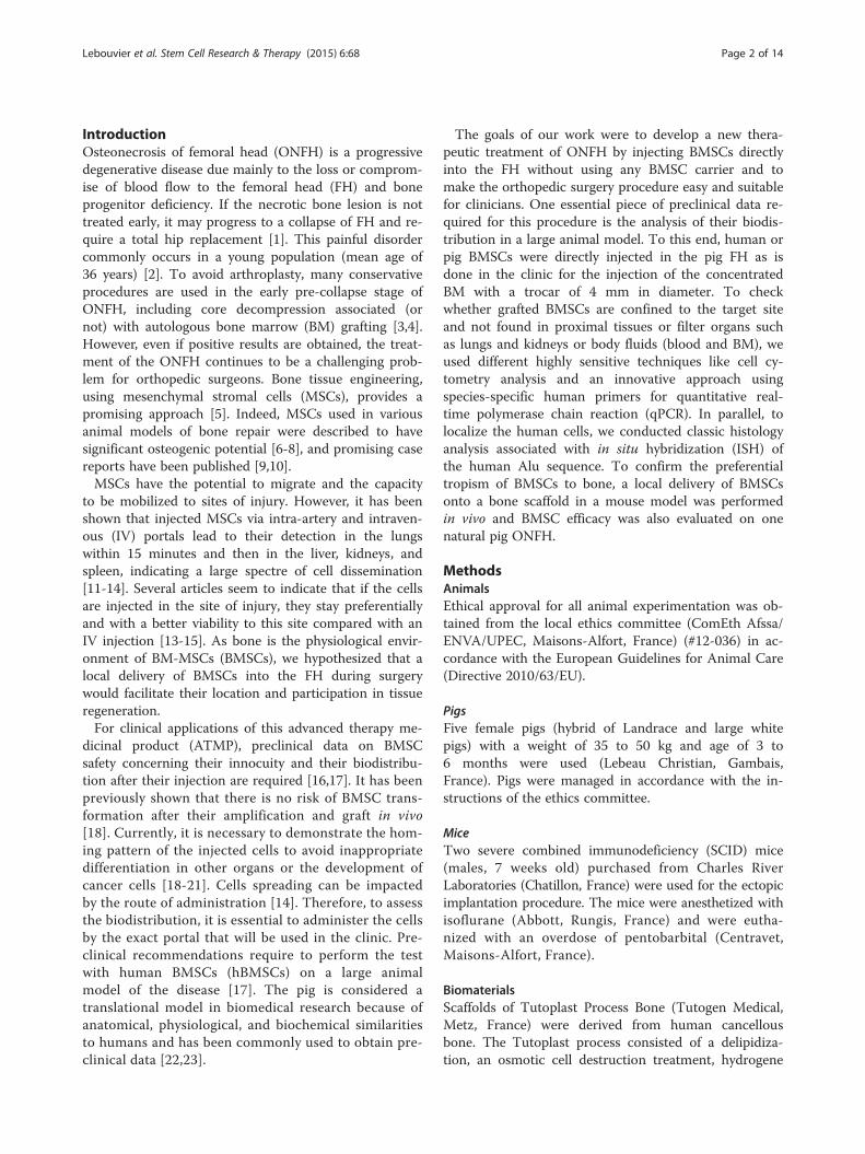
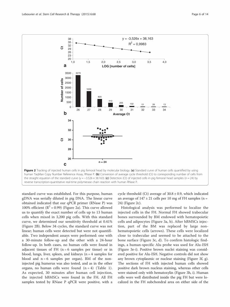
Figure 2 Tracking of injected human cells in pig femoral head by molecular biology. (a) Standard curve of human cells quantified by usinghuman TaqMan Copy Number Reference Assay, RNase P. (b) Conversion of average cycle threshold (Ct) to corresponding number of cells fromthe straight equation of the standard curve (y = −3.526 × 38.163). (c) Detection (Ct) of injected cells in pig femoral head samples (n = 24) byreverse transcription-quantitative real-time polymerase chain reaction with human RNase P.
Lebouvier et al. Stem Cell Research & Therapy (2015) 6:68 Page 6 of 14
standard curve was established. For this purpose, humangDNA was serially diluted in pig DNA. The linear curveobtained indicated that our qPCR primer (RNase P) was100% efficient (R2 = 0.99) (Figure 2a). This curve allowedus to quantify the exact number of cells up to 13 humancells when mixed in 3,200 pig cells. With this standardcurve, we determined our sensitivity threshold at 0.41%(Figure 2B). Below 34 cycles, the standard curve was notlinear; human cells were detected but were not quantifi-able. Two independent assays were performed: one witha 30-minute follow-up and the other with a 24-hourfollow-up. In both cases, no human cells were found inadjacent tissues of FH (n = 6 samples per tissue) or inblood, lungs, liver, spleen, and kidneys (n = 4 samples forblood and n = 6 samples per organ). BM of the non-injected pig humerus was also tested, and as in the otherorgans, no human cells were found (n = 4) (Table 1).As expected, 30 minutes after human cell injection,the injected hBMSCs were found in the FH. All FHsamples tested by RNase P qPCR were positive, with a
cycle threshold (Ct) average of 30.8 ± 0.9, which indicatedan average of 147 ± 21 cells per 10 mg of FH samples (n =24) (Figure 2c).Histological analysis was performed to localize the
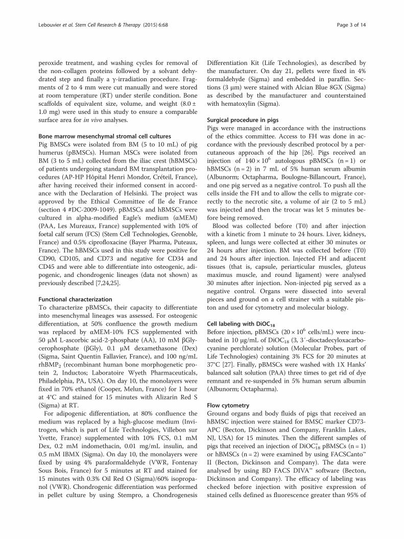
injected cells in the FH. Normal FH showed trabecularbones surrounded by BM endowed with hematopoieticcells and adipocytes (Figure 3a, b). After hBMSCs injec-tion, part of the BM was replaced by large non-hematopoietic cells (arrows). These cells were localizedclose to trabeculae and seemed to be attached to thebone surface (Figure 3c, d). To confirm histologic find-ings, a human-specific Alu probe was used for Alu-ISH(Figure 3e-i). Positive brown nuclei staining was consid-ered positive for Alu-ISH. Negative controls did not showany brown cytoplasmic or nuclear staining (Figure 3f, g).The sections of FH with injected human cells showedpositive dark brown nucleus staining, whereas other cellswere stained only with hematoxylin (Figure 3h, i). Humancells were well distributed inside the pig FH but were lo-calized in the FH subchondral area on either side of the
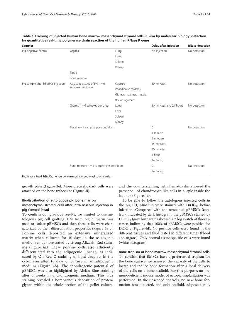
Table 1 Tracking of injected human bone marrow mesenchymal stromal cells in vivo by molecular biology: detectionby quantitative real-time polymerase chain reaction of the human RNase P gene
Samples Delay after injection RNase detection
Pig negative control Organs Lung No injection No detection
Liver
Spleen
Kidney
Blood
Bone marrow
Pig sample after hBMSCs injection Adjacent tissues of FH n = 6samples per tissue
Capsule 30 minutes No detection
Periarticular muscles
Gluteus maximus muscle
Round ligament
Organs n = 6 samples per organ Lung 30 minutes and 24 hours No detection
Liver
Spleen
Kidney
Blood n = 4 samples per condition 0 No detection
1 minute
5 minutes
15 minutes
30 minutes
1 hour
24 hours
Bone marrow n = 4 samples per condition 0 No detection
24 hours
FH, femoral head; hBMSCs, human bone marrow mesenchymal stromal cells.
Lebouvier et al. Stem Cell Research & Therapy (2015) 6:68 Page 7 of 14
growth plate (Figure 3e). More precisely, dark cells wereattached on the bone trabeculae (Figure 3i).
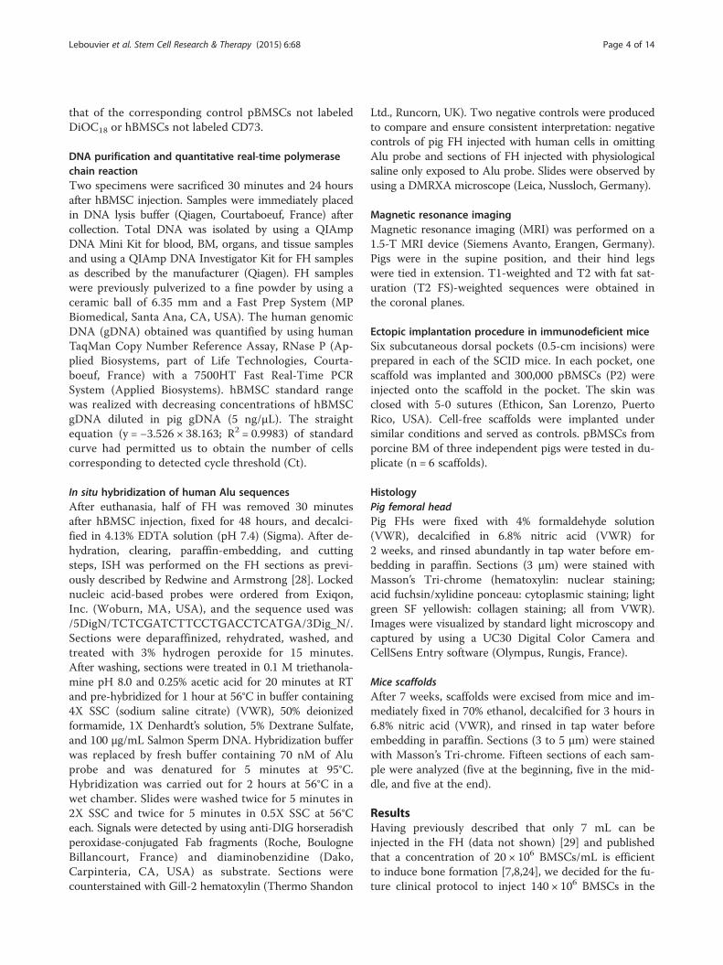
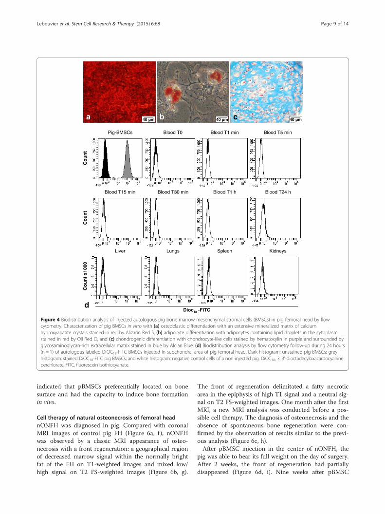
Biodistribution of autologous pig bone marrowmesenchymal stromal cells after intra-osseous injection inpig femoral headTo confirm our previous results, we wanted to use au-tologous pig cell grafting. BM from pig humerus wasused to isolate pBMSCs and then these cells were char-acterized by their differentiation properties (Figure 4a-c).Porcine cells deposited an extensive mineralizedmatrix when cultured for 10 days in the osteogenicmedium as demonstrated by strong Alizarin Red stain-ing (Figure 4a). These porcine cells also efficientlydifferentiated into the adipogenic lineage, as indi-cated by Oil Red O staining of lipid droplets in thecytoplasm after 10 days of culture in an adipogenicmedium (Figure 4b). The chondrogenic potential ofpBMSCs was also highlighted by Alcian Blue stainingafter 3 weeks in a chondrogenic medium. This bluestaining revealed a homogenous deposition of proteo-glycan within the whole section of the pellet culture,
and the counterstaining with hematoxylin showed thepresence of chondrocyte-like cells in purple inside thelacunae (Figure 4c).To be able to follow the autologous injected cells in
the pig FH, pBMSCs were stained with DiOC18 beforeinjection. Compared with the unstained pBMSCs (con-trol), indicated by dark histogram, the pBMSCs stained byDiOC18 (grey histogram) showed a 2 log switch of fluores-cence, indicating that 100% of pBMSCs were positive forDiOC18 (Figure 4d). No positive cells were found in thedifferent tissues and fluid tested in different times (bloodand organs). Only normal tissue-specific cells were found(white histogram).
Bone tropism of bone marrow mesenchymal stromal cellsTo confirm that BMSCs have a preferential tropism forthe bone surface, we assessed the capacity of the cells tolocate and induce bone formation after a local deliveryof the cells on a bone scaffold. For this purpose, an im-munodeficient mouse model of ectopic implantation wasperformed. In the unseeded controls, no new bone for-mation was detected, and only scaffold, adipose tissue,
h i
e
h
i
50µM50µM
50µM
f g
50µM
c
d
c d
a b
50µM
50µM
40µM
40µM
Figure 3 Detection of injected cells in pig femoral head (FH) 30 minutes after human bone marrow mesenchymal stromal cell (hBMSCs)injection. (a-e) Masson’s Tri-chrome staining. (a, b) Normal pig FH sections. (c-e) Injected pig FH sections. Arrows indicate the hBMSC area. (e)Localization of sections (c), (d), (h), and (i). (f-i) In situ hybridization of human Alu sequences (Alu-ISH). (f) Negative control tissue of pig FH injectedwith human cells in omitting Alu probe stained with the hematoxylin only. (g) Negative control tissue of pig FH injected with physiological salinewithout human cells, with Alu probe and hematoxylin counterstaining. (h, i) Injected pig FH with human cells detected by Alu-probestaining and counterstained with the hematoxylin. The positive nuclei for Alu-probe staining appear in dark brown (arrows). Magnifications:10× (a, c, f-h), 20× (b, d), 1× (e), and 40× (i).
Lebouvier et al. Stem Cell Research & Therapy (2015) 6:68 Page 8 of 14
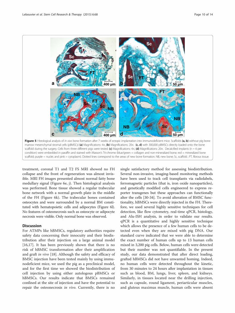
and loosely organized connective tissues were present(Figure 5a, b). In contrast, when pBMSCs were addedonto the scaffold during the surgery, histological analysis
revealed new bone formation with bone tissue contain-ing osteocyte-like cells and osteoblast-like cell liningat the surface (Figure 5c, d). Altogether, this result
40 µm40 µm40 µm ba c
Blood T0 Blood T1 min Blood T5 minPig-BMSCs
Blood T30 min Blood T1 h Blood T24 hBlood T15 min
Co
un
t
Lungs SpleenLiver
Co
un
t x1
000
Kidneys
Dioc18 -FITC
Co
un
t
d
Figure 4 Biodistribution analysis of injected autologous pig bone marrow mesenchymal stromal cells (BMSCs) in pig femoral head by flowcytometry. Characterization of pig BMSCs in vitro with (a) osteoblastic differentiation with an extensive mineralized matrix of calciumhydroxyapatite crystals stained in red by Alizarin Red S, (b) adipocyte differentiation with adipocytes containing lipid droplets in the cytoplasmstained in red by Oil Red O, and (c) chondrogenic differentiation with chondrocyte-like cells stained by hematoxylin in purple and surrounded byglycosaminoglycan-rich extracellular matrix stained in blue by Alcian Blue. (d) Biodistribution analysis by flow cytometry follow-up during 24 hours(n = 1) of autologous labeled DiOC18-FITC BMSCs injected in subchondral area of pig femoral head. Dark histogram: unstained pig BMSCs; greyhistogram: stained DiOC18-FITC pig BMSCs; and white histogram: negative control cells of a non-injected pig. DiOC18, 3, 3′-dioctadecyloxacarbocyanineperchlorate; FITC, fluorescein isothiocyanate.
Lebouvier et al. Stem Cell Research & Therapy (2015) 6:68 Page 9 of 14
indicated that pBMSCs preferentially located on bonesurface and had the capacity to induce bone formationin vivo.
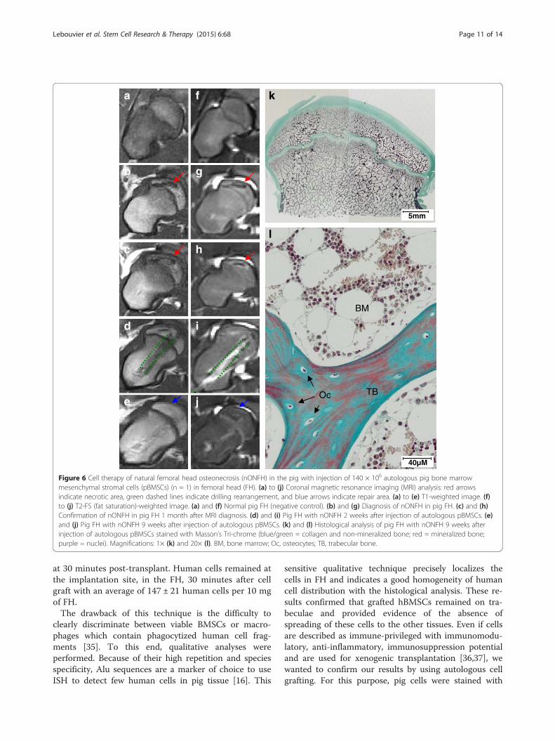
Cell therapy of natural osteonecrosis of femoral headnONFH was diagnosed in pig. Compared with coronalMRI images of control pig FH (Figure 6a, f ), nONFHwas observed by a classic MRI appearance of osteo-necrosis with a front regeneration: a geographical regionof decreased marrow signal within the normally brightfat of the FH on T1-weighted images and mixed low/high signal on T2 FS-weighted images (Figure 6b, g).
The front of regeneration delimitated a fatty necroticarea in the epiphysis of high T1 signal and a neutral sig-nal on T2 FS-weighted images. One month after the firstMRI, a new MRI analysis was conducted before a pos-sible cell therapy. The diagnosis of osteonecrosis and theabsence of spontaneous bone regeneration were con-firmed by the observation of results similar to the previ-ous analysis (Figure 6c, h).After pBMSC injection in the center of nONFH, the
pig was able to bear its full weight on the day of surgery.After 2 weeks, the front of regeneration had partiallydisappeared (Figure 6d, i). Nine weeks after pBMSC
dc 400 µm 50 µm
400 µm 50 µmba
Sc
NB
NB
FT
NB
NB
ScSc
FT
Sc
FTSc FT
Sc
Figure 5 Histological analysis of in vivo bone formation after 7 weeks of ectopic implantation into immunodeficient mice. Scaffolds (a, b) without pig bonemarrow mesenchymal stromal cells (pBMSCs) (a) Magnifications: 4×, (b) Magnifications: 20×. (c, d) with 300,000 pBMSCs directly loaded onto the bonescaffold during the surgery. Cells from three different pigs were tested. (c) Magnifications: 4×, (d) Magnifications: 20×. Decalcified implants (n = 6 percondition) were embedded in paraffin and stained with Masson’s Tri-chrome (blue/green = collagen and non-mineralized bone; red = mineralized bonescaffold; purple = nuclei; and pink = cytoplasm). Dotted lines correspond to the areas of new bone formation. NB, new bone; Sc, scaffold ; FT, fibrous tissue.
Lebouvier et al. Stem Cell Research & Therapy (2015) 6:68 Page 10 of 14
treatment, coronal T1 and T2 FS MRI showed no FHcollapse and the front of regeneration was almost invis-ible. MRI FH images presented almost normal fatty bonemedullary signal (Figure 6e, j). Then histological analysiswas performed. Bone tissue showed a regular trabecularbone network with a normal growth plate in the middleof the FH (Figure 6k). The trabecular bones containedosteocytes and were surrounded by a normal BM consti-tuted with hematopoietic cells and adipocytes (Figure 6l).No features of osteonecrosis such as osteocyte or adipocytenecrosis were visible. Only normal bone was observed.
DiscussionFor ATMPs like hBMSCs, regulatory authorities requiresafety data concerning their innocuity and their biodis-tribution after their injection on a large animal model[16,17]. It has been previously shown that there is norisk of hBMSC transformation after their amplificationand graft in vivo [18]. Although the safety and efficacy ofBMSC injection have been tested mainly by using immu-nodeficient mice, we used the pig as a preclinical model,and for the first time we showed the biodistribution ofcell injection by using either autologous pBMSCs orhBMSCs. Our results indicate that BMSCs remainedconfined at the site of injection and have the potential torepair the osteonecrosis in vivo. Currently, there is no
single satisfactory method for assessing biodistribution.Several non-invasive, imaging-based monitoring methodshave been used to track cell transplants via radiolabels,ferromagnetic particles (that is, iron oxide nanoparticles),and genetically modified cells engineered to express re-porter transgenes but these approaches can functionallyalter the cells [30-34]. To avoid alteration of BMSC func-tionality, hBMSCs were directly injected in the FH. There-fore, we used several highly sensitive techniques for celldetection, like flow cytometry, real-time qPCR, histology,and Alu-ISH analysis, in order to validate our results.qPCR is a quantitative and highly sensitive techniquewhich allows the presence of a few human cells to be de-tected even when they are mixed with pig DNA. Ourstandard curve indicated that we were able to determinethe exact number of human cells up to 13 human cellsmixed in 3,200 pig cells. Below, human cells were detectedbut their number was not quantifiable. In the presentstudy, our data demonstrated that after direct loading,grafted hBMSCs did not have unwanted homing. Indeed,no human cells were detected throughout the kinetic,from 30 minutes to 24 hours after implantation in tissuessuch as blood, BM, lungs, liver, spleen, and kidneys.Similarly, in tissues located near the drilling injection,such as capsule, round ligament, periarticular muscles,and gluteus maximus muscle, human cells were absent
TBOc
BM
40µM
l
k
5mm
a f
b g
d i
e j
c h
Figure 6 Cell therapy of natural femoral head osteonecrosis (nONFH) in the pig with injection of 140 × 106 autologous pig bone marrowmesenchymal stromal cells (pBMSCs) (n = 1) in femoral head (FH). (a) to (j) Coronal magnetic resonance imaging (MRI) analysis: red arrowsindicate necrotic area, green dashed lines indicate drilling rearrangement, and blue arrows indicate repair area. (a) to (e) T1-weighted image. (f)to (j) T2-FS (fat saturation)-weighted image. (a) and (f) Normal pig FH (negative control). (b) and (g) Diagnosis of nONFH in pig FH. (c) and (h)Confirmation of nONFH in pig FH 1 month after MRI diagnosis. (d) and (i) Pig FH with nONFH 2 weeks after injection of autologous pBMSCs. (e)and (j) Pig FH with nONFH 9 weeks after injection of autologous pBMSCs. (k) and (l) Histological analysis of pig FH with nONFH 9 weeks afterinjection of autologous pBMSCs stained with Masson’s Tri-chrome (blue/green = collagen and non-mineralized bone; red = mineralized bone;purple = nuclei). Magnifications: 1× (k) and 20× (l). BM, bone marrow; Oc, osteocytes; TB, trabecular bone.
Lebouvier et al. Stem Cell Research & Therapy (2015) 6:68 Page 11 of 14
at 30 minutes post-transplant. Human cells remained atthe implantation site, in the FH, 30 minutes after cellgraft with an average of 147 ± 21 human cells per 10 mgof FH.The drawback of this technique is the difficulty to
clearly discriminate between viable BMSCs or macro-phages which contain phagocytized human cell frag-ments [35]. To this end, qualitative analyses wereperformed. Because of their high repetition and speciesspecificity, Alu sequences are a marker of choice to useISH to detect few human cells in pig tissue [16]. This
sensitive qualitative technique precisely localizes thecells in FH and indicates a good homogeneity of humancell distribution with the histological analysis. These re-sults confirmed that grafted hBMSCs remained on tra-beculae and provided evidence of the absence ofspreading of these cells to the other tissues. Even if cellsare described as immune-privileged with immunomodu-latory, anti-inflammatory, immunosuppression potentialand are used for xenogenic transplantation [36,37], wewanted to confirm our results by using autologous cellgrafting. For this purpose, pig cells were stained with
Lebouvier et al. Stem Cell Research & Therapy (2015) 6:68 Page 12 of 14
DiOC18, a green fluorescent membrane dye which doesnot induce adverse cellular effects [38]. The only draw-back is that the cells can lose half of their fluorescenceafter doubling. However, human and pig BMSC doublingtime is between 50 and 55 hours [7,39], and our follow-up does not exceed 24 hours. One day after pBMSC in-jection in the FH, the flow cytometry analysis indicatedthat the injected cells were detected neither in blood norin filter organs.In contrast to studies in which cells are injected intra-
venously, our results confirm that injected cells prefer-entially stay in the injected tissue [13,14,40,41]. In ourcase, this can be explained by the fact that bone is thephysiological niche of BMSCs. We confirmed their trop-ism for the bone surface with the ectopic bone implant-ation model in immunodeficient mice as only bonescaffold receiving pBMSCs during surgery gives boneformation. Moreover, our results with pBMSCs are inaccordance with recent data we published showing thathBMSCs stay on the scaffold and do not migrate toother organs and this was the case throughout the6 weeks of the study [42]. On the other hand, our dataof grafted autologous pBMSCs in nONFH in the pigsupport that a local delivery of BMSCs into the FH dur-ing surgery facilitates their attachment and their partici-pation in tissue regeneration. Altogether, these resultsindicate that a direct injection of BMSCs on the bonesite is a safe procedure, even if a 4-mm diameter trocaris used to inject the cells into the FH. Clearly, these re-sults indicate that there is no need to use a cell carrierlike hydrogel or a plug after cell injection to obtain goodcell localization without dissemination throughout thebody. This is an important point as it was not possibleto inject the cells into the FH when cells were mixedwith hydrogel (data not shown). Finally, the feasibility ofthis approach is supported by the commonly used treat-ment of injection of concentrate BM in FH for the earlystages of ONFH. Effectively, no side effects and even apositive effect have been observed in patients [3,4,43].Our preliminary data for the efficacy of grafted autolo-
gous BMSCs in nONFH in the pig seem promising andshow a potential of BMSCs in the repair of necrosisin vivo. However, nONFH is rare in pigs. To have a sta-tistically relevant result of the effectiveness of BMSCs onthis painful disease, it is necessary to test this approachon one of the models of ONFH which has been devel-oped in either emu or pig and to conduct a study on alarge number of animals [26,44]. As the effectiveness ofBMSCs has been proven and we and others showed thesafety of this approach, the second strategy could be togo directly to the clinic [6,7,13,14,18,40,41].BMSC graft is a promising therapeutic approach for
treating ONFH but the other question is the effective-ness of the BMSCs from patients with ONFH. Previous
data from Yoo et al. described the good osteogenic abil-ities in vitro of BMSCs from patients with osteonecrosis[45]. Altogether, these data indicate that the use of au-tologous BMSCs for a therapeutic treatment of ONFH isa feasible strategy.
ConclusionsIn summary, we have demonstrated in a large animalmodel that the intra-osseous portal is a safe and promis-ing strategy for cell therapy treatment of FH osteonecro-sis. As it has been previously shown that there is no riskof transformation of hBMSCs after their amplificationand graft in vivo [18], this new study contributes to thepreclinical data which are required for the clinical appli-cation of hBMSCs in the context of ATMPs.
AbbreviationsαMEM: alpha-modified Eagle’s medium; Alu-ISH: in situ hybridization ofhuman Alu sequences; ATMP: advanced therapy medicinal product;BM: bone marrow; BMSC: bone marrow mesenchymal stromal cell; Ct: cyclethreshold; Dex: dexamethasone; DiOC18: 3, 3′-dioctadecyloxacarbocyanineperchlorate; FCS: foetal calf serum; FH: femoral head; FS: fat saturation;gDNA: genomic deoxyribonucleic acid; hBMSC: human bone marrowmesenchymal stromal cell; ISH: in situ hybridization; IV: intravenous;MRI: magnetic resonance imaging; MSC: mesenchymal stromal cell;nONFH: natural osteonecrosis of femoral head; ONFH: osteonecrosis of thefemoral head; pBMSC: pig bone marrow mesenchymal stromal cell;qPCR: quantitative real-time polymerase chain reaction; RT: room temperature;SCID: severe combined immunodeficiency; SSC: sodium saline citrate.
Competing interestsThe authors declare that they have no competing interests.
Authors’ contributionsAL carried out the BMSC culture and characterization, the biodistributionstudies in flow cytometry, molecular biology, the ectopic implantationprocedure, and all histological studies; performed data analysis andinterpretation; and drafted the manuscript. AP carried out the surgicalprocedure in pigs and BM collection, participated in MRI analysis, drafted thesurgical procedure part of the Methods section, and helped to revise themanuscript. MC performed the MRI studies and analyses and drafted the MRIpart of the Methods and Results sections. JA carried out the study of ISH ofhuman Alu sequences and drafted the part of Methods sectioncorresponding to this procedure. JL participated in the biology molecularand flow cytometry studies and helped to revise the manuscript. PHconceived the injection procedure in FH and participated in the surgicalprocedure in pigs and in human BM collection. AR performed the MRIstudies and analyses and helped to revise the manuscript. PB participated inthe design of the study and helped to revise the manuscript. PL participatedin the conception of the study and drafted the result section correspondingto the study of ISH of human Alu sequences. HR participated in conceptionand coordination of the study and in human BM collection and helped torevise the manuscript. NC designed and supervised the study, performeddata analysis and interpretation, and drafted the manuscript. All authors readand approved the final manuscript.
AcknowledgementsThis work was supported by the 7th Framework Program of the EuropeanCommission through the REBORNE (Regenerating BOne defects using Newbiomedical Engineering approaches) project (Health-2009-1.4.2-241879) andEFS Ile de France. We thank the Centre de Recherches ChirurgicalesDominique Chopin directed by René Yiou for the use of their animalplatform facilities. The authors are grateful to Philippe Druelle and PhilippeMario for their invaluable help during surgery and animal care. We thank thehistopathology of human and animal models service of the Pasteur Institute ofParis and give special thanks to Laurence Fiette.
Lebouvier et al. Stem Cell Research & Therapy (2015) 6:68 Page 13 of 14
Author details1Université Paris-Est, Faculté de médecine, Laboratoire de “Bioingénierie cellulaire,tissulaire et sanguine”, EA3952, 5 rue Gustave Eiffel, 94000 Créteil, France.2Etablissement Français du Sang d’Ile-de-France, Unité d’Ingénierie et de ThérapieCellulaire, 5 rue Gustave Eiffel, 94017 Créteil cedex, France. 3Inserm UMR955, IMRB,51 Avenue du Maréchal de Lattre de Tassigny, 94000 Créteil, France. 4Service dechirurgie orthopédique et traumatologie, AP-HP Hôpital Henri-Mondor, 51 Avenuedu Maréchal de Lattre de Tassigny, 94000 Créteil, France. 5Service de radiologieAlbert Chenevier, AP-HP Hôpital Henri-Mondor, 51 Avenue du Maréchal de Lattrede Tassigny, 94000 Créteil, France. 6Inserm U957, Laboratory for Pathophysiologyof Bone Resorption, Faculty of Medicine, University of Nantes, 1 rue Gaston Veil,44035 Nantes cedex 1, France. 7AP-HP Hôpital Henri-Mondor – A. Chenevier, Ser-vice hospitalier, 51 Avenue du Maréchal de Lattre de Tassigny, 94000 Créteil,France.
Received: 20 August 2014 Revised: 21 August 2014Accepted: 4 March 2015
References1. Levasseur R. Mechanisms of osteonecrosis. Joint Bone Spine. 2008;75:639–42.2. Hungerford DS, Jones LC. Asymptomatic osteonecrosis: Should it be
treated? Clin Orthop Relat Res. 2004;429:124–30.3. Hernigou P, Beaujean F. Treatment of osteonecrosis with autologous bone
marrow grafting. Clin Orthop Relat Res. 2002;405:14–23.4. Gangji V, De Maertelaer V, Hauzeur JP. Autologous bone marrow cell
implantation in the treatment of non-traumatic osteonecrosis of the femoralhead: five year follow-up of a prospective controlled study. Bone.2011;49:1005–9.
5. Wang X, Wang Y, Gou W, Lu Q, Peng J, Lu S. Role of mesenchymal stemcells in bone regeneration and fracture repair: A review. Int Orthop.2013;37:2491–8.
6. Petite H, Viateau V, Bensaid W, Meunier A, de Pollak C, Bourguignon M,et al. Tissue-engineered bone regeneration. Nat Biotechnol. 2000;18:959–63.
7. Chevallier N, Anagnostou F, Zilber S, Bodivit G, Maurin S, Barrault A, et al.Osteoblastic differentiation of human mesenchymal stem cells with plateletlysate. Biomaterials. 2010;31:270–8.
8. Leotot J, Coquelin L, Bodivit G, Bierling P, Hernigou P, Rouard H, et al.Platelet lysate coating on scaffolds directly and indirectly enhances cellmigration, improving bone and blood vessel formation. Acta Biomater.2013;9:6630–40.
9. Quarto R, Mastrogiacomo M, Cancedda R, Kutepov SM, Mukhachev V,Lavroukov A, et al. Repair of large bone defects with the use of autologousbone marrow stromal cells. N Engl J Med. 2001;344:385–6.
10. Marcacci M, Kon E, Moukhachev V, Lavroukov A, Kutepov S, Quarto R, et al.Stem cells associated with macroporous bioceramics for long bone repair:6- to 7-year outcome of a pilot clinical study. Tissue Eng. 2007;13:947–55.
11. Gao J, Dennis JE, Muzic RF, Lundberg M, Caplan AI. The dynamic in vivodistribution of bone marrow-derived mesenchymal stem cells after infusion.Cells Tissues Organs. 2001;169:12–20.
12. Lee RH, Pulin AA, Seo MJ, Kota DJ, Ylostalo J, Larson BL, et al. Intravenoushmscs improve myocardial infarction in mice because cells embolized inlung are activated to secrete the anti-inflammatory protein tsg-6. Cell StemCell. 2009;5:54–63.
13. Ramot Y, Meiron M, Toren A, Steiner M, Nyska A. Safety and biodistributionprofile of placental-derived mesenchymal stromal cells (plx-pad) followingintramuscular delivery. Toxicol Pathol. 2009;37:606–16.
14. Tolar J, O’Shaughnessy MJ, Panoskaltsis-Mortari A, McElmurry RT, Bell S,Riddle M, et al. Host factors that impact the biodistribution and persistenceof multipotent adult progenitor cells. Blood. 2006;107:4182–8.
15. Toupet K, Maumus M, Peyrafitte JA, Bourin P, van Lent PL, Ferreira R,et al. Long-term detection of human adipose-derived mesenchymalstem cells after intraarticular injection in scid mice. Arthritis Rheum.2013;65:1786–94.
16. Sensebe L, Fleury-Cappellesso S. Biodistribution of mesenchymal stem/stromalcells in a preclinical setting. Stem Cells Int. 2013;2013:678063.
17. Sharpe ME, Morton D, Rossi A. Nonclinical safety strategies for stem celltherapies. Toxicol Appl Pharmacol. 2012;262:223–31.
18. Tarte K, Gaillard J, Lataillade JJ, Fouillard L, Becker M, Mossafa H, et al.Clinical-grade production of human mesenchymal stromal cells: Occurrenceof aneuploidy without transformation. Blood. 2010;115:1549–53.
19. Breitbach M, Bostani T, Roell W, Xia Y, Dewald O, Nygren JM, et al. Potentialrisks of bone marrow cell transplantation into infarcted hearts. Blood.2007;110:1362–9.
20. Zhu W, Xu W, Jiang R, Qian H, Chen M, Hu J, et al. Mesenchymal stem cellsderived from bone marrow favor tumor cell growth in vivo. Exp Mol Pathol.2006;80:267–74.
21. Djouad F, Plence P, Bony C, Tropel P, Apparailly F, Sany J, et al.Immunosuppressive effect of mesenchymal stem cells favors tumor growthin allogeneic animals. Blood. 2003;102:3837–44.
22. Helke KL, Swindle MM. Animal models of toxicology testing: the role ofpigs. Expert Opin Drug Metab Toxicol. 2013;9:127–39.
23. Merrifield CA, Lewis M, Claus SP, Beckonert OP, Dumas ME, Duncker S, et al.A metabolic system-wide characterisation of the pig: A model for humanphysiology. Mol Biosyst. 2011;7:2577–88.
24. Coquelin L, Fialaire-Legendre A, Roux S, Poignard A, Bierling P, Hernigou P,et al. In vivo and in vitro comparison of three different allografts vitalizedwith human mesenchymal stromal cells. Tissue Eng Part A. 2012;18:1921–31.
25. Bouderlique T, Henault E, Lebouvier A, Frescaline G, Bierling P, Rouard H,et al. Pleiotrophin commits human bone marrow mesenchymal stromal cellstowards hypertrophy during chondrogenesis. PLoS One. 2014;9, e88287.
26. Poignard A, Lebouvier A, Cavet M, Rahmouni A, Flouzat Lachaniette CH,Bierling P, et al. New preclinical porcine model of femoral headosteonecrosis to test mesenchymal stromal cell efficiency in regenerativemedicine. Int Orthop. 2014;38:1837–44.
27. Ozdemir O. Evaluation of human mast cell-mediated cytotoxicity by dioc18target cell labeling in flow cytometry. J Immunol Methods. 2007;319:98–103.
28. Redwine JM, Armstrong RC. In vivo proliferation of oligodendrocyteprogenitors expressing pdgfalphar during early remyelination. J Neurobiol.1998;37:413–28.
29. Hernigou P, Homma Y, Flouzat Lachaniette CH, Poignard A, Allain J,Chevallier N, et al. Benefits of small volume and small syringe for bonemarrow aspirations of mesenchymal stem cells. Int Orthop. 2013;37:2279–87.
30. Bindslev L, Haack-Sorensen M, Bisgaard K, Kragh L, Mortensen S, Hesse B,et al. Labelling of human mesenchymal stem cells with indium-111 forspect imaging: Effect on cell proliferation and differentiation. Eur J NuclMed Mol Imaging. 2006;33:1171–7.
31. Nohroudi K, Arnhold S, Berhorn T, Addicks K, Hoehn M, Himmelreich U. Invivo MRI stem cell tracking requires balancing of detection limit and cellviability. Cell Transplant. 2010;19:431–41.
32. Schmidtke-Schrezenmeier G, Urban M, Musyanovych A, Mailander V,Rojewski M, Fekete N, et al. Labeling of mesenchymal stromal cells with ironoxide-poly(l-lactide) nanoparticles for magnetic resonance imaging: Uptake,persistence, effects on cellular function and magnetic resonance imagingproperties. Cytotherapy. 2011;13:962–75.
33. Love Z, Wang F, Dennis J, Awadallah A, Salem N, Lin Y, et al. Imaging ofmesenchymal stem cell transplant by bioluminescence and pet. J Nucl Med.2007;48:2011–20.
34. Wang F, Dennis JE, Awadallah A, Solchaga LA, Molter J, Kuang Y, et al.Transcriptional profiling of human mesenchymal stem cells transduced withreporter genes for imaging. Physiol Genomics. 2009;37:23–34.
35. Warncke B, Valtink M, Weichel J, Engelmann K, Schafer H. Experimental ratmodel for therapeutic retinal pigment epithelium transplantation–unequivocalmicroscopic identification of human donor cells by in situ hybridisation ofhuman-specific alu sequences. Virchows Arch. 2004;444:74–81.
36. Niemeyer P, Szalay K, Luginbuhl R, Sudkamp NP, Kasten P. Transplantationof human mesenchymal stem cells in a non-autogenous setting for boneregeneration in a rabbit critical-size defect model. Acta Biomater.2010;6:900–8.
37. Maumus M, Guerit D, Toupet K, Jorgensen C, Noel D. Mesenchymal stemcell-based therapies in regenerative medicine: Applications in rheumatology.Stem Cell Res Ther. 2011;2:14.
38. Piriou L, Chilmonczyk S, Genetet N, Albina E. Design of a flow cytometricassay for the determination of natural killer and cytotoxic t-lymphocyteactivity in human and in different animal species. Cytometry.2000;41:289–97.
39. Peterbauer-Scherb A, van Griensven M, Meinl A, Gabriel C, Redl H, Wolbank S.Isolation of pig bone marrow mesenchymal stem cells suitable for one-stepprocedures in chondrogenic regeneration. J Tissue Eng Regen Med.2010;4:485–90.
40. Detante O, Moisan A, Dimastromatteo J, Richard MJ, Riou L, Grillon E, et al.Intravenous administration of 99mtc-hmpao-labeled human mesenchymal
Lebouvier et al. Stem Cell Research & Therapy (2015) 6:68 Page 14 of 14
stem cells after stroke: In vivo imaging and biodistribution. Cell Transplant.2009;18:1369–79.
41. Allers C, Sierralta WD, Neubauer S, Rivera F, Minguell JJ, Conget PA.Dynamic of distribution of human bone marrow-derived mesenchymal stemcells after transplantation into adult unconditioned mice. Transplantation.2004;78:503–8.
42. Leotot J, Lebouvier A, Hernigou P, Bierling P, Rouard H, Chevallier N.Bone-forming capacity and biodistribution of bone marrow-derivedstromal cells directly loaded into scaffolds: A novel and easy approachfor clinical application of bone regeneration. Cell Transplant.2014 Oct 28. [Epub ahead of print].
43. Gangji V, Hauzeur JP, Matos C, De Maertelaer V, Toungouz M, LambermontM. Treatment of osteonecrosis of the femoral head with implantation ofautologous bone-marrow cells. A pilot study. J Bone Joint Surg Am.2004;86-A:1153–60.
44. Conzemius MG, Brown TD, Zhang Y, Robinson RA. A new animal model offemoral head osteonecrosis: One that progresses to human-like mechanicalfailure. J Orthop Res. 2002;20:303–9.
45. Yoo JJ, Song WS, Koo KH, Yoon KS, Kim HJ. Osteogenic abilities of bonemarrow stromal cells are not defective in patients with osteonecrosis. IntOrthop. 2009;33:867–72.
Submit your next manuscript to BioMed Centraland take full advantage of:
• Convenient online submission
• Thorough peer review
• No space constraints or color figure charges
• Immediate publication on acceptance
• Inclusion in PubMed, CAS, Scopus and Google Scholar
• Research which is freely available for redistribution
Submit your manuscript at www.biomedcentral.com/submit