research open access effects of uric acid on … of uric acid on endothelial dysfunction in early...
TRANSCRIPT

RESEARCH Open Access
Effects of uric acid on endothelial dysfunction inearly chronic kidney disease and its mechanismsYu Wang and Xiaorong Bao*
Abstract
Background: An increase in serum uric acid (UA) occurs during the early and middle stages of chronic kidneydisease (CKD) and aggravates the deterioration of kidney function. This study aims to explore the relation betweenUA and endothelial dysfunction in early CKD and its mechanisms in a murine model.
Methods: The experimental animals were randomly divided into three groups (n = 10): sham-operation group(control group), right nephrectomy only group (CKD group) and right nephrectomy with oxonic potassium group(CKD with hyperuricemia group). Furthermore, we analyzed the relation between UA and endothelial dysfunctionindices in early CKD as well as its mechanisms.
Results: Linear regression analysis showed that the level of serum UA had a significant positive correlation withserum endothelin-1 and the percentage of collagen I positive area, but a negative correlation with serum nitricoxide (NO) and NO/endothelin-1 ratio. In addition, the level of serum UA had significant positive correlations withserum malonaldehyde, serum C-reactive protein, serum oxidatively-modified low-density lipoprotein and serumlow-density lipoprotein, but a negative correlation with serum superoxide dismutase.
Conclusions: Endothelial dysfunction in the CKD group was significant and had a positive correlation with the levelof serum UA. Endothelial dysfunction in early CKD with hyperuricemia is perhaps related to oxidative stress,micro-inflammation and lipid oxidation.
Keywords: Early chronic kidney disease, Endothelial dysfunction, Lipid oxidation, Micro-inflammation, Oxidativestress, Uric acid
BackgroundSince previous studies have highlighted the role of serumuric acid (UA) in coronary heart disease [1], the effectand mechanism of UA in cardiovascular disease (CVD)has aroused widespread concern. Serum UA level is notonly the most significant predictor of occurrence of pri-mary hypertension [2], but it is also associated with car-diovascular morbidity and mortality [3-5].Chronic kidney disease (CKD) affects 10–13% of the
general population. CKD patients have an extremelyhigh risk of developing CVD compared with the generalpopulation. Patients in the early stages of CKD are morelikely to convert into CVD rather than progress towardsend-stage renal disease [6]; CVD is the major cause ofdeath in patients with CKD [7]. The increase in serum
UA occurs in the early and middle stages of CKDand aggravates with the deterioration of kidney func-tion [8]. At present, no publication has demonstratedthe effect and mechanism of UA in early CKD withCVD patients. Our earlier clinical study found thatserum UA was increased in patients with stage 2–3CKD and was related to CVD (e.g. left ventricularhypertrophy), indicating that hyperuricemia was asso-ciated with CVD in early CKD.Endothelial dysfunction is an early occurrence in
CVD. Vascular endothelial cells play an important rolein cellular functions, such as modulating angiokinesis,the proliferation and migration of vascular smoothmuscle cells, anti-platelet aggregation, and extracellularmatrix generation. Pathological changes in blood ves-sels, including intima hyperplasia, lumen straightnessand atherosclerosis, are a consequence of endothelialdysfunction. In the clinic, endothelial dysfunction is
* Correspondence: [email protected] of Nephrology, Jinshan Hospital affiliated to Fudan University,No.1508 Longhang Road, Jinshan District, Shanghai 201508, China
EUROPEAN JOURNAL OF MEDICAL RESEARCH
© 2013 Wang and Bao; licensee BioMed Central Ltd. This is an Open Access article distributed under the terms of the CreativeCommons Attribution License (http://creativecommons.org/licenses/by/2.0), which permits unrestricted use, distribution, andreproduction in any medium, provided the original work is properly cited.
Wang and Bao European Journal of Medical Research 2013, 18:26http://www.eurjmedres.com/content/18/1/26
general population. CKD patients have an extremelyhigh risk of developing CVD compared with the generalpopulation. Patients in the early stages of CKD are morelikely to convert into CVD rather than progress towardsend-stage renal disease [6]; CVD is the major cause of
mary hypertension [2], but it is also associated with car-diovascular morbidity and mortality [3-5].Chronic kidney disease (CKD) affects 10
general population. CKD patients have an extremelyhigh risk of developing CVD compared with the general
has aroused widespread concern. Serum UA level is notonly the most significant predictor of occurrence of pri-mary hypertension [2], but it is also associated with car-diovascular morbidity and mortality [3-5].
uric acid (UA) in coronary heart disease [1], the effectand mechanism of UA in cardiovascular disease (CVD)has aroused widespread concern. Serum UA level is notonly the most significant predictor of occurrence of pri-mary hypertension [2], but it is also associated with car-
Since previous studies have highlighted the role of serumuric acid (UA) in coronary heart disease [1], the effectand mechanism of UA in cardiovascular disease (CVD)
Early chronic kidney disease, Endothelial dysfunction, Lipid oxidation, Micro-inflammation, Oxidative
of serum UA. Endothelial dysfunction in early CKD with hyperuricemia is perhaps related to oxidative stress,
Early chronic kidney disease, Endothelial dysfunction, Lipid oxidation, Micro-inflammation, Oxidative
low-density lipoprotein, but a negative correlation with serum superoxide dismutase.
Endothelial dysfunction in the CKD group was significant and had a positive correlation with the levelof serum UA. Endothelial dysfunction in early CKD with hyperuricemia is perhaps related to oxidative stress,
Early chronic kidney disease, Endothelial dysfunction, Lipid oxidation, Micro-inflammation, Oxidative
oxide (NO) and NO/endothelin-1 ratio. In addition, the level of serum UA had significant positive correlations withserum malonaldehyde, serum C-reactive protein, serum oxidatively-modified low-density lipoprotein and serumlow-density lipoprotein, but a negative correlation with serum superoxide dismutase.
Endothelial dysfunction in the CKD group was significant and had a positive correlation with the levelof serum UA. Endothelial dysfunction in early CKD with hyperuricemia is perhaps related to oxidative stress,
Linear regression analysis showed that the level of serum UA had a significant positive correlation withserum endothelin-1 and the percentage of collagen I positive area, but a negative correlation with serum nitricoxide (NO) and NO/endothelin-1 ratio. In addition, the level of serum UA had significant positive correlations withserum malonaldehyde, serum C-reactive protein, serum oxidatively-modified low-density lipoprotein and serum
The experimental animals were randomly divided into three groups (n = 10): sham-operation group(control group), right nephrectomy only group (CKD group) and right nephrectomy with oxonic potassium group(CKD with hyperuricemia group). Furthermore, we analyzed the relation between UA and endothelial dysfunction
Linear regression analysis showed that the level of serum UA had a significant positive correlation with
disease (CKD) and aggravates the deterioration of kidney function. This study aims to explore the relation betweenUA and endothelial dysfunction in early CKD and its mechanisms in a murine model.
The experimental animals were randomly divided into three groups (n = 10): sham-operation group(control group), right nephrectomy only group (CKD group) and right nephrectomy with oxonic potassium group
An increase in serum uric acid (UA) occurs during the early and middle stages of chronic kidneydisease (CKD) and aggravates the deterioration of kidney function. This study aims to explore the relation between
An increase in serum uric acid (UA) occurs during the early and middle stages of chronic kidneydisease (CKD) and aggravates the deterioration of kidney function. This study aims to explore the relation between
early chronic kidney disease and its mechanisms

associated with CVD, and specifically with hyperten-sion, coronary heart disease, thrombosis, and cardiacinsufficiency [9-11]. Endothelial cells can synthesizeand excrete many important substances, the serumlevels of which will change in the condition of endothe-lial dysfunction, such as decreased nitric oxide (NO)level, increased endothelin-1 (ET-1) level, and de-creased NO/ET-1 ratio [12,13]. Thus, the content ofNO and ET-1 is important for evaluating the functionof endothelial cells. Collagen I deposition in the ar-tery is also related to the function of endothelial cells;endothelial cell dysfunction increases the excretion oftransforming growth factor-β (TGF-β) and other sub-stances of collagenous protein synthesis, leading tothe deposition of abundant collagen I in the artery.Endothelial cell injury is generally related to genetic
factors, lifestyle, age, obesity, smoking, blood pressure(pulse pressure), heart dysfunction, fasting hyperglycemia(impaired glucose tolerance) and insulin resistance. Theunique status of CKD, namely oxidative stress, micro-inflammation, and lipid oxidation [14], can also causeendothelial cell injury. At present, studies exploring themechanisms of endothelial cell injury in CKD patientsmainly focus on end-stage renal disease patients. Themechanism of UA-induced endothelial damage in earlyCKD is not well known.Despite recent advances in the treatment of CKD, the
disease remains an important public health challenge[15]. Traditional risk factors, such as hypertension andhypercholesterolemia, cannot explain the excess cardio-vascular mortality in CKD patients. Identifying andtreating risk factors of early CKD may be the best ap-proach to prevent and delay adverse outcomes [16].Through the establishment of early CKD animal modelswith elevated serum UA, we explored the relationshipbetween UA and vascular endothelial cell damage, andfurther investigated the mechanisms of injury in order toelucidate intervening CVD risk factors as early as pos-sible in CKD patients.
MethodsReagents and antibodiesNO, superoxide dismutase (SOD), and malondialdehyde(MDA) detection kits were purchased from NanjingKeyGEN Biotech. Co. Ltd. (Nanjing, China). Oxidativelymodified low-density lipoprotein (ox-LDL), ET-1, and C-reactive protein (CRP) detection kits were purchasedfrom ADL (Adlitteram Diagnostic Laboratories, USA).Diaminobenzidine chromogenic kit, rabbit anti-mousecollagen I polyclonal antibody, and goat anti-rabbit poly-clonal antibody were purchased from Fuzhou MaixinBiotechnology Co. Ltd. (Fujian, China). Beckman CX9biochemical analyzer and Beckman supporting reagentswere purchased from Beckman Coulter (USA).
The establishment of early CKD animal model withhyperuricemiaThirty male Sprague-Dawley rats, weighing 187 g to232 g and 6 to 7 weeks old, obtained from Xipuer-bikaiexperimental animal company (Shanghai, China), wereemployed in the present study. All experimental proce-dures were conducted in accordance with the GuidingPrinciples for the Care and Use of Animals in Researchand Teaching, approved by the Institutional AnimalCare and Use Committee of Jinshan Hospital affiliatedto Fudan University, China.The experimental animals were randomly divided into
three groups (n = 10): sham-operation group (controlgroup, Group A), right nephrectomy-only group (CKDgroup, Group B), and right nephrectomy with oxonic po-tassium group (CKD with hyperuricemia group, GroupC). The rats were housed in standard plastic cages; foodand water were freely available.The experimental animals were anesthetized using an
intraperitoneal injection at a dose of 5% ketamine(100 mg/kg). The surgical region was shaved and theshin was cleaned with 75% alcohol. The right kidneywas exposed through a longitudinal incision underthe right costal arch (proximal to the right side ofthe spine). For Groups B and C, the right kidney wasresected. The entire procedure was performed in thesham group, but nephrectomy was not applied. Afterone week of normal feeding, the rats in all threegroups were in good condition. The experimentalgroup was fed with uricase inhibitor (oxonic potas-sium) twice a day (800 mg/kg, at 8 a.m. and 5 p.m.)by gavage. During the experiment, rats were weighedevery two weeks and the administered dose was ad-justed based on body weight. Unilateral nephrectomygroup and the sham-operation group were fed withthe same amount of saline.
Sample collection and managementAfter ten weeks of gavage administration, rats werekilled and blood samples were collected from the heartinto non-heparinized tubes. The blood sera were thencollected via centrifugation and stored at −70°C for de-tection of UA, serum creatinine (Scr), NO, ET-1, CRP,MDA, SOD, ox-LDL, and LDL. For light microscopicexamination, left kidney tissues from each group werefixed with 10% formalin, stored at 4°C for 14 to 16 hours,and then embedded with paraffin. After routine process-ing, paraffin sections of each tissue were cut into 4-μmthickness and stained with periodic acid-Schiff. Theaorta tissue from descending aorta was cut, washed with0.9% saline, and embedded in paraffin. After routine pro-cessing, paraffin sections of each tissue were cut into 4-μm thickness for hematoxylin-eosin (HE) staining anddetermination of collagen I.
Wang and Bao European Journal of Medical Research 2013, 18:26 Page 2 of 10http://www.eurjmedres.com/content/18/1/26
Reagents and antibodiesNO, superoxide dismutase (SOD), and malondialdehyde(MDA) detection kits were purchased from NanjingKeyGEN Biotech. Co. Ltd. (Nanjing, China). Oxidativelymodified low-density lipoprotein (ox-LDL), ET-1, and C-
sible in CKD patients.
MethodsReagents and antibodiesNO, superoxide dismutase (SOD), and malondialdehyde(MDA) detection kits were purchased from Nanjing
further investigated the mechanisms of injury in order toelucidate intervening CVD risk factors as early as pos-sible in CKD patients.
with elevated serum UA, we explored the relationshipbetween UA and vascular endothelial cell damage, andfurther investigated the mechanisms of injury in order toelucidate intervening CVD risk factors as early as pos-
proach to prevent and delay adverse outcomes [16].Through the establishment of early CKD animal modelswith elevated serum UA, we explored the relationshipbetween UA and vascular endothelial cell damage, andfurther investigated the mechanisms of injury in order to
hypercholesterolemia, cannot explain the excess cardio-vascular mortality in CKD patients. Identifying andtreating risk factors of early CKD may be the best ap-proach to prevent and delay adverse outcomes [16].Through the establishment of early CKD animal models
Despite recent advances in the treatment of CKD, thedisease remains an important public health challenge[15]. Traditional risk factors, such as hypertension andhypercholesterolemia, cannot explain the excess cardio-vascular mortality in CKD patients. Identifying and
mechanism of UA-induced endothelial damage in early
Despite recent advances in the treatment of CKD, thedisease remains an important public health challenge[15]. Traditional risk factors, such as hypertension and
sham group, but nephrectomy was not applied. Afterone week of normal feeding, the rats in all three
mainly focus on end-stage renal disease patients. Themechanism of UA-induced endothelial damage in early
was exposed through a longitudinal incision underthe right costal arch (proximal to the right side ofthe spine). For Groups B and C, the right kidney wasresected. The entire procedure was performed in thesham group, but nephrectomy was not applied. After
intraperitoneal injection at a dose of 5% ketamine(100 mg/kg). The surgical region was shaved and theshin was cleaned with 75% alcohol. The right kidneywas exposed through a longitudinal incision underthe right costal arch (proximal to the right side of
C). The rats were housed in standard plastic cages; foodand water were freely available.The experimental animals were anesthetized using an
intraperitoneal injection at a dose of 5% ketamine(100 mg/kg). The surgical region was shaved and the
group, Group A), right nephrectomy-only group (CKDgroup, Group B), and right nephrectomy with oxonic po-tassium group (CKD with hyperuricemia group, GroupC). The rats were housed in standard plastic cages; foodand water were freely available.
three groups (n = 10): sham-operation group (controlgroup, Group A), right nephrectomy-only group (CKDgroup, Group B), and right nephrectomy with oxonic po-tassium group (CKD with hyperuricemia group, Group
The experimental animals were randomly divided intothree groups (n = 10): sham-operation group (controlgroup, Group A), right nephrectomy-only group (CKDgroup, Group B), and right nephrectomy with oxonic po-
Care and Use Committee of Jinshan Hospital affiliated
The experimental animals were randomly divided intothree groups (n = 10): sham-operation group (control
employed in the present study. All experimental proce-dures were conducted in accordance with the GuidingPrinciples for the Care and Use of Animals in Researchand Teaching, approved by the Institutional AnimalCare and Use Committee of Jinshan Hospital affiliated

The detection of serologic indexesThe detection of Scr, UA, serum NO, serum ET-1,serum SOD, serum MDA, serum CRP, serum LDL, andox-LDL was performed according to the instructions inthe kits.
Statistical analysisThe proportion of collagen I positive area was measuredby randomly selecting three fields in each slide and div-iding each field into 1,564 parts by Photoshop. Thenumber of positive points were counted; the proportionwas the number of positive points/1,564. Data were cal-culated by two examiners and the average values werecalculated.All data were expressed by x̄ ± s. Statistical analysis
was performed using the Statistical Package for SocialSciences (SPSS for windows, version 13.5). Comparisonsbetween groups were analyzed using the t-test. Variablecomparisons were assessed using one-way analysis ofvariance (ANOVA) and multiple stepwise regressionanalysis. A P value < 0.05 was considered as significant.
ResultsEarly CKD animal model with hyperuricemiaGroup A (sham operation) and group B (right nephrec-tomy only) served as controls. Group C (right nephrectomywith oxonate potassium) was the experimental group.Periodic acid-Schiff staining of kidney tissues from
each group showed mild glomerular mesangial prolifera-tion in groups B and C, compared to group A (Figure 1A,C,E). No obvious pathological change in renal tubule andrenal interstitium was visible in the three groups. Mean-while, there was no renal tubular epithelial cells necrosis,no inflammatory cells in the interstitium, and no smallvessel lesions in groups B and C (Figure 1D,F). In the ex-perimental group, there was no urate crystal deposition.Compared with groups A and B, the experimental grouphad a significantly higher level of UA. However, there wasno obvious difference in Scr among the three groups(Table 1), which was in accordance with the characteristicsof early CKD. The above results indicate the establishmentof an early CKD animal model with hyperuricemia.
Vascular endothelial cell injury in the experimental groupIn the light microscope, endothelial cells of group Awere arranged closely under the vascular intima and in-flammatory cells did not accumulate in the vascular wall(Figure 2A); smooth muscle cells were arranged in orderwith a spindle shape and an almost uniform morphology(Figure 2B). However, in group C, a foam-like interstitialedema of endothelial cells was visible (Figure 2E). Partialendothelial cells was shed from the vessel wall and thegap between them was broadened (Figure 2F). Further,inflammatory cells accumulated in the vascular intima
(Figure 2G,H) and several inflammatory cells infiltratedwithin the membrane (Figure 2I,J). The thickness of theblood vessel wall increased. Medial smooth muscle cellsproliferated and thickened with an irregular shape and adisordered arrangement (Figure 2K). The pathologicalchange of the right-side nephrectomy group (group B)was similar to the experimental group, but less marked(Figure 2C). Smooth muscle cell proliferation was notobvious and it was well arranged (Figure 2D). The re-sults confirmed significant vascular injury in the experi-mental group.Collagen I staining of the vascular wall in the three
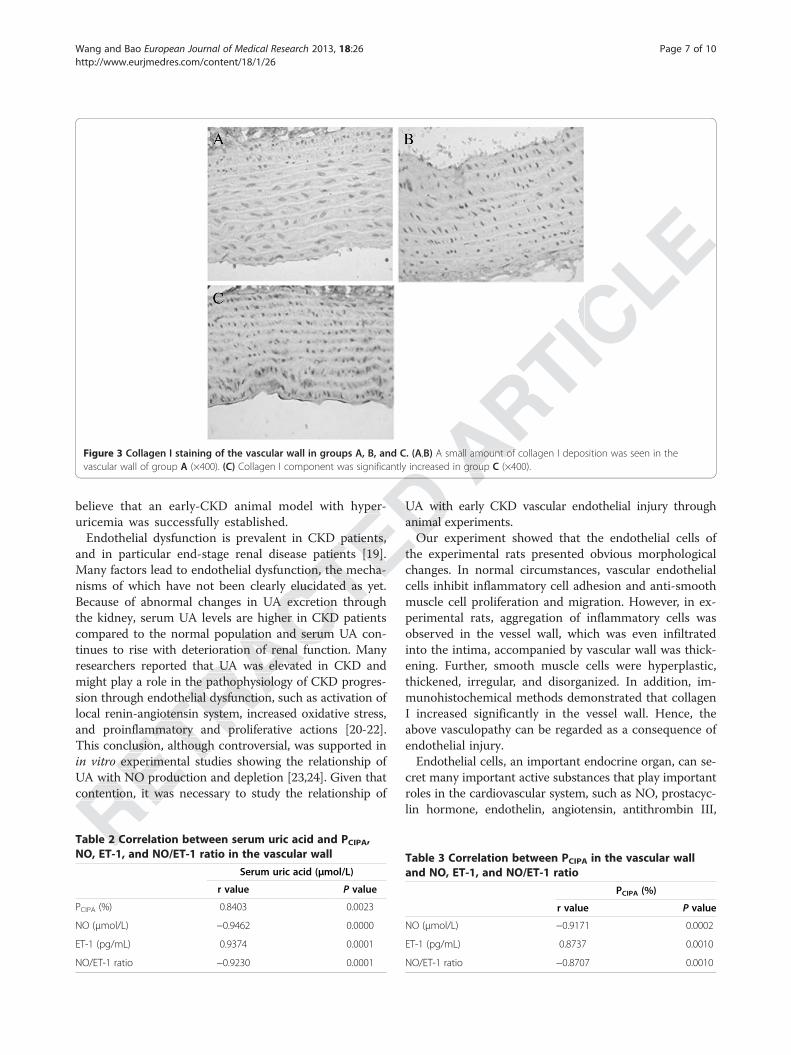
groups is shown in Figure 3. Several collagen I deposi-tions were visible in the vascular wall of groups A and B,while the collagen I component was significantly in-creased in group C. Statistical analysis showed thatthe percentage of collagen I positive area in the vesselwall of the experimental group was significantly higherthan that of group A and group B (group C vs. groupA, P < 0.01; group C vs. group B, P < 0.01) (Table 1).Blood NO and ET-1 were important values to reflect
the function of endothelial cells. Compared to groups Aand B, in the experimental group, serum NO level waslow (P < 0.01), serum ET-1 level was high (P < 0.05), andthe NO/ET-1 ratio was low (P < 0.01). The above resultsverified endothelial cell dysfunction and significant in-jury of vascular endothelial cells.
Uric acid-induced vascular endothelial cell injury inearly CKDThe possibility of a direct correlation between elevatedUA level and vascular disease as well as endothelial in-jury was also investigated. Linear regression analysisshowed that the level of serum UA had a significantpositive correlation with the percentage of collagen I posi-tive area (r = 0.8403, P < 0.01) and serum ET-1 (r = 0.9374,P < 0.01), but a negative correlation with serum NO(r = −0.9462, P < 0.01) and NO/ET-1 ratio (r = −0.9230,P < 0.01) (Table 2). The percentage of collagen I positivearea had a significant positive correlation with serumET-1 (r = 0.8737, P < 0.01), but a negative correlationwith serum NO (r = −0.9171, P < 0.01) and NO/ET-1ratio (r = −0.8707, P < 0.01) (Table 3). The results indi-cated that UA induced vascular endothelial cell injury inearly CKD and the production of collagen I in the vascularwall. Meanwhile, the production of collagen I was relativeto the injury of endothelial cells.
Uric acid caused vascular endothelial injury in early CKDby oxidative stress, micro-inflammation, and lipidoxidation mechanismsThe above experiments showed that UA was involved inearly CKD vascular endothelial cell injury; however, themechanism is unclear. Basic research demonstrated that
Wang and Bao European Journal of Medical Research 2013, 18:26 Page 3 of 10http://www.eurjmedres.com/content/18/1/26
of an early CKD animal model with hyperuricemia.
Vascular endothelial cell injury in the experimental groupIn the light microscope, endothelial cells of group Awere arranged closely under the vascular intima and in-
no obvious difference in Scr among the three groups(Table 1), which was in accordance with the characteristicsof early CKD. The above results indicate the establishmentof an early CKD animal model with hyperuricemia.
Vascular endothelial cell injury in the experimental group
Compared with groups A and B, the experimental grouphad a significantly higher level of UA. However, there wasno obvious difference in Scr among the three groups(Table 1), which was in accordance with the characteristicsof early CKD. The above results indicate the establishment
vessel lesions in groups B and C (Figure 1D,F). In the ex-perimental group, there was no urate crystal deposition.Compared with groups A and B, the experimental grouphad a significantly higher level of UA. However, there wasno obvious difference in Scr among the three groups
while, there was no renal tubular epithelial cells necrosis,no inflammatory cells in the interstitium, and no smallvessel lesions in groups B and C (Figure 1D,F). In the ex-perimental group, there was no urate crystal deposition.Compared with groups A and B, the experimental group
tion in groups B and C, compared to group A (Figure 1A,C,E). No obvious pathological change in renal tubule andrenal interstitium was visible in the three groups. Mean-while, there was no renal tubular epithelial cells necrosis,no inflammatory cells in the interstitium, and no small
with oxonate potassium) was the experimental group.Periodic acid-Schiff staining of kidney tissues from
each group showed mild glomerular mesangial prolifera-tion in groups B and C, compared to group A (Figure 1A,C,E). No obvious pathological change in renal tubule and
Group A (sham operation) and group B (right nephrec-tomy only) served as controls. Group C (right nephrectomywith oxonate potassium) was the experimental group.Periodic acid-Schiff staining of kidney tissues from
each group showed mild glomerular mesangial prolifera-
jury of vascular endothelial cells.
Group A (sham operation) and group B (right nephrec-
and B, in the experimental group, serum NO level waslow (Pthe NO/ET-1 ratio was low (verified endothelial cell dysfunction and significant in-jury of vascular endothelial cells.
< 0.01; group C vs. group B,Blood NO and ET-1 were important values to reflect
the function of endothelial cells. Compared to groups Aand B, in the experimental group, serum NO level was
< 0.01), serum ET-1 level was high (
the percentage of collagen I positive area in the vesselwall of the experimental group was significantly higherthan that of group A and group B (group C vs. group
< 0.01; group C vs. group B,Blood NO and ET-1 were important values to reflect
tions were visible in the vascular wall of groups A and B,while the collagen I componcreased in group C. Statistical analysis showed thatthe percentage of collagen I positive area in the vesselwall of the experimental group was significantly higher
groups is shown in Figure 3. Several collagen I deposi-tions were visible in the vascular wall of groups A and B,while the collagen I component was significantly in-creased in group C. Statistical analysis showed that
Collagen I staining of the vascular wall in the threegroups is shown in Figure 3. Several collagen I deposi-tions were visible in the vascular wall of groups A and B,
ent was significantly in-
sults confirmed significant vascular injury in the experi-
Collagen I staining of the vascular wall in the threegroups is shown in Figure 3. Several collagen I deposi-
change of the right-side nephrectomy group (group B)was similar to the experimental group, but less marked(Figure 2C). Smooth muscle cell proliferation was notobvious and it was well arranged (Figure 2D). The re-sults confirmed significant vascular injury in the experi-

UA crystals deposited in the intima could directly causeendothelial cell damage. Whether there are other im-portant mechanisms apart from this remains to beinvestigated.Our research showed that the experimental group had
lower level of serum SOD (U/mL) compared with groupsA and B (group C vs. group A, P < 0.01; group C vs. groupB, P < 0.01) and higher level of serum MDA (nmol/mL)(group C vs. group A, P < 0.01; group C vs. group B,
P < 0.05) (Table 4). Linear regression analysis showedthat the level of serum UA had a significant positivecorrelation with serum MDA (r = 0.8195, P < 0.01), but anegative correlation with serum SOD (r = −0.6885, P <0.05), which indicated that UA might lead to oxidativestress in early CKD. Further, the level of serum NOhad a significant positive correlation with serum SOD(r = 0.8179, P < 0.01), but a negative correlation withserum MDA (r = −0.9171, P < 0.01). The level of
Table 1 Concentration of Src, UA, PCIPA, NO, ET-1 and NO/ET-1 ratio in three groups (x±̅s)
Group Scr (μmol/L) UA (μmol/L) PCIPA (%) NO (μmol/L) ET-1 (pg/mL) NO/ET-1
A 30.20 ± 6.01 53.10 ± 8.62 12.90 ± 2.31 47.55 ± 5.39 5.89 ± 1.67 8.18 ± 2.32
B 31.70 ± 4.72 53.70 ± 11.52 12.97 ± 2.71 45.34 ± 4.76 5.92 ± 1.56 8.90 ± 3.55
C 30.80 ± 5.90 161.40 ± 28.04* 22.38 ± 3.14* 36.71 ± 3.45* 7.50 ± 1.06△ 5.07 ± 1.19*
Note:*: P <0.01 group C vs. groups A and B.△: P <0.05 group C vs. groups A and B.PCIPA: Percentage of collagen I positive area.
Figure 1 Pathological pictures of rat kidneys from groups A, B, and C. (A) The normal renal glomerulus of group A (×400). (B) No obviouspathological change in renal tubules of group A (×400). (C, E) Mild glomerular mesangial proliferation in renal glomerulus of groups B and C(×400). (D, F) There were no obvious pathological changes in renal tubules in groups B and C (×400).
Wang and Bao European Journal of Medical Research 2013, 18:26 Page 4 of 10http://www.eurjmedres.com/content/18/1/26
A and B (group C vs. group A,B, P < 0.01) and higher level of serum MDA (nmol/mL)(group C vs. group A,
Table 1 Concentration of Src, UA, P
Our research showed that the experimental group hadlower level of serum SOD (U/mL) compared with groupsA and B (group C vs. group A,
< 0.01) and higher level of serum MDA (nmol/mL)(group C vs. group A,
endothelial cell damage. Whether there are other im-portant mechanisms apart from this remains to be
Our research showed that the experimental group hadlower level of serum SOD (U/mL) compared with groups
UA crystals deposited in the intima could directly causeendothelial cell damage. Whether there are other im-portant mechanisms apart from this remains to be
pathological change in renal tubules of group A (×400).
UA crystals deposited in the intima could directly causeendothelial cell damage. Whether there are other im-
There were no obvious pathological changes in renal tubules in groups
Pathological pictures of rat kidneys from groups A, B, and C. (A)pathological change in renal tubules of group A (×400). (C
There were no obvious pathological changes in renal tubules in groups

Figure 2 (See legend on next page.)
Wang and Bao European Journal of Medical Research 2013, 18:26 Page 5 of 10http://www.eurjmedres.com/content/18/1/26

serum ET-1 had a significant positive correlation withserum MDA (r = 0.8658, P < 0.01), but a negative cor-relation with serum SOD (r = −0.7793, P < 0.01). NO/ET-1 ratio had a significant positive correlation withserum SOD (r = 0.8143, P < 0.01), but a negative cor-relation with serum MDA (r = −0.9143, P < 0.01).SOD entered the multiple stepwise regression equa-tion of NO and ET-1, indicating that oxidative stresscan cause vascular endothelial dysfunction in earlyCKD (Table 5).Our research also found that the experimental group
had a higher level of serum CRP (μg/mL) compared withgroups A and B (group C vs. group A, P < 0.01; group Cvs. group B, P < 0.05) (Table 4), which indicated that theexperimental group had a more obvious micro-inflam-mation state. Linear regression analysis showed that thelevel of serum UA had a significant positive correl-ation with serum CRP (r = 0.7251, P < 0.05), which in-dicated that UA was involved in the formation ofmicro-inflammation in early CKD. Further, the levelof serum NO had a significant negative correlationwith serum CRP (r = −0.7554, P < 0.05). Serum ET-1level had a significant positive correlation with serumCRP (r = 0.7447, P < 0.05). NO/ET-1 ratio had a signifi-cant negative correlation with serum CRP (r = −0.8042,P < 0.01). CRP entered the multiple stepwise regressionequation of NO and NO/ET-1 ratio, indicating that mi-cro-inflammation was involved in the formation of vascu-lar endothelial dysfunction in early CKD (Table 5).By studying the levels of serum LDL and ox-LDL, we
found that the experimental group had a higher level ofserum LDL (mmol/L) than groups A and B (group C vs.group A, P < 0.01; group C vs. group B, P < 0.05) and ahigher level of serum ox-LDL (mmol/L) (group C vs.group B, P < 0.05) (Table 4). Linear regression analysisshowed that the level of serum UA had a significant posi-tive correlation with serum ox-LDL (r = 0.8479, P < 0.01)and serum LDL (r = 0.6356, P < 0.05), which indicated thatUA can cause lipid metabolic disorder in early CKD. Fur-ther, the level of serum NO had a significant negative cor-relation with serum ox-LDL (r = −0.7459, P < 0.05), butno significant correlation with serum LDL (r = −0.5080,P > 0.05). Serum ET-1 levels had a significant positivecorrelation with serum ox-LDL (r = 0.7900, P < 0.01),but no significant correlation with serum LDL (r = 0.5734,
P > 0.05). NO/ET-1 ratio had a significant negative correl-ation with serum ox-LDL (r = −0.7949, P < 0.01), butno significant correlation with serum LDL (r = −0.4947,P > 0.05). Serum ox-LDL entered the multiple stepwise re-gression equation of ET-1 (Table 5). The results indicatedthat ox-LDL had strong endothelial cell toxicity.In addition, the percentage of collagen I positive area
in the vascular wall had significant positive correlationswith serum MDA (r = 0.8015, P < 0.01) and serum CRP(r = 0.6752, P < 0.05), a negative correlation with serumSOD (r = −0.8180, P < 0.01), and no significant correl-ation with serum ox-LDL (r = 0.5266, P > 0.05) or serumLDL (r = 0.5902, P > 0.05) (Table 5). Multiple stepwiseregression analysis showed that serum SOD entered theequation. According to the results, it was found that oxi-dative stress and micro-inflammation could lead to anincrease of collagen I deposition in the vessel wall. How-ever, whether lipid metabolism disorders had a relationwith increased collagen deposition in the vessel wall wasnot determined.In conclusion, UA caused endothelial dysfunction in
early CKD via mechanisms involved in oxidative stress,micro-inflammation, and abnormal lipid metabolism.
DiscussionIn this study, serum UA levels in rats were elevated withpotassium oxonate by gavage. In 1965, it was verifiedthat potassium oxonate had a strong ability to inhibituricase activity both in vivo an in vitro [17]. Many re-ports have established a hyperuricemia animal modelwith potassium oxonate [18]. In this study, we comparedthe morphology and biochemical changes of kidney cellsbetween the experimental group and the control group,as well as a unilateral nephrectomy group. The experi-mental group and the unilateral nephrectomy group hadno obvious glomerular lesions; only some of the glom-erulus presented mild mesangial proliferation. No obvi-ous abnormality of renal tubules and renal interstitiumwas visible. Meanwhile, hyperuricemia kidney diseasecaused by urate crystal deposition was not visible in theexperimental group. In serology, serum creatinine levelsamong the three groups had no significant difference(P > 0.05). Nevertheless, the serum UA level of theexperimental group was significantly higher than theother two groups (about three times). Therefore, we
(See figure on previous page.)
Figure 2 Pathological images of rat arteries from groups A, B, and C. (A) Endothelial cells arranged in order under the vascular intima ofgroup A (×400). (B) There was no obvious proliferation of medial smooth muscle cells in the vascular wall of group A (×200). (C) Themorphology of endothelial cells of group B was slightly abnormal (×400). (D) There was no obvious proliferation of medial smooth muscle cellsin the vascular wall of group B (×200). (E) Endothelial cells of group C had a foam-like change (×400). (F) Endothelial cells of group C shed fromthe vessel wall (×400). (G, H) Inflammatory cells accumulated in the vascular intima of group C (×400). (I) Neutrophil granulocytes were seenaround the endothelial cells of group C (×400). (J) Mononuclear cells were seen around the endothelial cells of group C (×400). (K) Smoothmuscle cells of group C proliferated, thickened, and had no order (×200).
Wang and Bao European Journal of Medical Research 2013, 18:26 Page 6 of 10http://www.eurjmedres.com/content/18/1/26
group A,higher level of serum ox-LDL (mmol/L) (group C vs.group B, P < 0.05) (Table 4). Linear regression analysisshowed that the level of serum UA had a significant posi-tive correlation with serum ox-LDL (r = 0.8479,
By studying the levels of serum LDL and ox-LDL, wefound that the experimental group had a higher level ofserum LDL (mmol/L) than groups A and B (group C vs.group A, P < 0.01; group C vs. group B,higher level of serum ox-LDL (mmol/L) (group C vs.
< 0.05) (Table 4). Linear regression analysis
cro-inflammation was involved in the formation of vascu-lar endothelial dysfunction in early CKD (Table 5).By studying the levels of serum LDL and ox-LDL, we
found that the experimental group had a higher level ofserum LDL (mmol/L) than groups A and B (group C vs.
< 0.01). CRP entered the multiple stepwise regressionequation of NO and NO/ET-1 ratio, indicating that mi-cro-inflammation was involved in the formation of vascu-lar endothelial dysfunction in early CKD (Table 5).By studying the levels of serum LDL and ox-LDL, we
< 0.05). NO/ET-1 ratio had a signifi-cant negative correlation with serum CRP (r =
< 0.01). CRP entered the multiple stepwise regressionequation of NO and NO/ET-1 ratio, indicating that mi-cro-inflammation was involved in the formation of vascu-
of serum NO had a significant negative correlation< 0.05). Serum ET-1
level had a significant positive correlation with serum< 0.05). NO/ET-1 ratio had a signifi-
cant negative correlation with serum CRP (r =
< 0.05), which in-dicated that UA was involved in the formation ofmicro-inflammation in early CKD. Further, the levelof serum NO had a significant negative correlation
< 0.05). Serum ET-1
mation state. Linear regression analysis showed that thelevel of serum UA had a significant positive correl-
< 0.05), which in-dicated that UA was involved in the formation ofmicro-inflammation in early CKD. Further, the level
ever, whether lipid metabolism disorders had a relationwith increased collagen deposition in the vessel wall was
experimental group had a more obvious micro-inflam-mation state. Linear regression analysis showed that the
regression analysis showed that serum SOD entered theequation. According to the results, it was found that oxi-dative stress and micro-inflammation could lead to anincrease of collagen I deposition in the vessel wall. How-ever, whether lipid metabolism disorders had a relation
SOD (r = −0.8180,ation with serum ox-LDL (r = 0.5266,LDL (r = 0.5902, Pregression analysis showed that serum SOD entered theequation. According to the results, it was found that oxi-
in the vascular wall had significant positive correlationswith serum MDA (r = 0.8015,
< 0.05), a negative correlation with serum0.8180, P < 0.01), and no significant correl-
ation with serum ox-LDL (r = 0.5266,
gression equation of ET-1 (Table 5). The results indicatedthat ox-LDL had strong endothelial cell toxicity.In addition, the percentage of collagen I positive area
in the vascular wall had significant positive correlationswith serum MDA (r = 0.8015,
> 0.05). Serum ox-LDL entered the multiple stepwise re-gression equation of ET-1 (Table 5). The results indicatedthat ox-LDL had strong endothelial cell toxicity.In addition, the percentage of collagen I positive area
0.7949,no significant correlation with serum LDL (r => 0.05). Serum ox-LDL entered the multiple stepwise re-
gression equation of ET-1 (Table 5). The results indicatedthat ox-LDL had strong endothelial cell toxicity.
> 0.05). NO/ET-1 ratio had a significant negative correl-0.7949, P < 0.01), but
no significant correlation with serum LDL (r => 0.05). Serum ox-LDL entered the multiple stepwise re-
> 0.05). NO/ET-1 ratio had a significant negative correl-
Smooth
believe that an early-CKD animal model with hyper-uricemia was successfully established.Endothelial dysfunction is prevalent in CKD patients,
and in particular end-stage renal disease patients [19].Many factors lead to endothelial dysfunction, the mecha-nisms of which have not been clearly elucidated as yet.Because of abnormal changes in UA excretion throughthe kidney, serum UA levels are higher in CKD patientscompared to the normal population and serum UA con-tinues to rise with deterioration of renal function. Manyresearchers reported that UA was elevated in CKD andmight play a role in the pathophysiology of CKD progres-sion through endothelial dysfunction, such as activation oflocal renin-angiotensin system, increased oxidative stress,and proinflammatory and proliferative actions [20-22].This conclusion, although controversial, was supported inin vitro experimental studies showing the relationship ofUA with NO production and depletion [23,24]. Given thatcontention, it was necessary to study the relationship of
UA with early CKD vascular endothelial injury throughanimal experiments.Our experiment showed that the endothelial cells of
the experimental rats presented obvious morphologicalchanges. In normal circumstances, vascular endothelialcells inhibit inflammatory cell adhesion and anti-smoothmuscle cell proliferation and migration. However, in ex-perimental rats, aggregation of inflammatory cells wasobserved in the vessel wall, which was even infiltratedinto the intima, accompanied by vascular wall was thick-ening. Further, smooth muscle cells were hyperplastic,thickened, irregular, and disorganized. In addition, im-munohistochemical methods demonstrated that collagenI increased significantly in the vessel wall. Hence, theabove vasculopathy can be regarded as a consequence ofendothelial injury.Endothelial cells, an important endocrine organ, can se-
cret many important active substances that play importantroles in the cardiovascular system, such as NO, prostacyc-lin hormone, endothelin, angiotensin, antithrombin III,
Figure 3 Collagen I staining of the vascular wall in groups A, B, and C. (A,B) A small amount of collagen I deposition was seen in thevascular wall of group A (×400). (C) Collagen I component was significantly increased in group C (×400).
Table 2 Correlation between serum uric acid and PCIPA,NO, ET-1, and NO/ET-1 ratio in the vascular wall
Serum uric acid (μmol/L)
r value P value
PCIPA (%) 0.8403 0.0023
NO (μmol/L) −0.9462 0.0000
ET-1 (pg/mL) 0.9374 0.0001
NO/ET-1 ratio −0.9230 0.0001
Table 3 Correlation between PCIPA in the vascular walland NO, ET-1, and NO/ET-1 ratio
PCIPA (%)
r value P value
NO (μmol/L) −0.9171 0.0002
ET-1 (pg/mL) 0.8737 0.0010
NO/ET-1 ratio −0.8707 0.0010
Wang and Bao European Journal of Medical Research 2013, 18:26 Page 7 of 10http://www.eurjmedres.com/content/18/1/26
UA with NO production and depletion [23,24]. Given thatcontention, it was necessary to study the relationship of
Table 2 Correlation between serum uric acid and P
and proinflammatory and proliferative actions [20-22].This conclusion, although controversial, was supported in
experimental studies showing the relationship ofUA with NO production and depletion [23,24]. Given thatcontention, it was necessary to study the relationship of
sion through endothelial dysfunction, such as activation oflocal renin-angiotensin system, increased oxidative stress,and proinflammatory and proliferative actions [20-22].This conclusion, although controversial, was supported in
experimental studies showing the relationship of
researchers reported that UA was elevated in CKD andmight play a role in the pathophysiology of CKD progres-sion through endothelial dysfunction, such as activation oflocal renin-angiotensin system, increased oxidative stress,and proinflammatory and proliferative actions [20-22].
compared to the normal population and serum UA con-tinues to rise with deterioration of renal function. Manyresearchers reported that UA was elevated in CKD andmight play a role in the pathophysiology of CKD progres-sion through endothelial dysfunction, such as activation of
nisms of which have not been clearly elucidated as yet.Because of abnormal changes in UA excretion throughthe kidney, serum UA levels are higher in CKD patientscompared to the normal population and serum UA con-tinues to rise with deterioration of renal function. Many
Endothelial dysfunction is prevalent in CKD patients,and in particular end-stage renal disease patients [19].Many factors lead to endothelial dysfunction, the mecha-nisms of which have not been clearly elucidated as yet.Because of abnormal changes in UA excretion through
believe that an early-CKD animal model with hyper-
Endothelial dysfunction is prevalent in CKD patients,and in particular end-stage renal disease patients [19].Many factors lead to endothelial dysfunction, the mecha-
the experimental rats presented obvious morphological
believe that an early-CKD animal model with hyper- UA with early CKD vascular endothelial injury throughanimal experiments.Our experiment showed that the endothelial cells of
A small amount of collagen I deposition was seen in theCollagen I component was significantly increased in group
and plasminogen activator. These substances are usefulfor homeostasis in the normal case; when the homeostasisbalance is broken, the secretion is abnormal. Therefore, toa certain extent, endothelial cell function can be assessedby detecting the concentration of these substances in theserum. In our study, the substances of contraction and re-laxation of vascular NO and ET-1 [25], synthesized andsecreted by endothelial cells respectively, were used to as-sess endothelial cell function. The study found that serumNO concentration of the experimental group was signifi-cantly low and ET-1 was significantly high compared tothe unilateral nephrectomy group, indicating that NO andET-1 secretion were out of balance. Therefore, we believethat there is a significant endothelial dysfunction in theexperimental group. Further, line correlation analysisshowed that serum UA level was significantly correlatedwith the endothelial function indicators NO and ET-1, in-dicating that UA has a relation to vascular endothelial celldysfunction and participates in early CKD vascular endo-thelial cell injury.Increased oxidative stress and reduced antioxidant
capacity are prevalent in CKD patients [26]. Even in pa-tients with mild renal impairment, oxidative stress levelhas increased more significantly than the normal popu-lation. The level of oxidative stress will intensify as renaldeterioration. Oxidative stress plays an important role inendothelial cell damage and functional changes [27]. At
present, studies on oxidative stress and endothelial in-jury mainly focus on end-stage renal disease patients[28]. The present study focuses on early CKD, and foundthat the experimental group has a significant increasedoxidative stress level related to serum UA concentration,indicating that UA is involved in the formation of theearly CKD oxidative stress status.Systemic micro-inflammation is widespread in CKD
patients, even accompanied by mild renal dysfunction[29]. At present, the degree of inflammatory response isclosely related to the incidence and mortality of cardio-vascular events in CKD patients [30,31]. CRP is an acutephase protein synthetized by liver in inflammation,which is recognized as a reliable marker reflecting theinflammatory state. Our study found that the CRP levelof the experimental group was significantly higher thanthe unilateral nephrectomy group, indicating that themicro-inflammatory state of the experimental group wasmore obvious. CRP level was significantly correlatedwith serum UA level, showing that UA is involved in theformation of micro-inflammation in early CKD. Furtheranalysis showed that serum CRP level was significantcorrelated with NO, ET-1, and NO/ET-1 ratio, all ofwhich are indicators of endothelial function. CRP en-tered the NO and ET-1 multiple regression equation, in-dicating that micro-inflammation is involved in vascularendothelial cell damage of in early CKD.
Table 4 Concentration of serum SOD, MDA, CRP, ox-LDL, and LDL in the three groups (x±̅s)
Group SOD MDA CRP ox-LDL LDL
(U/mL) (nmol/mL) (μg/mL) (mmol/L) (mmol/L)
A 249.80 ± 9.83 4.06 ± 0.28 10.43 ± 1.68 47.50 ± 11.51 0.18 ± 0.06
B 243.60 ± 8.11 4.04 ± 0.41 12.27 ± 2.76 53.31 ± 12.38 0.18 ± 0.06
C 224.40 ± 6.47*# 4.40 ± 0.23△# 14.68 ± 2.01△# 65.22 ± 10.91△# 0.25 ± 0.06△#
Note:*: P <0.01 group C vs. group B.△: P <0.05 group C vs. group B.#: P <0.01 group C vs. group A.
Table 5 Correlation between serum UA, NO, ET-1, and NO/ET-1 ratio as well as PCIPA, SOD, MDA, CRP, ox-LDL, and LDL
Serum uric acid (μmol/L) Serum NO (μmol/L) Serum ET-1 (pg/mL) NO/ET-1 ratio PCIPA (%)
r value P value r value P value r value P value r value P value r value P value
SOD (U/mL) −0.6885 0.0277 0.8179 0.0038 −0.7793 0.0079 0.8143 0.0041 −0.8180 0.0000
MDA (nmol/mL) 0.8195 0.0037 −0.9171 0.0002 0.8658 0.0012 −0.9143 0.0002 0.8015 0.0053
CRP (μg/mL) 0.7251 0.0177 −0.7554 0.0115 0.7447 0.0135 −0.8042 0.0050 0.6752 0.0322
ox-LDL (mmol/L) 0.8479 0.0019 −0.7459 0.0132 0.7900 0.0065 −0.7949 0.0060 0.5266 0.1179
LDL (mmol/L) 0.6356 0.0483 −0.5080 0.1339 0.5734 0.0831 −0.4947 0.1460 0.5902 0.0725
Note: NO as dependent variable and SOD, MDA, CRP, ox-LDL, LDL as independent variables, multiple stepwise regression analysis showed that serum SOD andCRP entered the equation, and the equation was y = 0.315 × 1-0.802 × 2-22.120 (y = NO, ×1 = SOD, ×2 = CRP; -22.120 was a constant). ET-1 as dependent variableand SOD, MDA, CRP, ox-LDL, and LDL as independent variables, multiple stepwise regression analysis showed that serum ox-LDL and SOD entered the equation,and the equation was y = 0.051 × 1-0.082 × 2 + 22.517 (y = ET-1, ×1 = ox-LDL, ×2 = SOD; 22.517 was a constant). NO/ET-1 ratio as dependent variable and SOD, MDA,CRP, ox-LDL, LDL as independent variables, multiple stepwise regression analysis showed that serum SOD and CRP entered the equation, and the equation wasy = 0.102 × 1-0.316 × 2-13.151 (y = NO/ET-1 ratio, ×1 = SOD, ×2 = CRP; 13.151 was a constant). PCIPA as dependent variable and SOD, MDA, CRP, ox-LDL, LDL asindependent variables, multiple stepwise regression analysis showed that serum SOD entered the equation, and the equation was y = 111.437-0.397 × 1
(y = PCIPA, ×1 = SOD; 111.437 was a constant).
Wang and Bao European Journal of Medical Research 2013, 18:26 Page 8 of 10http://www.eurjmedres.com/content/18/1/26
SOD (U/mL)
MDA (nmol/mL) 0.8195 0.0037
μg/mL) 0.7251 0.0177
Table 5 Correlation between serum UA, NO, ET-1, and NO/ET-1 ratio as well as P
Serum uric acid (
r value
deterioration. Oxidative stress plays an important role inendothelial cell damage and functional changes [27]. At
Table 5 Correlation between serum UA, NO, ET-1, and NO/ET-1 ratio as well as P
Serum uric acid (
has increased more significantly than the normal popu-lation. The level of oxidative stress will intensify as renaldeterioration. Oxidative stress plays an important role inendothelial cell damage and functional changes [27]. At
capacity are prevalent in CKD patients [26]. Even in pa-tients with mild renal impairment, oxidative stress levelhas increased more significantly than the normal popu-lation. The level of oxidative stress will intensify as renaldeterioration. Oxidative stress plays an important role in
dysfunction and participates in early CKD vascular endo-
Increased oxidative stress and reduced antioxidantcapacity are prevalent in CKD patients [26]. Even in pa-tients with mild renal impairment, oxidative stress level
showed that serum UA level was significantly correlatedwith the endothelial function indicators NO and ET-1, in-dicating that UA has a relation to vascular endothelial celldysfunction and participates in early CKD vascular endo-
that there is a significant endothelial dysfunction in theexperimental group. Further, line correlation analysisshowed that serum UA level was significantly correlatedwith the endothelial function indicators NO and ET-1, in-dicating that UA has a relation to vascular endothelial cell
of the experimental group was significantly higher thanthe unilateral nephrectomy group, indicating that the
ET-1 secretion were out of balance. Therefore, we believethat there is a significant endothelial dysfunction in the
vascular events in CKD patients [30,31]. CRP is an acutephase protein synthetized by liver in inflammation,which is recognized as a reliable marker reflecting theinflammatory state. Our study found that the CRP levelof the experimental group was significantly higher than
patients, even accompanied by mild renal dysfunction[29]. At present, the degree of inflammatory response isclosely related to the incidence and mortality of cardio-vascular events in CKD patients [30,31]. CRP is an acutephase protein synthetized by liver in inflammation,
indicating that UA is involved in the formation of theearly CKD oxidative stress status.Systemic micro-inflammation is widespread in CKD
patients, even accompanied by mild renal dysfunction[29]. At present, the degree of inflammatory response is
[28]. The present study focuses on early CKD, and foundthat the experimental group has a significant increasedoxidative stress level related to serum UA concentration,indicating that UA is involved in the formation of theearly CKD oxidative stress status.
jury mainly focus on end-stage renal disease patients[28]. The present study focuses on early CKD, and foundthat the experimental group has a significant increasedoxidative stress level related to serum UA concentration,
present, studies on oxidative stress and endothelial in-jury mainly focus on end-stage renal disease patients[28]. The present study focuses on early CKD, and foundthat the experimental group has a significant increased
present, studies on oxidative stress and endothelial in-jury mainly focus on end-stage renal disease patients
0.25 ± 0.06

CKD is often accompanied by lipid metabolic disor-ders, including increased plasma lipoprotein concentra-tions and/or lipoprotein composition changes [32]. Ourstudy found that in the experimental group LDL levels,as well as its oxidized form ox-LDL, were increasedcompared to the unilateral nephrectomy group, both ofwhich were significantly correlated with UA, suggestingthat UA leads to early CKD lipid metabolic disorders.Further analysis showed that LDL was not significantlycorrelated with endothelial function indicators NO andET-1, but its oxidized form was significantly associatedwith these indicators. Lipid metabolism disorders mightbe involved in endothelial cell damage. It is believablethat only ox-LDL has strong endothelial cell toxicitycompared to LDL. The higher the degree of oxidation,the stronger the damage is. Our findings also confirmedthis.Finally, a correlation study about the percentage of
collagen I positive area of the vessel wall and serum UAlevels, endothelial function indicators NO, ET-1, andNO/ET-1 ratio, as well as the activities of SOD, MDA,CRP, LDL, and ox-LDL indicated that the percentage ofcollagen I positive area of the vessel wall was signifi-cantly associated with serum UA level, NO, ET-1,NO/ET-1 ratio, SOD, MDA, and CRP, but had no ob-vious correlation with LDL and ox-LDL. Based on theabove, we believe that UA causes vascular endothelialcell injury in early CKD and leads to collagen prolif-eration in the vessel wall. Meanwhile, collagen hyper-plasia has a relation to endothelial cell injury. We foundthat oxidative stress and micro-inflammation can lead toincreased collagen I deposition in the vessel wall, butwhether lipid metabolism disorders are associated with in-creased collagen component deposition in the vessel wallis not yet determined. With the percentage of collagen Ipositive area as a dependent variable, multiple stepwise re-gression analysis showed that serum SOD entered theequation. Based on these results, we speculate that micro-inflammation can lead to collagen I proliferation, butoxidative stress may act in the final passage leading toincreased collagen deposition in the vessel wall.
ConclusionsIn summary, this study successfully established an early-CKD animal model with elevated serum UA by unilateralnephrectomy plus potassium oxonate intragastrically.With the animal model, we found UA had a role in earlyCKD vascular endothelial cell injury. UA is involved in theformation of oxidative stress, micro-inflammatory state,and abnormal lipid metabolism in early CKD, based onwhich UA may lead to early CKD vascular endothelialinjury. Therefore, strengthened control the UA level inearly CKD aiming to correct oxidative stress, micro-inflammatory state, and lipid metabolism disorders, could
improve vascular endothelial function, delay the processof atherosclerosis, and improve quality of life for CKDpatients.
AbbreviationsCKD: Chronic kidney disease; CRP: C-reactive protein; CVD: Cardiovasculardisease; ET-1: Endothelin-1; LDL: Low density lipoprotein;MDA: Malonaldehyde; NO: Nitric oxide; ox-LDL: Oxidatively modifiedlow-density lipoprotein; Scr: Serum creatinine; SOD: Super oxide dismutase;TGF-β: Transforming growth factor-β; UA: Uric acid.
Competing interestsThe authors declare that they have no competing interests.
Authors’ contributionsYW: Designed the experiments, and acquired and analyzed the data. XRB:Designed the experiments and drafted the manuscript. Both authors readand approved the final manuscript.
Received: 21 February 2013 Accepted: 18 July 2013Published: 30 July 2013
References1. Gertler MM, Garn SM, Levine SA: Serum uric acid in relation to age and
physique in health and in coronary heart disease. Ann Intern Med 1951,34:1421–1431.
2. Jossa F, Farinaro E, Panico S, Krogh V, Celentano E, Galasso R, Mancini M,Trevisan M: Serum uric acid and hypertension: the Olivetti heart study.J Hum Hypertens 1994, 8:677–681.
3. Freedman DS, Williamson DF, Gunter EW, Byers T: Relation of serum uricacid to mortality and ischemic heart disease: the NHANES Iepidemiologic follow-up study. Am J Epidemiol 1995, 141:637–644.
4. Liese AD, Hense HW, Lowel H, Doring A, Tietze M, Keil U: Association ofserum uric acid with all-cause and cardiovascular disease mortality andincident myocardial infarction in the MONICA Augsburg cohort: WorldHealth Organization monitoring trends and determinants incardiovascular diseases. Epidemiology 1999, 10:391–397.
5. Niskanen LK, Laaksonen DE, Nyyssonen K, Alfthan G, Lakka HM, Lakka TA,Salonen JT: Uric acid level as a risk factor for cardiovascular and all-causemortality in middle-aged men: a prospective cohort study. Arch InternMed 2004, 164:1546–1551.
6. Hajhosseiny R, Khavandi K, Goldsmith DJ: Cardiovascular disease in chronickidney disease: untying the Gordian knot. Int J Clin Pract 2013, 67:14–31.
7. Foley RN, Parfrey PS, Sarnak MJ: Clinical epidemiology of cardiovasculardisease in chronic renal disease. Am J Kidney Dis 1998, 32:S112–S119.
8. Ohno I: Relationship between hyperuricemia and chronic kidney disease.Nucleosides Nucleotides Nucleic Acids 2011, 30:1039–1044.
9. Loomans CJ, De Koning EJ, Staal FJ, Rabelink TJ, Zonneveld AJ: Endothelialprogenitor cell dysfunction in type 1 diabetes: another consequence ofoxidative stress? Antioxid Redox Signal 2005, 7:1468–1475.
10. Reinhart K, Bayer O, Brunkhorst F, Meisner M: Markers of endothelialdamage in organ dysfunction and sepsis. Crit Care Med 2002,30:S302–S312.
11. Goon PK, Lip GY, Boos CJ, Stonelake PS, Blann AD: Circulating endothelialcells, endothelial progenitor cells, and endothelial microparticles incancer. Neoplasia 2006, 8:79–88.
12. Drexler H, Hornig B: Endothelial dysfunction in human disease. J Mol CellCardiol 1999, 31:51–60.
13. Thorin E, Webb DJ: Endothelium-derived endothelin-1. Pflugers Arch 2010,459:951–958.
14. Abe M, Maruyama N, Okada K, Matsumoto S, Matsumoto K, Soma M: Effectsof lipid-lowering therapy with rosuvastatin on kidney function andoxidative stress in patients with diabetic nephropathy. J AtherosclerThromb 2011, 18:1018–1028.
15. Liao MT, Sung CC, Hung KC, Wu CC, Lo L, Lu KC: Insulin resistance inpatients with chronic kidney disease. J Biomed Biotechnol 2012,2012:691369.
16. Muntner P, He J, Hamm L, Loria C, Whelton PK: Renal insufficiency andsubsequent death resulting from cardiovascular disease in the UnitedStates. J Am Soc Nephrol 2002, 13:745–753.
Wang and Bao European Journal of Medical Research 2013, 18:26 Page 9 of 10http://www.eurjmedres.com/content/18/1/26
ConclusionsIn summary, this study successfully established an early-CKD animal model with elevated serum UA by unilateralnephrectomy plus potassium oxonate intragastrically.
inflammation can lead to collagen I proliferation, butoxidative stress may act in the final passage leading toincreased collagen deposition in the vessel wall.
ConclusionsIn summary, this study successfully established an early-
gression analysis showed that serum SOD entered theequation. Based on these results, we speculate that micro-inflammation can lead to collagen I proliferation, butoxidative stress may act in the final passage leading toincreased collagen deposition in the vessel wall.
is not yet determined. With the percentage of collagen Ipositive area as a dependent variable, multiple stepwise re-gression analysis showed that serum SOD entered theequation. Based on these results, we speculate that micro-inflammation can lead to collagen I proliferation, but
whether lipid metabolism disorders are associated with in-creased collagen component deposition in the vessel wallis not yet determined. With the percentage of collagen Ipositive area as a dependent variable, multiple stepwise re-gression analysis showed that serum SOD entered the
plasia has a relation to endothelial cell injury. We foundthat oxidative stress and micro-inflammation can lead toincreased collagen I deposition in the vessel wall, butwhether lipid metabolism disorders are associated with in-creased collagen component deposition in the vessel wall
above, we believe that UA causes vascular endothelialcell injury in early CKD and leads to collagen prolif-eration in the vessel wall. Meanwhile, collagen hyper-plasia has a relation to endothelial cell injury. We foundthat oxidative stress and micro-inflammation can lead to
NO/ET-1 ratio, SOD, MDA, and CRP, but had no ob-vious correlation with LDL and ox-LDL. Based on theabove, we believe that UA causes vascular endothelialcell injury in early CKD and leads to collagen prolif-eration in the vessel wall. Meanwhile, collagen hyper-
cantly associated with serum UA level, NO, ET-1,NO/ET-1 ratio, SOD, MDA, and CRP, but had no ob-
J Hum Hypertens3. Freedman DS, Williamson DF, Gunter EW, Byers T:
acid to mortality and ischemic heart disease: the NHANES Iepidemiologic follow-up study.
4. Liese AD, Hense HW, Lowel H, Doring A, Tietze M, Keil U:serum uric acid with all-cause and cardiovascular disease mortality and
1. Gertler MM, Garn SM, Levine SA:physique in health and in coronary heart disease.
–1431.2. Jossa F, Farinaro E, Panico S, Krogh V, Celentano E, Galasso R, Mancini M,
Trevisan M: Serum uric acid and hypertension: the Olivetti heart study.J Hum Hypertens
Published: 30 July 2013
1. Gertler MM, Garn SM, Levine SA:physique in health and in coronary heart disease.
Designed the experiments and drafted the manuscript. Both authors readand approved the final manuscript.
Received: 21 February 2013 Accepted: 18 July 2013
YW: Designed the experiments, and acquired and analyzed the data. XRB:Designed the experiments and drafted the manuscript. Both authors read
Received: 21 February 2013 Accepted: 18 July 2013
The authors declare that they have no competing interests.
YW: Designed the experiments, and acquired and analyzed the data. XRB:Designed the experiments and drafted the manuscript. Both authors read
The authors declare that they have no competing interests.
MDA: Malonaldehyde; NO: Nitric oxide; ox-LDL: Oxidatively modifiedlow-density lipoprotein; Scr: Serum creatinine; SOD: Super oxide dismutase;

17. Fridovich I: The competitive inhibition of uricase by oxonate and byrelated derivatives of S-triazines. J Biol Chem 1965, 240:2491–2494.
18. Mazzali M, Kanellis J, Han L, Feng L, Xia YY, Chen Q, Kang DH, Gordon KL,Watanabe S, Nakagawa T, Lan HY, Johnson RJ: Hyperuricemia induces aprimary renal arteriolopathy in rats by a blood pressure-independentmechanism. Am J Physiol Renal Physiol 2002, 282:F991–F997.
19. Choi JH, Kim KL, Huh W, Kim B, Byun J, Suh W, Sung J, Jeon ES, Oh HY, KimDK: Decreased number and impaired angiogenic function of endothelialprogenitor cells in patients with chronic renal failure. Arterioscler ThrombVasc Biol 2004, 24:1246–1252.
20. Edwards NL: The role of hyperuricemia in vascular disorders. Curr OpinRheumatol 2009, 21:132–137.
21. Filiopoulos V, Hadjiyannakos D, Vlassopoulos D: New insights into uric acideffects on the progression and prognosis of chronic kidney disease.Ren Fail 2012, 34:510–520.
22. Jalal DI, Chonchol M, Chen W, Targher G: Uric acid as a target of therapyin CKD. Am J Kidney Dis 2013, 61(1):134–146.
23. Gersch C, Palii SP, Kim KM, Angerhofer A, Johnson RJ, Henderson GN:Inactivation of nitric oxide by uric acid. Nucleosides Nucleotides NucleicAcids 2008, 27:967–978.
24. Zharikov S, Krotova K, Hu H, Baylis C, Johnson RJ, Block ER, Patel J: Uric aciddecreases NO production and increases arginase activity in culturedpulmonary artery endothelial cells. Am J Physiol Cell Physiol 2008,295:C1183–C1190.
25. Furchgott RF, Zawadzki JV: The obligatory role of endothelial cells in therelaxation of arterial smooth muscle by acetylcholine. Nature 1980,288:373–376.
26. Annuk M, Zilmer M, Lind L, Linde T, Fellstrom B: Oxidative stress andendothelial function in chronic renal failure. J Am Soc Nephrol 2001,12:2747–2752.
27. Fenster BE, Tsao PS, Rockson SG: Endothelial dysfunction: clinicalstrategies for treating oxidant stress. Am Heart J 2003, 146:218–226.
28. Vaziri ND, Ni Z, Oveisi F, Liang K, Pandian R: Enhanced nitric oxideinactivation and protein nitration by reactive oxygen species in renalinsufficiency. Hypertension 2002, 39:135–141.
29. Kundhal K, Lok CE: Clinical epidemiology of cardiovascular disease inchronic kidney disease. Nephron Clin Pract 2005, 101:c47–c52.
30. Arici M, Walls J: End-stage renal disease, atherosclerosis, andcardiovascular mortality: is C-reactive protein the missing link? Kidney Int2001, 59:407–414.
31. Menon V, Greene T, Wang X, Pereira AA, Marcovina SM, Beck GJ, Kusek JW,Collins AJ, Levey AS, Sarnak MJ: C-reactive protein and albumin aspredictors of all-cause and cardiovascular mortality in chronic kidneydisease. Kidney Int 2005, 68:766–772.
32. Majumdar A, Wheeler DC: Lipid abnormalities in renal disease. J R Soc Med2000, 93:178–182.
doi:10.1186/2047-783X-18-26Cite this article as: Wang and Bao: Effects of uric acid on endothelialdysfunction in early chronic kidney disease and its mechanisms.European Journal of Medical Research 2013 18:26.
Submit your next manuscript to BioMed Centraland take full advantage of:
• Convenient online submission
• Thorough peer review
• No space constraints or color figure charges
• Immediate publication on acceptance
• Inclusion in PubMed, CAS, Scopus and Google Scholar
• Research which is freely available for redistribution
Submit your manuscript at www.biomedcentral.com/submit
Wang and Bao European Journal of Medical Research 2013, 18:26 Page 10 of 10http://www.eurjmedres.com/content/18/1/26
Wang and Bao:dysfunction in early chronic kidney disease and its mechanisms.European Journal of Medical Research
doi:10.1186/2047-783X-18-26Wang and Bao: Effects of uric acid on endothelial
dysfunction in early chronic kidney disease and its mechanisms.European Journal of Medical Research 2013
predictors of all-cause and cardiovascular mortality in chronic kidney
Lipid abnormalities in renal disease.
Effects of uric acid on endothelial
31. Menon V, Greene T, Wang X, Pereira AA, Marcovina SM, Beck GJ, Kusek JW,C-reactive protein and albumin as
predictors of all-cause and cardiovascular mortality in chronic kidney
Clinical epidemiology of cardiovascular disease inc52.
End-stage renal disease, atherosclerosis, andcardiovascular mortality: is C-reactive protein the missing link? Kidney Int
31. Menon V, Greene T, Wang X, Pereira AA, Marcovina SM, Beck GJ, Kusek JW,
inactivation and protein nitration by reactive oxygen species in renal
Clinical epidemiology of cardiovascular disease in
Kidney Int