research open access pure curcumin decreases the ... · pure curcumin decreases the expression of...
TRANSCRIPT
RESEARCH Open Access
Pure curcumin decreases the expression of WT1by upregulation of miR-15a and miR-16-1 inleukemic cellsShen-meng Gao1†, Jun-jun Yang2†, Chi-qi Chen1, Jun-jie Chen1, Li-ping Ye1, Lu-yao Wang1, Jian-bo Wu1,Chong-yun Xing3 and Kang Yu3*
Abstract
Background: Pure curcumin has been reported to down-regulate the expression of WT1 in leukemic cells.However, the molecular mechanism underlying the down-regulation of WT1 by curcumin is not completelydelineated. The purpose of this present study is to identify a new miRNA-mediated mechanism which plays animportant role in the anti-proliferation effects of curcumin in leukemic cells.
Methods: K562 and HL-60 cells were treated with different concentrations of curcumin for 24 and 48 hours, thelevel of miR-15a/16-1 and WT1 were detected by qRT-PCR and Western blotting. WT1 expression and cellproliferation were detected by Western blotting and CCK-8, after curcumin treated-K562 and HL-60 cells weretransfected with anti-miR-15a/16-1 oligonucleotides.
Results: We found that pure curcumin upregulated the expression of miR-15a/16-1 and downregulated theexpression of WT1 in leukemic cells and primary acute myeloid leukemia (AML) cells. Overexpression of miR-15a/16-1 deduced the protein level of WT1 in leukemic cells, but downregulation of WT1 by siRNA-WT1 could notincrease the expression of miR-15a/16-1 in leukemic cells. These results reveal that curcumin induced-upregulationof miR-15a/16-1 is an early event upstream to downregulation of WT1. Furthermore, anti-miR-15a/16-1oligonucleotides (AMO) partly reversed the downregulation of WT1 induced by pure curcumin in leukemic cellsand AMO promoted the growth of curcumin treated-K562 and HL-60 cells.
Conclusion: Thus, these data suggest for the first time that pure curcumin downregulated the expression of WT1partly by upregulating the expression of miR-15a/16-1 in leukemic cells. miR-15a/16-1 mediated WT1downregulation plays an important role in the anti-proliferation effect of curcumin in leukemic cells.
Keywords: Curcumin, WT1, miR-15a, miR-16-1
IntroductionThe Wilms’ tumor 1 (WT1) gene, which is located atthe short arm of chromosome 11 and contains 10exons, encodes a DNA-binding transcription factoressential for embryonal development [1]. High level ofWT1, which is detected in most cases of acute humanleukemia and chronic myelogeous leukemia (CML) inblast crisis, is associated with a worse long-time
prognosis [2]. Downregulation of WT1 by special siRNAcan inhibit cell proliferation and induce apoptosis inK562 and HL-60 cells [3]. WT1 acts as a potent tran-scriptional regulation factor involved in cell growth anddevelopment due to the presence of zinc fingers [4].WT1 is firstly thought to function as tumor suppressor,but the following wildly studies support that WT1 actsas oncogene [5].Curcumin, a naturally occurring flavinoid and proa-
poptotic compound derived from the rhizome of Cur-cuma longa, has strong anti-inflammatory, antioxidant,anticarcinogen, anticancer properties through regulatingmultiple downstream cancer-related signaling molecules.
* Correspondence: [email protected]† Contributed equally3Department of Hematology, The First Affiliated Hospital of WenzhouMedical College, 2 FuXue Road, Wenzhou 325000, ChinaFull list of author information is available at the end of the article
Gao et al. Journal of Experimental & Clinical Cancer Research 2012, 31:27http://www.jeccr.com/content/31/1/27
© 2012 Gao et al; licensee BioMed Central Ltd. This is an Open Access article distributed under the terms of the Creative CommonsAttribution License (http://creativecommons.org/licenses/by/2.0), which permits unrestricted use, distribution, and reproduction inany medium, provided the original work is properly cited.
The molecular targets of curcumin include modulationof NF-kappaB, Jak/STAT, WT1, extracellular signalregulated kinase and other key molecules involved intumorigenesis [6-8]. The mechanisms underlying theanticancer activity of curcumin have been widely investi-gated. Bharti et al. showed curcumin decreased NF-kap-paB in human multiple myeloid cells, leading to thesuppression of proliferation and induction of apoptosis[7]. Recently more and more data have shown that WT1is a very important target gene by curcumin [9]. How-ever the exact mechanism by which curcumin downre-gulated the expression of WT1 is still not clear.MicroRNAs (miRNAs) are non-coding regulatory
RNAs of 21 to 25 nucleotides which regulate most ofbasal progress such as cell proliferation, survival, apop-tosis, and differentiation by triggering either transla-tional repression or mRNA degradation [10].Furthermore, computational prediction demonstratedthat each miRNA may target hundreds of genes, andthat more than 50% of human protein-coding genescould be modulated by miRNAs [11]. Recently somedata have indicated pure curcumin inhibited cancer cellproliferation though miRNAs mediated signal pathway.Michael et al. showed curcumin inhibited the prolifera-tion of pancreatic cancer cells through upregulation ofmiR-22 and downregulation of miR-199a* [12]. Yang etal. demonstrated that curcumin induced MCF-7 cellsapoptosis through miR-15a/16-1 mediated down-regula-tion of Bcl-2 [13]. These emerging results suggest thatspecific targeting of miRNAs by natural agents mayopen new avenues for the complete elucidation of anti-tumor activity by curcumin.In this study, we explored the potential modulation of
miR-15a and miR-16-1 by curcumin in leukemic cells.Our study aims to explain a new mechanism by whichcurcumin downregulates the expression of WT1 via theupregulation of miR-15a/16-1 in leukemic cells.
Material and methodsCell lines and primary AML cellsLeukemic cell lines (K562 and HL-60) were employedfor the present study. All cells were cultured in RPMI1640 supplemented with 10% heat-inactivated fetalbovine serum (Invitrogen, CA, USA) in humidified 37°Cincubator with 5% CO2. Primary leukemic cells wereobtained from 12 patients with acute myeloid leukemia(AML) (3 M2, 2 M3, 3 M4 and 4 M5, The FirstAffiliated Hospital of Wenzhou Medical College) withinformed consent. The detailed data of the patients wereshowed in Table 1. The diagnosis was establishedaccording to French-American-British classification. Allmanipulations were approved by the Medical ScienceEthic Committee of Wenzhou Medical College. Allthese patients did not receive any chemical therapy
treatments. Primary leukemic cells were isolated byFicoll density gradient centrifugation (GE Healthcare,Uppsala, Sweden). Pure curcumin (Sigma-Aldrich, StLouis, MO) was dissolved in DMSO as 20 mM stocksolution and kept at -20°C. For experiments, leukemiccells and primary AML cells were cultured in serial con-centrations of curcumin and control cultures were trea-ted with DMSO only.
Plasmids transfectionpRETROSUPER vector expressing miR-15a/16-1 (pRS-15/16) was constructed as previously described. Thesame empty plasmid (pRS-E) was served as negativecontrol. K562 and HL-60 cells were transiently trans-fected with 1 μg/mL (final concentration) pRS-15/16 orpRS-E vector mediated by Lipofectamine™ LTX andPLUS™ Reagents (Invitrogen) according to the manu-facturer’s instructions.
RNA extractionTotal RNA from curcumin-treated or untreated leuke-mic cells were extracted by TRIzol (Invitrogen) Follow-ing the manufacture’s protocol. RNA concentration andquality were quantified by measuring the absorbance at260 nm with Beckman DU6400 spectrophotometer(Beckman, USA) and gel analysis.
qPCR for miRNA and mRNA expressionQuantitative real-time polymerase chain reaction(qRT-PCR) analysis for miR-15a and miR-16-1 was performedin triplicate by the aid of the NCode™ miRNA First-strand cDNA synthesis (Invitrogen) and SYBR® GreenPCR Master Mix (Applied Biosystems, Foster City, CA)according to the manufacturer’s instructions. U6 snRNAlevel was used for normalization. The fold change foreach miRNA in curcumin-treated leukemic cells relativeto untreated cells was calculated using the 2-ΔΔCT
method [14]. WT1 transcript was determined by
Table 1 The data of acute myeloid leukemia patients
NO Sex Age(y) FAB subtype Chromosome karyotype
1 M 24 M5 46, XY
2 M 36 M3 46, XY PML-RARa+
3 F 47 M5 46, XX
4 F 53 M4 46, XX MYH11-CBFb+5 M 29 M3 46, XY PML-RARa+
6 F 48 M2 46, XX AML-ETO+
7 F 35 M4 46, XX MYH11-CBFb+8 M 41 M5 46, XY
9 F 58 M2 46, XX AML-ETO+
10 M 47 M4 46, XY
11 M 41 M2 46, XY
12 F 26 M5 46, XX
Gao et al. Journal of Experimental & Clinical Cancer Research 2012, 31:27http://www.jeccr.com/content/31/1/27
Page 2 of 9
quantitative real-time PCR using specific primer. ABLand GAPDH housekeeping genes were used for normali-zation [15,16]. The following primers were used respec-tively, miR-15a: 5’-TAG CAG CAC ATA ATG GTTTGT G-3’, miR-16-1: 5’-TAG CAG CAC GTA AATATT GGC G-3’, U6: 5’-CGC AAG GAT GAC ACGCAA ATT C-3’, WT1: sense strand: 5’-CAG GCT GCAATA AGA GAT ATT TTA AG CT-3’, antisense strand:5’-GAA GTC ACA CTG GTA TGG TTT CTC A-3’,Taqman probe: 5’-Fam-CTT ACA GAT GCA CAGCAG GAA GCA CAC TGA-Tamra-3’), ABL: (sensestrand: 5’-GAT GTA GTT GCT TGG GAC CCA-3’,antisense strand: 5’-TGG AGA TAA CAC TCT AAGCAT AAC TAA AGG T-3’, Taqman probe: 5’-Fam-CCA TTT TTG GTT TGG GCT TCA CAC CAT T-Tamra-3’). GAPDH: (sense strand: 5’-CCA GGT GGTCTC CTC TGA CTT C-3’, antisense strand: 5’-GTGGTC GTT GAG GGC AAT G-3’, Taqman probe: 5’-Fam-ACA GCG ACA CCC ACT CCT CCA CCT T-Tamra-3’).
Cell counting kit-8 (CCK-8) assayK562 and HL-60 cells were seeded into 96-well plates(6.0 × 10³ cells/well). Cell viability was assessed byCCK-8 assay (Dojin Laboratories, Kumamoto, Japan).The absorbance at 450 nm (A450) of each well was readon a spectrophotometer. Three independent experi-ments were performed in quadruplicate.
Western blottingProtein extracts from cell lines, patient samples pre-pared with RIPA lysis buffer (50 mM TrisHCl, 150 mMNaCl, 0.1% SDS, 1% NP-40, 0.5% sodiumdeoxycholate, 1mM PMSF, 100 mM leupeptin, and 2 mg/mL aprotinin,pH 8.0) were separated on an 8% SDS-polyacrylamidegel and transferred to nitrocellulose membranes. Afterblocking with 5% nonfat milk, the membranes wereincubated with an appropriate dilution (WT1 1:2000) ofthe primary antibody (Abcom, Cambridge, MA, USA),followed by incubation with the horseradish peroxidase(HRP)-conjugated secondary antibody (Abcom). The sig-nals were detected by chemiluminescence phototope-HRP kit (Cell Signaling, Danvers, MA, USA). Blots werestripped and reprobed with anti-GAPDH antibody(Abcom) as an internal control. All experiments wererepeated three times.
siRNA, mimics, and anti-miR-15a/16-1 oligonucleotide(AMO) transfectionSiRNA sequences targeting WT1: ccauaccagugugacuucacorresponds to positions 9-27 of exon 7 within the WT1coding sequence. SiRNA-WT1 and unspecific controlsiRNA (N.C) were synthesized from Invitrogen. 50 nMSiRNA-WT1 or N.C were transfected into K562 and HL-
60 cells using Hiperfect transfection reagent (Qiagen,Valencia, USA) according to manufacturer’s instructions.miR-15a or miR-16-1 mimics was synthesized from GenePharma (Shanghai, China). 40 uM miR-15a or miR-16-1mimics were transfected into K562 using Hiperfect trans-fection reagent (Qiagen). The sequences of AMO weredesigned according to the principle of sequences comple-mentary to mature miRNA-15a/16-1. AMO and scram-ble (SCR) were chemically synthesized by Qiagen. AMOand SCR (final concentration of 50 nM) were transfectedinto K562 and HL-60 cells using the Hiperfect transfec-tion reagent (Qiagen). All transfections were performedin triplicate for each time point.
Statistical analysisThe significance of the difference between groups wasdetermined by Student’s t-test. A P value of less than .05was considered statistically significant. All Statistical ana-lyses were performed with SPSS software (version 13).
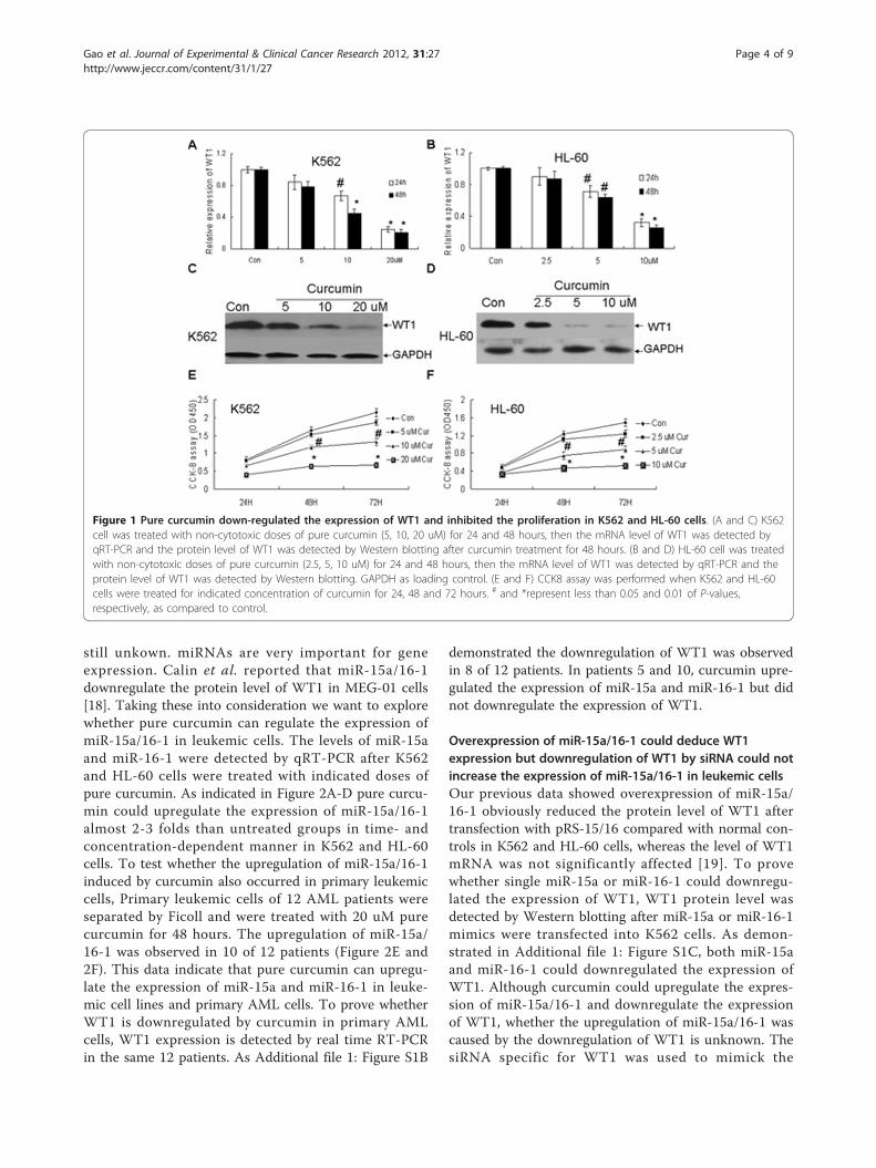
ResultsPure curcumin downregulated the expression of WT1 andeffectively inhibited cell proliferation in leukemic cellsAs reported previously [17], low concentration of purecurcumin could inhibit the growth of leukemic cells anddownregulate the expression of WT1. The mRNA andprotein levels of WT1 were detected by qRT-PCR andWestern blotting respectively after K562 and HL-60 cellswere treated with non-cytotoxic doses of pure curcumin(5, 10, 20 uM for K562 and 2.5, 5, 10 uM for HL-60) [17].As indicated in Figure 1A-D pure curcumin downregu-lated the expression of WT1 in time- and concentration-dependent manner. The mRNA levels of WT1 in theK562 cells were decreased by 12%, 55%, and 73% inresponse to treatment with 5, 10, and 20 μM curcumin at48 hours compared with the vehicle control (Figure 1A).To test whether ABL housekeeping gene was regulatedby curcumin, another widely used housekeeping geneGAPDH was used for normalization. As Additional file 1:Figure S1A demonstrated no difference occurred in WT1expression between GAPDH and ABL for normalization.Meanwhile the protein levels of WT1 in the k562 cellswere significantly decreased after 10 and 20 uM curcu-min treatment at 48 hours (Figure 1C). In HL-60 cells 5and 10 uM curcumin also significantly downregulatedthe mRNA and protein levels of WT1 (Figure 1B and1D). Finally CCK-8 assay showed that low concentrationsof pure curcumin could effectively inhibit the growth ofleukemic cells (Figure 1E and 1F).
Pure curcumin upregulated the expression of miR-15a/16-1 in leukemic cells and primary AML blastsAlthough pure curcumin decreased the expression ofWT1 in K562 and HL-60 cells, the exact mechanism is
Gao et al. Journal of Experimental & Clinical Cancer Research 2012, 31:27http://www.jeccr.com/content/31/1/27
Page 3 of 9
still unkown. miRNAs are very important for geneexpression. Calin et al. reported that miR-15a/16-1downregulate the protein level of WT1 in MEG-01 cells[18]. Taking these into consideration we want to explorewhether pure curcumin can regulate the expression ofmiR-15a/16-1 in leukemic cells. The levels of miR-15aand miR-16-1 were detected by qRT-PCR after K562and HL-60 cells were treated with indicated doses ofpure curcumin. As indicated in Figure 2A-D pure curcu-min could upregulate the expression of miR-15a/16-1almost 2-3 folds than untreated groups in time- andconcentration-dependent manner in K562 and HL-60cells. To test whether the upregulation of miR-15a/16-1induced by curcumin also occurred in primary leukemiccells, Primary leukemic cells of 12 AML patients wereseparated by Ficoll and were treated with 20 uM purecurcumin for 48 hours. The upregulation of miR-15a/16-1 was observed in 10 of 12 patients (Figure 2E and2F). This data indicate that pure curcumin can upregu-late the expression of miR-15a and miR-16-1 in leuke-mic cell lines and primary AML cells. To prove whetherWT1 is downregulated by curcumin in primary AMLcells, WT1 expression is detected by real time RT-PCRin the same 12 patients. As Additional file 1: Figure S1B
demonstrated the downregulation of WT1 was observedin 8 of 12 patients. In patients 5 and 10, curcumin upre-gulated the expression of miR-15a and miR-16-1 but didnot downregulate the expression of WT1.
Overexpression of miR-15a/16-1 could deduce WT1expression but downregulation of WT1 by siRNA could notincrease the expression of miR-15a/16-1 in leukemic cellsOur previous data showed overexpression of miR-15a/16-1 obviously reduced the protein level of WT1 aftertransfection with pRS-15/16 compared with normal con-trols in K562 and HL-60 cells, whereas the level of WT1mRNA was not significantly affected [19]. To provewhether single miR-15a or miR-16-1 could downregu-lated the expression of WT1, WT1 protein level wasdetected by Western blotting after miR-15a or miR-16-1mimics were transfected into K562 cells. As demon-strated in Additional file 1: Figure S1C, both miR-15aand miR-16-1 could downregulated the expression ofWT1. Although curcumin could upregulate the expres-sion of miR-15a/16-1 and downregulate the expressionof WT1, whether the upregulation of miR-15a/16-1 wascaused by the downregulation of WT1 is unknown. ThesiRNA specific for WT1 was used to mimick the
Figure 1 Pure curcumin down-regulated the expression of WT1 and inhibited the proliferation in K562 and HL-60 cells. (A and C) K562cell was treated with non-cytotoxic doses of pure curcumin (5, 10, 20 uM) for 24 and 48 hours, then the mRNA level of WT1 was detected byqRT-PCR and the protein level of WT1 was detected by Western blotting after curcumin treatment for 48 hours. (B and D) HL-60 cell was treatedwith non-cytotoxic doses of pure curcumin (2.5, 5, 10 uM) for 24 and 48 hours, then the mRNA level of WT1 was detected by qRT-PCR and theprotein level of WT1 was detected by Western blotting. GAPDH as loading control. (E and F) CCK8 assay was performed when K562 and HL-60cells were treated for indicated concentration of curcumin for 24, 48 and 72 hours. # and *represent less than 0.05 and 0.01 of P-values,respectively, as compared to control.
Gao et al. Journal of Experimental & Clinical Cancer Research 2012, 31:27http://www.jeccr.com/content/31/1/27
Page 4 of 9

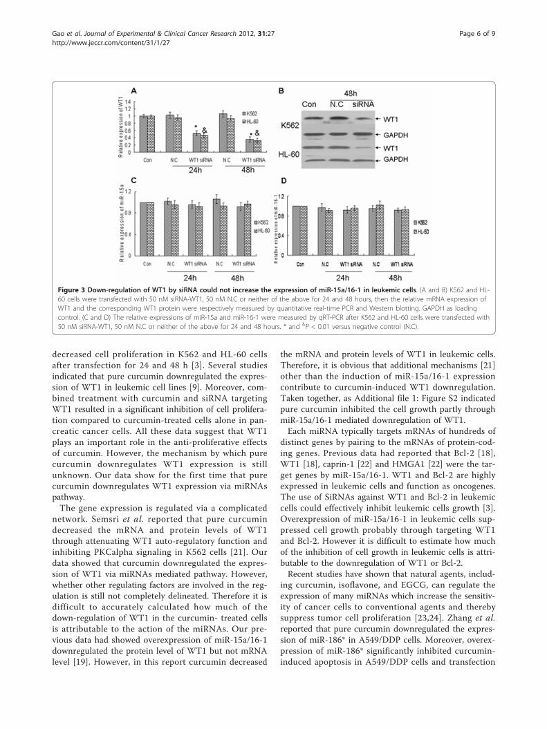
downregulation of WT1 by curcumin. WT1 mRNA andprotein levels were estimated by quantitative real-timePCR and Western blotting individually after K562 andHL-60 cells were transfected with siRNA-WT1 or nega-tive control for 24 and 48 hours. WT1 siRNA-treatedK562 and HL-60 cells showed a significant reduction ofWT1 mRNA level as compared to control cells (Figure3A). Furthermore the reduction of mRNA using siRNAresulted in a markedly decrease of WT1 protein levelafter 48 hours in K562 and HL-60 cells (Figure 3B).Finally we observed that the level of miR-15a and miR-16-1 were not significantly altered by siRNA-WT1 com-pared with normal control (Figure 3C and 3D). All thesedata demonstrate that downregulation of WT1 can notaffect the expression of miR-15a and miR-16-1 in K562and HL-60 cell lines. These data strongly indicate thatpure curcumin induced-upregulation of miR-15a/16-1 isan event upstream to the downregulation of WT1.
Anti-miR-15a/16-1 oligonucleotides (AMO) partly reversedthe down-regulation of WT1 induced by curcumin inleukemic cellsTo further confirm that pure curcumin down-regulatedthe expression of WT1 by up-regulation of miR-15a/16-
1, 20 uM curcumin treated-K562 and 10 uM curcumintreated- HL-60 cells were transfected with 50 nM anti-miR-15a/16-1 oligonucleotides for 48 hours. The levelsof WT1 protein were detected by Western blotting aftertransfection. As Figure 4A and 4B demonstrated thatanti-miR-15a/16-1 oligonucleotides could effectivelydecrease the expression of miR-15a and miR-16-1 inK562 and HL-60 cells. Moreover, anti-miR-15a/16-1 oli-gonucleotides partly abolished the inhibitory effect ofcurcumin on WT1 protein expression (Figure 4C and4D). Finally, as indicated in Figure 4E and 4F, 20 uMcurcumin treated-K562 and 10 uM curcumin treated-HL-60 cells were transfected with 50 nM of anti-miR-15a/16-1 oligonucleotides for 24, 48 and 72 hours, theCCK-8 assay revealed that anti-miR-15a/16-1 oligonu-cleotides effectively reversed the inhibition of cell prolif-eration caused by curcumin in K562 and HL-60 cells.
DiscussionWT1 is considered to play an important role in leuke-mogenesis because the expression of WT1 increases1000-10000 fold in primary leukemic cells than normalcells [20]. Glienke and Bergmann showed that siRNA-reduced WT1 mRNA expression was associated with a
Figure 2 Pure curcumin upregulated the expression of miR-15a/16-1 in leukemic cell lines and primary AML blasts. (A and C) Theexpression of miR-15a and miR-16-1 were detected by qRT-PCR after K562 and HL-60 cells were treated with different concentration ofcurcumin for 48 hours. (B and D) K562 and HL-60 cells were treated with 20 uM or 10 uM curcumin respectively for 24, 48, and 72 hours, thenthe relative expressions of miR-15a and miR-16-1 were detected by qRT-PCR. Data are shown as mean ± SD from three independentexperiments. (E and F) Primary leukemic cells were isolated by Ficoll density gradient centrifugation and were treated with 20 uM pure curcuminfor 48 hours, then the levels of miR-15a and miR-16-1 were detected by qRT-PCR. # and &represent less than 0.01 of P-values as compared tocontrol.
Gao et al. Journal of Experimental & Clinical Cancer Research 2012, 31:27http://www.jeccr.com/content/31/1/27
Page 5 of 9
decreased cell proliferation in K562 and HL-60 cellsafter transfection for 24 and 48 h [3]. Several studiesindicated that pure curcumin downregulated the expres-sion of WT1 in leukemic cell lines [9]. Moreover, com-bined treatment with curcumin and siRNA targetingWT1 resulted in a significant inhibition of cell prolifera-tion compared to curcumin-treated cells alone in pan-creatic cancer cells. All these data suggest that WT1plays an important role in the anti-proliferative effectsof curcumin. However, the mechanism by which purecurcumin downregulates WT1 expression is stillunknown. Our data show for the first time that purecurcumin downregulates WT1 expression via miRNAspathway.The gene expression is regulated via a complicated
network. Semsri et al. reported that pure curcumindecreased the mRNA and protein levels of WT1through attenuating WT1 auto-regulatory function andinhibiting PKCalpha signaling in K562 cells [21]. Ourdata showed that curcumin downregulated the expres-sion of WT1 via miRNAs mediated pathway. However,whether other regulating factors are involved in the reg-ulation is still not completely delineated. Therefore it isdifficult to accurately calculated how much of thedown-regulation of WT1 in the curcumin- treated cellsis attributable to the action of the miRNAs. Our pre-vious data had showed overexpression of miR-15a/16-1downregulated the protein level of WT1 but not mRNAlevel [19]. However, in this report curcumin decreased
the mRNA and protein levels of WT1 in leukemic cells.Therefore, it is obvious that additional mechanisms [21]other than the induction of miR-15a/16-1 expressioncontribute to curcumin-induced WT1 downregulation.Taken together, as Additional file 1: Figure S2 indicatedpure curcumin inhibited the cell growth partly throughmiR-15a/16-1 mediated downregulation of WT1.Each miRNA typically targets mRNAs of hundreds of
distinct genes by pairing to the mRNAs of protein-cod-ing genes. Previous data had reported that Bcl-2 [18],WT1 [18], caprin-1 [22] and HMGA1 [22] were the tar-get genes by miR-15a/16-1. WT1 and Bcl-2 are highlyexpressed in leukemic cells and function as oncogenes.The use of SiRNAs against WT1 and Bcl-2 in leukemiccells could effectively inhibit leukemic cells growth [3].Overexpression of miR-15a/16-1 in leukemic cells sup-pressed cell growth probably through targeting WT1and Bcl-2. However it is difficult to estimate how muchof the inhibition of cell growth in leukemic cells is attri-butable to the downregulation of WT1 or Bcl-2.Recent studies have shown that natural agents, includ-
ing curcumin, isoflavone, and EGCG, can regulate theexpression of many miRNAs which increase the sensitiv-ity of cancer cells to conventional agents and therebysuppress tumor cell proliferation [23,24]. Zhang et al.reported that pure curcumin downregulated the expres-sion of miR-186* in A549/DDP cells. Moreover, overex-pression of miR-186* significantly inhibited curcumin-induced apoptosis in A549/DDP cells and transfection
Figure 3 Down-regulation of WT1 by siRNA could not increase the expression of miR-15a/16-1 in leukemic cells. (A and B) K562 and HL-60 cells were transfected with 50 nM siRNA-WT1, 50 nM N.C or neither of the above for 24 and 48 hours, then the relative mRNA expression ofWT1 and the corresponding WT1 protein were respectively measured by quantitative real-time PCR and Western blotting. GAPDH as loadingcontrol. (C and D) The relative expressions of miR-15a and miR-16-1 were measured by qRT-PCR after K562 and HL-60 cells were transfected with50 nM siRNA-WT1, 50 nM N.C or neither of the above for 24 and 48 hours. * and &P < 0.01 versus negative control (N.C).
Gao et al. Journal of Experimental & Clinical Cancer Research 2012, 31:27http://www.jeccr.com/content/31/1/27
Page 6 of 9

of cells with a miR-186* inhibitor promoted A549/DDPapoptosis [25]. Mudduluru et al. demonstrated that inRko and HCT116 cells curcumin reduced the expressionof miR-21 in a dose-dependent manner by inhibitingAP-1 binding to the promoter of miR-21, and inducedthe expression of the tumour suppressor programmedcell death protein 4, which is a target of miR-21 [26].These data showed curcumin suppress tumor cellgrowth through downregulating a panel of onco-miR-NAs. Saini et al. showed curcumin increased the expres-sion of miR-203 via inducing the hypomethylation ofthe miR-203 promotes. This led to downregulation ofmiR-203 target genes Akt2 and Src resulting indecreased proliferation and increased apoptosis in blad-der cancer cells [27]. Bao et al. demonstrated that a
novel curcumin analog CDF inhibited pancreatic tumorgrowth and aggressiveness through upregulating a panelof tumor suppressive miRNAs let-7, miR-26a, miR-101and attenuating EZH2 expression [28]. In a word curcu-min suppress tumor cell growth through downregulatinga panel of onco-miRNAs or upregulating a panel oftumor suppressive miRNAs. However, very little datareported that miRNAs besides miR-15a/16-1 could regu-late the expression of WT1. More study were requiredto prove whether other miRNAs which target WT1were regulated by curcumin.Recently it has been reported that curcumin is an epi-
genetic agent. Curcumin inhibits the activity of DNAmethyltransferase I (DNMT1) through covalently block-ing the catalytic thiolate of C1226 of DNMT1. Global
Figure 4 Anti-miR-15a/16-1 oligonucleotides (AMO) partly reversed the downregulation of WT1 induced by curcumin in K562 and HL-60 cells. (A and B) The relative expressions of miR-15a/16-1 were measured by qRT-PCR after K562 and HL-60 cells were transfected with 50 nMof anti-miR-15a/16-1 oligonucleotides for 48 hours. * and &P < 0.01 versus negative control (SCR). (C and D) 20 uM curcumin treated-K562 and10 uM curcumin treated- HL-60 cells were transfected with 50 nM of anti-miR-15a/16-1 oligonucleotides for 48 hours, then the protein levels ofWT1 were measured by Western blotting. GAPDH as loading control. (E and F) 20 uM curcumin treated-K562 and 10 uM curcumin treated- HL-60 cells were transfected with 50 nM of anti-miR-15a/16-1 oligonucleotides for 24, 48, and 72 hours, then cell proliferation was measured byCCK-8 assay. # and $ represent less than 0.05 of p-values, compared respectively with pure curcumin treatment alone at the same time.
Gao et al. Journal of Experimental & Clinical Cancer Research 2012, 31:27http://www.jeccr.com/content/31/1/27
Page 7 of 9

DNA methylation levels were decreased by approxi-mately 20% in a leukemic cell line which is treated with30 uM curcumin compared with untreated basal methy-lation levels [29]. Curcumin can also modulates histoneacetyltransferases (HAT) and histone deacetylases(HDACs) [30]. Previous data had indicated that curcu-min upregulated the levels of miR-15a and miR-16-1 inMCF-7 and other cells [13]. Since curcumin is a DNAhypomethylation agent, epigenetic modulation of micro-RNA expression may be an important mechanismunderlying biological effects of curcumin. Curcuminprobably regulates the expression of miR-15a/16-1through epigenetic modulation.Overexpression of miR-15a and 16-1 downregulated
the expression of WT1. Calin et al. showed that WT1was a target gene of miR-15a/16-1 in MEG-01 cells bymicroarray and proteomics analysis [18]. However,whether WT1 was directly targeted by miR-15a andmiR-16-1 in leukemic cells was not verified in lab. Ourprevious data showed that overexpression of miR-15aand miR-16-1 in K562 and HL-60 cells significantlydownregulated the protein level of WT1. However themechanism of miR-15a/16-1 downregulating WT1 pro-tein level is not through targeting mRNAs according tothe degree of complementarity with their 3’untranlationregion. In conclusion, miR-15a and miR-16-1 probablyregulated WT1 expression through an indirect effect onWT1 [19].Anti-miR-15a/16-1 has the ability to efficiently and
specifically silence endogenous miR-15a and miR-16-1.Our data showed anti-miR-15a/16-1 could partly reversethe expression of WT1 in curcumin-treated K562 andHL-60 cells. These results suggest that the decrease ofWT1 expression is partly attributable to the increasedexpression of miR-15a and miR-16-1 in curcumin-treatedleukemic cells. Thus our data suggest that one of theimportant anti-proliferation effects of curcumin on leu-kemic cells is via miRNAs pathway. Given that manymiRNAs are regulated by pure curcumin, many furtherexperiments will be required to define other miRNAsbesides miR-15a/16-1 are regulated by curcumin andplay an important role in anti-tumor effects of curcumin.
ConclusionTherefore, we conclude that pure curcumin candecrease WT1 expression partly through upregulatingthe expression of miR-15a and miR-16-1. Our datashow for the first time that miRNAs pathway plays animportant role in the function of anti-proliferation bypure curcumin in leukemic cells.
Conflict of interestsThe authors declare that they have no competinginterests.
Additional material
Additional file 1: Figure S1. (A) K562 cells were treated with 5, 10, 20uM pure curcumin for 48 hours, then the mRNA level of WT1 wasdetected by qRT-PCR. ABL and GAPDH served as different housekeepingfor normalization. (B) Primary leukemic cells of 12 AML patients wereseparated by Ficoll and were treated with 20 uM pure curcumin for 48hours, then the mRNA levels of WT1 were detected by qRT-PCR. (C) Theprotein level of WT1 was detected by Western blotting after negativecontrol(N.C), miR-15a and miR-16-1 mimics were transfected into K562for 48 hours. Figure S2. An illustration of the potential mechanisms ofcurcumin action in leukemic cells. Curcumin upregulated the expressionof miR-15a/16-1 in leukemic cells. Overexpression of miR-15a/16-1obviously reduced the protein level of WT1. However, downregulation ofWT1 by siRNA could not increase the expression of miR-15a/16-1. Theseevents showed that curcumin induced-upregulation of miR-15a/16-1 wasan event upstream to the downregulation of WT1. Finally anti-miR-15a/16-1 oligonucleotides (AMO) partly reversed the down-regulation of WT1induced by curcumin in leukemic cells and reversed the inhibition of cellproliferation caused by curcumin in K562 and HL-60 cells.
AcknowledgementsThe project supported by National Natural Science Foundation of China(81172613), Zhejiang Provincial Natural Science Foundation of China(Y2101069, Y206383, Y12H080019), Scientifical Research Foundation(Y201119952) of Zhejiang Provincial Education Department.
Author details1Laboratory of Internal Medicine, The First Affiliated Hospital of WenzhouMedical College, 2 FuXue Road, Wenzhou 325000, China. 2Clinical Laboratory,The Second Affiliated Hospital of Wenzhou Medical College, 109 XuanyuanxiRoad, Wenzhou 325000, China. 3Department of Hematology, The FirstAffiliated Hospital of Wenzhou Medical College, 2 FuXue Road, Wenzhou325000, China.
Authors’ contributionsSMG and JJY contributed to samples collection, cell culture and draftedmanuscript. CQC and JJC carried out Western blotting. LPY and LYW carriedout plasmids, siRNA, and AMO transfection. JBW carried out CCK8 and qRT-PCR. CYX carried out clinical data collection. KY performed the study design,statistical analysis, and manuscript writing. All authors read and approvedthe final manuscript.
Received: 10 January 2012 Accepted: 27 March 2012Published: 27 March 2012
References1. Kreidberg JA, Sariola H, Loring JM, Maeda M, Pelletier J, Housman D,
Jaenisch R: WT-1 is required for early kidney development. Cell 1993,74:679-691.
2. Bergmann L, Miething C, Maurer U, Brieger J, Karakas T, Weidmann E,Hoelzer D: High levels of Wilms’ tumor gene (wt1) mRNA in acutemyeloid leukemias are associated with a worse long-term outcome.Blood 1997, 90:1217-1225.
3. Glienke W, Maute L, Koehl U, Esser R, Milz E, Bergmann L: Effectivetreatment of leukemic cell lines with wt1 siRNA. Leukemia 2007,21:2164-2170.
4. Dame C, Kirschner KM, Bartz KV, Wallach T, Hussels CS, Scholz H: Wilmstumor suppressor, Wt1, is a transcriptional activator of theerythropoietin gene. Blood 2006, 107:4282-4290.
5. Morrison AA, Viney RL, Ladomery MR: The post-transcriptional roles ofWT1, a multifunctional zinc-finger protein. Biochim Biophys Acta 2008,1785:55-62.
6. Kuttan R, Bhanumathy P, Nirmala K, George MC: Potential anticanceractivity of turmeric (Curcuma longa). Cancer Lett 1985, 29:197-202.
7. Bharti AC, Donato N, Singh S, Aggarwal BB: Curcumin (diferuloylmethane)down-regulates the constitutive activation of nuclear factor-kappa B andIkappaBalpha kinase in human multiple myeloma cells, leading to
Gao et al. Journal of Experimental & Clinical Cancer Research 2012, 31:27http://www.jeccr.com/content/31/1/27
Page 8 of 9

suppression of proliferation and induction of apoptosis. Blood 2003,101:1053-1062.
8. Glienke W, Maute L, Wicht J, Bergmann L: Wilms’ tumour gene 1 (WT1) asa target in curcumin treatment of pancreatic cancer cells. Eur J Cancer2009, 45:874-880.
9. Anuchapreeda S, Tima S, Duangrat C, Limtrakul P: Effect of pure curcumin,demethoxycurcumin, and bisdemethoxycurcumin on WT1 geneexpression in leukemic cell lines. Cancer Chemother Pharmacol 2008,62:585-594.
10. Bartel DP: MicroRNAs: genomics, biogenesis, mechanism, and function.Cell 2004, 16:281-297.
11. Lim LP, et al: Microarray analysis shows that some microRNAsdownregulate large numbers of target mRNAs. Nature 2005, 433:769-773.
12. Sun M, Estrov Z, Ji Y, Coombes KR, Harris DH, Kurzrock R: Curcumin(diferuloylmethane) alters the expression profiles of microRNAs inhuman pancreatic cancer cells. Mol Cancer Ther 2008, 7:464-473.
13. Yang J, Cao Y, Sun J, Zhang Y: Curcumin reduces the expression of Bcl-2by upregulating miR-15a and miR-16 in MCF-7 cells. Med Oncol 2010,27:1114-1118.
14. Livak KJ, Schmittgen TD: Analysis of relative gene expression data usingreal-time quantitative PCR and the 2(-Delta Delta C(T)) Method. Methods2001, 25:402-408.
15. Cilloni D, Gottardi E, De Micheli D, Serra A, Volpe G, Messa F, Rege-Cambrin G, Guerrasio A, Divona M, Lo Coco F, Saglio G: Quantitativeassessment of WT1 expression by real time quantitative PCR may be auseful tool for monitoring minimal residual disease in acute leukemiapatients. Leukemia 2002, 16:2115-2121.
16. Beillard E, Pallisgaard N, van der Velden VH, Bi W, Dee R, van der Schoot E,Delabesse E, Macintyre E, Gottardi E, Saglio G, Watzinger F, Lion T, vanDongen JJ, Hokland P, Gabert J: Evaluation of candidate control genes fordiagnosis and residual disease detection in leukemic patients using‘real-time’ quantitative reverse-transcriptase polymerase chain reaction(RQ-PCR) - a Europe against cancer program. Leukemia 2003,17:2474-2486.
17. Anuchapreeda S, Thanarattanakorn P, Sittipreechacharn S, Chanarat P,Limtrakul P: Curcumin inhibits WT1 gene expression in human leukemicK562 cells. Acta Pharmacol Sin 2006, 27:360-366.
18. Calin GA, Cimmino A, Fabbri M, Ferracin M, Wojcik SE, Shimizu M,Taccioli C, Zanesi N, Garzon R, Aqeilan RI, Alder H, Volinia S, Rassenti L,Liu X, Liu CG, Kipps TJ, Negrini M, Croce CM: MiR-15a and miR-16-1 clusterfunctions in human leukemia. Proc Natl Acad Sci USA 2008, 105:5166-5171.
19. Gao SM, Xing CY, Chen CQ, Lin SS, Dong PH, Yu FJ: miR-15a and miR-16-1inhibit the proliferation of leukemic cells by down-regulating WT1protein level. J Exp Clin Cancer Res 2011, 30:110.
20. Ostergaard M, Olesen LH, Hasle H, Kjeldsen E, Hokland P: WT1 geneexpression: an excellent tool for monitoring minimal residual disease in70% of acute myeloid leukaemia patients - results from a single-centrestudy. Br J Haematol 2004, 125:590-600.
21. Semsri S, Krig SR, Kotelawala L, Sweeney CA, Anuchapreeda S: Inhibitorymechanism of pure curcumin on Wilms’ tumor 1 (WT1) gene expressionthrough the PKCalpha signaling pathway in leukemic K562 cells. FEBSLett 2011, 585:2235-2242.
22. Kaddar T, Rouault JP, Chien WW, Chebel A, Gadoux M, Salles G, Ffrench M,Magaud JP: Two new miR-16 targets: caprin-1 and HMGA1, proteinsimplicated in cell proliferation. Biol Cell 2009, 101:511-524.
23. Davis CD, Ross SA: Evidence for dietary regulation of microRNAexpression in cancer cells. Nutr Rev 2008, 66:477-482.
24. Li Y, VandenBoom TG, Kong D, Wang Z, Ali S, Philip PA, Sarkar FH: Up-regulation of miR-200 and let-7 by natural agents leads to the reversalof epithelial-to-mesenchymal transition in gemcitabine-resistantpancreatic cancer cells. Cancer Res 2009, 69:6704-6712.
25. Zhang J, Zhang T, Ti X, Shi J, Wu C, Ren X, Yin H: Curcumin promotesapoptosis in A549/DDP multidrug-resistant human lung adenocarcinomacells through an miRNA signaling pathway. Biochem Biophys Res Commun2010, 399:1-6.
26. Mudduluru G, George-William JN, Muppala S, Asangani IA, Kumarswamy R,Nelson LD, Allgayer H: Curcumin regulates miR-21 expression and inhibitsinvasion and metastasis in colorectal cancer. Biosci Rep 2010, 31:185-197.
27. Saini S, Arora S, Majid S, Shahryari V, Chen Y, Deng G, Yamamura S, Ueno K,Dahiya R: Curcumin modulates microRNA-203-mediated regulation of
the Src-Akt axis in bladder cancer. Cancer Prev Res (Phila) 2011,4:1698-1709.
28. Bao B, Ali S, Banerjee S, Wang Z, Logna F, Azmi AS, Kong D, Ahmad A, Li Y,Padhye S, Sarkar FH: Curcumin analogue CDF inhibits pancreatic tumorgrowth by switching on suppressor microRNAs and attenuating EZH2expression. Cancer Res 2012, 72:335-345.
29. Liu Z, Xie Z, Jones W, Pavlovicz RE, Liu S, Yu J, Li PK, Lin J, Fuchs JR,Marcucci G, Li C, Chan KK: Curcumin is a potent DNA hypomethylationagent. Bioorg Med Chem Lett 2009, 9:706-709.
30. Bora-Tatar G, Dayangac-Erden D, Demir AS, Dalkara S, Yelekci K, Erdem-Yurter H: Molecular modifications on carboxylic acid derivatives aspotent histone deacetylase inhibitors: Activity and docking studies.Bioorg Med Chem 2009, 17:5219-5228.
doi:10.1186/1756-9966-31-27Cite this article as: Gao et al.: Pure curcumin decreases the expressionof WT1 by upregulation of miR-15a and miR-16-1 in leukemic cells.Journal of Experimental & Clinical Cancer Research 2012 31:27.
Submit your next manuscript to BioMed Centraland take full advantage of:
• Convenient online submission
• Thorough peer review
• No space constraints or color figure charges
• Immediate publication on acceptance
• Inclusion in PubMed, CAS, Scopus and Google Scholar
• Research which is freely available for redistribution
Submit your manuscript at www.biomedcentral.com/submit
Gao et al. Journal of Experimental & Clinical Cancer Research 2012, 31:27http://www.jeccr.com/content/31/1/27
Page 9 of 9