research open access yyfzbjs ameliorates colorectal cancer
TRANSCRIPT

RESEARCH Open Access
YYFZBJS ameliorates colorectal cancerprogression in ApcMin/+ mice by remodelinggut microbiota and inhibiting regulatory T-cell generationHua Sui1†, Lu Zhang1†, Kaijuan Gu2†, Ni Chai3, Qing Ji1, Lihong Zhou1, Yan Wang1, Junze Ren4, Limei Yang1,Bimeng Zhang5*, Jing Hu2* and Qi Li1,6*
Abstract
Background: Progression of Colorectal cancer (CRC) is influenced by single or compounded environmental factors.Accumulating evidence shows that microbiota can influence the outcome of cancer immunotherapy. T cell, one ofthe main populations of effector immune cells in antitumor immunity, has been considered as a double-edgedsword during the progression of CRC. Our previous studies indicate that traditional Chinese herbs (TCM) havepotential anticancer effects in improving quality of life and therapeutic effect. However, little is known about themechanism of TCM formula in cancer prevention.
Methods: Here, we used C57BL/6 J ApcMin/+ mice, an animal model of human intestinal tumorigenesis, toinvestigate the gut bacterial diversity and their mechanisms of action in gastrointestinal adenomas, and to evaluatethe effects of Yi-Yi-Fu-Zi-Bai-Jiang-San (YYFZBJS) on of colon carcinogenesis in vivo and in vitro. Through human-into-mice fecal microbiota transplantation (FMT) experiments from YYFZBJS volunteers or control donors, we wereable to differentially modulate the tumor microbiome and affect tumor growth as well as tumor immuneinfiltration.
(Continued on next page)
© The Author(s). 2020 Open Access This article is licensed under a Creative Commons Attribution 4.0 International License,which permits use, sharing, adaptation, distribution and reproduction in any medium or format, as long as you giveappropriate credit to the original author(s) and the source, provide a link to the Creative Commons licence, and indicate ifchanges were made. The images or other third party material in this article are included in the article's Creative Commonslicence, unless indicated otherwise in a credit line to the material. If material is not included in the article's Creative Commonslicence and your intended use is not permitted by statutory regulation or exceeds the permitted use, you will need to obtainpermission directly from the copyright holder. To view a copy of this licence, visit http://creativecommons.org/licenses/by/4.0/.The Creative Commons Public Domain Dedication waiver (http://creativecommons.org/publicdomain/zero/1.0/) applies to thedata made available in this article, unless otherwise stated in a credit line to the data.
* Correspondence: [email protected]; [email protected];[email protected]†Hua Sui, Lu Zhang and Kaijuan Gu contributed equally to this work.5Department of Acupuncture and Moxibustion, Shanghai General Hospital,Shanghai Jiao Tong University School of Medicine, No. 100 Haining Rd,Hongkou District, Shanghai 200080, P.R. China2Preclinical Medicine College of Shanghai University of Traditional ChineseMedicine, 1200 Cailun Rd, Shanghai 201203, P.R. China1Department of Medical Oncology, Shuguang Hospital, Shanghai Universityof Traditional Chinese Medicine, 528 Zhangheng Rd, Shanghai 201203, P.R.ChinaFull list of author information is available at the end of the article
Sui et al. Cell Communication and Signaling (2020) 18:113 https://doi.org/10.1186/s12964-020-00596-9
(Continued from previous page)
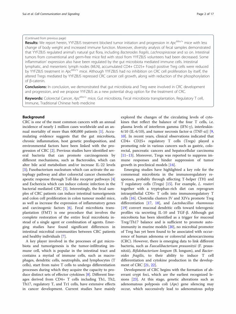
Results: We report herein, YYFZBJS treatment blocked tumor initiation and progression in ApcMin/+ mice with lesschange of body weight and increased immune function. Moreover, diversity analysis of fecal samples demonstratedthat YYFZBJS regulated animal’s natural gut flora, including Bacteroides fragilis, Lachnospiraceae and so on. Intestinaltumors from conventional and germ-free mice fed with stool from YYFZBJS volunteers had been decreased. Someinflammation’ expression also have been regulated by the gut microbiota mediated immune cells. Intestinallymphatic, and mesenteric lymph nodes (MLN), accumulated CD4+ CD25+ Foxp3 positive Treg cells were reducedby YYFZBJS treatment in ApcMin/+ mice. Although YYFZBJS had no inhibition on CRC cell proliferation by itself, thealtered Tregs mediated by YYFZBJS repressed CRC cancer cell growth, along with reduction of the phosphorylationof β-catenin.Conclusions: In conclusion, we demonstrated that gut microbiota and Treg were involved in CRC developmentand progression, and we propose YYFZBJS as a new potential drug option for the treatment of CRC.
Keywords: Colorectal Cancer, ApcMin/+ mice, Gut microbiota, Fecal microbiota transplantation, Regulatory T cell,Immune, Traditional Chinese herb medicine
BackgroundCRC is one of the most common cancers with an annualincidence of nearly 1 million cases worldwide and an an-nual mortality of more than 600,000 patients [1]. Accu-mulating evidence suggests that the gut microbiota,chronic inflammation, host genetic predisposition, andenvironmental factors have been linked with the pro-gression of CRC [2]. Previous studies have identified sev-eral bacteria that can promote carcinogenesis bydifferent mechanisms, such as Bacteroides, which canalter bile acid metabolism and/or increase IL-22 levels[3]; Fusobacterium nucleatum which can activate the au-tophagy pathway and alter colorectal cancer chemother-apeutic response through Toll-like receptor pathways [4]and Eschericia which can induce colonic infection in thebacterial mediated CRC [5]. Interestingly, the fecal sam-ples of CRC patients can induce intestinal tumorigenesisand colon cell proliferation in colon tumour model mice,as well as increase the expression of inflammatory genesand carcinogenic factors [6]. Fecal microbiota trans-plantation (FMT) is one procedure that involves thecomplete restoration of the entire fecal microbiota in-stead of a single agent or combination of agents. Emer-ging studies have found significant differences inintestinal microbial communities between CRC patientsand healthy individuals [7].A key player involved in the processes of gut micro-
biota and tumorigenesis is the tumor-infiltrating im-mune cell, which is popular in the intestinal tract andcontains a myriad of immune cells, such as macro-phages, dendritic cells, neutrophils, and lymphocytes (Tcells), start from naive T cells to undergo differentiationprocesses during which they acquire the capacity to pro-duce distinct sets of effector cytokines [8]. Different line-ages derived from CD4+ T cells including Th1, Th2,Th17, regulatory T, and Tr1 cells, have extensive effectsin cancer development. Current studies have mainly
explored the changes of the circulating levels of cyto-kines that reflect the balance of the four T cells, i.e.plasma levels of interferon gamma (IFN-γ), interleukin-6/10 (IL-6/10), and tumor necrosis factor-α (TNF-α)] [9,10]. In recent years, clinical observations indicated thatCD4+ CD25+ regulatory T cells (Tregs) played apromoting role in various cancers such as gastric, colo-rectal, pancreatic cancers and hepatocellular carcinoma[11–13]. Moreover, Tregs was reported to suppress im-mune responses and hinder suppression of tumorgrowth in preclinical models [14].Emerging studies have highlighted a key role for the
commensal microbiota in the immunoregulatory re-sponses, probably through affecting T-helper (TH) andT regulatory cells (Tregs) [15]. For example, L. reuteritogether with a tryptophan-rich diet can reprogramintraepithelial CD4+ T cells into immunoregulatory Tcells [16]. Clostridia clusters IV and XIVa promote Tregdifferentiation [17, 18], and Lactobacillus rhamnosus[19] convert mucosal dendritic cells toward tolerogenicprofiles via secreting IL-10 and TGF-β. Although gutmicrobiota has been identified as a trigger for mucosalTreg/Th17 balance and is sufficient to promote auto-immunity in murine models [20], no microbial promoterof Treg has yet been found to be associated with occur-rence of human adenoma or colorectal adenocarcinoma(CRC). However, there is emerging data to link differentbacteria, such as Faecalibacterium prausnitzii (F. praus-nitzii), Bifidobacterium longum (B. longum), and Bacter-oides fragilis, to their ability to induce T celldifferentiation and cytokine production in the develop-ment of CRC [21, 22].Development of CRC begins with the formation of ab-
errant crypt foci, which are the earliest recognized le-sions [23]. At this stage, genetic alterations such asadenomatous polyposis coli (Apc) gene silencing mayoccur, which successively lead to adenomatous polyp
Sui et al. Cell Communication and Signaling (2020) 18:113 Page 2 of 17

formation. As other mutations accumulate, the tumor ul-timately progresses to invasive adenocarcinoma. ApcMin/+
mice, a genetically engineered mouse model that has a mu-tation in the Apc gene, usually serve as a well-characterizedanimal model for human familial adenomatous polyposis[24]. Ki67 and Proliferating Cell Nuclear Antigen (PCNA)proteins are standard markers of cell proliferation, thuscommonly used to help assess malignancy grades of cancer[25]. The ApcMin/+ mice are often used as a well-recognizedspontaneous CRC model, highly expressing Ki67 andPCNA. Although studies highlighted the close involvementof Treg cells in CRC tumorigenesis in the ApcMin/+ mousemodel [26], the underlying molecular mechanism remainslargely enigmatic.Yi-Yi-Fu-Zi-Bai-Jiang-San (YYFZBJS), a thousand-
year-old prescription from the Golden Chamber, is com-monly used in traditional Chinese medicine (TCM) totreat gastrointestinal disorders [27, 28]. It is composedof three herbs: Yi-yi-ren (Semen Coicis), Fu-Zi (monks-hood), Bai-jiang-cao (Herba Patriniae), which are in a ra-tio of 30:6:15. Recently, Semen Coicis, Herba Patriniae,and monkshood are found to have multiple pharmaco-logical activities, including anti-cancer effect [28–30].Notably, Yi-yi-ren and Bai-jiang-cao, the most abundantingredients in the recipe, showed anti-proliferative effi-cacy in several human cancer cell lines, as well as a sup-pressive effect on the development of aberrant cryptforce (ACF) in Azoxymethane (AOM) treated mice [31].Our previous work demonstrated that some TCM inhib-ited the proliferation of CRC cells in vivo and in vitro[32, 33]. However, the anti-proliferation effect ofYYFZBJS on the intestinal tumor is poorly understood.In the current study, we investigated the effect of
YYFZBJS in a spontaneous intestinal tumor model ofApcMin/+ mice. Gavaging germ-free ApcMin/+ mice withstool from healthy controls and YYFZBJS volunteers, wedemonstrated that stool from YYFZBJS volunteers al-tered dysregulated inflammation and oncogenic path-ways and inhibited intestinal tumorigenesis. Wecharacterized the importance of Treg and expressionlevels of the related factors in spleen, MLN, LPL, andPBMC (Peripheral blood mononuclear cell) of the mice,in order to find the possible mechanisms involving inthe anti-cancer action of TCM prescriptions, and therole of Treg cells in spontaneous intestinalcarcinogenesis.
Materials and methodsCell culture and reagentsHuman colorectal adenocarcinoma HCT116 cell andMice colorectal adenocarcinoma MC-38 cell were pur-chased from the Shanghai Cell Collection (Shanghai,China). They were cultured in RPMI 1640, which wereall supplemented with 10% fetal bovine serum (Gibco,
NY, USA), 2 mM glutamine, 100 units/ml streptomycinand penicillin (Invitrogen, Carlsbad, CA). The cells weregrown at 37 °C in a humidified 5% CO2 atmosphere.Monoclonal antibodies specific for Ki67 (ab1667), PCNA(ab92552) and β-actin (ab179467) were obtained byAbcam plc., Cambridge, UK.
Mouse strains and breedingApcMin/+ mice on a C57BL/6 J background were origin-ally obtained from the Jackson Laboratory and bred inhouse as heterozygous wild type crosses to provide Apc-Min/+ mice and wild-type littermates [34]. All animalswere and kept under specific pathogen-free conditionsin filter-top cages. Genotyping was performed at 4 weeksby PCR [35]. Forty ApcMin/+ mice aged 6 weeks wererandomized into 5 groups (n = 8 per group). The micewere provided with YYFZBJS or Aspirin for 20 weeks aspreviously described [36]. Briefly, the intragastric admin-istration of YYFZBJS-L/M/H were taken at the doses of3.825 g/kg, 7.65 g/kg and 15.3 g/kg according to HED(human equivalent dose) [32]. In the clinical practice ofChinese herbal medicine, YYFZBJS is usually prescribedat a daily dose of 51 mg of herbal materials. When thishuman dose was converted into an animal dose (a per-son of 60 kg, and a conversion factor of 9 between hu-man and mouse), it was equivalent to the middle dose(7.65 g/kg) used in this study. Control group was oralgavaged with the same volume of sterile isotonic salineand fed with normal drinking water. The 20 week-oralgavage-protocol used in ApcMin/+ mice is presented inFig. 1a Signs of illness were monitored daily and bodyweight was recorded weekly.
Histology and immunohistochemistryMouse blood was collected from retinal venous plexus,centrifuged to harvest serum, which were stored at −80 °C. Mice were sacrificed by cervical dislocation. Thewhole intestine was removed immediately after sacrificeand opened longitudinally after washed with ice-coldPBS as previously described [37]. The number, location,and size of visible tumors throughout the intestine weremeasured to calculate the incidence of adenoma. Tumornumbers were counted and grouped based on sizes: < 2mm, 2–4 mm and > 4mm. Tissue sections were fixed in10% formalin followed by paraffin embedding. Then theywere stained with hematoxylin and eosin for pathologicalevaluation by a pathologist blinded to the experimentalgroups. Histological analysis for polyp, adenoma, andadenocarcinoma was performed by a board-certifiedpathologist (PV) as previously described [38]. The hist-ology scoring criteria is as follows: 0 = normal, 1 =mod-erate, 2 =marked and 3 = severe.For the murine samples, immunohistochemistry was
performed to detect total Ki67 (anti-mouse Ki67,
Sui et al. Cell Communication and Signaling (2020) 18:113 Page 3 of 17

Abcam), PCNA (anti-mouse PCNA, Abcam) and BrdU(anti-BrdU kit, Invitrogen); all stains used horseradishperoxidase-conjugated antibody, with chromogenic de-tection with the substrate 3–3′-diaminobenzidine, andfinally counterstained with hematoxylin.
Microbial analysis of mouse stoolFeces of all mice in the NS and YYFZBJS group werecollected for gut microbiota analyses. Briefly, (i) genomicDNA was extracted using a PowerSoil DNA IsolationKit (MO BIO Laboratories, Carlsbad, CA); (ii) the 16S
Fig. 1 Experimental design and effect of YYFZBJS in intestinal tumorigenesis. a Experimental design indicating the timing of intragastricadministration and organization of groups. b Macroscopic view of the representative mouse intestinal shows several polypoid and discoid colonictumors from different groups of ApcMin/+ mice after treatment with YYFZBJS for 20 weeks. c The number of intestinal polyps in small intestinalfrom different groups of ApcMin/+ mice after treatment with YYFZBJS for 20 weeks. d The number of intestinal polyps in the colon from differentgroups of ApcMin/+ mice after treatment with YYFZBJS for 20 weeks. The data are presented as the mean ± SD from at least three experiments. eThe tumor size distribution in the intestine was listed and compared with control. f Left: typical adenomatous polyp seen in infected ApcMin/+
mice showing high-grade dysplasia and carcinoma in situ. Middle: adenomatous intestinal polyp with the early invasion of neoplastic glands intothe muscular layers often seen in ApcMin/+ mice. Right: minute polyp with remnant dysplastic glands close to the surface epithelium. This typicalregressive intestinal cancer morphology is seen throughout the intestine in mice. Red arrows indicated adenocarcinoma cell. Magnification bars,100 μM. g&h Immunohistochemical staining with an antibody against PCNA, Ki67, BrdU in control group and YYFZBJS treatment group.Magnification bars, 50 μM. Data are given as means ± SD of 8 animals per experimental group, with Welch’s correction, one-tailed t-test. #P < 0.05,##P < 0.01; *P < 0.05, **P < 0.01, &P < 0.05, $P < 0.05 vs. control
Sui et al. Cell Communication and Signaling (2020) 18:113 Page 4 of 17

rDNA V4 region was amplified using the 515F and 806Rprimers; (iii) PCR product quantification, qualification,and purification were performed; (iv) library preparationand sequencing were performed on the MiSeq platform(Illumina, Inc., San Diego, CA). The 16S rRNA sequen-cing data were quality filtered using FLASH (Fast LengthAdjustment of Short reads, Version 1.2.11). Operationaltaxonomic units (OTUs) were picked at a 97% sequencesimilarity cut-off, and the purified amplicons were se-quenced on an Illumina MiSeq platform at MajorbioBio-pharm Technology Co. Ltd. according to the stand-ard protocols.
Antibiotic treatmentsMice were treated for four weeks with an antibiotic solu-tion (Abx) containing Ampicillin (1 mg/ml), Neomycin(1 mg/ml), Metronidazole (1 mg/ml), and Vancomycin(0.5 mg/ml) added to the sterile drinking water of miceadlibitum as previously described [39]. Solutions andbottles were changed 2 times a week. After four weeks,Abx treatment was stopped and the mice were recolo-nized by FMT.
Fecal microbiota transplantation (FMT)After receiving antibiotic cocktails for 4 weeks, a volumeof 200 μL suspension was gavaged to each mouse forfour weeks [39]. The ApcMin/+ mice were divided intotwo groups with 8 mice each: One group was gavagedfecal samples from healthy controls (Control-FMT),while the other group was gavaged fecal samples frompeople who eating YYFZBJS (YYFZBJS-FMT). Eachgroup of mice used a separate set of intragastricapparatus.
Electron microscopicThe intestinal tissue of ApcMin/+ mice treatment withFMT were excised and fixed in 0.1M phosphate buffercontaining 2.5% glutaraldehyde and 2.0% paraformalde-hyde (pH 7.4). Then the tissue were fixed, dehydrated,polymerized and then examined using the transmissionelectron microscope as previously described [40].
Spleen to body weight ratioBefore killing, mice body weight was measured, and thenmice spleens were collected, and spleen weight was mea-sured as previously described [41]. The ratios of thespleen to body weight were calculated as spleen weight/body weight.
Cytokine antibody arraysSerum samples were screened in duplicates using aMouse Cytokine Array QAM-INF-1 (RayBiotech) con-taining slides coated with 40 different cytokinesaccording to the manufacturer’s guidelines with some
modifications as previously described [42]. Briefly, thearrays were blocked, incubated with 100 mL of conditionmedium overnight, followed by biotin-conjugated anti-bodies (1/250) incubation for 2 h and with HRP-linkedsecondary antibody (1/1000) for 1 h. The membraneswere incubated with a peroxidase substrate, and the re-sults were documented using XAR films. Quantitativearray analysis was performed using Array Vision Evalu-ation 8.0 (GE Healthcare Life Science).
Quantitative real-time PCR and bioinformatics analyses ofRNA-SeqTumor tissues were homogenized with 1mL TRI re-agent to extract total RNA. cDNA was synthesized byreverse transcription of total RNA (Epicentre). Quantita-tive real-time PCR (qRT-PCR) was carried out as previ-ously described [43]. The Oligonucleotide primers fortarget genes (T-bet, Gata3, ROR-γt, Foxp3, c-Myc,Axin2, EphB3, β-catenin, TCF, LEF1, CyclinD1, Lgr5and GAPDH) were shown in Supplementary Table S1.RNA-Seq FASTQ files were processed using the RNA-Seq module implemented in the CLC Genomics Work-bench v8.0 software (Qiagen Bioinformatics) with defaultsettings.
Lymphocyte preparationSpleen, mesenteric lymph nodes (MLN) and small intes-tine and colon, were collected from ApcMin/+ and WTmice. The monoplast suspension was collected by pass-ing splenocytes through 70 μm cell strainers (BD Biosci-ences, Bedford, MA, USA). Red cell lysis was performedon splenic cells with 0.07M NH4Cl, pH 7.3, 37 °C for 5min. Lamina propria lymphocytes (LPL) from the smalland large intestine, and from adenomas were isolatedessentially as described before [44] but with the use ofcollagenase VIII (Sigma-Aldrich) for colon digestion.
Analysis of cytokine expression in serumMouse serum samples were analyzed for mouse cyto-kines by ELISA according to the manufacturer’s instruc-tions (eBioscience) and as previously described [45].
Flow CytometryPhenotype analysis of Tregs was performed with a BDFACS AriaII flow cytometer (BD, USA) as previously de-scribed [46]. Briefly, the cells were labeled with CD4-FITC, CD25-APC, and Foxp3-PE (eBioscience, SanDiego, CA) following the manufacturer’s protocol. Toanalyze the prevalence of Tregs, CD4 + Foxp3 + T cellswere evaluated after gating on CD4 + T cells and wereexpressed as a percentage of the total CD4+ T cells.
Sui et al. Cell Communication and Signaling (2020) 18:113 Page 5 of 17

Preparation of Chinese YYFZBJS herb formulaThe formula for creating one dose of YYFZBJS is pre-sented in Table 1. Chinese medicines were purchasedfrom Shanghai Hua Yu Chinese Herbs Co., Ltd. (Shang-hai, China). The Chinese medicines included Yi-yi-ren(lot# 180103), Fu-Zi (lot# 180709), Bai-jiang-cao (lot#180522). All herbs were authenticated by Associate Re-searcher Tao Yang according to the Pharmacopoeia ofthe People’s Republic of China (2015). The vouchers ofall components were deposited at the herbarium locatedin the College of Pharmacy, Shanghai University ofTCM (Shanghai, China).All herbs were added the appropriate amount of water
and then extracted twice, filtrated and dried into dry-extract according to as a validated method [32]. Forquality control, the fingerprint spectrum for YYFZBJSwas performed by UHPLC-Q Exactive system (Thermo,San Jose, CA, USA) equipped with a quaternary gradientpump, an autosampler, and high-resolution mass spec-trometry detector. The components were eluted with agradient system consisting of acetonitrile (A) and aque-ous 0.1% formic acid (B) in gradient (time, min/B%: 0/95, 12/5,14/5,14.1/95,16/5); flow rate: 0.3 ml/min). Thespectral data were recorded in the m/z range of 80–1200. Mass spectra were acquired in both negative andpositive modes with ion spray voltage 3.5 kV, capillarytemperature at 320 °C, auxiliary gas heater temperatureat 300 °C, sheath gas (nitrogen) flow at 35 AU, auxiliarygas (nitrogen) flow at 10 AU, Scan mode: Full MS (Reso-lution 70,000) and dd-MS2 (Resolution 17,500, NCE35,Stepped NCE50%). The chromatographic column wasACQUITY UPLC HSS T3 (2.1 mm × 100mm, 1.8 μm).The mobile phase flow rate was 0.3 ml/min and the col-umn temperature was maintained at 40 °C. Otherwise,the contents of liquiritigenin, luteolin, mesalamine,aconitine, and hypaconitine were detected by UPLC-MSmethod and were 1.71 mg/g, 311.22 mg/g, 5.32 mg/g,1.91 mg/g, and 74.71 mg/g in the extracts respectively.
Network constructionThe potential targets for the components of YYFZBJSwere retrieved from Therapeutic Targets Database in-cluding TCMSP (http://ibts.hkbu.edu.hk/LSP/tcmsp.php), TCM database @Taiwan (http://tcm.cmu.edu.tw), and TCM Integrated Database (TCMID) (http://www.megabionet.org/tcmid). In these networks,YYFZBJS and its targets are represented as nodes,
while the edges indicate interaction or relatedness(Supplementary Table S3).
Isolation of spleen Tregs and its effect on cancer cellabilitySpleen cells from ApcMin/+ mice were separated over col-umns for negative and positive selection for CD4+CD25+ Foxp3 MACS columns and separator (MiltenyiBiotech, CA) as previously described [47]. CD4+ CD25+Foxp3 T cells were co-cultured with Bacteroides fragilis(cell: bacterial = 1:10) in RPMI-1640 medium in presenceor absence of YYFZBJS (different concentrations) for 4 hand CD4+ CD25+ Foxp3 T cells were collected aftercentrifuging. For the analysis of Treg cell effect on can-cer cell ability, the MC-38 cells were inoculated in 24well plates with 1000 cells (per well) and co-culturedwith the Treg for 12, 24, 36 and 48 h. The proliferationof MC-38 cells was measured by trypan blue as previ-ously reported [48].
Bacterial attachment assayBacteroides fragilis (43858) were purchased from ATCCand cultured in lysogeny broth at 37 °C. The bacterial at-tachment assay was performed as described previously[49]; Treg cells were co-cultured with bacteria for 4 h(MOI = 10) under anaerobic conditions. After co-culture, medium was removed and cells were washedwith PBS three times. Then cells were lysed, and addedWilkins-Chalgren anaerobe broth to homogenize. Theattached Enterotoxigenic Bacteroides fragilis (ETBF) col-onies were recovered on Wilkins-Chalgren anaerobeagar plate under anaerobic conditions; the number ofcolonies was counted.
Western blot analysisWhole cell lysates for Western blot analysis of β-catenin(nuclear, cytoplasm), PCNA and β-actin expression wereprepared as previously reported [42]. Briefly, total lysatesfrom treated cells were prepared with RIPA buffer (50mM Tris, pH 7.2; 150 mM NaCl; 0.5% sodium deoxycho-late; 0.1% sodium dodecyl sulfate; 1% Nonidet P-40; 10mM NaF; 1 mM Na3VO4; protease inhibitor cocktail.Lysates were sonicated for 10 s and centrifuged at 14,000 rpm for 10 min at 4 °C. Protein concentration wasdetermined by bicinchoninic acid assay with BSA as astandard (Pierce, Rockford, IL, USA). Equivalentamounts of protein (50 μg/lane) were separated on 7.5–
Table 1 Formula of YYFZBJS (one dose)
Chinese medicine Plant origin Medicinal parts Origin (Province) Amount in Preparation (g)
Yi-yi-ren Coix lacrymajobi L.var.mayuen (Roman.)Stapf kernel Fujian Province 211,803
Fu-Zi Aconitum carmichaeli Debx. root Sichuan Province 221,304
Bai-jiang-cao Thlaspi arvense Linn aerial parts Henan Province 201,508
Sui et al. Cell Communication and Signaling (2020) 18:113 Page 6 of 17

12% SDS-polyacrylamide gel and transferred to polyviny-lidene difluoride membranes (Millipore, Bedford, MA,USA). Membranes were incubated with PBS containing0.05% Tween 20 and 5% nonfat dry milk to block non-specific binding and were incubated with primary anti-bodies, then with appropriate secondary antibodiesconjugated to horseradish peroxidase. Immunoreactivebands were visualized by using Renaissance chemilumin-escence reagent (Perkin-Elmer Life Science, Boston, MA,USA). Densitometric analysis was performed using theScion Imaging application (Scion Corporation), with β-actin as the internal reference.
ResultsYYFZBJS suppresses intestinal tumorigenesis andexpression of Ki67, PCNA, and reactivity to BrdU in theApcMin/+ mouse modelPreviously, we showed that traditional Chinese herbswere sufficient to inhibit colorectal carcinoma multidrugresistance (MDR) in nude mouse [28, 29]. In the presentstudy, we first sought to determine whether traditionalChinese herbs were beneficial for innate immunity andintestinal tumorigenesis in ApcMin/+ mice. The structuresof the determined experiment from the herbs are shownin Fig. 1a. Consistent with the clinical results, no differ-ence was noted in animal weight and hepatorenal tox-icity among treatment groups during the experiment(Supplementary Fig. 1&2), meaning the herbs were safefor the general health of the animals. Following a 20weeks intragastric administration of aspirin, ApcMin/+
mice contained much less intestinal adenomas comparedwith that in the no-aspirin group (Fig. 1b&c and Supple-mentary Fig. 3). Similarly, we discovered that ApcMin/+
mice treated with YYFZBJS carried fewer adenomas bothin the small and large intestine (Fig. 1c&d&e and Sup-plementary Fig. 4), compared with the normal salineControls(control). Notably, the numbers of polyps in allthree YYFZBJS groups were all much fewer than that ofthe non-treated control group (Fig. 1e).Control tumors in untreated ApcMin/+ mice were his-
tologically identified as polyps with severe atypia or earlycarcinoma with submucosal infiltration. Notably, earlycarcinomas in the colon were completely eradicated byYYFZBJS treatment (Fig. 1f). The poly adenomas insmall intestine, featuring moderate mucosal epitheliumarchitectural changes with some budding and branching,was much fewer in number in YYFZBJS treated ApcMin/+
mice than the untreated ones (Fig. 1f). However, high-grade dysplasia adenocarcinoma (including early carcin-oma), was only found in 0% (0/8), 25% (2/8), 37.5% (3/8)of mice in high-, middle-, and low-YYFZBJS dose-treated ApcMin/+ mice compared to 100% (8/8) of un-treated mice (Supplementary Table S2). These data
indicate that YYFZBJS slows down the intestinaladenoma-to-adenocarcinoma progression.Since Ki67, PCNA and BrdU are cell proliferation
markers, we then examined the localization and expres-sion levels of Ki67 and PCNA by immunohistochemistryin the tumors of ApcMin/+ mice with or withoutYYFZBJS treatment (Fig. 1g&h). Comparing with the un-treated mice, nuclear expression levels of Ki67 andPCNA, and BrdU reactivity in intestinal polyp epitheliawere reduced after YYFZBJS treatment (Fig. 1g, h).
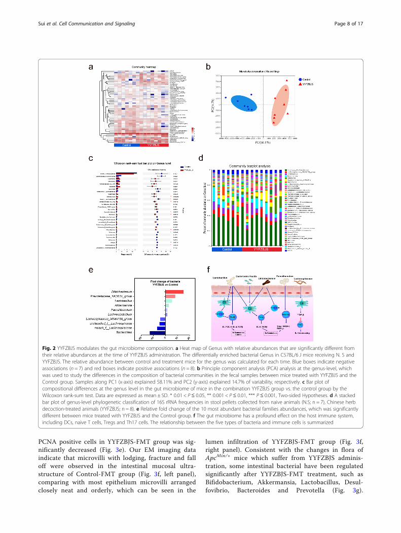
YYFZBJS modulates the gut microbiome compositionWe next sought to characterize the effects of YYFZBJStreatment on intestinal bacterial communities throughanalysis of bacterial 16S rRNA compositions. In the Apc-Min/+ mice model, YYFZBJS and NS group developeddifferent gut microbiota: we observed a significantlylower bacterial richness in the YYFZBJS group (Fig. 2a).Nonmetric multidimensional scaling analysis demon-strated the clear separation of bacterial OTU compos-ition (Fig. 2b). Alterations at the genus level were alsoassessed (Fig. 2c). While a significant elevation in abun-dance of several probiotic genera (Bifidobacterium andPrevotellaceae) was determined in response to YYFZBJStreatment, some genera (Bacteroides, Lachnospiraceae,unclassified lachnospiraceae among others) were nearlyeliminated. YYFZBJS administration resulted in reducedfrequencies of bacteria belonging to the Firmicutes, in-cluding Lactobacillus and Dubosiella (Fig. 2d). Consist-ent with the results of species richness, YYFZBJSadministration selectively blunted the relative expressionof the Bacteroides, Lachnospiraceae and so on (show sig-nificant changes in top 10) (Fig. 2e). Furthermore, basedon the published studies and our screening results, themechanisms of concentrated bacteria in regulatingCD4+ T cell-derived effectors were demonstrated inregulating host immunity (Fig. 2f).
Gut microbiota from YYFZBJS users delay the progressionof intestinal tumorigenesisThe impact of FMT with or without YYFZBJS on in-testinal adenoma was evaluated after treatment with12 weeks (Fig. 3a&b). No significant gross bloodystool was observed in the two groups (data notshown). All ApcMin/+ mice were well tolerated andsurvive after FMT. In the YYFZBJS-FMT group, afew scattered small polyps were observed, whilemore adenomas were observed in the Control-FMTgroup, especially the cribriform morphology ap-peared in the tumors (Fig. 3c&e). The total numberof intestinal tumors in mice receiving fecal samplesfrom YYFZBJS volunteers was decreased comparedwith the health controls (Fig. 3d). Comparing withthe Control-FMT group, the rate of Ki-67 and
Sui et al. Cell Communication and Signaling (2020) 18:113 Page 7 of 17
PCNA positive cells in YYFZBJS-FMT group was sig-nificantly decreased (Fig. 3e). Our EM imaging dataindicate that microvilli with lodging, fracture and falloff were observed in the intestinal mucosal ultra-structure of Control-FMT group (Fig. 3f, left panel),comparing with most epithelium microvilli arrangedclosely neat and orderly, which can be seen in the
lumen infiltration of YYFZBJS-FMT group (Fig. 3f,right panel). Consistent with the changes in flora ofApcMin/+ mice which suffer from YYFZBJS adminis-tration, some intestinal bacterial have been regulatedsignificantly after YYFZBJS-FMT treatment, such asBifidobacterium, Akkermansia, Lactobacillus, Desul-fovibrio, Bacteroides and Prevotella (Fig. 3g).
Fig. 2 YYFZBJS modulates the gut microbiome composition. a Heat map of Genus with relative abundances that are significantly different fromtheir relative abundances at the time of YYFZBJS administration. The differentially enriched bacterial Genus in C57BL/6 J mice receiving N. S andYYFZBJS. The relative abundance between control and treatment mice for the genus was calculated for each time. Blue boxes indicate negativeassociations (n = 7) and red boxes indicate positive associations (n = 8). b Principle component analysis (PCA) analysis at the genus-level, whichwas used to study the differences in the composition of bacterial communities in the fecal samples between mice treated with YYFZBJS and theControl group. Samples along PC1 (x-axis) explained 58.11% and PC2 (y-axis) explained 14.7% of variability, respectively. c Bar plot ofcompositional differences at the genus level in the gut microbiome of mice in the combination YYFZBJS group vs. the control group by theWilcoxon rank-sum test. Data are expressed as mean ± SD. * 0.01 < P≤ 0.05, ** 0.001 < P ≤ 0.01, *** P ≤ 0.001, Two-sided Hypotheses. d A stackedbar plot of genus-level phylogenetic classification of 16S rRNA frequencies in stool pellets collected from naive animals (N.S; n = 7), Chinese herbdecoction-treated animals (YYFZBJS; n = 8). e Relative fold change of the 10 most abundant bacterial families abundances, which was significantlydifferent between mice treated with YYFZBJS and the Control group. f The gut microbiome has a profound effect on the host immune system,including DCs, naive T cells, Tregs and Th17 cells. The relationship between the five types of bacteria and immune cells is summarized
Sui et al. Cell Communication and Signaling (2020) 18:113 Page 8 of 17

Therefore, the results suggested that gut microbiotafrom YYFZBJS users inhibited the progression of in-testinal adenoma in ApcMin/+ mice.
Effect of YYFZBJS on immunity of ApcMin/+ miceTo determine whether YYFZBJS changed levels of in-flammatory cytokines, we used a cytokine antibody
array to illustrate that, compared to normal controlmice, ApcMin/+ mice secreted higher levels of inflam-matory cytokines/chemokines including IL-17,Eotaxin-2, Leptin and PF4 (Supplementary Fig. 5). Itwas previously demonstrated that YYFZBJS negativelyregulated inflammatory cytokines IL-17, and IL-10 inmyeloid precursor differentiation [50]. Consistent with
Fig. 3 Gut microbiota from YYFZBJS volunteers delay the progression of intestinal tumorigenesis. a Design of stool gavage experiment toApcMin/+ mice. Mice were treatmented with Abx from week 6, and sacrificed at week 22 (n = 8). b Display of the fecal extracts of the ApcMin/+
mice with FMT treatment for 12 weeks. c Left: typical adenomatous polyp seen in infected ApcMin/+ mice showing high-grade dysplasia andcarcinoma in situ. Right: minute polyp with remnant dysplastic glands close to the surface epithelium. Blue arrows indicated adenocarcinoma cell.Magnification bars, 100 μM. Histological analysis of intestinal tumors applyed in the two FMT group mice (n = 8 for each group). d The tumor sizedistribution in the intestine was listed and compared with control-FMT (n = 8 for each group). Data shown represent means ± SD. *P < 0.05 vs.control-FMT. e Immunohistochemical staining with an antibody against PCNA and Ki67 in control-FMT group and YYFZBJS-FMT treatment group.Magnification bars, 100 μM. Data are given as means ± SD of 8 animals per experimental group, with Welch’s correction, one-tailed t-test. *P <0.05 vs. control-FMT. f Electron microscopy in the lumen infiltration of control-FMT group mice and YYFZBJS-FMT mice at age of week 22. Bothmicrovilli and goblet cells can also be seen. The black arrow refers to the intestinal microvilli; The red arrow indicates a tight connection.Magnification bars, 500 nM. g Fecal bacterial DNA was prepared from Control-FMT group and YYFZBJS-FMT treatment group. Relative genusabundance was shown as percentage of each OTU in the total OTUs (n = 5/group). Data shown represent means ± SD. *P < 0.05
Sui et al. Cell Communication and Signaling (2020) 18:113 Page 9 of 17
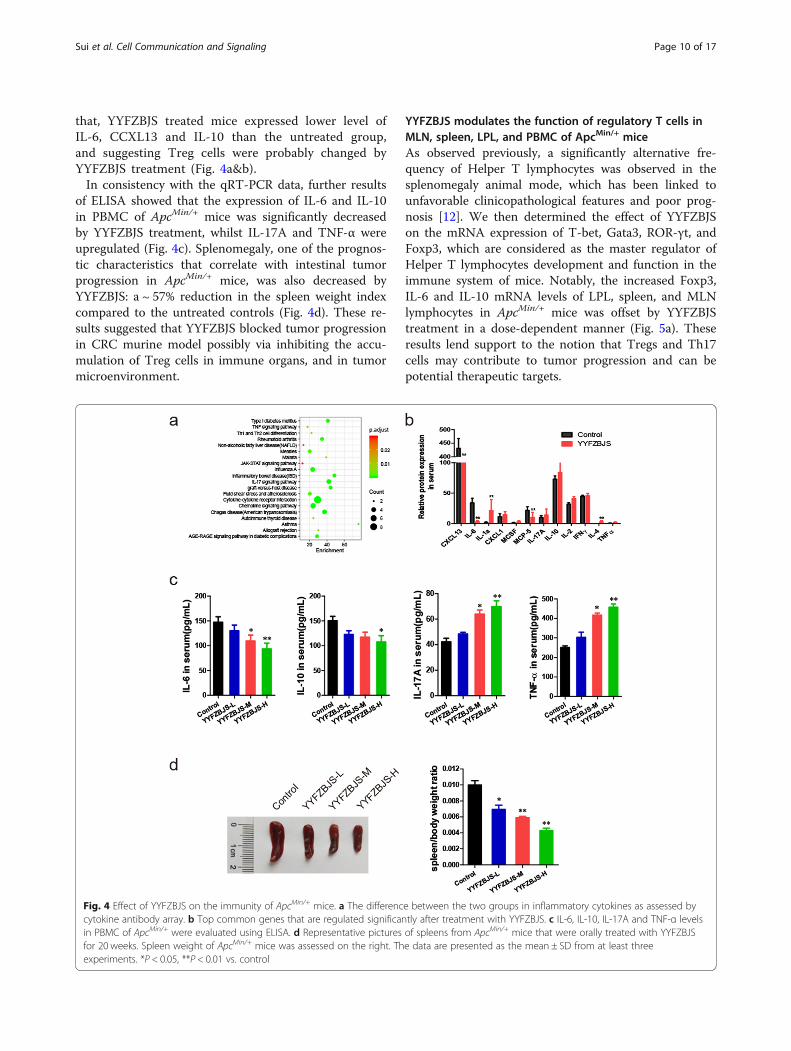
that, YYFZBJS treated mice expressed lower level ofIL-6, CCXL13 and IL-10 than the untreated group,and suggesting Treg cells were probably changed byYYFZBJS treatment (Fig. 4a&b).In consistency with the qRT-PCR data, further results
of ELISA showed that the expression of IL-6 and IL-10in PBMC of ApcMin/+ mice was significantly decreasedby YYFZBJS treatment, whilst IL-17A and TNF-α wereupregulated (Fig. 4c). Splenomegaly, one of the prognos-tic characteristics that correlate with intestinal tumorprogression in ApcMin/+ mice, was also decreased byYYFZBJS: a ~ 57% reduction in the spleen weight indexcompared to the untreated controls (Fig. 4d). These re-sults suggested that YYFZBJS blocked tumor progressionin CRC murine model possibly via inhibiting the accu-mulation of Treg cells in immune organs, and in tumormicroenvironment.
YYFZBJS modulates the function of regulatory T cells inMLN, spleen, LPL, and PBMC of ApcMin/+ miceAs observed previously, a significantly alternative fre-quency of Helper T lymphocytes was observed in thesplenomegaly animal mode, which has been linked tounfavorable clinicopathological features and poor prog-nosis [12]. We then determined the effect of YYFZBJSon the mRNA expression of T-bet, Gata3, ROR-γt, andFoxp3, which are considered as the master regulator ofHelper T lymphocytes development and function in theimmune system of mice. Notably, the increased Foxp3,IL-6 and IL-10 mRNA levels of LPL, spleen, and MLNlymphocytes in ApcMin/+ mice was offset by YYFZBJStreatment in a dose-dependent manner (Fig. 5a). Theseresults lend support to the notion that Tregs and Th17cells may contribute to tumor progression and can bepotential therapeutic targets.
Fig. 4 Effect of YYFZBJS on the immunity of ApcMin/+ mice. a The difference between the two groups in inflammatory cytokines as assessed bycytokine antibody array. b Top common genes that are regulated significantly after treatment with YYFZBJS. c IL-6, IL-10, IL-17A and TNF-α levelsin PBMC of ApcMin/+ were evaluated using ELISA. d Representative pictures of spleens from ApcMin/+ mice that were orally treated with YYFZBJSfor 20 weeks. Spleen weight of ApcMin/+ mice was assessed on the right. The data are presented as the mean ± SD from at least threeexperiments. *P < 0.05, **P < 0.01 vs. control
Sui et al. Cell Communication and Signaling (2020) 18:113 Page 10 of 17
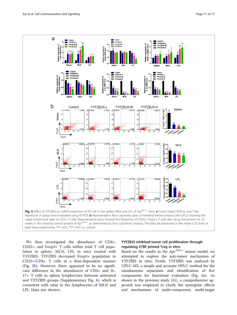
We then investigated the abundance of CD4+,CD25+, and Foxp3+ T cells within total T cell popu-lation in spleen, MLN, LPL in mice treated withYYFZBJS. YYFZBJS decreased Foxp3+ population inCD25+/CD4+ T cells in a dose-dependent manner(Fig. 5b). However, there appeared to be no signifi-cant difference in the abundances of CD4+ and IL-17+ T cells in spleen lymphocytes between untreatedand YYFZBJS groups (Supplementary Fig. 6), which isconsistent with what in the lymphocytes of MLN andLPL (data not shown).
YYFZBJS inhibited tumor cell proliferation throughregulating ETBF primed Treg in vitroBased on the results in the ApcMin/+ mouse model, weattempted to explore the anti-tumor mechanism ofYYFZBJS in vitro. Firstly, YYFZBJS was analyzed byUPLC-MS, a simple and accurate HPLC method for thesimultaneous separation and identification of fivecomponents for functional evaluation (Fig. 6a). Asshown in the previous study [51], a comprehensive ap-proach was employed to clarify the synergistic effectsand mechanisms of multi-component, multi-target
Fig. 5 Effect of YYFZBJS on mRNA expression of TH cell in the spleen, MLN and LPL of ApcMin/+ mice. a Foxp3, Gata3, ROR-γt, and T-betexpression in polyp were evaluated using RT-PCR. b Representative flow cytometry plots of intestinal lamina propria cells (LPCs) showing theviable lymphocyte gate on CD4 + T cells. Representative plots showed the frequency of CD25+ Foxp3+ T cells after drug intervention for 20weeks in the intestinal lamina propria of ApcMin/+, as determined by flow cytometric analysis. The data are presented as the mean ± SD from atleast three experiments. *P < 0.05, **P < 0.01 vs. control
Sui et al. Cell Communication and Signaling (2020) 18:113 Page 11 of 17

Fig. 6 (See legend on next page.)
Sui et al. Cell Communication and Signaling (2020) 18:113 Page 12 of 17
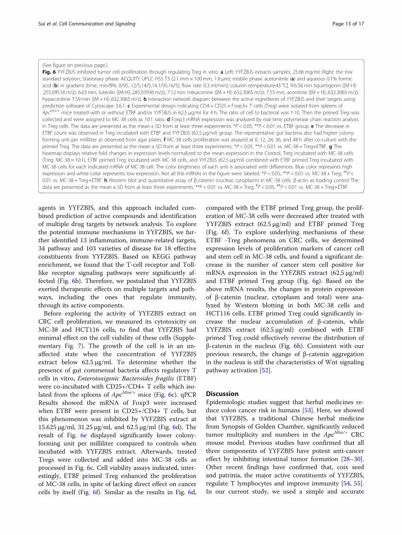
agents in YYFZBJS, and this approach included com-bined prediction of active compounds and identificationof multiple drug targets by network analysis. To explorethe potential immune mechanisms in YYFZBJS, we fur-ther identified 13 inflammation, immune-related targets,34 pathway and 103 varieties of disease for 18 effectiveconstituents from YYFZBJS. Based on KEGG pathwayenrichment, we found that the T-cell receptor and Toll-like receptor signaling pathways were significantly af-fected (Fig. 6b). Therefore, we postulated that YYFZBJSexerted therapeutic effects on multiple targets and path-ways, including the ones that regulate immunity,through its active components.Before exploring the activity of YYFZBJS extract on
CRC cell proliferation, we measured its cytotoxicity onMC-38 and HCT116 cells, to find that YYFZBJS hadminimal effect on the cell viability of these cells (Supple-mentary Fig. 7). The growth of the cell is in an un-affected state when the concentration of YYFZBJSextract below 62.5 μg/ml. To determine whether thepresence of gut commensal bacteria affects regulatory Tcells in vitro, Enterotoxigenic Bacteroides fragilis (ETBF)were co-incubated with CD25+/CD4+ T cells which iso-lated from the spleens of ApcMin/+ mice (Fig. 6c). qPCRResults showed the mRNA of Foxp3 were increasedwhen ETBF were present in CD25+/CD4+ T cells, butthis phenomenon was inhibited by YYFZBJS extract at15.625 μg/ml, 31.25 μg/ml, and 62.5 μg/ml (Fig. 6d). Theresult of Fig. 6e displayed significantly lower colony-forming unit per milliliter compared to controls whenincubated with YYFZBJS extract. Afterwards, treatedTregs were collected and added into MC-38 cells asprocessed in Fig. 6c. Cell viability assays indicated, inter-estingly, ETBF primed Treg enhanced the proliferationof MC-38 cells, in spite of lacking direct effect on cancercells by itself (Fig. 6f). Similar as the results in Fig. 6d,
compared with the ETBF primed Treg group, the prolif-eration of MC-38 cells were decreased after treated withYYFZBJS extract (62.5 μg/ml) and ETBF primed Treg(Fig. 6f). To explore underlying mechanisms of theseETBF -Treg phenomena on CRC cells, we determinedexpression levels of proliferation markers of cancer celland stem cell in MC-38 cells, and found a significant de-crease in the number of cancer stem cell positive formRNA expression in the YYFZBJS extract (62.5 μg/ml)and ETBF primed Treg group (Fig. 6g). Based on theabove mRNA results, the changes in protein expressionof β-catenin (nuclear, cytoplasm and total) were ana-lyzed by Western blotting in both MC-38 cells andHCT116 cells. ETBF primed Treg could significantly in-crease the nuclear accumulation of β-catenin, whileYYFZBJS extract (62.5 μg/ml) combined with ETBFprimed Treg could effectively reverse the distribution ofβ-catenin in the nucleus (Fig. 6h). Consistent with ourprevious research, the change of β-catenin aggregationin the nucleus is still the characteristics of Wnt signalingpathway activation [52].
DiscussionEpidemiologic studies suggest that herbal medicines re-duce colon cancer risk in humans [53]. Here, we showedthat YYFZBJS, a traditional Chinese herbal medicinefrom Synopsis of Golden Chamber, significantly reducedtumor multiplicity and numbers in the ApcMin/+ CRCmouse model. Previous studies have confirmed that allthree components of YYFZBJS have potent anti-cancereffect by inhibiting intestinal tumor formation [28–30].Other recent findings have confirmed that, coix seedand patrinia, the major active constituents of YYFZBJS,regulate T lymphocytes and improve immunity [54, 55].In our current study, we used a simple and accurate
(See figure on previous page.)Fig. 6 YYFZBJS inhibited tumor cell proliferation through regulating Treg in vitro. a Left: YYFZBJS extracts samples, 25.66 mg/ml; Right: the mixstandard solution; Stationary phase: ACQUITY UPLC HSS T3 (2.1 mm × 100mm, 1.8 μm); mobile phase: acetonitrile (a) and aqueous 0.1% formicacid (b) in gradient (time, min/B%: 0/95, 12/5,14/5,14.1/95,16/5); flow rate: 0.3 ml/min); column temperature:45 °C]. R:6.56 min liquiritigenin ([M-H]-,255.09518m/z); 6.63 min, luteolin ([M-H]-,285.03936m/z); 7.12 min mesaconine ([M + H]-,632.3065 m/z); 7.55 min, aconitine ([M + H]-,632.3065 m/z);hypaconitine 7.59 min ([M + H]-,632.3065 m/z). b Interaction network diagram between the active ingredients of YYFZBJS and their targets usingprediction software of Cytoscape 3.6.1. c Experimental design indicating CD4 + CD25 + Foxp3+ T cells (Treg) were isolated from spleens ofApcMin/+ mice treated with or without ETBF and/or YYFSBJS in 62.5 μg/ml for 4 h. The ratio of cell to bacterial was 1:10. Then the primed Treg wascollected and were assigned to MC-38 cells as 10:1 ratio. d Foxp3 mRNA expression was analyzed by real-time polymerase chain reaction analysisin Treg cells. The data are presented as the mean ± SD from at least three experiments. *P < 0.05, **P < 0.01 vs. ETBF group. e The decrease inETBF count was observed in Treg incubated with ETBF and YYFZBJS (62.5 μg/ml) group. The representative gut bacteria also had higher colony-forming unit per milliliter as observed from agar plates. f MC-38 cells proliferation was assayed at 0, 12, 24, 36, and 48 h after co-culture with theprimed Treg. The data are presented as the mean ± SD from at least three experiments. *P < 0.05, **P < 0.01 vs. MC-38 + Treg+ETBF. g Theheatmap displays relative fold changes in expression levels normalized to the mean expression in the Control, Treg incubated with MC-38 cells(Treg: MC-38 = 10:1), ETBF primed Treg incubated with MC-38 cells, and YYFZBJS (62.5 μg/ml) combined with ETBF primed Treg incubated withMC-38 cells for each indicated mRNA of MC-38 cell. The color brightness of each unit is associated with differences. Blue color represents highexpression and white color represents low expression. Not all the mRNAs in the figure were labeled. *P < 0.05, **P < 0.01 vs. MC-38 + Treg; ##P <0.01 vs. MC-38 + Treg+ETBF. h Western blot and quantitative assay of β-catenin (nuclear, cytoplasm) in MC-38 cells. β-actin as loading control Thedata are presented as the mean ± SD from at least three experiments. **P < 0.01 vs. MC-38 + Treg; #P < 0.05, ##P < 0.01 vs. MC-38 + Treg+ETBF
Sui et al. Cell Communication and Signaling (2020) 18:113 Page 13 of 17

UPLC-MS technology to simultaneously separate andidentify three drug components to evaluate YYFZBJS.Emerging evidence suggests that gut microbiota, along
with immune and metabolite factors, contribute to CRCcarcinogenesis [4, 56]. We found YYFZBJS treatmentchanged bacterial taxa in the colon of ApcMin/+ mice.OTUs results further showed that the bacteria of Lacto-bacillus, Dubosiella, might play an active role in bothpro- and anti-inflammatory T-cell regulatory pathways.Interestingly, several studies have highlighted the in-ducement of colon Treg cells is closely related to the gutmicrobiome [2, 6]. Reports showed that Treg cells canbe upregulated by certain bacterial strains and metabolicsubstances from B. fragilis [6, 57]. As expected, our dataalso found that YYFZBJS FMT administration modulatesmicrobial consortia on colorectal carcinogenesis and re-sults in a significant reduction in overall polyp numberand size. It also showed superiority in restoring gutmicrobiota diversity, which suggested that the anti-tumorigenesis effect of YYFZBJS was mediated mainlythrough the complex microbiome.Several studies have confirmed that gut microbiota
from CRC patients showed intestinal mucosal barrierdamage, low grade intestinal inflammation, activation ofadenomas progression [2, 7]. Previous research hasshown that FMT restored both the ratio and diversity ofgut microbiota, which promoted the CD4 + CD25 +Foxp3+ cells and attenuated T helper (Th)1/2/17 cells inCAC mice [6]. Similar to our bacteria analysis results,our microarray data suggest that the YYFZBJS evokemultiple inflammatory and oncogenic pathways in CRCcarcinogenesis, especially on Treg/Th17 signaling be-cause of significant impacts on IL-6, IL-10, IL-17 expres-sion. Several studies have shown the Treg involvementin colorectal tumorigenesis, e.g. IL-6 and IL-10 both en-hanced tumorigenesis in colitis-associated cancer models[58, 59], whereas blockade of IL17A inhibited tumorgrowth [28]. In animal experiments of colorectal car-cinogenesis, mechanisms of the effects of microbiota onimmune homeostasis have been studied extensively, withsome studies demonstrating that Lactobacillus orBacteroides fragilis coordinate Treg/Th17 balance toregulate carcinogenesis [16, 22].Accumulating data also indicated that the percentage
of Treg cells is inversely related to increasing the risk forthe progression of cancer [60, 61]. For CRC patients, in-creased numbers of Treg cells had been found in periph-eral blood, tumor-draining lymph node (DLN), andtumor microenvironment. Coincidently, Tregs have alsobeen reported by clinical observations and mechanisticstudies, to play an indispensable role as a promoter oftumor growth because of its suppressive effects on theautologous effector T-cell responses. However, othersfound that Tregs may inhibit the intestinal tumor
growth in adenomatous polyposis coli (Apc)-mice [62,63]. Nonetheless, in the early stages of tumor develop-ment, it is widely understood that the balance oflymphocyte-recruiting chemokines is altered, possiblycontributing to the observed shift toward higher abun-dance of Treg. What is more, Treg can inhibit the func-tion of effector T helper cells and cytotoxic T cells, andalso act on antigen presenting cells to reduce their cap-acity to activate naive T cells [64]. Recently, our groupdescribed an important involvement of the immune re-sponse in DSS-induced colitis in mice through regulat-ing Treg cell stability and function, to promote cancerdevelopment [65].In order to confirm our hypothesis that immune re-
sponses are responsible for the anticancer activities ofYYFZBJS, local lymphocyte accumulation in adenomaswas examined. YYFZBJS decreased expression levels ofFoxp3, IL-6 and IL-10 in conventional T cells in aden-omas. In the early stage of the disease, Treg cells andtheir effect molecule IL-10 serve an important, protect-ive role against cancer by maintaining immune homeo-stasis [56]. Therefore, as most clinical studies havefound, high intra-tumor Treg abundance correlate withimproved outcome in CRC [66, 67]. Consistent withthese observations, we found the expression of Treg as-sociated cytokines such as TNF-α, IL-6, IL-17A, and IL-10 was dysregulated in ApcMin/+ mouse. In support ofthis, abundance of CD4+ T Foxp3 Tregs was signifi-cantly reduced, especially in the lymphocytes of LPL, byYYFZBJS.To explore the anti-tumor mechanism of YYFZBJS in
the internal environment, it is the first time that theherbal extracts has been co-cultured with gut microbiotaand T cells in our study (Fig. 6c). Interestingly, YYFZBJSshowed insignificant changes in the cell viability of CRCHCT116 and MC-38 cells. However, the anti-proliferative effect of YYFZBJS (the same dose) wassignificantly enhanced through ETBF primed Tregs in-cubated with MC-38 cells. Further experiments indi-cated that the altered Tregs mediated by YYFZBJS couldinhibit cancer cell proliferation by alteration of nuclearβ-catenin in cancer cells [52], which have been taken asthe clinically crucial role in the activation of Wnt/β-ca-tenin signaling pathway. Of note, we hypothesized thatCRC carcinogenesis is due to unmitigated inflammatoryresponse and upregulation of immune cell in intestinaltissues. Since the effect of YYFZBJS in nude mouse,which lacks T cell immunity, was not obvious (data notshown).There remain some limitations to mention in this
work. ETBF is not the only microbial regulated byYYFZBJS, the other gut microbiome also play an import-ant role in the development of the colorectal cancer.However, our research is only a initial exploration of the
Sui et al. Cell Communication and Signaling (2020) 18:113 Page 14 of 17
mechanism of YYFZBJS in the complex microbiome,and no in-depth studies have been conducted on otherbacterial groups in vitro. Also, due to the limitation ofcurrent testing methods, we were not able to continuallymonitor the dynamic and interactive changes of gutmicrobiota, which indeed require further researches.
ConclusionsThe present study reports for the first time thatYYFZBJS markedly delays the progression of CRC inApcMin/+ mice. The observed effects were supported bythe tumor load change and gut tissue histology. Specific-ally, we demonstrate that growth of cancer cells can beinfluenced by the commensal microbiota via Treg cellinduction. This was supported by the fact that YYFZBJStreated lymphocyte–conditioned medium (LCM) inhib-ited MC-38 tumor cell proliferation through inhibitingthe phosphorylation of β-catenin.This discovery helps us better understand the antican-
cer effect of YYFZBJS and its ability to remodel the gutmicrobiota, leading to regulation of immunity and delayof carcinogenesis. Future studies will address functionalsignificance of loss of the Treg in the ApcMin/+ intestinaltumor microenvironment, to pave a way for the use ofYYFZBJS in CRC immunotherapy.
Supplementary informationSupplementary information accompanies this paper at https://doi.org/10.1186/s12964-020-00596-9.
Additional file 1: Table S1. PCR primers. Table S2. Histopathologicanalysis of neoplastic lesions and the degree of dysplasia. Table S4.Clinical characteristics of the human donors for stool gavage to mice.Figure S1. The effect of YYFZBJS on body weights of ApcMin/+ mice.Figure S2. The effects of YYFZBJS on the liver and kidney inApcMin/+mice. Figure S3. The effects of YYFZBJS in intestinaltumorigenesis. Figure S4. The effects of YYFZBJS in intestinal tumornumbers. Figure S5. Heatmap of inflammatory cytokines analysesbetween C57BL/6 J mice and ApcMin/+ mice. Figure S6. The phenotypeof IL-17-producing T cells Th17 in the spleen of ApcMin/+ mice was exam-ined. Figure S7. The effect of YYFZBJS on CRC cell proliferation.
Additional file 2: Table S3.
AbbreviationsCRC: Colorectal cancer; YYFZBJS: Yi-Yi-Fu-Zi-Bai-Jiang-San; FMT: Fecalmicrobiota transplantation; PBMC: Peripheral blood mononuclear cell;PCNA: Proliferating Cell Nuclear Antigen; IFN-γ: interferon gamma; IL-6/10: interleukin-6/10; TNF-α: tumor necrosis factor-α; Apc: adenomatouspolyposis coli; BrdU: 5-Bromo-2-deoxyuridine; ACF: aberrant crypt foce;AOM: Azoxymethane; TCM: Traditional Chinese Medicine; MLN: mesentericlymph nodes; LPL: Lamina propria lymphocytes; qRT-PCR: real timequantitative reverse transcription PCR; MDR: multidrug resistance;DLN: draining lymph node; LCM: lymphocyte–conditioned medium;NS: normal saline; EM: Electron microscopy; ETBF: Enterotoxigenic Bacteroidesfragilis
Authors’ contributionsConception and design: BMZ, JH and QL. Data collection and interpretation:NC, and JZR. Providing materials and technique supports: LZ and LMY.Drafting of the manuscript: HS and KJG. Critical revision of the article for
important intellectual content: QJ, LHZ, and YW. All authors have read andapproved the final manuscript.
FundingThis research was supported by the National Natural Science Foundation of China(No. 81830120, 81874399), the Science Foundation for Shanghai Committee ofScience Project (No.16XD1403600,2016039,19401972000,19401972200), three-yearPlan of Action for the Development of Traditional Chinese Medicine in Shanghai(ZY(2018-2020)-CCCX-2003-03), the National Natural Cultivation Project ofShuguang Hospital (No. SGKJ-201830).
Availability of data and materialsAll data generated or analyzed during this study are included in thispublished article.
Ethics approvalAll animal protocols were approved by the Institutional Animal Use and CareCommittee. All experiments and animal care procedures were approved bythe Shanghai Medical Experimental Animal Care Commission and wereconducted in accordance with the provisions and general recommendationsof the Chinese Experimental Animals Administration Legislation. All animalswere received humane care according to the National Institutes of Health(USA) guidelines.
Consent for publicationAll authors have read the manuscript and approved of the final version.
Competing interestsThe authors declare that they have no competing interests.
Author details1Department of Medical Oncology, Shuguang Hospital, Shanghai Universityof Traditional Chinese Medicine, 528 Zhangheng Rd, Shanghai 201203, P.R.China. 2Preclinical Medicine College of Shanghai University of TraditionalChinese Medicine, 1200 Cailun Rd, Shanghai 201203, P.R. China. 3YueyangHospital of Integrated of Traditional Chinese and Western Medicine,Shanghai University of Traditional Chinese Medicine, Shanghai 200437, P.R.China. 4Changhai Hospital of Traditional Chinese Medicine, Naval MedicalUniversity, Shanghai 200433, P.R. China. 5Department of Acupuncture andMoxibustion, Shanghai General Hospital, Shanghai Jiao Tong UniversitySchool of Medicine, No. 100 Haining Rd, Hongkou District, Shanghai 200080,P.R. China. 6Academy of Integrative Medicine, Shanghai University ofTraditional Chinese Medicine, Shanghai 201203, P.R. China.
Received: 11 October 2019 Accepted: 11 May 2020
References1. Siegel RL, Miller KD, Jemal A. Cancer statistics, 2019. CA Cancer J Clin. 2019;
69:7–34.2. Sunny H, Wong, Jun Yu. Gut microbiota in colorectal cancer: mechanisms of
action and clinical applications. Nat Rev Gastroenterol Hepatol 2019;16:690–604.
3. Qi X, Yun C, Sun L, Xia J, Wu Q, Wang Y, et al. Gut microbiota-bile acid-interleukin-22 axis orchestrates polycystic ovary syndrome. Nat Med. 2019;25:1225–33.
4. Yu T, Guo F, Yu Y, Sun T, Ma D, Han J, et al. Fusobacterium nucleatumPromotes Chemoresistance to Colorectal Cancer by Modulating Autophagy.Cell. 2017;170:548–63 e16.
5. Hopkins EGD, Roumeliotis TI, Mullineaux-Sanders C, Choudhary JS, FrankelG. Intestinal Epithelial Cells and the Microbiome Undergo SwiftReprogramming at the Inception of Colonic Citrobacter rodentium.Infection mBio. 2019;10:e00062–19.
6. Wang Z, Hua W, Li C, Chang H, Liu R, Ni Y, et al. Protective role of fecalmicrobiota transplantation on colitis and colitis-associated Colon Cancer inmice is associated with Treg cells. Front Microbiol. 2019;10:2498.
7. Li L, Li X, Zhong W, Yang M, Xu M, Sun Y, et al. Gut microbiota fromcolorectal cancer patients enhances the progression of intestinal adenomain Apcmin/+ mice. EBioMedicine. 2019;48:301–15.
Sui et al. Cell Communication and Signaling (2020) 18:113 Page 15 of 17

8. Gonzalez H, Hagerling C, Werb Z. Roles of the immune system incancer: from tumor initiation to metastatic progression. Genes Dev.2018;32:1267–84.
9. Braumüller H, Wieder T, Brenner E, Aßmann S, Hahn M, Alkhaled M,et al. T-helper-1-cell cytokines drive cancer into senescence. Nature.2013;494:361–5.
10. Watanabe MA, Oda JM, Amarante MK, Cesar VJ. Regulatory T cells andbreast cancer: implications for immunopathogenesis. Cancer Metastasis Rev.2010;29:569–79.
11. Sequeira I, Neves JF, Carrero D, Peng Q, Palasz N, Liakath-Ali K, et al.Immunomodulatory role of keratin 76 in oral and gastric cancer. NatCommun. 2018;9:3437.
12. Zhang L, Yu X, Zheng L, Zhang Y, Li Y, Fang Q, et al. Lineage trackingreveals dynamic relationships of T cells in colorectal cancer. Nature. 2018;564:268–72.
13. Jiang R, Tang J, Chen Y, Deng L, Ji J, Xie Y, et al. The long noncoding RNAlnc-EGFR stimulates T-regulatory cells differentiation thus promotinghepatocellular carcinoma immune evasion. Nat Commun. 2017;8:15129.
14. Wolf D, Sopper S, Pircher A, Gastl G, Wolf AM. Treg(s) in cancer: friends orfoe? J Cell Physiol. 2015;230:2598–605.
15. Frei R, Akdis M, O’Mahony L. Prebiotics, probiotics, synbiotics, and theimmune system. Curr Opin Gastroenterol. 2015;31:153–8.
16. Cervantes-Barragan L, Chai JN, Tianero MD, Di Luccia B, Ahern PP, MerrimanJ, et al. Lactobacillus reuteri induces gut intraepithelial CD4+CD8αα+ T cells.Science. 2017;357:806–10.
17. Atarashi K, Tanou T, Oshima K, Suda W, Nagano Y, Nishikawa H, et al. Treginduction by a rationally selected mixture of clostridia strains from thehuman microbiota. Nature. 2013;500:232–6.
18. Atarashi K, Tanoue T, Shima T, Imaoka A, Kuwahara T, Momose Y, et al.Induction of colonic regulatory T cells by indigenous Clostridium species.Science. 2010;331:337–41.
19. Mikulic J, Longet S, Favre L, Benyacoub J, Corthesy B. Secretory IgA incomplex with Lactobacillus rhamnosus potentiates mucosal dendritic cell-mediated Treg cell differentiation via TLR regulatory proteins, RALDH2 andsecretion of IL-10 and TGF-β. Cell Mole Immunol. 2016;14:546–56.
20. Shao S, Yu X, Shen L. Autoimmune thyroid diseases and Th17/Treglymphocytes. Life Sci. 2018;192:160–5.
21. Qiu X, Zhang M, Yang X, Hong N, Yu C. Faecalibacterium prausnitziiupregulates regulatory T cells and anti-inflammatory cytokines in treatingTNBS-induced colitis. J Crohn's Colitis. 2013;7:e558–68.
22. Geis AL, Housseau F. Procarcinogenic regulatory T cells in microbial-inducedcolon cancer. OncoImmunology. 2015;5:e1118601.
23. Chae WJ, Bothwell AL. Spontaneous intestinal tumorigenesis in Apc (/min+)mice requires altered T cell development with IL-17A. J Immunol Res. 2015;2015:860106.
24. Washington MK, Powell AE, Sullivan R, Sundberg JP, Wright N, Coffey RJ,et al. Pathology of rodent models of intestinal cancer: progress report andrecommendations. Gastroenterology. 2013;144:705–17.
25. Juríková M, Danihel Ľ, Polák Š, Varga I. Ki67, PCNA, and MCM proteins:markers of proliferation in the diagnosis of breast cancer. Acta Histochem.2016;118:544–52.
26. Faluyi OO, Fitch P, Howie SEM. An increased CD25-positive intestinalregulatory T lymphocyte population is dependent upon Cox-2 activity inthe Apcmin/+ model. Clin Exp Immunol. 2018;191:32–41.
27. Guo M, Ding S, Zhao C, Gu X, He X, Huang K, et al. Red ginseng and semenCoicis can improve the structure of gut microbiota and relieve thesymptoms of ulcerative colitis. J Ethnopharmacol. 2015;162:7–13.
28. Xia L, Zhang B, Yan Q, Ruan S. Effects of saponins of patrinia villosa againstinvasion and metastasis in colorectal cancer cell through NF-κB signalingpathway and EMT. Biochem Biophys Res Commun. 2018;503:2152–9.
29. Trinh TA, Park SC, Oh J, Kim CE, Kang KS, Yoo HS, et al. Preventive effect andsafety of a follicle stimulating hormone inhibitory formulation containing amixture of Coicis semen and Artemisia capillaris for precocious puberty: apreliminary experimental study using female rats. Evid Based ComplementAlternat Med. 2017;2017:2906014.
30. Huang XJ, Ren W, Li J, Chen LY, Mei ZN. Anti-inflammatory and anticanceractivities of ethanol extract of pendulous monkshood root in vitro. AsianPac J Cancer Prev. 2013;14:3569–73.
31. Tong JB, Zhang XX, Wang XH, Zeng SJ, Wang DY, Zhang ZQ, et al.Qiyusanlong decoction suppresses lung cancer in mice via Wnt/β-cateninpathway. Mol Med Rep. 2018;17:5320–7.
32. Sui H, Liu X, Jin BH, Pan SF, Zhou LH, Yu NA, et al. Zuo Jin wan, a traditionalChinese herbal formula, reverses P-gp mediate MDR in vitro and in vivo.Evid Based Complement Alternat Med. 2013;2013:957078.
33. Sui H, Pan SF, Feng Y, Jin BH, Liu X, Zhou LH, et al. Zuo Jin wan reverses P-gp-mediated drug-resistance by inhibiting activation of the PI3K/Akt/NF-κBpathway. BMC Complement Altern Med. 2014;14:279.
34. Dietrich WF, Lander ES, Smith JS, Moser AR, Gould KA, Luongo C, et al.Genetic identification of mom-l, a major modifier locus affecting min-induced intestinal neoplasia in the mouse. Cell. 1993;75:631–9.
35. Rao VP, Poutahidis T, Ge Z, Nambiar PR, Boussahmain C, Wang YY, et al.Innate immune inflammatory response against enteric Bacteria helicobacterhepaticus induces mammary adenocarcinoma in mice. Cancer Res. 2006;66:7395–400.
36. Poutahidis T, Rao VP, Olipitz W, Taylor CL, Jackson EA, Levkovich T, et al.CD4+ lymphocytes modulate prostate cancer progression in mice. Int JCancer. 2009;125:868–78.
37. Qiao X, Sun W, Wang C, Zhang L, Li P, Wen X, et al. Polyyne-enrichedextract from Oplopanax elatus significantly ameliorates the progression ofColon carcinogenesis in ApcMin/+ mice. Molecules. 2017;22:e1593.
38. Malik A, Sharma D, Malireddi RKS, Guy CS, Chang TC, Olsen SR, et al. SYK-CARD9 Signaling Axis Promotes Gut Fungi-Mediated InflammasomeActivation to Restrict Colitis and Colon Cancer. Immunity. 2018;49:515–30.e5.
39. Fu T, Coulter S, Yoshihara E, Oh TG, Fang S, Cayabyab F, et al. FXR RegulatesIntestinal Cancer Stem Cell Proliferation. cell. 2019;176:1098–112.e18.
40. Yu Q, Yuan L, Deng J, Yang Q. Lactobacillus protects the integrity ofintestinal epithelial barrier damaged by pathogenic bacteria. Front CellInfect Microbiol. 2015;5:26.
41. Zhong Z, Zhai Y, Bu P, Shah S, Qiao L. Papilloma-pseudovirus eradicatesintestinal tumours and triples the lifespan of ApcMin/+ mice. Nat Commun.2017;8:15004.
42. Lee HJ, Zhuang G, Cao Y, Du P, Kim HJ, Settleman J. Drug resistance viafeedback activation of Stat3 in oncogene-addicted Cancer cells. Cancer Cell.2014;26:207–21.
43. Sui H, Cai GX, Pan SF, Deng WL, Wang YW, Chen ZS, et al. miR-200cattenuates P-gp mediated MDR and metastasis by targeting JNK2/c-Junsignaling pathway in colorectal cancer. Mol Cancer Ther. 2014;13:3137–51.
44. Akeus P, Langenes V, von Mentzer A, Yrlid U, Sjöling Å, Saksena P, et al.Altered chemokine production and accumulation of regulatory T cells inintestinal adenomas of APC (min/+) mice. Cancer Immunol Immunother.2014;63:807–19.
45. Sui H, Zhao J, Zhou L, Wen H, Deng W, Li C, et al. Tanshinone IIA inhibits β-catenin/VEGF-mediated angiogenesis by targeting TGF-β1 in normoxic andHIF-1α in hypoxic microenvironments in human colorectal cancer. CancerLett. 2017;403:86–97.
46. Deng B, Zhu JM, Wang Y, Liu TT, Ding YB, Xiao WM, et al. Intratumorhypoxia promotes immune tolerance by inducing regulatory T cells via TGF-β1 in gastric cancer. PLoS One. 2013;8:e63777.
47. Janakiram NB, Mohammed A, Bryant T, Brewer M, Biddick L, Lightfoot S,et al. Adoptive transfer of regulatory T cells promotes intestinaltumorigenesis and is associated with decreased NK cells and IL-22 bindingprotein. Mol Carcinog. 2015;54:986–98.
48. Sui H, Xu H, Ji Q, Liu X, Zhou L, Song H, et al. 5-hydroxytryptamine receptor(5-HT1DR) promotes colorectal cancer metastasis by regulating Axin1/β-catenin/MMP-7 signaling pathway. Oncotarget. 2015;6:25975–87.
49. Yason JA, Liang YR, Png CW, Zhang YL, Tan KSW. Interactions between apathogenic Blastocystis subtype and gut microbiota: in vitro and in vivostudies. Microbiome. 2019;7(1):30.
50. Zhang SX, Shi RJ. Effect of Yiyi Fuzi Baijiang powder on Treg/Th17 in ratswith TNBS-induced colitis. World Chinese Journal of Digestology. 2014;22:1542–6.
51. Wu JJ, Guo ZZ, Zhu YF, Huang ZJ, Gong X, Li YH, et al. A systematic reviewof pharmacokinetic studies on herbal drug Fuzi: implications for Fuzi aspersonalized medicine. Phytomedicine. 2018;44:187–203.
52. Ji Q, Cai GX, Liu X, Zhang Y, Wang Y, Zhou LH, et al. MALAT1 regulates thetranscriptional and translational levels of proto-oncogene RUNX2 incolorectal cancer metastasis. Cell Death Dis. 2019;10(6):378.
53. McCulloch M, Broffman M, van der Laan M, Hubbard A, Kushi L, Abrams DI,et al. Colon cancer survival with herbal medicine and vitamins combinedwith standard therapy in a whole-systems approach: ten-year follow-updata analyzed with marginal structural models and propensity scoremethods. Integr Cancer Ther. 2011;10:240–59.
Sui et al. Cell Communication and Signaling (2020) 18:113 Page 16 of 17
54. Wang D, Yang C, Wang Z, Yang Y, Li D, Ding X, et al. Norcantharidincombined with Coix seed oil synergistically induces apoptosis and inhibitshepatocellular carcinoma growth by downregulating regulatory T cellsaccumulation. Sci Rep. 2017;7:9373.
55. He X, Luan F, Zhao Z, Ning N, Li M, Jin L, et al. The genus Patrinia: a reviewof traditional uses, phytochemical and pharmacological studies. Am J ChinMed. 2017;45:637–66.
56. Zhu W, Winter MG, Byndloss MX, Spiga L, Duerkop BA, Hughes ER, et al.Precision editing of the gut microbiota ameliorates colitis. Nature. 2018;553:208–11.
57. Round JL, Mazmanian SK. Inducible Foxp3+ regulatory T-cell developmentby a commensal bacterium of the intestinal microbiota. Proc Natl Acad Sci.2010;107:12204–9.
58. Serebrennikova OB, Tsatsanis C, Mao C, Gounaris E, Ren W, Siracusa LD,et al. Tpl2 ablation promotes intestinal inflammation and tumorigenesis inApcmin mice by inhibiting IL-10 secretion and regulatory T-cell generation.Proc Natl Acad Sci U S A. 2012;109:E1082–91.
59. Liu LQ, Nie SP, Shen MY, Hu JL, Yu Q, Gong D, Xie MY. Tea polysaccharidesinhibits colitis-associated colorectal Cancer via Interleukin-6/STAT3 pathway.J Agric Food Chem. 2018;66:4384–93.
60. Akeus P, Szeponik L, Ahlmanner F, Sundström P, Alsén S, Gustavsson B, et al.Regulatory T cells control endothelial chemokine production and migrationof T cells into intestinal tumors of Apc min/+ mice. Cancer ImmunolImmunother. 2018;67:1067–77.
61. Akeus P, Langenes V, Kristensen J, von Mentzer A, Sparwasser T, RaghavanS, Quiding-Järbrink M. Treg-cell depletion promotes chemokine productionand accumulation of CXCR3(+) conventional T cells in intestinal tumors. EurJ Immunol. 2015;45:1654–66.
62. Westendorf AM, Skibbe K, Adamczyk A, Buer J, Geffers R, Hansen W,et al. Hypoxia enhances immunosuppression by inhibiting CD4 effectorT cell function and promoting treg activity. Cell Physiol Biochem. 2017;41:1271–84.
63. Ren J, Sui H, Fang F, Li Q, Li B. The application of ApcMin/+ mouse model incolorectal tumor researches. J Cancer Res Clin Oncol. 2019;145:1111–22.
64. Kurup SP, Obeng-Adjei N, Anthony SM, Traore B, Doumbo OK, Butler NS,et al. Regulatory T cells impede acute and long-term immunity to blood-stage malaria through CTLA-4. Nat Med. 2017;23:1220–5.
65. Lu Y, Kim NM, Jiang YW, Zhang H, Zheng D, Zhu FX, et al. Camboginsuppresses dextran sulphate sodium-induced colitis by enhancing Treg cellstability and function. Br J Pharmacol. 2018;175:1085–99.
66. Frey DM, Droeser RA, Viehl CT, Zlobec I, Lugli A, Zingg U, et al. Highfrequency of tumor-infiltrating FOXP3+ regulatory T cells predicts improvedsurvival in mismatch repair-proficient colorectal cancer patients. Int JCancer. 2010;126:2635–43.
67. Pastille E, Wasmer MH, Adamczyk A, Vu VP, Mager LF, Phuong NNT, et al.The IL-33/ST2 pathway shapes the regulatory T cell phenotype to promoteintestinal cancer. 2019;12(4):990–1003.
Publisher’s NoteSpringer Nature remains neutral with regard to jurisdictional claims inpublished maps and institutional affiliations.
Sui et al. Cell Communication and Signaling (2020) 18:113 Page 17 of 17