research techniques made simple: assessing the in vivo epidermal barrier in mice – dye penetration...
TRANSCRIPT

Research Techniques Made Simple:Assessing the in vivo Epidermal Barrier in
Mice – Dye Penetration Assays• Annika Schmitz1, Elvira Lazić2, Dimitra Koumaki3, Francois Kuonen4,
Stamatina Verykiou5, Matthias Rübsam1
• 1Department of Dermatology, Cologne Excellence Cluster on Cellular Stress Responses in Aging-Associated Diseases (CECAD), SFB829 “Molecular Mechanism Regulating Skin Homeostasis”, University of Cologne, Cologne, Germany
• 2Department of Dermatology and Venereology, General Hospital "Dr Ivo Pedišić", Sisak, Croatia
• 32nd Department of Dermatology and Venereology, Attikon University Hospital, Medical School of Athens, Athens, Greece
• 4Department of Dermatology and Venereology, Hôpital de Beaumont, Lausanne University Hospital Center, Lausanne, Switzerland
• 5Department of Dermatology, Royal Victoria Infirmary, Newcastle upon Tyne, United Kingdom

Assessing the in vivo Epidermal Barrier in Mice – Dye Penetration Assays
• The skin barrier protects against harmful external influences and from water loss
• The skin barrier consists of two unique barriers – the stratum corneum and the tight junctions
• Cooperation of both barriers is required to provide proper skin barrier function
• Dye penetration assays can be used to test the functionality of each barrier
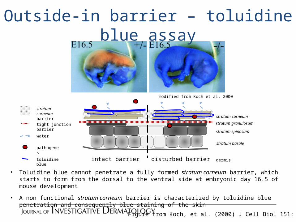
modified from Koch et al. 2000
Outside-in barrier – toluidine blue assay
intact barrier disturbed barrier
pathogenes
toluidine blue
tight junction barrier
stratum corneum barrier
water
stratum basale
stratum spinosum
stratum granulosum
stratum corneum
dermis
• Toluidine blue cannot penetrate a fully formed stratum corneum barrier, which starts to form from the dorsal to the ventral side at embryonic day 16.5 of mouse development
• A non functional stratum corneum barrier is characterized by toluidine blue penetration and consequently blue staining of the skin
Figure from Koch, et al. (2000) J Cell Biol 151: 389–400

Inside-out barrier – biotin diffusion assay
intact barrier disturbed barrier
tight junction barrier
stratum corneum barrier
biotin
water
stratum basale
stratum spinosum
stratum granulosum
stratum corneum
dermis
occludin biotin
Ctr Ecadepi-/-
modified from Tunggal et al. 2005
• Biotin diffuses through the paracellular space up to the tight junction barrier, which seals the paracellular space
• A non functional tight junction barrier is characterized by biotin positive cell membranes above the tight junctions
tight junction barrier (occludin)
Figure from Tunggal et al (2005) EMBO J 24: 1146–56

Assessing the in vivo Epidermal Barrier in Mice – Dye Penetration Assays
Benefits:Dye penetration assays are a powerful tool to assess potential leakiness of the epidermal barrier in vivoCombination of different assays indicates which of the two barriers is affectedApplication of different molecules can point to the nature of a barrier defect
Limitations: Does not provide any molecular explanation for a barrier defectUsage of a single method may not display the exact origin of a barrier defectTight junction penetration assays are often hard to evaluate in adult mice because their stratum granulosum is hardly visibleThe functional threshold of the assays and the physiological barrier may be different