research techniques made simple: identifying the stem cell krisztian nemeth, sarolta karpati...
TRANSCRIPT
Research Techniques Made Simple: Identifying the Stem Cell
Krisztian Nemeth, Sarolta Karpati
Department of Dermatology, Venerology, and Dermatooncology, Semmelweis University, Budapest,
Hungary
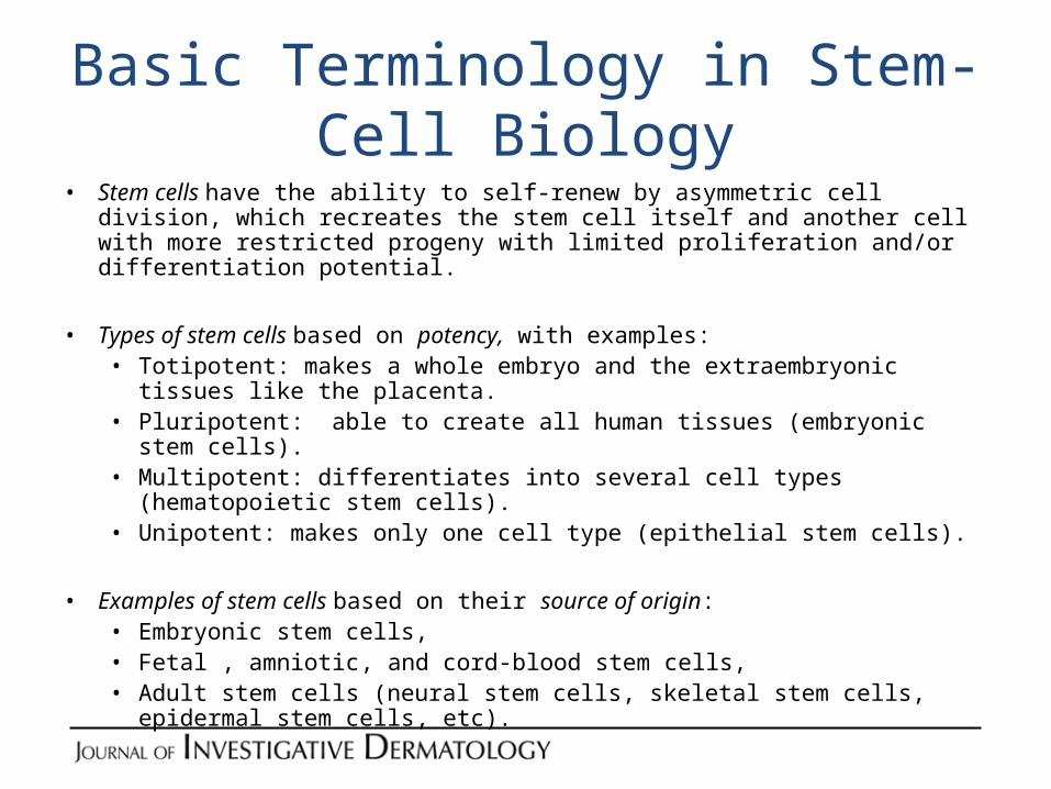
Basic Terminology in Stem-Cell Biology• Stem cells have the ability to self-renew by asymmetric cell division, which
recreates the stem cell itself and another cell with more restricted progeny with limited proliferation and/or differentiation potential.
• Types of stem cells based on potency, with examples: • Totipotent: makes a whole embryo and the extraembryonic tissues like the
placenta.• Pluripotent: able to create all human tissues (embryonic stem cells).• Multipotent: differentiates into several cell types (hematopoietic stem cells).• Unipotent: makes only one cell type (epithelial stem cells).
• Examples of stem cells based on their source of origin:• Embryonic stem cells,• Fetal , amniotic, and cord-blood stem cells,• Adult stem cells (neural stem cells, skeletal stem cells, epidermal stem cells,
etc).
Basic Terminology in Stem-Cell Biology
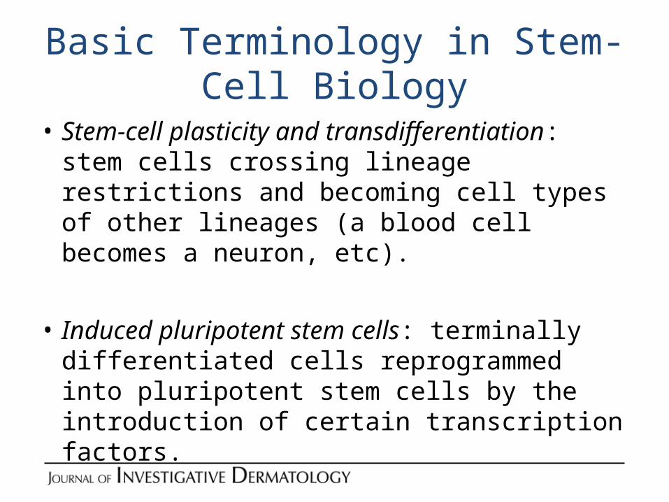
• Stem-cell plasticity and transdifferentiation: stem cells crossing lineage restrictions and becoming cell types of other lineages (a blood cell becomes a neuron, etc).
• Induced pluripotent stem cells: terminally differentiated cells reprogrammed into pluripotent stem cells by the introduction of certain transcription factors.
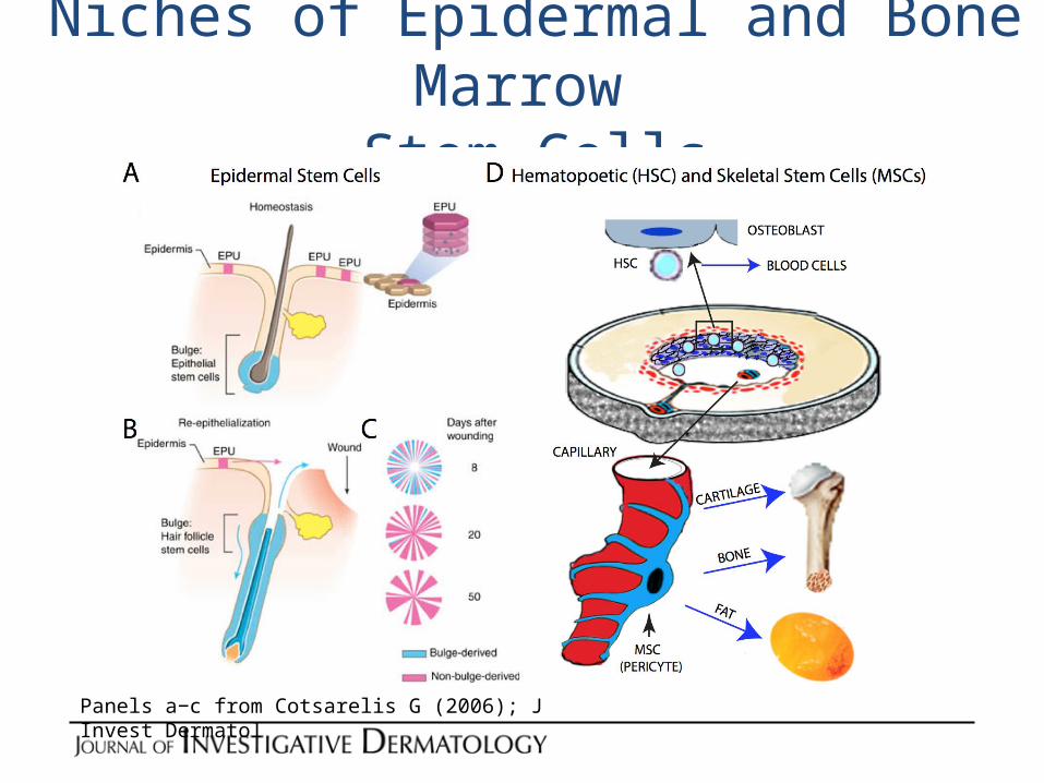
Niches of Epidermal and Bone Marrow Stem Cells
Panels a−c from Cotsarelis G (2006); J Invest Dermatol
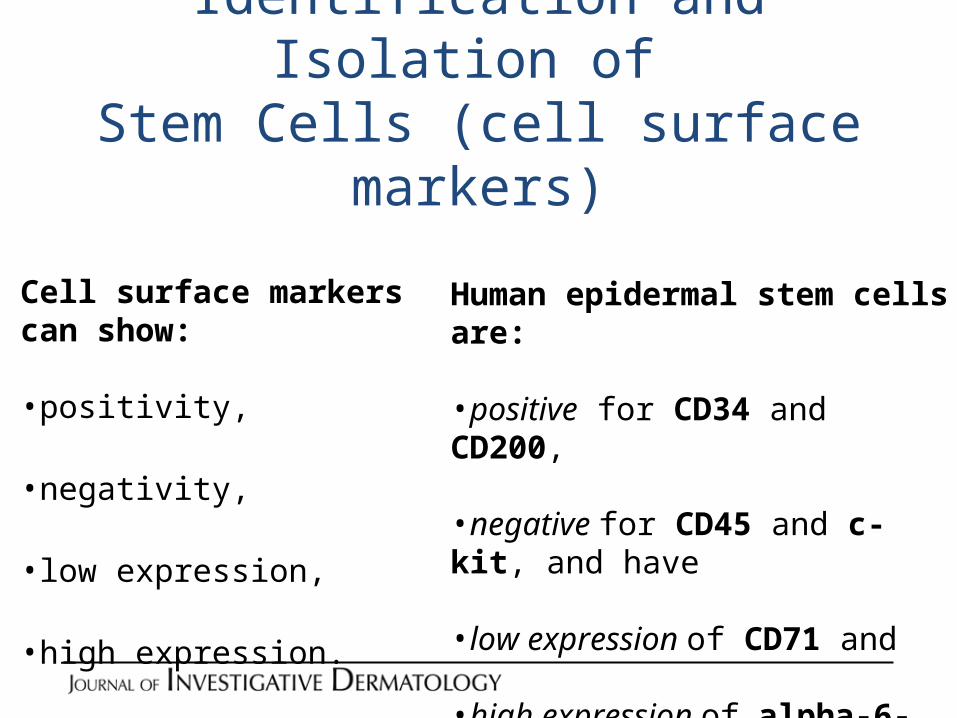
Identification and Isolation of Stem Cells (cell surface markers)
Cell surface markers can show: •positivity,
•negativity,
•low expression,
•high expression.
Human epidermal stem cells are: •positive for CD34 and CD200,
•negative for CD45 and c-kit, and have
•low expression of CD71 and
•high expression of alpha-6-integrin.
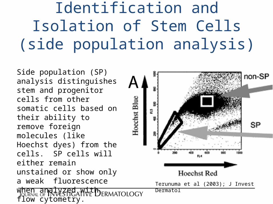
Identification and Isolation of Stem Cells (side population analysis)
Side population (SP) analysis distinguishes stem and progenitor cells from other somatic cells based on their ability to remove foreign molecules (like Hoechst dyes) from the cells. SP cells will either remain unstained or show only a weak fluorescence when analyzed with flow cytometry.
Terunuma et al (2003); J Invest Dermatol
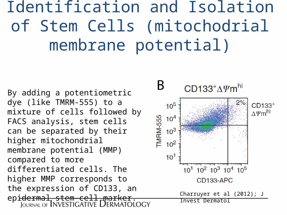
Identification and Isolation of Stem Cells (mitochodrial membrane potential)
By adding a potentiometric dye (like TMRM-555) to a mixture of cells followed by FACS analysis, stem cells can be separated by their higher mitochondrial membrane potential (MMP) compared to more differentiated cells. The higher MMP corresponds to the expression of CD133, an epidermal stem-cell marker.
Charruyer et al (2012); J Invest Dermatol
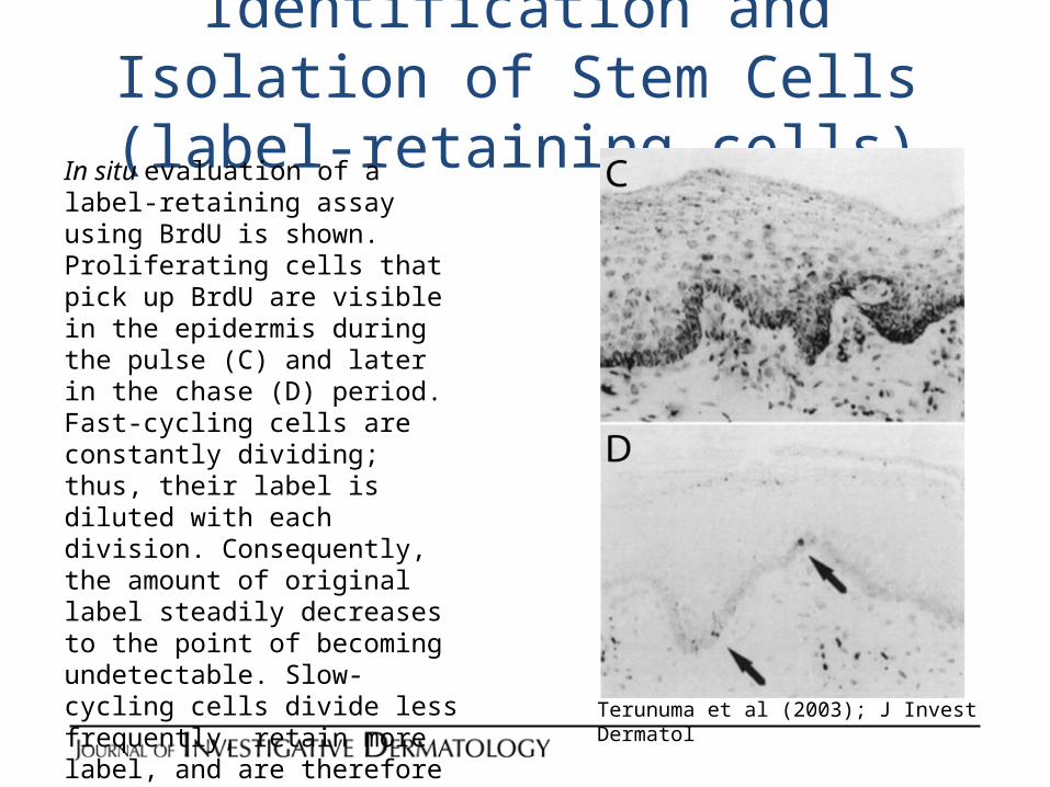
Identification and Isolation of Stem Cells(label-retaining cells)
In situ evaluation of a label-retaining assay using BrdU is shown. Proliferating cells that pick up BrdU are visible in the epidermis during the pulse (C) and later in the chase (D) period. Fast-cycling cells are constantly dividing; thus, their label is diluted with each division. Consequently, the amount of original label steadily decreases to the point of becoming undetectable. Slow-cycling cells divide less frequently, retain more label, and are therefore identified as label-retaining cells (LRCs). This cellular fraction is believed to be enriched in stem cells.
Terunuma et al (2003); J Invest Dermatol
Identification and Isolation of Stem Cells(colony-forming efficiency and holoclones)• The CFE assay is an in vitro method that helps enrich stem cells. It is
performed by first creating a single cell suspension from the target tissue, followed by a simple dilution step and subsequent inoculation of culture dishes with a low number of cells (typically 10-100 cells/cm2). Colonies that form are enriched in stem cells and the number of colonies is believed to correlate with the initial number of stem cells in the tissue.
• When epidermal stem cells are studied, colonies formed by keratinocytes can be further subcultured and colonies in this secondary culture reevaluated. Cells with high replicative capacity and low levels of terminal differentiation will form large colonies with smooth edges called holoclones. They represent a further enriched population of epidermal stem cells.
Identification and Isolation of Stem Cells(functional assays)
• The ultimate test for “stemness” is the ability of sorted/enriched cells to maintain tissue homeostasis indefinitely.
• Methods that assess in vivo functionality of transplanted epidermal and hematopoietic stem cells are called long-term repopulation assays.
• When long-term repopulation is assessed against a competitor cell type so that quantitative results can be achieved, the assay is called competitive repopulation assay.
• Skeletal stem cell stemness is evaluated by transplanting SSCs in vivo and evaluating their ability to form bone and support hematopoiesis within 12 weeks.