researcharticle complementfactorh-relatedprotein3 ... filetesting.togethertheseproteins form...
TRANSCRIPT
RESEARCH ARTICLE
Complement Factor H-Related Protein 3Serum Levels Are Low Compared to Factor Hand Mainly Determined by Gene CopyNumber Variation in CFHR3Richard B. Pouw1,2, Mieke C. Brouwer1, Judy Geissler3, Laurens V. van Herpen1, SachaS. Zeerleder4, Walter A. Wuillemin5, DianaWouters1*, TacoW. Kuijpers2,3
1 Department of Immunopathology, Sanquin Research and Landsteiner laboratory of the Academic MedicalCenter, University of Amsterdam, Amsterdam, the Netherlands, 2 Department of Pediatric Hematology,Immunology & Infectious Diseases, Emma Children’s Hospital, Academic Medical Center, Amsterdam, theNetherlands, 3 Department of Blood Cell Research, Sanquin Research and Landsteiner Laboratory of theAcademic Medical Center, University of Amsterdam, Amsterdam, the Netherlands, 4 Department ofHematology, Academic Medical Center, University of Amsterdam, Amsterdam, the Netherlands, 5 Division ofHematology and Central Hematology Laboratory, Luzerner Kantonsspital and University of Berne, Berne,Switzerland
AbstractThemajor human complement regulator in blood, complement factor H (FH), has several
closely related proteins, called FH-related (FHR) proteins. As all FHRs lack relevant comple-
ment regulatory activity, their physiological role is not well understood. FHR protein 3 (FHR-3)
has been suggested to compete with FH for binding toNeisseria meningitidis, thereby affect-ing complement-mediated clearance. Clearly, the in vivo outcome of such competition greatly
depends on the FH and FHR-3 concentrations. While FH levels have been established, accu-
rate FHR-3 levels were never unequivocally reported to date. Moreover,CFHR3 gene copy
numbers commonly vary, which may impact the FHR-3 concentration. Hence, we generated
five anti-FHR-3 mAbs to specifically measure FHR-3 in human healthy donors of which we
determined the gene copy number variation at theCFH/CFHR locus. Finally, we examined
the acute-phase response characteristics of FHR-3 in a small sepsis cohort. We determined
FHR-3 levels to have a mean of 19 nM and that under normal conditions the copy number of
CFHR3 correlates to a very large extent with the FHR-3 serum levels. On average, FHR-3
was 132-fold lower compared to the FH concentration in the same serum samples and FHR-
3 did not behave as a major acute phase response protein.
IntroductionComplement factor H (FH) is the major regulator of the complement activation cascades inblood, being produced in the liver and circulating at a concentration of approximately 2 μM[1–4]. Next to FH, humans possess several closely related proteins of which the function is stillunclear because of the lack of appropriate tools for their accurate detection and functional
PLOSONE | DOI:10.1371/journal.pone.0152164 March 23, 2016 1 / 13
OPEN ACCESS
Citation: Pouw RB, Brouwer MC, Geissler J, vanHerpen LV, Zeerleder SS, Wuillemin WA, et al. (2016)Complement Factor H-Related Protein 3 SerumLevels Are Low Compared to Factor H and MainlyDetermined by Gene Copy Number Variation inCFHR3. PLoS ONE 11(3): e0152164. doi:10.1371/journal.pone.0152164
Editor: Paul N Baird, Centre for Eye ResearchAustralia, AUSTRALIA
Received: October 14, 2015
Accepted: March 9, 2016
Published: March 23, 2016
Copyright: © 2016 Pouw et al. This is an openaccess article distributed under the terms of theCreative Commons Attribution License, which permitsunrestricted use, distribution, and reproduction in anymedium, provided the original author and source arecredited.
Data Availability Statement: All relevant data arewithin the paper and its Supporting Information files.
Funding: Research leading to these results hasreceived funding from the European Union’s seventhFramework program under EC-GA no. 279185(EUCLIDS; www.euclids-project.eu). The funders hadno role in study design, data collection and analysis,decision to publish, or preparation of the manuscript.
Competing Interests: The authors have declaredthat no competing interests exist.

testing. Together these proteins form the FH protein family, comprising FH, its splice variantFH-like 1, and 6 FH-related (FHR) proteins, numbered 1 to 5 including the splice variants 4Aand 4B. The genes of the FH protein family are located in tandem on chromosome 1q31 in thefollowing order: CFH, CFHR3, CFHR1, CFHR4, CFHR2 and CFHR5.
The FHR proteins show a remarkable sequence similarity to FH and all consist entirely ofso-called short consensus repeat (SCR) domains, each of approximately 60 amino acids. Recentinterest in FHR-3 stems from the association with meningococcal disease [5] and its potentialto interfere with FH binding to the meningococcal capsular protein, FH-binding protein(fHbp). By hijacking FH, fHbp protects the meningococcus from in vitro complement-medi-ated lysis, which could be countered by FHR-3 (unpublished observations) [6,7]. FHR-3 has 5SCR domains, each with a striking sequence identity with SCR domains of the complementregulator FH and other FHR proteins, in particular with FHR-4A/B [8]. The reported molecu-lar weight of serum-derived FHR-3 ranges from 37 to 50 kDa due to different glycosylation var-iants [8]. Together with FHR-4A/B, FHR-3 forms a subgroup of closely related FHRs, whereasFHR-1, FHR-2 and FHR-5 form a subgroup that is characterized by a dimerization motif inthe first two SCR domains resulting in homo- and hetero-dimerization, which is not present inFHR-3 and FHR-4A/B [9].
CFHR3 is commonly deleted due to homologous recombination–most often together withCFHR1. This deletion is reported to occur in about 15% of healthy individuals with ethnic vari-ations in its prevalence [10,11]. CFHR3/CFHR1 deletion is associated with a decreased risk forthe development of age-related macular degeneration (AMD) on the one hand, as well as withan increased risk for the development of atypical hemolytic uremic syndrome (aHUS), which,in the case of aHUS, seems to be partly explained by the appearance of anti-FH auto-antibodies[12–14].
FHR-3 has been reported to directly act as a complement regulator due to exhibiting weakco-factor activity for complement factor I, resulting in degradation of C3b [15]. In addition,FHR-3 can directly bind C3b via a seemingly similar mechanism as FH [15,16]. Currently,FHR-3 is hypothesized to act as a “de-regulator” of the complement system through competi-tion between FH and FHR-3 for the binding of either C3b or host surfaces, thus enhancing thecomplement activation in a positive manner [17]. This is explained by the fact that FHR-3, likeall FHRs, lacks any SCR domains identical to N-terminal SCR domains of FH reported to regu-late C3, while SCR domains identical to SCRs of FH associated with C3b and host surface bind-ing are present. The “de-regulator” hypothesis might also explain the association of CFHR3/CFHR1 deficiency with a decreased risk for AMD, as a lack of FHR-3 would thus allow for bet-ter surface binding and consequently complement regulation by FH [17].
Recently, Caesar et al. (2015) reported the binding of FHR-3 to meningococcal fHbp invitro, resulting in decreased survival of Neisseria meningitidis in human serum [7], althoughsuch competition on bacterial surfaces in vivo will strongly depend on the blood levels of bothproteins. Whereas FH serum levels have been established with an average concentration ofapproximately 2 μM [1–4], FHR-3 serum levels have only been estimated to circulate at a simi-lar molar concentration, but without accurate measurement due to the lack of specific reagentsfor accurate and reliable quantification [15]. Measuring FHR proteins remains challenging dueto the high degree of sequence identity between the FHR proteins as well as with FH. In thisstudy we report a FHR-3-specific ELISA with the use of monoclonal antibodies (mAbs) toestablish normal serum levels, describe the influence of copy number variation (CNV) inhealthy donors in relation to FHR-3 levels and finally, investigate the acute-phase characteris-tics of FHR-3 in sepsis.
FHR-3 Levels and CNV
PLOSONE | DOI:10.1371/journal.pone.0152164 March 23, 2016 2 / 13
Abbreviations: AMD, age-related maculardegeneration; CNV, Copy number variation; FH,complement factor H; fHbp, FH-binding protein; FHR-(1–5), FH-related protein 1 to 5; MLPA, Multiplexligation-dependent probe amplification.

Material and Methods
SamplesBlood samples were drawn from anonymous, healthy volunteers with informed, written con-sent in accordance with Dutch regulations and this study was approved by the Sanquin EthicalAdvisory Board in accordance with the Declaration of Helsinki. Serum was obtained by collect-ing blood, allowing it to clot for 1 hour at room temperature (RT) and collecting the superna-tant after centrifugation at 3000 rpm for 10 min. Plasma and peripheral blood leukocytes werecollected from EDTA blood samples and subsequently used for DNA extraction using theQIAamp DNA Blood Mini Kit (Qiagen, Hilden, Germany) according to the manufacturer’sinstructions. All samples were stored in small aliquots at -80°C until use to avoid repetitivefreeze/thawing. Serum samples of septic patients were collected as part of a previous study withinformed consent according to the local ethics committee in accordance with the Declarationof Helsinki [18]. Only samples taken upon inclusion into the original study (n = 39) were usedin this study. Patient characteristics and CRP levels were determined and reported as part ofthe original study [18].
Proteins and monoclonal antibodiesRat anti-mouse kappa (RM-19) mAb was from Sanquin Business Unit Reagents (Sanquin,Amsterdam, the Netherlands). Mouse mAbs directed against human FH SCR domains 16/17(anti-FH.16) were obtained as part of another study at our laboratory (manuscript in prepara-tion). Polyclonal goat anti-human FH was obtained from Quidel (San Diego, CA, USA) andconjugated with HRP. High Performance ELISA buffer (HPE) was provided by Sanquin. Pro-teins were biotinylated according to the manufacturer’s instructions using EZ-Link Sulfo-NHS-LC-Biotin, No-Weigh Format (Thermo Scientific), when indicated.
Expression of FHR proteinsConstructs containing the cDNA sequences of all FHR proteins, including both known splicevariants of CFHR4, were ordered at Invitrogen (Paisley, UK). Constructs were ordered with anin-frame C-terminal tag coding for six histidine residues (6xHis) and cloned into pcDNA 3.1vectors (Invitrogen). Proteins were transiently expressed in HEK293F cells with 293Fectin andOptiMEM (Invitrogen), using the Freestyle HEK293F expression system (Invitrogen) accord-ing to the manufacturer’s instructions. Five days after transfection, supernatants were collectedand recombinant human (rh) FHR proteins were purified by Ni2+ affinity chromatographywith the use of HisTrap High Performance 1 ml columns (GE Healthcare Life Sciences, Frei-burg, Germany) according to the manufacturer’s instructions. Subsequent filtrations usingAmicon Ultra Centrifugal Filter Devices (Merck Millipore, Darmstadt, Germany) were per-formed to obtain highly pure rhFHR-3. Throughout the purification process, purity of therhFHR proteins was assessed by SDS-PAGE under non-reducing conditions and visualized bysilver staining using the SilverQuest Staining kit (Invitrogen) according to the manufacturer’sinstructions.
Immunization and hybridoma generationPolyclonal anti-FHR-3 antibodies were obtained by immunizing a rabbit via i.m. injection with100 μg rhFHR-3 in montanide as adjuvant at four week intervals. Three days after the fourthbooster, an IgG fraction was obtained from serum with the use of a ProtG sepharose column.
Anti-FHR-3 mAbs were generated by immunizing BALB/c mice via i.p. injection with 25 μgrhFHR-3 in montanide at four week intervals. Three days after the fourth booster, spleen cells
FHR-3 Levels and CNV
PLOSONE | DOI:10.1371/journal.pone.0152164 March 23, 2016 3 / 13
were isolated and fused with the mouse myeloma cell line SP2/0. The presence of FHR-3 spe-cific antibodies in the supernatants of the hybridomas was tested by ELISA. In short, NuncMaxisorp 96-wells microtiter plates (Invitrogen) were coated overnight at room temperaturewith 1 μg/ml RM-19 mAb in PBS to capture mouse kappa immunoglobulins from the superna-tants. After coating, plates were washed five times with PBS + 0.02% (v/v) Tween-20 (PT), fol-lowed by incubation with 25% (v/v) supernatant, diluted in HPE, for 1 h. After washing,specificity of bound antibodies was determined by addition of 1 μg/ml biotinylated rhFHR-3(rhFHR-3-bt), diluted in HPE. Unbound rhFHR-3-bt was washed away and the plates wereincubated with 0.1% (v/v) streptavidin conjugated with HRP (strep-HRP, GE Healthcare) for20 min. After washing, the assay was developed by addition of 100 μl of 100 μg/ml 3,5,3’,5’-Tet-ramethylbenzidine (TMB) in 0.11 M sodium acetate containing 0.003% (v/v) H2O2, pH 5.5.Substrate conversion was stopped after approximately 10 min by addition of 100 μl 2 MH2SO4. Absorbance was measured at 450 nm and corrected for the absorbance at 540 nm witha Synergy 2 Multi-Mode plate reader (BioTek Instruments, Winooski, VT, USA). All ELISAsteps were performed with a volume of 100 μl per well. Positive wells were made monoclonalby multiple limiting dilution steps. Selected clones were allowed to grow and produce mAbsfor up to 21 days prior to purifying the mAbs from the supernatant using a ProtA sepharosecolumn. Isotypes of mAbs were determined by ELISA or with the use of the Mouse mAb Iso-typing Kit (Hycult Biotech, Uden, the Netherlands) according to the manufacturer’sinstructions.
Cross-reactivity ELISACross-reactivity of obtained anti-FHR-3 monoclonal and polyclonal antibodies was deter-mined by ELISA. Briefly, Nunc Maxisorp 96-wells microtiter plates (Invitrogen) were coatedovernight at RT with 2 μg/ml purified anti-FHR-3 monoclonal or polyclonal Abs in 0.1 M car-bonate-bicarbonate buffer, pH 9.6. After washing, 10 nM of biotinylated rhFHRs, FH, or a6xHis-tagged control protein, diluted in HPE, was added and incubated for 1 h, followed byanother washing step and incubation with 0.01% (v/v) strep-poly-HRP (Sanquin), in HPE. AllELISA steps were performed with a volume of 100 μl per well and the ELISA was developed asdescribed above.
CFHR gene copy number determinationFor the multiplex ligation-dependent probe amplification (MLPA) method, the SALSAMLPAprobemix P236-A3 ARMDmix-1 was used to genotype the CFH-CFHR gene region accordingto the manufacturer’s instructions (MRC-Holland, Amsterdam, the Netherlands). This kit waspreviously used and validated in several other studies [11,14,15,19–21].
ImmunoprecipitationFHR-3 was immunoprecipitated from human healthy donor serum using monoclonal antibod-ies against FHR-3. Immunoprecipitation (IP) was performed by incubating 200 μl serum with500 μl of 5 mg/ml CNBr-activated Sepharose (GE Healthcare) to which RM-19 was coupled(25 mg mAb per 1 gram sepharose), and 50 μl of 100 μg/ml anti-FHR-3 mAb, diluted in PBSsupplemented with 0.1% Tween-20, 0,1% BSA and 10 mM EDTA. Following overnight incuba-tion at 4°C, while rotating, the sepharose was washed 3 times with 1 ml PBS + 0.02% (w/v)Tween-20 (PT) and 2 times with 1 ml PBS. Precipitated proteins were eluted by addition of50 μl 1x NuPAGE Sample buffer solution (Invitrogen) and incubation at 70°C for 10 minutes.After spinning down the sepharose, SDS-PAGE under non-reducing conditions was performedusing a Novex NuPAGE 10% Bis-Tris gel followed by Western Blot onto a nitrocellulose
FHR-3 Levels and CNV
PLOSONE | DOI:10.1371/journal.pone.0152164 March 23, 2016 4 / 13

membrane (Novex iBlot Gel Transfer kit, Invitrogen). Membranes were blocked with 1% (v/v)Western Blocking Reagent (WBR, Roche, Basel, Switzerland) in PBS for 30 minutes and incu-bated with 1 μg/ml of either biotinylated polyclonal rabbit anti-FHR-3 or anti-FHR-3.1-bt inPBS + 0.5% (v/v) WBR, O/N. After washing three times with PT, membranes were incubatedwith 0.1% (v/v) Strep-HRP in PBS + 0.5% (v/v) WBR. After 1 hour, the membranes werewashed three times with PT followed by two washes with PBS. Western Blots were developedwith the Pierce ECL 2Western Blotting substrate kit (Thermo Scientific) according to the man-ufacturer’s instructions and analyzed using the ChemiDoc MP System (BioRad, Hercules, CA,USA).
Depletion of FHR-3 from human plasmaFHR-3 was depleted from normal human plasma using anti-FHR-3.1 or anti-FHR-3.4. Plasmawas run over a 3 ml column containing either anti-FHR-3.1 or anti-FHR-3.4 coupled to CNBr-activated Sepharose 4B (GE Healthcare, 10 mg mAb per 1 gram sepharose coupled, accordingto the manufacturer’s instructions). Flowthrough was collected and used for IP using poly-clonal anti-FHR-3 and ProtG-coupled sepharose (GE Healthcare) as described above.
FHR-3 ELISATo measure FHR-3 in serum, anti-FHR-3.1 was coated (2 μg/ml concentration in 0.1 M car-bonate-bicarbonate buffer, pH 9.6) onto Nunc Maxisorp 96-wells microtiter plates (Invitrogen)by O/N incubation at RT. After washing, samples, diluted in HPE, were added and incubatedfor 1 hour at RT. Following washing with PT, 0.5 μg/ml biotinylated anti-FHR-3.4 (in HPE)was incubated on the plate for 1 hour at RT. The ELISA was developed using 0.01% (v/v) strep-poly-HRP (Sanquin) and TMB solution as described above. All steps were performed with avolume of 100 μl per well.
FH ELISATomeasure FH in serum, anti-FH.16, directed against SCR 16/17, was coated (1 μg/ml concen-tration in PBS) onto Nunc Maxisorp 96-wells microtiter plates (Invitrogen) by incubating at RT,O/N. After washing, samples were diluted in HPE, added and incubated for 1 hour at RT. Afteranother wash step, 0.25 μg/ml HRP-conjugated polyclonal goat anti-human FH was incubatedon the plate for 1 hour at RT. Following washing, the ELISA was developed using TMB asdescribed above. All steps were performed with a volume of 100 μl per well. FH levels in healthydonor sera were calculated using a serum pool of 400 donors containing 288 μg/ml FH.
StatisticsData were analyzed and all statistical tests were performed using GraphPad Prism, version 6.04(GraphPad Software, La Jolla, CA, USA). Wilcoxon, Mann-Whitney and Kruskal-Wallis testswere used as indicated to assess significant differences. Correlation between FHR-3 and FH orCRP was assessed using a nonparametric Spearman’s correlation test.
Results
Generation of monoclonal and polyclonal antibodies against FHR-3Five mAbs directed against rhFHR-3 were obtained and named anti-FHR-3.1 to anti-FHR-3.5.All mAbs were identified to be IgG1 and none of the mAbs competed with each other for thebinding of FHR-3 (data not shown) indicating non-overlapping epitopes. All anti-FHR-3mAbs were able to bind rhFHR-3 but most showed, although to a different extent, cross-
FHR-3 Levels and CNV
PLOSONE | DOI:10.1371/journal.pone.0152164 March 23, 2016 5 / 13
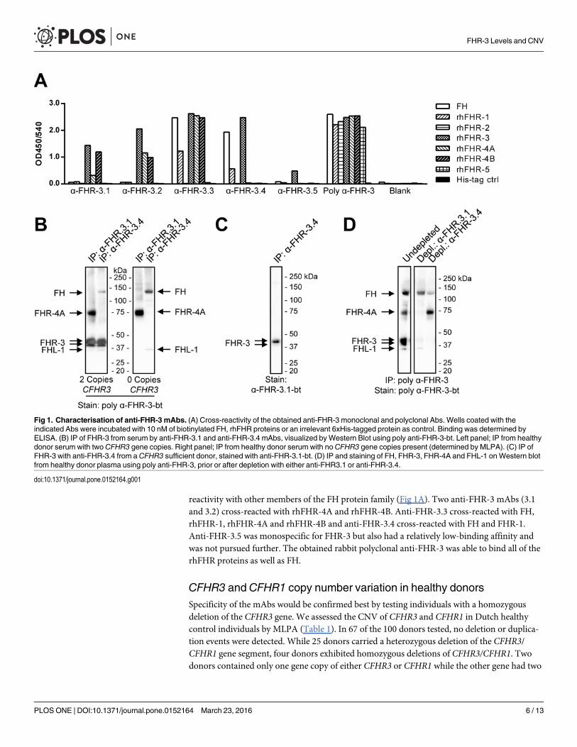
reactivity with other members of the FH protein family (Fig 1A). Two anti-FHR-3 mAbs (3.1and 3.2) cross-reacted with rhFHR-4A and rhFHR-4B. Anti-FHR-3.3 cross-reacted with FH,rhFHR-1, rhFHR-4A and rhFHR-4B and anti-FHR-3.4 cross-reacted with FH and FHR-1.Anti-FHR-3.5 was monospecific for FHR-3 but also had a relatively low-binding affinity andwas not pursued further. The obtained rabbit polyclonal anti-FHR-3 was able to bind all of therhFHR proteins as well as FH.
CFHR3 and CFHR1 copy number variation in healthy donorsSpecificity of the mAbs would be confirmed best by testing individuals with a homozygousdeletion of the CFHR3 gene. We assessed the CNV of CFHR3 and CFHR1 in Dutch healthycontrol individuals by MLPA (Table 1). In 67 of the 100 donors tested, no deletion or duplica-tion events were detected. While 25 donors carried a heterozygous deletion of the CFHR3/CFHR1 gene segment, four donors exhibited homozygous deletions of CFHR3/CFHR1. Twodonors contained only one gene copy of either CFHR3 or CFHR1 while the other gene had two
Fig 1. Characterisation of anti-FHR-3 mAbs. (A) Cross-reactivity of the obtained anti-FHR-3 monoclonal and polyclonal Abs. Wells coated with theindicated Abs were incubated with 10 nM of biotinylated FH, rhFHR proteins or an irrelevant 6xHis-tagged protein as control. Binding was determined byELISA. (B) IP of FHR-3 from serum by anti-FHR-3.1 and anti-FHR-3.4 mAbs, visualized byWestern Blot using poly anti-FHR-3-bt. Left panel; IP from healthydonor serum with two CFHR3 gene copies. Right panel; IP from healthy donor serum with noCFHR3 gene copies present (determined by MLPA). (C) IP ofFHR-3 with anti-FHR-3.4 from aCFHR3 sufficient donor, stained with anti-FHR-3.1-bt. (D) IP and staining of FH, FHR-3, FHR-4A and FHL-1 onWestern blotfrom healthy donor plasma using poly anti-FHR-3, prior or after depletion with either anti-FHR3.1 or anti-FHR-3.4.
doi:10.1371/journal.pone.0152164.g001
FHR-3 Levels and CNV
PLOSONE | DOI:10.1371/journal.pone.0152164 March 23, 2016 6 / 13
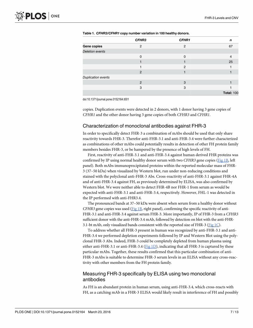
copies. Duplication events were detected in 2 donors, with 1 donor having 3 gene copies ofCFHR1 and the other donor having 3 gene copies of both CFHR3 and CFHR1.
Characterization of monoclonal antibodies against FHR-3In order to specifically detect FHR-3 a combination of mAbs should be used that only sharereactivity towards FHR-3. Therefor anti-FHR-3.1 and anti-FHR-3.4 were further characterizedas combinations of other mAbs could potentially results in detection of other FH protein familymembers besides FHR-3, or be hampered by the presence of high levels of FH.
First, reactivity of anti-FHR-3.1 and anti-FHR-3.4 against human derived FHR proteins wasconfirmed by IP using normal healthy donor serum with two CFHR3 gene copies (Fig 1B, leftpanel). Both mAbs immunoprecipitated proteins within the reported molecular mass of FHR-3 (37–50 kDa) when visualized by Western blot, run under non-reducing conditions andstained with the polyclonal anti-FHR-3 Abs. Cross-reactivity of anti-FHR-3.1 against FHR-4Aand of anti-FHR-3.4 against FH, as previously determined by ELISA, was also confirmed byWestern blot. We were neither able to detect FHR-4B nor FHR-1 from serum as would beexpected with anti-FHR-3.1 and anti-FHR-3.4, respectively. However, FHL-1 was detected inthe IP performed with anti-FHR3.4.
The pronounced bands at 37–50 kDa were absent when serum from a healthy donor withoutCFHR3 gene copies was used (Fig 1B, right panel), confirming the specific reactivity of anti-FHR-3.1 and anti-FHR-3.4 against serum FHR-3. More importantly, IP of FHR-3 from a CFHR3sufficient donor with the anti-FHR-3.4 mAb, followed by detection on blot with the anti-FHR-3.1-bt mAb, only visualized bands consistent with the reported size of FHR-3 (Fig 1C).
To address whether all FHR-3 present in human was recognized by anti-FHR-3.1 and anti-FHR-3.4 we performed depletion experiments followed by IP and Western Blot using the poly-clonal FHR-3 Abs. Indeed, FHR-3 could be completely depleted from human plasma usingeither anti-FHR-3.1 or anti-FHR-3.4 (Fig 1D), indicating that all FHR-3 is captured by theseparticular mAbs. Together, these results confirmed that this particular combination of anti-FHR-3 mAbs is suitable to determine FHR-3 serum levels in an ELISA without any cross-reac-tivity with other members from the FH protein family.
Measuring FHR-3 specifically by ELISA using two monoclonalantibodiesAs FH is an abundant protein in human serum, using anti-FHR-3.4, which cross-reacts withFH, as a catching mAb in a FHR-3 ELISA would likely result in interference of FH and possibly
Table 1. CFHR3/CFHR1 copy number variation in 100 healthy donors.
CFHR3 CFHR1 n
Gene copies 2 2 67
Deletion events
0 0 4
1 1 25
1 2 1
2 1 1
Duplication events
2 3 1
3 3 1
Total: 100
doi:10.1371/journal.pone.0152164.t001
FHR-3 Levels and CNV
PLOSONE | DOI:10.1371/journal.pone.0152164 March 23, 2016 7 / 13
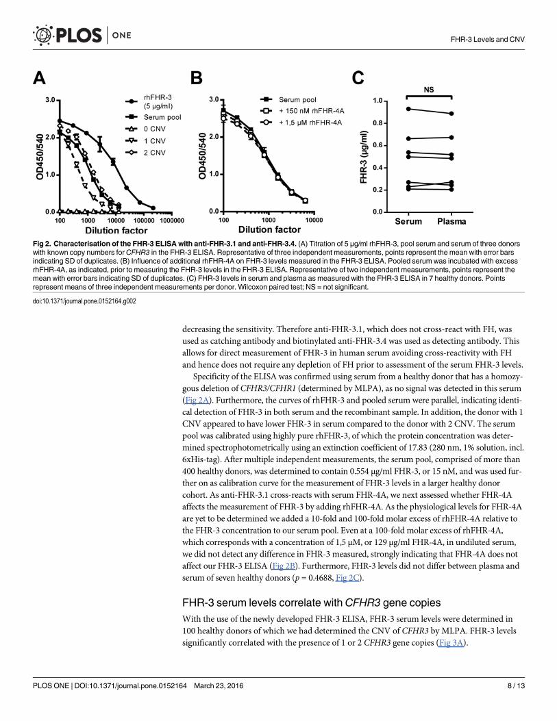
decreasing the sensitivity. Therefore anti-FHR-3.1, which does not cross-react with FH, wasused as catching antibody and biotinylated anti-FHR-3.4 was used as detecting antibody. Thisallows for direct measurement of FHR-3 in human serum avoiding cross-reactivity with FHand hence does not require any depletion of FH prior to assessment of the serum FHR-3 levels.
Specificity of the ELISA was confirmed using serum from a healthy donor that has a homozy-gous deletion of CFHR3/CFHR1 (determined by MLPA), as no signal was detected in this serum(Fig 2A). Furthermore, the curves of rhFHR-3 and pooled serum were parallel, indicating identi-cal detection of FHR-3 in both serum and the recombinant sample. In addition, the donor with 1CNV appeared to have lower FHR-3 in serum compared to the donor with 2 CNV. The serumpool was calibrated using highly pure rhFHR-3, of which the protein concentration was deter-mined spectrophotometrically using an extinction coefficient of 17.83 (280 nm, 1% solution, incl.6xHis-tag). After multiple independent measurements, the serum pool, comprised of more than400 healthy donors, was determined to contain 0.554 μg/ml FHR-3, or 15 nM, and was used fur-ther on as calibration curve for the measurement of FHR-3 levels in a larger healthy donorcohort. As anti-FHR-3.1 cross-reacts with serum FHR-4A, we next assessed whether FHR-4Aaffects the measurement of FHR-3 by adding rhFHR-4A. As the physiological levels for FHR-4Aare yet to be determined we added a 10-fold and 100-fold molar excess of rhFHR-4A relative tothe FHR-3 concentration to our serum pool. Even at a 100-fold molar excess of rhFHR-4A,which corresponds with a concentration of 1,5 μM, or 129 μg/ml FHR-4A, in undiluted serum,we did not detect any difference in FHR-3 measured, strongly indicating that FHR-4A does notaffect our FHR-3 ELISA (Fig 2B). Furthermore, FHR-3 levels did not differ between plasma andserum of seven healthy donors (p = 0.4688, Fig 2C).
FHR-3 serum levels correlate with CFHR3 gene copiesWith the use of the newly developed FHR-3 ELISA, FHR-3 serum levels were determined in100 healthy donors of which we had determined the CNV of CFHR3 by MLPA. FHR-3 levelssignificantly correlated with the presence of 1 or 2 CFHR3 gene copies (Fig 3A).
Fig 2. Characterisation of the FHR-3 ELISA with anti-FHR-3.1 and anti-FHR-3.4. (A) Titration of 5 μg/ml rhFHR-3, pool serum and serum of three donorswith known copy numbers for CFHR3 in the FHR-3 ELISA. Representative of three independent measurements, points represent the mean with error barsindicating SD of duplicates. (B) Influence of additional rhFHR-4A on FHR-3 levels measured in the FHR-3 ELISA. Pooled serum was incubated with excessrhFHR-4A, as indicated, prior to measuring the FHR-3 levels in the FHR-3 ELISA. Representative of two independent measurements, points represent themean with error bars indicating SD of duplicates. (C) FHR-3 levels in serum and plasma as measured with the FHR-3 ELISA in 7 healthy donors. Pointsrepresent means of three independent measurements per donor. Wilcoxon paired test; NS = not significant.
doi:10.1371/journal.pone.0152164.g002
FHR-3 Levels and CNV
PLOSONE | DOI:10.1371/journal.pone.0152164 March 23, 2016 8 / 13
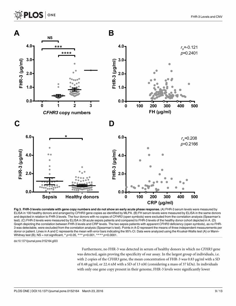
Furthermore, no FHR-3 was detected in serum of healthy donors in which no CFHR3 genewas detected, again proving the specificity of our assay. In the largest group of individuals, i.e.with 2 copies of the CFHR3 gene, the mean concentration of FHR-3 was 0.83 μg/ml with a SDof 0.48 μg/ml, or 22.4 nM with a SD of 13 nM (considering a mass of 37 kDa). In individualswith only one gene copy present in their genome, FHR-3 levels were significantly lower
Fig 3. FHR-3 levels correlate with gene copy numbers and do not show an early acute phase response. (A) FHR-3 serum levels were measured byELISA in 100 healthy donors and arranged by CFHR3 gene copies as identified by MLPA. (B) FH serum levels were measured by ELISA in the same donorsand depicted in relation to FHR-3 levels. The four donors with no copies of CFHR3 (open symbols) were excluded from the correlation analysis (Spearman’stest). (C) FHR-3 levels were measured by ELISA in 39 acute sepsis patients and compared to FHR-3 levels of the healthy donor cohort depicted in A. (D)Graph depicting the correlation between FHR-3 levels and CRP levels. The two sepsis patients with apparentCFHR3 deficiency (open symbols), as no FHR-3 was detectable, were excluded from the correlation analysis (Spearman’s test). Points in A-D represent the means of three independent measurements perdonor or patient. Lines in A and C represents the mean with error bars indicating the 95% CI. Data were analyzed using the Kruskal-Wallis test (A) or Mann-Whitney test (B); NS = not significant, * p<0.05, *** p<0.001, **** p<0.0001.
doi:10.1371/journal.pone.0152164.g003
FHR-3 Levels and CNV
PLOSONE | DOI:10.1371/journal.pone.0152164 March 23, 2016 9 / 13
(p<0.001) with a mean of 0.38 μg/ml and a SD of 0.23 μg/ml, or 10.3 nM (SD = 6.3 nM). Strik-ingly, FHR-3 levels in individuals with one CFHR3 copy were approximately 50% lower com-pared to the FHR-3 levels in individuals with two gene copies. In the only individual in whom3 copies of CFHR3 were found, FHR-3 serum levels were relatively high with levels of about2.3 μg/ml, but still within the normal range of the normal population carrying 2 copies ofCFHR3.
FH serum levels assessed with our monospecific FH ELISA in the same 100 healthy donorswere within the reported normal range of FH, with a mean of 284 μg/ml, or 1.8 μM, and a SDof 68 μg/ml (0.4 μM). There was no correlation with FHR-3 levels (rs = -0.121, p = 0.2401,Fig 3B).
Having established the normal serum levels for FHR-3 to be lower than expected, we subse-quently investigated whether FHR-3 levels increase as part of an acute-phase reaction duringsevere infectious disease by measuring FHR-3 levels in sepsis patients upon admission. Themean FHR-3 levels were significantly higher in the sepsis group (1.71-fold, p = 0.0173) com-pared to our healthy donor cohort (Fig 3C), but did not correlate with a classical marker for anacute phase reaction, i.e. CRP levels (rs = 0.208, p = 0.2166, Fig 3D). Note that two apparentFHR-3-deficient patients were identified with our FHR-3 ELISA in this small cohort; thesewere not included in the correlation analysis.
DiscussionWe developed and validated the first FHR-3 specific ELISA to accurately measure the serumlevels of FHR-3 in healthy donors as well as in severe disease. Unexpectedly, the serum FHR-3levels in healthy individuals were much lower than the previously reported estimates and ran-ged from 0 to 2.7 μg/ml with a mean of 0.69 μg/ml (18.7 nM). Since gene copy numbers ofCFHR3 vary, our data on the serum levels of FHR-3 were related to the genotype of healthyindividuals, demonstrating that the gene copy number in the individual’s genome determinesto a very large extent their steady state serum levels. The mean concentration was 22 nM indonors with two CFHR3 genes, whereas the FHR-3 levels were about 2-fold lower with a meanof 10 nM in donors with only one CFHR3 gene, showing a strong relation between CFHR3copy numbers and FHR-3 serum levels. In our cohort of healthy controls, the CFHR3/CFHR1deletion occurred with a frequency of 17%, which is in concordance with previous reported fre-quencies on CFHR3/CFHR1 CNV [11,13,15]. No FHR-3 could be detected in donors homozy-gous for the CFHR3/CFHR1 deletion.
Our finding that the FHR-3 levels in healthy controls were 100-fold lower than the previ-ously estimated values was remarkable. FHR-3 levels were previously estimated at 1–2 μM butthe data and assay on which this estimation was based had never been formally described [15].Based on our own results and the notorious cross-reactivity or lack of specificity of mAbsagainst FH and FHRs, it is conceivable that additional FH protein family members haveaffected previous estimations. As reported here for FHR-3, mAbs raised against FHR proteinscommonly show cross-reactivity with other FHR proteins as well as FH due to the highsequence identity within this protein family. By using two anti-FHR-3 mAbs that only sharetheir reactivity towards FHR-3, we were able to specifically distinguish FHR-3 from all otherFH protein family members, resulting in establishing lower but accurate levels of FHR-3. Thiswas confirmed by the lack of signal in healthy donors with a homozygous CFHR3/CFHR1 dele-tion, demonstrating that our FHR-3 ELISA was effective in specifically detecting FHR-3.
However, the difference in the previously estimated levels of FHR-3 and the levels reportedhere might alternatively be attributed to an acute-phase response resulting in much higher lev-els of FHR-3 then measured during healthy conditions. Therefore, we measured FHR-3 in a
FHR-3 Levels and CNV
PLOSONE | DOI:10.1371/journal.pone.0152164 March 23, 2016 10 / 13
small cohort of patients with sepsis. Although the mean FHR-3 levels were significantlyincreased in septic patients upon admission compared to healthy controls, this was only a1.7-fold increase. Moreover, this increase seemed to be mainly caused by 6 patients in whichwe could not find any clear association with disease activity, pathogen or other parameters. Alarger study in more homogeneous disease populations, which will include serum samplesdrawn after convalescence as well as establishing the corresponding CNV at the CFH/CFHRlocus, is required to further investigate the higher FHR-3 levels associated with sepsis. We can-not formally exclude an acute phase protein response in some of the sepsis patients that wetested as we were not able to measure FHR-3 under healthy conditions in these patients and wehad no DNA to verify the CFHR3 CNV status in these individuals. On the other hand since wedid not find any correlation of FHR-3 levels with CRP in samples drawn upon admission, ourfindings indicate that FHR-3 does not react as an early major acute phase protein, thus a majoracute-phase response of FHR-3 does not account for the difference between the previously esti-mated FHR-3 levels and the levels reported here.
It is important to note that previous reports on functional characteristics of FHR-3 haveused levels much higher than the physiological concentration of FHR-3 in human serum. Forinstance, Fritsche et al.(2010) demonstrated in vitro complement regulatory activity for FHR-3at concentrations ranging from 10 up to 80 μg/ml [15]. Whether these reported functions aretruly of physiological relevance remains to be elucidated but seems unlikely considering themuch lower serum concentration of FHR-3. Furthermore, in the recently demonstrated invitro competition between FH and FHR-3 for binding to meningococcal fHbp, Caesar et al.(2015) used up to a 10-fold molar excess of FHR-3 compared to FH [7], while in vivo the molarconcentration of FHR-3 is on average 132-fold lower compared to FH in serum. In addition,the most common variant group of meningococcal fHbp, V1, was reported by the same groupto demonstrate a ~20-fold lower binding affinity for FHR-3 compared to FH [7,22]. Nonethe-less, our data could not formally exclude that the demonstrated competition between FHR-3and FH in fHbp interactions could occur in vivo during meningococcal disease nor have weestablished local concentrations of FH and FHR-3 in tissues. Indeed, SNPs in both CFH andCFHR3 are associated with altered susceptibility for meningococcal disease [5]. It is possiblethat the reported SNPs result in a higher expression and serum levels of FHR-3, affecting thecompetition of the protein with FH for binding to fHbp. To further investigate whether theproposed competition does occur during meningococcal infection, FH and FHR-3 levelsshould be longitudinally determined in genotyped patients throughout the disease episode and—preferably—upon reconvalesence. Such prospective studies are ongoing at the moment.
Several groups have shown that the CFHR3/CFHR1 deletion is part of CFH haplotypes thatare associated with decreased risk for AMD [23–26]. As these haplotypes also include SNPs inCFH, it has proven to be difficult to attribute the association to a specific SNP or to theCFHR3/CFHR1 deletion itself. Some reports do indicate a direct link between the deletion andAMD [15,23]. Alternatively, the protective associations of the different haplotypes might alsobe explained by altered FH functionality and/or serum levels [25,27]. However, while MLPA isuseful to determine single gene copy numbers or SNPs, it is unsuited to determine completehaplotypes. Therefore we could not determine in this study whether individuals with a singledeletion of both CFHR3 and CFHR1 do indeed carry a haplotype previously associated withaltered FH levels. Establishing FH, FHR-3 and FHR-1 levels for all different haplotypes associ-ated with AMD is necessary to elucidate the underlying biological mechanism and the relativerole for each of the proteins in AMD.
In conclusion, we described for the first time the accurate assessment of serum levels ofFHR-3 in normal controls. We have demonstrated that the normal FHR-3 serum levels inhumans are mainly determined by CNV of CFHR3 and not by immediate early acute-phase
FHR-3 Levels and CNV
PLOSONE | DOI:10.1371/journal.pone.0152164 March 23, 2016 11 / 13
responses upon serious infection. Since FHR-3 levels are about 130-fold lower compared toFH, our data indicate that the determination of serum levels for all FHR proteins in patients isa crucial step to support and further elucidate any genetic association and physiological rele-vance of the FHR proteins in various complement-related diseases.
AcknowledgmentsWe thank the donors and the patients for their contribution.
Author ContributionsConceived and designed the experiments: RBP DW TWK. Performed the experiments: RBPMCB JG LVH. Analyzed the data: RBP MCB JG LVH. Contributed reagents/materials/analysistools: SSZ WAW. Wrote the paper: RBP DW TWK.
References1. Esparza-Gordillo J, Soria JM, Buil A, Almasy L, Blangero J, Fontcuberta J, et al. Genetic and environ-
mental factors influencing the human factor H plasma levels. Immunogenetics. 2004; 56: 77–82. doi:10.1007/s00251-004-0660-7 PMID: 15118848
2. Hakobyan S, Harris CL, Tortajada A, Goiecoechea De Jorge E, García-Layana A, Fernández-RobredoP, et al. Measurement of factor H variants in plasma using variant-specific monoclonal antibodies: appli-cation to assessing risk of age-related macular degeneration. Invest Ophthalmol Vis Sci. 2008; 49:1983–90. doi: 10.1167/iovs.07-1523 PMID: 18436830
3. Hakobyan S, Tortajada A, Harris CL, Rodríguez de Córdoba S, Morgan BP. Variant-specific quantifica-tion of factor H in plasma identifies null alleles associated with atypical hemolytic uremic syndrome. Kid-ney Int. 2010; 78: 782–8. doi: 10.1038/ki.2010.275 PMID: 20703214
4. Sofat R, Mangione PP, Gallimore JR, Hakobyan S, Hughes TR, Shah T, et al. Distribution and determi-nants of circulating complement factor H concentration determined by a high-throughput immunone-phelometric assay. J Immunol Methods. Elsevier B.V.; 2013; 390: 63–73. doi: 10.1016/j.jim.2013.01.009 PMID: 23376722
5. Davila S, Wright VJ, Khor CC, Sim KS, Binder A, Breunis WB, et al. Genome-wide association studyidentifies variants in the CFH region associated with host susceptibility to meningococcal disease. NatGenet. 2010; 42: 772–776. doi: 10.1038/ng.640 PMID: 20694013
6. Schneider MC, Prosser BE, Caesar JJE, Kugelberg E, Li S, Zhang Q, et al. Neisseria meningitidisrecruits factor H using protein mimicry of host carbohydrates. Nature. 2009; 458: 890–3. doi: 10.1038/nature07769 PMID: 19225461
7. Caesar JJE, Lavender H, Ward PN, Exley RM, Eaton J, Chittock E, et al. Competition between antago-nistic complement factors for a single protein on N. meningitidis rules disease susceptibility. Elife. 2014;3: 1–14. doi: 10.7554/eLife.04008
8. Skerka C, Kuhn S, Gunther K, Lingelbach K, Zipfel PF. A novel short consensus repeat-containing mol-ecule is related to human complement factor H. J Biol Chem. 1993; 268: 2904–2908. PMID: 8428964
9. Goiecoechea De Jorge E, Caesar JJE, Malik TH, Patel M, Colledge M, Johnson S, et al. Dimerizationof complement factor H-related proteins modulates complement activation in vivo. Proc Natl Acad Sci US A. 2013; 110: 4685–90. doi: 10.1073/pnas.1219260110 PMID: 23487775
10. Kubista KE, Tosakulwong N, Wu Y, Ryu E, Roeder JL, Hecker LA, et al. Copy number variation in thecomplement factor H-related genes and age-related macular degeneration. Mol Vis. 2011; 17: 2080–2092. PMID: 21850184
11. Holmes L V., Strain L, Staniforth SJ, Moore I, Marchbank KJ, Kavanagh D, et al. Determining the Popu-lation Frequency of the CFHR3/CFHR1 Deletion at 1q32. PLoS One. 2013; 8: 1–7. doi: 10.1371/journal.pone.0060352
12. Józsi M, Licht C, Strobel S, Zipfel SLH, Richter H, Heinen S, et al. Factor H autoantibodies in atypicalhemolytic uremic syndrome correlate withb CFHR1/CFHR3 deficiency. Blood. 2008; 111: 1512–1514.doi: 10.1182/blood-2007-09-109876 The PMID: 18006700
13. Schmid-Kubista KE, Tosakulwong N, Wu Y, Ryu E, Hecker LA, Baratz KH, et al. Contribution of copynumber variation in the regulation of complement activation locus to development of age-related macu-lar degeneration. Investig Ophthalmol Vis Sci. 2009; 50: 5070–5079. doi: 10.1167/iovs.09-3975
FHR-3 Levels and CNV
PLOSONE | DOI:10.1371/journal.pone.0152164 March 23, 2016 12 / 13
14. Abarrategui-Garrido C, Martínez-Barricarte R, López-Trascasa M, Rodríguez de Córdoba S, Sánchez-Corral P. Characterization of complement factor H-related (CFHR) proteins in plasma reveals novelgenetic variations of CFHR1 associated with atypical hemolytic uremic syndrome. Blood. 2009; 114:4261–71. doi: 10.1182/blood-2009-05-223834 PMID: 19745068
15. Fritsche LG, Lauer N, Hartmann A, Stippa S, Keilhauer CN, Oppermann M, et al. An imbalance ofhuman complement regulatory proteins CFHR1, CFHR3 and factor H influences risk for age-relatedmacular degeneration (AMD). HumMol Genet. 2010; 19: 4694–704. doi: 10.1093/hmg/ddq399 PMID:20843825
16. Hellwage J, Jokiranta TS, Koistinen V, Vaarala O, Meri S, Zipfel PF. Functional properties of comple-ment factor H-related proteins FHR-3 and FHR-4: binding to the C3d region of C3b and differential regu-lation by heparin. FEBS. 1999; 462: 345–352.
17. Józsi M, Tortajada A, Uzonyi B, Goiecoechea De Jorge E, Rodríguez de Córdoba S. Factor H-relatedproteins determine complement-activating surfaces. Trends Immunol. 2015; 1–11. doi: 10.1016/j.it.2015.04.008
18. Caliezi C, Zeerleder S, Redondo M, Regli B, Rothen H-U, Zürcher-Zenklusen R, et al. C1-inhibitor inpatients with severe sepsis and septic shock: Beneficial effect on renal dysfunction. Crit Care Med.2002;30.
19. Bernabéu-herrero ME, Jiménez-alcázar M, Anter J, Pinto S, Sánchez D, Garrido S, et al. Complementfactor H, FHR-3 and FHR-1 variants associate in an extended haplotype conferring increased risk ofatypical hemolytic uremic syndrome. Mol Immunol. Elsevier Ltd; 2015; 67: 276–286. doi: 10.1016/j.molimm.2015.06.021
20. Moore I, Strain L, Pappworth I, Kavanagh D, Barlow PN, Herbert AP, et al. Association of factor H auto-antibodies with deletions of CFHR1, CFHR3, CFHR4, and with mutations in CFH, CFI, CD46, and C3in patients with atypical hemolytic uremic syndrome. Blood. 2010; 115: 379–87. doi: 10.1182/blood-2009-05-221549 PMID: 19861685
21. Tortajada A, Yébenes H, Abarrategui-garrido C, Anter J, García-fernández JM, Martínez-barricarte R,et al. C3 glomerulopaty-associated CFHR1mutation alters FHR oligomerization and complement regu-lation. J Clin Invest. 2013; 123. doi: 10.1172/JCI68280DS1
22. Lucidarme J, Comanducci M, Findlow J, Gray SJ, Kaczmarski EB, Guiver M, et al. Characterization offHbp, nhba (gna2132), nadA, porA, Sequence Type (ST), and genomic presence of IS1301 in group Bmeningococcal ST269 clonal complex isolates from England andWales. J Clin Microbiol. 2009; 47:3577–3585. doi: 10.1128/JCM.00936-09 PMID: 19759227
23. Hughes AE, Orr N, Esfandiary H, Diaz-Torres M, Goodship THJ, Chakravarthy U. A common CFH hap-lotype, with deletion of CFHR1 and CFHR3, is associated with lower risk of age-related macular degen-eration. Nat Genet. 2006; 38: 1173–1177. doi: 10.1038/ng1890 PMID: 16998489
24. Hageman GS, Hancox LS, Taiber AJ, Gehrs KM, Anderson DH, Johnson LV, et al. Extended haplo-types in the complement factor H (CFH) and CFH-related (CFHR) family of genes protect against age-related macular degeneration: characterization, ethnic distribution and evolutionary implications. AnnMed. 2006; 38: 592–604. doi: 10.1080/07853890601097030 PMID: 17438673
25. Ansari M, McKeigue PM, Skerka C, Hayward C, Rudan I, Vitart V, et al. Genetic influences on plasmaCFH and CFHR1 concentrations and their role in susceptibility to age-related macular degeneration.HumMol Genet. 2013; 22: 4857–69. doi: 10.1093/hmg/ddt336 PMID: 23873044
26. McHarg S, Clark SJ, Day AJ, Bishop PN. Age-related macular degeneration and the complement sys-tem. Mol Immunol. Elsevier Ltd; 2015; doi: 10.1016/j.imbio.2015.02.032
27. Zhu L, Zhai Y-L, Wang F-M, Hou P, Lv J-C, Xu D-M, et al. Variants in Complement Factor H and Com-plement Factor H-Related Protein Genes, CFHR3 and CFHR1, Affect Complement Activation in IgANephropathy. J Am Soc Nephrol. 2014; 26: 1195–1204. doi: 10.1681/ASN.2014010096 PMID:25205734
FHR-3 Levels and CNV
PLOSONE | DOI:10.1371/journal.pone.0152164 March 23, 2016 13 / 13