respiratory care : the official journal of the american ... · mi48106. cauustou-freefor ......
TRANSCRIPT
August 1995
Volume 40, Number 8
ISSN 0020-1 324-RECACP
RE/PIRATORVI
^
A MONTHLY SCIENCE JOURNAL40TH YEAR—ESTABLISHED 1956
Broadening Our Outlook-Oxygen
Therapy In the People's Republic of
China-Editorial
Evaluation of Pulse-Dose and
Continuous-Flow Oxygen
Responses to Inhaled Bronchodilator
Multisite Evaluation of a Hemoglobin-
Based Control
Pulmonary Disease in AIDS
Psychological Issues in the Ventilator-
Dependent Patient
Tracheobronchial Fracture
Drug Therapy for Pulmonary
Hypertension
CRCE through the Journal-1995
41st Annual Convention and Exhibition
December 2-5 • Orlando, Florida

SIEMENS
Can you afford not to purchase
a Servo Ventilator 300?
Siemens Ventilators have always represented the state
of the art in patient care. We constantly strive to
develop new technologies which will improve patient
outcomes, reduce mortality rates, and provide cost
savings in your hospital.
Once again, Siemens sets the standard for mechanical
ventilation with the introduction of the Servo Ventilator
300. With the unique Servo Gas Delivery System pro-
viding the modes: Pressure Regulated Volume Control
(PRVC) and Volume Support (VS), experienced clinicians
report a decrease in mortality and morbidity resulting in
Circle 104 on reader service card
the reduction in the duration and cost of ventilator
care. In addition, the Servo 300 is the only ventilator
for adult, pediatric and neonatal patients, eliminating
the cost of purchasing different ventilators for different
clinical areas.
Can you afford to wait until other ventilators have
these features? For more information about howthe Servo 300 can reduce your hospital's cost of
ventilator care contact your Siemens representative
or call 1-800-803-0285.
The Servo
Ventilator 300
Siemens-Elema AB, Life Support Systems Division,
Sweden, is now certified to ISO 9001 standards.
Siemens Medical Systems, Inc., Electromedical Group
16 Electronics Avenue, Danvers, MA 01923 USATel. (508) 750-7500
Siemens and the Servo Ventilator300
System have received these
Awards of Excellence
4090/107/5A 37?3 «S«mens K

AARC Ri^onal SeminarSeptember 22 and 23, 1995
Chicago, Illinois
omorrows
Respiratory Guce Department,,
A special seminar designedto prepare managers andstaff to deal with today's
changes in health care
clear: respiratory care staff and services must change to
meet the demands of a new health care market.
But how do you make those changes? How do you
adapt? How do you survive? This seminar will describe
strategies and actual success stories to get you where
you need to be in tomorrow's (and today's) health care
system.
Program Overview
While most seminars dealing with changes in the health
care system are directed to managers, presentations at
this AARC Regional Seminar are designed for the
benefit of both managers and staff We have all felt the
change that marketplace health care reform has brought
to the operations of our respiratory care services. We all
know that the respiratory care profession is changing in
unprecedented ways. Reengineering, managed care,
outcomes management, utilization control, and
multicompetency are having a tremendous impact on
the way respiratory care is delivered. And the fact is ^A^ American Association for Respiratory Care
Continuing Education
The seminar is approved for up to 10 hours of
Continuing Respiratory Care Education (CRCE) credit
by the American Association for Respiratory Care.
For More Information
Call the AARC Executive Office at (214) 243-2272 or
fax (214) 484-2720 for program information.

Ih
V/ir patients
are mastering
SMI therapy in
almost no time.
Tliat'swliyril
choose Coach®
every time.
Thanks, DHD."Brenda Kalette, RRT
With the
DHD Coach
incentive
spirometer,
accurate SMI
therapy is
simplified
for respiratory patients and clinicians alike.
An exclusive combination of feahires helps
"coach" patients to inhale properly, maintain
correct inspiratory flowrate, and easily monitor
their own progress. For more information on
Coach, a free patient training video, or the
entire line of DHD qualit)' respiratory care
products, please call DHD toll-free at
1'800'847'8000
ODHDDIEMOLDING HEALTHCARE DIVISION
Comslolo,NV 13032 USA
18001 847 8000 FAX (31 SI 697 8083
(3IS)697 2221
Circle 94 on reader service card

RE/PIRATORy QVREA Monthly Science Journal. Established 1956. OITicial Journal of the American Association for Respiratory Care
Contents ...
EditorPat Brougher BA RRT
Associate EditorKaye Weber MS RRT
Editorial Office
11030 Abies Lane
Dallas TX 75229
(214)243-2272
Editorial Board
Chairman
James K Stoller MDCleveland Clinic Foundation
Cleveland, Ohio
Richard D Branson RRTUniversity of Cincinnati
Medical Center
Cincinnati, Ohio
Crystal L Dunlevy EdD RRTThe Ohio State University
Columbus, Ohio
Charles G Durbin Jr MDThe University of Virginia
Health Sciences Center
Charlottesville, Virginia
Thomas D East PhDLDS Hospital
University of Utah
Salt Lake City, Utah
Dean R Hess PhD RRTMassachusetts General Hospital
Harvard Medical School
Boston, Massachusetts
Neil R Maclntyre Jr MDDuke University Medical Center
Durham, North Carolina
Shelley C Mishoe PhD RRTMedical College of Georgia
Augusta, Georgia
Joseph L Rau PhD RRTGeorgia State University
Atlanta, Georgia
August 1995
Volume 40, Number 8
EDITORIALS
810 Broadening Our Outlook—Oxygen Therapy in the
People's Republic of China
Richard D Branson—Cincinnati. Ohio
Original Contributions
811 Clinical Evaluation of Pulse-Dose and Continuous-Flow
Oxygen Delivery
Lin Cai Yuan. Zeng Jun, and Lin Pei Min—Guangzhou. People's
Republic of China
815 Responses to Inhaled Bronchodilator in Patients with
Acute Airflow Obstruction
Joseph Limauro. Donald A Mahler, Elaine M Olmstead. and Anna NA
Tosteson—Lebanon, New Hampshire
820 Multisite Evaluation of a Hemoglobin-Based Control for
pH and Blood-Gas, Electrolyte, and CO-Oximetry
Instruments
Stanley M Liftman, Judith Holland. Maureen MacDonald, Patrick St
Louis, Elliott Prouty, Thomas Contolini. Celia Cavalier, Ronald E
Dechert. Debra K Amoldi. John S Law. and David H Lawson
Reviews, Overviews, & Updates
832 Pulmonary Disease in AIDS: Implications for
Respiratory Care Practitioners
Debra L Medin and Frederick P Ognibene—Bethesda. Maryland
855 Psychological Issues in the Ventilator-Dependent Patient
Gerard J Criner and Lisa Isaac—Philadelphia. Pennsylvania
Case Reports
866 Tracheobronchial Fracture from Blunt Chest Trauma:
A Case Report
Kenneth E Noblett and Jeffrey W Selby—Evansville. Indiana
Respiratory Care • August '95 Vol 40 No 8 795

ARMYRESPIRATORYTHERAPYSPECIALISTS
EXPERIENCEPLUS BENEFITS
Bring your skills as a certi-
fied Respiratory Therapist to an
organization that respects and
values your training: Today's
Army. Work with expert medical
staffs using the latest equipment.
Plus your skills earn you
great benefits you won't find in
many other places. Enlist for
4 years and receive an $8,000
bonus plus qualify for college
loan repayment of up to $55,000.
Enlist for 3 years or 4 years and
be eligible for the Army College
Fund of up to $30,000.
Call or visit your local ArmyNurse Recruiter for moreinformation on how to becomean Army Respiratory Therapy
Specialist.
1-800-USA-ARMYExtension 487
ARMY.BE ALLYOU CAN
This publication
is available in
microform.University Microfilms
International reproduces this
publication in microform:
microfiche and 16mm or
35mm film. For information
about this publication or anyof the more than 13.000 titles
we offer, complete and mail the coupon to: University
Microfilms International. 300 N. Zeeb Road. Axm Arbor.
MI 48106. CaU us toU-free for an immediate response:
800-521-3044. Or call coUect in Michigan, Alaska andHawaii: 313-761-4700.
Please send information about these titles:
Nan
Company/Institutic
Address
City
State_
UniversityMicrofilms
Internatbnal
Foryour convenience, and direct access, the advertisers in
this issue and theirphone numbers are listed below. Please
use this director}' for requesting written material or for any
question you may have.
Help Lines
AARC Information (214) 243-2272
DHD Diemolding Healthcare
Division (800) 847-8000
Driiger Inc (703) 817-0100
HealthScan Products Inc (800) 962-1266
Puritan-Bennett Corp (800) 255-6773
Sherwood Medical (800) 325-7472
Siemens (800) 803-0285
US Army Nurse Corps (800) USA-ARMY
Circle 98 on reader service card

Managing Editor
Ray Masferrer BA RRT
Assistant Editor
Kris Williams BA
Editorial Assistant
Linda Barcus BBA
Section EditorsRobert R Fluck Jr MS RRT
MS Jastremski MDBlood Gas Comer
Hugh S Mathewson MDDrug Capsule
Richard D Branson RRTRobert S Campbell RRT
Kittredge 's Comer
Charles G Irvin PhD
Jack Wanger MBA RPFT RRTPFT Corner
Patricia Ann Doorley MS RRTCharles G Durbin Jr MD
Test Your Radiologic Skill
Consulting EditorsFrank E Biondo BS RRTHoward J Birenbaum MDRobert L Chatbum RRTDonald R Elton MDRonald B George MDJames M Hurst MD
Robert M Kacmarek PhD RRTMichael McPeck BS RRT
David J Pierson MDJohn Shigeoka MD
Jeffrey J Ward MEd RRT
ProductionSteve Bowden
Gary Caywood
Donna Knauf
Karen Singleterry
MARKETINGDirector
Dale Griffiths
Advertising Assistant
Beth Binkley
ADVERTISINGWilliams & Wilkins
... ContentsAugust 1995
Volume 40, Number 8
Drug Capsule
871 Selective Drug Therapy for Pulmonary Hypertension
Hugh S Mathewson—Kansas City, Kansas
BOOKS, Films, Tapes, & Software
876 Comparative Review of Respiratory Care Equipment
Texts
reviewed by Timothy B Op't Holt—Mobile, Alabama
878 New Therapies for Neonatal Respiratory Failure
reviewed by Sherry E Courtney—Camden, New Jersey
Continuing Education Examination
889 CRCE through the Journal—1995
In This Issue
798
Abstracts s ol Penincnl ArtiLles m Other Journals
Editorials, Reviews, and Commentaries To Note
Childhood- versus Adult-Onset Asthma (NHLBI Workshop Summary)—W Busse, SP Banks-
Schlegel, GL Larsen. Am J Respir Crit Care Med 1995; 151: 1635-1639.
The Assessment and Management of Adults with Status Astluiiaticus( State of the Art)—TC Cor-
bridge.JB Hall. Am J Respir Crit Care Med 1995;151:1296-1316.
How Detrimental Is Chronic Use of Bronchodilators in Asthma and Chronic Obstructive Pul-
monary Disease? (Commentary)—CP VanSchayck. SGM Cloosterman. ID Hofland, CLA Van-
Herwaarden. Am J Respir Crit Care Med 1995;151:1317-13i9.
Is the Routine Use of InhaledyS-Adrenergic Agonists Appropriate in Asthma Treatment? YESand NO(editorials)— YES: A Wanner. Am J Respir Crit Care Med 1995;151:597-599. NO: MRSears. Am J Respir Crit Care Med 1995;151:600-601.
Monitoring Cardiopulmonary Resuscitation: Role of Blood and End-Tidal Carbon Dioxide Ten-
sion (editorial)—JD Gasman. RS Fishman. TA Raffin. Crit Care Med 1995;23(5):799-800.
Sleep in the Intensive Care Unit—SL Krach-
man. GE D'Alonzo. GJ Criner. Chest 1995;107
(6):1713.
The most critically ill patients in the hospital are
located in the ICU. Due to intensive individual-
ized care and monitoring, these patients often suf-
fer from severe sleep deprivation. The amount and
continuity of sleep as well as normal sleep ar-
chitecture are all affected. Moreover, by impairing
protein synthesis, cell division, and cellular im-
munity, sleep deprivation can affect the healing
process and thus contribute to an increased mor-
bidity and mortality. Reasons for sleep depriva-
tion appear to be multifactorial and include the
following: the patient's chronic underlying illness.
an acute superimposed illness or surgical pro-
cedure, medications used in treatment of the pri-
mary illness, and the ICU environment itself. Ther-
apeutic interventions need to address each of these
potential causes, with an emphasis placed on pro-
viding an environment that is both diurnal and fo
cused on the importance of uninterrupted sleep.
See related etlilorial: The Disruptive ICU: AnIssue to Lose Sleep Over? SE Eveloff. C'hesi
IW5:l()7tf)):l4H.fl-IH4.
Pulmonary Hanitruuma and Related Events
in Divers—LW Kaymond. Chesi 1995:107
(6): 1648.
Although pulmonary barotrauma (PBT) is a well-
known clinical entity, its recognition in divers
is sometimes delayed and its implications for fu-
ture diving often are unappreciated. The pul-
monary complications of diving activities range
from mere discomfort from mediastinal em-
physema or pneumothorax, or both, to life-threat-
ening gas embolization. In nine cases described
here, only minor manifestations were associated
with PBT which occurred at or close to the sur-
face; but three of the four divers [who developed
minor manifestations, explanation added] were
found to have abnormal pulmonary function.
More serious manifestations resulted from PBTwhich took place at depths of 16 to 120 ft. Even
minor forms of PBT should be considered a con-
traindication to further diving, since they are
prone to recur. Such recurrences—even at shal-
low depths—may cause serious complications.
Cerebral Air Embolism in Asthmatic Scuba
Divers in a Swimming Pool LD Weiss, KWVan Meter. Chest 1995; 107(6): 1653.
Significant shallow-water injuries can occur in
scuba divers, even in swimming pools. Two asth-
matic patients are presented who sustained cere-
bral air emboli during Scuba classes in a swim-
ming pool. Such injuries may be more common
in asthmatics. Asthma is a contraindication to
Scuba diving.
Endotracheal Intubation by Paramedics dur-
ing In-ilospital CI'R -.IK Snialc. K Kully. J
OhIert.r Cotter, Che.st I995;l()7(6): 1655.
SriJDY OBJECTIVE: To examine whether well-
trained paramedics can perform emergent, suc-
cessful, uncomplicated, endotracheal intubations
during in-hospital cardiopulmonary resuscitation
(CPR). DESIGN: Retrospective review of med-
ical records of inpatients undergoing emergent,
endotracheal intubations during in-hospital CPRover 8 months, with comparison of the perfor-
mance of the paramedics against that of other hos-
pital-based personnel. SETTING: A 437-bed Mid-
western community teaching hospital. PATIENTS:
Adult inpatients in general medical/surgical wards.
MAIN OUTCOME MEASURES: The rapidity
of response of paramedics and other medical per-
sonnel to a cardiorespiratory arrest (Code 4) an-
nouncement, and reported difficulties, success rate,
rapidity, and complications of endotracheal in-
tubation. RESULTS: In the 47 cardiorespirato-
ry arrests requiring intubation that we analyzed,
the median response times (with values in paren-
theses representing interquartile range 11QR|) for
paramedics, nurse anesthetists (CRNAs). anes-
thesiologists, and other physicians respectively,
were 2.00 (4.25). 4.00 (2.0), 4.00 ( 15.0). and 7.00
(8.0) min. requiring a median of 1.0 attempt for
all groups (mean values. 1.4, 1.125, 1.0, and 1.4
respectively) to place an endotracheal tube. The
paramedics were successful in 13 of 15 instances.
Median times (seconds) required for intubation
by various groups (same order as response times,
with IQR given in parentheses) were 60 (30). 150
(270), 45 (30). and 60 (30). Difficulties were re-
ported by all groups, including patients' resistance
to intubation, airway obstruction by extraneous
material, and difficulty in visualizing the glottis.
Reported complications (4%) were confined to
groups other than the paramedics. CONCLU-SIONS: Paramedics can successfully, and with-
798 RESPIRATORY CARE • AUGUST '^.S VOL 40 NO 8

CPAP AlwaysA Step Ahead!Evita makes work of breathing
even easier!
0.5 t(s)
Occlusion-pressure
10= •
Intrinsic PEEP
Now Drager provides you with two more
powerful tools to optimize weaning of your
patients.
Introducing Flowtrigger witfiout increase of
expiratory resistance, combined with P01
measurement to determine the patients
ventilatory drive.
To extend monitoring capabilities Evita now
includes the ability to measure Intrinsic
Peep with the display of Trapped Volume.
With Drager you can stay one step ahead in
providing safe, patient friendly ventilation.
For the difference your patients can feel,
choose...
Drager: Technology for Life
(irrrijit^ili^^ '^
DragerTechnology for life
4101 Pleasant Valley Road Suite 100 Ctiantilly. VA 22021
Tel (703) 817-0100 Fax (703) 817-0101
Circle 101 on reader service card

Abstracts
out undue difficulty or complicaiions, place en-
dotracheal tubes during in-hospital CPR. Ap-
propriately trained paramedics may be incorpo-
rated into hospital-based CPR teams in two con-
texts: (1 ) to provide an acceptable, long-term
solution to the scarcity of personnel highly skilled
in endotracheal tube placement during in-hospital
CPR, and (2) to fulfill the need for hospitals to
have on-site, qualified professionals to pertbmi
emergent endotracheal intubation during CPR. In
the latter situation, personnel skilled in airway man-
agement could supplement the paramedics on de-
mand. Further investigation in this area could be
fruitful in view of the small sample size covered
in this study.
Radiologic Evaluation of Emphysema in Pa-
tients with Chronic Obstructive Pulmonary Dis-
ease: Chest Radiography versus High Reso-
lution Computed Tomography—M Miniati. E
Filippi. F Falaschi. L Carrozzi. ENC Milne. HDSosunan. M Pistolesi. Am J Respir Crit Care Med
1995;151:1-V^9.
To objectively reappraise the role of the chest ra-
diograph (CXR) in the clinical assessment of em-
physema, we compared a standardized reading of
CXR with both a visual scoring and a quantita-
tive analysis of high resolution computed to-
mography (HRCT) of the chest in 46 consecutive
patients with chronic obstructive pulmonary dis-
ease (COPD) and fixed expiratory airflow lim-
itation. CXR were scored for signs of overinfla-
tion and pulmonary vascular deficiency by three
independent observers. HRCT scans were inde-
pendently scored for extent of emphysema and
for both severity and extent of emphysema. In 28
of 46 patients, inspiratory and expiratory HRCTscans were analyzed quantitatively by measuring
the mean CT number in Hounsfield Units (HU)
and the percentage of lung area with CT numbers
< -900 HU. Quantitative CT data were compared
with reference values obtained in seven normal
nonsmokers. The CXR score of emphysema
showed a highly significant interobserver re-
producibility and correlated linearly (p< 0.001
)
with HRCT visual scores and quantitative data
from both inspiratory and expiratory CT scan.
CXR score correlated with functional indices of
airflow obstruction, overinflation. and impaired
lung diffusing capacity in a way comparable to
that obtained by using qualitative and quantita-
tive CT data. PatienLs with no signs of emphysema
on CXR had mean expiratory CT numbers with-
in normal range and a fraction of lung area with
CT numbers < -900 HU on expiratory scan not
exceeding 15'/( of total cross-scclional area. The
latter value was consistently greater than 1 5% in
patients with CXR score > 0. Lung function im-
pairment was significantly less .severe (p < 0.0(X)1
)
in patients with CXR score = than in those with
CXR score > 0. For the clinical evaluation of em-
physema, the information derived from stan-
dardized reading of CXR is comparable to thai
obtained by qualitative and quantitative analysis
of HRCT.
\\ hich Index of Peak Expiratory Flow Is Most
Usetiil in the Management of Stable Asthma?—HK Reddel. CM Salome. JK Peat, AJ Woolcock.
Am J Respir Crit Care Med 1995; 15 1 : 1 .^20.
Calculation of dium;il peak expiratory flow (PEF)
variability using values before and after bron-
chodilator is no longer possible for many a.sthmatic
patients because they now use /i-agonists as need-
ed for symptoms rather than regularly. This study
assesses the usefulness of a number of alternative
PEF indices as markers of airway lability in sub-
jects with stable, although not necessarily well-
contfolled. asthma. Forty-six adult subjects com-
pleted a questionnaire about symptoms and treat-
ment in the previous 3 mo. Spirometric function
and airway hyperresponsiveness (AHR) were as-
sessed; AHR was expressed as dose response ratio
(DRR) (maximal percent fall in FEVj divided by
fmal dose of histamine). Subjects recorded PEF
morning and evening, before and after bron-
chodilator (if used) for 2 wk. Nine different PEF
indices were calculated. Diurnal variability (am-
plitude percent maximum) without bronchodilator
was significantly less than diurnal variability with
bronchodilator. Normal indices of PEF lability
were found in 42% of subjects with reduced max-
imal midexpiratory flow (MMEF). Most of the
PEF indices correlated strongly with DRR, and
less strongly with symptom score and airway ob-
struction. Minimum morning prebronchodilator
PEF over a week (expressed as percent recent best
or percent predicted) is recommended as the best
PEF index of airway lability in patients with sta-
ble asthma because it correlates strongly with
AHR, patients are more likely to comply with a
once-daily reading, the calculation is simple, and
regular use of ayi-agonist is not required.
A Prospective Study of Diet and Adult-Onset
Asthma—RJ Troisi, WC Willett, ST Weiss, DTrichopoulos, B Rosner, FE Speizer. Am J Respir
Cril Care Med 1995;151:1401.
A role for diet in the pathophysiology of asth-
ma may be mediated by altered immune or an-
tioxidant activity with consequent effects on air-
way inflammation. We evaluated associations
between several dietary factors assessed by a
semiquantitative food frequency questionnaire,
and incidence of asthma over a 10-yr period in
77,866 women 34 to 68 years of age. Womenin the highest quinlile of vitamin E intake from
diet, but not from supplements, had a risk of 0.53
(95% confidence interval [CI] = 0.33 to 0.86)
compared with women in the lowest quintilc.
This relationship, however, was attenuated when
the contribution from nuts, a major source of vi-
tamin E in these data and a possible allergen, was
removed (relative risk = 0.74 |0.50 to 1 .101. P
for trend = 0.007). Positive associations were
found for vitamins C and E from supplements,
but appeared to be explained by women at high
risk of asthma initiating use of vitamin sup-
plements prior to diagnosis. A nonsignificant in-
verse association with carotene intake was noted,
but no clear relations with asthma were demon-
strated for intake of linoleic acid or omega-3 fatty
acids. These data suggest that antioxidant sup-
plementation and intake of various fats during
adulthood are not important determinants of asth-
ma, although vitamin E from diet may have a
modest protective effect.
Plasma Elastase Levels and the Development
of the Adult Respiratory Distress Syndrome—SC Donnelly. I MacGregor. A Zamani. MWGGordon. CE Robertson. DJ. Steedman. K Lit-
tle. C Haslett. Am J Respir Crit Care Med 1995;
151:1428.
Inflammatory cells, particularly neutrophil gran-
ulocytes, have been implicated in the pathogenesis
of the adult respiratory distress syndrome (ARDS).
In this study, we investigated whether a relation-
ship exists between neutrophil elastase in the plas-
ma of multiple-trauma patients on initial hospital
presentation and the subsequent development of
lung injury and ARDS. Sixty-one multiple-trau-
ma patients were enrolled prospectively. Neu-
trophil elastase was measured by a specific ra-
dioimmunoassay, and analysis was performed by
nonparametric statistical methods. A highly sig-
nificantly elevated plasma elasta.se level was found
in patients who progressed to ARDS (median 217
ng/mL. range 127 to 480) (n = 8) compared with
those who did not (median 1 1 7 ng/mL. range 2 1 .4
to 685) (n = 53) (p = 0.009). Significant corre-
lation was found between initial elastase values
and subsequent requirement for mechanical ven-
tilation (p = 0.01 ). lowest arterial oxygen satu-
ration/oxygen supplementation recorded (p =
0.003), and organ failure score (p = 0.006). This
study shows that within minutes of the initiating
Q-auma event, there is evidence of enhanced neu-
trophil degranulation as manifested by elevated
levels of immunoreactive neutrophil elastase in
the peripheral bkxid. The level of this enzyme cor-
relates with the degree of subsequent lung injury
and ARDS. These findings reinforce the im-
portance of neutrophils and their secretory prod-
ucts in early ARDS disease pathogenesis.
Myocardial Injury in Critically III Patients: AFrequently Unrecognized Complication—TMGuest. AV Ramanathan. PG Tuteur. KB Schecht-
man, JH l.adcnson. AS Jaffe. JAMA 1995:273
(24): 1945.
OBJECTIVE: To detennine the incidence and ef-
fect of unrecognized cardiac injury in critically
ill patienls. DESIGN: Prospective blinded, sin-
gle-center study. SETTING: The medical and res-
piratory intensive care unit of an academic health
center. PATIENTS: Two hundred nine patienls
SOO Ri;si'ikA loRV Carl: • Augu.st "95 Vol 40 No 8

SSESS®P<
; Flow Meter
I5(
In status
isthmaticus:=8001
-;;i6oo
it/ ~i
^^3-500
U.400
; -300
' z
J5( :
;r-i.200
15C :::
NAEP Adult
EmergencyGuidelines:^
PEFR 40-70%of predicted
value after
4 hrs tx in ER.
Consider
hospitalization.
< 40%Definitely
hospitalize.
< 25%Admit to ICU.
guess.
Assess.Rapid initiation and close monitoring of therapy are vital to
successful ER management of severe asthma.' That's whythe ASSESS® Peak Flow Meter should be a vital part of
your armamentarium. ASSESS lets you—
Measure airway obstruction easily,
accurately, cost-effectively.
Peak expiratory flow rate (PEFR) provides an objective,
clinically relevant measurement of airflow— at a fraction
of the cost, bulk, and inconvenience of conventional
spii'ometry.- And when seconds count, you can count on
ASSESS to deliver those measurements vrith superior
accuracy and reproducibility.**
Evaluate response to therapy andneed for hospitalization.
How is your patient doing? What should you be doing?
From initial presentation through discharge, the rugged,
compact ASSESS gives you the hard data you need— as
often as you need it— to help you make informed treatment
decisions in line withNAEP recommendations.^
Help prevent future acute exacerbations.Patients don't come back when their maintenance therapy
stays on track. Keep it on track with ASSESS— and a
regular program of PEFR monitoring at home, work, or
school— as a routine part of your discharge orders.
Formore information about howASSESS can help
you deKver better asthma care, call HealihScan
Products at 1-800-962-1266.
Peak FlowMeter
REFERENCES; 1 Peters Jl»ssfucker J C.
/5(6) 829-849 1992 2 NalinalAsttima Ei > »,'»-
meniolAsthm Bethesda MD.US Dept oiHealiti&Hunaiibc >
3. Data on tile, HealthScan Products Ina 4 Shapiro SM Hwai? '
ttie accuracy ot Assess and IVIiniWnghtoeak Howmeters CtieU &-f
Crapo RO, Jacl<son BR et al Evaluation of accuracy are) rec odji c .,
CteS/Wr{4): 948-952 1992 AA710003-0 5/93 <© 1993
Circle 119 on reader service card
LOW RANGE30 to 390 L/min
Setting the standardfor peak flow monitoring.

Abstracts
(224 admissions). MAIN OUTCOME MEA-SURES: Daily measurement of levels of cardiac
troponin 1. a sensitive, highly specific, and long-
lived marker of myocardial injury. Concurrent-
ly signs and symptoms potentially related to my-
ocardial ischemia uerc tabulated hy blinded in-
vestigators .•\11 clinii-'al e\alualion and management
decisions were made by the physicians respon-
sible for the care of the patient. RESULTS: Thir-
ty-two (15% ) of the 209 patients had evidence of
myocardial damage based on elevated levels of
cardiac troponin I. Only 12 (37%) of these 32 pa-
tients were diagnosed as having acute myocardial
infarction by the intensive care unit staff. Cardiac
damage was unrecognized in the other 20 (63%).
Unrecognized cardiac injury was more common
in young patients and in blacks. Mortality in pa-
tients with m>ocardial injury that was recognized
(42%') or unrecognized (40% ) was higher than in
those without myocardial injury (15%) (P < 0.001 ).
Patients with cardiac injury were more frequently
hypotensive (75% vs 50%.; P = 0.007) and in need
of mechanical ventilation (66% vs 27% : P < 0.00 1
)
and had longer intensive care unit stays (5.3 vs
3. 1 days; P < 0.007) than patients without cardiac
injury. CONCLUSION: The incidence of my-
ocardial injury defined by elevated levels of car-
diac troponin I was unexpectedly high and as-
sociated with increased morbidity and mortality.
Clinically, it was often unrecognized.
Nasal Route for Infant Resuscitation by Motli-
ers—SL Tonkin. SL Davis, TR Gunn. Lancet
1995;345;1353.
In infants under 6 months of age. air normally en-
ters the trachea by the nose because the tongue
fills the oral cavity, and the oral route is open only
when the infant is making muscular efforts such
as crying or gasping. The present recommenda-
tion for infant resuscitation is for the resuscita-
tor's mouth to cover the mouth and nose of the
baby. We set out to test whether this recom-
mendation is feasible. We measured the dimen-
sions of the faces of 28 babies aged between 2 and
4 months (the age when resuscitation is mo.st often
needed ) and of the mouths of 25 of their moth-
ers. Only 2 mothers would have been able to cover
with their mouths the nose and closed mouth of
2 babies (not their own). The mannequins often
used to teach adults to resuscitate infanls are mis-
leading because they present a wide open mouth,
thus implying that that is the preferred route. Werecommend that the nasal route of air entry be
taught to parents for resuscitation of babies who
have stopped breathing.
Physician CliaracteriNlics Associated with De-
cisions To Withdraw IJfe Support—NA.
Christakis, DA Asch. Am J Public Health. 1995;
85(3):367.
OBJECTIVE: This study was undertaken to iden-
tify alUibutes of physicians associated with physi-
cians' decisions to withdraw life support. METH-ODS: Of the 862 Permsylvania intemists surveyed
and asked to make decisions in response to hy-
pothetical vignettes and to report their actual ex-
perience with the withdrawal of life support, 485
(56% ) responded. The data were analyzed with
regression models. RESULTS: With other fac-
tors controlled, physicians were more willing to
withdraw life support if they were young, prac-
ticed in a tertiary care setting, or spent more time
in clinical practice; they were less willing if they
were Catholic or Jewish. Physicians reported a
higher frequency of actually withdrawing life sup-
port if they were young, had more contact with
ICU patients, spent more time in clinical practice,
or were specialists. Physicians with a greater will-
ingness to withdraw were more likely to report
having done so. CONCLUSIONS: Physicians'
personal characteristics are associated with both
their preferences and their practice in the with-
drawal of life support, and a greater willingness
to withdraw is associated with a higher frequency
of withdrawal. The influence of physician char-
acteristics demonstrates that patient preferences
and clinical circumstances do not exclusively gov-
em such ethical decisions. See the related editorial:
Annotation: Physicians ' Attitudes and Decision
Making Regarding the Withdrawal of Life Sup-
port—BW Levin. Am J Public Health. 1995:85(3):
306-307.
Obstetrical Judgments of Viability and Peri-
natal Survival of Extremely Low Birthweight
Infants—ML Reuss, HR Gordon. Am J Public
Health 1995;85(3):362.
OBJECTIVES: The purpose of the study was to
determine whether the obstetrical judgment of vi-
ability makes a difference to fetal and neonatal
survival of extremely low birthweight infants (500-
749 g). METHODS: We assessed the effect of the
antenatal judgment of viability in a group of 66
infants bom weighing from 500 to 749 g. These
infants were alive at maternal hospital admission
and were subsequently live-bom or stillbom be-
tween January I, 1984, and December 3 1, 1985.
We related the antepartum assessment of viability
and other factors recorded in the medical record
to fetal survival and to postneonatal survival to
hospital discharge. RESULTS: The obstetrical
judgment of viability was strongly a.ssix:iated with
outcome. After birthweight and gestational age
were controlled, fetuses considered viable were
18 times more likely to survive (95%- confidence
interval = 2.175) than those considered nonviable.
CONCLUSIONS: The effects of obstetrical prac-
tices on perinatal mortality must be taken into con-
sideration in estimating the survival potential of
very small fetu,ses and in evaluating the relationship
between survival and disability. .Sec the related
ediidrial: Does Intensive Perinatal Care Improve
the Outcome of Extreme Prematurity? Ad-
dressing the Methodologic Issues— Am J Pub-
lic Health l995:<S5(3):300-302.
Safety of Propellants—DJ Alexander. J Aerosol
Med 1995:8(1, Suppl);S-29.
Chlorofiuorocartxjn (CFC) propellants used in me-
tered dose inhalers (MDl) must be replaced under
the temis of the Montreal Protocol. Following a
review of the available data. HFA 1 34a (1,1,1,2
tetrafluoroethane) was selected as a potential al-
temative propellant. However, because this data
was insufficient to satisfy the stringent require-
ments for pharmaceuticals, additional toxicological
assessments were performed. These included geno-
toxicology, animal inhalation studies, reproductive
toxicology, local tolerability and safety phar-
macology studies. A special grade of HFAI34a,
containing relatively high concentrations of all like-
ly impurities was used for all pivotal studies.
HFA 1 34a was devoid of genotoxicity and had ex-
ceptional low acute toxicity. No toxicity was seen
in rats or dogs exposed to concentrations of up
to 5% or 12% respectively for up to one year. Nofetotoxicity, effects on reproductive performance,
peri- or post-natal development was demonstrated.
HFA 1 34a was devoid of oncogenic potential in
rats and mice. There was no evidence of sensi-
tization or local irritation to the skin or eyes. All
species demonstrated high systemic HFA 1 34a con-
centrations. An extensive toxicological evalua-
tion, at very high multiples of the likely patient
exposure, has indicated that HFA 134a is a suit-
able altemative to CFC propellants for MDI's.
Clinical Evaluation ofCFC-Free Metered Dose
Inhalers—M Jenkins. J Aerosol Med 1995;8 (1,
Suppl):S-41.
Current metered dose inhalers (MDIs) contain
chlorofiuorocarbon (CFC) propellants. A new pro-
pellant HFA 1 34a, with no effect on ozone, may
be a .suiujble altemative. Four asthma medications,
salbutamol, salmeterol. tlutica-sone propionate (FP)
and beclomethasone dipropionate (BDP). currently
containing standard CFC propellants, were for-
mulated with HFA 134a for investigation. Single
doses of salbutamol (200 pg) and salmeterol (50
^Jg. lOO/jg) provided equivalent protection against
bronchial provocation, after histamine and metha-
choline respectively, compared with the current
preparation. A double-blind 4-week study com-
paring the two fomiulations of salbutamol, used
as required in inild to moderate asthma, showed
similar effects on morning peak expiratory flow
rates (PEER) and inhaler use. Salmeterol (50 ^g)
twice daily was compared with the current for-
mulation in a 4-week trial. Improvement in morn-
ing PEER was similar for both formulations. Adouble-blind study compared the two formula-
tions of FP (250 /ig) twice daily in mixJerate asth-
matics previously taking 400- 1 .(XX) /Jg of inhaled
corticosteroid daily. Morning PEFR improved in
both groups. Safety and tolerability of the
HFA 1 34a product were similar to current for-
mulations. The HFA 1 34a formulations of salbu-
tamol, salmeterol and FP provide equivalent ef-
802 Ri;spiRATORY Care • Augu.st '95 Vol 40 No 8

w^Face to -Fac*
with Chan<at the American Association for Respiratory Cai
41st Annual Convention and Exhibition|
Orlando, Florida, Deg^ber 2-5, 1995
^ Come to Orlando aVrd come fov.
B
I
respiratory care exhibition and 14455
=/ educational experience thdt\viir he.
= 7 Come experience the foUow^^gand
0(w interactive educational sessions—days packed with lectures on alWecialties
rehabiUtatioitiVIIWmjIrical ventilatio
pediatric care, PHsPni^tipnea, TB, horestructuring, ICU care, honje care, ECM
,,rocmciqernent, and more. 'C*
'iacti Willi NoiHi /Tiiiencd'si PRliK
lu. Iv is a l-oui--d.a.\' iv-spiiuion'
)'ou luce Uie cliaik-iiy es oi' c^
lucli iiioie.
continuing education ere
continuing education needs. ,1
Respiratory Care Open Forvm—More than 200
original research presentations by clinicians,
managers, and educators. Learn about the late|
research in one-on-one and group discussions. *
A dynamic exposition—See demonstrationiof the
newest products and services in the world's Idrgesfc^^
exhibit of the year for cardiopulmonary equipmerit,-
supplies, pharmaceuticals, publishers, and recruiters.
Mark your calendar and make your plans to attend
respiratory care's PREMIER event in North America^
.
(program, registration form, and hotel reservationibrr^ y|will mail in early September). \3
For more information, send your name arid^American Association for Respiratory <
11030 Abies Lane, Dallas, Texas 75229^

Abstracts
ficacy with a similar safety profile to the exist-
ing formulations at equivalent doses.
A Review of Cost Studies of Intensive Care
Units: Problems with the Cost Concept—
M
Gyldmark. Cnt Care Med iy95;23(5):964.
OBJECTIVE: To study methods for costing hos-
pital services, specifically in relation to multi-
unit studies of activity, case inix, severity of ill-
ness, outcome, and resource use in adult inten-
sive care units (ICUs). DATA SOURCES;Twenty published cost studies of adult ICUs. The
studies are all published in English and are both
European and American. STUDY SELECTION:
Cost studies of adult ICUs published in inter-
national journals (English language). DATA EX-
TRACTION: Literature survey, where the arti-
cles were obtained through MEDLINE and other
database searches. DATA SYNTHESIS: Cost of
intensive care therapy was compared across the
20 studies. However, as stressed in the article,
to compare costs of intensive care therapy across
units is not possible for a number of reasons. One
of the reasons for this limitation is that the stud-
ies employed different approaches to costing and
thereby introduced a methodologic bias. In ad-
dition, the costing methodology applied in the
majority of the studies was wrongly specified in
relation to the purpose and viewpoint of the stud-
ies. CONCLUSIONS: The methodologies for
costing ICU therapy are flawed and fail to pro-
vide correct answers. In most studies, the study
question is not adequately specified and the cost
concept used in the studies is not tailored to the
purposes of the study. Standardizing the cost
model would lead to better, faster, and more re-
liable costing. This standardized cost model
should not be rigid, but adaptable to different de-
cision situations. A decision tree or taxonomy
is proposed as a way toward better costing of ICU
activity. See rhe related editorial: Economic
Analysis ofthe Intensive Care Unit: A Dilem-
ma—Cril Care Med I995:23(5):t<05.
Simple Method To Measure Total Expiratory
Time Constant Based on the Passive Expira-
tory Flow Volume Curve—JX Brunner, TPLaubscher, MJ Banner, G lotti. A Braschi. Crit
Care Med 1995:23(6): 1 1 17.
OBJECTIVE: In intubated, mechanically venti-
lated patients, inspiration is forced by externally
applied positive pressure. In contrast, exhalation
is passive and depends on the time constant of the
total respiratory system. The expiratory time con-
stant is thus an important dctemiinant of mechanical
ventilation. The aim of this study was to evaluate
a simple method lor measuring the expiratory lime
constant in ventilated subjects. DESIGN: Prospec-
tive study using a lung simulator and ten dogs. SET-
TING: University hospital. SUBJECTS: Com-
mercially available lung simulator and ten grey-
hound dogs. INTERVENTIONS: Different
expiratory time constants were set on the lung sim-
ulator. In the dogs, the endou-acheal tube was
clamped to increase airways resistance by 22.5 cm
HiOAU sec I and the lungs were injured with hy-
drochloric acid to decrease total respiratory com-
pliance by 1 6 mUcm HjO. This procedure resulted
in a wide range of expiratory time constants. MEA-SUREMENTS AND MAIN RESULTS: Pneu-
motachography was used to measure flow and vol-
ume. The ratio of exhaled volume and peak flow
was calculated from these signals, corrected for
the limited exhalation time yielding the "calculated
expiratory time constant," and compared with the
actual expiratory time constant. The typical error
was ± 0. 1 9 sec for the lung simulator and ± 0. 1
5
sec for the dogs. CONCLUSIONS: The volume
and peak flow corrected for limited exhalation time
is a good estimate of the total expiratory time con-
stant in passive subjects and may be useful for the
titration of mechanical ventilation.
Probability of Survival after Prolonged Ex-
tracorporeal Membrane Oxygenation in Pe-
diatric Patients with Acute Respiratory Fail-
ure—TP Green. FW Moler. DM Goodman, for
the Extracorporeal Life Support Organization. Crit
Care Med 1995:23(6): 1 132.
OBJECTIVE: Extracorporeal membrane oxy-
genation (ECMO) has been used with increas-
ing frequency for respiratory failure that is un-
responsive to conventional therapy. We exam-
ined the relationship between duration of ECMOand outcome to understand whether prolonged
ECMO (duration of the procedure for > 14 days)
was more commonly associated with futile ther-
apy or eventual recovery. DESIGN: A cohon
study of all patients reported to the Pediatric
ECMO Registry for Acute Respiratory Failure
of the Extracorporeal Life Support Organization.
SETTING: Teniary hospitals (n = 83) capable
of providing extracorporeal support for pediatric
patients. PATIENTS: Children (n = 382) between
the ages of 1 wk and 18 yrs of age with severe
respiratory failure. INTERVENTIONS: Extra-
corporeal membrane oxygenation. MEA-SUREMENTS AND MAIN RESULTS: The
death or live hospital discharge of ECMO-treat-
ed patients, together with the post-ECMO me-
chanical ventilation course, were examined as a
function of duration of ECMO and of pre-ECMO
respiratory status. The occurrence of complica-
tions and the causes of death were also noted. The
criteria used to initiate ECMO. as well as the de-
termination of the futility of further ECMO. were
determined by local practice at individual cen-
ters. There were 382 patients treated with ECMO,of whom 184 (48%) survived. The proportion-
al survival in the patients treated for the longest
duration was similar to the overall group. The
cause of death was given for 168 patients: 32 neu-
rologic deaths; nine deaths due to ECMO com-
plications; and 30 deaths due to nonpulmonary
organ failure. There were 97 deaths due to elec-
tive ECMO termination: 80 of these deaths oc-
curred after the determination of the futility of
anticipating pulmonary recovery. The latter deaths
occurred at widely varying durations of ECMO.with a median of 282 hrs. However, at that same
duration. 47 eventual survivors (26^^ of all sur-
vivors) continued to receive ECMO. By dis-
criminant analysis, the survival rate was inde-
pendently related (r= = 0. 1 8;p < 0.000 1 ) to peak
ventilator inspiratory pressure before ECMO and
duration of intubation before ECMO. patient age.
and the occurrence of several complications.
CONCLUSIONS: While the survival rate in pe-
diatric patients receiving ECMO appears relat-
ed to the severity of lung disease and to the oc-
currence of ECMO complications, the survival
rate in patients treated with ECMO courses of >
2 wks was similar to the survival rate of patients
treated for shorter periods of time. ECMO was
terminated in some patients for pulmonary futility
at durations ofECMO associated with survival
in substantial numbers of patients in whomECMO was continued.
Noninvasive Monitoring of End-Tidal Car-
bon Dioxide Tension via Nasal Cannulas in
Spontaneously Breathing Children with Pro-
found Hypocarbia—JFK Flanagan. JS Garrett.
A McDuffee. JD Tobias. Crit Care Med 1995:23
(6):1140.
OBJECTIVE: To determine the correlation be-
tween end-tidal CO; and Paco: measured via nasal
cannulas in spontaneously breathing children with
profound hyptx;arbia ( Pjco; < -^0 torr |< 4.0 kPa] ).
DESIGN: Prospective evaluation. SETTING Pe-
diatric intensive care unit (ICU) in a tertiary care
referral center. INTERVENTIONS: None. PA-
TIENTS: Patients admitted to the ICU with a di-
agnosis of diabetic ketoacidosis in whom inva-
sive arterial access was deemed necessary for clin-
ical care. The patients were spontaneously
breathing, without intubation. The study included
nine patients, with an average age of 9.9 yrs (range
4 to 17) and weight of 38.7 kg (range 17 to 68).
MEASUREMENTS AND MAIN RESULTS:
End-tidal CO: was sampled from nasal cannu-
las by a sidestream aspirator and estimated by in-
frared spectroscopy. The correlation between ar-
terial and end-tidal CO; was compared using lin-
ear regression analysis. A total of 65 arterial blood
gases were obtained from the nine patients. The
PaCO; was < 30 torr (< 4.0 kPa) in 38 of the sam-
ples. The PacO: 10 end-tidal CO: gradient was <
4 torr (< 0.5 kPa) in 64 of 65 samples and 4.8 torr
(0.6 kPa) in one sample. Linear regression anal-
ysis of arterial vs end-tidal CO: yielded a slope
of 0.99, an r- value of 0.97, and a p value of
0.0001. CONCLUSIONS: End-tidal CO: mea-
surement by infrared spectroscopy provides an
accurate estimation of PaCO;. even during episodes
of severe hypocarbia. Its use may limit the need
for invasive monitoring and/or repeated arteri-
al blood gas analyses.
804 RESPIRATORY CARE • AllGUST "95 VOL 40 NO 8

^V>*»*k'-.r^''-.V
Celebrate
RCWcekT-shirtshow pride in your profession, both
coming and going, with this newfront-and-back design. This 50/50
cotton/polyester blend T-shirt
comes in a fashionable stonewashed-
denim color. The design is displayed
in navy blue, cream, and white. Wear
it with pride! Medium, large, extra
large, and extra-extra large available.
Item R16 $9.75
($1 1 .50 nonmembers)Add S2J0for extra-extra large.
SweatshirtThis luxurious, heavyweight, 50/50
blend navy-blue sweatshirt sports a
plaid applique accented with gold
embroidery. Medium, large, extra
large, and extra-extra large available.
ltemR19 $25 ($30 nonmembers)Add $2JO for extra-extra large.
CapTop off your RCWeek ensemble with
this embroidered
navy-blue cap.
One size fits all.
ItemRIZ $6.50
($9 nonmembers)
To order by credit card or
purchase order, call the RC WeekHotline at (214) 243-1478.
Shipping

Tn Order hy Credit Card or Purchase Order, Call the RC Week Hothnc at (214) 243-1478.
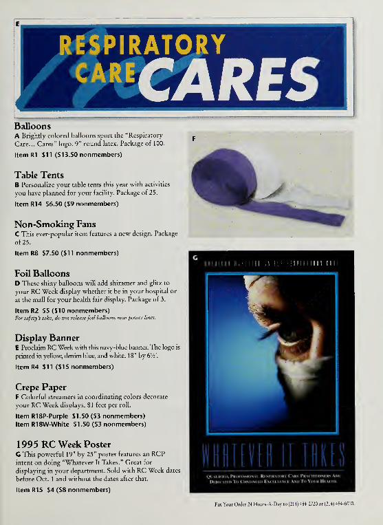
RESPIRATORY
ViiCARESBalloonsA Brightly colored balloons sport the "Respiratory
Care... Cares" logo. 9" round latex. Package of 100.
Item R1 $11 ($13.50 nonmembers)
Table TentsB Personalize your table tents this year with activities
you have planned for your facility. Package of 25.
Item R14 $6.50 ($9 nonmembers)
Non-Smoking FansC This ever-popular item features a new design. Package
of 25.
Item R8 $7.50 ($11 nonmembers)
Foil BalloonsD These shiny balloons will add shimmer and glitz to
your RC Week display whether it be in your hospital or
at the mall for your health fair display. Package of 3.
Item R2 $5 ($10 nonmembers)For safety's sake, do not release foil balloons nearpower lines.
Display BannerE Proclaim RC Week with this navy-blue banner. The logo is
printed in yellow, denim blue, and white. 18" by 6'/2'.
Item R4 $11 ($15 nonmembers)
Crepe PaperF Colorful streamers in coordinating colors decorate
your RC Week displays. 81 feet per roll.
Item RISP-Purple $1.50 ($3 nonmembers)
Item R18W-White $1.50 ($3 nonmembers)
1995 RC Week PosterC This powerful 19" by 25" poster features an RCPintent on doing "Whatever It Takes." Great for
displaying in your department. Sold with RC Week dates
before Oct. 1 and without the dates after that.
ltemR15 $4 ($8 nonmembers)
Fax Your Order 24 Hours-A-Day to (214) 484-2720 or (214) 484-6010.

Abstracts
The Effect of Bronchopulmonary Dysplasia on
Growth at School Age—LA Vrlenich. MEABozynski, Y Sh> r. MA Schork. DW Roloff. MCMcCormick. Pediatrics 1995:95(6):855.
OBJECTIVE: To determine the effect of bron-
chopulmonary dysplasia (BPD) on growth at
school age. DESIGN; A prospective cohort study.
METHODS: The sample included 406 children
selected from a reconsUTicted cohort of infants of
very low birthweight previously enrolled in a
multisite. randomized, controlled clinical trial. The
children were contacted at 8 to 10 years of age.
Height, weight, and head circumference (HO were
measured. Possible confounders including so-
ciodemographic data, and neonatal factors were
also recorded. RESULTS: The children in the BPDgroup were significantly smaller in weight (; score,
-0.50 ± 1 . 19 SD vs -0.06 ± 1 .30 SD) and HC (;
score. -1 .41 ± 1 .32 SD vs -0.63 + 1 .62 SD) than
those without BPD. However, after controlling
for confounders (using analysis of covariance).
no significant differences were demonstrated be-
tween the two groups. Power analyses showed that
a difference of at least 0.43 r-score units could have
been detected. The previously documented as-
sociations between BPD and suspected con-
founders were reconfirmed. CONCLUSIONS:Significant differences were noted between chil-
dren with and without BPD for weight and HCbut not height. When possible confounders were
taken into account, the differences were no longer
appreciated. Thus, the previously reported poor
growth in children with BPD may have been re-
lated to other factors and not necessarily to BPD.
Prolonged Episodes of Hypoxemia in Preterm
Infants Undetectable by Cardiorespiratory
Monitors—CF Poets, VA Stebbens. D Richard.
DPSouthall. Pediatrics 1995:95(6):860.
OBJECTIVE: To determine whether episodes of
prolonged hypoxemia occur without prolonged
apneic pauses (> 20 seconds) and without brady-
cardia (pulse rate, < 100 beats per minute) in ap-
parently well preterm infants. METHODS: Long-
term recordings of arterial oxygen saturation as
measured by pulse oximetry (SpO:), photo-
plethysmographic (pulse) waveforms from the
oximeter, and breathing movements were per-
formed in 96 preterm infants (median gestational
age at birth. 34 weeks; range, 28 to 36 weeks) who
were breathing room air. Recordings started at a
median age of 4 days (range. 1 to 60 days). RE-
SULTS; During a median duration of recording
of 25 hours. 88 episodes in which Spo, fell to 80'7r
or less and remained there for 20 seconds or longer
were identified in \5 infants. The median dura-
tion of these prolonged desaturations was 27 sec-
onds (range, 20 to 81 seconds). In 73 episodes
(83%). SpOj continued to fall to (tO% or less. Twen-
ty-three desaturations were associated with pro-
longed apneic pauses and 54 with bradycardia;
19 of these were associated with both apnea and
bradycardia. Thirty desamrations (34%; 10 infants)
occurred without bradycardia and without pro-
longed apnea. CONCLUSIONS; These results in-
dicate that a proportion of apparently well preterm
infants exhibit episodes of severe prolonged hy-
poxemia unaccompanied by prolonged apneic
pauses or bradycardia. Such episodes, therefore,
would be difficult to detect if only breathing move-
ments and heart rate are monitored. Indications
for the use of oxygenation monitors in preterm
infants should be reconsidered.
Prevention Of Atelectasis during General
Anaesthesia—HV Rothen, B Sporre, G Engberg.
G Wegenius, A Reber, G Hedenstierna. Lancet
1995:345:1387.
Atelectasis is an important cause of impaired gas
exchange during general anaesthesia; it causes pul-
monary shunting. We studied the effects of gas
composition on the formation of atelectasis and
on gas exchange during the induction of gener-
al anaesthesia. In 1 2 adult patients, the lungs were
ventilated with 30% oxygen in nitrogen during
anaesthesia induction, and in another 12, a con-
ventional technique was used ( 100% oxygen dur-
ing induction and 40% oxygen in nitrogen there-
after). Extent of atelectasis was estimated by com-
puted tomography and the ventilation-perfusion
relation (VvQ) by the multiple inert gas elimi-
nation technique. After anaesthesia induction, there
was little atelectasis in the 30% oxygen group
(mean 0.2 [SD 0.4| cm-) and a significandy greater
amount (4.2 [5.6] cm-; p < 0.001 ) in the 100% oxy-
gen group. Patients in the 30% oxygen group were
observed for another 40 min. 6 continued to re-
ceive 30% oxygen (subgroup A) and 6 were ven-
tilated with 100% oxygen (subgroup B). During
this time, the amount of atelectasis increased to
1 .6 ( 1 .6) cm- in subgroup A and to 4.7 (4.5) cm-
in subgroup B (p = 0.047 for difference between
groups). In subgroup A, the shunt (V^/Q < 0.005)
increa.sed from 1 .6 (2.0)% of cardiac output to 3.2
(2.7)%, but the arterial oxygen tension did not
change. In subgroup B. the shunt increased from
2.6 (5.2)% to 9.8 (5.7)% of cardiac output. These
results suggest that the composition of inspired
gas is important in atelectasis formation during
general anaesthesia. Use of a lower oxygen con-
centration than is now standard practice might pre-
vent the early formation of atelectasis.
Continuous versus Intermittent Nebulized
Terbutaline: Plasma Levels and Effects—FWMoler. CE Johnson. C VanLuanen. JM Palmi-
sano, SZ Nasr, O Akingbola. Am J Rcspir Crit
Care Med 1995:151:602.
The purpose of this investigation was to compare
continuous versus intermittent nebulization of a
/i-2-agoni.st, terbutaline, to determine whether dif-
ferences exist in plasma concentrations or adverse
cardiovascular effects of the drug with these two
lL'chnii|Ucs lor ils administration. Sixteen children
6 to 1 6 years of age, admitted for acute asthma,
were enrolled in this randomized double-blind clin-
ical trial. Nebulization of 1 6 mg of terbutaline over
an 8-h period was performed either continuous-
ly or intermittently, with a dose of 4 mg given over
20 min every 2 h. The peak plasma terbutaline con-
centration for the intermittent nebulization treat-
ment (INT) group (5.1 ± 2.1 ng/mL) occurred 1
h after the fourth inhalation treatment and was sim-
ilar to the peak concentration for the continuous
nebulization treatment (CNT) group, which was
reached at the end of the 8 h period (4.7 ± 2.3
ng/mL). The maximum heart rate increase for the
INT group ( 19.6+ 18.3 beats/min) occurred I h
after the fourth dose and was similar to the peak
observed in the CNT group ( 19.6 ± 19.2 beats/
min), which occurred after 3 h. Similar increas-
es in systolic and decreases in diastolic pressures
were observed for the INT and CNT groups. No
evidence of serious adverse myocardial compli-
cations was seen in either group, as evidenced by
measurements of the MB fraction of creatine phos-
phokinase (CPK-MB) and Holter-monitor record-
ings. Continuous nebulization of the terbutaline
produces similar plasma concenfrations and car-
diovascular physiologic responses as intermittent
nebulization. These findings suggest that the im-
proved therapeutic efficacy of frequent or con-
tinuous,yj-2-agonist administration is not secondary
to a greater systemic drug concentration achieved
with this technique but rather to more optimal di-
rect local pulmonary effects.
Prospective Study of Hospitalization for Asth-
ma: A Preliminary Risk Factor Model—D Li.
D German. S Lulla, RG Thomas, SR Wilson. AmJ Rcspir Crit Care Med 1995:151:647.
We conducted an exploratory analysis of sever-
al prospectively obtained objective measures of
disease activity to derive a predictive model of
hospitalization for asthma among 310 adults, ages
1 8 to 50 years, with moderate to severe asthma.
Baseline characteristics associated with increased
risk of hospitalization in the succeeding year in-
clude (1 ) prior year hospitalization, (2) moder-
ate or severe respiratory impairment, (3) a med-
ication regimen consistent with severe asthma (4)
a history of significant systemic steroid use, (5)
maximum overnight PEE variability > 40%, and
(6) mean evening PEE < 60% of predicted (rel-
ative risk = 6.5, 6.9. 8.1, 3.7, 3.0. and 3.2. re-
spectively). Recursive partitioning analysis, de-
picted as a "classification tree," provided a more
sensitive (94%) and specific (68%) multivariate
description of the data set than either logistic re-
gression (87 and 48%, respectively) or a simple
additive risk model (46 and 93%. respectively).
Patients with very high (> 50%), moderately el-
evated ( 10 to 15%'). and very low (< 5%) risk of
hospitalization were identified on the basis of par-
ticular combinations of prior hospitalization his-
tory, level of respiratory impairment, and med-
ication regimen. Ovcrnighl variability and mean
808 RESPIRATORY CARE • AUGUST "95 VOI. 40 NO 8
Abstracts
evening PEF measured at home over a 2-wk pe-
riod proved less informative for risk stratification
than respiratory impairment determined once at
baseline by office spirometry. The findings war-
rant replication and extension in other populations
with the goal of developing decision rules for risk
stratification and effective interventions for risk
reduction.
Sleep Apnoea and Nocturnal Angina—KAFranklin, JB Nilsson. C Sahlin, U Naslund. Lancet
1995:345:1085.
Hypoxaemia occurs with sleep apnoea and might
induce noctumal angina. Sleep apnoea was found
in 9 of 10 patients with noctumal angina pectoris.
Noctumal angina diminished during treatment of
sleep apnoea by continuous positive airway-pres-
sure, and the number of noctumal myocardial is-
chaemic events measured by computerised vec-
tor-cardiography was reduced.
The Familial Aggregation of Obstructive Sleep
Apnea—S Redline, PV Tishler, TD Tosteson.
J Williamson, K Kump, I Browner, V Ferrette,
P Krejci. Am J Respir Crit Care Med 1995:
151:682.
An inherited basis for sleep-disordered breath-
ing (SDB ) has been suggested by reports of fam-
ilies with multiple affected members and by a pre-
vious study of the familial aggregation of symp-
toms of SDB. In this study, we quantify and
characterize the aggregation of SDB and assess
the degree to which familial similarities may be
independent of obesity. This was a genetic-epi-
demiologic study that assessed the distribution
of SDB in families identified through a proband
with diagnosed sleep apnea and among families
in the same community with no relative with
known sleep apnea. SDB was assessed with
overnight in-home monitoring of airflow, oxy-
gen saturation, chest wall impedance, heart rate,
and body movement. Standardized questionnaires
were used to assess symptoms, and weight, height,
and neck circumference were measured direct-
ly. Intergenerational and intragenerational cor-
relation coefficients and pairwise odds ratios
(ORs) were calculated with adjustment for
proband sampling. In toto, 561 members of 91
families were studied: ( 1 ) 47 subjects with lab-
oratory-confimied SDB (index probands), (2) 44
community control subjects, and (3) the spous-
es and relatives of 1 and 2. Of all 9 1 families, 32
(35%) had two or more members with SDB, 30
(33%) had one affected member, and 29 had no
affected members. SDB was more prevalent in
the relatives of index probands (21%) than among
neighborhood control subjects (12%) (p = 0.02).
After adjusting for age, sex, race, and obesity, the
odds of SDB, defined on the basis of age-specific
threshold values for the respiratory disturbance
index (RDI), was 1.3 greater in subjects with one
affected relative, and 2.3 greater in subjects with
three affected members when compared with sub-
jects without affected family members. Inter-
generational and intragenerational coefficients,
describing the similarities in RDI levels among
family members, were each 0.21 (p < 0.005), and
they were not reduced significantly with ad-
justment for obesity. These results suggest that
SDB significantly aggregates within families and
that risk increases progressively with increasing
numbers of affected relatives. The familial ag-
gregation of SDB is not explained entirely by fa-
milial similarities in body mass index (BMI) or
neck circumference, suggesting the importance
of other familial factors in increasing suscepti-
bility to the disorder.
Assessment of the Role of Inheritance in Sleep
Apnea Syndrome—G Pillar, P Lavie. Am J
Respir Crit Care Med 1995:151:688.
Several reports have suggested a genetic im-
portance in the pathogenesis of the sleep apnea
syndrome (SAS). In this study, all adult (> 16 yr
of age) offspring of 45 randomly selected parents
with previously diagnosed SAS were asked to un-
dergo a whole-night polysomnographic study. One
hundred and five of 1 20 candidates participated
in the study (66 M:39 F). with a high rate of com-
pliance. Forty-seven percent of the offspring (36
males and 13 females: mean age, 32 years) were
found to have SAS. These results appear con-
siderably higher (p < 0.001 ) than the common es-
timation of the prevalence of SAS in the popu-
lation (4%). Another 21 .9% of the offspring were
"simple snorers" ( 1 7 males and 6 females: mean
age, = 26 years). Thirty-one percent were unaf-
fected ( 1 3 males, 20 females: mean age, 29 years).
Only 1 6% of males over 35 years of age were un-
affected. In the single family studied in which both
parents were affected, all three of their children
(sons) had SAS: two grandsons older than 16 years
were simple snorers. Considering the well-es-
tablished prevalence of SAS in the general pop-
ulation ( 1 to 4%), these results may suggest that
SAS is an inherited syndrome.
Effect of Synchronized, Systolic, Lower Body,
Positive Pressure on Hemodynamics in Human
Septic Shock: A Pilot Study—JP Gunter, BP de-
Boisblanc, BS Rust, WD Johnson, WR Summer.
Am J Respir Crit Care Med 1995:151:719.
The pathophysiologic disturbance observed in vol-
ume-resuscitated patients with septic shock is pri-
marily that of hyperdynamic circulation with a
markedly reduced systemic vascular resistance.
We hypothesized that extemal, mechanically ap-
plied, phasic lower body positive pressure could
increase systemic vascular resistance and, thus,
blood pressure in patients with refractory septic
shock. A total of nine studies were performed on
seven patients with septic shock refractory to vol-
ume resuscitation and vasopressors. All pre-ex-
isting therapies were continued unaltered during
the study period. Phasic lower body positive pres-
sure was produced by rapid synchronized infla-
tion of pneumatic fabric cuffs fitted around each
lower extremity. The cuffs were inflated for 200
ms to a pressure of 150 mm Hg, and the timing
of inflation was adjusted to coincide with the peak
systolic arterial pressure. Hemodynamic mea-
surements were obtained at baseline and after 1
5
min of phasic lower body positive pressure. This
off-on cycle was repeated twice for each study.
Phasic lower body positive pressure increased
mean arterial pressure by 12% and cardiac index
by 14% (p = 0.01 ) over baseline (p < 0.001 ). Heart
rate, central venous pressure, pulmonary capil-
lary wedge pressure, arterial pH, arterial Pq;, and
mixed-venous Pq; were unchanged. Synchronized
extemal systolic compression of the lower ex-
u-emities increased mean arterial pressure and car-
diac output in seven patients with refractory sep-
tic shock. This hemodynamic improvement was
independent of changes in calculated systemic vas-
cular resistance.
Chest Physical Therapy Management of Pa-
tients with Cystic Fibrosis: A Meta-Analysis—J Thomas, DJ Cook, D Brooks. Am J Respir Crit
Care Med 1995:151:846.
The purpose of this overview is to quantitative-
ly assess the conflicting body of literature con-
ceming the efficacy of physical therapy modal-
ities for clearing bronchial secretions in the treat-
ment of patients with cystic fibrosis (CF). The
modalities examined included positive expiratory
pressure (PEP) mask, forced expiratory technique
(FET), exercise (EX), autogenic drainage (AD),
and standard physical therapy (STD). consisting
of postural drainage, percussion, and vibration.
Computerized searches of the MEDLINE and Cu-
mulative Index for Nursing and Allied Health
databases were performed for the years from 1966
to 1993. The authors of relevant papers were con-
tacted for unpublished information. Studies were
considered relevant if they met the following cri-
teria: randomized trials in patients with a definite
diagnosis of CF: an intervention of any combi-
nation of PEP, FET, STD, AD, or EX: and an out-
come of FEV|, sputum weight, or sputum clear-
ance. A review of 456 citations yielded 65 po-
tentially relevant trials and 8 review articles: of
these, 35 met the inclusion criteria and were in-
corporated into the overview. These studies eval-
uated different combinations of physical thera-
py modalities: therefore, we performed seven sep-
arate meta-analyses comparing the independent
techniques using the pooled effect size technique.
Standard physical therapy resulted in a significantly
greater sputum expectoration than no treatment
(effect size of 0.61 SD units, p < 0.0001 ). The com-
bination of standard therapy with EX was asso-
ciated with a statistically significant increase in
FEVi over STD alone (effect size of 0.48 SD units,
p = 0.04). No other differences between physi-
cal therapy modalities were found.
RESPIRATORY CARE • AUGUST '95 VOL 40 NO 8 809

Editorials
Broadening Our Outlook
—
Oxygen Therapy in the People's Republic of China
Technology has made the world a much smaller place,
and we should indeed consider ourselves a part of the glob-
al health-care system. Western medical technology is cer-
tainly spreading around the world, carrying with it the ex-
citement of advances as well as the economic difficulties with
which we in the United States are learning to live.
As part of the global health-care system, the AARC has
an active International Council including members from the
Pacific Rim, Italy. Mexico, and Central and South America.'
A topic of particular interest to Council members and other
AARC members is the application of technology from the Unit-
ed States in different economic, environmental, and gov-
ernmental climates.
In this issue of the Journal we get a glimpse of how med-
ical technology affects the use of home oxygen therapy in the
People's Republic of China (PRC).- Dr Lin Cai Yuan and col-
leagues report their experience with pulse-dose oxygen ther-
apy versus continuous oxygen therapy in the home. The au-
thors have done an excellent study that is practical and ap-
plicable, and they deserve to be congratulated. There are,
however, some interesting contrasts between the practice of
home oxygen therapy in the United States and in China, which
deserve mention.
Quick perusal of the paper brings the first clue—cylinders
are always used in China because oxygen concentrators and
liquid systems are unavailable. Of course, it is important to
remember that the PRC remains a Communist state, which
confuses the issue of reimbursement. Whereas in America it
may seem that most home oxygen therapy is paid for by the
Federal Government, in the PRC that is certainly the case. So,
whereas we might be concerned with reimbursement relat-
ed to home oxygen therapy and the convenience afforded by
the reduced oxygen use that the pulse-dose devices provide,
Dr Yuan and colleagues face other issues. If the use of pulse-
dose oxygen delivery increa.ses the length of cylinder life by
300% as Dr Yuan demonstrates, then fewer cylinders per pa-
tient are used over time, allowing more patients to receive oxy-
gen therapy for a given number of available cylinders. Thus,
the pulse-dose device, which in the United States is used to
save a fairly inexpensive commodity (oxygen) and to extend
the time that a patient can be away from his primary oxygen
source, in the PRC provides a unique and valuable benefit to
Dr Yuan's patients. In the PRC, the same device provides an
advantage to patients that may previously have had little mean-
ing for those caring for patients in the United States.
I want to extend my personal congratulations to Dr Yuan
and his colleagues for a work well done. I also look forward
to a more international character of RESPIRATORY CARE and
more opportunities to ponder the unique problems faced by
our colleagues in the global respiratory care community.
Richard D Branson RRTAssistant Professor of Surgery
University of Cincinnati Medical Center
Cincinnati, Ohio
REFERENCES
Bunch D. The international medical community and respiratory care
professionals: a global partnership for the future. AARC Times
1995;19:18-22.
Yuan LC, Jun Z. Min LP. Clinical evaluation of pulse and contin-
uous oxygen delivery. RespirCare 1995;40(8):81 1-814.
810 Rk,spirat()RY Care • Augh.st '95 Voi. 40 No 8
Original Contributions
Clinical Evaluation of Pulse-Dose and Continuous-Flow Oxygen Delivery
Lin Cai Yuan MD, Zengjun MD, and Lin Pei Min MD
INTRODUCTION: Several methodologies have been introduced to reduce
oxygen (Oi) usage during home O2 therapy. Pulse-dose O2 therapy has been
reported to reduce O2 usage and costs. PURPOSE: Our purpose was to com-
pare the clinical effectiveness and cost of pulse-dose O2 therapy to contin-
uous-flow O2 therapy via nasal cannula in patients with chronic obstructive
pulmonary disease (COPD), who were living at home. METHODS: We an-
alyzed arterial blood samples drawn from 20 patients with hypoxemia on
room air (mean PaO: 52 torr) while they were using continuous-low O2 at 2
L/min and while they were using pulse-dose O2 at a dose of 33 mL/pulse. In
addition, total O2 usage was determined by monitoring cylinder usage while
patients used each delivery method at home. RESULTS: We found no dif-
ference in mean Pao- (90 ± SD 13 vs 91 ± SD 11 torr) or Paco: (44 ± SD 8 vs
42 ± SD 6 torr) between the two delivery methods during home use. The cylin-
ders lasted nearly three times longer with pulse-dose usage than with con-
tinuous-flow O2 (86 ± 2 vs 30 ± 1 h). Patients did not express a preference
for one device over the other. CONCLUSION: Pulse-dose O2 therapy pro-
vides oxygenation equivalent to continuous-flow O2 while using only one third
the amount of O2. This may reduce O2 cost and delivery costs and provide
therapy for more patients when supplies are limited. (Respir Care 1995;40(8):
811-814).
Introduction
The safety and efficacy of long-term home oxygen (Oi)
therapy in patients with chronic obstructive pulmonary dis-
ease (COPD) is well documented.'- However, the cost of home
O2 therapy remains a concern.
Recent techniques to decrease O2 therapy costs include the
use of reservoir devices, transtracheal O2, and demand delivery
devices.'""" Each of these techniques seeks to reduce O2 costs
by allowing a lower or intermittent flow while maximizing
efficiency by delivering O2 as early as possible in the inspi-
ratory phase.
The purpose of this study was to compare the clinical ef-
fectiveness and cost of O2 therapy delivered by a pulse-dose
The authors are associated with The First Municipal Hospital of
Guangzhou, Guangzhou, P.R. China.
Reprints: Richard D Bran.son RRT, Assistant Professor of Surgery.
Department of Surgery, University of Cincinnati Medical Center, 2.^1
Bethesda Avenue, ML 558, Cincinnati, Ohio 45267-0558.
device to continuous low-flow O: delivery, in the home of pa-
tients with hypoxemia in the Peoples Republic of China (PRC).
Methods
Twenty patients currently receiving home O2 therapy were
studied. The study population consisted of 14 men and 6
women with a mean (SD) age of 58 (± 1 1 years, range 31-
83 ). Prior to the initiation of O2 therapy, all patients had ar-
terial blood drawn and analyzed while they were breathing
room air. The mean (SD) arterial O2 tension (PaO:) was 52
(±10 torr). Four patients also had hypercarbia with mean (SD)
arterial carbon dioxide tension (Paco:) of 51 (5) torr (range
47-57 torr). Table 1 lists the sex, age, diagnosis, and room-
air PaO: of patients in the study.
Patients were initially placed on continuous-flow O2 via
a nasal cannula at 2 L/min. After a period of stabilization,
they were asked to sit quietly for 1 hour. At the end of this
1-hour period, blood was obtained from the radial artery using
standard aseptic technique. Patients were then placed on pulse-
dose O2 therapy using a Pulsair OMS50 device (Devilbiss,
RESPIRATORY CARE • AUGUST '95 VOL 40 NO 8 811

Pulse-Dose vs Continuous-Flow Oxygen
Table 1 . Clinical Data on 20 Patients before Oxygen Therapy Began
Case
Pulse-Dose vs Continuous-Flow Oxygen
demonstrate the cost-savings associated with pulse-dose O2
delivery. Cylinders lasted three times longer with the pulse-
dose device than with continuous flow. This may have im-
portance beyond cost alone in situations in which the total oxy-
gen supply is limited.
The impetus behind the development of pulse-dose O2 de-
livery devices was cost." Early providers realized the waste-
fulness of continuous-flow O2 delivery, which is exacerbated
by the breathing pattern of the COPD patient who typically
has a prolonged expiratory phase and an inspiratory-to-ex-
piratory-time ratio of 1 :3 or even 1 :4. In this instance, the O2
flowing during the expiratory phase (65% or more of the total
respiratory-cycle time) is lost to the atmosphere.
Other methods to reduce O2 waste include conserving de-
vices (eg, reservoir cannulas) and transtracheal O2 (TTO) de-
livery. The pulse-dose O2 therapy device allows oxygenation
comparable to continuous-flow O2 without the obtrusive ap-
pearance of a reservoir device or the surgical procedure ofTTO.
In our experience, the pulse-dose device allows for the max-
imum efficiency and cost-savings of 02-conserving technologies.
Others have demonstrated the cost-savings of pulse-dose
oxygen therapy. Mecikalski and Shigeoka found that O2 use
was reduced by a mean of 28% compared to continuous flow
in a group of stable COPD patients.'" Tremper et al" found
that O2 use was reduced by 17% in a group of 8 COPD pa-
tients at home. Several papers on pulse-dose O2 therapy'""''"'-''
report short-term results obtained in the laboratory with no
follow-up on patients at home ie, they establish only effica-
cy, limiting the extrapolation of the results. Our study was
performed in the home in an effort to detect actual im-
provements in O2 conservation ie, effectiveness.
The pulse-dose device operates by sensing patient inspi-
ration and delivering a 'burst' of O2 early in the inspiratory
phase. The device setting for flow is equivalent to 10 L/min,
and the pulse period is 0.2 seconds. This provides an O2 vol-
ume of 33 mL/pulse, which approximates a continuous flow
of O2 at 2 L/min. Another advantage of the pulse-dose device
is the rate-dependent delivery of O2. As the patient's respi-
ratory rate decreases, O2 usage is further reduced.
In several studies, the effectiveness of pulse-dose oxygen
therapy during exercise has been questioned.**" '^ McDon-
nell et al" and Tiep et al"" have shown decreased oxygena-
tion associated with use of a pulse-dose delivery device dur-
ing exercise. Hoffinan has suggested that patients using a pulse-
dose delivery device have separate prescriptions, one for rest
and one for exercise.'"* In our series, no patient complained
of dyspnea while using the device, and no periods of arteri-
al O2 desaturation were recorded. We did not test our patients
during exerci.se.
Another issue related to pulse-dose O2 therapy is the change
in sensation of flow experienced by the patient. Hoffman has
suggested some patients might find the burst of oxygen early
in the inspiratory phase uncomfortable, which might result
in lower acceptance of the device.''' This burst might cause
irritation of the nasal cavity and interrupt patient rest. Our pa-
tients did not specify a preference for either type of delivery
method, and no patient complained about the sensation cre-
ated by the pulse-dose device.
A unique facet of our study is the environment in which
the study was performed. In the PRC, all patients on home
O2 therapy use cylinders of gas; liquid systems and con-
centrators are not available. During use of pulse-dose O2 ther-
apy, cylinder life was extended nearly threefold. Based on cost
(one cylinder is 33 yuan), pulse-dose O2 therapy results in a
cost of 8.8 yuan per day vs 26.4 yuan per day for continuous-
flow O2. More importantly, in a country as large as the PRCunder a universal health-care system, the reduction in O2 use
reduces the number of cylinder deliveries further reducing costs.
In addition, because costs are borne by the government, the
reduction in cylinder usage may allow more patients to be treat-
ed. The initial cost of the pulse-dose device appears to be made
up for in O2 and service cost reductions.
In summary, our results suggest that pulse-dose oxygen
delivery can maintain equivalent oxygenation to continuous-
flow oxygen at lower costs without changes in patient ac-
ceptance in a group of COPD patients in the home. We be-
lieve that in our population pulse-dose therapy has signif-
icant cost and clinical advantages over continuous-flow
oxygen administration.
ACKNOWLEDGMENT
We thank Professor Jun Long Zhu and the staff of Yamagawa Pro-
motion Ltd for their encouragement and support and Richard D Branson
RRT for his assistance with the manuscript.
REFERENCES
L Nocturnal Oxygen Therapy Trial Group. Continuous or nocturnal
oxygen therapy in hypoxemic chronic obstructive lung disease: a clin-
ical trial. Ann Intern Med 1980;93:391-398.
2. Report of the Medical Research Council Working Party. Long term
domiciliary oxygen therapy in chronic bronchitis and emphysema.
Lancet 1981;1:681-686.
3. Tiep BL, Lewis MI, Oxygen conservation and oxygen conserv-
ing devices in chronic lung disease—a review. Chest 1987:92(2):
263-272.
4. Tiep BL, Belman MJ, Mittman C, Phillips R, Otsap B. A new pen-
dant storage oxygen-conserving nasal cannula. Chest I985;87(3):
381-383.
5. Heimlich HG. Respiratory rehabilitation with transtracheal oxygen
system. Ann Otol Rhin Laryngol 1982;91:643-647.
6. Tiep BL, Christopher KL. Spoffard BJ. Goodman JR. Worley PD,
Macy SL. Pulsed nasal and tracheal oxygen delivery. Chest 1990:97(2):
364-368.
7. Braun SR, Spran G. Scolt GC, Ellersieck M. Comparison of six oxy-
gen delivery systems for COPD patients at rest and during exercise.
Chest 1992:102:694-698.
8. Carter R, Tashkin D, Djahed B. Hathaway E. Nicotra MB. Tiep BL.
Demand oxygen delivery for patients with restrictive lung disease.
Chest 1989;96:1307-1311.
Respiratory Care • August '95 Vol 40 No 8 813

Pulse-Dose vs Continuous-Flow Oxygen
Auhcrbach D. Flick MR. Block AJ. A new oxygen cannula system
using intermittent-demand nasal flow. Chest 1978;74:39-44.
Mecikalski M. Shigeoka JW. A demand valve consei^'es oxygen in
subjects with chronic obstructive pulmonary disease. Chest 1984;86:
667-670.
McDonnell TJ. Wanger JS, Senn S, Chemiack RM. Efficacy of
pulsed oxygen delivery during exercise. RespirCare 1986;31(10):
883-888.
Bower JS. Brook CJ, Zimmer K. Davis D. Performance of a demand
o.xygen saver system during rest and exercise. Chest 1988;94(1):
77-80.
13. Tiep BL. Nicotra MB, Caner R. Phillips R, Otsap B. Low concen-
tration oxygen therapy via a demand oxygen delivery system. Chest
1985;87:636-638.
14. Hoffman LA. Novel strategies for delivering oxygen: reservoir can-
nula, demand flow, and transtracheal oxygen administration. Respir
Care 1994;39(4l:363-377.
15. Tremper JC. Campbell SC. Kelly SJ. Durbin RG. Reliability of the
Oxymatic electronic oxygen conserver (abstract). Am Rev Respir
Dis 1987;135(Part2):A194.
1 6. Tiep BL, Carter R, Nicotra B, Berry J. Phillips RE. Otsap B. Demand
oxygen delivery during exercise. Chest 1987;91:15-20.
FACE TO FACE WITH CHANGE
AARC 41st Annual Convention and Exhibition
December 2-5 • Orlando, Florida
814 RiiSPiR.vroRi Carl; • Augu.st "95 Vol 40 No 8

Responses to Inhaled Bronchodilator in Patients with
Acute Airflow Obstruction
Joseph Limauro MEd RRT, Donald A Mahler MD,Elaine M Olmstead BA, and Anna NA Tosteson ScD
BACKGROUND: Subjective and physiologic responses to inhaled bron-
chodilator (BD) therapy in patients with obstructive airway disease are vari-
able. Our objectives were (1 ) to compare changes in breathlessness and lung
function based on randomization to either nebulized albuterol or normal saline;
(2) to examine the stability of these responses in patients who received albuterol
therapy on consecutive days. METHODS: Thirty-nine patients with obstructive
airways disease who experienced an acute exacerbation and required hos-
pitalization for therapy were studied before and after inhaled bronchodila-
tor (BD) therapy to evaluate the responses in breathlessness and lung func-
tion. On Day 1, outcomes were measured before and after inhaled BD ther-
apy. On Day 2, patients were randomized into a single-blind parallel-design
study to receive either inhaled normal saline (placebo) or inhaled albuterol.
The same outcomes were measured before and after the two different treat-
ments. Main outcomes were ratings of breathlessness provided by the patient
on the to 10 category scale and spirometry—forced vital capacity (FVC)
and volume exhaled in the first second ofFVC (FEV,). RESULTS: Two pa-
tients were excluded. Mean (SD) age of the 37 patients was 60 ± 18 years. Nine-
teen patients had chronic obstructive pulmonary disease, 15 had asthma, and
3 had cystic fibrosis. On Day 1. inhaled albuterol caused significant increases
in both FVC and FEV| and a significant reduction in the perception of breath-
lessness. On Day 2, there were no significant changes in the outcome variables
with inhaled normal saline, whereas inhaled albuterol produced bronchodilation
and a reduction in the perception of breathlessness. There was no significant
correlation (rj = 0.26, p = 0.12) between the changes in FEVi and the changes
in ratings of breathlessness following treatment with inhaled BD. Most pa-
tients (62% ) hospitalized for an exacerbation of obstructive airway disease
reported some relief of breathlessness with inhaled albuterol. CONCLUSIONS:Because no significant relationship between improvement in spirometry and
reduction in breathlessness was evident, it may be important to assess the pa-
tient's level of breathlessness along with physiologic variables to gain a more
comprehensive assessment of the response to inhaled BD therapy. (Respir Care
1995:40(8): 8 15-819]
Mr Limauro is Assistant Manager of the Respiratory Care Department.
Dartmouth-Hitchcock Medical Center. Dr Mahler is Associate Professor
of Medicine. Ms Olmstead is Senior Research Assistant, and Dr Tosteson
is Assistant Professor of Medicine and Community and Family Medicine.
Dartmouth Medical School. Lebanon, New Hampshire.
The authors have no financial interests in any of the products mentioned
in the paper or in competing products. The study was supported in part by
a research grant from Schering-Plough Corp, Kenilworth, New Jersey,
Introduction
Inhaled albuterol, a p-2 adrenergic agonist, is recommended
to dilate constricted airways in the treatment of acute and chron-
ic airflow obstruction in patients with asthma and chronic ob-
structive pulmonary disease (COPD),'- Bronchodilation should
lead to reduced respiratory symptoms, particularly the patient's
perception of breathlessness. Turner and colleagues' have shown
RESPIRATORY CARE • AUGUST '95 VOL 40 NO 8 815

Responses to Inhaled Bronchodilator
that metaproterenol, a /3-2 agonist, improved lung function
and relie\ ed breathlessness in patients witli asthma or COPDwho de\eloped acute airway obstruction and sought emergency
care. Wolkove et al^ showed similar overall benefits with al-
buterol in stable outpatients with obstaictive lung disease, but
repeirted that the change in FEV; (volume exhaled in the first
second of a forced vital capacity [FVC]) following inhaled
BD did not correlate with the change in breathlessness (r =
0.05). However, neither of these studies included a placebo
group to compare with the inhaled BD group. Furthermore,
we have heard the anecdotal descriptions of patients who, at
times, report marked relief of dyspnea with inhaled BD but
demonstrate little if any improvement in lung function.
The major purpose of this study was to evaluate breath-
lessness after inhaled BD therapy in hospitalized patients with
obstructive airways disease (asthma, COPD, and cystic fibrosis).
The hypothesis of the investigation was that patients would
experience reduced breathlessness following inhaled BD but
would report no change with placebo therapy. Based on the
data by Wolkove et aH as well as our clinical observations,
we proposed as a secondary hypothesis that changes in breath-
lessness would not significantly correlate with changes in
spirometry. Specific objectives were ( 1 ) to compare responses
of breathlessness and spirometry in patients randomized to
receive either nebulized BD or normal saline and (2) to ex-
amine stability of subjective and objective responses in pa-
tients who received BD therapy on consecutive days.
Methods
Patients admitted to the Pulmonary Service at Mary Hitch-
cock Memorial Hospital for an exacerbation of their under-
lying obstructive airways disease were recruited to partici-
pate in the study. Sequential patients were considered and en-
rolled based on the availability of one of the authors (JL) to
collect data on each subject for two successive days. Inclu-
sion criteria were previous diagnosis of asthma, COPD, or
cystic fibrosis based on the patient's outpatient medical record;
acute deterioration leading to an increase in respiratory symp-
toms; a decision by an attending physician to hospitalize the
patient for treatment; and an FEVi/FVC < 0.7 at initial test-
ing. Exclusion criteria included an unstable cardiovascular
condition; inability to perform spirometi-y; and inability to un-
derstand and provide ratings of breathlessness. In order to de-
tect a 1-unit difference in the change of breathlessness rat-
ings (post-BD minus pre-BD values), given the change in the
placebo group is 0, a sample size of 17 patients is required
for each group ( 1 -/3 = 0.80, a 0.05 ).
The study involved two consecutive days of testing. Day
1 included baseline measures of ratings of breathlessness,
FVC and FHV| measurements, oxyhemoglobin saturation
using pulse (>ximetry (Spo,), and heart rate (HR). On Day 2,
subjects were randomized in a single-blind parallel-design
study to receive either inhaled normal saline (placebo) or in-
haled BD. The same outcome variables were measured be-
fore and after the two different therapies.
Patients entered the investigational protocol on the day
of admission to the hospital (Day 1 ); diagnostic testing and
therapy was instituted by the house staff under the supervision
of the attending pulmonologist. Inhaled BD was prescribed
via a nebulizer as initial therapy in addition to other medi-
cations (eg, intravenous corticosteroids, inhaled anticholinergic
therapy, oral theophylline, and/or antibiotics). One of the au-
thors (JL) was notified and then interviewed each patient with-
in 6 hours of hospitalization to describe the study and test-
ing procedure. All subjects who were interviewed agreed to
participate in the study and provided written consent. The
protocol had been approved by the Committee for the Pro-
tection of Human Subjects.
Initial testing was performed to coincide with the next
scheduled administration of nebulized albuterol. Subjects were
shown the 0- 1 category-ratio (CR- 10) scale developed by
Borg-"* and were asked to read the following instructions;
This is a scale for rating breathlessness (difficult
or labored breathing). The number represents no
breathlessness. The number 1 represents the strongest
or greatest breathlessness that you have ever experi-
enced. Before receiving therapy you will be asked to
select a number from the scale that represents your per-
ceived level of breathlessness at that moment. After
therapy has been administered you will be asked to re-
peat the above procedure.
The patient was asked whether he/she had any questions
and was then instructed to .select a number on the CR- 10 scale.
Next, SpO: and HR were measured using a pulse oxime-
ter (Nonin Medical Incorporated, Model 8500; Minneapolis
MN). Spirometry was performed by each subject in a seat-
ed position according to the guidelines of the American Tho-
racic Society.'' Three to five FVC maneuvers were obtained
using a portable spirometer (SpiromeUics Incoiporated, Model
2500; Auburn ME). Predicted nomial values were taken from
Crapo et al.^ Nebulized albuterol |2.5 mg (0.5 mL) in 2.5 mLnormal saline] was given according to the previously writ-
ten medical orders. Fifteen minutes after completion of neb-
ulized BD therapy, ratings of breathlessness, Spo,, HR, and
spirometry were measured in sequential order.
Testing on Day 2 was coordinated to occur 24 hours after
Day-1 testing. Baseline values for breathlessness, SpO;, HR,
and spirometry were obtained. Using a table of random num-
bers, patients were randomized to receive either nebulized al-
buterol or nebulized normal saline. Posttreatment outcome
measures were obtained 15 minutes later. For subjects who
received normal saline, nebulized BD was then administered
as part of their medical therapy.
Baseline data were compared using an unpaired t-tcst, as
were data from initial testing on Day 1 and the absolute ch;mge
(post-BD minus prc-BD values) on the variables on Day 2,
between groujis. Pre- and post-BD values and absolute change
816 RiispiRAioK^ Carp • AnGtisi "45 Voi, 40 No 8

Responses to Inhaled Bronchodilator
within each group on Days 1 and 2 were tested using a paired
t-test. Because we were interested primarily in differences
in the ratings of dyspnea on the CR- 1 scale, the data most
closely approximated a continuous measure. Therefore, mean
values and t-tests were considered appropriate. Spearman's
rank-order correlation coefficient was calculated to exam-
ine the relationship between the absolute changes in breath-
lessness and lung function with albuterol on Day 1. Differ-
ences were considered to be statistically significant only for
p < 0.05.
Results
A total of 39 patients were recruited for the study and agreed
to participate. Complete data are available for 37 subjects. Twoindividuals were excluded; one could not provide a reproducible
effort for spirometry at initial testing, and the other person
had difficulty understanding and using the CR-10 scale.
Characteristics of the entire sample as well as the albuterol
and placebo groups are listed in Table 1 . The values for FEV|
indicate that, as a group, the patients had severe airflow ob-
struction at the time of hospitalization. No differences between
the 2 groups at baseline were statistically significant for any
of the variables (independent t-test, p > 0.05).
Pre-BD and post-BD values for the five outcome variables
for each subgroup are given in Table 2. On Day 1 nebulized
BD caused significant increases in FEV| and P/C, a significant
reduction in ratings of breathlessness, and minimal changes
in HR and SpO: in the 2 groups. On average, FEV| improved
by 9.3% and 10.6% with nebulized BD in the placebo and BDgroups, respectively.
Baseline values on Day 2 showed modest improvement in
lung function (H-lOO mL and -1-90 mL in FEVi in placebo and
BD groups, respectively) and modest reduction in ratings of
breathlessness (-0.4 and -0.7 units on the CR-10 scale in place-
bo and BD groups, respectively) compared with baseline val-
ues taken on Day 1 . On Day 2. there were no significant changes
in any of the outcome variables with the placebo group, where-
as inhaled BD produced significant improvements in spirom-
etry, SpO:. and ratings of breathlessness (BD group). None of
the absolute changes (post-BD minus pre-BD values) for any
of the variables between Days 1 and 2 in the BD group were
statistically significantly different.
None of the changes in FEVior ratings of breathlessness
with inhaled BD on Day 1 for the entire group were signif-
Table 1. Patient Characteristics, Medications, Baseline Spirometry,
and Ratings of Breathlessness.
Characteristic

Responses to Inhaled Bronchodilator
0.0 0.1 0.2
Change in FEV, (L) (Post-BD
0.3 0.4
Pre-BD)
Fig. 1. Relationship of changes in FEVi and changes in ratings of
breathlessness before and after inhaled bronchodilator (BD) on
Day 1 for 37 patients with obstructive ainway disease. Spearman's
correlation coefficient = -0.26; p = 0.12.
Table 3. Number (Percent) of Patients Showing Improvement and No
Improvement in FEV| Relative to Breathlessness Ratings on
Day 1.
FEVi Improved

Responses to Inhaled Bronchodilator
haled BD treatment on Day 1 . These results demonstrate that
inhaled albuterol provides an acute but modest decrease in
breathlessness in the majority of patients hospitalized with
an exacerbation of airways disease. It is interesting that there
was no difference in the overall improvement in breathless-
ness ratings between Days 1 and 2 in the BD group.
Even though albuterol produced significant changes in
lung function and breathlessness, the corresponding changes
in these variables did not significantly correlate (Fig. 1 ). These
results are consistent with previous data showing that lung
function and dyspnea ratings are separate dimensions or con-
structs in patients with chronic respiratory disease. "'- Fur-
thermore, some patients may not perceive or recognize a
change in breathlessness with alteration in respiratory func-
tion. For example, in a study of stable out-patients with COPDNoseda et al" found that 50% (8 of 16) reported significant
improvement in shortness of breath after inhaled terbutaline
(800 ng) via nebulizer and were considered 'high perceivers,"
whereas the other 50% showed no significant response and
were called 'low perceivers'; yet, there were no significant
differences in FEV) or FVC after bronchodilator therapy be-
tween the groups. The potential consequences for patients
who are poor percievers are serious. Kikuchi et al''* showed
that patients who experienced near-fatal asthma attacks had
a blunted perception of dyspnea while breathing through re-
sistive loads.
This collective information emphasizes the importance of
measuring subjective ratings as well as physiologic function
in evaluating patients with respiratory disease. Symptomat-
ic patients are most concerned about reduction or relief of
breathlessness, and therefore routine measurement of dysp-
nea is appropriate and indicated. Although it is possible to sim-
ply ask the patient whether he/she has less, no change, or more
breathlessness after inhaled bronchodilator therapy, the use
of the CR-10 scale or another measurement instrument en-
ables the health care provider to quantify the subjective re-
sponse of the patient. For those individuals who may have dif-
ficulty in perceiving the intensity of dyspnea or any change,
routine measurement of airway function (eg, peak expiratory
flow) is important.
REFERENCES
American Thoracic Society. Standards for the diagnosis and care of
patients with chronic obstructive pulmonary disease (COPD) and asth-
ma (official statement). Am Rev Respir Dis 1987;136( 1 ):22.';-244.
Expert Panel Report, National Asthma Education Program. Guidelines
for the diagnosis and management of asthma. Publication No. 91-3042A,
U.S. Depiirtment of Heath and Human Services. Public Health Ser-
vice, National Institute of Health. Bethesda MD; 1991; 21-27.
Turner JR, Corkery KJ, Eckman D, Gelb AM, Lipavsky A. Shep-
pard D. Equivance of continuous flow nebulizer and metered-dose
inhaler with reservoir bag for treatment of acute airflow obstruction.
Chest 1988;93(3):476-481.
Wolkove N. Dajczman E, Colacone A, Kreisman H. The relation-
ship between pulmonary function and dyspnea in obstructive lung
disease. Chest 1989;96(6):1247-125l.
Borg GAV. Psychophysical bases of perceived exertion. Med Sci
Sports Exerc 1982;14(5):377-381.
American Thoracic Society. Standardization of spirometry- 1 987 up-
date. Am Rev Respir Dis 1987;I36(5):1285-1298.
Crapo RO. Morris AH, Kanner RE, Gardner RM. Clinical pulmonary
function testing, a manual of uniform laboratory procedures, 2nd Ed.
Intermountain Thoracic Society, Salt Lake City UT 1984.
American Thoracic Society. Lung function testing: selection of ref-
erence values and interpretative strategies. Am Rev Respir Dis
1991;144(5):1202-1218.
Karpel JP, Pesin J. Greenberg D, Gentry E. A comparison of the ef-
fects of ipratropium bromide and metaproterenol sulfate in acute ex-
acerbations of COPD. Chest 1990;98(4):835-839.
Hay JG. Stone P. Carter J, Church S, Eyre-Brook A, Pearson MG,
et al. Bronchodilator reversibility, exercise performance, and breath-
lessness in stable chronic obstructive pulmonary disease. Eur Respir
J 1992;5(6):659-664.
Mahler DA, Harver A. A factor analysis of dyspnea ratings, respi-
ratory muscle strength, and lung function in patients with chronic
obstructive pulmonary disease. Am Rev Respir Dis 1992; 145( 2. Part
1 ):467-470.
Ries AL. Kaplan RM, Blumberg E. Use of factor analysis to con-
solidate multiple outcome measures in chronic obstructive pulmonary
disease. J Clin Epidemiol 199I:44(6):497-503.
Noseda A, Schmerber J, Prigogine T, Yemault JC. Perceived effect
on shortness of breath of an acute inhalation of saline or terbutaline:
variability and sensitivity of a visual analogue scale in patients with
asthma or COPD. Eur Respir J 1992;5(9): 1043-1053.
Kikuchi Y, Okabe S, Tamura G, Hida W, Homma M. Shirato K. Tak-
ishima T. Chemosensitivity and perception of dyspnea in patients
with a history of near-fatal asthma. New Engl J Med 1994;330( 19):
1329-1334.
Respiratory Care • August '95 Vol 40 No 8 819

Multisite Evaluation of a Hemoglobin-Based Control
for pH and Blood-Gas, Electrolyte, and CO-Oximetry Instruments
Stanley M Liffmann MS, Judith Holland BS, Maureen MacDonald ART, Patrick St Louis PhD,
Elliott Prouty BS, Thomas Contolini BA, Celia Cavalier MPA MT, Ronald E Dechert MS RRT,
Debra K Ainoldi MHA,John S Law BS RRT, and David H Lawson BS RRT RPFT
BACKGROUND: Aqueous and fluorocarbon emulsion controls do not have
the same oxygen carrying capacity as human blood, and at Po, levels between
25-100 torr do not adequately assess either analyzer inaccuracy or imprecision.
MATERIALS: EQUIL Plus Control (EPC) is a bovine hemoglobin (Hb) so-
lution prepared at 3 levels of Hb and electrolyte concentration (P50 range from
26-31 torr). The EQUILibrator Tonometer is a 3-channel bubble tonometer
that uses a 6-mL syringe as a tonometry vessel. Tonometry of EPC with ap-
propriate gas mixtures yields clinically relevant values for pH (7.2-7.6), P02
(40-100 torr), and Pcoi (20-70 torr) and provides clinically useful values for
Na* ( 1 16-160 mmol/L), K* (2.9-7.4 mmol/L), iCa" (0.65-1.5 mmol/L), CI" (76-
120 mmol/IJ. tHb (90-175g/L), and Hb fractions. EVALUATION METH-ODS: Tonometered EPC was evaluated on 11 different blood-gas analyzers
at 7 sites. Day-to-day imprecision was compared to imprecision obtained with
aqueous and fluorocarbon controls. CO-oximetry and electrolyte impreci-
sion were evaluated with EPC only. Post-tonometry stability of blood-gas val-
ues was assessed by analysis of 10 samples from a single syringe over 30-minute
interval. RESULTS: From 40-100 torr, EPC monitors P02 inaccuracy with-
in 5% or 3 torr of the theoretical (calculated) value for most analyzers test-
ed. Day-to-day Po, standard deviations were 35-400% lower than the stan-
dard deviations found for aqueous controls in the same Po, range (on all in-
struments tested except one). From 20-70 torr, EPC also monitors the Pco2
inaccuracy in most analyzers tested. Imprecision for Pcoi and pH was sim-
ilar for all 3 types of controls. Electrolyte was generally comparable to aque-
ous controls. CO-oximetry imprecision was also good for most analyzers test-
ed. Blood-gas values sampled 10 times from a syringe and measured on an
ABL 330 changed by < 3% over 30 minutes. CONCLUSION: Tonometered
EPC can monitor the Po: and Pco: inaccuracy of many blood-gas analyzers
and improve the observed Po, imprecision compared to aqueous controls. EPCelectrolyte imprecision was comparable to aqueous controls for iCa*+, and
K*, and slightly greater for Na*. With standard gas mixtures, a single syringe
of tonometered EPC can be used on multiple pH and blood-gas and electrolyte
instruments for up to 30 minutes. [Respir Care 1 993 ;4()( 8): 820-831
1
Mr Liffmann is employed by Bionostics Inc. His co-authors have no fi-
nancial interest in Bionostics Inc and received no reimbursement for par-
liLipaticiii in ihis study.
Mr Liffmann is associated with llic Department of Research, Bionostics Inc, Reprints: Stanley M Liffmann MS. Dept of Research, Bionostics Inc, 2
Aclon, Massachusetts; Ms Holland with llie Pulmonary Laboratory, Emory Craig Rd, Acton, Massachusetts 01720.
Clinic, Atlanta, Georgia; Ms MacDonald and Dr Si Louis with the Clinical
BiiKhemistry Laboratory, Hospital for Sick Children, Toronto, Ontario,
Canada; Mr Prouty with the Department of Pathology, Victoria General
Hospital, Halifax. Nova Scotia, Canada; Mr Contolini with ihc DcparlnK-ril
of Pulmonary Medicine, Maine Medical Center, Portland, Maine. Ms
Cavalier with Department of Clinical Chemistry, Milton S Hershey Medical
Center, Hershey, Pennsylvania; Mr Dechert and Ms Amoldi with the An abstract based on this study was presented by William Donohue at the
Department of Surgery, University of Michigan Medical Center, Ann International Symposium on "Blood Gases and pH, Electrolytes, and
Arbor, Michigan; and Mr Law and Mr Lawson with Respiratory Care Other Critical Analytes: Clinical Usefulness" held in Linkoping, Sweden,
Services, Cobb Hospital and Medical Center, Austell, Georgia, in May 1994.
820 RiiSPiRATORY CAR!-; • AuciuST '95 Vol 40 No 8

Hemoglobin-Based Analyzer Controls
Introduction
The need for improvements in the matrix of commercially
available blood-gas quality control materials has been well
documented.'- The commonly used aqueous buffers and tluoro-
carbon emulsions have lower oxygen buffering capacities than
whole human blood in the diagnostically important Pq: range
of 25-100 torr.' As a result, these materials must be handled
differently from whole blood and provide Pq: test results that
vary by as much as 10-15 torr (at a target value of 50 torr)
depending upon the make and model of blood-gas analyzer.'*
These limitations prompted the National Committee for Clin-
ical Laboratory Standards (NCCLS-USA) Sub-Committee
on Blood Gas and pH to state that "commercial controls can-
not yield a true check for instrument inaccuracy.'"^
The recommended method for assessing the performance
of blood-gas measurements is tonometry of leukocyte-poor
human blood.''* This advice has not been widely followed be-
cause human blood is unstable, nonuniform, metabolically
active, and a potential hazard for laboratory workers. TTie de-
velopment of a hemoglobin-, or Hb-, based blood substitute
for quality control of blood-gas analyzers has, therefore, been
actively pursued.^**
Erythrocyte suspensions and hemoglobin solutions equi-
librated to known CO2 and O2 tensions have been used as con-
trol materials.''* However, these products have been shown
to have lower P50S (the O2 tension at which 50% of the func-
tional Hb is HbOi) than normal human blood,'" " and, thus,
show inadequate oxygen buffering when compared to nor-
mal human blood.'* Also, formation of methemoglobin in these
products may result in unstable P02 values.'""
EQUIL Plus Control (EPC)* a trilevel, Hb-based, blood-
gas, electrolyte, and CO-oximeter control, has been shown
to have oxygen buffering properties similar to those of whole
human blood.'- EPC is supplied deoxygenated and is prepared
for use by tonometry (ie, by bringing its gas tension to equi-
librium with the tensions of a known gas mixture). Previous
studies have indicated that tonometered EPC provides blood-
gas values that are similar to tonometered human blood. '- Fur-
ther studies showed that, like human blood, the Hb solutions
had P50S ranging from 26-31 torr." Unlike human blood, how-
ever, the pH values for each level after tonometry were found
to be predictable and reproducible.'*
Gas equilibration of EPC can be achieved with commer-
cially available tonometers. However, in this study EQUI-
Librator Tonometers, specifically designed for use with EPC,
were used throughout. According to the manufacturer, the de-
velopment of this tonometer followed previously published
guidelines for achieving proper thermal control (37.0 ±0.1°
C), gas humidification, and complete gas-liquid equilibration."
In addition, the tonometer design'* was altered to minimize
* Suppliers of commercial products are listed in the Products Sources
section at the end of the text.
evaporation and condensation in EPC to permit the repeatable
measurement of pH, electrolytes, and total hemoglobin (tHb).
In this paper, we report the results of a multisite evalua-
tion of tonometered EPC as a control for monitoring the in-
accuracy of blood-gas analyzers. In addition, the day-to-day
blood-gas, pH, and electrolyte imprecision of tonometered
EPC was compared to the imprecision achieved with aque-
ous and fluorocarbon-based controls. Also, the day-to-day im-
precision of tHb and Hb fractions of tonometered EPC was
evaluated on 4 different designs or models of CO-oximeter.
Finally, post-tonometry stability was evaluated by sequen-
tially testing 10 samples from a single syringe of tonometered
EPC for blood-gas values (across a 30-minute period).
Description of the Products
The process for preparing EPC from bovine blood has been
previously described by the manufacturer." Stromata are re-
moved from lysed bovine blood by cross-flow filtration through
a 0.2 jU filter. The Hb is then deoxygenated, sterile filtered,
and subdivided in an atmosphere of N2-CO2 gas into glass
ampules. The volume of liquid in each ampule is 2.5 ± 0.1
mL. All ampules are stored at temperatures between 2 and
8° C but can be satisfactorily maintained at room tempera-
ture for up to 5 days according to the manufacturer. Before
use, EPC should be brought to room temperature.
The tonometry system includes a capped, antifoam-treat-
ed, 6-mL Monoject syringe. The syringe serves as a tonom-
etry chamber, delivery device, and a storage vessel when capped.
A 1.5-in. blunt-tipped needle is mounted on the syringe. Anampule of EPC is opened, and the contents are aspirated from
the ampule into the syringe. A gas mixture of known O2, CO2,
and N2 composition (%) is warmed, humidified, and bubbled
into the sample using a stainless steel probe.
The tonometer is illustrated in Figure 1. The syringe is
mounted on the tonometer with the plunger pulled down to
the 6-mL mark. The Plexiglas door on the tonometer is closed,
pressing the syringe against the heating block of the tonome-
ter. As the sample warms to 37.0 ± 0. 1° C (approximately 10
min), the gas flows through the sample and vents through the
syringe tip into the atmosphere. The gas flow is controlled
by a jeweled orifice and is about 75 mL/min at an inlet pres-
sure of 12 psi.
To verify that EPC is being tonometered at 37.0 ± 0. 1° C,
a thermocouple calibrated to a thermometer meeting National
Institute of Standards and Technology (NIST) specifications
is mounted on the rear panel of each tonometer. If the syringe
supplied by the manufacturer is used, the thermocouple can
be immersed in the liquid being tonometered and the tem-
perature can be monitored to 0. 1° C. Thermal adjustments to
the operating temperature of the tonometer can be made on
the rear panel.
For most studies, 20 minutes is sufficient for gas equili-
bration, but with larger volumes ofEPC (ie, 2 ampules/syringe)
Respiratory Care • August '95 Vol 40 No 8 821

Hemoglobin-Based Analyzer Controls
Table L Gas Mixtures Used in Evaluation of Hemuglobin-Based
Controls and Tonometer
Gas Mixtures
J CO2 % O: N:
Fig. 1. Sketch of tonometer evaluated for use with hemoglobin-
based controls.
longer gas equilibration times are required. After equilibra-
tion, the syringe is removed from the tonometer and the gas
in the head-space of the syringe is expelled. At ambient tem-
perature, the syringe is capped and transported to instruments
for analysis.
Evaluation Methods
Gas Mixtures
Certain gas mixtures are recommended by the manufac-
turer because after tonometry each level of EPC yields a known
pH value. These 'standard' gas mixtures were supplied by Pu-
ritan-Bennett, and are shown in Table 1 . The gas concentrations
were verified by Puritan-Bennett using gas chromatography
against NIST traceable gas standards to within 0.03 mole%
of the values shown in Table 1 . The target values for Pq: and
Pco: are provided in Table 1 with the formula for the target
values illustrated in the footnote. At one site, the laborato-
ry staff wanted to use EPC for verification of the upper limit
of blood-gas analyzer linearity ( ie, Po: of about 350 toir; Pco:
of about 100 torr). Without this daily verification, the labo-
ratory could not report values above the highest blood-gas-
control values analyzed. By using an extended-range gas mix-
ture of 50% O2, 15% CO2, balance N2 to tonometer EPC, they
were able to create a high-Po; and high-Pco: sample. TTiis con-
centration was verified by the manufacturer.
Barometric Pressure
Al 6 siics, barometric pressure (P|j) was measured daily
on mercury barometers, both for calculations used in the study
and to verify the internal barometers of the blood-gas in-
struments. At 1 site, a mercury barometer was not available,
iind Pn was obtained liom the bkxxl-gas analyzer. The accuracy
Tonometered Samples Target Values*
PcO; Po.
(torr) (torr)
ty.82 14.0 Balance EQUIL Plus Level I 70
t5.61 9.82 Balance EQUIL Plus Level 2 40
t2.83 5.61 Balance EQUIL Plus Level 3 20
15.0 50.0 Balance EQUIL Plus Level I 107
100
70
40
356
* Target values in torr calculated according to the equation
target value = (Gas fraction)(76() - 47)
where target values is expressed in torr. 47 = water vapor pressure al 37°
C and 760 = barometric pressure at sea level.
t Indicates standard gas mixtures
was verified daily by comparison with the Pb obtained from
the National Weather Service, which is located at the same
altitude and within 10 miles of the participating laboratory.
Evaluation of EPC
Tonometry of EPC. A single ampule of EPC was tonome-
tered for 20-35 minutes. Within 30 minutes after completion
of tonometry, EPC was analyzed first on 2 or 3 blood-gas an-
alyzers and then by CO-oximeters, spectrophotometric an-
alyzers capable of determining total Hb and the fractional per-
centage of each Hb derivative, and stand-alone electrolyte an-
alyzers. For the post-tonometry stability study, 2 ampules from
the same EPC level were tonometered together in a single sy-
ringe (to provide the sample volume necessary for 10 anal-
yses) for 35-45 minutes. These samples were analyzed 10 times
for blood-gas values, within 30 minutes of preparation.
Blood-Gas Inaccuracy. Twice daily for 10 days (at 2 sites
the 20 samples were analyzed during an 1 1-day period), am-
pules of EPC (Levels 1, 2, and 3) were tonometered with the
gases shown in Table 1 . The material was then analyzed on
the instruments shown in Table 2, and the Pb was recorded.
All instalments used in the evaluation were operated according
to the manufacturers' specifications. Each laboratory deter-
mined the time of day at which the 2 control runs were to be
performed and the order in which the controls were to be an-
alyzed. Because the blood-gas values obtained from tonom-
etry are affected by Pb, all blood-gas values were corrected
to nonnal sea-level Pb of 760 torr using the formula based on
Henry's Law:
(760 - 47)/(Pn - 47)(ohscrvcii BGV) = cdrrccled BGV.
where HOV = bk)(Kl-s;;is \ ;ikic ;ind 47 = (he p;inial pics-
siMV (Ion ) of water \apor al 37 (".
822 Rl•:sl'lRAl<)K^ CarI' • Aucitisr "95 Vol 40 No 8

Hemoglobin-Based Analyzer Controls
'Normalization' of blood-gas values to 760 torr allows the
comparison of blood-gas values obtained from different in-
struments, at different site elevations, and under different weath-
er conditions to be compared to the target value" (Table 1 ).
The difference between the normalized blood-gas value and
the target blood-gas value is what is defined in this paper as
blood-gas inaccuracy.
Table 2. pH-Blood Gas and pH-Blood Gas-Electrolyte Analyzers and
Aqueous and Fluorocarbon Controls Used in the Evaluation
and Comparison
Site Instruments Used Control (in ampule)
1 IL1312
ILBGE2 Radiometer ABL 330
NOVA Stat Profile 5"
3 Coming 288t
Coming 288t
Coming 278
4 Radiometer ABL 500
Radiometer ABL 500
5 ILI310
Radiometer ABL 505
6 Mallinckrodt GEM Premier
Mallinckrodt GEM Premier
7 AVL995AVL 995
IL ABC Control
RNA Medical Qualidata 623 Control
O.T. Blood G.A.S. Control
Stat Profile Control
Ciba Coming Certain + Control
Ciba Coming Certain + Control
Ciba Coming Certain + Control
RNA Medical Qualidata 123 Control
RNA Med. Qualidata 123 Control
RNA Med. Qualidata 623 Control
RNA Med. Qualidata 623 Control
GEM Check and Critchek Control
GEM Check and Critchek Control
RNA Med. Qualidata 623 Control
RNA Med. Qualidata 623 Control
Electrolyte values were not corrected electronically
t Only Na* values were corrected to match flame photometer
Day-to-Day Imprecision of Tonometered EPC, & Aque-
ous & Fluorocarbon Controls. Twice a day for 10 days ei-
ther aqueous or fluorocarbon controls and tonometered EPCwere analyzed for pH, blood gases, and, in some cases, electro-
lytes. Tonometered EPC samples were prepared, analyzed,
and the values corrected for Pb as we have described. (At 1
site, a total of 20 aqueous controls were analyzed over the
course of 8 days. The 8 days fell on the same days that tonome-
tered EPC samples were analyzed. From 1 to 4 aqueous blood-
gas controls were analyzed. At 2 other sites, the 20 controls
were analyzed during a period of 1 1 days.)
Aqueous or fluorocarbon-based controls in ampules were
used according to the established procedures of each labo-
ratory. Four laboratories discarded each ampule after a sin-
gle use on only 1 blood-gas analyzer, whereas 3 laboratories
used special devices supplied by the analyzer manufacturer
or a syringe to inject the control into 1 or more analyzers. The
blood-gas and pH and combination blood-gas and pH and elec-
trolyte analyzers and the aqueous and fluorocarbon controls
used in this study are shown in Table 2.
Day-to-Day Electrolyte & CO-Oximetry Imprecision. Twice
daily for 10 days tonometered EPC was analyzed for electro-
lytes, tHb, and Hb fractions on the insuuments shown in Table
3. Tonometered EPC samples were prepared and analyzed
as described earlier.
Table 3. Instmments Used To Evaluate Tonometered EPC as an
Electrolyte and CO-Oximeter Control
Electrolyte Analyzers CO-Oximeter Analyzers
IL BGE'AVL 984t
None
None
NOVA 6t
Radiometer ABL 505
Mallinckrodt GEM Premier ( 1 )t
Mallinckrodt GEM Premier (2)t
None
AVL 912
Coming 2500
Radiometer OSM3IL282
Radiometer 0SM3None
None
AVL 912
* Na* and K* values were readjusted to match flame photometer.
t Electrolyte values were not corrected electronically.
Post-Tonometry Blood-Gas Stability. Tonometered EPCsamples were prepared as described previously, except for
2 ampules from each level of EPC that were tonometered in
a single syringe. After tonometry, those samples of EPC were
analyzed 10 sequential times on the same blood-gas analyzer,
during a 30-minute period. Between each analysis, air in the
syringe head-space was expelled, and the syringe was recapped.
All values were also corrected for Pb-
Statistical Methods. The imprecision of all measurements
of tonometered EPC and of aqueous and fluorocarbon con-
trols was evaluated from the means and standard deviations
calculated from the day-to-day imprecision data.'** Using the
method described in Tietz,'*^ all highly statistically significant
oudiers (p < 0.01 ) were eliminated from the database and were
not used in the analysis of imprecision.
The elimination of any data point is either noted in the table
or is reflected in a decreased n value for that group of data.
Evaluation Results
Blood-Gas Inaccuracy
Figures 2 and 3 illustrate the Pq: and Pco: recovery of 1
1
different blood-gas analyzers, from 6 different manufactur-
ers, at 7 different sites using tonometered EPC. Table 4 il-
lustrates the Pqt and Pco: recovery of 2 different blood-gas
analyzers, from a single manufacturer, at 1 site, using EPC(Level 1 ) tonometered to elevated P02 (356 torr) and Pco: ( 107
torr) target values.
Day-to-Day pH, Blood-Gas, & Electrolyte Imprecision
Site-to site comparisons of means and standard deviations
Respiratory Care • August '95 Vol 40 No 8 823
Hemoglobin-Based Analyzer Controls
44
? "2
~ 40O0- 38
36
f^r^
rfl
f^f^Fb flA*
Site 1 Site 2 Site 3 Site 4 Site 5 Site 6 Site 7
rfl
^tn^rfl*^l
r*Tf*if*i
rt*
rii*
Site 1 Site 2 Site 3 Site 4 Site 5 Site 6 Site 7
— 7n -t'
60
US-
no-
105-
^100-O0.
95-
rfl
*- \h^rii rfl
^** rfl Target
Site 1 Site 2 Site 3 Site 4 Site 5 Site 6 Site 7
J-, T Target^ r h i Value
S.te 2 Site 3 Site 4 Site 5 Site 6 Site 7
78-
76-
ft.-^rfrrfir^
Arfl*
r^*
Site 1 Site 2 Site 3 Site 4 Site 5 Site 6 Site 7
:-J^*
A ^**-
n*A
Site 2 Site 3 Site 4 Site 5 Site 6 Site 7
Fig. 2A-2C. Graphs of Pq, accuracy of blood-gas analyzers, using
tononnetered hemoglobin-based control (Equil Plus). Twenty sam-
ples were tonometered at 3 levels (P02 40 torr, 70 torr. and 100
torr) dunng a 10-day period. Two samples were analyzed each day
on the instruments identified In each figure. The error bars repre-
sent ± 1 standard deviation.
Fig. 3A-3C. Graphs of Pco2 accuracy of blood-gas analyzers,
using tonometered hemoglobin-based control (Equil Plus). Twenty
samples were tonometered at 3 levels (Pcoj 20 torr, 40 torr, and
70 torr) during a 10-day period. Two samples were analyzed each
day on the instruments identified in each figure. The error bars rep-
resent ± 1 standard deviation.
Table 4. Accuracy of Tonometered EPC at Elevated Pq, and Pco;
Values
Instrument Variahle Target Mean
Value (torr) (torr)*
IL BGE

Hemoglobin-Based Analyzer Controls
Table 5. Po, Day-to-Day Imprecision of Instrumenls Using Tonometered EPC and Aqueous or Fluorocarbon Controls

Hemoglobin-Based Analyzer Controls
Table 6. Pco; Day-to-Day Imprecision of Instruments Using Tonometered EPC and Aqueous or Fluorocarbon Controls

Hemoglobin-Based Analyzer Controls
Table 7. pH Day-to-Day Imprecision of Instruments Using Tonometered EPC and Aqueous or Fluorocarbon Controls

Hemoglobin-Based Analyzer Controls
Mean (SD) Electrolyte Values (in mmol) Reflecting Day-lo-Day Imprecision of Different Instruments with Aqueous and Tonometered EPC
Controls
Instrument
Hemoglobin-Based Analyzer Controls
Table 10. Mean (SD) CO-Oximetry Values Reflecting Day-to-Day Imprecision of Tonometered EPC on Different Instruments
tHb(g/dL)
Coming 2500
Radiometer OSM3 (I)
Radiometer OSM3t (2)
AVL912(2)

Hemoglobin-Based Analyzer Controls
analyzed randomly or at specific times each day. and (2) be-
cause one site did not have a barometer to check the built-in
barometers of the blood-gas instruments, both the blood-gas
readings and the calculations used to normalize the blood-gas
readings might have more error associated with them than those
of the other sites.
Additional studies are planned with EPC to evaluate whether
( 1 ) the improved Pq, imprecision of tonometered EPC tests
can statistically justify a reduction in blood-gas quality con-
trol testing, (2) EPC can be tonometered twice without los-
ing its effectiveness as a combination control, and (3) EPCcompares favorably with other control materials for instru-
ment error detection of CO-oximetry and electrolyte analyzers.
Tonometer:
EQUILibrator™. RNA Medical Division of Bionostics IC. Acton MA
Combination Analyzers:
IL L^IO. 1312. and BGE'". Instrumentation Laboratory, l^xington MACorning 278 and 288, Ciba Corning Diagnostics. Medfield MARadiometer ABL™ 330. 500. and 505. Radiometer A/S Copenhagen Den-
mark
Mallinckrodt GEM Premier, Mallinckrodt Sensor Systems, Ann Arbor
MI
NOVA Stat Profile 5, NOVA Biomedical, Waltham MAAVL 995, AVL Scientific Corp, Roswell GA
Electrolyte Analyzers:
AVL 984, AVL Scientific Corp. Roswell GANOVA 6. NOVA Biomedical. Waltham MA
Conclusions
We have demonstrated that EPC can be used to monitor
the Pq: and Pco: inaccuracy between 40-100 and 20-70 torr
on a variety of blood-gas analyzers. We have also demon-
strated that on some blood-gas analyzers tonometered EPCcan monitor P02 inaccuracy at 356 torr and Pco: inaccura-
cy at 1 07 torr.
In addition, our data suggest that the day-to-day Pqi im-
precision in the 40- 1 00 torr range is better with tonometered
EPC than with aqueous controls on nearly all of the analyz-
ers tested. The pH and Pco: imprecision with EPC was as good
as that obtained with aqueous or fiuorocarbon-based blood-
gas controls. Day-to-day electrolyte imprecision with tonome-
tered EPC seemed to be about as good as aqueous controls
for K* and iCa**, but not as good for Na*. The CO-oximeter
imprecision results were somewhat variable. On 4 of the 6
analyzers tested, the imprecision was good, but the other 2
gave more variable tHb results.
Because the number of useful blood-gas control mea-
surements that can be made from a single syringe of tonome-
tered EPC depends on different variables, post-tonometry sta-
bility experiments should be interpreted con.servatively. Webelieve that for best results (using standard gas mixtures),
tonometered EPC should be used within 30 minutes of prepa-
ration on a maximum of 3 to 5 blood-gas analyzers. This would
allow the user to monitor multiple pH and blood-gas and pHand blood-gas and electrolyte analyzers with a single ampule
of tonometered EPC.
PRODUCT SOURCES
Controls:
EQUIL'" Plus Control. Qualidala" 1 23, Qualidata 623. RNA Medical
Division of Bionoslics IC. Aclon MAABC" Control. Inslrunienlalion Laboratory IC, Lexington MAStat Pronie'" Control, NOVA Biomedical, Waltham MACertain™ +, Ciba Coming Diagnostics IC. Medfield MABlood G.A.S. Control'", Organon Teknika Corp. Durham NCGEM'" Check and GEM Critchck. Mallinckrodt Sensor Systems. Ann
Arbor MI
CO-Oximctcrs:
IL 282. Instrumentation Laboratory. Lexington MAComing 2500, Ciba Coming Diagnostics, Medfield MAAVL 912, AVL Scientific Corp, Roswell GA0SM3, Radiometer A/S, Copenhagen, Denmark
Syringes & Needles:
Sherwood Medical, St Louis MO
Tonometry Gases:
Puritan-Bennett, Marlborough MAHolox, Union City GA
REFERENCES
Steiner MC, Shapiro BA. Kavanaugh J. Walton JR. Johnson W. Astable blood product for pH-blood-gas quality control. Clin Chem1978;24(5):793-795.
Leary ET. Graham G. Kenny MA. Commercially available blood-
gas quality controls compared with tonometered blood. Clin Chem1980:26(9):1309-1316.
Hansen JE. Stone ME. Ong ST. Van Kessel AL. Evaluation of blood
gas quality control and proficiency testing materials by tonometry.
Am Rev Respir Dis 1 982; 1 25(4):480-483.
Hansen JE. Clausen JL. Levy SE. Mohler JG. Van Kessel AL. Pro-
ficiency testing material for pH and blood gases: the California Tho-
racic Society experience. Chest 1986:89(21:214-217.
National Committee for Clinical Laboratory Standards. Blood gas
preanalytic considerations: specimen collection, calibration, and con-
trols. Approved guideline. C-27A. Villanova PA: NCCLS. 1993.
Burnett RW. Covington AK. Maas AHJ. Muller-Plathe O. Weisberg
HP. Wimberiey PD. Zijlstra WG. Siggard-Andersen O. Durst RA.
IFCC inethod ( 1 988 ) for tonomeuy of blood: reference materials for
Pco; and Pq;. Ann Biol Clin 1989:47:373-376.
Louderback AL. Blood control standard. US Patent 3,973.913. Au-
gust 10. 1976.
Simmonds RS. Owen WP. Blood gas calibration and control fluid
utilizing stroma-free hemoglobin. US Patent 4.469.792. September
4. 1984.
Sprokholl R. van Ooik S. van den Camp RA. Bouma BN. Zijlstra
WG. Maas AHJ. Quality control material containing hemoglobin
for blood gas and pH measurement: preparation of stroma-free
hemoglobin solution (Supplement). .Scand J Clin Lab Invest 1987:188:
69-82.
Veefkind AH. M;uis AHJ. V;m den Camp RA. Jansen AP. Comparison
of three liquid-filled ampoules for quality control of pH, Pro.- and
Po: measurements (Letter). Clin Chem 1979;2.5( 10):I864-1865.
830 Ri;SPIRAT()RY CARH • AUGUST '95 VOL 40 No 8
Hemoglobin-Based Analyzer Controls
1 1
.
Sprockholt R. Maas AHJ. Evaluation of ampouled pH and blood gas
quality control material (Prime) based on a su-oma-free hemoglobin
solution (SFHS). In: Maas AHJ, Kofstad J. Siggard-Andersen O,
Kokholm G, editors. Physiology and methodology of blood gases
and pH. Copenhagen: Private Press, 1984:67-97.
1 2. Chiang C. Liffmann S, Russell J, Beardsley K. Stable nonhazardous
bovine hemoglobin substitute for human blood in tonometry. Clin
Chem 1992:38(9): 1649-1650.
Van Kessel AL, Mahoney JJ, Donohue W. Evaluation of xHb-50 as
a blood substitute in tonometry. In: D'Orazio P. Burritt MF. Sena
SF, editors. Electrolytes, blood gases, and other critical analytes: the
patient, the measurement, and the government. Madison, WI: Om-
nipress, 1992:340-345.
Mahoney JJ. Wong RJ, Van Kessel AL. Reduced bovine hemoglobin
solution evaluated for use as a blood gas quality conu-ol material. Clin
Chem 1993;39(5):874-879.
Wallace WD, Cutler CA, Clark JS. New gas-liquid equilibration
method: syringe tonometry. Clin Chem 198 1 ;27(5):68 1 -686.
16. Iwasaki DH, Iliff MD. Syringe tonometric apparatus and method.
US Patent 4.668,634, May 26, 1987.
13
14
15
1 7. Chiang C, Liffmann SM. Stable hemoglobin reference solution. US
Patent 5,304,491, April 19, 1994.
18. Weisbrot IM. Statistics for the clinical laboratory. Philadelphia: JB
Lippincott, 1985:1-43.
19. Kringle RO. Statistical Procedures. In: Burtis CA, Ashwood ER, ed-
itors. Tietz textbook of clinical chemistry. Philadelphia: WB Saun-
ders, 1994:384-453.
20. Mahoney JJ. Vreman HJ, Stevenson DK. Van Kessel AL. Mea-
surement of carboxyhemoglobin and total hemoglobin by five spe-
cialized spectrophotometers (CO-oximeters) in comparison with ref-
erence methods. Clin Chem 1993;39(8):1693-I700.
2 1
.
Clausen JL. Murray KM. Clinical applications of arterial blood gases;
how much accuracy do we need? (commentary). J MedTechnol 1985;
2(I);19-21.
22. Foster JM. Terry ML. Studies on the energy metabolism of human
leukocytes. I. Oxidative phosphorylation by human leukocyte mi-
tochondria. Blood 1967;30(2):168-175.
23. Meyerhoff ME. New in vitro analytical approaches for clinical chem-
istry measurements in critical care. Clin Chem 1990;36(8. Part 2):
1567-1573,
Don't miss the
Research Symposiumat the 1 995
Annual Conventionof the American Association
for Respiratory Care
Orlando, Florida • Dec. 2-5, 1 995
RESPIRATORY CARE • AUGUST '95 VOL 40 NO 8 831

Reviews, Overviews, & Updates
Pulmonary Disease in AIDS:
Implications for Respiratory Care Practitioners
Debra L Media BS RRT and Frederick P Ognibene MD
III.
IV.
V.
VI.
V.
VI.
Introduction
Adult Pulmonary Disease
A. Pneumocystis carinii Pneumonia
B. Mycobacterial Infection
C. Community-Acquired Infection
D. Viral Infection
E. Fungal Infection
F. Parasitic Infection
G. Malignancy
H. Inflammatory Disease
Pediatric Pulmonary Disease
A. Lymphocytic Interstitial Pneumonitis
B. Viral Infection
C. Mycobacterium tuberculosis Infection
Diagnosis of Pulmonary Pathologic Processes
A. Radiography
B. Sputum Analysis
C. Bronchoscopy
D. Tests of Pulmonary Function
E. Laboratory Tests
ManagementMinimizing Occupational Risk
Bioethics
In Conclusion
IRespirCare 1995:40(8):832-854]
Introduction
The awareness of human immunodeficiency virus (HIV)
infection and acquired immunodeficiency syndrome (AIDS)
is into its second decade, and respiratoiy care practitioners are
increasingly called upon to care for those with this disease.
Ms Medin is a critical care respirulory Ihcrapisl in ihe Critical Care
Medicine Departmeni and Dr Ognibene is Medical Director ol Ihe
Critical Care Therapy Section, Warren Ci Magnuson Clinical Center.
National Institutes ol Health. Bethesda. Maryland.
Reprnits: Irederick 1' Ognibene MD. National Insimites ol Heallli. Warren
G Magnuson Ccnlcr, Critical Care Medicine Department, Buikhng 111.
7D4.1. 10 Center Drive MSC 1662. Bethesda MD 20892-1662.
Since the syndrome was first described in 1981, knowl-
edge has been gained and made available to health-care work-
ers involved in the care of patients with AIDS. Because the
lungs are either a primary site of infection or the portal of entry
formiiny life-threatening infections in AIDS, and because 65%of AlDS-defining illnesses are pulmonary opportunistic in-
fections.' - respiratory care professionals must become as fa-
miliar as possible with this disease.
In this review, we present the variety of pulmonary in-
fections and diseases common in patients with HIV infection.
Pathophysiology is described, and the differences between
pediatric and adult care are highlighted. Further, we exam-
ine the role of respiratory care practitioners in the diagnosis
of pulmonary diseases and also discuss cuirent therapies, ef-
8.12 Re.si'Ira roRY Cari-; • Alkui.st "95 Vol 40 No 8

Pulmonary Disease in AIDS
ficacy issues, safety in the workplace, and ethical issues. Fi-
nally, we explore the future role of respiratory c;ire practitioners
in the care of the patient with AIDS.
Adult Pulmonary Disease
Pneumocystis carinii Pneumonia
Pneumocystis curliili (P. carinii) pneumonia is the most
common of the pulmonary complications of AIDS. In at least
bOVc of AIDS patients, P. carinii pneumonia is the first man-
ifestation of illness, and up to 85% of those with the syndrome
contract the disease.' Further, as many as one quarter of pa-
tients with AIDS die from complications of this pneumonia.
P. carinii is an opportunistic pathogen first identified in
malnourished infants during World War 11."" The small num-
ber of cases reported each year before the AIDS epidemic began
typically involved patients with primary immunodeficiencies,
cancer patients receiving chemotherapy, and organ-transplant
recipients. These conditions share a defect in T-cell immu-
nity that predisposes the host to certain opportunistic infec-
tions. An important factor predisposing the AIDS patient to
P. carinii pneumonia is a decline in the number of T-helper
(CD4*) lymphocytes in the blood to fewer than 209c of total
T lymphocytes or an absolute number of fewer than 200
cells/mm'.-'' The CD4+ lymphocyte count is also a predictor
of the progression of HIV infection to AIDS* because the
human immunodeficiency virus is responsible for the reduction
of these cells.
P. carinii was once considered to be a protozoan; how-
ever, recent examination of the organism has determined that
it is more similar to a fungus or an ascomycete.' '" The or-
ganism has a predilection for the lung and possibly is trans-
mitted via aerosolization. It has also been found in the soil."*
P. carinii is ubiquitous, and many immunologically com-
petent children are exposed to the organism but never de-
velop pulmonary symptoms. In studies conducted in the mid-
1970s, 100% of 2-year-old children in The Netherlands" and
two thirds of 4-year-olds in Tennessee'- had documentation
of antibodies to P. carinii. Infection in the AIDS patient is
postulated to be an induction of dormant organisms or an in-
ability to suppress the organisms upon re-exposure. Both of
these mechanisms reflect the immunologic compromise of
HIV infection.-*
Once inside host lungs, P. carinii adheres to type-I alve-
olar cells, develops into cysts, and produces trophozoites that
are released when the pneumocytes burst. " Progression of in-
fection produces consolidation of the alveoli as the tropho-
zoites proliferate, inflammatory cells congregate, and alve-
olar cells are destroyed."
In patients with P. carinii pneumonia without HTV infection,
symptoms typically tend to be much more severe, with tachyp-
nea in most cases.'"* Symptoms of P. carinii pneumonia in HIV-
infected patients may be subtle or may include chest or ster-
nal discomfort, nonproductive cough, dyspnea on exertion,
and fever. Patients may even present with symptoms consistent
with acute asthma, confounding the diagnosis of P. carinii
pneumonia,'^ and 50% of patients have a nonnal physical ex-
amination on presentation."* Some AIDS patients subsequently
diagnosed with P. carinii pneumonia have presented with pneu-
mothoraces."' ''' In these cases, there had been no prior trau-
ma to the lungs, such as might occur with mechanical ven-
tilation or transbronchial biopsy.
When examined histologically, P. carinii-infecled pul-
monary tissue typically shows interstitial thickening due to
hyperplasia and hypertrophy of type-II pneumocytes and in-
filtration of lymphocytes and plasma cells.-" As described,
the alveoli are usually filled with a proteinaceous exudate.
In addition, there may be evidence of tissue destruction in
the form of cysts, cavitations, bullae (where the intra-alve-
olar septae have been destroyed), and subpleural emphyse-
matous blebs." -' In some cases, P. carinii affects the vas-
culature by obliterating vessel walls. The results of progressive
infection are ventilation-perfusion mismatching, oxygena-
tion defects, and reduced pulmonary compliance.-- In addition,
Travis et al-' found interstitial fibrosis in 63% of the cases
they examined.
Many diseases are clinically indistinguishable from P.
carinii pneumonia, and some may actually occur as co-in-
fections. These pneumonias may be caused by bacteria such
as Streptococcus pneumoniae. Hemophilus influenzae. Staphy-
lococcus aureus, and Mycobacterium species; the fungi, Ciyp-
tococcus neoformans and Histoplasma capsulatwn; the par-
asites. Toxoplasma gondii and Strongyloides stercoralis: and
viruses such as herpes simplex, cytomegalovirus, and ade-
noviruses. In addition, Kaposi's sarcoma or lymphoma; drug
toxicity; and, particularly in children, lymphocytic interstitial
pneumonitis may be observed in HIV-infected patients in as-
sociation with P. carinii.
Mycobacterial Infection
From 1953 to 1984, Mycobacterium tuberculosis infec-
tions declined about 5% per year in the United States. In 1986,
however, this trend reversed, and tuberculosis cases increased
by 2-6%.-' From 1984 to 1990, there were about 28.000 more
cases than expected, and the largest increases in incidence oc-
curred in areas with the largest HIV-infected population.-^ Data
indicate that the general population in the United States demon-
strates a 0. 1 % prevalence of active disease, whereas among
the HIV-infected, the prevalence is 3.8%.-''
The HIV-infected groups who are most vulnerable to in-
fection with M. tuberculosis are blacks from Africa, Hispanics,
Haitians, and intravenous drug users.-*-'' Additionally, data
support a greater trend for active disease in those who exhibit
positive-purified-protein-derivative (PPD) reactions (a pos-
itive tuberculin skin test) and HIV seropositivity or who later
become HIV-infected.-*-^ It is important to note that the air-
Respiratory Care • August "95 Vol 40 No 8 833

Pulmonary Disease in AIDS
borne transmission of tuberculosis renders the entire HFV po|>
ulation susceptible. Moreo\er. because of its greater virulence.
M. tuherailosis may cause disease earlier in the course of HIV
infection than P. ccihnii. if the CD4* lymphocyte count is used
as a marker of disease progression.'"
M. tuberculosis appears to cause active disease in two dis-
tinct ways." The alterations in cell-mediated immunity ren-
der the host vulnerable to latent reactivation of M. tubercu-
losis infection, and new exposures that progress to active in-
fection may also occur. It is possible that the growing incidence
of tuberculosis in prisons, homeless shelters, and hospitals
is attributable to the increased incidence of HIV infection.-''
HIV-infected persons with tuberculosis present with fever,
cough, and pulmonary infiltrates, but the chest radiograph-
ic presentation may not be consistent with the presentation
of tuberculosis in the non-HlV-infected population.-''"-'- Fur-
thermore, some extrapulmonary manifestations of tubercu-
losis may be more commonly observed: brain abscesses and
meningitis.'' '''pericarditis,'""'^ pleural effusions.'^ '** and my-
cobacteremia in 40'yf of cases.'**
Several other species of mycobacteria may cause disease
in these patients. Mycobacterium aviuni-intrcwellulare com-
plex (MAC) produces systemic bacterial infection most often.'''
Up to 33% of HIV-infected patients are diagnosed with MACbacteremia before death, and more than 60% have the di-
agnosis established at autopsy. This opportunistic pathogen
resides in water and soil, and usually gains access to the host
through either the respiratory or gastrointestinal tracts.*' Symp-
toms are nonspecific and may include fever, weight loss, and
crampy diarrhea that may persist for months. Infection with
MAC typically has a slowly progressive course.'''""''- The
other atypical mycobacterial pathogens are M. chelonei, M.
fortuitum. M. gordouae. M. haemophilum. M. katisasii. M.
malmoerse, and M. xenopi. all of which have a clinical pre-
sentation similar to MAC.''' All of these atypical mycobacteria
are frequently cultured from pulmonary specimens, but the
significance of this in terms of pulmonary morbidity has not
been clearly established. '-
Community-Acquired Infection
Community-acquired bacterial pathogens may also play
a role in HIV disea.se. Pneumococcal pneumonia occurs with
an incidence of 1 7.6 cases per 1 .000 persons with HIV infection
compared to the incidence in the general population of only
2.6 cases per 1 .000 persons."" Recurrences of this infection
and of infections with H. influenzae are reportedly higher in
HIV-infected patients, and bacteremia develops more frequently
with both of these pathogens."*"* ""' Other bacterial agents com-
monly causing pneumonia include Staphylococcus aureus.
Branhanu'lla catarrhalis, Legioiwlla pneumophila, and My-
coplasma pneumoniae. All of these infections produce symp-
toms characteristic of bacterial pneumonias in the non-HlV-
infected subject (ie, fever, productive cough, pleuritic chest
pain, and physical signs consistent with pulmonary consol-
idation). Patients tend to be more acutely symptomatic com-
pared to patients with P. carinii pneumonia."*' H. influenzae
and S. pneumoniae are most frequently responsible for tra-
cheitis in HIV-infected patients.'- although they also cause
bacterial tracheitis in non-HIV-infected patients.
Morbidity secondary to bacterial pathogens is probably
the result of both impaired T-cell and B-cell immunities. HIVdisease adversely affects humoral immunity by a decreased
production of antibodies. Homosexual men with AIDS have
been found to demonstrate a decreased response to pneu-
mococcal vaccine when compared with HIV-negative ho-
mosexual men."*' T-cell functional abnormalities include im-
paired phagocytosis, impaired chemotaxis. and 6-interferon
deficiency."*^"*'
Two rare bacterial pathogens that occasionally cause dis-
ease are Nocardia asteroides and Rhodococcus equi. R. equi
(formerly Corynebacterium equi) is an aerobic Gram-posi-
tive coccobacillus found originally as the causative pathogen
of pneumonia in foals.''"''' In HIV-infected patients, it is be-
lieved that the organism thrives inside phagocytes, and HIV
infection has recently become the most important underly-
ing condition for R. equi infection. On radiograph, it presents
as a unilateral or bilateral cavitary pneumonia, and hemop-
tysis is also a prominent clinical feature.""'''" Nocardial pul-
monary infections present with pleuritic chest pain in up to
one third of cases.""^ and this may be a distinguishing feature
of that pathogen.
Viral Infection
Cytomegalovirus (CMV) is probably the most important
viral pathogen in HTV disease. Mortality from P. carinii pneu-
monia is 14%: however, this rate increases to 92% when there
is concurrent CMV pneumonia. "*"* CMV is a herpes virus usu-
ally acquired in childhood, at which time it only infrequently
causes clinical disease. Because of the immunosuppressive
effects of HIV, CMV is often reactivated. It has been estimated
that up to 90% of HIV-infected patients will have evidence
ofCMV infection at autopsy. ''''*'
Making the distinction between CMV infection of the lungs
and true CMV pneumonia is difficult.'''' Tlie frequency ofCMVpneumonia in HIV-infected patients is not entirely clear. The
isolation ofCMV from bronchoalveolar lavage fluid may be
due to the severity of immunosuppression rather than to ac-
tual pneumonia.""'' However, systemic disease, including pneu-
monia and other organ dysfunction such as retinitis or coli-
tis, is quite common in the late stages of HIV infection.
Herpes simplex virus is also capable of producing either
a localized or diffuse pneumonitis. When mucocutaneous
organisms are reactivated with subsequent aspiration into
the tracheobronchial tree, a focal necrotizing process may
occur.''- If viremia ensues, the host may develop a diffuse
interstitial pneumonia.
8.34 Respiratory Care • August "9,^ voi. 40 No 8

Pulmonary Disease in AIDS
Fungal Infection
Cnpiococcus neoformans is probably the most commonpulmonary fungal pathogen in HIV disease, occurring as a
primary manifestation in 5% of HIV-infected patients.*-' The
organism is present in the excreta of pigeons and chickens
and is easily inhaled into the peripheral airways. Meningitis
is the most common presentation (70-85%); concurrent pneu-
monia occurs in 309^ of patients with meningitis.''''^ A per-
sistent fever is present in 60-70% of cases, and pleuritic chest
pain is common.*'''
Histoplasma capsulatiim is spread via the droppings of birds
and bats, and in its endemic areas the majority of the popu-
lation has been exposed through inhalation.'''' Histoplasmo-
sis can be a disseminated disease in up to 4-5% of HIV-in-
fected patients living in endemic areas, and in half of these,
primary pulmonary histoplasmosis is the first manifestation
of AIDS.''"'* Symptoms are fairly typical and may include
pleuritic chest pain.
Coccidioides immitis is endemic to the southwestern Unit-
ed States. In Tucson, Arizona, C. immitis is responsible for
50,000 new infections per year.*' In patients with immun-
odeficiency, the disease may be disseminated and life-threat-
ening.™ Symptoms are typical, and infections usually occur
when the CD4+ lymphocyte count is less than 150/mm\™
Saccharomyces cerevisiae (common brewer's yeast) has
been reported to cause pulmonary infection in AIDS.^' The
organism produces pulmonary histopathologic changes in-
cluding hyaline membranes, alveolar septal damage, and
bronchial cell hyperplasia.^' Pulmonary aspergillosis is like-
wise destructive to the lungs and may cause an obstruct-
ing, invasive bronchitis as well as pneumonitis.^-^' Pul-
monary infection with Aspergillus species carries a very high
mortality. Finally, Candida species, although rarely a pri-
mary pulmonary pathogen, have potential for life-threat-
ening disease, such as esophagitis and fungemia, as well
as tracheobronchitis.''^
Parasitic Infection
Strongyloides stercoralis is a parasite endemic to tropi-
cal and subtropical areas. When S. stercoralis causes pul-
monary morbidity, infection is typically a late sign of multi-
organ involvement. '"' The lung is the most common ex-
traintestinal organ involved, and when the lung is infected,
the worm induces a necrotizing bronchopneumonia.'* Clin-
ical signs of disease include dyspnea, cough, wheezing, and
hemoptysis. Airway constriction is probably the result of larva-
induced bronchial hyperreactivity or mediators released from
eosinophils.'* One of the most unusual aspects of this dis-
ease is the latency of infectious symptoms. The organism may
be harbored in the gut for decades before immunologic com-
promise leads to morbidity.'^ Mortality from this disease has
been estimated at 86%.'' Another protozoan. Toxoplasma
gondii, may also cause a necrotizing process in the lungs'^
and may occasionally cause pharyngitis.'"^
Malignancy
Two primary malignancies cause important pulmonary mor-
bidity. Kaposi's sarcoma manifests most commonly as pink,
red, or violet lesions of the skin. The lesions are frequently
located on the head, neck, trunk, or extremities, and on the
mucous membranes of the gingiva, tonsils, and palate.**'^ Al-
though one third of the patients originally identified with AIDS
had Kaposi's sarcoma as the only manifestation of disease,
by 1 987 only about 1 0% of patients were noted to have Ka-
posi" s sarcoma as the primary indicator of AIDS.*" Of those
with both HIV infection and Kaposi's sarcoma, 20-40% man-
ifest signs of pulmonary involvement due to Kaposi's sar-
coma.**-"*' Risk factors for the disease are not well established
but may include concurrent infection with cytomegalovirus
or another virus and the use of nitrite inhalational vapors.**"
Patients with Kaposi's sarcoma in association with dys-
pnea, fever, dry cough, hemoptysis, and/or hypoxemia should
be evaluated for pulmonary Kaposi's sarcoma.^" However,
pulmonary involvement may occur in the absence of skin le-
sions. In addition, hoarseness may occur if there are lesions
on the vocal cords. Endobronchial or laryngotracheal lesions
may cause wheezing or stridor. If lesions obstruct the lym-
phatic system, a pleural effusion may develop, and the pa-
tient may have concomitant pleuritic chest pain.*"*
The prognosis for pulmonary Kaposi's sarcoma is poor,
with life expectancy averaging from 2-15 months.'*"*'* Death
is often due to concurrent infections or to airway obstruction,
pulmonary hemorrhage, or massive pleural effusions caused
by Kaposi's sarcoma.
The other major HIV-associated malignancy is non-
Hodgkin's lymphoma. Its incidence in the HIV-infected pop-
ulation is 60 times greater than in the general population.*''
When found in an HIV-positive person, non-Hodgkin's lym-
phoma is diagnostic for AIDS.' The Epstein-Barr virus (EBV)
has been postulated to be a causative agent of this B-cell lym-
phocyte malignancy.***' The overall incidence of this ma-
lignancy in the lungs is unknown, but pulmonary symptoms
may be the only manifestafion of lymphoma.** Most often,
however, patients present with disseminated lymphoma af-
fecfing the gastrointestinal tract, central nervous system, and
liver. Median survival after diagnosis of pulmonary lymphoma
is 6 months.^
Inflammatory Disease
Like other immunocompromised patients, such as bone-
marrow or organ-transplant recipients, those with AIDS may
develop an idiopathic inflammatory process in the lungs. Three
of these processes have been described in the literature. Lym-
phocytic interstitial pneumonitis (LIP) most commonly oc-
Respiratory Care • August '95 Vol 40 No 8 835

RiLMONARY Disease in AIDS
curs in children. A second process, nonspecific interstitial pneu-
monitis, has been diagnosed in 1 1-38% of AIDS patients with
pulmonary symptoms.'"'*'' The disease is characterized by
histopathologic evidence of interstitial inflammation with
mononuclear ceils, typically lymphocytes, in the absence of
infection or malignancy. Symptoms are nonspecific, may in-
clude mild cough, dyspnea, and fever, and may either persist,
progress, or resolve spontaneously.'*'' No consistent etiolog-
ic agent has been identified, although there is a suggestion
that HIV may be responsible.""'
The third of these inflammatory disorders is the diffuse
infiltrative CD8* lymphocytosis syndrome. This disease oc-
curs most frequently in non-Caucasians." The parotid glands
are typically enlarged; adenopathy is apparent throughout the
body; the liver and spleen are enlarged; and the lungs show
a pattern of interstitial inflammation. Dryness of the mouth
is a common symptom."*'
In addition, several other noninfectious complications have
been associated with HIV disease. Pulmonary hypertension
may occur leading to abnormalities in gas exchange.'*- * Polos
et al"** found that the most common pathologic finding as-
sociated with pulmonary hypertension was plexogenic ar-
teriopathy. The etiology of this vascular destruction is unclear;
however, it may represent another manifestation of an im-
munologic response to the virus.
Foreign body granulomatosis is an inflammatory process
in the lung that occurs in response to antigenic invasion, fre-
quently a solid substance that has been crushed and inject-
ed peripherally. Progression of this granulomatous process
leads to bullous changes, pulmonary edema, and bronchitis.''^'^
Cocaine injection has the potential to cause chronic bronchitis
and hemoptysis,'''' and heroin abuse is associated with pul-
monary edema.'"" Other, less common pulmonary compli-
cations have been described in the literature, including aspiration
pneumonias, lung abscesses, septic thromboemboli, endo-
carditis, and pulmonary artery aneurysms. '"'•'"-
Pediatric Pulmonary Disease
Pediatric HIV-infected patients with life-threatening com-
plications are being seen more frequently in intensive care
units. Up to one third of children horn to HIV-infected moth-
ers demonstrate seropositivity and develop AIDS.'"' TTie vims
can be transmitted through the placenta, during delivery, or
through breast milk. Like adults, children are at risk for ac-
quiring HIV infection through transfusions of blood products,
although this risk has been greatly reduced through screen-
ing of the blood supply.
Clinical features of AIDS in children are similar to those
in adults, and include failure to thrive, persistent fevers, lym-
phadenopathy, respiratory disease and infection, and neu-
rologic abnormalities. The Children's Hospital AIDS Pro-
gram'"^ noted that 68% of children with AIDS had en-
cephalopathy. These are chronic manifestations of HIV disease.
However, children with HIV infection are susceptible to acute
life-threatening illnesses. In addition to pulmonary oppor-
tunistic infections, these include dilated cardiomyopathy and
congestive heart failure;'"'' arteriopathies of the heart, lungs,
kidneys, spleen, intestines, and brain progressing to ruptured
aneurysm or infarction;'"*' renal disease including glomeru-
lonephritis with hypoalbuminemia, proteinuria, azotemia, and
edema;'"^ neoplasms of the lungs, brain, and intestines;'""
and thrombocytopenia.
Lymphocytic Interstitial Pneumonitis
One of the most important pulmonary problems in pe-
diatric HIV-infected patients is lymphocytic interstitial pneu-
monitis (LIP). Of children with so-called vertically acquired
HIV (ie, transmitted from a parent), 30-50% develop this
disease,'"''"" and in the pediatric population with HIV in-
fection, LIP is an AIDS-defining illness.- Usually presenting
in the second or third year of life, the disease typically man-
ifests with cough, bronchospasm, and, later, digital club-
bing.'" In the lungs, the interstitium is infiltrated with lym-
phocytes causing a diffuse thickening that involves the alve-
olar septa, the interlobular septa, and the subpleural and
peribronchial lymphatic channels."- Fibrosis is the result.
Thus, the patient typically demonstrates both obstructive and
restrictive pulmonary mechanics.
The etiology of LIP is not known, but HIV is one expla-
nation."^"'* Some investigators believe that the EBV acts syn-
ergistically with HIV to produce the histopathologic changes
of LIP."-"'"'* It has been reported that EBV has been recov-
ered from up to 80% of pulmonary histopathologic specimens
from patients with LIP.'"*
Other disorders of immune dysfunction may compromise
the pulmonary status of children with HIV. Reactive airways
disease is common among children with immunodeficiencies,
probably as a result of the chronic airway inflammation as-
sociated with persistent infection.'"* Lymphoid hypertrophy
affecting the tonsils and adenoids may be severe enough to
cause obstructive sleep apnea in young children.
Viral Infection
Viruses more common to children may produce signifi-
cant pulmonary disease. Varicella-zoster virus, the etiolog-
ic agent of chicken pox, may be associated with pneumonia.
The rate at which respiratory compromise with pneumonia
occurs in HIV-infected patients exceeds that of varicella-zoster
infections in other immunocompromised hosts."" Additionally,
the infection recurs often in the pediauic HIV population. The
respiratory syncytial virus may also produce a much more se-
vere and protracted course in the HIV-infected host. The
measles virus, likewise, may prcxluce a devastating pneumonia,
and vaccination against the virus may not confer immunity
to these children.'"* With any of these viruses, respiratory fail-
836 Respiratory Care • Augu.st '95 Voi. 40 No 8

Pulmonary Disease in AIDS
ure oftentimes ensues requiring ventilatory support.
Mycobacterium tuberculosis Infection
Mycobacterium tuberculosis has become a particularly men-
acing pathogen for HIV-infected children. In large cities, pul-
monary tuberculosis has presented in children in epidemic pro-
portions. From 1987 to 1991, the incidence of pulmonary tu-
berculosis in children increased over 300%,'-° and in the past
2 years, pediatric cases have increased by another 40%.''' Typ-
ically, 60% of childhood njberculosis occurs in children under
5 years of age. '--'-' HIV-infected children are at risk because
they may be living with or may be exposed to adults who are
themselves HIV-infected and who have other risk factors for
tuberculosis.
Unlike adults, pulmonary tuberculosis in children most
often represents a new infection.'-' Children typically do not
present with clinical findings suggestive of infectious tu-
berculosis, eg, fever, cough, and a chest radiograph demon-
strating pulmonary infiltrates; instead, variable or self-lim-
ited symptoms such as fatigue, anorexia, and skin rash may
occur.'-' Enlarged lymph nodes surrounding the tracheo-
bronchial tree may cause symptoms of airway compression
and atelectasis or obstructive emphysema. In one study, lym-
phadenopathy, particularly of the hilar and paratracheal nodes,
was found in 92% of pediatric tuberculosis cases. '-'* Extra-
pulmonary tuberculous disease, such as meningitis and joint
disease, is more common in children than in adults. '^^
Diagnosis of Pulmonary Pathologic Processes
The key to improved treatment and longer survival from
most of these pulmonary pathogens is the rapid verification
of a diagnosis. Several diagnostic procedures require the ex-
pertise and aid of respiratory care practitioners.
Radiography
The usual initial diagnostic step is the chest radiograph,
although, as indicated in Table 1, its utility in establishing a
diagnosis is not always readily apparent. Several distinct pro-
cesses may present similarly on the chest radiograph.
The presentation of P. carinii pneumonia is usually in-
sidious. Of patients diagnosed with P. carinii pneumonia, 5-
10% present with a normal chest radiograph.'-*'-^ More com-
monly, however, the infection produces a bilateral intersti-
tial and/or alveolar infiltrative pattern that typically begins
in the perihilum and progresses symmetrically towards the
periphery of the lungs. In addition, there appear to be two types
of cysts noted on radiographs of the P. carinii-'mfecxed lung:
intrapulmonary cysts and subpleural bullous emphysematous
blebs. '-^ These parenchymal changes can be seen early on
computed tomography (CT) scans of the chest. '^^ There is
considerable potential for rupture of these cysts and blebs;
Table 1. Radiographic Patterns in HIV-Associated Pulmonary Disease
Normal Findings
Diffuse reticulonodular
infiltration
Focal airspace
consolidation
Primary upper lobe
infiltrates
Adenopathy
Pleural effusion
Cavitations and
pneumatoceles
Pneumothorax
Disease
P. carinii pneumonia
Disseminated Mycobacterium avium-com-
plex(MAC)
Disseminated histoplasmosis
Tuberculosis
Kaposi's sarcoma
Nonspecific interstitial pneumonitis
P. carinii pneumonia
Disseminated tuberculosis
Disseminated histoplasmosis
Disseminated coccidioidomycosis
Lymphocytic interstitial pneumonitis
Nonspecific interstitial pneumonitis
Kaposi's sarcoma
Non-Hodgkin's lymphoma
Cryptococcosis
Community-acquired bacterial pneumonia
Strongyloidosis
Respiratory syncytial virus pneumonia
Atypical tuberculosis
Lymphoma
P. carinii pneumonia
Bacterial pneumonia
Kaposi's sarcoma
Cryptococcosis
Legionellosis
Mycoplasmal pneumonia
Community-acquired bacterial pneumonia
Strongyloidosis
P. carinii pneumonia
Tuberculosis
Rhodococcus equi pneumonia
P. carinii pneumonia
Tuberculosis
Disseminated MACKaposi's sarcoma
Non-Hodgkin's lymphoma
Cryptococcosis
Kaposi's sarcoma
Tuberculosis
Cryptococcosis
Non-Hodgkin's lymphoma
Histoplasmosis
P. carinii pneumonia
Tuberculosis
Rhodococcus equi pneumonia
Strongyloidosis
P. car i pneumonia
therefore, pneumothoraces are an important clinical threat.
Another important radiologic observation is that infection may
localize to the upper lobes of the lungs, particularly in pa-
Respiratory Care • August '95 Vol 40 No 8 837

Pulmonary Disease in AIDS
tients receiving aerosolized pentamidine as chemoprophy-
laxis against P. carinii.
The radiographic picture of tuberculosis may be either iden-
tical to that of classic tuberculosis of the non-AIDS popu-
lation or it may be atypical."'^'-"'"- Those patients who ac-
quire M. tuberculosis infections early in the course of HIV
may present with more typical chest radiographs showing
upper lobe infiltrates with cavitation. Those who become in-
fected during more advanced stages of AIDS typically show
diffuse interstitial infiltration with lower lobe involvement,
intrathoracic adenopathy, and pleural effusions. Because many
patients with advanced immunosuppression are anergic, cau-
tion must be applied in assessing skin tests in patients with
chest radiographs showing diffuse pulmonary disease; the
disease may be tuberculosis despite lack of a positive response
to the antigen.
Primary pulmonary tuberculosis of childhood may present
with one of four different radiologic patterns: lobar or seg-
mental parenchymal di.sease, lymphadenopathy, pleural ef-
fusions, or miliary involvement. '-' Atelectasis or hyperinflation
may be complications of airway compression by enlarged
lymph nodes. Pleural effusions are less common in young chil-
dren than in adolescents. Miliary disease is more common in
older children.'-' The chest roentgenogram of the child with
pulmonary tuberculosis returns to normal slowly, and hilar
adenopathy may be apparent for years.'''
Although CT of the chest can detect the miliary disease
of pulmonary tuberculosis prior to the chest radiograph. '
'^
the value of the chest CT scan is probably greater in the as-
sessment of HIV-related neoplasms. When the nodules or
pleural effusions due to pulmonary Kaposi's sarcoma or non-
Hodgkin's lymphoma are small, a CT scan may provide ear-
lier evidence for malignancy than a chest radiograph.'*" Le-
sions inside or outside the airways causing compression may
also be detected by CT scan. If intrathoracic adenopathy is
evident, without the diagnosis of Kaposi's sarcoma or lym-
phoma, then the chest CT may suggest an infectious process
(Table 1).'"'
Sputum Analysis
Sputum analysis is another essential .step for proper iden-
tification of pulmonary pathogens. Although collection of
a sputum specimen may seem routine, obtaining a good sam-
ple requires diligence. For some infectious agents, a pre-
scribed collection procedure may enhance the diagnostic
power of sputum.
The diagnosis of bacterial infections of the lung is made
froin culture of pulmonary specimens in approximately 50%of cases. ^'* When a patient's chest radiograph is free of in-
filtrates, yet purulent sputum is produced, one should suspect
tracheobronchitis due to either a bacterial or fungal pathogen.
In most situations, if the patient has an effective cough, a spu-
tum specimen is simply collected in a sterile container and
sent for analysis. Occasionally, collection of a good sample
requires induction of sputum with an aerosolized hyperton-
ic saline solution.
The diagnosis of pulmonary M. tuberculosis in a child is
not usually based on the bacteriologic analysis of sputum be-
cause speciinens are generally difficult to obtain, especially
in young children. Instead, gastric aspirates obtained on three
consecutive mornings may either confinn or eliminate the di-
agnosis of pulmonary tuberculosis.'-'^ However, this proce-
dure has a reported sensitivity of only 30-40%.'-' The diag-
nosis of pediatric pulmonary tuberculosis is most likely to be
based on a careful history of exposure, tuberculin skin-test re-
sults, the chest radiograph, and the physical examination.'-'
In patients with suspected P. carinii infection, sputum in-
duction becomes essential. Expectorated sputum obtained witli-
out the aid of sputum induction from those with P. carinii will
most often yield negative results, even when special stains
are used to detect this organism. As described previously, Pneu-
mocystis organisms dwell primarily within alveoli, and, con-
sequently, are not easily expectorated with pulmonary se-
cretions. Thus a proper procedure for sputum induction should
be applied in those patients suspected of having P. carinii pneu-
monia. Such a procedure has been described using ultrasonic
aerosolization of hypertonic (3%) sodium chloride solu-
tion."'''"' In our institution, an optimal specimen is one that
results from a maximal patient effort and a deep cough. '"' Pa-
tients are instructed to gargle and to rinse with the 3% solu-
tion prior to collecting any sputum in order to minimize oral
contaminants in the sample. This method of induction yields
positive results in more than 92% of patients not on aerosolized
pentamidine chemoprophylaxis."' We believe that this pro-
cedure should become standard practice for all respiratoiy care
practitioners caring for HIV-infected patients.
Though seemingly benign, sputum induction procedures
may be associated with complications. The hypertonicity of
the solution coupled with the aerosol volume and paiticle size
may invoke bronchospasm in patients with reactive airways.
This is especially true in pediatric patients who have small-
er airways and are more likely to develop bronchospasm.
Miller et al"** reported significant arterial desaturation that
persisted for 20 minutes after the procedure. This desaturation
was attributed to ventilation-perfusion mismatch; however,
it could not be predicted from patients' baseline arterial sat-
uration, chest radiographs, or alveolar-arterial oxygen gra-
dients. Close monitoring of patients undergoing sputum in-
duction is recommended.
Bronchoscopy
If spulimi induction fails to yield a specimen, if the spec-
imen is negative for P. carinii. or if the procedure could not
be pertbmied adequately, then filieroptic bronchoscopy should
be considered as the next diagnostic procedure. Respiratory
care practitioners often assist with the procedure as well as ad-
838 Respira lORY Care: • Auculst '95 Vol 40 No 8
Pulmonary Disease in AIDS
minister supplemental oxygen to patients during the procedure.
Bronchoscopy may be performed solely to visualize the air-
ways. However, the procedure is most often performed to ob-
tain pulmonary specimens. Lavage of the bronchial tree with
normal saline (bronchoalveolar lavage, or BAL) yields an aspi-
rate of fluid, which is analyzed for pathogens. The organisms
for which BAL is particularly useful include most bacteria, fiingi,
viruses, and especially Pneumocystis species. The diagnostic
yield for P. carinii with a BAL procedure ranges from 84-
97%.'''''"''*' Occasionally, a histopathological specimen is re-
quired for the accurate diagnosis of these opportunistic
pathogens. A biopsy forceps is inserted through the bronchoscope
and is used to obtain samples of endobronchial or pulmonary
parenchymal tissue. The biopsy procedure is associated with
an incidence of pneumothorax ranging from 1-9%.''
BAL has been performed recently without the aid of a bron-
choscope, using a specially developed catheter. In a study of
28 patients suspected of having P. carinii pneumonia, Bus-
tamante and Levy showed a superiority of yield of catheter
BAL over sputum induction for diagnosing P. carinii}*- The
authors also reported less cough with the catheter as compared
to sputum induction, greater cost-effectiveness, and easier per-
formance of the procedure, allowing nonpulmonary and non-
physician personnel to obtain BAL specimens.'''- Addition-
al clinical trials are needed before the use of the catheter for
lavage becomes routine.
Airway appearance may be assessed with the bronchoscope.
Kaposi's sarcoma lesions may be visualized by their char-
acteristic appearance. If no such lesions are seen, however,
BAL cannot establish the diagnosis of pulmonary Kaposi's
sarcoma. Occasionally, a diagnosis can be made from en-
dobronchial biopsies. ''*-''''''' However, biopsies of Kaposi's le-
sions in the lung may cause hemorrhage leading to compromise
of the airway. Parenchymal lesions of Kaposi's sarcoma are
even more difficult to diagnose. Definitive diagnosis is usu-
ally based on characteristic histology from a large piece of
tissue obtained at open-lung biopsy, or most often, at au-
topsy.^-''*'*"'''* The role of bronchoscopy and transbronchial
biopsy for establishing the diagnosis of non-Hodgkin's lym-
phoma is similar to that for Kaposi's sarcoma.
LIP may be presumptively diagnosed from BAL results.
Lavage specimens may demonstrate an overabundance of white
blood cells, particularly CD8* lymphocytes.'''^ The diagno-
sis is typically established after a transbronchial or an open-
lung biopsy. Current clinical practice recommends that open-
lung biopsy be limited to patients with progressive pulmonary
disease and nondiagnostic BAL who are not candidates for
transbronchial biopsy.'^ This includes patients with bleeding
disorders or refractory hypoxemia. In addition, patients sus-
pected of having multiple disease processes and who are not
improving on current therapy may be candidates for open-
lung biopsy. However, because of its morbidity, and because
most diagnoses can be established by bronchoscopy, open-
lung biopsy has limited diagnostic utility.
Tests of Pulmonary Function
Although not useful for diagnosis, pulmonary function test-
ing is sometimes useful in determining the severity of pul-
monary disease or the need for follow-up diagnostic proce-
dures. Spirometry measurements demonstrated an important
decrease (less than 80% of predicted) in total lung capacity
and vital capacity in AIDS patients with pulmonary disease,
in a report by Stover et al.'''*'
P. carinii pneumonia was the diagnosis most often re-
sponsible for the changes. Expiratory flowrates (as reflect-
ed by the FEVi/FVC) increased beyond the 80%-normal value
in those patients with P. carinii pneumonia. ''*''-''' This find-
ing was postulated to be due to increased elastic recoil sec-
ondary to interstitial lung disease. Conversely, in one study,
21% of those with P. carinii pneumonia and 42% of those with
other diseases demonsu-ated airflow obstruction as manifested
byaFEV|/FVC<75%.""In this same study, all patients with endobronchial Kaposi's
sarcoma demonstrated airflow obstruction. In such patients,
the combination of peribronchiolar lesions and large tumors
at bifurcations of the major airways is probably responsible
for clinically important obstruction.''"* However, other patients
with no evidence of endobronchial masses demonstrate low
FEVi/FVC. An immunologic etiology has been postulated
for the ventilatory obstruction: reactive airways may occur
due to high levels of circulating immunoglobulins, such as
IgE, or to local deposition of immune complexes. '''-
The single-breath measurement of diffusing capacity (Dlco)
has proven to be a highly sensitive test for the detection of F.
carinii pneumonia.'"'''^''''"'* The Dlco was less than 80% of
predicted in 89-100% of patients with documented P. carinii.
The test has been used as an adjunctive, noninvasive tool for
the evaluation of HIV-infected patients who have a persistent
cough but normal chest roentgenograms and normal arteri-
al blood-gas values. This decreased diffusion is attributed to
the thickening of the alveolar-capillary membrane caused by
the attachment of P. carinii trophozoites to the epithelium.'''*
Diffusion capacity improved after successful treatment of P.
carinii pneumonia, leading investigators to conclude that fewer
intra-alveolar parasites produced a thinner alveolar-capillary
membrane."' Although it is a sensitive technique, the test is
not specific. Both HFV-negative intravenous drug abusers and
patients with pulmonary parenchymal Kaposi's sarcoma may
demonstrate low diffusion capacities.'''''
Sophisticated assessments of oxygenation may be performed
when pulmonary disease is suspected. Hypoxemia at rest oc-
curs often; however, it is an unreliable predictor of the pres-
ence of disease. Stover and Meduri''*'* reported that although
64% of AIDS patients with pulmonary symptoms had an ab-
normal alveolar-arterial oxygen difference [PcA-ajO:] for oxy-
gen at rest, 9 1 % exhibited a greater than normal difference
after 1.5 minutes of exercise on a 2-step staircase.'''** More-
over, in patients with P. carinii pneumonia, the P(A-a)Oi was
Respiratory Care • August '95 Vol 40 No 8 839

Pulmonary Disease in AIDS
frequently abnt)niial despite a nomial chest roentgenogram.
Increases in \enlilation-perfusion mismatch are the most like-
ly explanation for this observation. The specificity of the P(A-a)0;
for P. carina pneumonia is greater than that of the Dico- and
because it requires less technical sophistication, measurement
of the P(A-aiO: gradient may be useful for evaluating patients
with normal chest radiographs for P. carinii pneumonia. Sev-
eral other HIV-related pulmonary diseases do not demonstrate
abnormal differences, although they can present with a nor-
mal chest radiograph (Table 1 ).
The pulse oximeter can also provide useful information.
Smith et al'"^' measured arterial saturation by pulse oxime-
try (SpO;) in AIDS patients with P. carinii pneumonia and
found that 85% had an SpO; of < 90% after exercise. Patients
with other pulmonary diagnoses and those recovering from
P. carinii pneumonia did not demonstrate such findings. Saule-
da et al"** found that transcutaneous oxygen saturation fell
by a mean of4% in patients with P. carinii pneumonia after
2 minutes of pedaling on a stretcher bed. Conversely, patients
with other causes of pneumonia demonstrated a mean satu-
ration improvement of 2% after pedaling.
Laboratory Tests
As already noted, leukopenia may result from HIV infec-
tion. As patients become more lymphocytopenic, opportunistic
infections and other diseases may become more prevalent. Con-
versely, in those with LIP or other lymphoproliferative diseases,
CDS* lymphocyte counts are elevated, and polyclonal hy-
pergammaglobulinemia predominates. "-Thrombocytopenia
(< 1 00.(X)0 platelets/mm' blood) may also be noted as the dis-
ease progresses.
In patients with P. carinii pneumonia, measurement of the
serum lactate dehydrogenase (LDH) level may be helpful.
LDH, the enzyme that catalyzes the interconversion of lac-
tate and pymvate, is liberated from injured lung tissue. In one
series, only 7% of patients with P. carinii pneumonia had nor-
mal LDH levels at diagnosis.'"^" An LDH level of > 223 lU/L
coupled with an increase in P(A-a)0: of > 10 torr after exer-
cise yield a 97% specificity for the diagnosis of P. carinii pneu-
monia.'"' However, elevations of LDH may be seen in other
diseases that cause lung injury.
Another important diagnostic tool is the tuberculin skin test.
Because the ability to respond to an intradennally placed anti-
gen is dependent on cell-mediated immunity, HIV-infected in-
dividuals may exhibit no reactivity in the late stages of disease.
Studies estimate that only about 40% of HIV-infected patients
with tuberculosis have positive PPD tests for tuberculosis."
As a consequence, this may be a .serious issue for infection con-
trol as well as for patient and health-worker safety.
Management
One of the goals in ihe medical management ol the HIV-
infected patient is the preservation of quality of life for as long
as possible. Treatment objectives include: ( 1 ) delay of the pro-
gression of immune deficiency and improvements in immune
compromise; (2) prevention of opportunistic infections; and
(3) early diagnosis and treatment of the complications of im-
mune deficiency."'' Many of these treatment objectives have
implications for the respiratory care practitioner.
Pneumocystis carinii Pneumonia
Therapeutic regimens for the treatment and prophylax-
is of Pneumocystis infections include single-drug therapy or
combinations of the following: trimethoprim, sulfametho-
xazole, dapsone. clindamycin, primaquine, atovaquone. trime-
trexate. intravenous pentamidine, and aerosolized penta-
midine."'- The present standard of care for treatment of P.
carinii infection is the combination of trimethoprim and sul-
famethoxazole for patients who do not have a sensitivity to
these agents. The other antimicrobial agents have the potential
for significant toxicity. Parenteral pentamidine can produce
hypotension, alterations in glucose metabolism that may lead
to diabetes, hemolytic anemia, thrombocytopenia, leukope-
nia, hepatic and renal dysfunction, pancreatitis, ventricular
tachycardia, and the Stevens-John.son syndrome.'*'"^ Up to
40% of patients who have received systemic pentamidine have
subsequently developed renal toxicity, glucose imbalances,
or pancreatitis."'''
The sulfonamides, dapsone and sulfamethoxazole, may
cause hematologic disorders, including methemoglobinemia,
hemolytic anemia, agranulocytosis, leukopenia, thrombo-
cytopenia, and aplastic anemia. In addition, they may cause
peripheral neuropathy, hepatitis, pancreatitis, renal failure,
toxic epidermal necrolysis, and allergic myocarditis. Med-
ina et al"''' reported severe methemoglobinemia in 20% of pa-
tients receiving dapsone therapy. Primaquine, clindamycin,
and trimetrexate may also be responsible for blood disorders.
One of the newer antipneumocystis agents, atovaquone, may
cause a rash in some patients, but it does not seem to produce
bone marrow suppression. ""'
In addition to antimicrobial agents, glucocorticosteroids
are advocated for the management of severe P. carinii pneu-
monia to help treat the pulmonary inflammation. For rapid-
ly progressive infections, early use of steroids has been as-
scx;iated with increased survival and more rapid improvements
in pulmonary function."'**
Nebulized pentamidine alone is not indicated for the treat-
ment of acute Pneumocystis infections. Current practice rec-
ommends its use for prophylaxis but not as first-line thera-
py. Because of alveolar tilling with Pneumocystis organisms,
the drtig may not reach some infected areas of the lung."''' Sim-
iliuiy, because it fails to reach therapeutic levels in the blood,
aerosolized pentamidine is not effective against extrapulmon;u"y
disease. It does, however, have a role in the prevention ol' re-
infection of a susceptible patient with P. carinii. It is pi>siu-
840 Rl-SI'IKATOR^- CAR1-: • AtlCillST '9.S VoL 40 No 8
Pulmonary Disease in AIDS
lated that patients who have multiple P. carinii infections have
never had the organism completely eradicated, and thus a good
chemoprophylaxis regimen is necessary.""
In 1989. the U.S. Public Health Service'^' established guide-
lines for the use of aerosolized pentamidine in HIV-positive
patients. For those patients with a known sulfa intolerance and
a previous episode of Pneumocystis infection, aerosolized pen-
tamidine should be instituted as secondary prophylaxis regardless
of CD4+ counts. Primary prophylaxis should be instituted for
anyone with a CD4+ count of < 200/mm\ A recent multicenter
trial found a smaller risk of Pneumocystis infections among
those patients receiving aerosolized pentamidine prophylax-
is who had a CD4+ count of at least 100/mm' as compared to
all other patients receiving prophylaxis with fewer than 200
CD4+cells/mm\'^2
What further defines the effectiveness of these treatments?
What role can the respiratory care practitioner play in effecting
better treatment outcomes? Because delivery of a drug to the
lungs depends on nebulizer design and on the concentration
of the solution, several studies have compared types and
brands of nebulizers as well as varied dosing regimens to de-
termine which nebulizer-dose combination yields the great-
est benefit for pentamidine chemoprophylaxis. ''''*' Based
on these studies, the current United States standard for pen-
tamidine prophylaxis is the nebulization of 300 mg of pen-
tamidine isethionate suspended in 6 mL of sterile water via
the Respirgard II nebulizer (Marquest Medical Products, En-
glewood CO) administered once monthly. In Canada and in
some European countries, however, an alternative approach
is utilized. Rather than a jet nebulizer, the Fisoneb (Fisons,
Rochester NY), a hand-held, patient-triggered ultrasonic neb-
ulizer, is used to administer 60 mg of pentamidine every 2
weeks. The smaller and more consistent particle size gen-
erated by an ultrasonic nebulizer may ensure greater deposi-
tion and better distribution of the drug. Additionally, some
feel it is better suited for use at home because it is not gas
powered and is reusable.
Several issues further complicate the determination of an
optimal treatment method. It is difficult to prescribe aerosolized
pentamidine as primary prophylaxis when particle size, de-
livery method, total dose, and nebulizer efficiency are not stan-
dardized. These variables also may be patient dependent. Thus,
the occurrence of a good and bad treatment cannot always be
predicted. For example, the breathing pattern greatly affects
particle deposition and may contribute to some of the com-
plications associated with aerosolized pentamidine therapy.
The coordination of the phases of inspiration and expiration,
especially inspiration through a mouthpiece and one-way valve,
requires a level of comprehension and skill that some younger
patients may lack. The bad taste of the drug and the length
of each aerosolization therapy may also contribute to poor com-
pliance. Therefore, it is important that practitioners careful-
ly instruct and closely monitor all patients during treatment.
Adverse reactions are associated with aerosolized pen-
tamidine. Complaints include increased salivation, unpleasant
taste, nausea, and rash."*^ As many as 90% of patients ex-
perience a cough or some form of airway irritation, such as
sore throat or bronchoconstriction."*^ Airway irritability maybe caused by sulfides in the isethionate, the sterile water dilu-
ent, the pH of the solution, and/or the nebulized particle size,
such that larger particles delivered to large airways activate
the cough mechanism."*' For patients with hypersensitive air-
ways, pretreatment with a bronchodilator may minimize bron-
choconstriction.'*^ McSharry et al"*'' studied the spirometric
changes in patients who complained of bronchospasm with
aerosolized pentamidine before and after P-agonist admin-
istration. Bronchodilator prophylaxis eliminated the reduc-
tions in FEV\ as well as symptoms in these patients. An ad-
ditional benefit of bronchodilator prophylaxis may be improved
drug delivery to distal airways. In our institution, inhaled al-
buterol delivered via a metered dose inhaler prior to aerosolized
pentamidine has become the standard for those patients who
have documented airway hyperreactivity after the adminis-
tration of aerosolized pentamidine, as well as for patients with
reactive airways disease.
Additional adverse pulmonary effects of aerosolized pen-
tamidine include crystallization of the drug,"** activation of
a granulomatous process,"*'^ and apical bullous changes and
accelerated emphysema.''^" There are also reports of spon-
taneous pneumothorax during aerosolized pentamidine treat-
ments"*''* and pulmonary hemorrhage occurring in relation
to bronchoalveolar lavage.''*' An exception was the report of
Camus et al'**- who did not find changes in pulmonary func-
tions in patients prophylactically treated with aerosolized pen-
tamidine compared to patients treated with dapsone, over a
2-year period. Other effects associated with more frequent
administration include pancreatitis and hypoglycemia'*" and
extrapulmonary pneumocystosis.''*' Finally, aerosolized pen-
tamidine contributes to the difficulty of diagnosing P. carinii
in those receiving the drug as chemoprophylaxis. The un-
characteristic presentation off. carinii pneumonia in just the
upper lobes is well documented.-'''*'* ''"' This is attributed to
the poorer apical distribution of aerosolized medications. Chron-
ic aerosolized pentamidine reduces the diagnostic yield from
induced sputum and BAL'*^' for P. carinii.
A United States Public Health Service Task Force and an-
other more-recent multicenter trial suggest that trimethoprim-
sulfamethoxazole is superior to aerosolized pentamidine in pre-
venting recurrences of P. carinii pneumonia. '^-''*^ However,
17% of the patients in these clinical trials discontinued sulfa
prophylaxis because of severe side effects. Only 3.5% of the
patients discontinued pentamidine prophylaxis. Recently, Che-
ung et al'**** retrospectively studied intramuscular pentamidine
for prophylaxis against P. carinii. This may be an effective
alternative for patients who do not tolerate either trimethoprim-
sulfamethoxazole or aero.solized pentamidine. Fallat et al''*'*
investigated metered-dose-inhaler delivery of pentamidine and
concluded that pentamidine levels in BAL fluid were signif-
Respiratory Care • August '95 Vol 40 No 8 841

Pulmonary Disease in AIDS
icantly elevated over levels when the drug is delivered via the
Respirgard II nebulizer. Finally, the aerosolization of re-
combinant interferon 5 for treatment of severe P. carinii pneu-
monia is being studied by Sattler et al.-""
Tuberculosis
Since the beginning of the AIDS epidemic, multidrug-re-
sistant tuberculosis has become more prevalent. In addition, ad-
verse reactions to antituberculous drugs are more frequent in
the HIV population. In one study, 18% of patients receiving ri-
fampin for their pulmonary tuberculosis withdrew from ther-
apy due to side effects.-"' This and other complicating factors
have necessitated the development of new therapeutic regimens.
Primary treatment regimens for tuberculosis include com-
binations of isoniazid. ethambutol. rifampin, and para-aminos-
alicylic acid. Clinical experience has shown that HlV-asso-
ciated aiberculosis usually responds to these regimens.-"' How-
ever, because there have been reports of treatment failures.''
the Centers for Disease Control and Prevention recommends
a protracted treatment time for anyone with tuberculosis who
is HIV positive.-"' Among the more important side effects of
these drugs are hepatic dysfunction that may be severe or fatal,
renal toxicity, hematologic disturbances, and shock.
Secondary treatment regimens for pulmonary tuberculosis
have included the agents: ethionamide, cycloserine, pyrazi-
namide, viomycin sulfate, and streptomycin. These, too, may
cause liver or renal toxicity and problems with hearing and vi-
sion. As more effective agents are developed to battle multidrug-
resistant tuberculosis, additional complications may manifest.
Other Microbes
Eradication of other pulmonary infectious agents remains
difficult. Amphotericin B remains the drug of choice for fun-
gal infections, but its renal toxicity occasionally limits its util-
ity. In addition, the relapse and mortality rates for fungal pneu-
monias remain high."" Treatment forCMV pneumonia does
not change outcome, nor does treatment for disseminated Mv-
cohactehiim avium-intracelliilcire complex."" Acyclovir for
symptomatic herpes simplex pneumonitis appears effective,
but an effective antiviral therapy for EBV, an agent possibly
responsible for lymphocytic interstitial pneumonitis, is not
available. Glucocoilicosteroids have led to clinical im-
provements in patients with LIP, however."
'
The American Academy of Pediatrics recommends
aerosolized ribavirin therapy for all HIV-infected patients hos-
pitalized with lower respiratory tract disease due to respira-
tory syncytial virus (RSV) and for those with RSV who re-
quire mechanical ventilation.'"' The procedure for aerosoliza-
tion of ribavirin to treat bronchiolitis resulting from RSV was
originally described by Taber et al.-"'' However, controver-
sy regarding the efficacy of aerosolized ribavirin for RSV in-
fection continues.
Neoplastic Disease
Treatment for neoplastic diseases of the airways or lungs,
eg, Kaposi's sarcoma or non-Hodgkin"s lymphoma, is only
palliative at this time. Tracheotomy may be required for re-
lief of an airway obstruction due to tumor or hemorrhage. '-
Chemotherapy or radiation therapy are used primarily to re-
lieve symptoms and have been associated with prolongation
of life.
Several chemotherapeutic agents should be of particular
interest to respiratory care practitioners because of their po-
tential pulmonary toxicity. One of the most important of these
is bleomycin. Because of its pulmonary cytotoxic effects, spe-
cial considerations are necessary in caring for patients whohave received bleomycin. The primary pulmonary effects of
bleomycin on lung tissue include the development of hyaline
membranes, bleb formation in the endothelium of alveolar
capillaries resulting in interstitial edema, and proliferation of
Type-II pneumocytes with migration into the alveoli.-"'' The
result is irreversible pulmonary fibrosis characterized by de-
creased lung coinpliance and restrictive lung disease. The in-
cidence of clinically important bleomycin pulmonary toxi-
city is 10% with a mortality of 1%.-"' Early signs of bleomycin
toxicity include a nonproductive cough and exertional dys-
pnea that may progress to respiratory distress, fever, and
cyanosis. The chest roentgenogram is characterized by bibasi-
lar interstitial infiltrates early and lobar consolidation later.
Hypoxemia is usually noted.-"''
Pulmonary function tests may be more sensitive than phys-
ical findings for the toxic effects. Decreases in the Dlco and
in the forced vital capacity may precede clinically detectable
pulmonary toxicity.-"'' For this reason, pulmonary function
is evaluated at regular intervals in order to assess for early ev-
idence of bleomycin toxicity.
Oxygen must be administered with caution to these pa-
tients as it may potentiate the effects of bleomycin toxici-
ty in the lungs.-"'' Postoperative patients who have received
bleomycin are especially at risk for pulmonary toxicity at
oxygen concentrations not normally thought to be particu-
larly toxic,-"'' Other chemotherapeutic agents commonly as-
sociated with pulmonary toxicity include mitomycin, car-
mustine (BCNU). cytosine arabinoside (ara-C), interleukin-2,
and methotrexate.-"''
Respiratory Failure
When acute infections progress to respiratory failure, mak-
ing the decision to support the patient with mechanical ven-
tilation is difficult. The initial reports from the early 1980s
documented up to 100% mortality from P. carinii pneumo-
nia after the initiation of mechanical ventilation.-"^ More re-
cent literature has shown some improxcmcnt in mortality de-
spite ventilatory failure.-"** -'" A contributing factor to this im-
provement in survival may be the more discreet u.se of
842 Respirator^' Cari-; • Auoust "^.'S Vol 40 No 8

Pulmonary Disease in AIDS
mechanical ventilation. Beginning in 1986, criteria were set
forth at San Francisco General Hospital to help dit't'erentiate
those patients who would be intubated from those who would
not. Criteria for mechanical ventilatory support included: pa-
tients who were experiencing their first episode of P. carinii
pneumonia, those patients in whom deterioration was preceded
by an invasive procedure such as bronchoscopy, patients with
a rapid clinical deterioration that occurred prior to diagno-
sis, patients who specifically expressed a desire to be intu-
bated, and those who had advance directives in place indi-
cating that desire.-'" Prognosis also seemed more predictable.
El-Sadr et al-"** differentiated survivors from nonsurvivors.
Survivors were those who ( 1 ) were battling their first infection
of P. carinii. (2) had a shorter duration of symptoms, (3) were
less hypoxic, (4) required mechanical ventilation just 1-2 days
after bronchoscopy precipitated respiratory failure, and (5)
demonstrated rapid improvement in PaO: after mechanical ven-
tilation was initiated.
More recently, noninvasive support of oxygenation and
ventilation has demonstrated some success. Miller and Sem-
ple-" applied continuous positive airway pressure, via nasal
or face mask, to those in acute respiratory failure from P. carinii
infection. The use of noninvasive ventilation may be important
—by allowing time for ethical discussions with the patient
concerning life-support measures.-'- Although the use of pos-
itive end-expiratory pressure (PEEP) may be beneficial, PEEP
should be applied with caution because of the baseline high
incidence of pneumothorax associated with the disease pro-
cess, and the possible aggravation or precipitation of baro-
trauma by high pressures.-'''
Surgical Intervention
When a persistent air leak complicates the medical man-
agement of patients with P. carinii, surgical intervention may
be beneficial. Two groups have reported an increased stability
of pulmonary status after resections of large bullae"* or sur-
gical closure with sutures or staples.-''* Asboe et al-" have
recommended eariy surgical intervention in successful man-
agement of recurrent pulmonary air leaks.
Minimizing Occupational Risli
Human Immunodeficiency Virus
Respiratory care practitioners face frequent occupation-
al risks when caring for HIV-infected patients. Among the
more important threats are exposure to HIV, exposure to
aerosolized microbes, and exposure to potentially toxic ther-
apeutic agents and safety devices. Although the major threat
is direct contact with the blood of HIV-infected individuals,
the virus has been recovered from .semen, cervicovaginal fiuid,
saliva, bronchoalveolar lavage, tears, cerebrospinal fluid, and
breast milk.-"' In 1987, the Centers for Disease Control pub-
lished recommendations for the prevention of HIV transmission
to health-care workers.-'^ These recommendations are con-
sidered part of Universal Precautions and are now the stan-
dard of care for all patients.
In an effort to assess the risk to health-care workers of con-
tracting HIV infection through patient care, the Centers for
Disease Control and Prevention (CDC) has a voluntary surveil-
lance program for employees of United States health-care fa-
cilities. Since the Needlestick Surveillance Study's inception
in 1983. the HIV seroconversion rate for those who sustained
percutaneous exposure to HIV-infected blood is only 0.36%,
or 4 of 1 , 103 workers enrolled in the program.-"* Workers who
reported only mucous membrane or skin contacts had no con-
versions. British and Canadian studies conducted prior to Uni-
versal Precautions showed no seroconversions after either type
of exposure. -''--" Thus, the risk of occupationally acquired
HIV is small, and is significantly less than the risk of acquiring
hepatitis-B virus infection. One health-care worker who suf-
fered an accidental needlestick from an HIV-infected patient
who also had the hepatitis-B virus subsequently contracted
hepatitis B but not HIV.--' --- Studies conducted in the 1970s
and 1980s have demonstrated that the overall transmission
rate of hepatitis B after a parenteral exposure is 12%.-"'
If accidental exposure occurs, the respiratory care prac-
titioner should be tested at that time for the serologic pres-
ence of HIV antibodies. Because most people seroconvert
in the first 6 to 12 weeks following exposure, acute symp-
toms of fever, rash, and lymphoadenopathy during the first
12 weeks must be reported, and .serologic testing repeated.-"'
In some centers, zidovudine prophylaxis is initiated at the
fime of significant exposure in an effort to minimize the risk
of seroconversion. However, in 1990, the U.S. Public Health
Service issued a statement that neither supported nor refuted
chemoprophylaxis with zidovudine for occupational expo-
sure to HIV.--- The CDC reported eight instances of chemo-
prophylaxis failure, including one in which the HIV strain
was zidovudine-sensitive, in its more recent surveillance
study.-'* The zidovudine regimen was discontinued or the
dose reduced due to adverse symptoms in 3 1% of health-care
workers, 12% missing work for 1 to 49 days due to the symp-
toms.-'* Two other drugs, zalcetabine (ddC) and didanosine
(ddl) are being advocated for HIV prophylaxis in health-care
workers after significant exposure, but information about their
efficacy and toxicity is not yet available.--'
In 1991. in an effort to protect patients from potential
HIV exposure, the CDC published recommendations to limit
the performance of invasive procedures by HIV-positive
health-care workers.--'' The recommendations were pub-
lished after reports of an HIV-infected dentist who allegedly
infected five patients. Despite the possible evidence in that
case, chart reviews of several thousand patients treated by
other HIV-infected health-care workers have failed to re-
veal a single transmission.--'*--'' The 1991 recommendadons
remain controversial.
Respiratory Care • August '95 Vol 40 No 8 843

Pulmonary Disease in AIDS
Cytomegalovirus
The high incidence ofCMV infection among AIDS pa-
tients warrants a discussion of exposure risk to respiratory care
practitioners. Like HIV. CMV is carried in the blood, semen,
tears. saHva. breast milk, vaginal secretions, and cerebrospinal
fluid of infected persons.-"' The greatest risk of infection is
to the unborn babies of pregnant health-care workers. Fetuses
exposed to CMV during the first half of gestation are at risk
of developing CMV-related birth defects. Healthy. non-HIV-
infected individuals who contract CMV develop a nonspe-
cific, mild, flu-like illness that is self-limited, but they remain
asymptomatic carriers of the virus. Adherence to Universal
Precautions provides adequate protection to health-care work-
ers from CMV infection.-'"
Tuberculosis
M. tiiherciilosis exposure is one of the greatest risks for
respiratory care practitioners caring for the HIV population.
With the HIV-positive population expanding in the inner cities,
with the emergence of new strains of multidrug-resistant tu-
berculosis, and with the performance of crucial cough-pro-
voking tasks related to the diagnosis and treatment of tu-
berculosis, respiratory care practitioners are particularly at
risk for nosocomial transmission. In 1989, in a Florida health
clinic. 30 health-care workers who were previously PPD neg-
ative converted to positive after two patients with unsuspected
tuberculosis were treated with aerosolized pentamidine.--^ Sev-
eral other incidents have necessitated an updated policy for
dealing with the new generation of tuberculosis infection. Thus,
the CDC published revised guidelines for tuberculosis con-
trol in December 1990.--'
Although it may be difficult in the HIV population, early
diagnosis of contagious persons probably represents the most
effective means of controlling the nosocoinial spread of this
disease. Proper isolation precautions and appropriate an-
timicrobial therapy are essential. In addition, the use of phys-
ical barriers to limit contamination and the use of methods
for procedural containment of microbes are necessary. Finally,
an aggressive program of transmission surveillance and treat-
ment for health-care workers is vital.
What specific relevance do these goals have for the res-
piratory care practitioner? Because the emergency room or
clinic is usually the site of initial entry into the health-care
system for patients with active tuberculosis, respiratory care
practitioners who work in these areas should be aware of the
signs of active disease. Any persistent, productive cough that
is of longer than 3-weeks duration should be considered sus-
picious for tuberculosis, especially when accompanied by
weight loss, fever, anorexia, and fatigue.--'* When a cough is
productive, patients are most contagious, and they must be
instructed to cover all coughs and sneezes with tissues. This
simple method contains most aerosolized particles licforc cvajv
oration occurs. Persons identified as being potential transmitters
of tuberculosis should be removed from common waiting iireas.
It may even be appropriate for such patients to use surgical
masks or valveless particulate respirators until proper isolation
can be achieved. We believe that respiratory care practitioners
must be unwavering in their use of particulate respirators and
cominitted to assuring that adequate inventories of masks are
maintained in all areas of the health-care facility or home-care
setting where exposures can occur.
For any procedures involving the provocation of a cough,
a patient should be in a negative-flow room or booth equipped
with exhaust fans for removal of airborne particles.--** All pro-
cedures related to the diagnosis of suspected pulmonary tu-
berculosis or to the removal of pulmonary secretions, eg, spu-
tum induction, BAL. tracheal intubation, or suctioning, as well
as therapeutic interventions for assisting cough and aerosolized
pentamidine treatments, must be performed in such rooms.
More specific recommendations for ventilation of acid-fast
bacillus isolation rooms have been detailed by the American
Society of Heating, Refrigerating, and Air Conditioning En-
gineers Inc and the Health Resources and Services Admin-
istration.--''-* It is imperative that the doors to such rooms
remain closed at all times, and that the close fit of all doors
be maintained. Cough-provoking procedures that must be per-
formed in the home should be done in well ventilated areas
away from other household members.
In addition to the isolation techniques described, the CDCrecommends the use of ultraviolet light as a supplement to
ventilation in areas where the risk of transmission is high.--'*
Ultraviolet irradiation has germicidal properties, although
its efficacy for disinfection of exhaled air has not been proven.
Respiratoiy care practitioners working under ultraviolet lights
should be aware of the risks involved. Short-term overex-
posure can cause keratoconjunctivitis and erythema of the
skin, although when the lights are properly used and main-
tained, the risk is small.-" Long-term overexposure to ul-
traviolet light is associated with an increa.sed risk of basal
cell carcinoma of the skin and with cataract forination.-'- Ul-
traviolet lamps are not appropriate for use in small rooms
or booths.--**
Particulate respirators are standard equipment for all
health-care workers caring for those with tuberculosis. By
design, the inasks filter droplet nuclei in the size of aero-
solized tuberculous particles. However, wearing them does
not guarantee protection from transmission of tuberculo-
sis. Because gaps between the mask and the face create a
funnel for air flow, impropeiiy fitted masks provide virtually
no protection.-" It is especially important that respiratory
care practitioners be expert wearers of the masks, and that
retraining and periodic review of the proper technique for
fit of particulate respirators be a priority. Health-care work-
ers with asthma or other airflow limitations may expeilence
difllculty due to the increased work of breathing associat-
ed with these masks.
844 Ri;si'iRATOi?Y Care • Augu.st '9.'^ Voi, 40 No 8
Pulmonary Disease in AIDS
Surveillance of health-care workers at risk for tuberculosis
should be ongoing. Respiratory care practitioners who work
with the HIV population should have documentation of the
PPD skin test prior to employment and should be tested every
6 months.--* PPD-negative practitioners should also be test-
ed after unprotected exposure to a potentially infectious pa-
tient. Chest radiographs are indicated for those who have new
reactions > 10 mm or who exhibit symptoms of infection. FVac-
titioners diagnosed with active tuberculous disease should not
resume work until drug therapy has suceeded in resolving the
cough and three consecutive sputum smears have yielded neg-
ative results for tuberculosis. Those with new positive skin
tests who are without clinical disease should be evaluated and
counseled for preventive therapy against tuberculosis. Pro-
phylaxis is not imperative, and those who choose not to take
therapy may not necessarily be excluded from work unless
active disease is diagnosed. However, the risk of transmis-
sion to the HIV population is great and must be accounted for
in determining work status and assignments.--'^
For HIV-positive health-care workers, prophylactic ther-
apy is indicated whenever contact is made with an individ-
ual with infectious tuberculosis.'" Preventive therapy is ap-
propriate despite a negative PPD, and repeated courses are
appropriate with each subsequent exposure. For those who
show PPD reactivity, treatment with isoniazid for 12 months
is indicated.-'-" In all HIV-infected individuals, a reaction of
> 5 mm is considered positive as opposed to the 10-mm in-
duration considered positive in others.--** This is due to the
relative hyporeactivity to skin antigens demonstrated by those
with HIV.
with ribavirin exposure include irritation of the eyes, nose,
throat, and airways.
Studies evaluating the teratogenicity of pentamidine are
not available. A decreased diffusing capacity has been doc-
umented in 1 respiratory care practitioner chronically exposed
to pentamidine for 1 8 months.-"'-' Other complaints noted by
practitioners administering pentamidine treatments without
barrier masks include shortness of breath, cough, chest tight-
ness, burning of eyes, nose and throat, sinus irritation, sneez-
ing, nausea, lightheadedness, and headache.
Because it is unclear to what degree exposure to either rib-
avirin or pentamidine is important, we believe that respiratory
care practitioners must be responsible during aerosol ad-
ministration. Particulate respirators must be worn during aerosol
delivery, and gloves should be worn when medications are pre-
pared and nebulization equipment is cleaned. Treatments should
be given only in rooms with negative airflow of at least 6 air
exchanges/hour, and with high-efficiency particulate air (HEPA)
filters, or in booths or hoods with aerosol exhaust capabilities.
The optimal nebulizer design includes exhalation filters and
hand control of aerosolization. Ribavirin aerosol delivery should
be interrupted for at least 5 minutes before tents, hoods, and
ventilation circuits are entered. When ribavirin is being de-
livered via tent or hood, vacuum containment systems should
be applied-"" -""' or the Aerosol Delivery Hood System (ICN
Pharmaceuticals Inc, Costa Mesa, CA) should be utilized. Preg-
nant health-care workers should be discouraged from any ex-
posure to either ribavirin or pentamidine.
Cross-Contamination
Pentamidine & Ribavirin Exposure
In addition to the potential for infection, there are toxic
aerosols that must be dealt with appropriately. Pentamidine
and ribavirin have received a great deal of attention. Ribavirin
has been shown to produce teratogenic effects in hamsters,
rats, and rabbits.-'-''-'** These effects are dramatic and include
malformations of the skull and skeleton, gastrointestinal dis-
turbances, and decreased survival of offspring. A study using
baboons did not show similar effects.-"* The .studies involving
the rodent models have been criticized because ribavirin was
given orally, and the blood levels achieved were much greater
than those achieved through aerosolization.-''^
In studies of occupational exposure to ribavirin in respi-
ratory care practitioners and nurses,-""' -"*- only one nurse has
demonstrated a detectable ribavirin concentration in the
blood.-"" Kacmarek cautions against a flippant treatment of
this data, however.-"" First, investigators failed to monitor the
caregivers over a period that accurately represents the res-
piratory syncytial virus "'season" when chronic exposure oc-
curs. Second, the detection limit in blood used in these stud-
ies may not be appropriate in that it does not account for the
high solubility of ribavirin in water. Complaints associated
The last major area of concern regarding patient safety is cross-
contamination from dirty equipment. The importance of strict
adherence to proper procedures for cleaning and disinfection
of all respiratory therapy equipment, pulmonary function lab-
oratory equipment, and bronchoscopic equipment must be reem-
phasized. The potential is high for respiratory therapy equip-
ment to harbor agents of nosocomial pneumonia, such as Pseu-
doinonas aeruginosa and other Gram-negative bacilli.
The opportunistic pathogens associated with HIV disease
pose a much greater threat to the immunocompromised host.
Failure to properly disinfect flexible bronchoscopes result-
ed in tuberculosis infection in subsequent patients who un-
derwent bronchoscopy.-"*^ -"'"The ability of HIV to survive for
prolonged periods under various environmental conditions is
also a cause for concern. -"''^ Several studies have demonstrated
that a variety of disinfecting agents are capable of inactivat-
ing HIV.-^' --''' Most ofthe.se agents are commonly used by
respiratory care practitioners. It is important that specific CDCrecommendations for prevention of HIV cross-contamination
be followed.-'^ All equipment that touches mucous membranes
should be sterilized or receive high-level disinfection, ie, the
equipment should be subjected to an agent such as glutaralde-
hyde, hydrogen peroxide, ethyl alcohol, or chlorine, capable
Respiratory Care • August '95 Vol 40 No 8 845

Pulmonary Disease in AIDS
of killing all microorganisms except bacterial endospores.-'^
Before disinfection can take place, however, it is essential that
organic material (such as serum, sputum, blood, and pus) be
removed from equipment. These substances may act as bar-
riers to the chemical properties of the disinfectant. As an al-
teniati\e. equipment may be g;is sterilized with ethylene oxide,
but this may remove equipment from service for much longer
periods of time. It is imperative that specific instructions re-
garding exposure time, concentration, temperature, and pHbe followed to ensure optimal disinfection. In our institution,
bronchoscopes are disinfected with a Hibiclens solution (Stu-
art Pharmaceuticals. Wilmington DE) prior to ethylene oxide
sterilization. Pulmonary function testing equipment is best main-
tained by first cleaning disassembled devices with a detergent
in water, then disinfecting with a high-level disinfectant, and
autoclaving or gas sterilizing.-'* Disposable mouthpieces and
tubing should be used whenever possible.
Bioethics
In her pursuit to better understand issues involved in the
care of AIDS patients, one resident physician stated that "for
all the dizzying medical difficulties of the disease, its eth-
ical difficulties were even greater."-'- Most health-care pro-
fessionals would probably agree. Respiratory care practitioners
deal with a number of ethically challenging issues in the care
of AIDS patients including transmission risks and manda-
tory HIV testing, decisions regarding life support measures,
and concerns over the limits of one's ability to comfort the
sick and the dying. On a larger scale, the scarcity and allo-
cation of health-care resources, the uncertainties of exper-
imental therapies, and confrontations with one's own mor-
tality, prejudice, and fears challenge health-care providers
daily. These issues are substantial stressors that, in some in-
stances, may limit the practitioner's effectiveness in caring
for AIDS patients.
Fear of transmission of the virus is a potent stressor. Anger
stemming from the feeling of being placed in a treacherous
situation, in which one does not have control, may inhibit good
care of the patient. O'Donnell and colleagues-'' found that
the job categories of health-care workers who felt themselves
to be at greatest risk for contracting AIDS also represented
those who felt that they could be infected through casual con-
tact. Respiratory care practitioners possess the knowledge and
understanding to allay these fears. We believe that clearing
the confusion about the transmissibility of AIDS will improve
communication and reduce discriminatory behaviors on the
part of health-care workers towards infected patients. In this
same study, the employees surveyed described a reduction
in stress with increasing patient contact, especially when such
contact involved interactions of a social nature.-"
Another stressor is the helplessness that health-care pro-
fessionals feel in their care of AIDS patients. Respiratory care
practitioners may be especially prone to such feelings when
they play a role in the mechanical ventilatory support of pa-
tients. Pathogens resistant to chemotherapeutic agents and pul-
monary physiology unresponsive to the "cutting-edge' tech-
nology of ventilators can discourage exploration into thera-
peutic options. Yet. aggressive medical intervention does not
always constitute good care. A difficult ethical dilemma ex-
ists between sustaining a patient in the hope of successful treat-
ment and maintaining comfort and dignity.
In 1990. two groups described guidelines to use in deter-
mining who would be mechanically supported for respiratory
failure secondary to P. carinii.^-^-^'* Based on the available
data characterizing survivors and nonsurvivors, these groups
recommended mechanical ventilation for patients with a first
infection of P. carinii who were in good general condition and
who wished to be intubated. More recently, further study by
Dohn et al-'' has shown that there is no statistically signifi-
cant difference in survival between the first episode of P. carinii
pneumonia and the second episode of P. carinii progressing
to respiratory failure. Montaner et al-'* determined that serum
LDH levels and a scoring system of organ failure predicted
mortality in patients with P. carinii pneumonia progressing
to respiratory failure. The study concluded that patients with
LDH levels > 750 U/L and failure of at least one other organ
system besides the lungs had greater mortality than those pa-
tients with LDH levels < 750 U/L and only pulmonary in-
volvement. All patients in that series with involvement of 4
or more organs and LDH levels > 2.500 U/L died.
As we are all aware, multiple organ system failure is fre-
quently associated with a significant mortality, and the sec-
ondary feeling of futility is noted by health-care workers. Fu-
tility itself is a great stressor for health-care workers. Whentherapies are perceived to be futile, health-care profession-
als may feel helpless, discouraged, and inadequate. A lack of
understanding regarding the boundaries of what constitutes
futility may also be frustrating. Schneiderman et al-" have
suggested that a therapy that has failed 100 times is futile. Fur-
thermore, if a treatment merely preserves unconsciousness
or fails to free one from life-support measures, thereby pre-
serving biologic life without autonomy, it should be considered
futile. The Bioethics Task Force of the American Thoracic
Society endorsed this view, unless a prior statement by the
patient expressed a contrary desire.-'" The withdrawal of life
support measures may be perceived by the health-care provider
as a violation of the commitment to care for the patient.-'" Some
may find it easier not to begin a therapy, such as mechani-
cal ventilation, than to withdraw it later. The obvious con-
sequences of withdrawing treatment render it a less attrac-
tive option. Application of mechanical ventilation may be seen
as simply prolonging the inevitable. However, ethicists gen-
erally agree that it is ethically and morally preferable to try
a treatment and then withdraw it if it proves futile than not
to try it at all.-"*
The use of the Durable Power of Attorney for Health Care
(DPAHC) may help clarify the ethical issues surrounding
846 Ri:spiRAT<)RY Care • Augu.st '95 Vol 40 No 8

Pulmonary Disease in AIDS
life support in critically ill patients. This type of advance
directive leaves value judgments and decision making to
the patient and his or her proxy, thus placing less pressure
on the health-care team to decide on the best therapeutic
plan. The DPAHC grants legal recognition of another (a
proxy or agent) appointed by the patient to make decisions
on his or her behalf when the patient is unable to do so. The
agent or proxy is a person who should be familiar with the
expressed desires of the patient regarding aggressive care.
Since the implementation of the Patient Self-Determina-
tion Act in 1991, hospitals have been required to provide
education and choice to patients regarding the execution
of advance directives.
One final issue that may be contrary to the element of choice
among treatment options is the issue of the allocation of health-
care services. Indeed, the AIDS epidemic has placed a bur-
den on the health-care system. One example is multidrug re-
sistance, which requires additional resources for the devel-
opment of alternative chemotherapeutic agents, a greater
surveillance of patients for treatment compliance, and pro-
longed hospital stays and surgeries.-'' Some patients with AIDS
present to health-care facilities in the advanced stages of dis-
ease,'^ which may greatly increase costs to the system by hav-
ing to provide more expensive intervention at a time when
patients are unable to pay, and poses a greater likelihood that
communicable diseases may be spread more widely. Will the
existing health-care system manage these additional pressures?
How much money can be allocated for AIDS education, treat-
ment, and basic research?
It is clear that the future holds greater challenges to our
national resources. The epidemic is spreading. Between 1984
and 1990, the case burden for five major U.S. cities decreased
from 63% to 38%,-''" and the percentage of new cases in the
homosexual population has been decreasing, but there has been
a dramatic increase in the number of intravenous drug abusers,
women, and teen-aged youth with the disease.-''' Current pro-
jections show that African-Americans, Hispanics, homosexual
men, and adolescents will continue to constitute the major-
ity of those affected.-*'" Projecdons of the number of cases in
the United States by the 2 1 st century vary, ranging from 1 .5
million to 14.5 million. Though the number may not be ac-
curately predicted, experts agree that the opening of the new
century will be "the time of AIDS."-''-
By 1990, 24 million dollars had been spent to support 1
1
different vaccine trials.-'^'' More funds were spent on the test-
ing of 70 drug protocols. The merits of basic research in the
HIV arena should not be questioned; however, despite the
monies spent, curative treatments and/or vaccines remain elu-
sive.-'*' Successes with treatments aimed at early intervention
are providing optimism for patients and health-care providers
alike. Dr Mathilde Krim, one of the founders of the Ameri-
can Federation for AIDS Research (AMFAR) has stated,
"Within my lifetime, people with AIDS may live long, nor-
mal lives, receiving regular treatments. "-''-
In Conclusion
As the public policy debate over health-care reform con-
tinues, respiratory care practitioners may find themselves prac-
ticing in tuberculosis sanitariums not unlike those of yesteryear.
In an effort to minimize costs without compromising quali-
ty of care, specialized AIDS-care institutions may become the
norm. In a similar way, greater involvement for practitioners
may be realized in outpatient clinics, hospice care. AIDS day-
care centers, and in home-health care. Regardless of the fa-
cility, the respiratory care practitioner should assure the main-
tenance of a good therapeutic environment for AIDS care. As
patient advocates, informed caregivers may also contribute
greater understanding to the ongoing challenges of medical
science, ethics, and public policy. Participation in determining
the effectiveness of medical intervention for AIDS patients
provides a significant ongoing role for practitioners. Whether
aerosolized pentamidine will continue to be standard thera-
py for P. carina prophylaxis, and whether diagnosis of pul-
monary infections can be improved using newer, less cost-
ly techniques are just two of the avenues of exploration to which
practitioners may contribute. As we move toward a new mil-
lennium, respiratory care practitioners can and must be ex-
pected to participate in the challenge of the AIDS epidemic.
REFERENCES
1
.
Centers for Disease Control. Revision of the case definition of ac-
quired immunodeficiency syndrome for national reporting—Unit-
ed States. MMWR 198.'i;34:37-S-.^75.
2. Centers for Disease Control. Revision of the CDC surveillance case
definition of acquired immunodeficiency syndrome. MMWRI987;36(suppl U:IS-15S.
3. Glatt AE. Chirgwin K. Pneumocystis carinii pneumonia in human
immunodeficiency virus infected patients. Arch Intern Med
1990;150(2):271-279.
4. Meduri GU. Stein DS. Pulmonary manifestations of acquired im-
munodeficiency syndrome. Clin Infect Dis 1 992; 1 4(1 ):98- 1 1 3.
5. Masur H, Ognibene FP. Yarchoan R, Shelhamer JH, Baird BF, Travis
W. et al. CD4 counts as predictors of opportunistic pneumonias in
human immunodeficiency virus (HIV) infection. Ann Intern Med
1989;111(3);223-231.
6. Moss AR. Bacchetti P. Osmond D, Krampf W. Chaisson RE, Stites
D, et al. Seropositivity for HTV and the development of AIDS or AIDS-
related condition: three year follow-up of the San Francisco General
Hospital cohort. Br Med J Clin Res Ed 1988;296(6624):745-750.
7. Edman JC. Kovacs JA. Masur H. Santi DV. Elwood HJ. Sogin MC.
Ribosomal RNA sequence shows P. carinii to be a member of the
fungi. Nature 1988;334(6l82):519-522.
8. Edman U. Edman JC, Lundgren B, Santi DV. Isolation and expression
of the Pneumocystis carinii thymidylate synthase gene. Proc Natl Acad
Sci USA 1989;86(17):65O3-6507.
9. Stringer SL, Hudson K. Blase MA. Walzer PD. Cushion MT, Stringer
JR. Sequence from ribosomal RNA of Pneumocystis carinii com-
pared to those of four fungi suggests an ascomycetous affinity. J Pro-
tozool 1989l36(1):14S-16S.
10. Frenkel JK. Bartlett MS. Smith JW. RNA homology and the re-
classification of Pneumocystis carinii. Diagn Microbiol Infect Dis
1990;13(I):l-2.
Respiratory Care • August "95 Vol 40 No 8 847

Pulmonary Disease in AIDS
1 1
.
Meuwissen JH. Tauber I, Leeuwcnberg AD. Beckers PJ. Sieben M.
Parasitologic and serologic observations of infection with Piifwiiocystis
in humans. J Infect Dis l977;136(l):43-49.
12. Pifer LL. Hughes WT, Stagno SS. Woods D. Pneiimocysris carinii
infection: evidence for high prevalence in normal and immuno-
suppressed children. Pediatrics 1978;61( 1 ):35-41.
\}i. Weber WR. Askin FB. Dehner LP. Lung biopsy in Pneumocystis
carinii pneumonia: a histopathologic study of typical and atypical
features. Am J Clin Pathol 1977;67( 1 ): 1 1 - 1 9.
14. Kovacs JA. Hiemenz JW. Macher AM. Stover D, Murray HW. Shel-
hamer J, et al. Pneumocystis carinii pneumonia: a comparison be-
tween patients with the acquired immunodeficiency syndrome and
patients with other immunodeficiencies. Ann Intern Med 1 984; 1 00(5):
663-671.
15. Schnipper S, Small CB. Lehach J. Kaufman A, Grizzanti JN, Roth-
stein M. et al. Pneumocystis carinii pneumonia presenting as asth-
ma: increased bronchial hyperresponsiveness in Pneumocystis carinii
pneumonia. Ann Allergy 1 993;70(2): 14 1 - 146.
16. Afessa B. Green WR, Williams WA, Hagler NG, Gumbs RV, Hack-
ney RL, Frederick WR. Pneumocystis carinii pneumonia complicated
by lymphadenopathy and pneumothorax. Arch Intern Med 1988:148
(121:2651-2654.
17. Beers MF. Sohn M. Swartz M. Recurrent pneumothorax in AIDS
patients with Pneumocystis pneumonia, a clinicopathologic report
of three cases and review of the literature. Chest 1990;98(2):266-270.
18. Sepkowitz KA, Telzak EE, Gold JW. Bernard EM, Blum S. Car-
row M, et al. Pneumothorax in AIDS. Ann Intern Med 1991;1 14(6):
455-459.
19. Bernard EM, Sepkowitz KA, Telzak EE, Armstrong D. Pneumo-
cystosis. Med Clin North Am 1992:76(1):107-1 19.
20. Su TH, Martin WJ. Pathogenesis and host response in Pneumocys-
tis carinii pneumonia. Annu Rev Med 1994:45:261-272.
2 1
.
Travis WD, Pittaluga S, Lipschik GY, Ognibene FP, Suffredini AF,
Masur H, et al. Atypical pathologic manifestations of Pneumocys-
tis carinii pneumonia in the acquired immunodeficiency syndrome,
review of 123 lung biopsies from 76 patients with emphasis on cysts,
vascular invasion, vasculitis, and granulomas. Am J Surg Pathol 1 990;
14(7):615-625.
22. Henry SB, Holzemer WL. Critical care management of the patient
with HIV infection who has Pneumocystis carinii pneumonia. Heart
Lung 1992;21(3):243-249.
23. Centers for Disease Control. Tuberculosis, final data—United Slates,
1986. MMWR 1988:.36(50-5I):817-820.
24. Centers for Disease Control. Cases of specified notifiable diseases
-United States. MMWR 1991:39:944.
25. Centers for Disease Control. Tuberculosis and human immunode-
ficiency virus infection: recommendations of the Advisory Committee
for the Elimination of Tuberculosis (ACET). MMWR 1989:38( 14):
236-238, 243-250.
26. Pitchenik AE, Fertel D, Block AB. Mycobacterial disease: epi-
demiology, diagnosis, treatment, and prevention. Clin Chest Med1988;9(3):425-441.
27. Rieder HL, Cauthen GM, Comslock GW, Snider DE Jr. Epidemi-
ology of tuberculosis in the United States. Epidemiol Rev 1989:1 1:
79-98.
28. Selwyn PA, I land D, Lewis VA, Schoenbaum EE, Vcrmund ST,
Klein RS.et al. A prospective study of the risk of tuberculosis among
intravenous drug users with human immunodeficiency virus infec-
tion. N Engl J Med l989;320(9):.545-550.
29. Nolan CM. Human immunodeficiency syndrome—associated tu-
berculosis: a review with an emphasis on infection control issues.
Am J Infect Control 1992:20(1 ):.30-34.
.30. Theuer CP, Hopewell PC, Elias D, Scheclcr GF, Rutherford GW.Chaisson RE. Human immunodeficiency vims infcclion in liihcrculosis
patients. J Infect Dis 1990;I62( l):8-12.
Hopewell PC. Impact of human immunodeficiency virus infection
on the epidemiology, clinical features, management, and control of
tuberculosis. Clin Infect Dis 1992:15(3):540-547.
Ognibene FP. Upper and lower airway manifestations of human im-
munodeficiency virus infection. Ear, Nose, Throat J 1990:69(6):
424-431.
Fischl MA, Pitchenik AE, Spira TJ. Tuberculosis brain abscesses and
toxoplasma encephalitis in a patient with the acquired immunode-
ficiency syndrome. JAMA 1985;253(23):3428-3430.
Bishburg E, Sunderam G, Reichman LB, Kapila R. Central nervous
system tuberculosis with the acquired immunodeficiency syndrome
and its related complex. Ann Intern Med 1986; 105(2 1:210-2 13.
Sunderam G, McDonald RJ, Maniatis T, Oleske J. Kapila R, Reichman
LB. Tuberculosis as a manifestation of the acquired immunodeficiency
syndrome (AIDS). JAMA 1986;256(3):362-366.
D'Cruz lA, Sengupta EE, Abrahams C, Reddy HK. Turlapati RV.
Cardiac involvement, including tuberculous pericardial effusion, com-
plicating acquired immunodeficiency syndrome. Am Heart J 1 986; 1 1
2
(51:1100-1102.
Harries AD. Tuberculosis and human immunodeficiency virus in de-
veloping countries. Lancet 1990;335(8686):387-390.
Barnes PF, Bloch AB, Davidson PT. Snider Jr DE. Tuberculosis in
patients w ith human immunodeficiency virus infection. N Engl J Med199 1:324(23): 1644- 1650.
Coker RJ, Hellyer TJ, Brown IN, Weber JN. Clinical aspects of my-
cobacterial infections in HIV infection. Res Microbiol 1992; 143(4):
377-381.
MacDonell KB, Glassroth J. Mycobacterium avium complex and other
nontuberculous mycobacteria in patients with HIV infection. Semin
Respir Infect 1989:4(2): 123- 132.
Hawkins CC, Gold JW, Whimbey E, Kiehn TE, Brannon P, Cam-
marata R. el al. Mycobacterium avium complex infections in patients
with the acquired immuncxleficiency syndrome. Ann Intern Med 1986;
105(2):I84-188.
Horsburgh CR Jr, Selik RM. The epidemiology of disseminated non-
tuberculous mycobacterial infection in the acquired immunodeficiency
syndrome (AIDS). Am Rev Respir Dis 1989:139(1 ):4-7.
Chaisson RE. Bacterial pneumonia in patients with human im-
munodeficiency virus infection. Semin Respir Infect 1989:4(2):
133-138.
Stover DE, White DA, Romano PA, Gellene RA, Robeson WA. Spec-
trum of pulmonary diseases associated with the acquired immune
deficiency syndrome. Am J Med 1985;78(3):429-437.
Polsky B, Gold JW, Whimbey E, Dryjanski J, Brown AE, Schiffman
G. Armstrong D. Bacterial pneumonia in patients with the acquired
immunodeficiency syndrome. Ann Intern Med 1986:104(1): 38-41.
Witt DJ, Craven DE, McCabe WR. Bacterial infections in adult pa-
tients with the acquired immunodeficiency syndrome (AIDS) and
AIDS-relaled complex. Am J Med 1987;82(5):90()-9()6.
Janoff EN. Douglas JM Jr, Gabriel M. Blaser MJ, Davidson AJ, Cohn
DL, Judson FN. Class-specific antibody response to pneumococcal
capsular polysaccharides in men infected with human immunode-
ficiency virus type I. J Infect Dis l988;158(5):983-990.
Murray HW. Rubin BY, Masur H, Roberts RB. Impaired produc-
tion of lymphokines and immune (gamma) interferon in the acquired
immunodeficiency syndrome. N Engl J Med 1984:3 1()(14):883-889,
Ellis M, Gupta S, Galant S, Hakim S, VandeVen C, Toy C, Cairo
MS. Impaired neutrophil function in patients with AIDS or AIDS-
related complex: a comprehensive evaluation. J Infect Dis 1 988; 1 58(6):
1268-1276.
Thomsen VF, Henriques U. Magnusson M. Corynebacterium equi
magnuss<m isolated from a tuberculoid lesion in a child with adeni-
fis colli. Dan Med Bull 1968:15:135-138.
Prescott JF. Rhodococcus equi: an animal and human pathogen.
Clin Microbiol Rev l99l:4(l):20-34.
848 Ri:SPlRAT{)KY CARI- • AUGUST "95 Voi. 40 No 8

Pulmonary Disease in AIDS
52. Martens RJ. Fiske RA, Renshaw HW. Experimental subacute foal
pneumonia induced by aerosol administration of Corynebacteri-
umequi. Equine Vet J 1982;14(2):1 1 1-1 16.
53. Johnson JA. Prescott JF. Markham RJ. The pathology of experimental
Coiynehuclerium eijui infection in foals following inlrabronchial chal-
lenge. Vet Pathol 1983;20(4):450-459.
54. Weingarten JS. Huang DY, Jackman JD Jr. RhoJococcus ec/iii pneu-
monia; an unusual early manifestation of the acquired immunode-
ficiency syndrome (AIDS). Chest 1988;94:195-196.
55. Vestbo J, Lundgren JD. Gaub J. Roder B, Gutschik E. Severe
Rhodococciis eqiii pneumonia; a case report and literature review.
Eur J Clin Microbiol Infect Dis 1991;10(9):762-768.
56. Kramer MR. Uttamchandani RB. The radiographic appearance of pul-
monary nocardiosis associated with AIDS. Chest 1990;98(2);382-385.
57. Reichert CM. O'Uary TJ, Levens DL. Simrell CR, Macher AM. Au-
topsy pathology in the acquired immunodeficiency syndrome. AmJ Pathol 1983;1 12(31:357-382.
58. McKenzie R, Travis WD. Dolan SA, Pittaluga S. Feuerstein IM. Shel-
hamer J, et al. The causes of death in patients with human im-
munodeficiency virus infection: a clinical and pathologic study with
emphasis on the role of pulmonary diseases. Medicine Baltimore
l991:70(5):326-343.
59. Jacobson MA, Mills J. Cytomegalovirus infection. Clin Chest Med1988;9(3);443-448.
60. Broaddus C. Dake MD. Stulbarg MS, Blumenfeld W, Hadley WK.Golden JA. Hopewell PC. Bronchoalveolar lavage and transbronchial
biopsy for the diagnosis of pulmonary infections in the acquired im-
munodeficiency syndrome. Ann Intern Med I985;102(6l:747-752.
6 1
.
Drew WL. Cytomegalovirus infection in patients with AIDS. J In-
fect Dis 1988;158(2):449-456.
62. Corey L. Spear PG. Infections with herpes simplex viruses, (part 2).
N Engl J Med 1986;3I4(I2):749-757.
63. Zuger A. Louie E, Holzman RS. Simberkoff S, Rahal JJ. Crypto-
coccal disease in patients with the acquired immunodeficiency syn-
drome. Diagnostic features and outcome of treatment. Ann Intern
Med I986;104(2):234-240.
64. Kovacs JA. Kovacs AA, Polls M. Wright C. Gill VJ. Tuazon CU.
et al. Cryptococcosis in the acquired immunodeficiency syndrome.
Ann Intern Med l985:I03(4);533-538.
65. Clark RA. Greer D, Atkinson W. Valainis GT. Hyslop N. Spectrum
of Ciyptocoiaix iwofomums infection in 68 patients infected with human
immunodeficiency syndrome. Rev Infect Dis 1990;l2(5);768-777.
66. Edwards LB. Acquaviva FA. Livesay VT, Cross FW, Palmer CE.
An atlas of sensitivity to tuberculin, PPD-B and histoplasmin in the
United States. Am Rev Respir Dis 1969:99(4. Part 2): 1 - 1 32.
67. Johnson PC. Khardori N. Najjar AF. Butt F, Mansell PW. Sarosi GA.
Progressive disseminated histoplasmosis in patients with acquired
immunodeficiency syndrome. Am J Med 1988:85(2): 152-158.
68. Nightingale SD. Parks JM. Pounders SM. Burns DK. Reynolds J.
Hernandez JA. Disseminated histoplasmosis in patients with AIDS.
South Med J 1990;83(6):624-630.
69. Yozwiak ML. Lundergan LL. Kerrick SS. Galgiani JN. Symptoms
and routine laboratory abnormalities associated with coccidioido-
mycosis. West J Med I988:I49(4):419-421.
70. Galgiani JN, Ampel NM. Coccidioidomycosis in human im-
munodeficiency virus infected patients. J Infect Dis 1990:162(5):
1165-1169.
7 1
.
Tawfik OW. Papasian CJ, Dixon AY. Potter LM. Saccharomyces
cerevisiae pneumonia in a patient with acquired immunodeficien-
cy syndrome. J Clin Microbiol 1989;27(7):1689-1691.
72. Pervez NK. Kleinerman J. Kattan M. Freed JA. Harris MB. Rosen
MJ. Schwartz, IS. Pseudomembranous necrotizing bronchial as-
pergillosis, a variant of invasive aspergillosis in a patient with hemophil-
ia and acquired immune deficiency syndrome. Am Rev Respir Dis
I985;I3l(6):96l-963.
73. Eels AO. Bacterial and fungal pneumonias. Clin Chest Med 1988:9(3):
449-457.
74. Saral R. CandUla and Aspergillus infections in immunocompromised
patients: an overview. Rev Infect Dis l991;13(3):487-492.
75. Gompels MM, Todd J. Peters BS. Main J, Pinching AJ. Disseminated
strongyloidiasis in AIDS; uncommon but important. AIDS 1991:5(3):
329-332.
76. Kramer MR. Gregg PA. Goldstein M. LLamas R. Krieger BP. Dis-
seminated strongyloidiasis in AIDS and non-AIDS immunocom-
promised hosts: diagnosis by sputum and bronchoalveolar lavage.
South Med J 1990;83( I0):I226-1229.
77. Igra-Siegman Y. Kapila R. Sen P, Kaminski ZC, Louria DB. Syn-
drome of hyperinflation with Strongyloides stercoralis. Rev Infect
Dis 198l;3(3):397-407.
78. Travis WD. Roth DB. Histopathologic evaluation of lung biopsy spec-
imens. In; Shelhamer J. Pizzo PA, Parrillo JE, Masur H, editors. Res-
piratory disease in the immunocompromised host. Philadelphia: JB
Lippincott. 1991:210.
79. Wald ER. Infections of the sinuses, ears, and hypopharynx. In; Shel-
hamer J, Pizzo PA, Parrillo JE. Masur H. editors. Respiratory dis-
ease in the immunocompromised host. Philadelphia: JB Lippincott,
1991:462.
80. Ognibene FP. Evaluating pulmonary neoplasm associated with HIVinfection. J Respir Dis 1993:14:296-310.
8 1
.
DesJarlais DC. Stonebumer R, Thomas P. Friedman SR. Declines
in proportion of Kaposi's sarcoma among cases of AIDS in mul-
tiple risk groups in New York City (letter). Lancet 1987:2(8566):
1024-1025.
82. Ognibene FP, Steis RG, Macher Am, Liotta L, Gelmann E. Pass HI,
et al. Kaposi's sarcoma causing pulmonary infiltrates and respira-
tory failure in the acquired immunodeficiency syndrome. Ann In-
tern Med 1985;102(4):471-475.
83. Zibrak JD, Silvestri RC. Costello P. Marlink R. Jensen WA. Robins
A. Rose RM. Bronchoscopic and radiologic features of Kaposi's sar-
coma involving the respiratory system. Chest 1 986:90(4 ):476-479.
84. Meduri GU. Stover DE. Lee M. Myskowski PL. Caravelli JF. Zaman
MB. Pulmonary Kaposi's sarcoma in the acquired immune deficiency
syndrome, clinical, radiographic, and pathologic manifestations. AmJMed I986:81(l):ll-18.
85. Safai B. Diaz B. Schwartz J. Malignant neoplasms associated with
the human immunodeficiency virus infection. CA Cancer J Clin
1992;42(2):74-95. Erratum. CA Cancer J Clin 1992;42(4):254.
86. Lane HC, Fauci ES. Immunologic abnormalities in the acquired im-
munodeficiency syndrome. Ann Rev Immunol 1985:3:477-500.
87. Birx DL, Redfield RR, Tosato G. Defective regulation of Epstein-
Barr virus infection in patients with acquired immunodeficiency syn-
drome (AIDS) or AIDS-related disorders. N Engl J Med 1986;3I4( 14):
874-879.
88. White DA, Matthay RA. Noninfectious pulmonary complications
of infection with the human immunodeficiency virus. Am Rev Respir
Dis 1989;I40(6);I763-I787.
89. Suffredini AF. Ognibene FP. Lack EE. Simmons JT. Brenner M. Gill
VJ. et al. Nonspecific interstitial pneumonitis; a common cause of
pulmonary disease in the acquired immunodeficiency syndrome. Ann
Item Med 1987:I07(I):7-13.
90. Travis WD, Fox CH, Devaney KO. Weiss LM. O'Leary TJ. Ognibene
FP. et al. Lymphoid pneumonitis in 50 adult patients infected with
the human immunodeficiency virus: lymphocytic interstitial pneu-
monitis versus nonlymphocytic interstitial pneumonitis. Hum Pathol
1992;23(5);529-541.
9 1
.
Itescu S. Brancato J. Buxbaum LJ. Gregersen PK, Rizk CC, Crox-
son TS, et al. A diffuse infiltrative CD8 lymphocytosis syndrome
in human immunodeficiency virus (HIV) infection: a host immune
response associated with HLA-DR5. Ann Intern Med I990;l 12(1):
3-10.
RESPIRATORY CARE • AUGUST '95 VOL 40 NO 8 849
Pulmonary Disease in AIDS
92. Kim KK, Factor SM. Membranopioliferulive glomeruUmephritus 1 13.
and pie.xogenic pulmonary arleriopathy in a homosexual man with
the acquired immunodeficiency disease. Hum Pathol 1987; 1 8(1 2):
1293-1296. 114.
93. Goldsmith Jr GH, Baily RG. Brettler DB. Davidson Jr WR. Bal-
lard JO. Driscol TE, et al. Primary pulmonary hypertension in 115.
patients with classic hemophilia. Ann Intern Med I988;108{6):
797-799.
94. Himelman RB. Dohrmann M, Goodman P. Schiller NB. Starksen
NF. Wamock M. Cheitlin MD. Severe pulmonary hypertension and 1 16.
cor pulmonale in the acquired immunodeficiency syndrome. Am J
Cardiol 1989:64(1 9): 1396- 1 399.
95. Coplan NL. Shimong RY. loachim HL, Wilentz JR. Posner DH. Lip- 117.
schitz A. el al. Primary pulmonary hypertension associated with the
human immunodeficiency virus infection. Am J Med I990;89(l):
96-99. 118.
96. Polos PG. Wolfe D. Harley RA. Strange C, Sahn SA. Pulmonary hy-
pertension and human immunodeficiency virus infection. 2 reports 1 19.
and a review of the literature. Chest 1992;101(2):474-478.
97. Radow SK. Nachamkin I. Morrow C, Fairman RP. Fratkin M, Ro-
driguez GE. Glauser FL. Foreign body granulomatosis. Clinical and
immunologic findings. Am Rev Respir Dis 1983;127:575-580. 120.
98. Pare' JP. Cote G. Eraser RS. Long-term follow-up of drug abusers
with intravenous talcosis. Am Rev Respir Dis 1989:139(1): 233-241.
99. Tashkin DP. Simmons MS. Coulson AH. Clark VA. Gong H Jr. Res-
piratory effects of cocaine "freebasing" among habitual users of mar-
ijuana with or without tobacco. Chest 1987;92(4):638-644. 121.
100. Louria DB. Hensle T. Rose J. The major medical complications of
heroin addiction. Ann Intern Med 1967:67:1-22. 122.
101
.
O'Donnell AE. Pappas LS. Pulmonary complications of intravenous
drug abuse, experience at an inner-city hospital. Chest 1988:94(2):
251-253. 123.
1 02. Heffner JE. Harley RA. Schabel SI. Pulmonary reactions from illicit
substance abuse. Clin Chest Med 1 990; 1 1 ( I ): 1 5 1 - 1 62. 1 24.
103. Joshi VV. Pathology of AIDS in children. Pathol Ann 1989;24(Part
1):355-381.
104. Connor E. Morrison S. Boland M, Epstein L, Joshi V, Oleske J. Chil- 125.
dren's Hospital AIDS Program (CHAP): I. Demographic and clin-
ical data 1984-1986 (abstract). Proceedings of the Third International 126.
Conference on AIDS. Washington. DC. 1987 June:37.
105. Joshi VV. Gadol C, Connor E. Oleske JM. Mendelson J, Marin-Gar-
cia J. Dilated cardiomyopathy in children with acquired immun- 127.
odeficiency syndrom: a pathologic study of five cases. Hum Pathol
1988:19(1 ):69-73.
106. Joshi VV. Pawel B. Connor E. Sharer L, Oleske JM. Morrison S. 128.
Marin-Garcia J. Arteriopalhy in children with acquired immunod-
eficiency syndrome. Pediatr Pathol 1987:7(31:261-275.
107. Connor E. Gupta S, Joshi V. DiCarlo F. Offenberger J. Minnefor A.
et al. Acquired immunodeficiency syndrome-associated renal dis-
ease in children. J Pediatr 1988:1 13(1. Part l):39-44.
108. Connor EM. Joshi VV. Morrison S. Neoplastic disease in children 129.
with HIV infection (abstract). Proceedings of the 27th Interscience
Conference on Antimicrobial Agents and Chemotherapy 1987:216.
109. Jason JM, Stehr-Green J. Holman RC, Evatt BL. Human immun- 130.
(xleficiency virus infection in hemophilic children. Pediatrics I988;82
(4):565-570.
I 10. V\//.o A. Eddy J. Faloon J. Acquired immunodetlciency syndrome
m children: current problems and therapeutic considerations (sup- 131.
plemeni). Am J Med 198X:85(2. SuppI): 195-202.
Ill Rubinstein A. Morccki R. Silverman B. Charytan M. Krieger BZ. 132.
Andiman W. et al. Pulmonary disease in children with acquired im-
mune deficiency syndrome and AIDS-rclated complex, J PediaU 1986: 1 33.
108(4):498-.5()3.
1 1 2. Teirstcin AS. Rosen MJ. Lymphocytic interstitial pneumonia. Clin 1 34.
Chest Med 1 988:9(3 ):467-471.
Plata F. Autran B. Martins LP. Wain-Hobson S. Raphael M. Mayard
C, et al. AIDS virus-specific cytotoxic T lymphocytes in lung dis-
orders. Nature I987;328(6128):348-35I.
Ho DD. Pomerantz RJ. Kaplan JC. Pathogenesis of infection with
mimunodeficiency virus. N Engl J Med 1987:3I7(5):278-286.
Andiman WA. Eastman R. Katz BZ. Martin K. Rubinstein A. Pitt
J, et al. Opportunistic lymphoproliferations associated with Epstein-
Barr viral DNA in infants and children with AIDS. Lancet 1985:2:
1 390- 1 393.
Fackler JC. Nagel JE. Adler WH. Mildvan PT. Ambinder RF. Ep-
stein-Barr virus infection in a child with acquired immunodeficiency
syndrome. Am J Dis Child 1 985: 1 39( 1 0): I (.)()0- 1 004.
Wallace JM. Pulmonary infection in human immunodeficiency dis-
ease: viral pulmonary infections. Semin Respir Infect 1989:4(2):
147-154.
Church JA. Clinical aspects of HIV infection in children. Pediatr Ann
l993:22(7):417-427.
Jura E. Chadwick EG, Josephs SH. Steinberg SP. Yogev R. Ger-
shon AA. et al. Varicella-zoster virus infections in children infected
with human immunodeficiency virus. Pediatr Infect Dis J 1989:8(9):
586-590.
Lyon L. Thomas PA, Caldwell B, Admassur E. Pediatric Spectrum
of Disease Clinical Consortium. Tuberculosis in children bom HIV-
infected to mothers in New York City (abstract Po B3649). Eighth
International Conference on AIDS:July 19-24. 1992. Amsterdam.
The Netherlands.
Agrons GA, Markowitz RI. Kramer SS. Pulmonary tuberculosis in
children. Semin in Roentgenol I993;28(2):158-172.
Snider Jr DE, Rieder HL, Combs D, Bloch AB, Hayden CH.
Smith MH. Tuberculosis in children. Pediatr Infect Dis J 1988:7
(4):27I-278.
Starke JR. Modern approach to the diagnosis and treatment of tu-
berculosis in children. Pediau-Clin North Am I988;35(3): 441-464.
Leung AN. Muller NL. Pineda PR. FitzGerald JM. Primary tuber-
culosis in childhood: radiographic manifestations. Radiology 1992:182
(1):87-91.
Schutze GE. Jacobs RF. Tuberculosis in children: where we stand
today. J Respir Dis 1993;14:1326-1341.
Hopewell P. Pneumocystis carinii pneumonia. In: Sande MA. Vol-
berding PA. editors. The medical management of AIDS. Philadel-
phia:WB Saunders. 1990:216.
Murray JF. Mills J. Pulmonary infectious complications of human
immunodeficiency virus, part 2. Am Rev Respir Dis 1990:141(6):
1582-1598.
Travis WD. Pittaluga S. Lipschik GY. Ognibene FP. Suffredini AF.
Masur H. et al. Atypical pathologic manifestations of Piwumocys-
ris carinii pneumonia in the acquired immunodeficiency syndrome:
review of 1 23 lung biopsies from 76 patients with emphasis on cysts,
vascular invasion. va.sculitis, and granulomas. Am J Surg Pathol 1 990:
14(7):615-625.
Kuhlman JE. Knowles MC, Fi.shman EK, Siegelman SS. Premature
bullous pulmonary damage in AIDS: CT diagnosis. Radiology 1989;
l73(l):23-26.
Pitchenik AE, Rubinson HA. The radiographic appearance of tu-
berculosis in patients with the acquired immunodeficiency syn-
drome (AIDS) and pre-AIDS. Am Rev Respir Dis 1985:131(3):
393-396.
Chaisson RC, Slutkin G. Tuberculosis and human imiiunuxleficiency
virus infection. J Infect Dis 1989; 1 59( I ):96- 1 00.
Murray JF. The white plague: down and out or up and coming'.' AmRev Respir Dis 1 989; 1 40(6): 1788- 1 795.
Starke JR. Childhood tuberculosis during the 199()'s. Pediatr Rev
1992;l.^(9):.343-.353.
Optical! RJ, Osl A. Ravin CE. High-resolution computed tomography
in the diagnosis of miliiuy tuberculosis. Chest 1 992:1 02(3 ):94 1 -943.
H5() Respiratory Care • August '95 Vol 40 No 8

Pulmonary Disease in AIDS
135. Zaman MK, Wooten OJ, Suprahmanya B. Ankobiah W, Finch
PJP. Kamholz SL. Rapid noninvasive diagnosis of Pneumocys-
tis carina from induced liquified sputum. Ann Intern Med 1988;I09
(1):7-10.
1 36. Godwin CR. Brown DT, Masur H.Gill VJ, Ognibene FP. Sputum
induction: a quick and sensitive technique for diagnosing Pneumo-
cystis carina pneumonia in immunosuppressed patients. Respir Care
1991;36:33-39.
1 37. Levine SJ, Masur H. Gill VJ. Feuerstein I, Suffredini AF, Brown D.
et al. Effect of aerosolized pentamidine prophylaxis on the diagnosis
of Pneumocystis carina pneumonia by induced sputum examination
in patients infected with the human immunodeficiency virus. Am Rev
Respir Dis 1991;I44(4):760-764.
138. Miller RF. Buckland J. Semple SJ. Arterial desaturation in HIV-
positive patients undergoing sputum induction. Thorax 1991 ;46(6):
449-451.
139. Ognibene FP, Shelhamer JH. Gill V, Macher AM. Loew D, Park-
er MM, et al. The diagnosis of Pneumocystis carinii pneumonia in
patients with the acquired immunodeficiency syndrome using sub-
segmental bronchoalveolar lavage. Am Rev Respir Dis 1984:129(61:
929-932.
1 40. Stover DE, White DA, Romano PA, Gellene RA. Diagnosis of pul-
monary disease in acquired immunodeficiency syndrome (AIDS):
role of bronchoscopy and bronchoalveolar lavage. Am Rev Respir
Dis l984;l30(4):659-662.
141. Golden JA, Hollander H, Stulbarg MS, Gamsu G. Bronchoalveolar
lavage as the exclusive diagnostic modality for Pneumocystis carina
pneumonia, a prospective study among patients with acquired im-
munodeficiency syndrome. Chest 1986:90(1): 18-22.
142. Bustamante EA, Levy H. Sputum induction compared with bron-
choalveolar lavage by Ballard catheter to diagnose Pnewnocystis carinii
pneumonia. Chest 1994:l05(3):816-822.
143. Au JP. Krauthammer M. Lau KY, Rubin A. Kaposi's sarcoma pre-
senfing with endobronchial lesions. Heart Lung 1986:15(4):41 1-413.
144. Garay SM, Belenko M. Fazzini E, Schinella R. Pulmonary mani-
festations of Kaposi's sarcoma. Chest 1987:91( I ):39-43.
1 45. Pass HI, Potter DA, Macher AM, Reichert C, Shelhamer JH, Masur
H, et al. Thoracic manifestations of the acquired immune deficien-
cy syndrome. J Thorac Cardiovasc Surg 1984:88(5, Part 1 ):654-658.
146. Purdy U, Colby TV, Yousem SA, Battifora H. Pulmonary Kaposi's
sarcoma, premortem histologic diagnosis. Am J Surg Pathol 1 986: 1
(5):301-31l.
147. Solal-Celigny P, Couderc U, Herman D, Herve P, Schaffar-Deshayes
L, Brun-Vezinet F, et al. Lymphoid interstitial pneumonitis in ac-
quired immunodeficiency syndrome-related complex. Am Rev Respir
Dis 1985:1 3 1(6):956-960.
148. Stover DE, Meduri GU. Pulmonary function tests. In: White DA,Stover DE, editors. CHn Chest Med. Pulmonary effects of AIDS.
Philadelphia: WB Saunders, 1988:473-479.
1 49. Coleman DL, Dodek PM, Golden JA, Luce JM, Golden E, Gold WM,Murray JF. Correlation between serial pulmonary function tests and
fiberoptic bronchoscopy in patients with Pneumocystis carina pneu-
monia and the acquired immune deficiency syndrome. Am Rev Respir
Dis l984:129(3):49l-493.
1 50. Gagliardi AJ, White DA, Meduri GU, Stover DE. Pulmonary func-
tion abnormalities in AIDS patients with respiratory illness (abstract).
Am Rev Respir Dis 1 985: 1 3 1 ( SuppI ):A75.
151. Gagliardi AJ, White DA, Stover DE, Zaman MK. A noninvasive index
for the diagnosis of Pneumocystis carinii pneumonia (PCP) in pa-
tients with the acquired immunodeficiency syndrome (AIDS) (ab-
stract). Am Rev Respir Dis 1986:133(Suppl):A183.
152. Schroff RW, Gottlieb MS, Prince HE, Chai LL, Fahey JL. im-
munological studies of homosexual men with immunodeficiency
and Kaposi's sarcoma. Clin Immunol Immunopathol 1983:27(3):
300-314.
153. Murray JF, Felton CP, Garay SM, Gottlieb MS, Hopewell PC, Stover
DE, Teirstein AS. Pulmonary complications of the acquired im-
munodeficiency syndrome, repwrl of a National Heart, Lung, and Blood
Institute workshop. N Engl J Med 1984:3I0(25):1682-1688.
154. Hopewell PC, Luce JM. Pulmonary involvement in the acquired im-
munodeficiency syndrome. Chest 1985:87(1 ): 104-1 12.
155. Sankary RM, Turner J, Lipavsky A, Howes Jr EL, Murray JF. Alve-
olar-capillary block in patients with AIDS and Pneumocystis carinii
pneumonia. Am Rev Respir Dis l988:137(2):443-449.
156. Kvale PA, Rosen MJ, Hopewell PC, Markowitz N, Hansen N, Re-
ichman LB, et al. A decline in the diffusing capacity does not in-
dicate opportunistic lung disease in asymptomatic persons infected
with the human immunodeficiency virus, pulmonary complications
of HIV infection study group. Am Rev Respir Dis 1993:148(2):
390-395.
157. Smith DE, McLuckie A, Wyatt J, Gazzard B. Severe exercise hy-
poxaemia with nonmal or near normal x-rays: a feamre of Pneumocystis
ran>7;7 infection. Lancet 1988:2(86 19): 1049- 1 05 1.
158. Sauleda J, Gea J, Aran X, Aguar MC, Orozco-Levi M, Broquetas
JM. Simplified exercise test for the differential diagnosis of Pneu-
mocystis carinii pneumonia in HIV antibody positive patients. Tho-
rax 1994:49(2):1!2-114.
1 59. Zaman MK, White DA. Semm lactate dehydrogenase levels and Pneu-
mocystis carinii pneumonia: diagnostic and prognostic significance.
Am Rev Respir Dis 1988:l37(4):796-800.
1 60. Tietjen PA, Stover DE. The use of exercise blood gas measurements
and lactate dehydrogenase in the diagnosis of Pneumocystis carinii
pneumonia (abstract). Am Rev Respir Dis 1990:I4I:A274.
161. Gold JW. HlV-1 infection: diagnosis and management. Med Clin
North Am 1992:76(1): 1-18.
162. Amsden GW, Kowalsky SF, Morse GD. Trimetrexate for Pneu-
mocysns carinii pneumonia in patients with AIDS. Ann Pharmacother
1992:26(2):2 18-226.
1 63. Kovacs JA, Hiemenz JW, Macher AM, Stover D, Murray HW, Shel-
hamer J, et al. Pneumocystis carinii pneumonia: a comparison be-
tween patients with the acquired immunodeficiency syndrome and
patients with other immunodeficiencies. Ann Intem Med 1984:100(5):
663-671.
164. Henry SB. Holzemer WL. Critical care management of the patient
with HIV infection who has Pneumocystis carinii pneumonia. Heart
Lung 1992:21{3):243-249.
165. Salamone FR, Cunha BA. Update on pentamidine for the treatment
of Pneumocystis carina pneumonia. Clin Pharm 1988:7(7):501-5I0.
1 66. Medina 1, Mills J, Leoung G, Hopewell PC, Lee B, Modin G, et al.
Oral therapy for Pneumocystis carinii pneumonia in the acquired im-
munodeficiency syndrome. N Engl J Med 1990:323( 12):776-782.
167. Artymowicz RJ, James VE. Atovaquone: a new anfipneumocystis
agent. Clin Pharm l993:l2(8):563-570.
1 68. The National Institutes of Health - University of California Expert
Panel for Corticosteroids as Adjunctive Therapy for Pneumocys-
tic Pneumonia. Consensus statement on the use of corticosteroids
as adjunctive therapy for Pneumocystis carinii pneumonia in the ac-
quired immunodeficiency syndrome. N Engl J Med 1990:323(21 ):
1500-1504.
169. Armstrong D, Bernard EM. Aerosol pentamidine (editorial). Ann In-
tern Med 1988:109(ll):852-854.
1 70. Kovacs JA, Masur H. Pneumocystis carinii pneumonia: therapy and
prophylaxis. J Infect Dis 1988:l58(l):254-259.
171. United States Public Health Service. Guidelines for prophylaxis
against PCP for persons infected with HIV. MMWR 1989:38(5,
SuppI): I -9.
1 72. Bozzette SA, Finkelstein DM, Spector SA, Frame P, Powderly WG,He W, et al. A randomized trial of three antipneumocystis agents in
pafients with advanced human immunodeficiency virus infection.
N Engl J Med 1995:332( 1 1 ):693-699.
Respiratory Care • August '95 Vol 40 No 8 851

Pulmonary Disease in AIDS
173. Bernard EM, Pagel L. SchmitI HJ. Donnelly H. Armstrong D. Clin-
ical trials with aerosolized pentamidine for prevention of Pneumo-
cystis ccirinii pneumonia (abstract). Clin Res 1987;35:468A.
1 74. Godfrey-Faussett P. Miller RF. Semple SJG. Nebulised pentamidine
(letter). Lancet l988;l(8586):645-646.
1 75. Montgomery AB, Debs RJ, Luce JM. Corkery KJ, Turner J. Brunette
EN. et al. Selective delivery of pentamidine to the lung by aerosol.
Am Rev Respir Dis l988;l37(2):477-478.
176. O'Doherty MJ. Thomas S. Page C. Barlow D. Bradbeaer C,
Nunan TO. Bateman NT. Differences in relative efficiency of
nebulisers for pentamidine administration. Lancet I988;2(8623):
1283-1286.
177. Smaldone GC. Fuhrer J. Steigbigel RT. McPeck M. Factors deter-
mining pulmonary deposition of aerosolized pentamidine in patients
with human immunodeficiency virus infection. Am Rev Respir Dis
I99I;I43(4. Part l):727-737.
1 78. Leoung GS. Montgomery AB, Abrams DI. Wardlaw L. Corkery K.
Busch D. et al. Aerosolized pentamidine for PCP prophylaxis: a three-
arm randomized trial ( abstract ). Abstracts of Fifth International AIDS
Conference. Montreal 1989:196.
179. O'Doherty M, Thomas S, Page C, Nunan T. Bateman N. The effect
of initial pentamidine solution volume on total pulmonary deposi-
tion (abstfact). Abstracts of the Fifth International AIDS Conference.
Montreal 1989:298.
1 80. Leoung GS. Feigal DW Jr. Montgomery AB, Corkery K, Wardlaw
L. Adams M, et al. Aerosolized pentamidine for prophylaxis against
Pneiimticystis carinii pneumonia, the San Francisco community pro-
phylaxis trial. N Engl J Med 1990;323(l2):769-775.
181. Montaner JS. Lawson LM. Gervais A. Hyland RH. Chan CK, Fa-
lutz Jm, et al. Aerosol pentamidine for secondary prophylaxis of AIDS-
related Pneimwcysiis carinii pneumonia, a randomized, placebo-con-
trolled study. Ann Intern Med 1991:1 14(1 1):948-953.
182. Murphy RL. Lavelle JP. Allan JD, Gordin FM, Dupliss R. Boswell
SL, et al. Aerosol pentamidine prophylaxis following Pneumocys-
tis carinii pneumonia in AIDS patients: results of a blinded dose-com-
parison study using an ultrasonic nebulizer. Am J Med 1991:90(4):
418-426.
1 83. Van Gundy KP. Akil B. Bill R. Boylen CT. The effect of inhaled pen-
tamidine on the prevention of PCP in patients with AIDS (abstract).
Am Rev Respir Dis I988;137:AI20.
1 84. Smith DE. Hills DA, Harman C, Hawkins DA, Gazzard BG. Neb-
ulized pentamidine for the prevention of Pneumocystis carinii pneu-
monia in AIDS patients: experience of 1 73 patients and a review of
the literature. Q J Med 1991:80(291 ):6 19-629.
18.5. Woolman PS. Coutts CT. Lowry RH, Higenbottam TW. The dif-
ferential cough response to aerosol particle size (abstract). Clin Sci
1987:73(17, Suppl):37P.
1 86. Montgomery AB, Debs RJ, Luce JM, Corkery KJ, Turner J. Brunette
F.N, et al. Aerosolized pentamidine as sole therapy for Pneumocystis
carinii pneumonia in patients with acquired immunodeficiency syn-
drome. Lancet 1987:2(K.'i.S7):48()-483.
187. McSharry RJ, Kirsch CM, Jensen WA. Kagawa FT. Prophylaxis of
aerosolized pentamidine-induced bronchospasm: a symptom-based
approach. Am J Med Sci I993:.306( I ):20-22.
188. McGowan 1. Potter M. George RJ, Michaels L, Sinclair E, Scar-
avilli F, Miller RI-. HIV encephalopathy presenting as hypoma-
nia: clinico-pathologic conference. Genitourin Med 1991:67(5):
420-424.
1 89. Birley HD, Buscombe JR. Griffiths MH, .Semple SJ, Miller RF. Gran-
ulomatous Pneumocystis carinii pneumonia in a patient with act|uired
immunodeficiency syndrome. Thorax I990:45( 10): 769-771.
1 90. Miller Rl-', Semple SJ. Lucas SB. Premature bullous pulmonary dam-
age in AIDS. Genitourin Med 1991 ;67( I ):4-9.
191. Miller RF. Semple .SJ. Bronchial bleeding with nebulised |X'nlaniidine
(letter). Lancet 1988:2(8626-8627): 1488.
Camus F. DePicciotto C. Landman R. Gerbe J. Matheron S, Girard
PM. Lung tolerance of aerosolized pentamidine during long term pri-
mary prophylaxis o( Pneumocystis carinii pneumonia (abstract). In-
temauonal Conference on AIDS 1993 June 6-1 l:9:3821PO-BI0-!4841.
Telzak EE, Cote RJ. Gold JW. Campbell SW. Armstrong D. Ex-
ffapulmonary Pneumocystis carinii Mecuom. Rev Infect Dis 1990:12
(3): 380-386.
Chaffey MH, Klein JS, Gamsu G, Blanc P, Golden JA. Radiographic
disuibution o( Pneumocystis carinii pneumonia in patients with AIDS
treated with prophylactic inhaled pentamidine. Radiology 1990:175(3):
715-719.
Jules-Elysee KM. Stover DE. Zaman MB, Bernard EM, White DA.
Aerosolized pentamidine: effect on diagnosis and presentation of Pneu-
mocystis carinii pneumonia. Ann Intern Med 1990:1 12( IO):750-757.
Levine SJ. Masur H. Gill VJ. Feuerstein I. Suffredini AF. Brown D.
et al. Effect of aerosolized pentamidine prophylaxis on the diagnosis
of Pneumocystis carinii pneumonia by induced sputum examination
in patients infected with the human immunodeficiency virus. Am Rev
Respir Dis l991:l44(4):76t)-764.
United States Public Health Service Task Force on Antipneumocystis
Prophylaxis in Patients with Human Immunodeficiency Vims Infection.
Recommendations for prophylaxis against Pneumocystis carinii pneu-
monia for persons infected with human immunodeficiency virus. J
Acquir Immune Defic Syndr 1993:6( I ):46-55.
Cheung TW. Malta R, Neiban E, Hammer G, Chusid E. Sacks HS.
et al. Intramuscular pentamidine for the prevention of Pneimwcys-
tis carinii pneumonia in patients infected with human immunode-
ficiency virus. Clin Infect Dis I993;I6( I ):22-25.
Fallat RJ, Kandal K. Brown CR. Harris RA. Price KJ, Wesley AM.Pulmonary deposition of pentamidine deliv ered by metered dose in-
haler compared to Respirgard II nebulizer (abstract PO-BlO-1436).
International Conference on AIDS 1993 June 6-1 1:9:374.
Sattler F. Walser J. Golden J. Safrin S. Starko K, Locksley R. Re-
combinant interferon gamma aerosol for treatment of Pneumocys-
tis pneumonia (absQ-act PO-B09-1366). International Conference on
AIDS 1993 June 6-1 1:9:363.
Small PM. Schecter GF. Goodman PC. Sande MA. Chaisson RE.
Hopewell PC. Treatment of tuberculosis in patients with advanced
human immunodeficiency virus infection. N Engl J Med 1991:324(5):
289-294.
Centers for Disease Control. Tuberculosis and human immunode-
ficiency virus infection: recommendations of the Advisory Committee
for the Elimination of Tuberculosis (ACET). MMWR 1989:38:236-
238.243-250.
American Academy of Pediatrics Committee on Infectious Disease:
Use of ribavirin in the treatment of respiratory syncytial virus infection.
Pediatrics l993;92(3):50l-504.
Taber LH. Knight V. Gilbert BE. McClung HW. Wilson SZ. Nor-
ton HJ, et al. Ribavirin aerosol treatment of bronchiolifis associat-
ed with respiratory syncytial virus infection in infants. Pediatrics 1983:
72(5):6I3-6I8.
Wiemann M. Calabresi P. Phamiacology of antineoplastic agents.
In: Calabresi P. Schein P, Rosenberg S, editors. Medical oncology:
basic principles and clinical management of cancer. New York:
MacMillan Publishing Company, 1985:338-340.
Kreisman H, Wolkove N. Pulmonary toxicity of antineoplastic ther-
apy. In: Perry MC, editor. The chemotherapy source txxik. Baltimore:
Williams and Wilkins. 1992:598-619.
Wachter RM. Luce JM, Turner J, Volbcrding P, Hopewell PC. In-
tensive care of patients with the acquired immunodeficiency syndrome:
outcome and changing patterns of utilization. Am Rev Respir Dis
l986:l.34(5):89l-896.
El-Sadr W, Simberkoff MS. Survival and prognostic factors in se-
vere Pneumocystis carinii pneumonia requiring mechanical \enti-
lation. Am Rev Respir Dis 1 988; 1 37(6): 1 264- 1 267.
852 Rhspikator'i- ("ARI-; • AiKiLisT '95 Vol 40 No 8

Pulmonary Disease in AIDS
209. Efferen LS, Nadarajah D, Palat DS. Survival following mechanical
ventilation for Pneumocyslis carinii pneumonia in patients with the
acquired iminunodeficiency syndrome: a different perspective. AmJ Med 1989;87(4):401-404.
2 1 0. Wachter RM, Russi MB, Hopewell PC. Luce JM. The improving sur-
vival rate after intensive care for P. carinii pneumonia and respiratory
failure (abstract). Fifth International Conference on AIDS, Montreal,
1989:127.
211. Miller RF, Semple SJG. Continuous positive airway pressure ven-
tilation for respiratory failure associated with Pneumocystis carinii
pneumonia. Respir Med 1 99 1 :85(2 ): 1 33- 138.
212. DeVita MA. Friedman Y, Peu-ella V. Mask continuous positive air-
way pressure in AIDS. Crit Care Clin 1993;9( 1 ): 137-15L
213. Maxfield RA, Sorkin IB, Fazzini EP, Rapoport DM, Stenson WM,Goldring RM. Respiratory failure in patients with acquired im-
munodeficiency syndrome and Pneumocystis carinii pneumonia. Crit
Care Med 1986;l4(5):443-449.
214. Horowitz MD, Oliva H. Pneumothorax in AIDS patients: operative
management. Am Surg 1993;59(3):200-204.
215. Asboe D, Fisher MJ, Nelson MR, Gazzard BG. Pneumothorax in
AIDS: medical or surgical management (abstract WS-B 19-4)? In-
ternational Conference on AIDS 1993 June 6-1 1:9:62.
216. Garay SM, Plottel CS. Nosocomial transmission. ClinChest Med 1 988;
9(3):5 19-533.
217. Centers for Disease Control. Recommendations for prevention of
HIV transmission in health-care settings. MMWR 1987:36(2,
Suppl):lS-18S.
218. Tokars Jl, Marcus R. Culver DH, Schable CA. McKibben PS, Ban-
dea CI, Bell DM. Surveillance of HIV infection and zidovudine use
among health care workers after occupational exposure to HIV-in-
fected blood. Ann Intern Med 1993:1 18(12):913-919.
219. McEvoy M, Porter K, Mortimer P. Simmons N, Shanson D. Prospec-
tive study of clinical, laboratory, and ancillary staff with acciden-
tal exposures to blood or body fluids from patients infected with HIV.
Br Med J Clin Res Ed 1987:294(6587): 1595-1597.
220. Centers for Disease Control. Update: acquired immunodeficiency
syndrome and HIV infection among health care workers. MMWR1988:37:229.
221. Gerberding JL. Hopewell PC, Kaminsky LS. Sande MA. Transmission
of hepatitis B without transmission of AIDS by accidental needle-
stick (letter). N Engl J Med 1985;312(l):56-57.
222. Centers for Disease Control. Public health service statement on man-
agement of occupational exposure to human immunodeficiency virus,
including considerations regarding zidovudine postexposure use.
MMWR 1990:39:1.
223. Gerberding J. Is antiretroviral therapy after percutaneous HIV
exposure justified (editorial)? Ann Intern Med I993;l 18(12): 979-
980.
224. Centers for Disease Control. Recommendations for preventing trans-
mission of human immunodeficiency virus and hepatitis B virus to
patients during exposure-prone invasive procedures. MMWR 1 99 1
;
40:RR-8.
225. Bell DM, Martone WJ. Culver DH, Margolis HS, Shapiro CN. Tokars
JI, et al. Risk of endemic HIV and hepatitis B virus (HBV) transmission
to patients during invasive procedures (abstract). Seventh Interna-
tional Conference on AIDS, Florence 1991; 1:37.
226. Lowenfels AB. Wormser GP. Transmission of HIV infection from
surgeon to patient: estimating the risk (abstract). Seventh Interna-
tional Conference on AIDS, Florence 1991:1:37.
227. Centers for Disease Control. Mycobacterium tuberculosis trans-
mission in a health clinic: Florida. MMWR 1989;38( 1 5):256-258,
263-264.
228. Centers for Disease Control. Guidelines for preventing the transmission
tuberculosis in health care settings, with special focus on HlV-re-
lated issues. MMWR 1990;39:RR-17.
229. American Society of Heating. Refrigeration, and Air-Condilioning
Engineers. Heating, ventilating, and air-conditioning systems and
applications. In: 1987 ASHRAE Handbook. Atlanta, GA: American
Society of Heating, Refrigeration, and Air-Conditioning Engineers.
Inc.. 1987;23:1-23.
230. Health Resources and Ser\'ices Administration. Guidelines for con-
struction and equipment of hospitals and medical facilities. In: Pub-
lic Health Service publication number (HRSA) 84-14500. Rockville.
MD: U.S. Department of Health and Human Services. Public Health
Service. 1984.
231. National Institute for Occupational Safety and Health. Criteria for
a recommended standard: occupational exposure to ultraviolet ra-
diation. In: National Institute for Occupational Safety and Health pub-
lication number (HSM) 73-1 10009. Washington, D.C.: National In-
stitute for Occupational Safety and Health, 1972.
232. International Society of Biometereology (England). The biological
effects of ultraviolet radiation (with emphasis on skin). In: Urbach
F. editor. Proceedings of the First International Conference of Biome-
tereology. Oxford, England: Pergamon Press, 1969.
233. Pippin DJ. Verderame RA. Weber KK. Efficacy of face masks in pre-
venting inhalation of airborne contaminants. J Oral Maxillofac Surg
1987:45(4):3 19-323.
234. Centers for Disease Control. Screening for TB and TB infection in
high risk populations, and the use of preventative treatment for TB
infection in the U.S. Recommendations of the Advisory Commit-
tee for Elimination of TB. MMWR 1 990;39(RR-8 ): 1
.
235. Kilham L. Fenii VH. Congenital anomalies included in hamster em-
bryos with ribavirin. Science 1977;195(4276):413-414.
236. Perm VH, Willhite C, Kilham L. Teratogenic effects of ribavirin on
hamster and rat embryos. Teratology 1978;17(I):93-I02.
237. Kochhar DM, Penner JD. Knudsen TS. Embryotoxic. teratogenic,
and metabolic effects of ribavirin in mice. Toxicol AppI Pharmacol
1980;52(I):99-1I2.
238. Hillyard IW. The preclinical toxicology and safety of ribavirin. In:
Kirkpatrick W. Smith RA. editors. Ribavirin: a wide spectrum agent.
New York: Academic Press. 1980:59-71.
239. Infectious Diseases and Immunization Committee, Canadian Pae-
diatric Society. Ribavirin: is there a risk to hospital personnel? Can
Med Assoc J 199I;144(3):285-286.
240. Rodriguez WJ, Bui RH, Connor JD, Kim HW, Brandt CD, Parrott
RH, etal. Environmental exposureof primary care personnel to rib-
avirin aerosol when supervising treatment of infants with respira-
tory syncytial virus infections, Antimicrob Agents Chemother I987;3I
(7):1 143-1 146.
241
.
Harrison R. Reproductive risk assessment with occupational ex-
posure to ribavirin aerosol. Pediatr Infect Dis J 1990:9(9 suppi):
S102-S105.
242. Arnold SD, Buchan RM. Exposure to ribavirin aerosol. Appl Occup
Environ Hyg l991;6(4):271-279.
243. Kacmarek RM. Care-giver protection from exposure to aerosolized
pharmacologic agents, is it necessary (editorial)? Chest 1991:100(4):
1104-1105.
244. Gude JK. Selective delivery of pentamidine to the lung by aerosol
(letter). Am Rev RespirDis 1989: 1 39(4): 1060.
245. National Institute for Occupational Safety and Health. Ribavirin meet-
ing. Atlanta. GA: Centers for Disease Control. June 30, 1989.
246. Torres Jr A. Krilov LR, Jacobson JM, Kelly KJ, Havens PL. Reduced
environmental exposure to aerosolized ribavirin using a simple con-
tainment system. Pediatr Infect Dis J 1 99 1 : 1 0( 3 ):2 1 7-22 1
.
247. Leers WD. Disinfecting endoscopes: how not to transmit My-
cobacterium tuberculosis by bronchoscopy. Can Med Assoc J 1 980; 1 23
(I):275-283.
248. Nelson KE, Larson PA, Schraufnagel DE, Jackson J. Transmission
of tuberculosis by flexible fiberbronchoscopes. Am Rev Respir Dis
1983;127(1):97-I00.
RESPIRATORY CARE • AUGUST "95 VOL 40 NO 8 853

Pulmonary Disease in AIDS
Resnick L. Veren K. Salahuddin SZ, Tondreau S. Markhani PD. Sta- 256.
bility and inactivily of HTLV-III/LAV under clinical and laboratory
environments. JAMA 1 y86;255( 1 4 (: 1 887- 1 89 1
.
Spire B, Barre-Sinoussi F. Montagnier L, Chermann JC. Inactiva-
tion of lymphadenopathy associated virus by chemical disinfection. 257.
Lancet I984;2(8408):899-901.
Martin LS. McDougal JS, Loskoski SL. Disinfection and inactiva- 258.
lion of the human T lymphotropic virus type lll/lymphadenopathy-
associated virus. J Infect Dis 1985:152(2):400-403. 259.
Zuger A. AIDS on the wards: a residency in medical ethics. Hast-
ings Cent Rep 1987;17(3):16-2().
O'Donnell L. O'Donnell CR. Pleck JH. Snarey J. Rose RM. Psy- 260.
chosocial responses of hospital workers to acquired immunode-
ficiency syndrome (AIDS). In: Temoshok L, Baum A, editors. Psy-
chosocial perspectives on AIDS: etiology, prevention, and treat- 261.
ment. Hillsdale, NJ: Lawrence Erlbaum Associates, Inc., 1990:
287-300.
Singer P. Askanazi J, Akiva L, Burzstein S. Kvetan V. Reassessing 262.
intensive care for patients with the acquired immunodeficiency syn-
drome. Heart Lung 1990;19(4):387-394.
Dohn MN, Baughman RP, Vigdorth EM, Frame DL. Equal survival
rates for first, second, and third episodes of Pneumocystis carinii pneu- 263
.
monia in patients with the acquired immunodeficiency syndrome.
Arch Intern Med I992;I52(I2):2465-2470. 264.
Montaner JSG, Hawley PH, Ronco JJ, Russell JA. Quieffin J, Law-
son LM, Schecter MT. Multisystem organ failure predicts mortal-
ity of ICU patient.s with acute respiratory failure secondar)- to AIDS-
related PCP. Chest 1 992; 1 02(6): 1 823- 1 828.
Schneiderman U. Jecker NS. Jonsen AR. Medical futility: its mean-
ing and ethical implications. Ann Intern Med 1990:1 12( 12):949-954.
American Thoracic Society. Withholding and withdrawing life-sus-
taining therapy. Am Rev Respir Dis 1991:144(3. Part 1):726-731.
Orlowski JP, Collins RL, Cancian SN. Forgoing life-supporting or
deatfi-prolonging therapy: a policy statement, Cleve Clin J Med 1993:
60(I):81-85.
The United States Conference of Mayors. Task Force on AIDS. The
57th Annual Meeting of the United States Conference of Mayors,
1989 Report.
United States Department of Health and Human Services. HIV/AIDS
surveillance: U.S. AIDS cases reported through January 1990. Wash-
ington, D.C.: February 1990.
Walker RS. Into the twenty-first century with AIDS. In: From AIDS
today, tomorrow: an introduction to the HIV epidemic in Ameri-
ca. Atlantic Highlands, NJ: Humanities Press International, Inc.
1991:157-173.
Capri J. The AIDS vaccine front expanding. Medical Tribune 1990
March 8.
Cherfas J. Hope for AIDS vaccines. Science 1989;246(4926):23-24.
41st Annual Convention and Exhibition
December 2-5 • Orlando, Florida
854 Rfspiratorv Care • Aucukst '95 Vol 40 No 8

Psychological Issues in the Ventilator-Dependent Patient
GerardJ Criner MD and Lisa Isaac PhD
I. Introduction
A. Nature of Psychological Problems in ICU Patients
B. Incidence
II. Etiologies of Psychological Disturbances
A. The Severity of Illness
B. Sleep Deprivation
C. Sensory Deprivation
D. Contributing Special Factors
III. Effect of Lung Disease on Psychological Function
A. Personality & the Pulmonary Patient
B. Gas Exchange Abnormalities
C. Cognitive Deficits
D. Chronic Dyspnea
E. Pulmonary Medications
F. Impact of Prolonged Mechanical Ventilation
Evaluation of Psychological Disturbances
Treatment of Psychological Problems
A. General Approach
B. Communication & Swallowing
C. Medications
D. Comprehensive MultidiscipHnary Programs
Psychological Treatment To Facilitate Weaning
A. Psychotropic Medications
B. Biofeedback Training
C. Cold Facial Stimulation
In Conclusion
[RespirCare 1995:40(8):855-865]
IV.
V.
VI.
VII.
Introduction
It is commonly accepted that psychological problems fre-
quently have an adverse effect on weaning patients from me-
chanical ventilation; however, data examining psychologi-
Dr Criner is Associate Professor of Medicine and Director, Pulmonary &Critical Care Medicine, Division of Pulmonary & Critical Care Medicine,
Department of Medicine. Temple University School of Medicine.
Philadelphia. Pennsylvania; Dr Isaac is Clinical Psychologist. Kaiser Per-
manente Medical Center. Occupational Medical Center. Santa Clara Div-
ision. Santa Clara. California.
Reprints; Gerard J Criner MD, Pulmonary & Critical Care Medicine.
3401 North Broad Street, Suite 931, Philadelphia PA 19140
cal function in patients receiving ventilatory support are ex-
tremely limited.' This is somewhat surprising because many
factors reported to have an adverse effect on psychological
function (eg. ICU environment, dependence on machines for
life support, and the presence of underlying chronic illness)
are commonly present in the ventilator-dependent patient. In
addition, underlying cardiopulmonary disorders that cause
chronic hypoxemia or hypercapnia or that require medications
that impair psychological function are routinely found in the
ventilator-dependent patient. Finally, the implementation of
mechanical ventilation itself impedes natural communication,
renders the patient immobile, promotes the loss of indepen-
dent function, and, thereby, contributes to a patient's increased
sense of apprehension and/or feelings of isolation. All of these
RESPIRATORY CARE • AUGUST '95 VOL 40 NO 8 855

Psychological Issues in the Ventilator-Dependent Patient
factors, either singly or in aggregate may enhance the de-
velopment of psychological disorders.
In this re\iew. we attempt to integrate information about
psychological problems in patients with pulmonary disorders,
critical illnesses, and chronic diseases with the limited data
that exist in patients receiving prolonged ventilation. More-
over, we attempt to use this infomiation to develop ;m effective
strategy to aid the respiratory therapist and other members
of the treatment team in the evaluation and therapy of psy-
chological issues in the ventilator-dependent patient.
Psychological Problems Encountered in ICU Patients
Because most, if not all, patients who receive mechani-
cal ventilation have at least a portion, if not all of their care
provided in the ICU, psychological problems arising in ICUpatients may provide insight into the psychological disturbances
that arise in those receiving mechanical ventilation. It is re-
ported that non-ICU patients experience psychological dis-
turbances less than 1% of the time. In comparison, 14-72%
of ICU patients experience psychological problems—rang-
ing from anxiety, apathy, and agitation to fear, depression,
and delirium. Delirium is an acute organic brain syndrome
characterized by progressive disorientation to person, place,
and time, and is associated with varying levels of cognitive
embarrassment. Delirium is a serious problem when it aris-
es and is a result of cerebral insufficiency secondary to metabol-
ic, toxic, anoxic, or infectious injuries to the brain that if left
untreated account for a mortality of 30%. Overall, the etiology
of psychological disturbances in critically ill ICU patients is
thought to be multifactorial and includes psychological de-
rangements resulting from the severity of the underlying ill-
ness, the person's age, the adverse effect of medications, poor
baseline psychological function, or the effect of the ICU en-
vironment on sleep and sensory deprivation.
Incidence
Komfeld and colleagues ~ were among the first to describe
the incidence and types of psychological disturbances that arise
in ICU patients. In postoperative adult open-heart-surgery pa-
tients, they found that 38% of patients developed delirium, char-
acterized by progressive disorientation to person, place, and
time, that was asscx'iated with various degrees of cognitive im-
pairment. The authors noted that delirium was more likely to
develop in the patients who were most ill or in those who re-
quired a longer time on circulatory bypass. In most cases, delir-
ium arose within the first 3 to 5 postoperative days and even-
tually resolved I to 2 days following transfer to a routine hos-
pital bed. Egerton and Kay,' Blachy and Starr,'' and Rubinstein
and Thomas^ confirmed these findings and further reported
that delirium or other psychological disturbances complicated
the postoperative period of open heart surgery in 25-57% of
their patients. Hackett et a^ found that 47% of their patients
admitted to a CCU for 5 days or longer became temporally dis-
orientated to time of day, day of week, or time of year. Although
the exact causes for the development of cognitive disturbances
were not provided in any of these studies, several factors that
they believe important to the development of psychological
problems were highlighted by the authors. The most impor-
tant of these etiologies included the severity of underiying ill-
ness, sleep deprivation, and sensory deprivation.
Etiologies of Psychological Disturbances
The Severity of Illness
Blachy and Starr* found severity of illness to be closely as-
sociated with the onset of psychological disturbances. They
found that patients with American Heart Association Class I
congestive heart failure (CHF) had a 29% incidence of delir-
ium after cardiotomy; however, the incidence increased to 100%
in patient's with perioperative Class-IV CHF. Moreover, they
found that the duration of cardiopulmonary bypass and the com-
plexity of the underlying operative procedure further increased
the risk for development of postoperative delirium. Although
they were unclear as to the exact causes of the higher incidence
of psychological disturbances in the more ill patients, they spec-
ulated that subclinical infection, anoxia, and a higher incidence
of electrolyte disturbances may have been present.
Sleep Deprivation
The ICU is designed to provide optimum critical care with
a multitude of monitoring equipment that includes many vi-
sual and auditory alarnis and frequent patient assessment, per-
formed in a clean but stark and sterile environment. The un-
fortunate result is that this type of environment can be an im-
portant contributor to sleep deprivation, a condition reported
to enhance the development of psychological problems.'* In
most studies, restless sleep, nightmares, and the total absence
of dreaming during sleep are commonly reported in ICU pa-
tients who develop delirium or other psychological problems.
Sleep disorders also appear to be a common finding in a sig-
nificant number of ICU patients despite the absence of psy-
chological problems. In patients from the general population
who present for evaluation of psychological disorders, it is
estimated that approximately one third suffer from an un-
derlying sleep disorder.' Berlin and colleagues'* have reported
that over 80% of patients who require hospitalization for acute
medical problems and psychological consultation arc found
to have underlying sleep disorders.
Although, data regiuding sleep and psychological function
in critically ill patients are limited, several in\ estigators have
shown that disturbed sleep patterns frequently precede the onset
of psychological disorders. Sveinsson'' found sleep depriva-
tion commonly present before the onset of delirium and per-
ceptual problems in his patients following cardiac surgery.
8.56 RhspiraTORY Care • AlKiU.ST '95 Voi. 40 No S
Psychological Issues in the Ventilator-Dependent Patient
Egerton and Kay' found that one to two sleepless nights pre-
ceded the onset of delirium that developed in 40-60% of their
postoperative open-heart-surgery patients. Hackett and col-
leagues'' observed that 75% of patients admitted to an ICU for
an average of 5 days or greater denied any dreaming and that
7 of 10 patients who did dream, reported teirifying nightmares.
Komfeld- and Hackett and colleagues'' found nightmares most
vivid and intense in patients who had survived cardiac arrest;
and these dreams were frequently associated with the develop-
ment of insomnia. Most critically ill patients who recover re-
call the content of their dreams as persecutional in nature and
find that the substance of their dreams usually arises as dis-
tortions of treatment regimes encountered in the ICU.'""
Several factors inherent in the care of the critically ill con-
tribute to the development of disturbed sleep. Egerton and Kay'
found that the level of noise generated in the ICU by patient-
staff conversation and staff activity contributed greatly to in-
terrupted sleep. In addition to staff activity, ICU equipment (eg,
monitors, ventilators) significantly contribute to elevated ICU
noise levels. In one study that measured mean noise levels at
the head of ICU beds,'- it was found that the noise levels gen-
erated by monitors, ventilators, and oxygen outlets, were equiv-
alent to a radio playing at full volume. 90% of the time.
In addition to ongoing noise, nighttime nursing and med-
ical interventions required to treat the patient's underlying ill-
nesses are not conducive to restful sleep. Sleep is a cyclical
process. The normal adult alternates between nonrapid-eye-
movement (NREM) and rapid-eye-movement (REM) sleep.
Sleep phases cycle approximately 5 times over an 8-hour pe-
riod, with approximately 25% of that time spent in REM sleep.
Critically ill ICU patients who require multiple interventions
may not experience even one full unintenupted sleep cycle,
depending on the nature of their underlying illness, and this
pattern may continue for several consecutive nights. Aurell
and Elmquist" performed sleep studies in 9 ICU patients for
2-4 consecutive nights and documented that all patients had
fewer than 2 hours of total sleep time, no patient demonstrated
REM sleep, and all patients showed severe reductions in the
duration of Sleep Stages 3 and 4.
Medications frequently used in treating critically ill ICUpatients may also adversely affect sleep quality. Barbiturates,
narcotic analgesics, catecholamines, and long-acting ben-
zodiazepines have all been reported to either significantly re-
duce or delay the onset of REM sleep.'*
Another important factor that may affect sleep quality in
the ICU patient is the common failure of physicians and nurs-
es to recognize the importance of sleep quality and to make
appropriate interventions to ensure the adequacy of sleep. In
the study by Aurell and Elmquist," nurses in the ICU reported
that patients had slept all night despite concurrent polysomno-
graphic studies demonstrating that no patients had slept more
than 2 hours. Similar findings were reported in hospitalized
non-ICU patients studied by Berlin and colleagues'* who found
that 54% of the patients who had significant sleep disorders
had no documentation of sleep quality in physician or nurs-
ing notes. Greater awareness of the necessity for restful sleep
and the knowledge of the appropriate measures needed to en-
sure adequate sleep are required by all practitioners caring
for critically ill patients.
Sensory Deprivation
Besides contributing to sleep deprivation, the ICU enforces
sensory deprivation that may contribute to the development
of psychological problems. Wilson" compared the incidence
of delirium in 50 postoperative patients treated for 3 or more
days in an ICU location without windows to 50 postopera-
tive patients treated in an ICU with windows providing a view.
Although, patients in both units did not differ in age. oper-
ative procedure, or nursing care, patients treated in the win-
dowless ICU had a threefold greater incidence of postoper-
ative delirium. It is conceivable that the absence of windows
fostered a sense of isolation and limited the ability of post-
operative critically ill patients to orient themselves to time
of day and season of the year.
Besides sensory deprivation, monotonous sensory input has
also been reported to induce disordered thought processes and
even hallucinations.'* For the most part, an ICU patient's vi-
sual stimulus is restricted to the monochromatic overhead ceil-
ing tiles or surrounding unadorned plain white walls. Acous-
tic stimulation is also limited in the ICU and consists almost
entirely of the monotonous sounds of beeping monitors and feed-
ing pumps and the harsh noises emitted from ventilators and
oxygen and gas outlets. Neher" reported in normal subjects
that intermittent monotonous auditory stimulation may induce
auditory and visual hallucinations in normal subjects. Egerton
and Kay' speculated that exposing open-heart-surgery patients
to auditory driving by one to six clicks of sound per second may
have provoked disordered thought processes and hallucinations.
Thus, it appears that a variety of abnormal auditory and visual
stimulation routinely encountered during the process of long-
term ventilation in the ICU may predispose susceptible patients
to the development of psychological disturbances.
Contributing Special Factors
In addition to factors in the ICU that may predispose ven-
tilator-dependent patients to psychological problems, the un-
derlying pulmonary disease, its medical treatment, and the
process of mechanical ventilation itself may all independently
contribute to the development of psychological problems.
Effect of Lung Disease on Psychological Function
Personality & the Pulmonary Patient
In an attempt to better understand the psychological func-
tion of patients with underlying pulmonary diseases, sever-
Respiratory Care • August '95 Vol 40 No 8 857
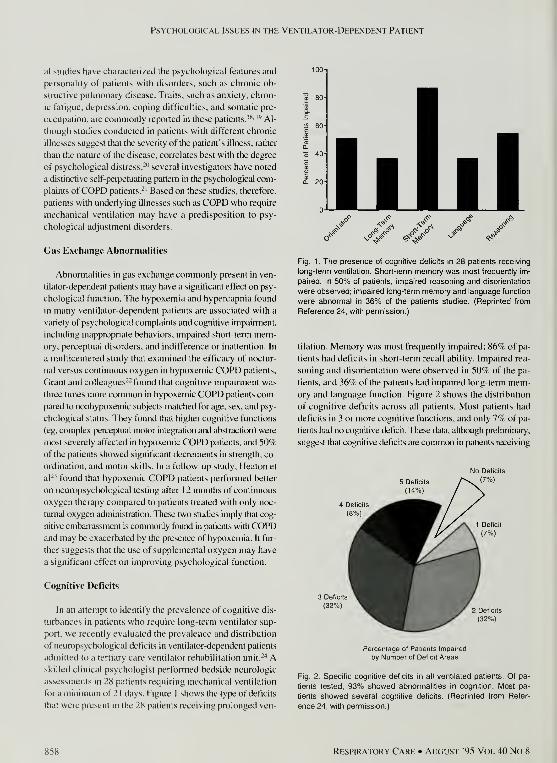
Psychological Issues in the Ventilator-Dependent Patient
al studies have chaiacterized the psychological features and
personality of patients with disorders, such as chronic ob-
structive pulmonary disease. Traits, such as anxiety, chron-
ic fatigue, depression, coping difficulties, and somatic pre-
occupation, are commonly reported in these patients."*' '"^ Al-
though studies conducted in patients with different chronic
illnesses suggest that the severity of the patient's illness, rather
than the nature of the disease, correlates best with the degree
of psychological distress,-" several investigators have noted
a distinctive self-peipeUiating pattern in the psychological com-
plaints ofCOPD patients.-' Based on these studies, therefore,
patients with underiying illnesses such as COPD who require
mechanical ventilation may have a predisposition to psy-
chological adjustment disorders.
Gas Exchange Abnormalities
Abnormalities in gas exchange commonly present in ven-
tilator-dependent patients may have a significant effect on psy-
chological function. The hypoxemia and hypercapnia found
in many ventilator-dependent patients are associated with a
variety of psychological complaints and cognitive impaimient,
including inappropriate behaviors, impaired short-temi mem-ory, perceptual disorders, and indifference or inattention. In
a multicentered study that examined the efficacy of noctur-
nal versus continuous oxygen in hypoxemic COPD patients.
Grant and colleagues-- found that cognitive impairment was
three times more common in hypoxemic COPD patients com-
pared to nonhypoxemic subjects matched for age, sex, and psy-
chological status. They found that higher cognitive functions
(eg, complex percepaial motor integration and abstraction) were
most severely affected in hypoxemic COPD patients, and 50%of the patients showed significant decrements in strength, co-
ordination, and motor skills. In a follow-up study, Heaton et
al-' found that hypoxemic COPD patients performed better
on neuropsychological testing after 1 2 months of continuous
oxygen therapy compared to patients treated with only noc-
turnal oxygen administration. These two studies imply that cog-
nitive embarrassment is commonly found in patients with COPDand may be exacerbated by the presence of hypoxemia. It fur-
ther suggests that the use of supplemental oxygen may have
a significant effect on improving psychological function.
Cognitive Deficits
In an attempt to identify the prevalence of cognitive dis-
turbances in patients who require long-term ventilator sup-
port, we recently evaluated the prevalence and distribution
of neuropsychological deficits in ventilator-dependent patients
admitted to a tertiary care ventilator rehabilitation unit.-'* Askilled clinical psychologist performed bedside neurologic
assessments in 28 patients requiring mechanical ventilation
for a minimum of 2 1 days. Figure 1 shows the type of deficits
that were present in the 28 patients receiving prolonged ven-
^"^ <3->«
Fig. 1. The presence of cognitive deficits in 28 patients receiving
long-term ventilation. Short-term memory was most frequently im-
paired. In 50% of patients, impaired reasoning and disorientation
were observed; impaired long-term memory and language function
were abnormal in 36% of the patients studied. (Reprinted from
Reference 24, witti permission.)
tilation. Memory was most frequently impaired; 86% of pa-
tients had deficits in short-term recall ability. Impaired rea-
soning and disorientation were observed in 50% of the pa-
tients, and 36% of the patients had impaired long-term mem-
ory and language function. Figure 2 shows the distribution
of cognitive deficits across all patients. Most patients had
deficits in 3 or more cognitive functions, and only 7% of pa-
tients had no cognitive deficit. These data, although preliminary,
suggest that cognitive deficits are common in patients receiving
Percentage of Patients Impaired
by Number of Deficit Areas
Fig. 2. Specific cognitive deficits in all ventilated patients. Of pa-
tients tested, 93% showed abnormalities in cognition. IVIost pa-
tients showed several cognitive deficits. (Reprinted from Refer-
ence 24, with permission.)
858 Respiratory Care • August "95 Vol 40 No 8
Psychological Issues in the Ventilator-Dependent Patient
prolonged ventilation and may be important factors in the de-
velopment of psychological problems.
Chronic Dyspnea
In some patients, the cause for dyspnea may be unclear but
may be significant in limiting their ability to tolerate ventila-
tor support or wean efficiently. The lack of clear-cut causes for
failed weaning may be misinterpreted by caregivers as a lack
of patient cooperation or submaximal effort. The sensation of
dyspnea may also be important in the patient's psychological
perception of breathing or weaning from ventilator support. Dys-
pnea is a subjective complaint that may produce a feeling of
anxiety that, in turn, increases the sensation of dyspnea inde-
pendent of any physiologic changes. In many patients, the feel-
ing of dyspnea leads to a decreased expression of any emotion
because even positive emotions, such as joy, can lead to changes
in oxygen demand and increase the sensation of dyspnea. In
response to this vicious cycle, patients may decrease the ex-
pression of any emotion and the patient is typically described
as living in an "emotional straight jacket."-' Preceding the im-
plementation of mechanical ventilation, some pulmonary pa-
tients may have developed a decreased tolerance for breath-
lessness over time and display anxiety or avoidance in antic-
ipation of any physical activity that they feel will increase their
sensation of dyspnea. In some patients receiving long-term ven-
tilation, the initiation of weaning trials or physical exercise pro-
grams can provoke anxiety and fear that is more related to the
perception of dyspnea than to the stress of the activity itself.
Pulmonary Medications
Medications routinely used to treat pulmonary illnesses
can produce a host of psychological and behavioral side ef-
fects. For example, agitation and excitation may result from
the use of sympathomimetic agents, and affective disturbances
and inappropriate behaviors can occur with steroid treatment.
Aminophylline has been reported to affect patient mood,
behavior, and performance on neuropsychological testing.
Young et al-'^ demonstrated that the administration of theo-
phylline produced a substantial drop in regional cerebral blood
flow (rCBF) in five patients with severe COPD—an average
decrease of 267c following a loading dose and of 22% with
maintenance doses. The investigators speculated that a de-
crease in rCBF contributed to neuropsychological disturbances
in COPD patients; however, they failed to report the patient's
baseline psychological function, and no correlation was made
between neuropsychological function and rCBF. On the other
hand, Lindgren et al-'' reported that chronic theophylline ad-
ministration had no significant effect on academic achieve-
ment in 85 school-aged asthmatic children compared with a
normal sibling control group. At the present time, data regarding
theophylline use and psychological function is contradicto-
ry and awaits further study.
The administration of steroids has been implicated in the
development of neuropsychological disturbances in a num-
ber of case reports. Schraa and Dirks-^ studied the effects of
ACTH on cerebral function and found that exogenous cor-
ticosteroid therapy has a significant negative effect on attention
span and memory function. A comprehensive review of more
than 700 consecutively hospitalized patients treated with cor-
ticosteroids found a dose-dependent effect of corticosteroid
administration on psychological disturbances.-'* In those pa-
tients who received < 40 mg of prednisone/day, the incidence
of psychiatric problems was only 1 %, compared with an in-
cidence of psychological dysfunction of > 5% in patients re-
ceiving 40-80 mg of prednisone and 1 8% in patients receiving
> 80 mg of prednisone/day. Although, the effect of chronic
corticosteroid therapy on cognitive function in patients with
pulmonary disease is limited, Smyllie and Connolly-'^ found
a < 2% incidence of psychological problems in 520 patients
who received > 20 mg of prednisone/day. These data suggest
that corticosteroid therapy may have an important effect on
memory and attention span in some patients, but, in most cases,
noteworthy psychological problems were minimal when pred-
nisone doses were < 20 mg/day.
Impact of Prolonged Mechanical Ventilation
In addition to the stress facing every critically ill patient,
ventilator-dependent patients experience the unique stress of
depending on a machine for breathing moment to moment,
hour to hour, and day to day. Some recent studies have re-
viewed the feelings reported by patients who have experienced
prolonged ventilatory support. Riggio and colleagues'" de-
scribed the perceptions of ventilated patients about the sources
for their psychological stress and found that the inability to
communicate, disorientation, memory loss, and pain and dis-
comfort were all perceived as major problems.
The patient's recall of reactions to mechanical ventilation
after hospital discharge was further assessed by Bergbom-En-
gberg and Haljamae." They found that 47% of patients re-
ported feeling anxious or fearful during mechanical ventilation,
and 90% recalled the experience as stressful or unpleasant as
long as 4 years after the episode.
A number of emotional stressors specific to ventilator treat-
ment have been recognized. The process of mechanical ven-
tilation necessitates the loss of independence and control over
the most vital, voluntary life-sustaining activity—breathing.
Endotracheal and tracheostomy tubes impair the ability of
patients to speak, but patients also cannot sigh, gasp, sneeze
or produce other emotional expressions that are part of the
usual body-language vocabulary. Gale and O'Shanick'- have
characterized a patient's inability to communicate while being
ventilated as communicator isolation. The inability of ven-
tilated patients to fully communicate their physical and emo-
tional needs to others can create further feelings of isolation
and abandonment.
Respiratory Care • August '95 Vol 40 No 8 859

Psychological Issues in the Ventilator-Dependent Patient
Other aspects of ventilator care have also been described
as additional sources of stress. Most ventilated patients de-
scribe endotracheal suctioning as extremely unpleasant and
compare suctioning with gagging or suffocating. The
monotonous noises emitted by mechanical ventilators are un-
natural and further contribute to aberrant sensory input and
disturbed sleep. Limitations in ambulation induced by ven-
tilator support may further contribute to feelings of patient
isolation. Finally, patients on ventilator support are faced with
the possibility that equipment failure at any moment might
result in their death. The incessant sounding of ventilator alarms
may, therefore, be perceived as a serious development (eg,
mechanical failure) and contribute greatly to their feelings of
apprehension and anxiety and even, in some, a sense of im-
pending death.
In summary, the stresses faced by the ventilator-dependent
patient are similar to those encountered by other critically ill
patients but includes the added stress of continuous dependence
on a machine for breathing. The complex interactions between
physiologic changes caused by the underlying pulmonary dis-
order and dependence on life-sustaining technology that lim-
its the ability to communicate results in a combination of fac-
tors that may precipitate or amplify the development of psy-
chological disturbances specific to the ventilated patient.
Evaluation of Psychological Disturbances
The assessment of psychological function and the impact
of psychological stress in the patient receiving prolonged ven-
tilation falls well within the evaluation skills of the clinical
management team. Psychological assessment can be incor-
porated into the routine assessments conducted by physicians,
nurses, and respiratory and rehabilitation therapists. A psy-
chologist or psychiatrist can provide important additional in-
formation to the management team, but participation by a men-
tal-health professional is not a prerequisite for measuring and
addressing psychological issues.
The evaluation of psychological function begins with a com-
prehensive history and physical exam. In contrast to the rou-
tine examination, however, the quality of the psychological
assessment is largely influenced by the communication skills
of the interviewer. An initial successful patient encounter will
have a positive impact on establishing a good rapport with
the patient and facilitate the completion of a thorough psy-
chological examination. Throughout the initial assessment,
the interviewer should be supportive and reassuring and use
nonverbal skills, such as, direct eye contact with the patient.
The main goal of the psychological exatnination is to iden-
tify factors that may contribute to or exacerbate the patient's
present psychological state. Three broad areas should be as-
sessed during the psychological exam: features of the un-
derlying disease; aspects of the underlying psychological sta-
tus, and reactions specific to the medical treatment.
The course of a patient's disease plays a large role in de-
termining how the patient reacts to the stresses of the illness
in general and to specific treatments and medical interven-
tions. For example, there may be a marked difference in the
reaction of the patient receiving mechanical ventilation for
the first time compared to the reaction of the patient who has
received mechanical ventilation several previous times for a
chronic illness, such as COPD. The patient experiencing the
initial bout of respiratory failure is usually fearful and be-
wildered by ventilator support but has a hopeful outlook and
high expectations for weaning and functional improvement.
By contrast, the previously ventilated patient is more familiar
with ventilator support but is less hopjeful and more discouraged
about the eventual outcome.
An additional consideration is the premorbid level of func-
tion of the patient, prior to being placed on ventilatory sup-
port. Many patients with chronic respiratory disorders, severe-
ly restrict their daily activities and level of independence as
dyspnea and fatigability increase. TTiis may result in patients'
lowering their expectations about optimal functioning once
discharged. It is important for the clinical management team
to establish realistic goals for the patient and family about the
highest level of activity that can be achieved relative to the
underlying disease process.
A good psychological history should include questions about
the patient's past learning behavior, marital discord, family
support, socioeconomic status, prior education background,
living arrangement, work history, and alcohol and drug use.
The interviewer should also query patients about psychiatric
symptoms in themselves or their family and their style of cop-
ing with and adjustment to physical illness or death.
The interview should also detemiine whether the patient
is experiencing frank psychiatric symptoms, such as visual or
auditory hallucinations, delusions, or paranoid ideations. Men-
tal status and cognitive function can be objectively assessed
using standardized bedside neuropsychological tests (eg, the
Mini-Mental Status Examination"). Facets of function to be
evaluated include orientation to person, place, and time, ca-
pacity for sustained attention, immediate and delayed mem-
ory function, and reasoning ability. Other skills that can be as-
sessed in the interview process include motor function, long-
tenn memory, receptive and expressive language function, visual
skills, reading ability, and the patient's thought process. Psy-
chological issues should also be evaluated in relationship to
the process of mechanical ventilation. It is important to assess
symptoms of delirium associated with sleep and sensory de-
privation and to establish the patient's level of orientation to
person, place, and time, but it is also important to assess symp-
toms in response to aspects of the daily management plan in-
cluding scheduled treatments, rehabilitation therapies, and in-
teractions with members of the management team.
Psychological problems related to the process of wean-
ing are especially important. An as.sessment of the patient's
perception of the ventilator apparatus, the process of wean-
ing from ventilation, and goals for weaning should be re-
K6() Ri:spiRAr()RY Care • Augu.st '95 Vol 40 No 8

Psychological Issues in the Ventilator-Dependent Patient
viewed. Weaning may be perceived differently by individ-
ual patients, and it is important to ascertain the patient's per-
spective prior to implementing weaning trials. For example,
weaning may be viewed as a significant improvement in some
patients because their ability to verbally communicate will
become enhanced and they will become more mobile. By con-
trast, other patients may view weaning as uncomfortable and
anxiety provoking without any clear-cut gains to be obtained.
Patients may also misunderstand the weaning process, lack
confidence in the ability to breath without the ventilator, feel
unable to regulate breathing, or, in some respects, feel aban-
doned by the treatment staff. The patient's perception of the
weaning process, goals for weaning outcome, and the like-
lihood of weaning success should be established beforehand
to minimize the development of psychological dysfunction
during the weaning process.
Treatment of Psychological Problems
General Approach
The general approach to the treatment of psychological
problems in the ventilator-dependent patient combines the
basic principles used to treat psychological disorders in pa-
tients suffering from advanced lung disease or critical illnesses
coupled with the treatment plan directed at the special prob-
lems posed by the process of mechanical ventilation. Fac-
tors contributing to the development of psychological prob-
lems precipitated by the process of mechanical ventilation
(eg, limited mobility, impaired communication, ICU envi-
ronment) should be identified and systematically resolved
in order to improve the patient's sense of control over the
situation and ability to communicate appropriately with the
treatment staff. It is paramount to provide an environmen-
tal setting that minimizes patient seclusion and maximizes
interaction with the stafi" and the patient's family. As previously
discussed, several studies have documented the intellectu-
al impainnent observed in ICU patients, which is characterized
by perceptual distortions, confusion, and disorientation that
may arise in large part from the environmental constraints
of the ICU.
To decrease the incidence of environmentally mediated psy-
chological problems, Kornfeld-"* has proposed several mod-
I ifications in patient care: ( 1 ) maximizing periods of unin-
terrupted sleep by minimizing nighttime interventions; (2) plac-
ing each patient in an individual room; (3) placing nonessential
monitoring equipment outside of the patient's rooms to reduce
the amount of monotonous auditory input; (4) placing ori-
entation aids, such as calendars, clocks, and artifacts in the
patient's room; (5) making an outside window visible to each
venfilated patient and; (6) increasing patient mobility.
An additional goal of psychological treatment is mini-
mization of conditions that contribute to disorientation and
copJiision by providing compensatory strategies for major cog-
nitive problems. As previously described, ventilator-dependent
patients are prone to cognitive disorders including impaired
memory and orientation, as well as deficits in abstract rea-
soning and verbal functioning.-^ Compensatory strategies for
these problems include repeating information in a siinplified
manner, delivering information both verbally and visually,
using illustrative aids and gestures, and continuing to mon-
itor the patient's recall and understanding of instructions and
information provided. By systematically providing cues and
compensatory strategies for these cognitive impairments,
deficits that could interfere with teaching and rehabilitation
activities can be substantially minimized.
Communication & Swallowing
An important step to involve patients in their ciue and min-
imize cognitive disturbances is the re-establishment of the abil-
ity to communicate. Previous studies have shown that impaired
communication is one of the most common features that con-
tribute to poor tolerance of mechanical ventilation. In addi-
tion, improved communication among ventilator-dependent
patients, their families, and the staff decreases stiess, improves
decision-making, and decreases the likelihood of developing
feelings of isolation and/or abandonment. Several modalities
are now available to facilitate written or verbal communication
in patients receiving mechanical ventilation. Some of these
methods can be employed while patients ai'c on full ventilatory
support (eg. buccal resonator, 'talk trach'. electrolarynx) or
during periods of spontaneous breathing by using devices that
redirect airflow through the vocal cords and permit more nat-
ural speech (fenestrated tracheostomy tube. Passy-Muir valve).
IiTespective of the method chosen, the entire staff should en-
courage the patient to use the device consistently to facilitate
verbal expression and decrease feelings of isolation.
Besides communication, one of the most important nat-
ural processes to restore in recovering, critically ill ventila-
tor-dependent patients is eating. Besides the obvious important
role in providmg nutrition, eating rather than being fed by ar-
tificial means fosters a sense of independence, social inter-
action, patient control, and enjoyment that can provide a sig-
nificant emotional uplift for patients who have been receiv-
ing prolonged ventilatory support. Although swallowing
dysfunction is commonly encountered in patients receiving
prolonged ventilator support,'^ swallowing reconditioning and
treatment measures implemented after evaluation by an ex-
perienced speech pathologist can help to restore successful
oral intake in most patients. In our experience, the majority
of our long-tenn ventilator-dependent patients have improved
swallowing and are able to eat after the implementation of
compensatory strategies (eg. altering the consistency of food,
making repetitive swallows after a bolus of food, and adjusting
the position of the head during eating). In most cases, the re-
institution of feeding provides an emotional uplift for patients
that becomes an important milepost in their recovery.
Respiratory Care • August '95 Vol 40 No 8 861
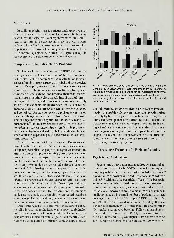
Psychological Issues in the Ventilator-Dependent Patient
Medications
In addition to behavioral techniques and supportive psy-
chotherapy, some patients receiving long-tenri ventilation may
benefit from the selective use of psychotropic medications.-'
Anxiolytics, such as, benzodiazepines may be useful in some
patients who suffer from extreme anxiety. In other ventilat-
ed patients, small doses of neuroleptic agents may be help-
ful in controlling agitation. In others, antidepressant agents
may be needed to treat extreme fatigue and apathy.
Compreliensive Multidisciplinary Programs
Studies conducted in patients with COPD"' and those re-
ceiving chronic mechanical ventilation" have demonstrated
that involvement in a comprehensive rehabilitation program
can significantly improve a patient's physical and psychological
function. These programs usually involve both pulmonary and
whole body rehabilitation and use a multidisciplinary team
composed of occupational and physical therapists, respira-
tory therapists, psychologists, speech therapists, nutritionists,
nurses, social workers, and physicians working collaboratively
with patients and their families to reach jointly declared re-
habilitative goals. The impact of such units on the outcome
and cost of care for patients receiving prolonged ventilation
is currently being examined in the Chronic Ventilator Demon-
stration Project conducted by the Health Care Financing Ad-
ministration."* Preliminary data from one of the four sites in-
volved in this project suggest that a significant improvement
in patient's physiological and psychological state is obtained
when ventilator-dependent patients are enrolled in such treat-
ment programs.'''
As participants in the Chronic Ventilator Demonstration
Project, we have studied the effects of a comprehensive multi-
disciplinary rehabilitation program on cognitive function and
affective disorders in patients receiving prolonged ventilation
treated in a noninvasive respiratory care unit. As shown in Fig-
ure 3, patients and their families reported an overall reduc-
tion in cognitive problems when treated in the ventilator step-
down unit (VSDU) using specific strategies to minimize dis-
orientation and compensate for memory lapses. Patients in the
VSDU were provided with clocks and calendars to maintain
orientation and were actively encouraged to perfonn as many
activities of daily living as possible. Ongoing psychological
suppi)rt wa.s used to enhance patient's weaning success in order
to reduce emotional stress—by providing encouragement to
participate maximally and counseling when needed to address
adjustment problems. In addition, steps were taken to decrease
noise and to avoid unnecessary nocturnal interventions.
Despite the need for long-term ventilator support, patient
mobility is required to facilitate whole body rehabilitation
and to maintain maximal functional status. Secondary to re-
cent advances in medical technology, patient mobility is en-
hanced by using portable ventilators as much as possible. In
Patients
Fig. 3. The perceptions of patients and families of care given in the
Ventilator Step Down Unit (VSDU) compared to the ICU setting. A5-point scale was used to rate each item corresponding to how the
patient or family member rated the perceived feelings: = never, 1
= occasionally, 2 = sometimes, 3 = often, 4 = very often (reprinted
from Reference 39a).
our unit, patients receive mechanical ventilation predomi-
nately via portable volume ventilators that provide patient
mobility by liberating patients from large stationary venti-
lators and permit patient ambulation and out-of-hospital ac-
tivities to enhance a sense of independence and limit feel-
ings of isolation. Preliminaiy data from multidisciplinary treat-
ment programs for long-temi ventilated patients, such as ours.
suggest that a significant improvement in patient function-
al status is obtained when they are treated in such multi-
disciplinary treatment programs.
Psycliologic Treatments To Facilitate Weaning
Psychotropic Medications
Several studies have attempted to reduce dyspnea and im-
prove exercise capacity in COPD patients by employing a
range of psychotropic medications, which includes diiizepam.''"
alprazolam.'*'^- promethazine,^' dihydrocodeine.^ and mor-
phine.'*'^'"' Although the beneficial effects of the benzodia-
zepines are contradictory and limited, the administration of
opiates has been significantly associated with reduced breath-
lessness and improved exercise tolerance when examined in
studies conducted in a small number of patients. Light and
colleagues'*^ reported that 13 eucapnic COPD patients (FEVi
= 0.99 ± 0.48 L) increased maximal workload by 1 87r and
oxygen consumption by 19% after ingesting oral morphine
(0.8 mg/kg) compared to baseline. Following morphine in-
gestion at end exercise, mean ISD PaO: was lower (66 ± 1
2
torr vs 72 torr) and P„co: was higher (44 ± 8 torr vs 38 ± ^.5
torr). Despite a higher level of ventilation achieved at ex-
862 Respiratory Cari; • Auolist '95 Vol 40 No 8
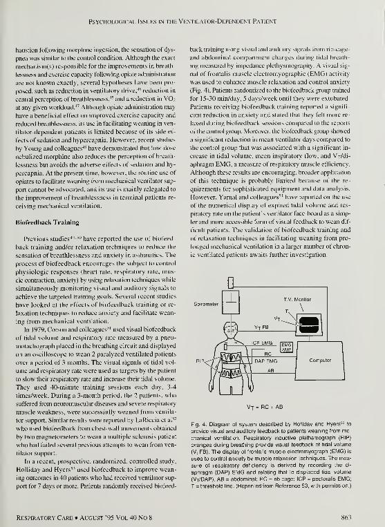
Psychological Issues in the Ventilator-Dependent Patient
haustion following morphine ingestion, the sensation of dys-
pnea was similar to the control condition. Although the exact
mechanism(s) responsible for the improvements in breath-
lessness and exercise capacity following opiate administration
are not known exactly, several hypotheses have been pro-
posed, such as reduction in ventilatory drive,^*" reduction in
central perception of breathlessness,^' and a reduction in VO2
at any given workload.''' Although opiate administration may
have a beneficial effect on improved exercise capacity and
reduced breathlessness. its use in facilitating weaning in ven-
tilator-dependent patients is limited because of its side ef-
fects of sedation and hypercapnia. However, recent studies
by Young and colleagues'*'* have demonstrated that low-dose
nebulized morphine also reduces the perception of breath-
lessness but avoids the adverse effects of sedation and hy-
percapnia. At the present time, however, the routine use of
opiates to facilitate weaning from mechanical ventilator sup-
port cannot be advocated, and its use is mainly relegated to
the improvement of breathlessness in terminal patients re-
ceiving mechanical ventilation.
Biofeedback Training
Previous studies'*^- '" have reported the use of biofeed-
back training and/or relaxation techniques to reduce the
sensation of breathlessness and anxiety in asthmatics. The
process of biofeedback encourages the subject to control
physiologic responses (heart rate, respiratory rate, mus-
cle contraction, anxiety) by using relaxation techniques while
simultaneously monitoring visual and auditory signals to
achieve the targeted training goals. Several recent studies
have looked at the effects of biofeedback training or re-
laxation techniques to reduce anxiety and facilitate wean-
ing from mechanical ventilation.
In 1979, Corson and colleagues" used visual biofeedback
of tidal volume and respiratory rate measured by a pneu-
motachograph placed in the breathing circuit and displayed
on an oscilloscope to wean 2 paralyzed ventilated patients
over a period of 3 months. The visual signals of tidal vol-
ume and respiratory rate were used as targets by the patient
to slow their respiratory rate and increase their tidal volume.
They used 40-minute training sessions each day, 3-4
times/week. During a 3-month period, the 2 patients, who
suffered from neuromuscular diseases and severe respiratory
muscle weakness, were successfully weaned from ventila-
tor support. Similar results were reported by LaRiccia et al"
who used biofeedback from chest-wall movements obtained
by two magnetometers to wean a multiple sclerosis patient
who had failed several previous attempts to wean from ven-
tilator support.
In a recent, prospective, randomized, controlled study.
Holliday and Hyers'' used biofeedback to improve wean-
ing outcomes in 40 patients who had received ventilator sup-
port for 7 days or more. Patients randomly received biofeed-
back training using visual and auditory signals from rib-cage-
and abdominal-compartment changes during tidal breath-
ing measured by impedance plethysmography. A visual sig-
nal of frontalis muscle electromyographic (EMG) activity
was used to enhance muscle relaxation and control anxiety
(Fig. 4). Patients randomized to the biofeedback group trained
for 15-30 min/day, 5 days/week until they were extubated.
Patients receiving biofeedback training reported a signifi-
cant reduction in anxiety and stated that they felt more re-
laxed during biofeedback sessions compared to the reports
of the control group. Moreover, the biofeedback group showed
a significant reduction in mean ventilator days compared to
the control group that was associated with a significant in-
crease in tidal volume, mean inspiratory flow, and Vj/di-
aphragm EMG, a measure of respiratory muscle efficiency.
Although these results are encouraging, broader application
of this technique is probably limited because of the re-
quirements for sophisticated equipment and data analysis.
However, Yamal and colleagues'"" have reported on the use
of the numerical display of expired tidal volume and res-
piratory rate on the patient's ventilator face-board as a simp-
ler and more accessible form of visual feedback to wean dif-
ficult patients. The validation of biofeedback training and
of relaxation techniques in facilitating weaning from pro-
longed mechanical ventilation in a larger number of chron-
ic ventilated patients awaits further investigation.
Spirometer
Vj = RC + AB
Fig. 4. Diagram of system described by Holliday and Hyers^^ to
provide visual and auditory feedback to patients weaning from me-
chanical ventilation. Respiratory inductive plethsmograph (RIP)
changes during breathing provide visual feedback of tidal volume
(V, FB). The display of frontalis muscle electromyograph (EMG) is
used to control anxiety by muscle relaxation techniques. The mea-
sure of respiratory deficiency is derived by recording the di-
aphragm (DAP) EIVIG and relating that to displaced tidal volume
(Vt/DAP). AB = abdominal; RC = nb cage; ICP = pectoralis EMG;
T = threshold line. (Reprinted from Reference 53, with permission.)
Respiratory Care • August '95 Vol 40 No 8 863

Psychological Issues in the Ventilator-Dependent Patient
Cold Facial Stimulation
Patients w ith extreme dyspnea due to a variety of under-
lying lung disorders note symptomatic relief when cool air
is directed to the face from a fan or open window. Studies con-
ducted in animals" " and humans''*'*'" have shown that air-
flow or cold solutions applied to the pharynx, nasal mucosa,
or face may result in an alteration in breathing pattern''' or re-
duction in total minute ventilation.^^"* Schwartzstein and col-
leagues''' studied the effect of 4°-10° C of cold air directed
against the cheeks at 4 km/h on the sensation of dyspnea in
16 normal subjects breathing against elevated inspiratory loads
while Paco: was maintained at 53 torr. Cold air directed to
the face produced a significant reduction in breathlessness,
although, minute ventilation remained unchanged. Early in-
vestigators cited by Freedman''- have shown that dyspnea is
reduced even when air at ambient temperature is directed across
the face during loaded breathing. The mechanisms respon-
sible for the relief of breathlessness when airflow is direct-
ed across the face remain unclear but may include a decrease
in minute ventilation, elicitation of the diving respon.se, or stim-
ulation of the trigeminal nerve. Whatever the responsible mech-
anism, however, the utility of this inexpensive modality for
relieving dyspnea in the ventilator-dependent patient deserves
further investigation.
In Conclusion
The clinical course of patients receiving long-term ven-
tilator support may frequently be complicated by the devel-
opment of psychological problems. We believe that treatment
plans that focus on isolating the cause of psychological dis-
orders in a comprehensive biopsychosocial approach have the
best chance of improving the patient's functional status and
facilitating the successful withdrawal of ventilator support.
Newer approaches that foster the independence of patients,
decrease isolation, and restore their control over their own des-
tiny, such as comprehensive ventilator-dependent rehabili-
tation programs and respiratory biofeedback training may be-
come important treatment options in the future but deserve
further investigation at the present time.
REFERENCES
LaFond L, Horner J. Psychosocial issues related to long-term ven-
tilatory support. Prob Respir Care. l988;l(2):24l-256.
Komfeld DS, Zimbers S. Malm JR. Psychiatric complications of open
heart surgery. N Engl J led 196.'i:273:2«7-292.
Egenon N. Kay JH. Psyi ological disturbances asstK'iatcd with open
hean surgery. Br J Psycniatry 1964;1 \0-M?,-4W.
Blachy PH. Starr A. Post-cardiotomydelirium. Am .1 Psychiatry
l%4;12l:.37l-.37.'i.
Rubinstein D. Thomas JK. Psychiatric findings in cardioiomy pa-
tients. Am J Psychiatry I969;I26(.1):360-.168.
6. Hackett TP. Cassem NH. Wishnie. HA. The coronary care unit: an
appraisal of its psychologic hazards. N Engl J Med 1968:279(25):
1365-1.370.
7. Bi.xler EG. Kales A. Soldatos CR. Kales JD. Healey S. Prevalence
of sleep disorders in the Los Angeles metropolitan area. Am J Psy-
chiatry 1979;136(10):1257-I262.
8. Berlin RM, Litovitz GL. Diaz MA, Ahmed SW. Sleep disorders on
a psychiatric consultation service. Am J Psychiatry I984;141(4):
582-584.
9. Sveinsson I. Postoperative psychosis after heart surgery. J Thorac
Cardiovasc Surg. l975:70(4):7l7-726.
1 0. Schroeder HG. Psycho-reactive problems of intensive therapy. Anaes-
thesia 1971:26(l):28-35.
1 1
.
Parker MM, Schubert W, Shelhamer JH, Parrillo JE. Perceptions of
a critically ill patient experiencing therapeutic paralysis in an ICU.
Crit Care Med 1984:12(11:69-71.
1 2. Woods NA, Falk SA. Noise stimuli in the acute care area. Nurs Res
1974:23(2): 144-150.
13. Aurell J. Elmquist D. Sleep in the surgical intensive care unit: Con-
tinuous polygraphic recording of sleep in nine patients receiving post-
operative care. Br Med J 1985:290(6474): 1029-1032.
1 4. Nicholson AN, Bradley CM. Pascoe PA. Medications: Effect on sleep
and wakefulness. In: Principles and practice of sleep medicine. Kryger
MH, Roth T. Dement WC (editors). Philadelphia: WB Saunders, 1989:
228-236.
15. Wilson LM. Intensive care delirium, the effect of outside depriva-
tion in a windowless unit. Arch Intern Med 1972;l30(2):225-226.
16. Davis JM. McCourt WF. Solomon P. The effect of visual stimula-
tion on hallucinations and other mental experiences during senso-
ry deprivation. Am J Psychiatry 1960:1 16:889-892.
1 7. Neher A. Auditory driving observed with scalp electrodes in normal
subjects. Electroencep L Clin. Neurophysiol 1961;13:449-451.
18. Dudley DL, Glaser EM, Jorgenson BN, Logan DL. Psychosocial con-
comitants to rehabilitation in chronic obstructive pulmonary disease.
Part I. Psychosocial and psychological considerations. Chest 1980:77
(31:413-420.
19. Agle DP. Baum GL. Psychological aspects of chronic obstructive
pulmonary disease. Med Clin North Am I977:61(4):749-758.
20. Cassileth BR, Lush EJ, Strouse TB, Miller DS, Brown LL, Cross
PA, Tenaglia AN. Psychosocial status in chronic illness: a com-
parative analysis of six diagnostic groups. N Engl J Med 1984;3I 1(8):
506-511.
2 1
.
Sandhu HS. Psychosocial issues in chronic obstructive pulmonary
disease. Clin Chest Med 1986:7(4):629-642.
22. Grant I, Heaton RK, McSweeney AJ, Adams KM, Timms R. Neu-
ropsychologic findings in hypoxemic chronic obstructive pulmonary
disease. Arch Intern Med 1 982: 1 42( 8 ): 1 470- 1 476.
23. Heaton RK, Grant 1, McSweeny AJ, Adams KM, Petty TL. Psy-
chologic effects of continuous and nocturnal oxygen therapy in hy-
poxemic chronic obstructive pulmonary disease. Arch Intern Med
1983;I43(I0):I941-I947.
24. Isaac L, Hungerpillar J, Criner G. Neuropsychologic deficits in
chronic ventilator-dependent patients (abstract). Chest I989;96(2):
255S.
25. Young LD. Bowton D, Alford P, McLees B. Do medications reduce
cerebral blood How in COPD patients? Psychosomatics 1986;27
(4):24().
26. Lindgren S, Lokshin B, Stroniquist A. et al. Does asthma or treat-
ment with Iheopyhylline limit children's academic peiformance? NEngl J Med 1992:327:926-9.30.
27. Schraa JL. Dirks JF. The influence of corticosteroids and theophylline
on cerebral function, a review. Chest 1982;82(2):181-I85.
28. Boston Collaborative Dnig Surveillance Program. Acute adverse re-
actions to prednisone in relation to dosage. Clin Pharmacol
1972:13:694-698.
864 RESPIRATORY CARF • AUGU.ST "95 VOL 40 NO 8
Psychological Issues in the Ventilator-Dependent Patient
Smyllie H, Connolly C. The incidence of serious complications
of corticosteroid therapy in respiratory disease, a retrospective
survey of patients in the Brompton hospital. Thorax 1968:23(6):
571-581.
Riggio RE. Singer RD, Hartman K, Sneider R. Psychological is-
sues in the care of critically ill respirator patients: differential per-
ceptions of patients, relatives, and staff. Psychol Rep 1982:51(2):
363-369.
Bergbom-Engberg I. Haljamae H. Assessment of patients' experi-
ence of discomforts during respirator therapy. Crit Care Med 1989: 1
7
(10): 1068-1072.
Gale J. O'Shanick GJ. Psychiatric aspects of respirator treatment and
pulmonary intensive care. Adv Psychosom Med 1985:14:93-108.
Molloy DW. Alemayehu E. Roberts R. A standardized mini-men-
tal state examination compared with the traditional mini-mental state
examination. Am J Psychiatry 1991:148(1 1:102-105.
Komfeld DS. Psychiatric view of the intensive care unit. Br Med J
1969:1(636):108-110.
Hungerpiller J. Leonard C. Criner GJ. Swallowing function in tra-
ched patients receiving chronic ventilation. Chest 1989:96(2):296S.
Fishman DB. Petty TL. Physical, symptomatic and psychological im-
provement in patients receiving comprehensive care for chronic air-
way obstruction. J Chronic Dis I971:24( 12):775-785.
Make BJ, Gilmartin ME. Rehabilitation and home care for ventila-
tor assisted individuals. CIm Chest Med I986:7(4):679-69 1
.
O'Donohue Jr WJ. Chronic ventilator-dependent units in hospitals:
attacking the front end of a long-term problem (editorial). Mayo Clin
Proc. 1992:67(2): 198-200.
Criner GJ, Kreimer DT, Pidloan L. Outcome of patients receiving
prolonged mechanical ventilation (abstract). Am Rev Respir Dis 1993:
147:A874.
Isaac L, Criner GJ. Patient and family perceptions on weaning from
mechanical ventilation in a specialized unit. Am Rev Respir Dis
1990:141(4, Part 2):A411.
Mitchell-Hegges P, Murphy K. Minty K. Guz A. Patterson SC. Minty
PS. Rosser RM. Diazepam in the treatment of dyspnoea in the "pink
puffer" syndrome. Q J Med 1980:49(I93):9-20.
Man GC. Hsu K. Sproule BJ. Effect of alprazolam on exercise and
dyspnea in patients with chronic obstructive pulmonary disease. Chest
1986:90(6):832-836.
Shivaram V. Cash M, Finch P. Acute effects of alprazolam on gas
exchange, work of breathing and exercise tolerance in severe em-
physema (abstract). Chest 1987:92:82S.
Woodcock AA, Gross ER, Geddes DM. Drug treatment of breath-
lessness: contrasting effects of diazepam and promethazine in pink
puffers. Br Med J Clin Res Ed 1981:283(6287):343-345.
Woodcock AA, Gross ER, Gellert A, Shah SO, Johnson MD, Ged-
des DM. Effects of dihydrocodeine, alcohol, and caffeine on breath-
lessness and exercise tolerance in patients with chronic obstructive
lung disease and normal blood gases. N Engl J Med 1981:305(27);
16I1-I616.
45. Light RW. Muro JR, Sato Rl, Slansbury DW, Fischer CE, Brown
SE. Effects of oral morphine on breathlessness and exercise toler-
ance in patients with chronic obstructive pulmonary disease. Am Rev
Respir Dis 1 989: 1 39( I ): 1 26- 1 33.
46. Weil JV, McCullough RE, Kline JS, Sodal IE. Diminished ventilatory
response to hypoxia and hypercapnia after morphine in normal man.
N Engl J Med 1975:292(21 ):1 103-1 106.
47. Santiago TV. Johnson J. Riley DJ, Edelman NH. Effects of mor-
phine on ventilatory response to exercise. J AppI Physiol 1979:47( 1 ):
112-118.
48. Young IH. Daviskas E, Keena VA. Effect of low dose nebulized mor-
phine on exercise endurance in patients with chronic lung disease.
Thorax 1989:44(5):387-390.
49. Feldman GM. The effect of biofeedback training on respiratory
resistance of asthmatic children. Psychosomatic Med 1976:38(1 ):
27-34.
50. Erskine J, Schonell M. Relaxation therapy in bronchial asthma. J Psy-
chosomatic Res 1979:23:131-139.
51. Corson JA, Grant JL, Moulton DP, Green RL. Dunkel PT. Use of
biofeedback in weaning paralyzed patients from respirators. Chest
1979:76(5):543-545.
52. LaRiccia PJ. Katz RH, Peters JW, Atkinson GW, Weiss T. Biofeed-
back and hypnosis in weaning from mechanical ventilators. Chest
1985:87(2):267-269.
53. Holliday JE, Hyers TM. The reduction of weaning time from me-
chanical ventilation using tidal volume and relaxation biofeedback.
Am Rev Respir Dis 1990:141(5 Part 1 ):I214-1220.
.54. Yarnal J, Herrell D, Sivak E. Routine use of biofeedback in wean-
ing patients from mechanical ventilation ( letter). Chest 1 98 1 ;79( 1 ):
127-128.
55. Sant'AmbrogioG, Matthew OP, Sam" AmbrogioPTJ, Fisher JT. La-
ryngeal cold receptors. Respir Physiol 1 985:59( 1 ):35-44.
56. Mortola JP, Al-Shway S, Noworaj A. Importance of upper airway
airflow in the ventilatory depression of laryngeal origin. Pediatr Res
1983:17(7):550-552.
57. Sessle BJ. Greenwood LF, Lund JP, Lacier GE. Effects of upper res-
piratory tract stimuli on respiration and single respiratory neurons
in the adult cat. Exp Neurol 1978:61(2):245-259.
58. McBride B. Whitelaw WA. A physiological stimulus to upper air-
way receptors in humans. J Appl Physiol 1 98 1 :5 1 (5 ): 1 1 89- 1 1 97.
59. Burgess KR. Whitelaw WA. Reducing ventilatory response to car-
bon dioxide by breathing cold air. Am Rev Respir Dis 1 984: 1 29(5 ):
687-690.
60. Folgering H. Olivier O. The diving response depresses ventilation
in man. Bull Eur Physiolpathol Respir 1985:21(2):143-147.
6 1
.
Schwartzstein RM, Lahive K, Pope A, Weinberger SE, Weiss JW.
Cold facial stimulation reduces breathlessness induced in normal sub-
jects. Am Rev Respir Dis 1987:136(1 ):58-61.
62. Freedman S. Cold facial stimulation reduces breathlessness in-
duced in normal subjects (letter). Am Rev Respir Dis 1988:137(2):
492-493.
RESPIRATORY CARE • AUGUST '95 VOL 40 NO 8 865

Case Reports
Tracheobronchial Fracture from Blunt Chest Trauma:
A Case Report
Kenneth E Noblett MA RRT andJeffreyW Selby MD
Introduction
Tracheobronchial fracture is a serious condition occurring
in less than 3% of victims of clinically important chest trau-
ma.' In the past mortality has been reported as high as 30%;-
however, in recent years mortality has declined markedly,
doubtless due to improvements in emergency care, life-sup-
port systems, and surgical techniques. Tliis type of injury from
blunt chest trauma can present some challenging diagnostic
problems. Because of the infrequency and possible difficulty
in early recognition, this condition may not become appar-
ent until the subsequent onset of pulmonary problems. Wepresent the case of a traumatic tracheobronchial fracture with
a delayed clinical manifestation.
Case Study
A 35-year-old man presented to the emergency department
with pain in the right side of his chest and severe shortness
of breath. Examination revealed respiratory rate 34 breaths/min;
heart rate 1 00 beats/min: blood pressure 1 32/88 mm Hg; ar-
terial blood gas values pH 7.45; Pco: ^ ' torr; Pq: 47 torr on
oxygen at 5 L/min by nasal cannula. His electrocardiogram
was normal but his chest radiograph revealed complete opaci-
fication of the right lung with marked loss of volume in the
right hemithorax and shift of the mediastinum to the right. The
left JLing appeared normal (Fig. 1 ). Breath sounds were great-
ly diminished on the right but clear on the left. The patient
was placed on oxygen at 10 L/min via a nonrebreathing mask.
His pulse oximetry saturation (SpOi) rose to 85-90%, and his
dyspnea improved markedly.
Mr Noblett is Clinical instructor and Dr .Selby is Medical Director.
Department of Respiratory Care. Welborn Baptist Hospital, Evans-
ville. Indiana.
TTie authors received no yranis or other financial support lor the prepara-
tion of this paper.
Reprints: Kenneth H Noblett MA RRT. Clinical Instructor. Department
of Respiratory Care, Welborn Baptist Hospital. 41)1 Si; 6th .St. l-vansville
IN 4771.VI 299.
Fig. 1 . Chest radiograph (anteroposterior) of a 35-year-olcl man on
presentation to the emergency department with pain in the right
chest and severe dyspnea. The film demonstrates almost com-
plete volume loss in the right lung, with marked tracheal shift to-
ward the right.
The patient had been hospitalized 6 weeks earlier following
a motorcycle accident in which he sustained extensive sub-
cutaneous emphysema, bilateral pneuinothoraces. pneumo-
mediastinum, bilateral pulmonary contusions, fracture of the
second rib on the left and possible fracture of the first and sec-
ond ribs on the right, laceration of the tongue, contusion of
the left periorbital area, and abrasions of the knees. He was
treated with bilateral chest tubes, rest, pain control, and pro-
phylactic antibiotics but did not require iriechanical ventilatory
support. He was released from the hospital after 7 days and
had been doing well at home until his current symptoms began
and increased over the 24 hours prior to his current hospi-
talization. He had been increasing his exercise level and, in
fact, had walked 3 miles the day before his admission!
The patient underwent flexible bronchoscopy shortly after
this second admission, which revealed that the right main
bronchus was more than 99% obstructed from what appeared
to be scar forination, probably at the site of a bronchial in-
jury (Fig. 2).
The patient was treated with ceftriaxone sodium ( 1 g l.V.)
for possible pneuinonia distal to the obstruction. Purulent se-
8Af-) Ri;si'iR,AT()RY Cari- • Auc.tiST "95 Vol 40 No 8

Tracheobronchial Fracture from Blunt Chest Trauma
Fig. 2. Image(s) obtained during bronchoscopy shortly following
admission. Image(s) reveal almost complete stenosis of the right
mainstem bronchus.
cretions were noted during bronchoscopy, and pneumonia was
confirmed when Staphylococcus aureus was grown from the
bronchial washing.
After 3 days of antibiotic treatment to allow infection and
inflammation to subside, the patient underwent a right tho-
racotomy, with resection of the bronchial stenosis and anas-
tomosis of the right main-stem bronchus to the trachea. The
patient tolerated the surgical procedure well and required no
mechanical ventilatory support following surgery. He un-
derwent daily bronchoscopies for the next 5 days for secre-
tion clearance and was discharged 2 days later with no ap-
parent complications. A follow-up bronchoscopy performed
approximately 10 weeks following surgery revealed dramatic
improvement in the lumen of the right main bronchus, which
appeared essentially normal (Fig. 3).
Discussion
Tracheobronchial fracture is one of the rarer injuries as-
sociated with blunt thoracic trauma. Early diagnosis is im-
portant for optimal management of this life-threatening con-
dition. Failure to diagnose can lead not only to higher mor-
tality but also to delayed complications that may require
extensive intervention.
Presentation
vival rate among victims of more serious trauma as a con-
sequence of effective early treatment. It will be interesting
to see what impact recent developments in passenger restraint
systems (ie, mandated seat belt compliance and air bags) will
have on the occurrence of major chest trauma.
Although the exact mechanism of tracheobronchial frac-
ture is unknown several possible mechanisms may cause this
injury. First, with blunt trauma there can be a flattening of the
thorax and a widening of the transverse diameter and because
the lungs 'follow' the chest wall, a 'pulling apart' may occur
at the carina. Second, if the glottis is closed at the moment
of impact and the trachea and major airways are trapped be-
tween the sternum and vertebral column, the sudden buildup
of intrabronchial pressure can result in bronchial rupture. Third,
during the rapid deceleration that occurs during blunt chest
trauma, shearing forces may be exerted at points of fixation,
namely the carina and cricoid cartilage.' ** Some combination
of these factors is probably involved in the development of
tracheobronchial fracture.
About 80% of such injuries involve the bronchi. The in-
jury is usually parallel to the cartilage rings, occurring with-
in 2 cm of the carina. The right side is affected more often
than the left, but pulmonary vessels are rarely damaged.'' The
patient in our case report fits this pattern.
The patient with tracheobronchial fracture may present
with a variety of symptoms, but in some instances few, if any,
signs or symptoms are present. This latter condition makes
early diagnosis difficult, and early diagnosis can be critical
to a good patient outcome. A review of the literature reveals
The rarity of tracheobronchial injuries is well documented
although an apparent increase in occurrence may be attributed
to increases in high-speed travel^'* or perhaps to a higher sur-
Fig. 3. Image(s) obtained during bronchoscopy approximately 12
weeks following surgical repair of bronchial stenosis resulting from
tracheobronchial fracture received duhng blunt chest trauma.
Image(s) reveal an essentially normal right mainstem bronchus.
Respiratory Care • August '95 'Vol 40 No 8 867

TRArHFOBRONCHIAI- FRACTURE FROM Bl.UNT CHEST TRAUMA
the priiiian tnulings to include cer\ico-mediastinal em-
physema and/or generalized subcutaneous emphysema, pneu-
mothorax, acute dyspnea, hemoptysis, sternal tenderness, and
the Hamman's sign (a crushing sound with each heartbeat,
heard on auscultation).' ''''*
Two findings should cause one to suspect tracheobronchial
injury. If the patient requires positive pressure ventilation, not
only will there be a pneumothorax (in the presence of open
communication of the airway with the pleura) but massive
generalized subcutaneous emphysema is also likely. These
conditions persist and even increase despite the placement of
a chest tubeCs).''
The second important observation involves patients who
do not require positive pressure ventilation. Such patients, with
either an airway that is in open communication with the pleu-
ra or an airway that has some of its tissues intact, will develop
mediastinal and deep cervical emphysema—conditions that
may persist and even increase until the damaged airway has
had time to seal, which may occur in as early as 1 week after
the injury.'' The first finding would make itself known in the
early stages of patient stabilization; the second is somewhat
more subtle and may be overlooked.
Diagnosis
Delay in diagnosis can be detrimental, even fatal, and com-
plications from delay include atelectasis, empyema, hilar ab-
scess, pneumonia, mediastinitis, sepsis, and possible multi-
system organ failure.^ Delayed diagnosis can produce one of
two likely clinical courses depending on the extent of the in-
jury to the airway. With a partial laceration of the airway, the
normal healing process can produce stricture that can vary
in the extent of lumen reduction produced. Recurring atelec-
tasis, infection, and bronchiectasis can result with possible
irreversible parenchymal damage being the final result, a con-
sequence of the fibrosis that results from long standing at-
electasis and/or chronic infection.'''"
If the injury results in a complete transection of the air-
way, then as each end of the severed airway heals hypertrophic
granulation tissue can completely seal the airway lumen. This
results in complete atelectasis of the lung distal to the injury
site. If the stricture occurs quickly and completely there is lit-
tle chance for infection, but absorption atelectasis quickly en-
sues.^ Although there was some infection noted in our sub-
ject, the speed at which complete atelectasis occuned and the
extent of the airway stricture lead us to believe that our pa-
tient suffered complete obstruction.
The primary diagnostic tools involved are the history,
physical examination, a high index of suspicion, chest radio-
graphs, and bronchoscopy. The chest radiograph is important
because it is usually obtained early in the patient's presentation
to the hospital. Radiographic findings with tracheobronchial
fracture can include pneumothorax, bilateral pneumotho-
races, pneuinomediastinum. rib I'ractures, clavicle fractures.
pneumopericardium, and widened mediastinum.^ Because
of the sometimes occult nature and varied presentation of
tracheobronchial fracture, several attempts have been made
to find a focal injury that might serve as the primary diag-
nostic indicator of tracheobronchial fracture.
One of the primary findings that would cause one to sus-
pect tracheobronchial fracture is a pneumothorax.' especially
one that does not respond to chest tube evacuation.'' In fact,
application of suction to the chest tube may actually increase
the air leak and the patient's respiratory distress. The in-
creased distress can be attributed to evacuation of air that,
prior to the application of suction, was involved in gas ex-
change.'" Another abnormality once thought to be a primary
indicator is the presence of fractures involving the first three
ribs, however, later research seems to show that this can-
not be a definite diagnostic indicator." However, one does
have to suspect that the chest has been subjected to a great
atnount of blunt force for fractures involving the first three
ribs and/or clavicle to result. The patient in our case did fit
this pattern, with fracture of the first rib on the right and the
second ribs bilaterally.
Although these abnormalities may suggest airway dam-
age, two findings have been suggested as reliable indicators
of airway injury—first, abnormalities in the appearance of
the endotracheal tube, either an overdistention of the cuff or
extraluminal position of the tip, and second, the findings of
the "fallen lung sign," which is the collapse of the lung to-
ward the lateral chest wall.'- Both of these radiographic find-
ings may be rare: yet, they stand as important observations
that can easily be overlooked or forgotten.
The difficulty in making the diagnosis of bronchial injury
is well documented. Burke- in an early review of 167 cases
found that 687r of these patients were stabilized from their
trauma but did. in fact, have airway injury. Burke observed
These trivial, observable injuries and the initially
satisfactory response to treatment give no clue to
bronchial injury and no further diagnostic efforts
are made. The patient is peacefully allowed to de-
velop a bronchial stricture with all of its hazards.
It has been reported that about \07c of patients with tra-
cheobronchial fracture have no radiographic or physical find-
ings, which adds to the difficulty in making an early diag-
nosis. This absence of findings is probably the result of the
bronchial sheath's remaining intact: thus, air does not enter
the pleura or mediastinum." A delayed diagnosis is made
after the late onset of pulmonary problems due to bronchial
stenosis—reported from I month up to .^2 years following
the trauma.- It remains extremely impoilant that an early di-
agnosis be made. Although repair is possible at a later dale,
a delay in diagnosis may result in a serious loss of functioning
liuig tissue.-
'
The most effective procedme to confirm a diagnosis re-
868 Rl SI'IRA lOK^ CAR! • AUOU.ST '95 Voi. 40 No S

Tracheobronchial Fracture from Blunt Chest Trauma
mains direct visualization of the tracheobronchial tree via
flexible or rigid bronchoscopy.'''^ ''" While all agree that bron-
choscopy is the best procedure for diagnosis of tracheo-
bronchial fracture, two problems are noteworthy. First, is an-
swering the question.Who needs to have the procedure done?
The second is the many cases of the injury that may be missed
during bronchoscopy.
There are no specific criteria for determining who receives
a bronchoscopic examination following blunt chest trauma.
Hara and Prakash'"" have evaluated the value of fiberoptic bron-
choscopy within the first 3 days following acute chest trau-
ma. They established that routine early fiberoptic bronchoscopy
is useful in the evaluation and diagnosis of 53% of patients
involved in acute blunt trauma to the chest.''* The usefulness
of bronchoscopy in this classification of patients has been
confirmed by several clinician-authors;'* 'yet, the decision
to perform bronchoscopy ultimately relies on the clinical judg-
ment and experience of the physician. A review of the lit-
erature reveals several reasonable indicators that suggest con-
sideration of bronchoscopy in the patient following blunt chest
trauma (Table 1).
Table 1 . Indicators for Bronchoscopy following Blunt Trauma to the
Chest.
Primary
Subcutaneous emphysema:
1. Generalized (massive amounts during positive pressure
ventilation)
2. Generally confined to the medial anterior chest and cervical areas
(especially when not subjected to positive pressure ventilation)
Pneumothorax:
1
.
Pneumothorax that poorly responds to chest-tube placement
2. Pneumothorax with which the patient gets more dyspneic when
suction is applied to the pleura! space
,1. Pneumothorax with which the "air leak" increases when suction
is applied or increased within the pleural space
"Fallen lung' sign on chest radiograph
Endotracheal-tube abnormalities on chest radiograph
Overdistention of the cuff
Extraluminal position of the tip
Sternal tenderness
Pneumomediastinum on chest radiograph
Hemoptysis
Hamman's sign
Secondary
Clavicle fracture
Rib fracture(s)
Pulmonary contusion
We believe these reasonable indicators, based on clinical
findings could be helpful in establishing who needs early bron-
cho,scopy following chest trauma.
The second problem encountered is complex and diffi-
cult to verify. Baumgartner et aV showed that even when
bronchoscopy is carried out there is a significant correla-
tion between the skill and experience of the endoscopists
and actually finding the injury under direct visualization.
Repeat bronchoscopies may be indicated in this patient pop-
ulation especially if clinical symptoms persist. ''"' Therefore,
although bronchoscopy is a mainstay for proper diagnosis,
it is not foolproof.
Management
Whether the tracheobronchial fracture is found early or late.
the treatment involves surgical repair. Since the first reported
successful surgical repair in 1948,- numerous advances in the
surgical techniques for repair of tracheobronchial fracture have
been made. Reports indicate that early repair produces good
long-term results and preservation of lung function in about
90% of the cases undergoing surgery.'" In most cases the site
of the injury establishes the surgical intervention plan of ac-
tion. Injuries involving the trachea require urgent restoration.
Injury to the mainstem bronchi can be done in a somewhat
more conservative manner, mainly via resection. If onset of
signs and symptoms and diagnosis are delayed, disinfection
of the tracheobronchial tree may be needed. If the injury in-
volves the smaller airways, treatment may involve resection
or laser repair.""
One of the most important measures is airway control dur-
ing or prior to surgical intervention. If the injury is in the tra-
chea, it is imperative that tracheal intubation be done distal
to the injury preferably under direct visualization via bron-
choscopy. If the injury involves a bronchus, intubation with
a dual-lumen tube may be indicated; however, a single-lumen
tube may be preferable when the main bronchus of the un-
affected lung is to be intubated. This may allow more free-
dom for surgical repair of the injured airway. When the af-
fected lung needs to be ventilated during the surgical procedure,
high-frequency ventilation may be very useful. By placing
the tip of the orotracheal tube cephalad to the injury and the
tip of the gas inflow catheter distal to the injury site, gas ex-
change can take place in the affected lung. This method of
ventilation also allows for a less obstructed field during the
surgical repair.'
In Summary
Although tracheobronchial fracture is rare, detection or
exclusion require careful diagnosis under the sometimes ad-
verse conditions surrounding the trauma patient. The injury
can be treated effectively especially when an early diagno-
sis is made. Our case had an ultimately excellent outcome,
but it suggests one possible scenario following tracheo-
bronchial fracture. Those who treat victims of blunt chest trau-
ma are urged not to forget about this sometimes elusive and
dangerous injury.
Respiratory Care • August "95 Vol 40 No 8 869

Tracheobronchial Fracture from Blunt Chest Trauma
REFERENCES
Bertelsen S. Howitz P. Injuries of the trachea and bronchi. Thorax
1972;:7(2):188-194.
Burke JF. tiarly diagnosis of traumatic rupture of the bronchus. JAMA1962;18L682-686.
Symbas PN. Justicz AG, Ricketts RR. Rupture of the airways from
blunt trauma: treatment of complex injuries. Ann Thorac Surg 1992;
.S4( 1 ): 1 77- 1 8.\
Velly JF. Martigne C. Moreau JM, Dubrez J, Kerdi S. Couraud L.
Post traumatic tracheobronchial lesions. Eur J Cardiothorac Surg
l991;5(7):352-355.
Vukich DJ, Markovchick V. Thoracic U'auma. In: Rosen P, ed. Emer-
gency medicine concepts and clinical practice. 3rd edition. St. Louis
MO:Mosby 1992:437.
Eraser RG, Pare. Pare JD. Eraser RS. Generenx GP. editors. Diag-
nosis of diseases of the chest. 3rd ed. Philadelphia: WB Saunders.
1991:2480-2495.
Baumgartner F. Sheppard B. de Virgilio C. Esrig B, Harrier D. Nel-
son RJ. Robertson JM. Tracheal and main bronchial disruptions after
blunt chest tfauma: presentation and management. Ann Thorac Surg
I99():50(4):569-574.
Tsui SS. Kendall SW. Wells EC. Combined suhclax ian artery rup-
ture and delayed bronchial stenosis from blunt chest trauma. Eur J
Cardiothorac Surg 1 99 1 i.-^d 1 ):6 1 0-6 1 2.
Lotz PR. Martel W. Rohwedder JJ. Green RA. Significance of pneu-
momediastinum in blunt trauma lo the thora,\. Am J Roentgenol 1979;
132(5):817-819.
Kirsh MM. Orringer MB, Behrendt DM, Sloan H. Management of
tracheobronchial disruption secondary to nonpenetrating trauma. Ann
Thorac Surg 1976;22( 1 ):93-10I.
Woodring JH, Fried AM, Hatfield DR. Stevens RK. Todd EP. Frac-
tures of first and second ribs: predictive value for arterial and bronchial
injury. Am J Roentgenol I982;I38(2):21 1.
Unger JM, Schuchmann GG, Grossman JE. Pellett JR. Tears of the
trachea and main bronchi caused by blunt trauma: radiologic find-
ings. Am J Roentgenol 1989.153(61:1 175-1 180.
Hartley C, Morritt GN. Bronchial rupture secondary to blunt chest
trauma. Thorax 1993;48(2):I83-I84.
Hara KS, Prakash UBS. Fiberoptic bronchoscopy in the evaluation
of acute chest and upper airway trauma. Chest 1989;96(3):627-30.
FACE TO FACE WITH CHANGE
AARC 41st Annual Convention and Exhibition
December 2-5 • Orlando, Florida
870 [^i;si'iRAT()RV Care • August "95 Vol 40 No 8

Hugh S Mathewson MD, Section Editor
DrugCapsule
Selective Drug Therapy for Pulmonary Hypertension
Hugh S Mathewson MD
Introduction
The pathogenesis of pulmonary hy-
pertension can be obvious, as it is in hy-
poxemic states, or obscure, as is the case
with primary pulmonary hypertension.
Decreased airway Pqi is the main stim-
ulus for hypoxic pulmonary vasocon-
striction, which is presumed to be an au-
toregulatory mechanism for maintenance
of optimal ventilation-perfusion ratios.
Although the physiologic basis of this
mechanism is unknown, the obvious va-
sodilator drug is oxygen, which exerts a
marked preferential effect on the pul-
monary vasculature in the presence of hy-
poxemia. Restoration of a normal airway
P02 is usually the sole therapeutic re-
quirement for relieving hypoxic pul-
monary hypertension, although oxygen
may in some instances fail to improve car-
diac output.
There are other conditions, howev-
er, in which pulmonary hypertension is
not directly caused by hypoxemia. In-
tracardiac defects with residual left-to-
right shunts comprise the major caus-
es of precapillary pulmonary hyper-
tension. Postcapillary causes include left
ventricular failure, mitral stenosis, and
obstructive conditions of the pulmonary
venous system. Often these disorders are
amenable to drug therapy (eg, inotropic
agents) or to surgical correction. Man-
agement of patients with operable de-
fects may include vasodilator admin-
istration for diagnostic purposes or to
Dr Mathewson is Professor Emeritus of
Anesthesiology and Professor of Respiratory
Care Education—University of Kansas
Medical Center, Kansas City, Kansas.
relieve pulmonary vasoconstriction in
the perioperative period.
The foregoing comprise about 95%of conditions causing pulmonary hy-
pertension, indicating that increased pul-
monary vascular resistance (PVR) is
nearly always secondary to underlying
respiratory or cardiovascular disease.
Primary disease of the pulmonary vas-
culature can be due to veno-occlusive
disease or chronic thrombotic ob-
struction or may constitute a diagnos-
tic entity known as primary pulmonary
hypertension (PPH). Because there is
no specific identifying test for PPH this
is a diagnosis made by exclusion when
all secondary causes of pulmonary
hypertension have been ruled out.'- The
principal lesion in PPH is an obliterating
vasculitis, which is progressive in most
cases. The expected time of survival of
patients with PPH has, until recently,
been less than 5 years.
Certain varieties of pulmonary hy-
pertension are confined to newborns.
Persistent pulmonary hypertension'-'' is
a neonatal disorder in which there is a
continuation of the fetal circulatory con-
dition, with pulmonary vasoconstriction
and a marked curtailment of pulmonary
blood flow. Blood is shunted from right
to left, either through the foramen ovale
or the ductus arteriosus, producing ve-
nous admixture sufficient to cause pro-
found tissue hypoxia. The pathogene-
sis is obscure, but on postmortem ex-
amination many infants have shown
abnormal muscular hypertrophy of the
pulmonary arterial walls. Congenital di-
aphragmatic hernia is commonly as-
sociated with this condition. A stressed
newborn may revert to a fetal-type cir-
culation, especially if he has a low-birth-
weight or shortened gestational age. Aneffective pulmonary vasodilator may
preserve blood flow to the lungs until
the cause of the stress is relieved.
The principal goal of treatment in all
cases is preservation of right ventric-
ular (RV) function, that is, to reduce af-
terload to RV ejection. The RV is a rel-
atively thin-walled, highly compliant
and distensible structure. Unlike the left
ventricle, which usually is able to with-
stand marked rises in afterload, even
small increases in RV afterload cause
significant reduction in stroke volume.
RV output is usually maintained up to
a mean pulmonary artery pressure of
about 40 mm Hg.^
Vasodilator drugs usually affect both
the systemic and pulmonic vasculature.
In the past, attempts have been made
to treat pulmonary hypertension with
sodium nitroprusside or nitroglycerin.
Although both are potent pulmonary
vasodilators, the concomitant decrease
in systemic arterial pressure serious-
ly limits their clinical usefulness. Sim-
ilar experiences have been reported
with hydralazine and with the alpha-
adrenergic blocking agents tolazoline
and phentolamine.
Drug Mechanisms
Current research is directed toward
the development of drugs that are spe-
cific vasodilators of the pulmonary vas-
culature. Two physiologic mechanisms
are targeted. The first is the receptor-cou-
pled adenylate cyclase system. This is
the major mechanism by which ^adren-
ergic catecholamines (epinephrine, iso-
Respiratory Care • August '95 Vol 40 No 8 871

Drug Capsule
proterenol) accomplish both bronchiolar
and arteriolar relaxation. Occupancy of
the receptor coupled to adenylate cyclase
leads to the production of cyclic adeno-
sine monophosphate (cAMP). The in-
creased levels of cAMP activate a
cAMP-dependent protein kinase that
augments the phosphorylation of myosin
and results in relaxation of vascular
smooth muscle. Allied to this mecha-
nism is the action on prostanoid re-
ceptors, which is linked to the adeny-
late cyclase system. Agents such as
prostaglandin PGEj and prostacyclin
(PGI2) exert their vasodilator effects via
this pathway.*-^
The second major mechanism is the
guanylate cyclase pathway, leading to
the formation of endothelium-derived
relaxant factor (EDRF). Although this
sequence is less well understood, it has
been shown that activation of guanylate
cyclase by EDRF or other nitroso-con-
taining compounds leads to an increase
in cyclic guanosine monophosphate
(cGMP), activating a family of cGMP-dependent protein kinases, which pre-
sumably cause phosphorylation of
myosin as part of the relaxant process.*
It has been established that nitric oxide
appears to have activity identical to
EDRF," and that it is synthesized from
/-arginine in endothelial cells.'"
Nitric Oxide
Nitric oxide (NO) is an unstable,
highly reactive gas characterized chem-
ically as a free radical. Its pulmonary
specificity is entirely due to its phar-
macokinetics." When given by in-
halation in concentrations of 20-40 ppm,
it diffuses rapidly through the alveolo-
capillary membrane and exerts its re-
laxant effect on the pulmonary vascu-
lature. Then it is immediately bound to
the circulating hemoglobin, forming ni-
trosyl-hemoglobin that is then rapidly
oxidized to methemoglobin. The re-
moval is virtually quantitative, so that
practically none of the inhaled NOreaches the systemic circulation.'- The
nitrosyl residue is oxidized to form ni-
trite and nitrate."
Studies have shown that endogenous
NO is an important modulator of peri-
natal pulmonary vascular tone.'"* New-
borns with persistent pulmonary hy-
pertension,'^ and pediatric patients with
congenital heart disease and pulmonary
hypertension" react favorably to NO,
with prompt reduction of pulmonary
artery pressure. Similar responses have
also been demonstrated in adults with
PPH"' and in those with mitral valve dis-
ease." Of particular interest is the po-
tential for improving gas exchange in
patients with severe acute respiratory
distress syndrome (ARDS)."*-- Im-
provement in oxygenation occurs be-
cause the inhaled NO is distributed ac-
cording to ventilation; thus, the asso-
ciated vasodilatation increases blood
flow to well-ventilated alveoli. In ad-
dition, inhaled NO may, to a limited ex-
tent, relieve bronchoconstriction.-'
There are obvious limitations to the
therapeutic use of NO.-'' Its preparation,
storage, and delivery present technical
problems that can be difficult and ex-
pensive. Because its effective half-life
is only a few seconds, administration
must be continuous, and the obtainable
benefits are temporary. Also there is con-
siderable toxic potential associated with
the high reactivity of the gas.-'' In ad-
dition to the destruction of hemoglobin,
there also is production of nitrogen diox-
ide (NO2)—a potent alveolar irritant."
Inhaled concentrations must be accu-
rately metered; up to 80 ppm have been
employed, but this approaches the limit
of human tolerance. For perinatal and
perioperative pulmonary hypertensive
crises, NO has established applicabil-
ity,-'' but its use will continue to be con-
fined to well-monitored operating rooms
and critical care units.-^-*'
Current Treatment of PPH
This uncommon but serious illness
is the subject of a review in the Febru-
ary 1994 Supplement to Chest.-' The
main features of the pathogenesis, in-
cluding genetic factors, are sunnna-
ri/cd,'"'
" and methods of treatment re-
viewed.'-" The series presents .sepa-
rate reports of more than 100 cases each
from France,''' the United States,- and
the United Kingdom'^ with their re-
spective therapeutic regimens and clin-
ical outcomes. Two components of drug
treatment appear to be well established:
the use of anticoagulants and the ad-
ministration of large doses of calcium-
channel blockers."' Of the anticoagu-
lant agents, warfarin is the most wide-
ly used, although it is suggested that
heparin, given its inhibitory effects on
sinooth-muscle-cell proliferation, might
be more suitable. '-
Pulmonary vasodilatation has been
successfully achieved with calcium-
channel blockers in high dosage, such
as 720 mg of diltiazem or 240 mg of
nifedipine daily. ^'' The risks of systemic
cardiovascular depression are obvious,
and the drugs must be titrated to effect
with careful hemodynamic monitoring.
Several new calcium-channel blockers
that have more selective vascular effects
and are less negatively inotropic'- are
under investigation.
Surgical interventions, such as atri-
al septostomy and, in extreme cases,
heart-lung transplantation, are also eval-
uated in the Chest supplement.-'' An in-
ternational registry has recently been or-
ganized that is expected to monitor PPH
patients with greater accuracy, partic-
ularly with emphasis on the outcomes
of drug trials."
Prostacyclin (PGI2)
Extensive use of this agent in Europe,
both for diagnosis and therapy, has
demonstrated its eftlcacy as a pulmonary
vasodilator.^ Prostacyclin is an eicosa-
noid, a 20-carbon atom lipoidal com-
pound that is an arachidonic acid meta-
bolite. It is a potent vasodilator with a
half-life of about 5 minutes. It is com-
monly employed (in Europe) to assess
the vasodilatory capacity of the pul-
monary vasculature. Given ina~dvenously
by infusion at rates of 2 to 4 ng • kg '
min ' it can be useful in predicting re-
sponse to long-term vasodilator thera-
py. '''It may prove to be especially ef-
fccti\ c in controlling pulmonary vascular
872 Rlv.SPIRAI'ORY CARI-; • AtlGU.ST '9.^ Voi. 40 No 8
Drug Capsule
resistance during and after some surgical
procedures for congenital heart dis-
ease.'''""' Continuous PGI2 infijsions have
been shown to improve exercise toler-
ance and decrease symptoms in patients
with PPH.^'"'- Side actions are common,
including headache, flushing, and hy-
potension. Prostacyclin is not yet avail-
able for clinical use in the United States,
but its relea.se is anticipated.
Iloprost
Prostacyclin must be given intra-
venously; it is relatively unstable and
must be protected from light. Its solu-
tions lose their efficacy if allowed to
stand for more than 12 hours. Iloprost.
a synthetic analogue of PGI2. has an ap-
proximate half-life of 13 min."" It can
be stored at room temperature and has
an oral bioavailability of 1 Wc. indicating
that an oral preparation could be de-
veloped. Preliminai7 reports on the use
of iloprost for treatment of PPH have
been favorable.^
Adenosine
It is not surprising that adenosine,
a component of the cAMP molecule,
has smooth-muscle-relaxant properties.
Some evidence suggests that it exerts
a preferential vasodilator effect on the
pulmonary circulation."'-"' At least three
papers report on its use for treatment
of PPH. ^^"'^ In one of them, adenosine
was combined with nifedipine, which
reduced PVR by 49%.^'' Another in-
vestigation compared these two drugs,
finding that adenosine reduced PVR but
nifedipine did not."*^ All three studies
found adenosine to be immediately ben-
eficial. However, adenosine has a half-
life of less than 10 minutes and must
be given intravenously. It has potent
negative inotropic, dromotropic. and
chronotropic properties, and can cause
pacemaker dysfunction, coronary steal.
and systemic hypotension.'** Its even-
tual applicability may be confined to
administration for diagnostic purpos-
es or in emergency situations pending
corrective surgery.
Bipyridine Derivatives
The physiologic action of cAMP is
limited by its almost immediate con-
version to the inactive AMP. This is cat-
alyzed by an intracellular enzyme called
phosphodiesterase-fraction three (PDE-
III). Inhibition of this enzyme prolongs
the action of cAMP and extends the du-
ration of smooth muscle relaxation.
Theophylline and its derivatives are
PDE-III inhibitors, although in ordinary
dose ranges this effect is probably minor.
Two bipyridine derivatives, amrinone
and milrinone, are PDE-III inhibitors
that have positive inotropic as well as
va.sodilator properties. Experimental data
indicate that PDE-III inhibitors of this
type cause the release of NO from the
vascular endothelium."''' Both amrinone
and milrinone reduce PVR in animal
models with pulmonary hypertension.
In one study, milrinone substantially de-
creased pulmonary ailei-y pressure with-
out significant change in systemic ar-
terial pressure.''"
Two clinical studies of milrinone, em-
ployed for its inotropic eftect in patients
with congestive heart failure, showed
that in addition to improving hemody-
namics the drug significantly reduced
PVR." ••''- This is in marked contrast to
the action of established inotropic agents
such as dobutamine. which does not re-
duce right ventricular afterload.-^' Mil-
rinone is currently under study in Eu-
rope as an agent to provide inotropic
support to patients following open heart
surgery.''*-''^ A consistent finding is re-
duction of PVR in a population of pa-
tients that cannot withstand large
changes in systemic hemodynamics.
Endothelin Antagonists
Much of the research on the patho-
physiology of PPH has centered on en-
dothelial dysfunction, which leads to in-
timal thickening and eventual obliter-
ation of pulmonary arterioles. The
respective roles of prostacyclin and NOhave focused attention on endothelium-
derived vasoactive metabolites. A group
of such compounds are the endothelins.
which are 2 1 -amino acid peptides that
exist in 3 known isofonns: ET-1. ET-2.
and ET-3. It appears that ET- 1 is one of
the most powerful vasoconstrictors yet
identified.''*' Antagonistic biochemical
mechanisms between ET- 1 and NO have
been demonstrated in vascular en-
dothelial cells.''
A cyclic neuropeptide (BE-18257B)
has been isolated from Streptomyces mis-
akieuesis, which can displace ET-1 from
the receptors on vascular smooth mus-
cle and antagonize its pressor effects.''''
Va.soconstrictor antagonists of this type
represent a new line of investigation in
elucidating the nature of PPH and offer
promising leads for therapy.
Conclusion
The vasodilator compounds now
known are far from specific for the pul-
monary vasculature, except for NO.
which does not gain entry to the systemic
circulation when administered by in-
halation. From a broad spectrum of com-
pounds, ranging from prostacyclin and
other eicosanoids to calcium-channel
blockers and PDE-III inhibitors, a drug
with strong pulmonary and weak sys-
temic vasodilator properties may yet ap-
pear. At this point, investigators express
confidence that long-standing relief from
pulmonary hypertension will soon be at-
tainable with pharmacotherapy.
REFERENCES
1
.
Pietra GG. Histopathology of primary pul-
monary hypertension. Chest 1994; 105(2,
Suppl):2S-6S.
2. Dantzker DR. Primary pulmonary hy-
pertension: the American experience.
Chest I994;105(2. Suppl):26S-28S.
3. Kinsella JP. Neish SR. Shaffer E, Abman
SH. Low-dose inhalational nitric oxide
in persistent pulmonary hypertension of
the newborn. Lancet 1992;340(8823):
819-820.
4. Roberts JD Jr. Polaner DM, Lang P. Zapol
WM. Inhaled nitric oxide in persistent pul-
monary hypertension of the newborn.
Lancet I992;340(8823):818-819.
5. Sibbald WJ. Driedger AA. Myers ML.
Short Al, Wells GA. Biventricular func-
tion in the adult respiratory distress syn-
drome. Chest 1983;84(2): 126-134.
Respiratory Care • August '95 Vol 40 No 8 873
Drug Capsule
Melot C. Lejeune P. Leeman M. Moraine
JJ. Naeije R. Prostaglandin Ei in the adult
respirjlory distress syndrome: benefit for
pulrnonaTN' hypertension and cost forpul-
monar\ gas exchange. Am Rev Respir Dis
I yS9; 139(1): 106- 110.
Radermacher P, Santak B. Wust HJ,
Tamow J. Faike KJ. Prostacyclin for the
treatment of pulmonary hypertension in
the adult respiratory distress syndrome:
effects on pulmonary capillary pressure
and ventilation-perfusion distributions.
Anesthesiology 1990;72(2):238-244.
Morley D, Maragos CM, Zhang XY,
Boignon M, Wink DA. Keefer LK. Mech-
anism of vascular relaxation induced by
the nitric oxide (NO) nucleophile com-
plexes, a new class of NO-based va-
sodilators. J Cardiovasc Pharmacol 1993;
2!(4):670-676.
Ignarro LJ. Buga GM, Wood KS. BymsRE. Chaudhuri G. Endothelium-derived
relaxing factor produced and released from
artery and vein is nitric oxide. Proc Natl
AcadSci USA 1987;84(24):926.'i-9269.
Palmer RM, Ashton DS, Moncada S. Vas-
cular endothelial cells synthesize nitric
oxide from L-arginine. Nature 1988:333
(6174):664-666.
Moncada S, Palmer RM. Higgs EA. Ni-
tric oxide: physiology, pathophysiology,
and pharmacology. Pharmacol Rev 1991;
43(2):109-I42.
Frostell C. Blomqvist H, Hedenstiema G,
Lundberg J, Zapol WM. Inhaled nitric
oxide selectively reverses human hypoxic
pulmonary vasoconstriction without caus-
ing systemic vasodilation. Anesthesiol-
ogy 1993:78(2):427-435.
Gaston B, Drazen JM, Loscalzo J. Stam-
ler JS. The biology of nitrogen oxides in
the airways. Am J Respir Crit Care Med1994:149(2, Part 1):538-551.
Snyder SH, Bredt DS. Biological roles of
nitric oxide. Sci Am 1992;266(5):68-71,
74-77.
Roberts JD Jr, Lang P, Bigatello LM, Vla-
hakes GJ, Zapol WM. Inhaled nitric oxide
in congenital heart disease. Circulation
1993:87(2):447-453.
Pepke-Zaba J. Higenbottam TW, Dinh-
Xuan AT, Stone D, Wallwork J. Inhaled
nitric oxide as a cause of selective pul-
monary vasodilatation in pulmonary hy-
pertension. Lancet 1991:338(8776):
1173-1174.
Girard C, U-hot JJ, Pannetier JC, Filley S,
French P. Estanove S. Inhaled nitric oxide
after mitral valve replaccinent in patients
with chronic pulmonary artery hyperten-
sion. Anesthesiology 1992;77(5):880-883.
Rossaint R, FaIke KJ. Lopez F. Slama K,
Pison U, Zapol WM. Inhaled nitric oxide
for the adult respiratory distress syndrome.
N Engl J Med l993;328(6):.399-40.'i.
19. Bigatello LM, Hurford WE, Kacmarek
RM, Roberts JD Jr. Zapol WM. Pro-
longed inhalation of low concentrations
of nitric oxide in patients with severe
adult respiratory distress syndrome: ef-
fects on pulmonary hemodynamics and
oxygenation. Anesthesiology 1994:80(4):
761-770.
20. Zapol WM. Rimar S, Gillis N. Marlette
M, Bosken CH. Nitric oxide and the lung.
Am J Respir Crit Care Med 1994:149(5):
1375-1380.
21. Gerlach H, Pappert D, Lewandoski K,
Rossaint R. FaIke KJ. Long-term in-
halation with evaluated low doses of ni-
tric oxide for selective improvement of
oxygenation in patients with adult respi-
ratory distress syndrome. Intensive Care
Med 1993;19(8):443-449.
22. Puybasset L, Stewart T, Rouby J-J.
Cluzel P. Mourgeon E, Belin M-F, et
al. Inhaled nitric oxide reverses the in-
crease in pulmonary vascular resistance
induced by permissive hypercapnia in
patients with acute respiratory distress
syndrome. Anesthesiology 1994:80(6):
1254-1267.
23. Barnes PJ. Nitric oxide and airways. Eur
Respir J 1 993:6(2): 163- 165.
24. Davidson D. NO bandwagon, yet. Inhaled
nitric oxide (NO) for neonatal pulmonary
hypertension (editorial). Am Rev Respir
Dis 1993:147(5):1078-1079.
25. Stamler JS, Singel DJ, Loscalzo J. Bio-
chemistry of nitric oxide and its redox-ac-
tivated forms. Science 1992;258(5090):
1898-1902.
26. Rich GF, Murphy GD, Roos CM, Johns
RA. Inhaled nitric oxide: selective pul-
monary vasodilation in cardiac surgical
patients. Anesthesiology 1993:78(6):
1028-1035.
27. Foubert L, Fleming B. Latimer R, Jonas
M, Oduro A. Borland C. Higenbottam
T. Safety guidelines for use of nitric
oxide (letter). Lancet 1992:339(8809):
1615-1616.
28. Miller CC. Miller JWR. Pulmonary vas-
cular smooth-muscle regulation: the role
of inhaled nitric oxide gas. Respir Care
I992;37(10):l 175-1 185.
29. Rich S. Abenhaim L. editors, Primjiry pul-
monary hypertension (supplement). Chest
1994(2. Suppl):l()5:lS-41S.
30. Higenbottain T. Pathophysiology of pul-
monary hypertension: a role for endo-
thelial dysfunction. Chest 1994:105(2
.Suppl):7S-l2S.
31. I jnglcben D. Familial priinary pulmonary
hypertension. Chest 1994:105(2, Suppl):
13.S-16S.
32. Rich S. The medical treatment of primary
pulmonary hypertension: proven and
promising strategies. Chest 1994:105(2,
Suppl): 17.S-20S.
33. Butt AY, Higenbottam T. New therapies
for primary pulmonary hypertension.
Chest 1994:105(2, Suppl):21S-25S.
34. Brenot F. Primary pulmonary hyperten-
sion: case series from France. Chest 1994:
105(2, Suppl):33S-.36S.
35. Oakley CW. Primary pulmonary hy-
pertension: case series from the United
Kingdom. Chest 1994:105(2. Suppl):
29S-32S.
36. Rich S. Kaufmann E. Levy PS. The effect
of high doses of calcium-channel block-
ers on survival in primary pulmonary hy-
pertension. N Engl J Med 1992:327(2):
76-81.
37. The International Primary Pulmonary
Hypertension Study Group. The inter-
national primary pulmonary hyperten-
sion study (IPPHS). Chest 1994:105(2,
Suppl):37S-41S.
38. Rozkovec A, Stadling JR, Shepherd
G, MacDermot J. Oakley CM, Dollery
CT. Prediction of favorable response
to long-term va.sodilation of pulmonary
hypertension by short-term adminis-
tration of epoprostenol (prostacyclin)
or nifedipine. Br Heart J 1988:59(6):
696-705.
39. Schranz D, Zepp F, Iversen S, Wipper-
man C. Huth R, Zimmer B. et al. Effects
of tolazoline and prostacyclin on pul-
monary hypertension in infants after car-
diac surgery. Crit Care Med 1992:20(9):
1243-1249.
40. Bush A, Busst C. Knight WB, Shine-
bourne EA. Modification of pulmon-
ary hypertension secondary to con-
genital heart disease by prostacyclin
therapy. Am Rev Respir Dis 1987;136:
767-769.
4 1
.
Jones DK, Higenbottam TW. Wallwork
J, Treatment of primary pulmonary hy-
pertension with intravenous epoprostenol
(prostacyclin). Br Heart J 1987:57(3):
270-278.
42. Rubin U. Mendoza J, Hood M, McGoon
M, Barst R, Williams WB. et al. Treat-
ment of primary pulmonary hypertension
with continuous intravenous prostacy-
clin (epoprostenol). Ann Intern Med1990:1 12 (2):485-49l.
43. Kaukinen S. Ylitalo P. Pessi T. Vapaatolo
H. Hemodynamic effects of iloprost. a
prostacyclin analogue. Clin Pharmacol
Ther 1984:36(4):464-469.
44. Scott JP. Higenbottam TW. Wallwork J.
The acute effect of the synthetic prosta-
cyclin analogue iloprost in primary pul-
monary hypertension. Br J Clin Phiirmacol
199();44(6):231-2.M.
45. Morgan JM. McComiack DG. Griffiths
MJ. Morgan CJ. Barnes PJ. Evans TW.
Adenosine as a vastxlilator in priniitry pul-
Tnonary hypertension. Circulation 1 99 1 :84
(3):1 145-1 149,
874 RESPIRATORY CARE • AUGUST '95 VOL 40 NO 8

Drug Capsule
Inbar S, Schrader BJ, Kaufmann E, Vestal
RE, Rich S. Effects of adenosine in combi-
nation with calcium channel blockers in pa-
tients with primary pulmonary hypertension.
J Am Coll Cardiol I993:21(2):413-418.
Schrader BJ. Inbar S, Kaufmann L. Vestal
RE, Rich S. Comparison of the effects of
adenosine and nifedipine in pulmonary hy-
pertension. J Am Coll Cardiol 1992; 19
(51:1060-1064.
Ogilby JD, Iskandrian AS, Untereker WJ,
Heo J, Nguyen TN, Mercuro J. Effect of
intravenous adenosine infusion on my-
ocardial perfusion and function. Hemo-
dynamic/angiographic and scintigraphic
study. Circulation 1992;86(3):887-895.
Morray J, Powers K, Clarke W. Amrinone
reduces elevated pulmonary vascular re-
sistance in isolated perfused rabbit lungs
(abstract). Anesthesiology 1990:73:A1 162.
Tanaka H, Tajimi K. Moritsune O, Koba-
yashi K, Okada K. Effects of milrinone
on pulmonary vasculatirre in normal dogs
and in dogs with pulmonary hypertension.
Crit Care Med 1991;19(l):68-74.
Monrad ES. Baim DS. Smith HS, Lanoue
A, Braunwald E, Grossman W. Effects of
milrinone on coronary hemodynamics and
myocardial energetics in patients with con-
gestive heart failure. Circulation 1985;71
(5):972-979.
Benotti JR, Lesko LJ. McCue JE, Alpert
JS. Pharmacokinetics and pharmacody-
namics of milrinone in chronic conges-
tive heart failure. Am J Cardiol 1985;56
(10):683-689.
Eichhom EJ, Konstam MA. WeilandDS,
Roberts DJ, Martin IT, Slransky NB, Salem
DN. Differential effects of milrinone and
dobutamine on right ventricular pnsload, af-
terload. and systolic performance in con-
gestive heart failure secondary to ischemic
or idiopathic dilated cardiomyopathy. AmJ Cardiol 1987;60(16):1329-1333.
Feneck RO and the European Milrinone
Multicentre Trial Group. Intravenous mil-
rinone following cardiac surgery: I. effects
of bolus infusion followed by variable dose
maintenance infusion. J Cardiothorac Vase
Anesth 1992;6(5):554-562.
55. Feneck RO and the European Milrinone
Multicentre Trial Group. Intravenous
milrinone following cardiac surgery: II.
influence of patient factors and baseline
haemodynamics on therapeutic response.
J Cardiothorac Vase Anesth 1992:6(5):
563-567.
56. Inoue A, Yanagisawa M, Kimura S, Ka-
suya Y, Miyauchi T. Goto K. Masaki T.
The human endothelin family: three struc-
turally and pharmacologically distinct iso-
peptides predicted by the separate genes.
Proc Natl Acad Sci USA 1989:86(8):
2863-2867.
57. Stewart DJ. Levy RD. Cemacek P. Lan-
gleben D. Increased plasma endothelin-
1 in pulmonary hypertension: marker or
mediator of disease? Ann Intern Med1991:1 14(6):464-469.
58. Ihara M. Fukuroda T, Saeki T. Nishik-
ibe M. Kojiri K. Suda H, Yano M. An
endothelin receptor antagonist (ETA) iso-
lated from Sireplomyces misakiensis.
Biochem Biophys Res Commun 1991:
178(1):132-137.
FACE TO FACE WITH CHANGE
AARC 41ST Annual Convention and Exhibition
December 2-5 • Orlando, Florida
RESPIRATORY CaRE • AUGUST '95 VOL 40 NO 8 875

Books, Films,
Tapes, & Software
Listing and Reviews ur Books and Other Media
Note to publishers: Send review copies of booi^s. t'llnis, tapes, and softwa
RESPIRATORY CARE. 1 1 030 Abies Lane, Dallas TX 75229-459.1.
Comparative Review of Respiratory
Care Equipment Texts
Respiratory Care Equipment, edited by
Richard D Branson. Dean R He.ss. and
Robert L Chatburn. with 19 contributors.
Hardbound. 568 pages. Philadelphia: JB
Lippincolt, 1995. $49.95.
Principles and Applications of Cardio-
respiratory Care Equipment, edited by
Da\ id H Eubanks and Roger C Bone, with
14 contributors. Hardbound, .^94 pages.
Chicago: Mosby-Yearbook, 1994. $48.95.
Respiratory Care Equipment, 5th ed, by
Steven P McPherson with a 5-niember ed-
itorial-review board. Hardbound, 413 pages.
Chicago: Mosby-Yearbook, 1995. $54.95.
Equipment Theory for Respiratorj' Care,
by Gary C White. Hardbound, 505 pages.
Albany: Delmar. 1995. $48.95.
For years, Steven McPherson's Respi-
ratory Therapy Equipment has been a
mainstay for respiratory care programs. This
book has been joined in the market by oth-
ers with similar content. The purpose of this
review is to look at four respiratory thera-
py equipment texts descriptively and ana-
lytically with recommendations for authors
as they consider future editions.
These texts cover the equipment com-
monly associated with respiratory care as
it is practiced in most institutions (Table 1 ).
McPherson does not cover hyperbaric oxy-
gen, a therapy that is administered by many
respiratory therapists; nor does McPherson
cover blood gas electrodes and the aspects
of quality control that now pervade all as-
pects of laboratory medicine. Eubanks and
Bone fail to cover home-care and transport
ventilators. Transport ventilators are not
covered by While either, although many
hospitals have adopted transport ventilators
for good reason, as detailed in the text by
Branson et al. Because the field has made
significant inroads into home care, these
ventilators are important. Perhaps, the Eu-
banks and Bone text intentionally covers
only hospital-use equipment, leaving other
authors lo cover homc-carc equipment. Only
While has omitted coverage of high-fre-
Tahle 1 . Chapter Title and Content Comparison
B
Text

Books, Films, Tapes, & Software
Table 2. Special Features of Each Text
Branson, Hess, AARC Clinical Practice Guidelines; chapter outlines and objectives at the be-
& Chatburn ginning of each chapter; a shaded box of key terms in each chapter; numerous
shaded equation boxes; extensive referencing to the medical literature; a 9-
page glossary; chapters on computer use and equipment approval; renowned
contributors.
Eubanks & Bone AARC Clinical Practice Guidelines; major section on unconventional ventilation
(eg, high-frequency positive pressure ventilation |HFPPV| and high-frequency
oscillation [HFO]); section on metabolic monitoring; major section on
plethysmographs; emphasis on cardiopulmonary laboratory equipment;
objectives at the beginning of each chapter, extensive information on extra-
corporeal membrane oxygenation (ECMO) and intravenous oxyenators (IVOX);
extensive referencing to the medical literature; sections of suggested readings in
some chapters; renowned contributors.
McPherson Each chapter begins with a chapter outline, objectives, and key terms; shaded
boxes emphasize key points; an 1 1-page glossary; a knowledgeable contributor.
White Shaded boxes showing asseinbly and troubleshooting; self-assessment quizzes at
the end of each chapter; introduction and objectives at the beginning of each
chapter; a 9-page glossary.
Content Comparison
To make this comparison, I have chosen
to look specifically at the chapter on aerosol
and humidity devices and the discussion of
the Puritan-Bennett 7200 ventilator, which
are discussed in each of these texts.
Aerosol & Humidity
The Branson text uses the title, "Hu-
midification: Humidifiers and Nebulizers."
The chapter is 29 pages in length, with 63
illustrations and 61 references. Line draw-
ings are sharp and instructive. By this I mean
that the illustrations are of equipment cur-
rently in use or are generic and are clearly
labeled, so the student can understand the
piece of equipment when using the illus-
tration and the written material (eg. Figs. 5-
5 to 5-8). The photographs are usually clear,
in that the purpose of control knobs is leg-
ible. Photographs are such that the product
does not "disappear" into the background.
Several illustrations from the medical lit-
erature are provided to demonstrate current
practices regarding the use of metered-dose
inhalers, and three AARC Clinical Practice
Guidelines are included. There is a partic-
ularly good, referenced table on artificial
noses. Mist tents are not discussed. Other-
wise, all the current equipment is more than
adequately covered. I particularly like the
emphasis on referring to the medical liter-
ature, rather than to equipment suppliers for
pertbrmance and evaluation data. In general,
the equipment is discussed in a generic sense,
rather than as specific devices. This proba-
bly gives the book a longer applicable life
because device names may change faster than
the principles upon which the devices are
based. The application of specific devices
is left to the instructor. This is congruent with
the NBRC examinations, which seldom men-
tion brand names but rather emphasize the
application of categories of devices.
The McPherson text uses the title. ""Hu-
midifiers and Nebulizers."" The chapter is 33
pages in length, with 5 1 illustrations and 19
references. Line drawings are clear but not
as instructive as in the text by Branson and
colleagues ( Figs. 4-6 and 4-7 ). Another ex-
ample is that for the small particle aerosol
generator { SPAG )( Fig. 5-57 in Branson et
al versus Fig. 4-51 in McPherson). Many of
the photographs are identical to those in Bran-
son, Hess, and Chatburn, so, clarity is sim-
ilar. McPherson does not cite the medical lit-
erature to the extent that Branson et al do.
I question the need to discuss floating-island
and centrifugal nebulizers, due to their ob-
solescence and the infection hazards involved.
The Eubanks and Bone text uses the title,
'"Humidity and Aerosols. ""It is 68 pages in
length, with 74 illustrations and 163 refer-
ences including those cited in the AARCClinical Practice Guidelines. Line drawings
are similar to those in the McPherson text.
Photographs are generally clear so that the
reader can detemiine the function of control
knobs. This chapter combines the generic
description with specific references to in-
dividual devices in some detail. For exam-
ple, 10 different heated humidifiers are de-
scribed. One unique feature is the section on
aerosol particle scavenging and hospital per-
sonnel protection from aerosol particles.
The White text uses the title, ""Humidi-
ty and Aerosol Therapy Equipment." It is 74
pages in length, with 44 illustrations and no
reference to the medical literature; it does in-
clude 1 1 titles in a selected bibliography. Line
drawings are similar to those in McPherson'
s
book. The quality of the photographs makes
it difficult to determine the function of con-
trol knobs; therefore, they are not very in-
structive. There is little in the way of gener-
ic description, but there are detailed descrip-
tions of 6 heated humidifiers, mostly in terms
of assembly and troubleshooting. A discus-
sion of the Bird 500 mL micronebulizer may
be unnecessary, as this device is probably ob-
solete. There is a discussion of the Devilbiss
65 ultrasonic nebulizer, but the unit pictured
is not the Devilbiss 65 nebulizer, rather it is
the Devilbiss 35 nebulizer,' a device that is
rarely used. No discussion of metered-dose
inhalers, aerosol spacer devices, or dry-pow-
der inhalers is offered. This is a serious omis-
sion because these are some of the most
common ways to administer inhaled medica-
tions. This chapter is basically a compila-
tion of manufacturers" data and manuals.
Each of these chapters is well-organized.
The differences are the emphases. The books
by Eubanks and Bone and Branson and col-
leagues clearly emphasize aerosol therapy
based upon the medical literature. The chap-
ter in Eubanks and Bone includes extensive
device-specific information. McPherson has
an adequate generic description and a small
number of examples. White is more device-
specific and includes trouble-shooting sec-
tions for each device, but these tend to be
repetitious among devices of the same type.
Puritan-Bennett 7200
The texts have adopted the Chatburn .sy.s-
tem of classifying ventilators, first published
in Respiratory Care,' and all have in-
cluded the same illustrations to present this
classification scheme.
The text by Branson et al has a chapter en-
titled ""Mechanical Ventilators," that begins
with a table describing each of the ventila-
tors detailed in the text (21 machines). The
discussion of the 7200 is 6 pages in length and
RESPIRATORY CARE • AUGUST '95 VOL 40 NO 8 877
Books, Films, Tapes, & Software
includes 4 illustrations. One is a sharp, clear
picture of the whole ventilator, ventilating a
test lung. Two illustrations ans excellent, clear
line drawings of different configurations of
the control panel. Students could easily de-
termine the function of the control keys by
viewing these drawings. This type of draw-
ing is present for each of the ventilators in the
text. The fourth illustration is a line drawing
simplified froin the actual internal schema.
The text describes the ventilator" s systems and
operation, without going into the detail pro-
vided by the user's manual.
The McPherson text covers the 7200 in
5 pages with 3 illustrations. One illustration
is of the ventilator, but it is not in operation
and has no circuit attached. The next illus-
tration is a poor quality photograph of the
control panel, such that many of the control
key functions are illegible; therefore, this il-
lustration is not very instructive. The third
illustration is a diagram furnished by Puri-
tan-Bennett. Gas flow through the ventila-
tor can easily be traced using the text, which
adequately describes the operation of the ven-
tilator. A shaded box lists the specifications
of this, and each ventilator discussed.
The Eubanks and Bone text handles ven-
tilators differently. Only a brief description
is given of each ventilator, and then several
tables of data for each ventilator are pre-
sented. The picture of the ventilator is of poor
quality-not at all instructive. A 3-page table,
that is basically copied from tlie manual, lists
the specifications of the ventilator. The line
drawings of the ventilator keyboard display
panel are equally unintelligible and not in-
structive. One photograph of the actual key-
board is partially legible; the other is almost
entirely illegible. No discussion or schema
of the operation of the machine is provid-
ed. The information given is intemiixed with
infomiation about several ventilators, which
makes it very reader-unfriendly. I would not
consider this treatment of the machine an
adequate instructional tool.
The White text covers the ventilator in
14 pages. One noninstructive photograph
shows the ventilator with a circuit attached;
however, several other pieces of equipment
are attached, making the picture t(X) cluttered.
The schema is the same as that in McPher-
son except it is kirger. covering two adjacent
pages. Tables of monitors and alarms are
given, but the limits of the alamis are not list-
ed. Apnea-monitoring parameters are list-
ed in a separate tabic, with allowable ranges.
A photograph of the exhalation filler is not
instructive. An additional photograph of the
inspiratory side filter is present, but prob-
ably unnecessary. No illustration of the con-
trol panel is provided. The drive system and
control functions are adequately discussed.
Summary & Recommendations
The text by Branson. Hess, and Chatbum
features excellent illustrations and is ex-
tensively referenced to the literature. The Eu-
banks and Bone text features good illus-
trations, excellent references, and detailed
coverage of cardiopulmonary laboratory pro-
cedure; however, coverage of ventilators is
disappointing. McPherson has good illus-
trations, fair referencing, and good cover-
age of ventilators. White is a basic text with
adequate illustrations. It emphasizes as-
sembly and troubleshooting but lacks ref-
erences to the medical literature and has sev-
eral key omissions and inaccuracies.
Regarding readability, the Branson et al.
Eubanks and Bone, and McPherson texts are
similar. As I reviewed the description of the
ultrasonic nebulizer and the properties of par-
ticles. I could detennine no difference in the
style or readability. The White text tends to
be somewhat more basic with terms such as
frequency and amplitude defined and il-
lustrated, as if to present these terms to in-
dividuals who were not familiar with them.
The White text goes on to describe the same
principles as the other texts using similar lan-
guage.
The field of respiratory care is under con-
siderable pressure from hospital adminis-
trators to become multiskill competent.
Therefore, books that are broader in scope
might be seen as contributing towards the
goal of producing this multiskilled practi-
tioner. According to the recent consensus
conferences held by the American Associ-
ation for Respiratory Care.- the role of res-
piratory care practitioners is expanding to
include the use of equipment not addressed
in these texts, such as instruments used to
perform noninvasive cardiac monitoring:
electrocardiography, echocardiography or
doppler va.sculography. Equipment for hemo-
dynamic monitoring and measurement of
cardiac output, either invasivcly or nonin-
vasively, is not included except in a few
pages in the book by Eubanks and Bone.
Most educational programs are therefore
Ibrced to have students purchase a separate
text for hemodynamic monitoring. Equip-
ment for extracorporeal membrane oxy-
genation is mentioned only in Eubanks and
Bone's book. Equipment for sleep labora-
tories (polysomnography) is mentioned on
one page in Eubanks and Bone and is not
covered in the other texts. Likewise, elecU-o-
encephalography equipment is not covered.
Cardiopulmonary laboratory personnel are
increasingly responsible for assisting the
bronchocopist with lavage, biopsy, and care
of the bronchoscope. However, broncho-
scopy is mentioned only in Eubanks and
Bone's book. It has been suggested that in-
travenous-line insertion should be another
role of the multiskilled therapist, a topic that
none of these texts even mentions. Topics
to cover should include intravenous solutions,
types of lines, the many catheters inserted,
and infusion pumps.
Perhaps, one of these authors should con-
sider a "big book." similar to those edited
by Barnes, Eubanks. Dantzker. or Scanlan.
which are more comprehensive. Such a
text would encompass equipment used
by the therapist who has an 'expanded
role,' as described by the AARC consen-
sus conferences.
-
Timothy B Op't Holt EdD RRTAssociate Professor
Cardiorespiratory Care
University of South Alabama
Mobile, Alabama
REFERENCES
1
.
Chatbum RL. Classification of mechan-
ical ventilators. RespirCare 1992:37(9);
1009-1025.
2. American Association for Respiratory
Care. Year 2001 : an action agenda. Pro-
ceedings of the second national consen-
sus conference on respiratory care edu-
cation. Dallas: Daedalus, 1993.
New Therapies for Neonatal Respirato-
ry Failure edited by Bruce R Boynton,
Waldemar A Carlo, and Alan H Jobe, with
42 contributors. Hardcover, 357 pages, il-
lustrated. New York and Melbourne: Cam-
bridge. 1994. $110.
During the past few years, neonatolo-
gy has experienced a burgeoning of new
techniques for the Ueatment of neonatal res-
piratory diseases. Five years ago. an under-
standing of intermittent mandatory venti-
lation was about all that was necessary to
878 Re.spirm'ory Cari; • Aliglisp '95 Vol 40 No 8

Books, Films, Tapes, & Software
manage neonatal respiratory failure. Now,
those caring for infants must also know how
to manage surfactant therapy, high-fre-
quency ventilation, synchronized venti-
lation, and volume-cycled ventilation. In
addition, intensive care professionals in
many nurseries must also understand how
to manage infants on extracorporeal mem-
brane oxgenation (ECMO), nitric oxide, or
negative-pressure ventilation. Modalities
such as liquid ventilation have gone beyond
laboratory curiosities and into the realm of
clinical trials.
This book. New Therapies for Neona-
tal Respiratory Failure, fills an otherwise
nearly empty niche by providing an overview
of this rapidly changing field, while providing
a sound physiologic basis upon which to
build a deeper understanding of the tech-
niques described. Drs Boynton, Carlo, and
Jobe are superbly qualified to edit such a
book because they have been intimately in-
volved in various aspects of neonatal res-
piratory research for many years. All have
contributed extensively to the book's con-
tent. Additionally, they have chosen an equal-
ly well-qualified group of individuals to con-
tribute to the chapters, including Jeffrey
Whitsett (Composition and Structure of Pul-
monary Surfactant); Anne Greenough and
Robert Chatbum (Advances in Conventional
Mechanical Ventilation); Gregory Heldt and
Graham Bernstein (Patient-Initiated Me-
chanical Ventilation); Thomas Shaffer, Jay
Greenspan, and Maria Wolfson (Liquid Ven-
tilation); and Billie Lou Short (Clinical Use
of ECMO). Useful and interesting com-
mentaries are added throughout by other
noted researchers and clinicians in neona-
tology such as Mary Ellen Avery, Victor
Chemick, and EOR Reynolds.
The text is divided into four parts. In Part
I—Surfactant, chapters on surfactant struc-
ture, function, and metabolism precede an
excellent review of the clinical trials and cur-
rent use of surfactant by Roger Soil, T Allen
Merritt, and Mikko Hallman. Future use of
surfactant for abnormalities other than res-
piratory distress syndrome is addressed in
the closing chapter of this section. In Part U
—
Mechanical Ventilation, the first chapters re-
view respiratory physiology and mechani-
cal ventilation and build a solid background
for the following chapters about patient-ini-
tiated mechanical ventilation, high frequency
ventilation, negative pressure ventilation, con-
tinuous flow oxygenation and ventilation, and
liquid ventilation. Four chapters are dedicated
to high-frequency ventilation and include a
review of physiology and pertinent literature
as well as practical, clinically oriented chap-
ters on high-frequency oscillation, high-fre-
quency flow interruption, and high-frequency
jet ventilation. Part III includes two chapters
on the theory and clinical use of ECMO. The
book concludes in Part IV with a chapter on
the physiology behind the current clinical in-
terest in nitric oxide inhalation.
This book is not a primer; it requires a
background in neonatal pulmonary care tech-
niques and physiology. It is most valuable
to physicians and neonatal caregivers who
treat infants requiring these therapies or to
those who plan to do so. For these profes-
sionals it is invaluable because it provides
a review of pertinent physiology and liter-
ature in addition to providing in-depth in-
formation on the various modalities re-
viewed. It is a valuable teaching tool as well
as a useful reference.
Most of this text is quite readable and il-
lustrations are used amply. However, the ed-
itorial decision to reference works using au-
thors and dates instead of reference numbers
does interfere with readability, particular-
ly when tliese references are placed mid-sen-
tence [eg, "Collagen is not very distensible
(Stromberg & Weiderhielm, 1969; Hoppin
& Hildebrandt, 1977), but elastin is (Carton,
Dainauskas, & Clark, 1962).", page 97].
The chapter by Jose Venegas and Jeffrey
Fredberg on oscillatory mechanics is elegant
work but conceptually difficult. Not enough
attenfion is paid to definition of terms and
abbreviations; for example.
q =Vl/C
Raw
on page 194. The concept of q is not well ex-
plained, except in mathematical terms. Fur-
thermore, by this time I had forgotten about
the list of symbols and abbreviations in the
front of the book, as previous chapters had
defined all terms.
Despite these relatively minor problems,
respiratory care practitioners who are actively
involved in the care of neonatal patients re-
quiring any of these new technologies should
find this book of great value. Because neona-
tal respiratory care is rapidly changing, the
editors of this book will need to revise and
update this textbook frequently. Given the
quality of this first ediUon, I certainly hope
they choose to do so.
Sherry E Courtney MD MSAssociate Professor
Department of Pediatrics
Division of Neonatology
Robert Wood Johnson School of Medicine
Cooper Hospital
Camden, New Jersey
Respiratory Care • August "95 Vol 40 No 8 879

RE/PIRATORy QVRE
Manuscript Preparation Guide
General Information
Respiratory Care welcomes original manuscripts related to
respiratory care and prepared according to these Instructions.
Manuscripts are blinded and reviewed by professionals who are
experts in their fields. Authors are responsible for all aspects of
the manuscript and receive galleys to proofread before publica-
tion. Each accepted manuscript is copyedited so that its message
is clear and it conforms to the Journal's style. Published papers
are copyrighted by Daedalus Inc and may not be published else-
where without permission.
Editorial consultation is available at any stage of planning or writ-
ing. On request, specific guidance is provided for all publication
categories. These Instructions and related materials are available.
Write to Respiratory Care, 11030 Abies Lane, Dallas TX75229-4593, call (214) 243-2272, or fax (214) 484-6010.
Publication Categories & Structure
Research Article: A report of an original investigation (a study).
It includes a Title Page, Abstract, Introduction, Methods, Results,
Discussion, Conclusions, Product Sources. Acknowledgments,
References, Tables, Appendices, Figures, and Figure Captions.
Evaluation of Device/Method/Technique: A description and
evaluation of an old or new device, method, technique, or mod-
ification. It has a Title Page, Abstract, Introduction, Descrip-
tion of Device/Method/Technique, Evaluation Methods,
Evaluation Results, Discussion, Conclusions, Product Sources,
Acknowledgments, References. Tables, Appendices, Figures,
and Figure Captions.
Case Report: A report of a clinical case that is uncommon, or
was managed in a new way. or is exceptionally instructive. All
authors must be associated with the case. A case-managing
physician must either be an author or furnish a letter approving
the manuscript. Its components are Title Page, Abstract, Intro-
duction, Case Summary, Discussion, References. Tables,
Figures, and Figure Captions.
Update: A report of subsequent developments in a topic that has
been critically reviewed in this Journal or elsewhere.
Point-of-View Paper: A paper expressing personal but substan-
tiated opinions on a pertinent topic. Title Page, Text, References,
Tables, and Illustrations may be included.
Special Article: A pertinent paper not fitting one of the forego-
ing categories may be acceptable as a Special Article. Consult
with the Editor before writing or submitting such a paper.
Editorial: A paper drawing attention to a pertinent concern; it
may present an opposing opinion, clarify a position, or bring a
problem into focus.
Letter: A signed communication about prior publications in this
Journal or about other pertinent topics. Tables and illustrations
may be included. Mark "For publication."
Blood Gas Corner: A brief, instructive case report involving
blood-gas values—with Questions, Answers, and Discussion.
Drug Capsule: A mini-review paper about a drug or class of
drugs that includes discussions of phai^macology, pharmacoki-
netics, and pharmacotherapy.
Kittredge's Corner: A brief description of the operation of res-
piratory care equipment—with information from manufacturers
and editorial comments and suggestions.
PFT Corner: Like Blood Gas Comer, but involving pulmonary
function tests.
Test Your Radiologic Skill: Like Blood Gas Corner, but involv-
ing pulmonary medicine radiography and including one or more
radiographs, may involve imaging techniques other than conven-
tional chest radiography.
Review of Book, Film, Tape, or Software: A balanced, critical
review of a recent release.
Review Article: A comprehensive, critical review of the litera-
ture and state-of-the-art summary of a pertinent topic that has
been the subject of at least 40 published research articles. Title
Page, Outline. Introduction. Review of the Literature, Summary,
Acknowledgments, Relerences. Tables. Appendices, and Figures
and Captions may be included.
Overview: A critical review of a pertinent topic that has fewer
than 40 published research articles.
Preparing the Manuscript
Print on one side of white bond paper. 8.5 in. \ 1 I in. (2 Id x 279
mm) with margins of at least 1 in. (25 mm) on all sides of the
page. Vx double-spacing throughout [he entire manuscript. Use
a standard font (eg. Times, Helvetica, or Courier) at least 10
points in size, and do not use italics except for special emphasis.
Number all pages in upper-right corners. Indent paragraphs 5
spaces. Do not justify. Do not pul authors' names or other ideii-
Manuscript Preparation Guide
tification anywhere except on the title page. Repeat title only (no
authors) on the abstract page. Begin each of the following on a
new page: Title Page, Abstract, Text, Product-Sources List,
Acknowledgments, References, each Table, and each Appendix.
Use standard English in the first person and active voice.
Center main section headings on the page and type them in cap-
ital and small letters (eg. Introduction, Methods, Results,
Discussion). Begin subheadings at the left margin and type
them in capital and small letters (eg. Patients, Equipment,
Statistical Analysis).
References. Cite only published works as references.
Manuscripts accepted but not yet published may be cited as ref-
erences: designate the accepting journal, followed by (in press).
Please provide 3 copies of the in-press article for reviewer
inspection. Cite references in the text with superscript numerals.
Assign numbers in the order that references are first cited. On the
reference page, list the cited works in numerical order. Follow
the Journal's style for references. Abbreviate journal names as in
Index Medicus. List all authors.
Article in a journal carrying pagination throughout volume:
1
.
Rau JL. Harwood RJ. Comparison of nebulizer delivery methods
through a neonatal endotracheal tube; a bench study. Respir Care
1992:37(11):1233-1240.
Article in a publication that numbers each issue beginning with
Page 1
:
2. Bunch D. Establishing a national database for home care. AARCTimes 1991;15(Mar):61,62,64.
Corporate author journal article:
3. American Association for Respiratory Care. Criteria for estab-
lishing units for chronic ventilator-dependent patients in hospi-
tals. Respir Care 1988;33(ll):1044-1046.
Article in journal supplement: (Journals differ in their methods of
numbering and identifying supplements. Supply sufficient infor-
mation to promote retrieval.)
4. Reynolds HY. Idiopathic interstitial pulmonary fibrosis. Chest
1986:89(3, Suppl):139S-143S.
Abstract in journal: (Abstracts citations are to be avoided. Those
more than 3 years old should not be cited.)
5. Stevens DP. Scavenging ribavirin from an oxygen hood lo reduce
environmental exposure (abstract). Respir Care 1990:35(11):
1087-1088.
Editorial in journal:
6. Enright P. Can we relax during spirometry? (editorial). Am Rev
Respir Dis 1993;148(2):274.
Editorial with no author given:
7. Negative-pressure ventilation for chronic obstructive pulmonary
disease (editorial). Lancet 1992;340(8833):1440-1441.
Letter in journal:
8. Aelony Y. Ethnic norms for pulmonary function tests (letter).
Chest 199 1:99(4): 1051.
Paper accepted but not yet published:
9. Hess D. New therapies for asthma. Respir Care (year, in press).
Personal author book: (For any book, specific pages should be
cited whenever possible.)
10. DeRemee RA. Clinical profiles of diffuse interstitial pulmonary
disease. New York: Futura, 1990:76-85.
Corporate author book:
1 1
.
American Medical Association Department of Drugs. AMA drug
evaluations, 3rd ed. Littleton CO: Publishing Sciences Group, 1977.
Chapter in book with editor(s):
12. Pierce AK. Acute respiratory failure. In: Guenter CA, Welch MH,
editors. Pulmonary medicine. Philadelphia: JB Lippincott. 1977.
Tables. Use consecutively numbered tables to display informa-
tion. Start each table on a separate page. Number and title the
table and give each column a brief heading. Place explanations in
footnotes, including all nonstandard abbreviations and symbols.
Key the footnotes with conventional designations (asterisk, dag-
ger, double dagger, etc) in consistent order, placing them super-
script in the table body. Do not use horizontal or vertical rules or
borders. Do not submit tables as photographs, reduced in size, or
on oversize paper. Use the same typeface as in the text.
Drugs. Identify precisely all drugs and chemicals used, giving
generic names, doses, and routes of administration. If desired,
brand names may be given in parentheses after generic names.
Drugs should be listed on the product-sources page.
Commercial Products. In parentheses in the text, identify any
commercial product (including model number if applicable) the
first time it is mentioned, giving the manufacturer's name, city,
and state or country. If four or more products are mentioned, do
not list any manufacturers in the text; instead, list them on a
Product Sources page at the end of the text, before the
References. Provide model numbers when available and manu-
facturer's suggested price, if the study has cost implications.
Ethics. When reporting experiments on human subjects, indicate
that procedures were conducted in accordance with the ethical
standards of the institution's committee on human experimenta-
tion. State that informed consent was obtained. Do not use
patient's names, initials, or hospital numbers in text or illustra-
tions. When reporting experiments on animals, indicate that the
institution's policy, a national guideline, or a law on the care and
use of laboratory animals was followed.
Statistics. Identify the statistical tests used in analyzing the data,
and give the prospectively determined level of significance in the
Methods section. Cite only textbook and published article refer-
ences to support choices of tests. Identify any general-use or com-
Respiratory Care

Manuscript Preparation Guide
mercial computer programs used, naming manufacturers and their
locations. These should be listed on the product-sources page.
Units of Measurement. Express measurements of length, height,
weight, and volume in metric units appropriately abbreviated;
temperatures in degrees Celsius; and blood pressures in millime-
ters of mercury (mm Hg). Report hematologic and clinical-chem-
istry measurements in conventional metric and in SI (Systeme
Internationale) units. Show gas pressures (including blood gas
tensions) in torr. List SI equivalent values, when possible, in
brackets following non-SI values—for example. "PEEP. 10 cmHiO [0.981 kPa]." For conversion to SI, see RESPIRATORY CARE
1988;33(10);861-873 (Oct 1988) and 1989;34(2):145 (Feb 1989).
Conflict of Interest. Authors are asked to disclose any liaison or
financial arrangement they have with a manufacturer or distribu-
tor whose product is part of the submitted manuscript or with the
manufacturer or distributor of a competing product. (Such
arrangements do not disqualify a paper from consideration and
are not disclosed to reviewers.) A statement to this effect is
included on the cover-letter page.
Abbreviations and Symbols. Use standard abbreviations and
symbols. Avoid creating new abbreviations. Avoid all abbrevia-
tions in the title and unusual abbreviations in the abstract. Use an
abbreviation only if the term occurs several times in the paper.
Write out the full term the first time it appears, followed by the
abbreviation in parentheses. Thereafter, employ the abbreviation
alone. Never use an abbreviation without defining it. Standard
units of measurement can be abbreviated without explanation
(eg, 10 L/min, 15 torr, 2.3 kPa).
Please use the following forms: cm HiO (not cmH20), f (not
bpm), L (not 1), L/min (not LPM. l/min. or Ipm), mL (not ml),
mm Hg (not mmHg), pH (not Ph or PH), p > 0.001 (not p>0.001),
s (not sec), SpO: (pulse-oximetry saturation).
Submitting the Manuscript
Mail three copies of the manuscript, figures, diskette, and the
Cover Letter & Checklist to RESPIRATORY Care, 1 1030 Abies
Lane, Dallas TX 75229-4593. Do not fax manuscripts. Protect
figures with cardboard. Keep a copy of the manuscript and fig-
ures. Receipt of your manuscript will be acknowledged.
Computer Disltettes. A manuscript may be submitted on a
Macintosh or IBM-DOS diskette. Macintosh documents on 3.5
in. diskettes written in Microsoft Word versions 4.0 and 5.0 are
preferred. However, we can convert most documents (including
PC-DOS format) to our format. Label each diskette with date;
author's name; name of word-processing program and version
used to prepare documents; and filename(s). Do not write on a
diskette except with a felt-tipped pen. Tables and figures must
be in their own separate files, with software identified. Supply 3
hard copies of the manuscript with the diskette.
Prior and Duplicate Publication. Work that has been published
or accepted elsewhere should not be submitted. In special
instances, the Editor may consider such material, provided that
permission to publish is given by the author and original pub-
lisher. Please consult the Editor before submitting such work.
Authorship. All persons listed as authors should have participat-
ed in the reported work and in the shaping of the manuscript; all
must have proofread the submitted manuscript; and all should be
able to publicly discuss and defend the paper's content. A paper
with corporate authorship must specify the key persons responsi-
ble for the article. Authorship is not justified solely on the basis
of solicitation of funding, collection or analysis of data, provision
of advice, or similar services. Persons who provide such ancillary
services exclusively may be recognized in an Acknowledgments
section.
Permissions. The manuscript must be accompanied by copies of
permissions to reproduce previously published material (figures
or tables); to use illustrations of, or report sensitive personal
information about, identifiable persons; and to name persons in
the Acknowledgments section.
Reviewers. Please supply the names, credentials, affiliations,
addresses, and phone/fax numbers of three professionals whomyou consider expert on the topic of your paper. Your manuscript
may be sent to one or more of them for blind peer review.
Editorial Office:
Respiratory Care11030 Abies Lane
Dallas TX 75229-4593
(214) 243-2272 (telephone)
(214) 484-6010 (fax)
Respiratory Care

Cover Letter & ChecklistA copy of this completed form must accompany all manuscripts submitted for publication.
Title of Paper:
Publication Category:
.
Authors:
Author to be Contacted:
.
Mailing Address of Contact Author: ^
Reprints: D Yes D No
"We, the undersigned, have all participated in the work reported, proofread the accompanying manuscript, and approve its
submission for publication." (Please print and include credentials, title, institution, academic appointments, city and state. If
more that 4 authors, please use another copy of this form.)
Author Name:
Author Name:
Author Name:
Author Name:
Author Signature/Date
Author Signature/Date
Author Signature/Date
Author Signature/Date
Has this research been presented in a public forum? D Yes D No
If yes, where, when and by whom?
Has this research received awards? D Yes D No
If yes, please describe.
Do any of the authors have a financial interest in the products mentioned in this paper or competing products?
D Yes D No If yes, please disclose: __^
Checklist:
D Is double-spacing used throughout entire manuscript?
D Are all pages numbered in upper-right corners?
D Are all references, figures, and tables cited in the text?
D Has the accuracy of the references been checked, and are they correctly formatted?
D Have SI values been provided?
D Has all arithmetic been checked?
D Have generic names of drugs been provided?
D Have necessary written permissions been provided?
D Have authors' names been omitted from text and figure labels?
D Have copies of 'in press' references been provided?
n Has the manuscript been proofread by all the authors?
Respiratory Care

New Products
& Services
News releases aboul new pruducls and services \^ill be considered for publication in this section. There is no charge for
these listings. Send descriptive release and glossy black and white photographs to Rbspiratorv Care Journal. New
Products and Services DepI, 1 1030 Abies Lane. Dallas TX 75229-4593.
issue of Respiratory Care, call Mar-
quette Electronics at (800) 917-6000
ext. 8812.
Respiration/Apnea Simulator.
Fogg System Company Inc announces
the Fogg Model 98C Respiration Sim-
ulator for quantitative testing of mon-
itors that detect changes in thoracic
impedance. According to Fogg, the de-
vice dynamically simulates impedance
over a range of respiratory rates from
3 to 150 breaths per minute. In addition,
an unlimited number of apnea intervals
can be manually produced in either in-
spiration or expiration. This instrument
also produces impedance variations se-
lectable in 1 1 steps from 0. 1 to 10 ohms
including zero, with an independent se-
lection of 1 1 basal iinpedances from
100 to 4,000 ohms. The simulator is
powered by an internal 9-volt battery
or from an external power source and
connects directly to the respiration mon-
itor via its lead wires. The Model 98C
may also be used with an ECG simu-
lator to test monitors that detect both
respiration and ECG/heart rate. Con-
tact Fogg System Company Inc. Dept
RC, 15.592 East Batavia Drive, Auro-
ra CO 8001 1 or phone (800) 525-0292
for more details. Remember to mention
Re.spiratory Care.
Multi-Gas Analyzer. For infor-
mation about the Smart Anesthesia
Multi-Gas module featured in the June
Educational Video. HealthScan
Products Inc, maker of the Personal
Best® Peak Flow Meter, now provides
a new patient education video. Crisis
versus Control: Slaying on Top of Your
Asthma with Peak Flow Monitoring.
The 8-minute video, according to the
producer, teaches patients how to de-
tect changes in their lung function by
using the peak flow meter and appro-
priately reading the measurements.
Topics also include details about the
3-zone method of interpreting peak
flow readings and the proper care and
cleaning of peak tlow meters. The
video is free to physicians and respi-
ratory care professionals. For more de-
tails, call HealthScan Products" Cus-
tomer Service Dep;ulment at (8(X)) 962-
1266. Please mention RESPIRATORY
Care when you call.
quiz"'' Prescreen '''^ system in Spanish.
Now Spanish-speaking patients can give
clinicians more accurate and complete
information for preoperative assessments,
according to Nellcor. The questionnaire
can be tailored to match an organization's
preoperative assessment fomis and clin-
ical protocols. The system uses a
Healthquiz device with Prescreen soft-
ware. Patients complete the questionnaire
in Spanish by pushing a button to answer
a series of 'si" or 'no" questions. The clin-
ician can print a one-page summary re-
port in English that highlights importiint
health issues and calculates a risk score.
The system can also suggest appropri-
ate preoperative laboratory tests. In ad-
dition the Healthquiz device creates an
electronic record for each patient" s med-
ical history and stores up to 125 histo-
ries on a small data card. A data appli-
cation, PC Link, provides data man-
agement, custom reporting, and data
integration. For more information, con-
tact Nellcor Incorporated, Dept RC, 4280
Hacienda Drive, Pleasanton CA 94588,
or call (800) NELLCOR. Please mention
Respiratory Care.
Pkescreeninc; System. Nellcor
corporated launches the new Health-
Portable Oxygen SY.sTE^Ks.
DeVilbiss, a division of Sunrise Med-
ical, introduces a new line of portable
oxygen systems—the Walkabout"'^
Mini, ! , and 2. The new systems com-
bine a compressed oxygen cylinder
with PulscDoseT''', an oxygen delivery
technology that ensures oxygen flow
deep into the lungs for maximum ben-
efit, according to DeVilbiss. The Walk-
884 Ri;si>iratory Care; • Atic.u.sT "95 Voi, 40 No 8

New Products & Services
about systems deliver oxygen at a rate
of 10 L/min. Also, this technology al-
lows for smaller, lighter, more attrac-
tive systems and increases ambulato-
ry range for patients. Each Walkabout
unit is packaged in a dark blue, padded
cordura cany bag. The complete sys-
tem includes one OMS 50 PulseDose
unit, an oxygen cylinder and regulator,
an AC charger/adaptor, and a cany bag.
For details, write to DeVilbiss Health
Care Inc, Dept RC, PO Box 635, Som-
erset PA 15501-0635 or mention RES-
PIRATORY Care when you call (814)
443-7499.
NELLCOR. Don't forget to mention
Respiratory Care.
Pulse Oximeter Sensor. Nellcor
expands its line of pulse-oximeter sen-
sors to include the D-YSE ear clip.
When used with the DURA-Y®reusable multisensor, the clip can be
attached to the ear pinna as an alter-
native monitoring site. Nellcor claims
this is helpful when a patient's hands
and feet have low blood flow or if ex-
tremities are unavailable. The clip is
designed for patients weighing 30 kg
or more. To resume conventional mon-
itoring, detach the clip from the sen-
sor and use on a finger, toe, hand, or
foot. Write to Nellcor Incorporated,
Dept. RC, 4280 Hacienda Drive.
Pleasanton CA 94588 or call (800)
14

With The
Information
Service
Card. You
Can Stop
Searching
And Start
Buying.
Get the facts on all
the products and
services
advertised in this
issue easily and
quickly.
The computerized
Information
Service Card does
it all. Simply fill in
your name and
address, check the
appropriate boxes,
and mail or fax it.
Newly Revised! The Uniform Reporting Manual
provides you with nationally recognized standards for
documenting workload units and time standards. Included
are patient assessment activities, bronchial hygiene,
supplemental oxygen, airway care, diagnostic tests,
and cardiovascular
diagnostics.
In addition, there
are chapters on
"Clinical Activities
without Time
Standard" and
"Management
Support
Activities." By the
AARC. 1990,
updated 1993.
Binder, 165 pages.
Item BK1 $65
($85 nonmembers)
Texas Customen only, please add S.2i% sales lax (including shipping charges). Texas
Don't miss the
Research Symposium
at the 1995
Annual Convention
of the American Association
for Respiratory Care
Orlando, Florida • Dec. 2-5, 1995

AARC Clinical Practice GuidelinesCPG 1
CPG 2-
CPGCPGCPGCPGCPG
CPG 8-
CPG 9-
CPGIO-
CPGl 1
CPGl 2 -
CPGl 3
-
CPGl 4
-
CPGl 5
-
CPGl 6
CPGl 7
-
CPGl 8-
CPG19-
CPG20
-
CPG21 -
CPG22
• Spirometry • $1
- Oxygen Therapy in Acute Care Hospital • $1
Nasotracheal Suctioning • $1
• Patient-Ventilator System Checks • $1
Directed Cough • $1
In-Vitro pH ancJ Blood Gas Analysis and Hemoximetry • $1
Use of Positive Airway Pressure Adjuncts to Bronchial
Hygiene Therapy • $1
Sampling for Arterial Blood Gas Analysis • $1
Endotracheal Suctioning of Mechanically Ventilated Adults
and Children with Artificial Airways • $1
- Incentive Spirometry • $1
- Postural Drainage Therapy • $1
Bronchial Provocation • $1
Selection of Aerosol Delivery Device • $1
- Pulse Oximetry • $1
- Single-Breath Carbon Monoxide Diffusing Capacity • $1
• Oxygen Therapy in the Home or Extended Care Facility • $1
Exercise Testing for Evaluation of Hypoxemia
and/or Desaturation • $1
• Humidification during Mechanical Ventilation • $1
Transport of the Mechanically Ventilated Patient • $1
Resuscitation in Acute Core Hospitals • $1
- Bland Aerosol Administration • $1
• Fiberoptic Bronchoscopy Assisting • $1
CPG23 — Intermittent Positive Pressure Breathing (IPPB) • $1
CPG24 — Application of CPAP to Neonates Via Nasal Prongs or
Nasopharyngeal Tube • $1
CPG25 — Delivery of Aerosols to the Upper Airway • $1
CPG26 — Neonatal Time-Triggered, Pressure-Limited, Time-Cycled
Mechanical Ventilation • $1
CPG27 — Static Lung Volumes • $ 1
CPG28 — Surfactant Replacement Therapy • $1
CPG29 — Ventilator Circuit Changes • $1
CPG30 — Metabolic Measurement using Indirect Calorimetry
during Mechanical Ventilation • $1
CPG31 — Transcutaneous Blood Gas Monitoring for
Neonatal & Pediatric Patients • $1
CPG32 — Body Plethysmography • $1
CPG33 — Capillary Blood Gas Sampling for Neonatal &
Pediatric Patients • $1Pediatric Patients • $1
CPG99 — Complete Set in Binder •
(+$3.25 for Shipping and Handling)
$25
American Association for Respiratory Care
1 1030 Abies Ln. • Dallas, TX 75229-4593
Call (214) 243-2272 or fax to (214) 484-2720
with MasterCard, Visa, or Purchase Order Number
J>&siipiie
t^h^lpAmericsi^ prepare for
theiuture.
Support America's
colleges. Because college is
more than a place where
young people are preparing
tor their future. It's where
America — and Americanbusiness — is preparing
for its future.
Give tothe college ofyour choice.
By Tony DalNogare, MD
ARDS was originally described in 1967 and has since
undergone much scrutiny. This review of ARDS discusses
the latest developments in risk factors and treatment. Also
covers the 5 diagnostic criteria that must be present to make
an accurate diagnosis of ARDS, including clinical,
radiographic, and physiologic criteria.
Item VT31 - VHS (60 minutes)
$35 (nonmembers $40)
Add $-4.50 for shipping.
Call (214) 243-2272 or
Fax your order to (214) 484-6010
American Association for Respiratory Care
11030 Abies Ln.
Dallas, TX 75229-4593
Prices subject to cliange without notice.
Texas customers only, please add 8%% sales tax (including sliipping cliarges). Texas
customers that are exempt from sales tax must attach an exemption certificate.

The AARC HumanResources Survey:A Study ofRespiratory CareHuman Resourcesin Hospitals(AncTi .1 wide r.ingo ot human resource
issues, including compensaiion, numbers
ot full-time equivalents, job vacancy
rates, education, credentialing, and
licensure. Even includes mformation on
age, sex, and years of experience. Includes comprehensive summary,
position profiles, salaries, education, experience, credentials, and rcgiona
demographics. Vacancies arc inventoried. 68 pages, 66 tables.
Item BK12 $35 ($50 nonmembers)
A Study ofChronic VentilatorPatients in theHospitalChronic ventilator-dependent patients
are costing American hospitals morethan $9 million per day according to
this Gallup study conducted for the
AARC. This important study provides
information on patients who depend on
life-support systems; why, how, and
where they are being treated; and the
cost of treatment. 47 pages, 9 tables, 12
figures.
Item BK20 $25 ($50 nonmembers)
A Study ofRespiratory CarePracticeThis study e.\amincs the practice of
respiratory care in today's health care
environment and how hospital
reorganization is affecting the
profession. Includes chapters on medical
direction, current respiratory care
services, nontraditional services,
respiratory care protocols, and hospital
service reorganization. ."^8 pages,
34 tables, 15 charts.
Item BK17 $20 ($40 nonmembers)
RE/PIRATORy C&REWe are pleased to offer CRCE through the Journal for 1995. We have changed the
format somewhat, and we hope the changes represent enhancements.
• The grids for answers to questions, name, and AARC member number are
found on the other side of this page. Using such grids enables a certain degree
of automation and accuracy that would not otherwise be possible.
• This page is perforated to make it easy to complete and mail and to prevent
tearing.
However, the rules of previous years apply.
• Only original Answer Sheets will be graded. No facsimiles or photocopies will
be accepted. Six hours of CRCE credit is awarded to those who answer 35 or
more questions correctly.
• You may photocopy the Answer Sheet for your personal record to check your
answers against the Answer Key, which will be published in the October issue
of Respiratory Care.
• Obtain a check or money order for $5.00 payable to AARC (this is for
processing; your Answer Sheet will not be graded if the fee is not included).
• Read and sign the statement below.
• Place the Original Answer Sheet and processing fee in a stamped #10 envelope
and mail to:
CRCE through the Journal
11030 Abies Lane
Dallas TX 75229-4593
• Deadline is September 30, 1995. Responses postmarked after September 30,
1995 will not be processed.
• Follow all instructions explicitly. Failure to do so may cause technical problems
that could disqualify you.
• Questions relating to completing the examination should be directed to the
CRCE Coordinator, (214) 243-2272.
I attest that I completed the Answer Sheet independently, without help from others.
Signature Date


CRCE through theJournal-1995
We are delighted to bring you the sixth edition of CRCE through the Jour-
nal, a program for AARC members to gain credit for continuing education de-
rived from careful reading, thoughtful study, and test completion. The acceptance
of these credits for the fulfillment of license-mandated continuing education is
dictated solely by the licensure law of each individual state.
This year, we are indebted to the efforts of Crystal Dunlevy EdD RRT*and F
Herbert Douce MS RRT who authored the test. This 50-item multiple-choice ex-
amination is based on papers published in RESPIRATORY CARE from July 1994
through June 1995. The issue and page numbers of the paper on which a ques-
tion is based are shown in brackets following the question. You are free to con-
sult the cited paper as you complete the answer sheet. However, we encourage
you to read the paper in its entirety before answering the questions. Then, choose
the single most-correct answer, and carefully mark the answer sheet. RememberCRCE through the Journal is an honor system, and the benefits you derive are
directly proportional to the effort you invest.
Mail your completed answer sheet by September 30, 1995. Answer sheets post-
marked after September 30 will not be processed. The Answer Key for CRCEthrough the Journal will be published in the October issue of RESPIRATORY CARE.
No scores will be available from the AARC until the 1995 CRCE Transcripts are
mailed in early 1996.
* Dr Dunlevy is Associate Professor and Director of Clinical Education and Mr Douce is
Assistant Professor and Program Director—The Ohio State University, Columbus, Ohio.
Respiratory Care • August "95 Vol 40 No 8 889

CRCE THROUGH THE JOURNAL
Questions:
1. In the respiratory care assessment-treatment
program evaluated by Shrake and others, which
of the following proved to be true?
a. Overall utilization of respiratory therapy ser-
vices increased.
b. Cost-savings were reported only for the
3-month evaluation period.
c. Cost savings were reported for both the
3-month evaluation period and for the
3-month period following the study.
d. No significant cost-savings were reported.
e. Patient care proved to be significantly im-
proved.
[July 94:715-724]
2. Which of the following findings reported by
Palmisano and others is most likely to influence
patient outcome during a cardiac arrest?
a. variation in response times of individual
members of the an^est team
b. delay in providing epinephrine
c. delay in administering oxygen
d. delay in establishing intravenous access
e. delay in activation of the cardiac-arrest pag-
ing sy.stem
[July 94:725-729]
3. Acceptable indicators for bronchodilator thera-
py utilized by Wollam and colleagues include
all but which of the following?
a. FEVi< 50% predicted
b. diagnosis of CORD or asthma
c. previous use of corticosteroids or broncho-
dilators
d. presence of auto-PEEP
e. presence of wheezing
[July 94:730-735]
4. Which of the following was not demonstrated
in Padman and associates' case report of nonin-
vasive positive-pressure ventilation for end-
stage cystic fibrosis patients?
a. improved oxygenation
b. improved ventilation
c. diminished sensation of dyspnea
d. improved quality of life
e. improved ability to perform daily activities
[July 94:736-739]
5. According to Valeri et al, what factor influenced
the compliance of mechanical ventilator breath-
ing circuits after the first hour of use?
a. type of plastic composition
b. duration of use up to 48 hours
c. circuit temperature up to 34°C
d. flow setting on the ventilator
e. No factor was found to be clinically impor-
tant in influencing compliance.
[August 94:793-796]
6. Which of the following aerosolized corticos-
teroids (when used on a chronic basis) has been
associated with systemic side effects, including
adrenal suppression?
a. beclomethasone dipropionate
b. triamcinolone acetonide
c. betamethasone valerate
d. budesonide
e. dexamethasone sodium phosphate
[August 94:837-840]
7. Who discovered penicillin?
a. Pasteur
b. Fleming
c. Curie
d. Salk
e. LaVeque
(August 94:850-852)
890 Ri;si>iKATOK'i Cari; • AUGU.ST '95 Vol. 40 No 8

CRCE THROUGH THE JOURNAL
When administering albuterol via metered-dose
inhaler (MDI) to an intubated, mechanically
ventilated adult-lung model, which method de-
livers the largest amount of drug to the distal
end of the endotracheal tube (ETT)?
a. MDI directly attached to ETT between the
Y-connector and the 15-mm adapter
b. MDI attached to the inspiratory limb prox-
imal to the Y-connector with the Aero-
chamber
c. MDI attached to the inspiratory limb proxi-
mal to the Y-connector with the Aerosol
Cloud Enhancer
d. MDI attached to the inspiratory limb just dis-
tal to the humidifier
e. MDI attached to the inspiratory limb just
proximal to the humidifier
[September 94:886-891]
9. Which of the following statements summarizes
the findings of Jager, Tweeddale, and Holland
regarding intubated COPD patients'?
a. Flow-triggered CPAP significantly reduced
the work of breathing in this patient popu-
lation.
b. There was no difference in inspiratory work
of breathing between flow-triggered CPAPand CPAP triggered by a preset pressure
change.
c. CPAP triggered by a preset pressure change
significantly reduced the work of breathing
in this patient population.
d. Intubated COPD patients who are receiving
sedation exhibited an increased work of
breathing when flow-triggered CPAP was
used.
e. Inspiratory airway pressures were signifi-
cantly lower in patients receiving CPAP trig-
gered by a preset pressure change.
[September 94:892-896]
10. For measurements of work of breathing, how
does the Bicore CP-100 with updated software
compare to conventional laboratory methods?
a. Correlation coefficients and coefficients of
determination are nearly equal and highly
significant.
b. Bias is excessive.
c. Precision is unacceptable.
d. Correlation coefficients are not statistically
significantly different from zero.
e. Reliability and validity are not acceptable.
[September 94:897-905]
11. In the case report on unilateral lung disease
chronicled by Schmitt, Mang, and Kirmse, what
can be concluded?
a. Independent lung ventilation was not benefi-
cial due to the patient's increased airway re-
sistance.
b. Independent lung ventilation resulted in cost-
savings.
c. High levels of PEEP dramatically decreased
the patient's work of breathing.
d. Independent lung ventilation greatly im-
proved the patient's oxygenation.
e. Independent lung ventilation should only be
used in an operating room.
[September 94:906-911]
12. According to cases reported by Thomas and
others, exogenous surfactant therapy for ARDSdue to aspiration of gastric contents has resulted
in which of the following?
a. improved oxygenation
b. increased P(A-a)0:
c. reduced dynamic compliance
d. decreased lung volumes
e. increased alveolar surface tension
[September 94:9 12-9 16]
Respiratory Care • August '95 Vol 40 No 8 891

CRCE THROUGH THE JOURNAL
13. Which of the following statements is true about
pulmonary artery catheterization in patients ad-
mitted to the hospital for COPD exacerbation,
according to Smith and Pesce?
a. Pulmonary artery catheterization is not cost
effective.
b. Pulmonary artery catheterization decreases
morbidity and mortality.
c. Pulmonary artery catheterization improves
5-year survival rates.
d. This patient population demonstrates the
highest incidence of sepsis, associated with
pulmonary artery catheterization.
e. Long-term quality of life dramatically im-
proves for patients who undergo pulmonary-
artery catheterization.
[October 94:961-967]
14. Which of the following statements is not true
about the caregiver education program institut-
ed at Harborview Medical Center?
a. Each nursing unit received an educational
presentation on oxygen therapy during trans-
port.
b. Guidelines for oxygen use were posted on
each nursing unit.
c. A respiratory therapist was required to be pre-
sent during all transports involving oxygen.
d. Tables were posted, showing how to calculate
the expected duration of cylinder oxygen.
e. Memos were circulated to nursing units and
the transportation department, stressing the
importance of continuous oxygen therapy for
hypoxemic patients.
[October 94:968-972]
16. In the absence of auto-PEEP. what is the prima-
ry factor that determines the distribution of vol-
ume during mechanical ventilation?
a. lung time constants
b. lung compliance
c. airways resistance
d. ventilator mode
e. chest-wall compliance
[October 94:979-986]
17. The nomogram created by Chatburn and
Anderson to predict blender settings required to
deliver any CO2-O2 combination
a. is useful in detemiining the PVR:SVR.b. is only valid when combinations of C02:02
are 10:90 or 5:95.
c. is within 10% of CO2 and O2 values 99% of
the time.
d. is based on the technique described by
Morray and Jobes.
e. is within 1% of actual measured CO2 values;
and within 7% of actual measured Ot values,
95% of the time.
[November 94: 1039-1046]
18. In adult asthmatic patients receiving albuterol,
which aerosol holding chamber produces the
best clinical response?
a. Aerosol Cloud Enhancer
b. Aerochamber spacer
c. 100 mL of corrugated tubing
d. Aerovent actuator amplifier
e. the Aerosol Cloud Enhancer and the Aero-
chamber spacer functioned equally well
[November 94: 1047- 1050]
15. In Weber and associates' study performed on
surfactant-deficient rabbits receiving high fre-
quency oscillation, which %IT resulted in the
largest tidal volume?
a. 10%
b. 14%c. 30%d. 50%e. 70%
[October 94:973-9781
19. In patients who present with soft tissue swelling
accompanied by bleeding into the retropharyn-
geal space, the most appropriate initial interven-
tion is
a. suctioning.
b. fiberoptic bronchoscopy to control bleeding.
c. administration of racemic epinephrine.
d. delivery of oxygen therapy.
e. assurance of airway patency.
[November 94: 105 1-1056]
892 Rh.spiraTORY Carh • AUGUST '95 Voi. 40 No 8

CRCE THROUGH THE JOURNAL
20. According to a study by Mason, Miller, and
Small, the Circulaire system of aerosol delivery
a. exhibited greater extrapulmonary deposi-
tion than small-volume nebulizer aerosol
delivery.
b. reduced escape of the aerosol into the room
by tenfold compared to small-volume nebu-
lizer delivery.
c. resulted in greater aerosol deposition in the
lung than did small-volume nebulizers.
d. did not show significantly greater lung depo-
sition than small-volume nebulizers.
e. produced an aerosol of significantly greater
diameter than that produced by the conven-
tional small-volume nebulizer.
[December 94: 1157-1 161]
21. A bench model used by O'Riordan and associ-
ates to predict aerosol deposition during neona-
tal ventilation utilizes what type of aerosol-de-
livery system?
a. small-volume ultrasonic
b. small-volume pneumatic
c. small particle aerosol generator (SPAG)
d. metered-dose inhaler (MDI)
e. large-volume pneumatic
[December 94: 11 62- 11 68]
22. Risk factors for nosocomial pneumonia include
all but which of the following?
a. immunosuppression
b. extremes of age
c. underlying chronic lung disease
d. prone positioning of patients
e. immobilization due to illness or trauma
[December 94: 1191-1236]
23. According to a study by Torres and colleagues,
which of the following methods of measuring
volume during bias-flow ventilation is most ac-
curate and associated with the fewest technical
problems?
a. addition of an in-line isolation valve
b. open-circuit pneumotachograph attached di-
rectly to the ETTc. single pneumotachograph
d. dual pneumotachographs
e. triple pneumotachographs
[January 95:22-27]
24. Which method of mechanical ventilation pro-
duces the least imposed work of breathing?
a. Flow-triggering CPAPb. Pressure-triggering CPAPc. Flow-triggering PS
d. Pressure-triggering PS
e. None of the methods listed above produced a
significant decrease in WOB.[January 95:28-34]
25. Which of the following statements about the
safety module study reported by Lowe is true?
a. Posttest results for students were not signifi-
cantly better than pretest results.
b. All groups demonstrated an improvement in
posttest scores, following completion of the
safety module.
c. Respiratory care employees previously ex-
posed to safety issues at other institutions
demonstrated the greatest improvement from
pretest to posttest.
d. Data were collected over a 6-month period.
e. Median improvement from pretest to posttest
among all 3 groups tested was 50%.
[January 95:35-38]
Respiratory Care • August '95 Vol 40 No 8 893

CRCE THROUGH THE JOURNAL
26. Which of the following statements about the
asthma management program described by
Lawrence is not true?
a. Neither the partial nor full program partici-
pants had any hospital or emergency room
visits in the year following intervention.
b. The partial asthma-management program in-
cluded peak-flow monitoring and action-plan
instruction.
c. The full asthina-manageinent program in-
cluded peak-flow monitoring and action-plan
instruction, as well as 12 hours of additional
instruction.
d. Both the partial and full programs follow the
Guidelines of the National Asthma Educa-
tion Panel.
e. Both the partial and full asthma management
programs proved to be cost-effective.
[January 95:39-43]
27. Which of the following statements is not true of
cromolyn sodium?
a. Cromolyn sodium is available in capsule
form.
b. Cromolyn sodium is available in nebulizer
solution.
c. Cromolyn sodium is available in metered-
dose inhaler.
d. It is associated with a low incidence of side
effects.
e. It should not be prescribed for children for
prolonged periods.
[January 95:108-1 13]
29. What is the most common method of research
instruction in advanced respiratory care practi-
tioner education programs?
a. formal courses in research
b. journal studies
c. reading research
d. evaluating research
e. conducting independent research
[February 95:152-155]
30. Which method of oxygen enrichment to a port-
able volume ventilator should not be recom-
mended, according to a study by McFalls?
a. oxygen entrained into the air-inlet filter using
an oxygen adapter with a 50-mL reservoir
b. the manufacturer's oxygen-enrichment kit
c. oxygen entrained into the air-inlet filter using
an oxygen adapter
d. oxygen bled into the circuit
e. There was no clinically significant differ-
ence between any method listed above
—
they are all acceptable methods of oxygen
enrichment.
[February 95:156-161]
28. What measurement of lung function suggests
which chronically ventilated preterm infant
may have a negative response to inhaled bron-
chodilator therapy?
a. FRCb. R,o,
d. Vre. R|
[February 95:145-151]
3 1 . Which of the following did Shrake not empha-
size as an important skill or knowledge area for
the RCP to possess?
a. computer literacy
b. knowledge of the principles of managed care
c. basic considerations of hospital finances
d. contacts outside the respiratory care depart-
ment
e. adequacy of the physical plant
[February 95:162-1701
894 Rhspiratorv Cari- • Aucui.ST '95 Vol. 40 No 8

CRCE THROUGH THE JOURNAL
32. Which of the following statements is true of the
chest radiograph of a lung that is contused?
a. The radiograph should be taken on full expi-
ration to adequately visualize free air.
b. The affected side may show a raised di-
aphragm.
c. Well-defined lobar density is a commonfinding.
d. The contused lung may appear smaller than
the unaffected lung.
e. The affected area appears as solid white, with
visible fissures.
[February 95: 181-183]
35. According to Sassoon and Mahutte, which of
the following is true of flow-triggering?
a. It imposes greater work of breathing than
pressure-triggering.
b. Base flow often produces high levels of
PEEP.
c. During weaning, it is more advantageous
than pressure-triggering.
d. Significantly greater patient effort is required
to overcome intrinsic PEEP, compared to
pressure-triggering.
e. The patient initiates a breath while both the
inhalation and exhalation valves are closed.
[March 95:249-256]
33. When compared to physician-directed weaning,
what is an outcome of respiratory-therapist-di-
rected protocols for weaning cardiac patients
froiTi mechanical ventilation?
a. an increase in patient complications
b. fewer median total ventilation hours
c. greater median total ventilation hours
d. 100% of patients successfully weaned re-
gardless of group
e. More patients required reintubations in the
therapist-directed-protocol group.
[March 95:219-224]
36. Of the following factors, which one does not
strongly affect a patient's ability to be weaned
from mechanical ventilation?
a. baseline functional status
b. health status of other organs and systems
c. duration of the patient's critical illness
d. socioeconomic status
e. duration of mechanical ventilation
[March 95:263-270]
34. According to Marini, weaning priorities include
all but which of the following?
a. Maintain an adequate FRC with PEEP of 3-5
cm H2O.
b. Minimize breathing workload with pressure
support sufficient to overcome endotracheal
tube resistance.
c. Provide large tidal breaths at least once every
minute.
d. Gradually prolong T-piece trials.
e. Administer CPAP.
[March 95:233-238]
37. What is the most prevalent cognitive disorder
among COPD patients in ICU receiving pro-
longed mechanical ventilation?
a. short-term memory deficit
b. language deficit
c. long-temi memory loss
d. diminished ability to reason
e. lack of orientation to place and time
[March 95:277-281]
Respiratory Care • August "95 Vol 40 No 8 895

CRCE THROUGH THE JOURNAL
38. Which ol' the following statements is not true
about the cunent advanced cardiac Hfe support
(ACLS) cuiriculum?
a. Students must complete 9 case scenarios.
b. Students must go through the CPR defibrilla-
tion station.
c. Successful completion does not authorize the
student to perform procedures that were
learned during the course.
d. Current ACLS courses have moved away
from passive lectures toward interactive
learning.
e. Students must pass a written ACLS test.
[April 95:338-345]
39. Regarding chest compression during cardiopul-
monary resuscitation (CPR), which of the fol-
lowing is not true?
a. The arms of the rescuer should be locked rel-
atively straight.
b. Abdominal compressions should be imple-
mented as soon as possible.
c. Compression rate should be 80-100/min.
d. Compression duration should be around 50%of the total time of compression and decoin-
pression.
e. Airway inflation should be used between
chest compressions.
[April 95:380-392]
40. Which of the following statements has been
proposed by Pepe regarding ACLS?
a. ACLS has been proven to be highly effective.
b. ACLS has been shown to significantly in-
crease survival rates for patients who are in
ventricular fibrillation.
c. ACLS has not been shown to be cost-effective.
d. Early automated defibrillation should not be
performed by first-responders.
e. The number of ACLS courses should be in-
creased nationwide.
[April 95:427-43(1
1
41. In the case reported by Milisch, Rho,
Schell, which of the following is true?
and
a. Use of the closed-system suction catheter al-
lowed for maintenance of the patient's venti-
lation and oxygenation.
b. The patient was extubated, the suction
catheter was removed, and the patient was
successfully reintubated.
c. Significant laryngeal damage occurred to the
patient's airway.
d. The practitioner advanced the catheter too
far, causing the suction catheter to kink at the
carina.
e. The closed-system suction catheter actually
perforated the wall of the patient's trachea.
[April 95:438-440]
Of those discussed by Branson and Johannig-
man, which inethod of ventilation provides the
most consistent tidal volumes, with the least
amount of gastric insufflation?
a. transport ventilator with a fixed flow
b. bag-valve-mask ventilation
c. mouth-to-mouth ventilation
d. mouth-to-mask ventilation
e. transport ventilator with an adjustable flow
control
[May 95:479-497]
43. Which of the following is true of do-not-resus-
citate (DNR) policies?
a. There has been an increase in the documenta-
tion of the rationale for withholding CPR.
b. In only a few cases is the decision to withhold
CPR made after admission to the hospital.
c. Fewer than 50% of patients who die in the
hospital have DNR orders.
d. Patients are not required to receive informa-
tion about advanced directives at the time of
admission.
e. Patients themselves participate in the vast
majority of DNR decisions.
[May 95:528-5371
896 Rt'.SPIRATORY Carh • AUGUST '95 VOL 40 No 8

CRCE THROUGH THE JOURNAL
44. The American Heart Association's (AHA) ra- 47.
tionale for early defibrillation performed by
first responders trained in BCLS includes all
but which of the following?
a. Ventricular fibrillation tends to deteriorate
into asystole within a few minutes.
b. Ventricular fibrillation is rarely seen in sud-
den cardiac arrest, so first responders would
not encounter it often.
c. Ventricular fibrillation of the heart has no
pumping action.
d. The probability of successful defibrillation
diminishes rapidly over time.
e. The most effective treatment for ventricular
fibrillation is electrical defibrillation.
[May 95:538-549]
45. Which of the following topics is not mandatory
in the Pediatric Advanced Life Support course?
a. emergency department delivery and resus-
citation
b. skill stations
c. dysrhythmia recognition and management
d. critical stress debriefing
e. recognition of respiratory failure
[May 95:575-587]4g_
46. Which of the following is true of the face-shield
CPR barrier devices evaluated by Simmons and
associates?
a. The Res-Cue Key face shield delivered the
AHA's suggested tidal volumes.
b. Back pressure of the Res-Cue Key face
shield was the greatest of any of the face
shields studied.
c. Mouth-to-mouth rescue breathing delivered
the greatest tidal volume.
d. Mouth-to-mouth rescue breathing provided
the greatest amount of back flow.
e. Neither the face shields nor mouth-to-mouth
breathing met AHA guidelines for acceptable
tidal volumes during rescue breathing.
[June 95:618-623]
Which of the following conclusions was not
drawn by Buff and colleagues about circadian
patterns of emergency asthma presentations?
a. For all patients, emergency department visits
peaked between 8:00 PM and 1 1:59 PM.
b. For all patients, emergency department visits
reached a low point between 4:00 AM and
7:59 am.
c. Adults most often presented to the emergen-
cy department in the late morning.
d. Patterns of circadian rhythms were similar
for all emergency department admissions, re-
gardless of the diagnosis.
e. Children most often presented to the emer-
gency department between 8:00 PM and
11:59 pm.
[June 95:624-630]
Which of the following is true about adminis-
tration of aerosolized synthetic surfactant to
dogs suffering from wood-smoke inhalation?
a. Surfactant treatment dramatically improved
Pa03.
b. Hemodynamic values were not significandy
different from dogs who received aerosolized
saline.
c. Surfactant-treated dogs exhibited a lower
shunt fraction than saline-treated dogs.
d. Synthetic surfactant is just as effective as
natural surfactant in the treatment of smoke
inhalation.
e. The synthetic surfactant was acutely effec-
tive.
[June 95:631-637]
Respiratory Care • August '95 Vol 40 No 8 897
CRCE THROUGH THE JOURNAL
49. All but which of the following represent "new
and improved" features of the Ciba Corning 840
blood gas analyzer?
a. Peristaltic pumps have replaced vacuum
pumps.
b. The sample syringe is held in place by an au-
tomated aspiration assembly.
c. A sampling device aspirates the precise
blood volume necessary for analysis.
d. Fiberoptic cables allow the user to view the
blood sample during analysis.
e. All of the above are new features of the Ciba
Corning 840.
[June 95:638-643]
50. Which of the following is true about cysteinyl
leukotrienes?
a. They mimic pathological changes seen in
asthma.
b. They cause bronchodilation.
c. They decrease capillary permeability.
d. Asthmatic patients are deficient in producing
them.
e. Therapeutic intervention has been aimed at
increasing their production artificially.
[June 95:655-657]
Malce sure to double checl: your worlc and to mark the answer
sheet clearly. IVIail your completed answer sheet by September 30,
1995 to
CRCE through the Journal
11030 Abies Lane
Dallas TX 75229-4593
Look for the answers in the October 1995 issue of RESPIRATORY
Care.
898 Ri;siMRAr()RY Cark • AUGU.ST '95 Vol 40 No 8
DontForget!
Send for the latest edition of the
free Consumer Information Cata-
log. D The Catalog lists over 200
selected federal publications of
consumer interest on subjects like
health, nutrition, federal benefits,
money management. D The Cata-
log is free and so are many of the
booklets. Just send your nameand address, no strings attached.
Write today:
Consumer Information CenterDepartment DFPueblo, Colorado 81009
A Study ofRespiratoryCare PracticeThis study examines the
practice of respiratory care
in today's heakh care
environment and howhospital reorganization is
affecting the profession.
Includes chapters on
medical direction, current
respiratory care services,
nontraditional services,
respiratory care protocols,
and hospital service
reorganization. 38 pages,
34 tables, 15 charts.
ltemBK17$20($40 nonmembers)
Add $3.75 for shipping.
Notices
Notices of competitions, scholarships, fellowships, examination dates, new educational programs, and the like will be listed here free of
charge. Items for the Notices section must reach the Journal 60 days before the desired month of publication (January 1 for the March issue.
February I for the April issue, etc). Include all pertinent information and mail notices to RE.SPIRATORY CARE Notices Depl. 1 1030 Abies
Lane. Dallas TX 75229-A5^J.
1995 Publication Awards
$1,000
Radiometer Awards for Best Feature Articles
from Test Your Radiologic Skill,
Blood Gas Comer, Kittredge's Comer,
and PFT Comer
accepted for publication from
November 1994 through October 1995
Sponsored by Radiometer America
(Divided equally among three winners)
Respiratory Care Week
October 1-7. 1995
Ideas and products for Respiratory Care Weekare available in the
1995 RC Week Catalog,
mailed with the June issue ofAARC Times.
Call the RC Week hotline at
(214)243-1478.
$1,000
Best Papers by 1994 OPEN FORUM Participants
Who Have Never Published as First Author
Authors must present the abstract at the Annual Meeting
and submit a paper based on the abstract to the
Editorial Office by November 1. 1995
Sponsored by Allen & Hanburys
(Two winners - $500 each)
$2,000
Best Papers on OPEN FORUM Presentation
Papers accepted for publication from
November 1994 through October 1995
based on any Open Forum presentation
Sponsored by Allen & Hanburys
(Two winners - $ 1 ,000 each)
$2,000
Best Paper Published in RESPIRATORY CARE
Original Study. Evaluation, or Case Report
are the categories for consideration for papers
accepted for publication
December 1994 through October 1995
Sponsored by Allen & Hanburys
$2,000
Dr Allen DeVilbiss Technology Paper Award
Best paper published from
November 1994 through October 1995
that addresses new technology or a new application of
current technology in respiratory care
(Plus travel expenses to the
AARC Annual Convention & Exhibition)
AARC 41st Annual Convention
& Exhibition
FACE TO FACE WITH CHANGE
Orlando, Florida
December 2-5, 1995
Look for this year's program in the
September issue of the AARC Times.
Advance programs will be mailed
September 13, 1995.
The registration deadline is
November II, 1995.
The National Board for
Respiratory Care
8310 Niemann Road
LenexaKS 66214
For information about
1995 or 1996
Examination & Fee Schedules
call
(913)599-4200
or FAX(913)541-0156
900 Rli.SPIRATORY Cari-; • AUGlLST "95 Voi. 40 No 8

THfc H>A MIDUAI 1' K O l> U ( 1 '^ kMM)RTIN(; 1' k ( K - R A M
For VOLUNTARY reporting
by health professionals of adverse
events and product problems
Form Approved' OMB ^
Use Only (RESP CARE)
A. Patient information1 Patient identifier 2 Age at time
of event:
Date
of birth:
3 Sex
I I
female
I I
male
Page
4 Weight
B. Adverse event or product problem1. \_\ Adverse event and/or
||
Product problem (eg . defects/malfunctions)
2 Outcomes attributed to adverse event|
—
,
(check all thai apply) I—I
disability
I I
congenital anomaly
I I
required intervention to preventQ death
I I
life-threatening permanent impairment/damage
I I
hospitalization - initial or prolonged LH other:
3. Date of
event
4 Date of
this report
5 Describe event or problem
6. Relevant tests/laboratory data, including dates
7. Other relevant history, including preexisting medical conditions (e.g.. allergies,
race, pregnancy. smoi(ing and alcohol use, hepatic/renal dysfunction, etc.)
Mail to: MEDWatch or fax to:
5600 Fishers Lane 1-800-FDA-0178Rockville, MD 20852-9787
C. Suspect medication(s)

ADVICE ABOUT VOLUNTARY REPORTING
Report experiences with:
• medications (drugs or biologies)
• medical devices (including in-vitro diagnostics)
• special nutritional products (dietary
supplements, medical foods, infant formulas)
• other products regulated by FDA
Report SERIOUS adverse events. An event
is serious when the patient outcome is:
• deatti
• life-threatening (real risk of dying)
• hospitalization (initial or prolonged)
• disability (significant, persistent or permanent)
• congenital anomaly
• required intervention to prevent permanentimpairment or damage
Report even if:
• you're not certain the product caused the
event
• you don't have all the details
Report product problems - quality, performanceor safety concerns such as:
• suspected contamination
• questionable stability
• defective components
• poor packaging or labeling
How to report:
• just fill in the sections that apply to your report
• use section C for all products except
medical devices
• attach additional blank pages if needed
• use a separate form for each patient
• report either to FDA or the manufacturer
(or both)
Important numbers:• 1-800-FDA-0178 to FAX report
• 1-800-FDA-7737 to report by modem• 1-800-FDA-1088 for more information or to
report quality problems
• 1 -800-822-7967 for a VAERS form
for vaccines
If your report involves a serious adverseevent with a device and it occurred m a facility out-
side a doctor's office, that facility may be legally required
to report to FDA and/or the manufacturer. Please notify
the person in that facility who would handle such reporting.
Confidentiality: The patient's identity is held m strict
confidence by FDA and protected to the fullest extent of
the law. The reporter's identity may be shared with the
manufacturer unless requested otherwise. However,FDA will not disclose the reporter's identity in response to
a request from the public, pursuant to the Freedom of
Information Act.
The public reporting burden for this collection of information
Not-for-profit organizations are offered a free advertisement of up to eight lines to appear, on a space-available basis, in Calendar of Events in
Respiratory Care. Ads for other meetings are priced at $5 .50 per line and require an insertion order. Deadline is the 20th of the month two
months preceding the month in which you wish the ad to run. Submit copy and insertion orders to Calendar of Events, RESPiRATOR'i' CARE.
1 1030 Abies Lane, Dallas TX 75229-4593.
Calendar
of Events
AARC & AFFILIATES
August 23-25 in Albuquerque, New Mexico. The NMSRCpresents its 29th Annual Convention "Sail the Caribbean,"
at the Albuquerque Convention Center. Health-care reform,
respiratory care education, post acute care, bedside assess-
ment techniques, and pressure-controlled ventilation are fea-
tured topics. Other events include a golf tournament, casino
night, and Sputum Bowl. The meeting is approved for up to
9 CRCE Credit 1 hours. Contact Bill Oden at (505) 884-7721
or John Blewett at (505) 224-4138.
September 21-22 in Napa, California. Chapter 10 of the
CSRC. the American Lung Association of the Redwood Em-
pire and the respiratory therapy program at Napa Valley Col-
lege present the 1 3th Annual Napa Valley Conference. Top-
ics include therapist-driven protocols, primary care concerns
in HIV, sleep-related breathing disorders, pediatric trauma
triage, and ethics in ventilator termination. Contact Kate Ben-
scoter at (707) 253-3145.
September 21-22 in Pittsburgh, Pennsylvania. The South-
west District of the PSRC announces the 22nd Annual Phys-
iology and Medicine Meeting at the Sheraton Hotel, Sta-
tion Square. Specialty tracts include management, nurs-
ing, and a new continuum-of-care tract. Contact St Francis
Medical Center's Continuing Medical Education at (412)
622-4100.
September 26-27 in Honolulu, Hawaii. The HSRC and the
American Lung Association of Hawaii host the 22nd An-
nual Respiratory Care Conference at the Hilton Hawaiian
Village Hotel. Topics include treatment of airway obstruction
by therapist-driven protocol, clinical trials in liquid venti-
lation, an update on high-frequency ventilation, newer forms
of mechanical ventilation by protocol, and metabolic mon-
itoring. Contact Wendell Inouye RRT, at Kapiolani Med-
ical Center for Women and Children, (808) 973-8689, fax
(808)973-4730.
October 20 in Uniondale, New York. The Southeastern
Chapter of the New York State Society for Respiratory Care
hosts its 27th Annual Symposium at the Marriott Hotel. The
symposium, "The Wheel of Change—Are We Prepared?"
features a managers' breakfast, 50 exhibitor booths, and the
annual awards luncheon. Contact Ralph Cavallo at (212) 523-
3101 or Michael Karol at (718) 470-7680.
OTHER MEETINGS
September 15-17 in Kansas City, Missouri. The Missouii-
Kansas Association of Cardiovascular and Pulmonary Re-
habilitation announces its annual conference, entitled "Car-
diac and Pulmonary Rehabilitation: Issues and Advance-
ments," at the Holiday Inn Crowne Plaza. Participants can
earn 9 CRCE Credit I hours. Contact Mary Cullen, RCP, at
(816)276-8711.
September 22-23 in Nagoya, Japan. The Asia-Pacific As-
sociation for Respiratory Care ( APARC) presents its 4th
Congress. A stellar international faculty addresses new modes
of treatment and mechanical ventilation and the latest in pa-
tient management. For registration information and trav-
el arrangements, contact Travel Plaza International Chubu
Inc. Ishida Bldg 9F, 4-25-21 Meieki, Nakamura-ku, Nagoya
450, Japan. Phone: -1-81-52-561-9880/8655; Fax: -1-81-52-
541-2520.
October 13 in Boise, Idaho. The Respiratory/Critical Care
Section, VA Medical Center, and the John Butler Lung Foun-
dation announce the 2nd Annual John Butler Lung Conference,
a free 1 -day symposium on recent advances in critical care
medicine. Register early; seating is hmited. CRCE credit has
been requested. Contact Nola Darling at (208) 389-7956, fax
(208)338-7219.
October 19-20 in Fresno, California. The Respiratory Caie
Services Department of Valley Medical Center (VMC) and
the Central Valley VMC Foundation present their 6th An-
nual Seminar and Equipment Fair, entitled "Controversies
in Respiratory Care." Contact John Nausley or Karen Boswell
at (209) 453-5197.
October 21 in Dearborn, Michigan. The University of Michi-
gan Medical School Department of Internal Medicine an-
nounces its 1-day course, "Current Initiatives in the Care and
Treatment of Asthma." The course's 3 objectives are to doc-
ument cunent concepts of a.sthma pathogens, to give current
perspectives on care of asthmatic patients, and to explain treat-
ment with inflammatory drugs. Participants can earn 8 cred-
it hours in Category I of the Physician's Recognition Award
of the American Medical Association. Contact Registrar,
Towsley Center for Continuing Medical Education, Depaitment
of Postgraduate Medicine and Health Care Professions, Uni-
versity of Michigan Medical School, PO Box 1 157, Ann Arbor
MI 48106-1 157.
October 30-November 2 in San Antonio, Texas. The Amer-
ican Association of Homes and Services for the Aging
(AAHSA) hosts its 34th Annual Meeting and Exposition -
"Embrace the Power of Global Vision"- and offers 168 ed-
ucational sessions, 4 general sessions, and an exposition of
more than 350 companies. Call the AAHSA Annual Meet-
ing Information Line at (202) 508-9400.
Respiratory Care • August '95 Vol 40 No 8 903

Authors
in This Issue
Arnoldi. Debra K 820
Branson. Richard D 810
Cavalier. Celia 820
Contolini. Thomas 820
Courtney. Sherry E 878
Criner. Gerard J 855
Dechert. Ronald E 820
Douce, Herbert 889
Dunlevy. Crystal 889
Holland. Judith 820
Isaac. Lisa 855
Jun. Zeng 81
1
Law. John S 820
Lawson. David H 820
Liffman. Stanley M 820
Limauro. Joseph 815
MacDonald. Maureen 820
Mahler. Donald A 815
Mathewson. Hugh S 871
Medin, Debra L 832
Min. Lin Pei 81
1
Noblett. Kenneth E 866
Ognibene, Frederick P 832
Olmstead. Elaine M 815
Op'l Holt. Timothy B 876
Prouty. Elliott 820
St Louis. Patrick 820
Selby, Jeffrey W 866
Tosteson. Anna NA 815
Yuan. Lin Cai 81
1
Advertisers
in This Issue
To advertise in RESPIRATORY Care. conCacl Williams & Wilkins, 428 E Preston St. Baltimore MD 21202-3993.
Fax (410) 528-4452. For rates and media kits, call Gregory J Pessagno. (410) 528-4218. For recruitment, contact Jason
Point, (800) 528-1843. Dale Griffiths is RESPIRATORY Care's M.irketing Director, and Beth Binkley is .'Advertising
Assistant, (214) 243-2272. Fax (214) 484-6010.
DHD Diemolding Healthcare Division 794
Drager Inc 799
HealthScan Products Inc 801
Puritan-Bennett Corp Cover 3
Siemens Cover 2
Sherwood Medical Cover 4
US Army Nurse Corps 796
Copyright Information. Rkspiratory Carb is copyrighted by
Daedalus Enterprises Inc. Reproduction in whole or in part without the
express written permission of Daedalus Enterprises Inc is prohibited.
Permission to photocopy material in this Journal for noncommercial pur-
poses of scientific or educational advancement is granted. Anyone may.
without permission, quote up to 500 words of material in this journal pro-
vided the quotation is for noncommercial use and RESPIRATORY CARE is
credited. Longer quotation requires written approval by the author and
publisher. Single reprints are available only from the authors. Reprints for
commercial use may be purchased from Daedalus Enterprises Inc. For
information and prices call (214) 243-2272.
DI.SCLAIMKR. The opinions expressed in any article or editorial are those
of the author and do not necessarily retlecl the views of the Editors, the
American Association for Respiratory Care (AARC). or Daedalus
Enterprises Inc. Neither are the Editors, the AARC or the Publisher
responsible for the consequences of the clinical applications or use of any
methods or devices described in any article or advertisement.
SUB.SCRIPTION Rates. Individual subscription rates are $50 per year ( 1
2
issues), $95 for 2 years, and $140 for 3 years in the US and Puerto Rico.
Rates arc $70 per year. $135 for 2 years, and $2(X) for 3 years in all other
countries (add $84 per year for air mail ). Single copies when available cost
$5; add $7 for air mail postage to overseas countries. Checks should be
made payable to RFLSPIRATORV CaRF. and sent to the subscription office at
1 1030 Abies Lane. Dallas TX 75229-45'»3
Subscription Rates for Associations. An association may offer
individual subscriptions of RespiratoR'i' Care to its members at a
reduced rate. The rates based on membership are: $6 per year for 101-500
members; $5.50 for 501-1.500 members; $5 for 1.501-10.000 members;
$4 for more than 10.000 members. For information, contact Ray Masferrer
at (214) 243-2272.
Change of Address. Notify Respiratory Care as soon as possible of
any change in address. Note the subscription number (from the mailing
label) and your name, old address, and new address. Allow 6 weeks for the
change. To avoid charges for replacement copies of missed issues, requests
must be made within 60 days in the US and 90 days in other countries.
Maniiscripts. The Journal publishes clinical .studies, method/device
evaluations, reviews, and other materials related to cardiopulmonary med-
icine and research. Manuscripts may be submitted to the Editorial Office.
Respiratory Care. 11030 Abies Lane, Dallas TX 75229-4593.
Instructions for authors are printed in every issue. An expanded version of
the Instructions is available from the editorial office.
Advertising. For information, contact Gregory J Pessagno of Williams
& Wilkins. 428 E Preston St, Baltimore MD 21202, (410) 528-4218, or fax
(410) 528-4452. Dale Griffiths is RESPIRATORY Care's Marketing
Director, and Beth Binkley is Advertising Assistant. (214) 243-2272 or fax
(214)484-6010.
Oi/'xriglil €> /y95. hy D.ioldliis Hnkrprisi's Im:
904 Ri;sPiR.vr()RV Carh • Augllst "9.5 Vol 40 No 8
For faster
service, FAXyour reader
service card to
(609)786-4415
81 AARC
Membership
Information
82 Respiratory Care
Subscription
Information
94 DHD
Incentive Spirometer
101 Drager Inc
Flowtrigger Ventilation
119 HealthScan Products
Peak Flow Meter
102 Puritan-Bennett Corp
7200 Series Ventilator
157 Sherwood Medical
Pulmonary Function
Ventilation Monitor
104 Siemens
Servo Ventilator 300
98 US Army Nurse Corps
Recruitment
RE/PIRATORy C&REAugust Reader Service Expires November 30, 1995
_Ph#
Institution
Street
City/State/Zip
.

NO POSTAGENECESSARY IF
MAILED IN THEUNITED STATES
BUSINESS REPLY MAILFIRST-CLASS MAIL PERMIT NO. 439 RIVERTON, NJ
POSTAGE WILL BE PAID BY ADDRESSEE
AARC PublicationsPO BOX 11605
RIVERTON NJ 08076-7205
I...I..I.II...I...I.II..I.mI..I,III,.„I.I..I.I.I
NO POSTAGENECESSARY IF
MAILED IN THEUNITED STATES
BUSINESS REPLY MAILFIRST-CLASS MAIL PERMIT NO. 439 RIVERTON, NJ
POSTAGE WILL BE PAID BY ADDRESSEE
AARC PublicationsPO BOX 11605
RIVERTON NJ 08076-7205
l...l..i.ll...l...l.ll..l...l..l.lll„Ml.l.,l.l.l
Return Address
AMERICAN ASSOCIATION FORRESPIRATORY CARE11030 ABIES INDALLAS TX 75229-4593
Place
Stamp
Here
liiilililnilihiM ll.l...l..l.l.l.l.l....ll..l..il

We'd like to introduce a special newfeature of our 7200* Series Ventilator:
Insight.
The best
clinical decisions
require insight.
And the best insight
comes from the best
tools. Like the new 7200
Series Ventilatory System
You've always known the 7200
Series for its reliability and upgradeability.
Now we're adding a whole new dimension -
five enhancements that allow greater insight into the
patient's status.
Graphics 2.0
allows you to
select from a
menu of over
30 parameters to
be continuously
trended for up to
72 hours. With
just two simple
key presses, the
7250~ Metabolic Monitor accurately and continuously
monitors whole body O2 consumption, CO2 production
and calculates energy expenditure for ventilated patients.
Tlie 7250 Metabolic Monitor continuously
measures and calculates several metabolic
parameters including energy expenditure.
Its unique sampling method is designed to
improve measurement accuracy on
mechanically ventilated patients.
The rapid shallow breathing index (RR/Vt) is
among over 30 trendable parameters in the ner
Graphics 2.0 option.
Auto-PEEP
provides reliable
ntrinsic PEEP and
circuit PEEP
measurements. DCI 2.0
allows two host devices
such as bedside monitors and
computers to be linked to the
ventilator simultaneously. And finally, the
Etthanced-Plus keyboard allows direct access to options
and shows you at a glance which are enabled.
Together, the 7200
Ventilator and these new
upgrades give you a greater
ability than ever before to
observe patient responses
to ventilatory titration.
The new 7200 Series.
Now turning information
into insight.
For more information
about our New Tools for
Greater Insight ~ contact
your Puritan-Bennett sales
representative, or call 1-800-255-6773
1994 Zenith Award Winner
mm&mmMm^mm
m

RESPIRADYNE^n
vPUJ^lVIONAfn'.FUjSlCTlON/VENTILATION MONITORraphig^fint6ut9g:^uiti-Patient Memory...and Easy to Use
Results-Oriented Features At Cost Effective Prices
New Graphic Forced Vital Capacity (FVC) document printout of Flow vs Volume andVolume vs Time New 10 patient memory with 8 pre-bronchodilator and 8 post-bronchodilator tests per patient and automatic calculation of % change New customizingsoftware package New Slow Vital Capacity (SVC) monitoring Automatic determinationof "best test" Knudson, ITS and ECCS reference nomograms Easy to operate
Performs A Complete Range Of lest iVieasurements
Forced Exhalation ParametersForced Vital Capacity (FVC) Forced Expiratory Volume in One Second (FEV,)
FEV,/FVCI?atio FVC Time Peak Flow Forced Expiratory Flow Between 25%and 75% of Vital Capacity (FEF 25.7h) Percent Extrapolated Volume (Vol. extras,)
Weaning/ Extubation ParametersRespiratory l^te (RR) Tidal Volume (TV) Minute Volume (iVIV) Slow Vital
Capacity (SVC) Maximum Voluntary Ventilation (MVV) Negative Inspiratory
Force (NIF)
For further information, call: 1-800-325 7472 (outside Missouri)
y^Sheriuood
Circle 157 on reader service card
I 800-392-7318 (In IV\lssourl)