respiratory distress syndrome in...
TRANSCRIPT
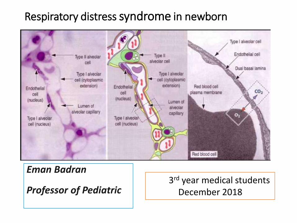
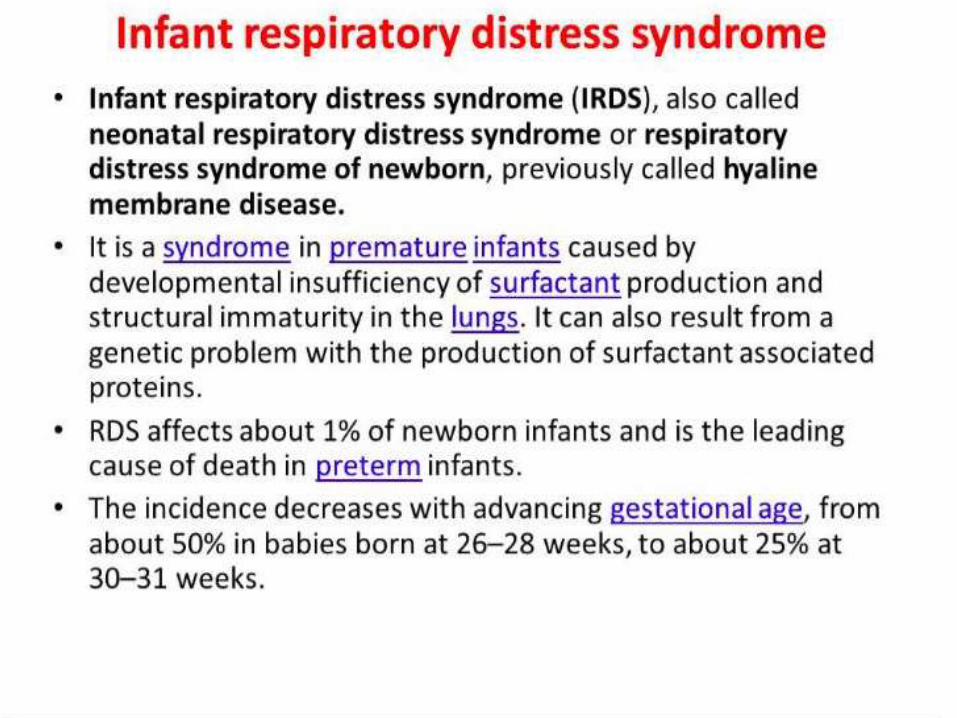
Respiratory distress syndrome in newborn
Eman Badran
Professor of Pediatric3rd year medical students
December 2018
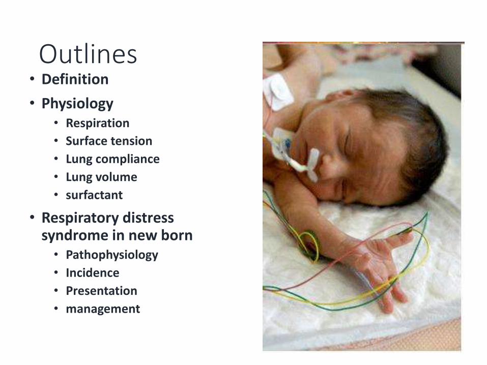
Outlines• Definition
• Physiology• Respiration
• Surface tension
• Lung compliance
• Lung volume
• surfactant
• Respiratory distress syndrome in new born • Pathophysiology
• Incidence
• Presentation
• management

Disclosure
Nothing
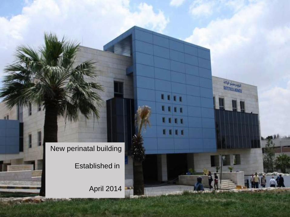
New perinatal building
Established in
April 2014
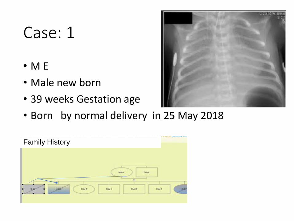
Case: 1
• M E
• Male new born
• 39 weeks Gestation age
• Born by normal delivery in 25 May 2018
Family History
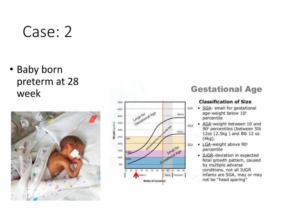
Case: 2
• Baby born preterm at 28 week

Some review Respiratory physiology
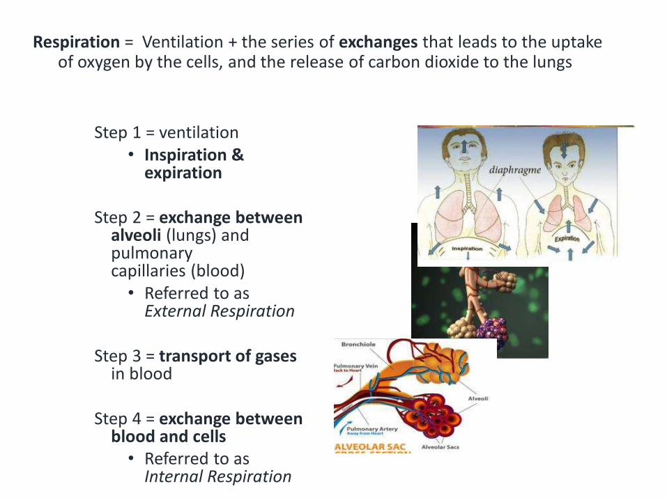
Respiration = Ventilation + the series of exchanges that leads to the uptake of oxygen by the cells, and the release of carbon dioxide to the lungs
Step 1 = ventilation• Inspiration &
expiration
Step 2 = exchange between alveoli (lungs) and pulmonary capillaries (blood)• Referred to as
External Respiration
Step 3 = transport of gases in blood
Step 4 = exchange between blood and cells• Referred to as
Internal Respiration
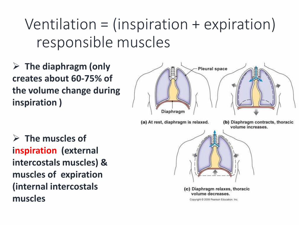
Ventilation = (inspiration + expiration)responsible muscles
The diaphragm (only creates about 60-75% of the volume change during inspiration )
The muscles of inspiration (external intercostals muscles) & muscles of expiration (internal intercostals muscles
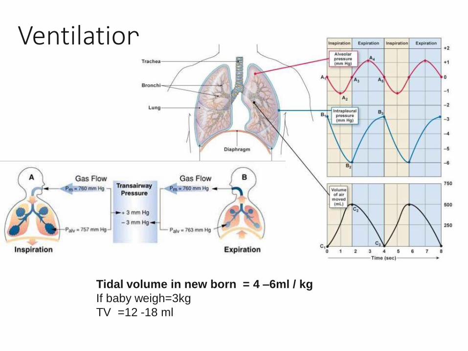
Ventilation
Tidal volume in new born = 4 –6ml / kg
If baby weigh=3kg
TV =12 -18 ml
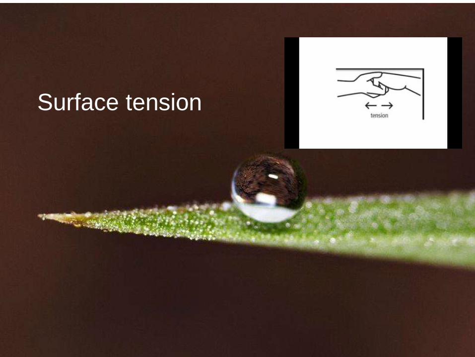
Surface tensionSurface tension
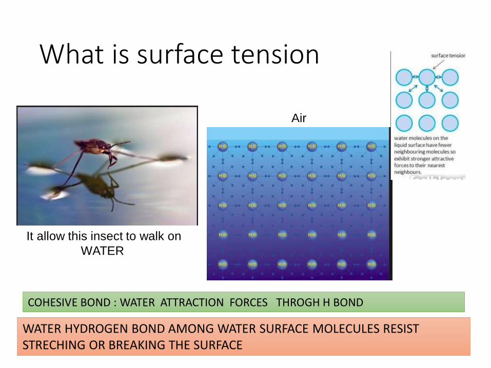
What is surface tension
It allow this insect to walk on
WATER
COHESIVE BOND : WATER ATTRACTION FORCES THROGH H BOND
WATER HYDROGEN BOND AMONG WATER SURFACE MOLECULES RESIST STRECHING OR BREAKING THE SURFACE
Air
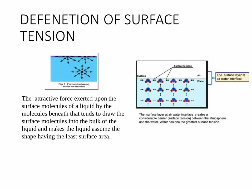
DEFENETION OF SURFACE TENSION
The attractive force exerted upon the
surface molecules of a liquid by the
molecules beneath that tends to draw the
surface molecules into the bulk of the
liquid and makes the liquid assume the
shape having the least surface area.
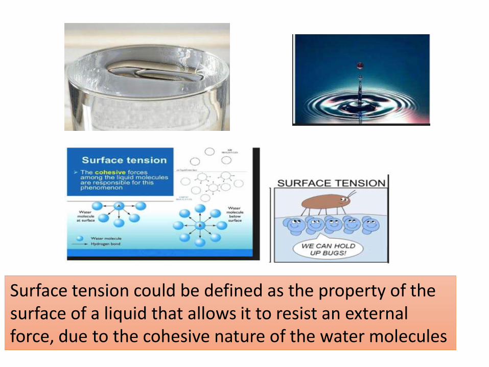
Surface tension could be defined as the property of the surface of a liquid that allows it to resist an external force, due to the cohesive nature of the water molecules
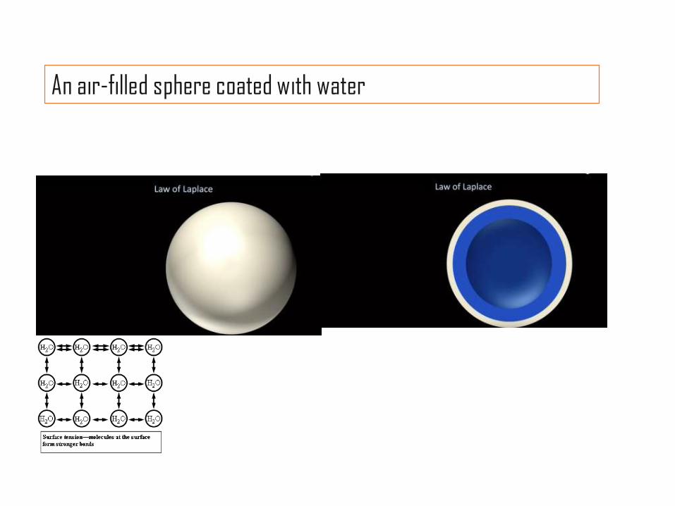
An air-filled sphere coated with water
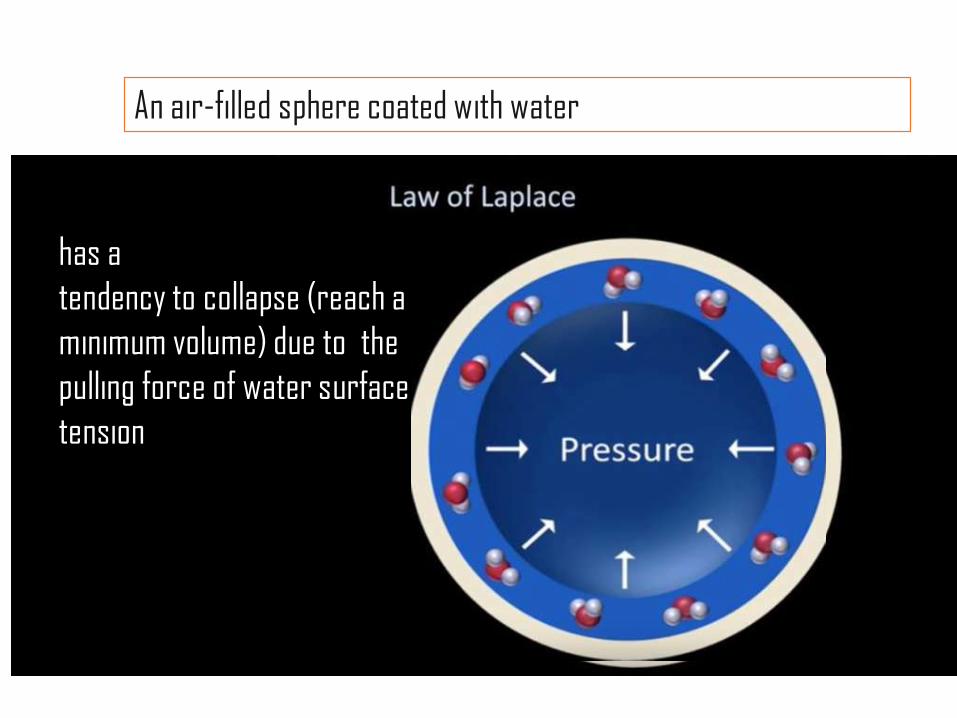
has a
tendency to collapse (reach a
minimum volume) due to the
pulling force of water surface
tension
An air-filled sphere coated with water
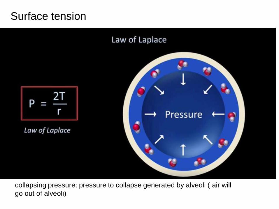
Surface tension
collapsing pressure: pressure to collapse generated by alveoli ( air will
go out of alveoli)
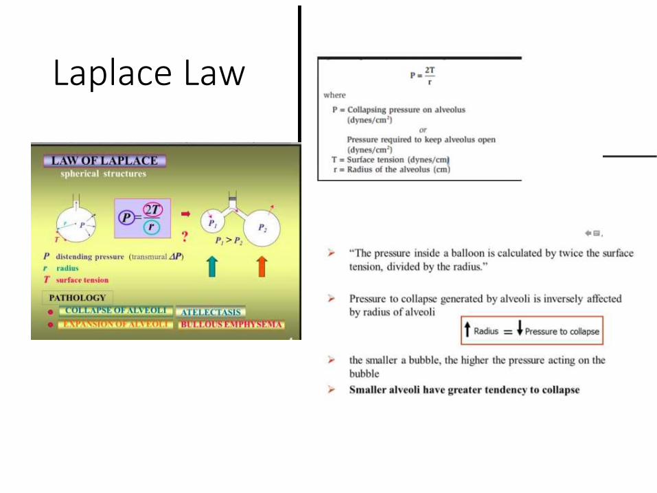
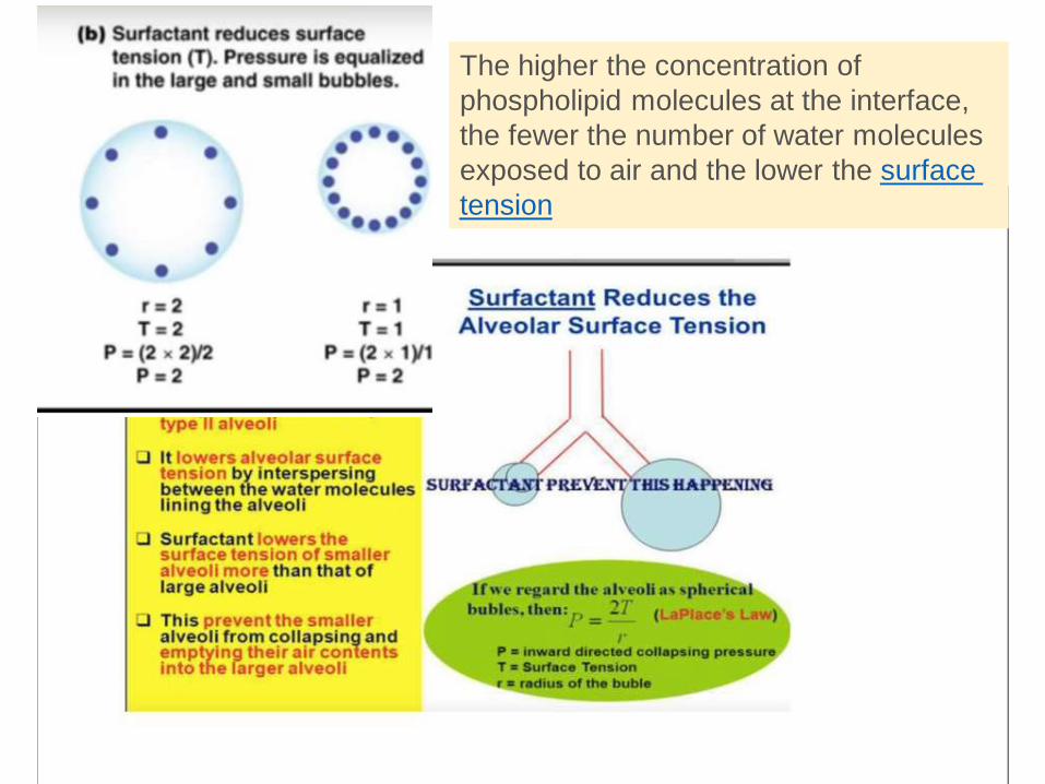
Laplace Law
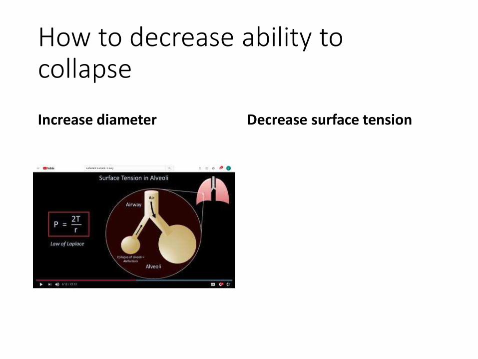
How to decrease ability to collapse
Increase diameter Decrease surface tension
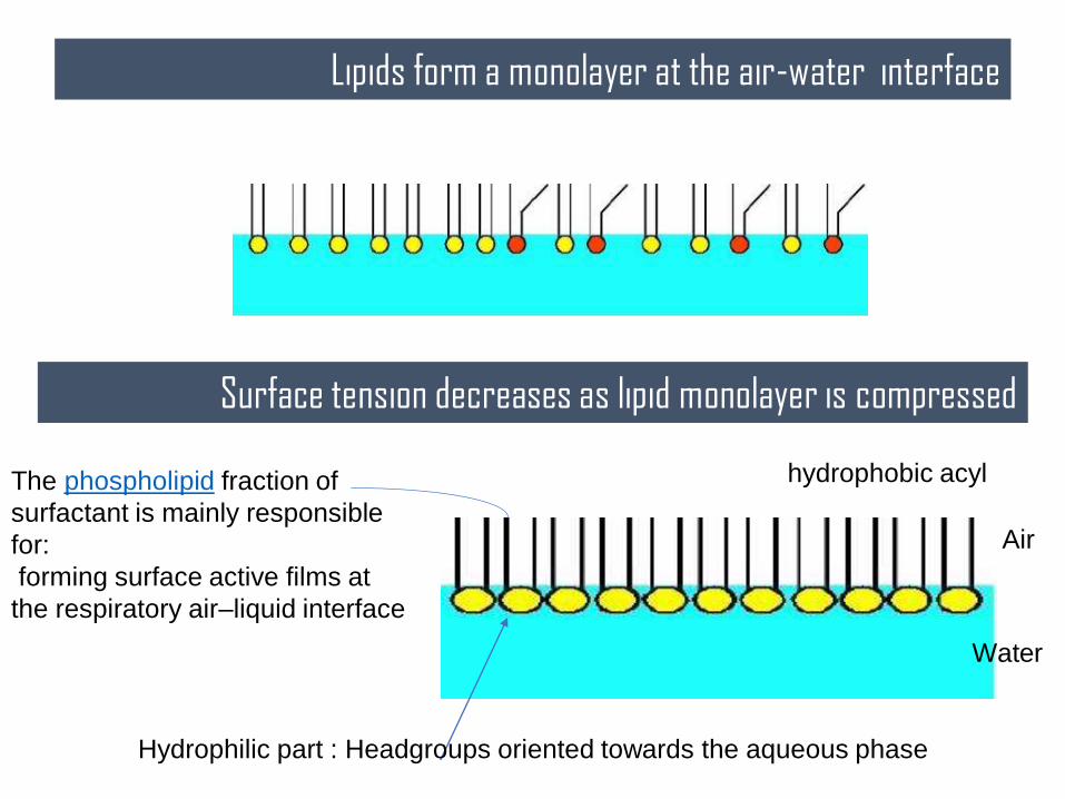
Lipids form a monolayer at the air-water interface
Surface tension decreases as lipid monolayer is compressed
The phospholipid fraction of
surfactant is mainly responsible
for:
forming surface active films at
the respiratory air–liquid interface
Air
Water
hydrophobic acyl
Hydrophilic part : Headgroups oriented towards the aqueous phase
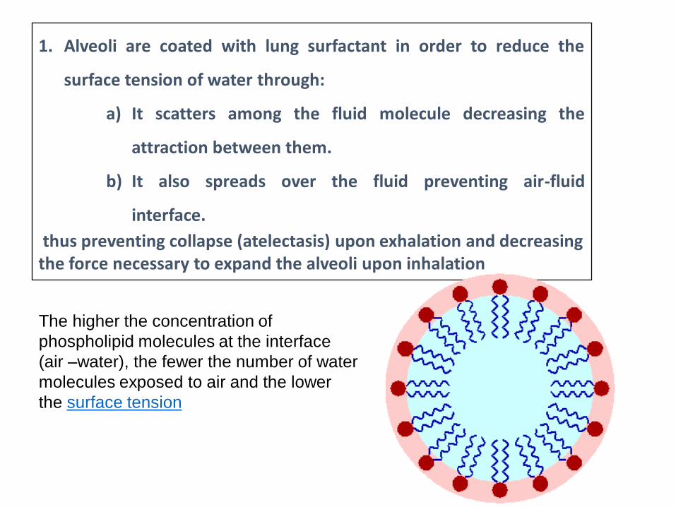
1. Alveoli are coated with lung surfactant in order to reduce the
surface tension of water through:
a) It scatters among the fluid molecule decreasing the
attraction between them.
b) It also spreads over the fluid preventing air-fluid
interface.
thus preventing collapse (atelectasis) upon exhalation and decreasing the force necessary to expand the alveoli upon inhalation
The higher the concentration of
phospholipid molecules at the interface
(air –water), the fewer the number of water
molecules exposed to air and the lower
the surface tension
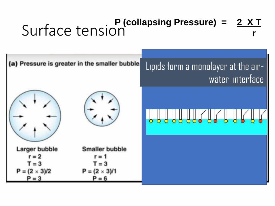
Surface tensionP (collapsing Pressure) = 2 X T
r
Lipids form a monolayer at the air-
water interface
The higher the concentration of
phospholipid molecules at the interface,
the fewer the number of water molecules
exposed to air and the lower the surface
tension
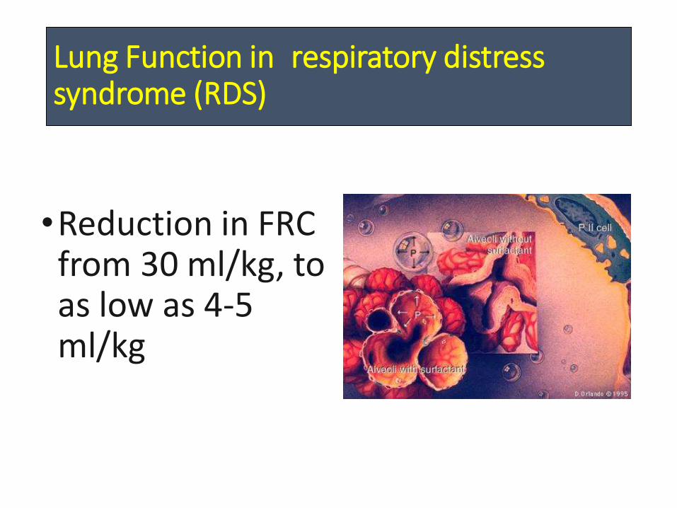
Lung Function in respiratory distress syndrome (RDS)
•Reduction in FRC from 30 ml/kg, to as low as 4-5 ml/kg
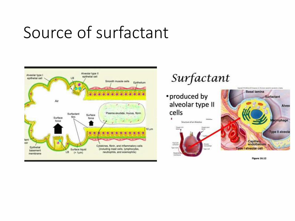
Source of surfactant

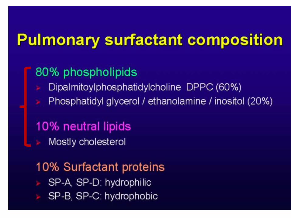
Endogenous Surfactantcomposition and functions
• Proteins (~10%)
• SP-ASHost defenseSurfactant homeostasis
P-D: ? Phagocytic function
SP-B is the most important protein in surfactant required for the biogenesis of pulmonary surfactant and its packing
into lamellar bodies For , Spreading, surface tension
SP-C , Adsorption

Surfactant pulmonary mutations disorders
-Mutations in one of the genes encoding SP-A (SFTPA2) have been reported as a cause of pulmonary fibrosis and lung cancer in adults.
-- Mutation in SP-B gene and neonatal RDS.
-- Mutations in the gene encoding ABCA3 (
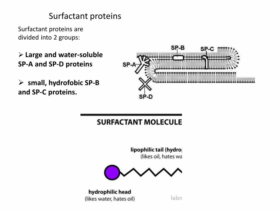
Surfactant proteins are divided into 2 groups:
Large and water-soluble SP-A and SP-D proteins
small, hydrofobic SP-B and SP-C proteins.
Surfactant proteins
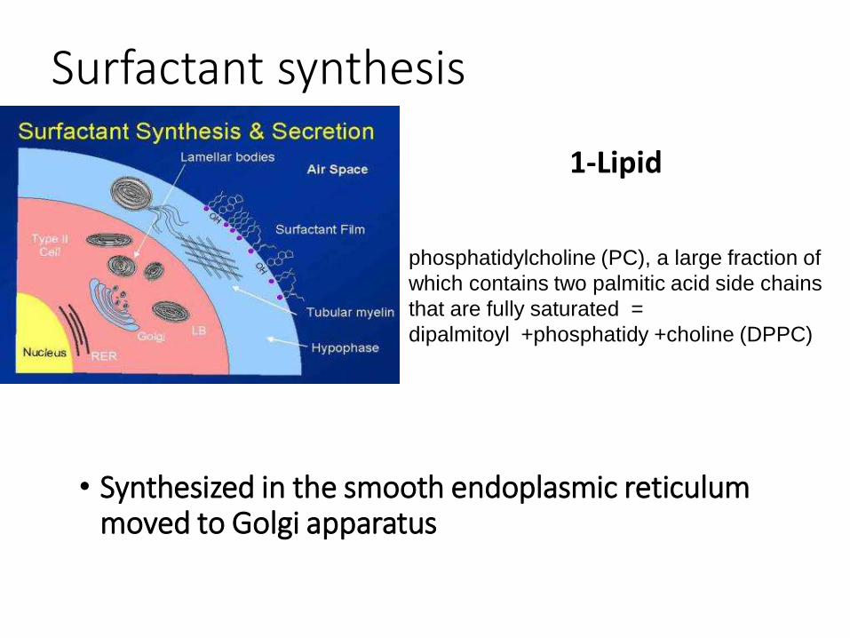
Surfactant synthesis
1-Lipid
• Synthesized in the smooth endoplasmic reticulum moved to Golgi apparatus
phosphatidylcholine (PC), a large fraction of
which contains two palmitic acid side chains
that are fully saturated =
dipalmitoyl +phosphatidy +choline (DPPC)
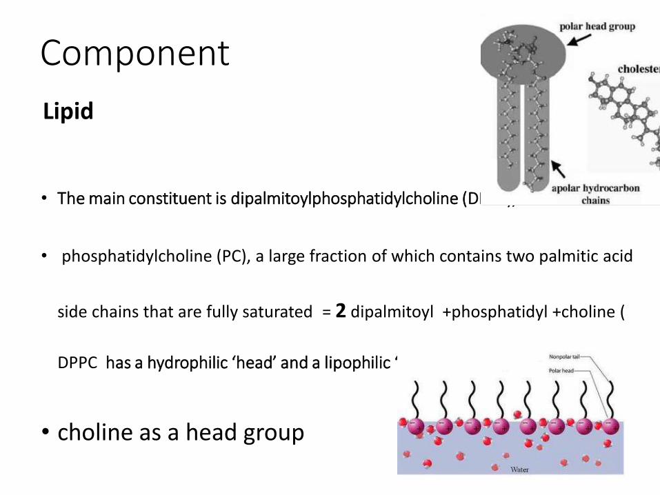
Component
Lipid
• The main constituent is dipalmitoylphosphatidylcholine (DPPC),
• phosphatidylcholine (PC), a large fraction of which contains two palmitic acid
side chains that are fully saturated = 2 dipalmitoyl +phosphatidyl +choline (
DPPC has a hydrophilic ‘head’ and a lipophilic ‘tail’
• choline as a head group
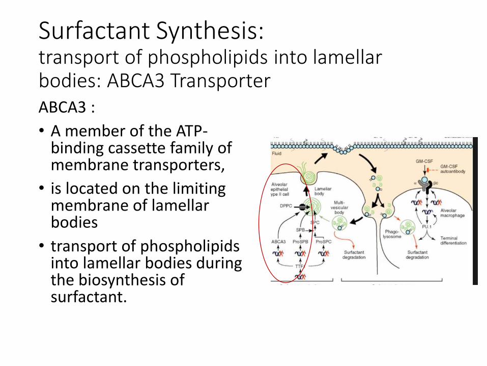
Surfactant Synthesis:transport of phospholipids into lamellar bodies: ABCA3 Transporter ABCA3 :
• A member of the ATP-binding cassette family of membrane transporters,
• is located on the limiting membrane of lamellar bodies
• transport of phospholipids into lamellar bodies during the biosynthesis of surfactant.
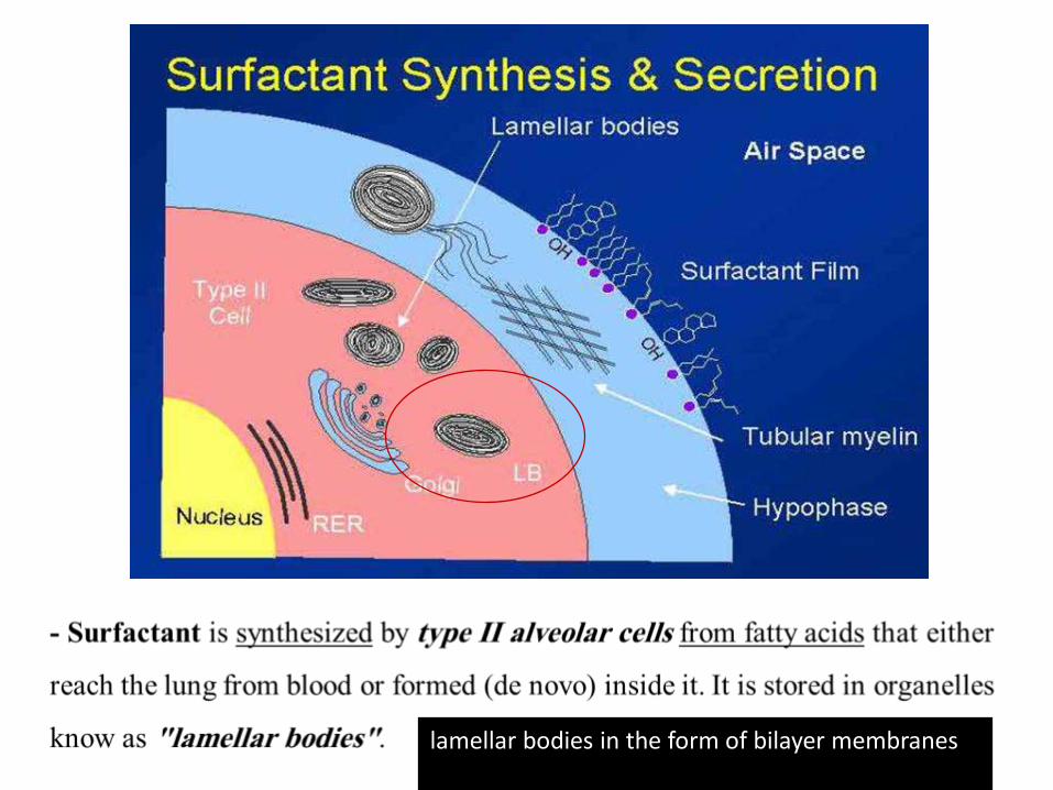
lamellar bodies in the form of bilayer membranes
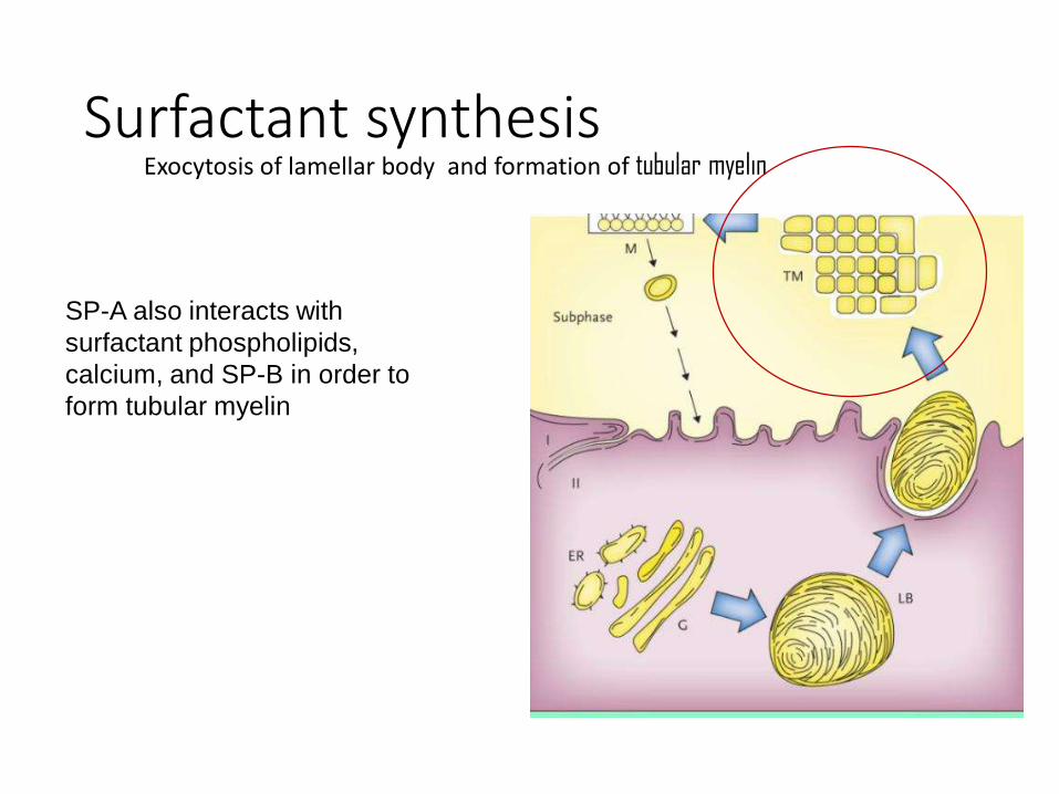
Surfactant synthesisExocytosis of lamellar body and formation of tubular myelin
SP-A also interacts with
surfactant phospholipids,
calcium, and SP-B in order to
form tubular myelin

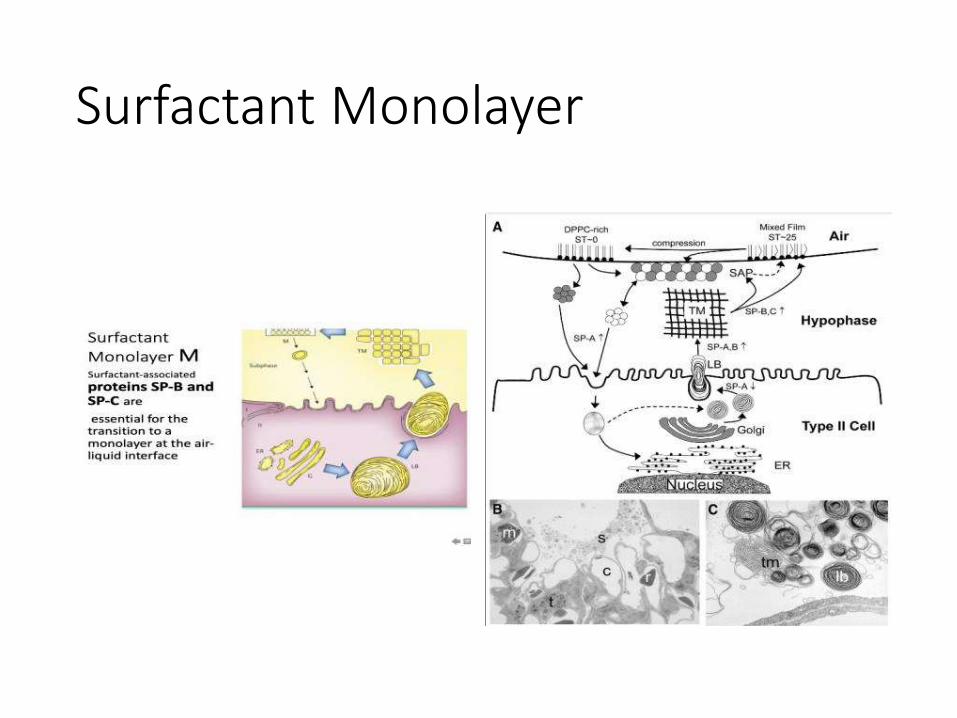
Surfactant
Monolayer MSurfactant-associated
proteins SP-B and SP-C are
essential for the transition to a monolayer at the air-liquid interface
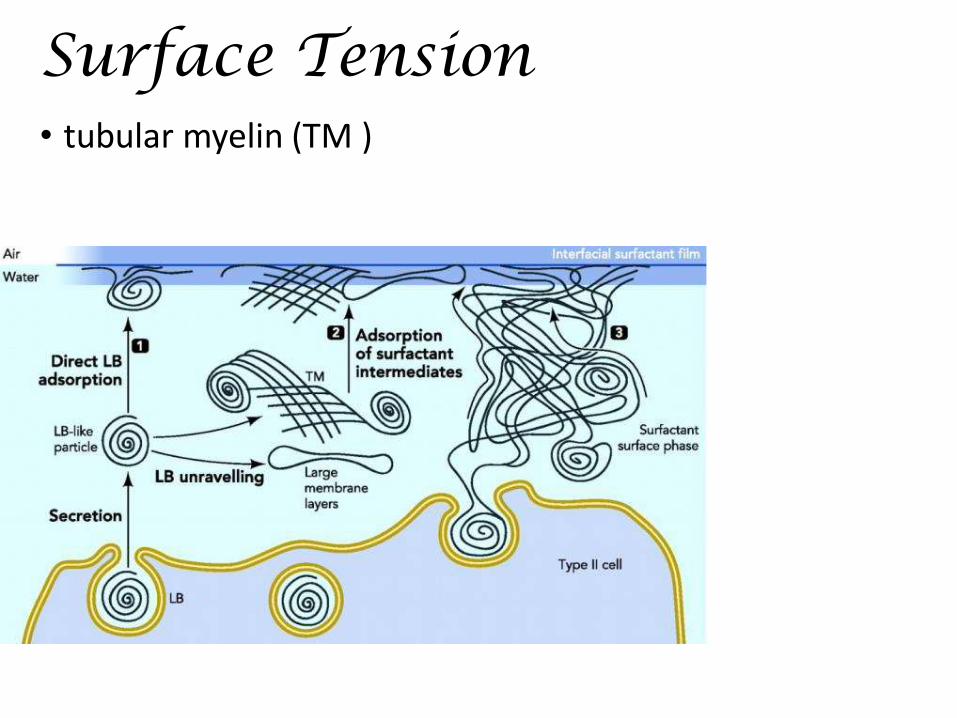
Surface Tension• tubular myelin (TM )
Surfactant Monolayer
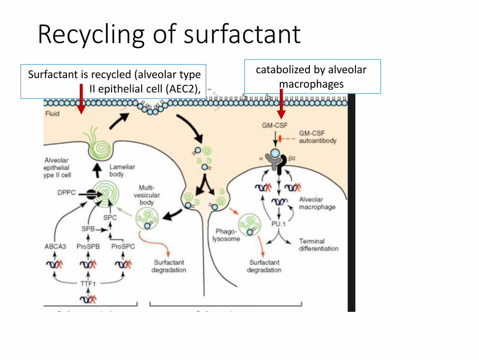
Recycling of surfactantSurfactant is recycled (alveolar type
II epithelial cell (AEC2),
catabolized by alveolar macrophages
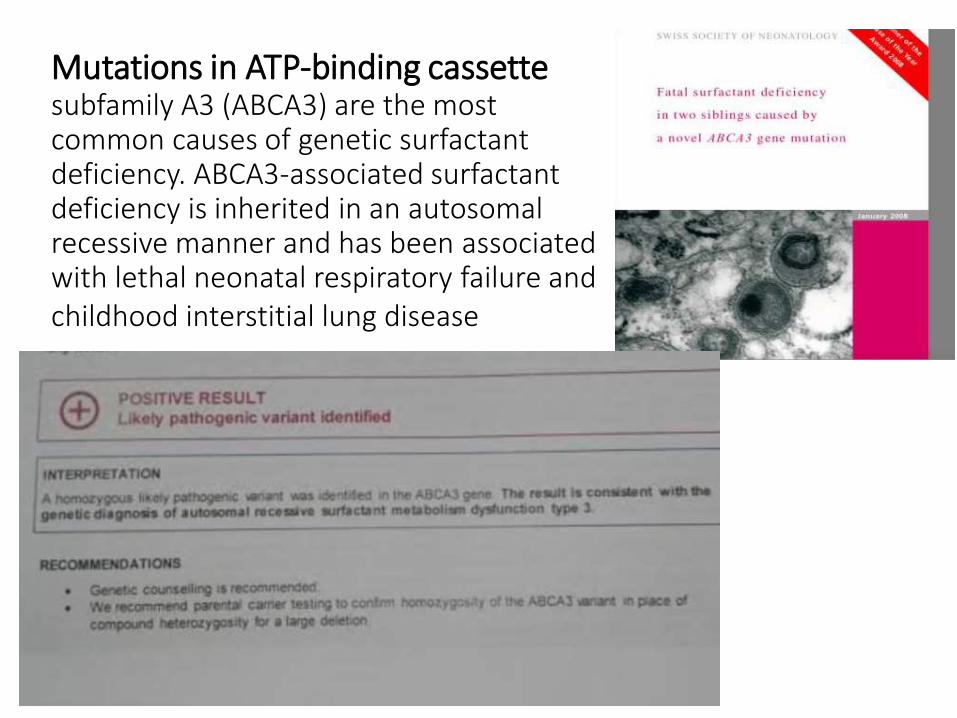
Mutations in ATP-binding cassette subfamily A3 (ABCA3) are the most common causes of genetic surfactant deficiency. ABCA3-associated surfactant deficiency is inherited in an autosomal recessive manner and has been associated with lethal neonatal respiratory failure and childhood interstitial lung disease

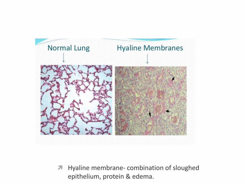
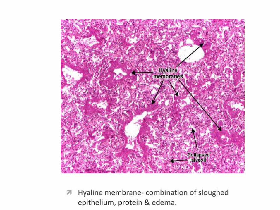
Pathology reportDysfunction of lamellar Body
• Functions of surfactant:
Function of Surfactant
• function to protect the lungs from injuries and infections caused by inhaled particles and microorganisms
SP _A
SP-D
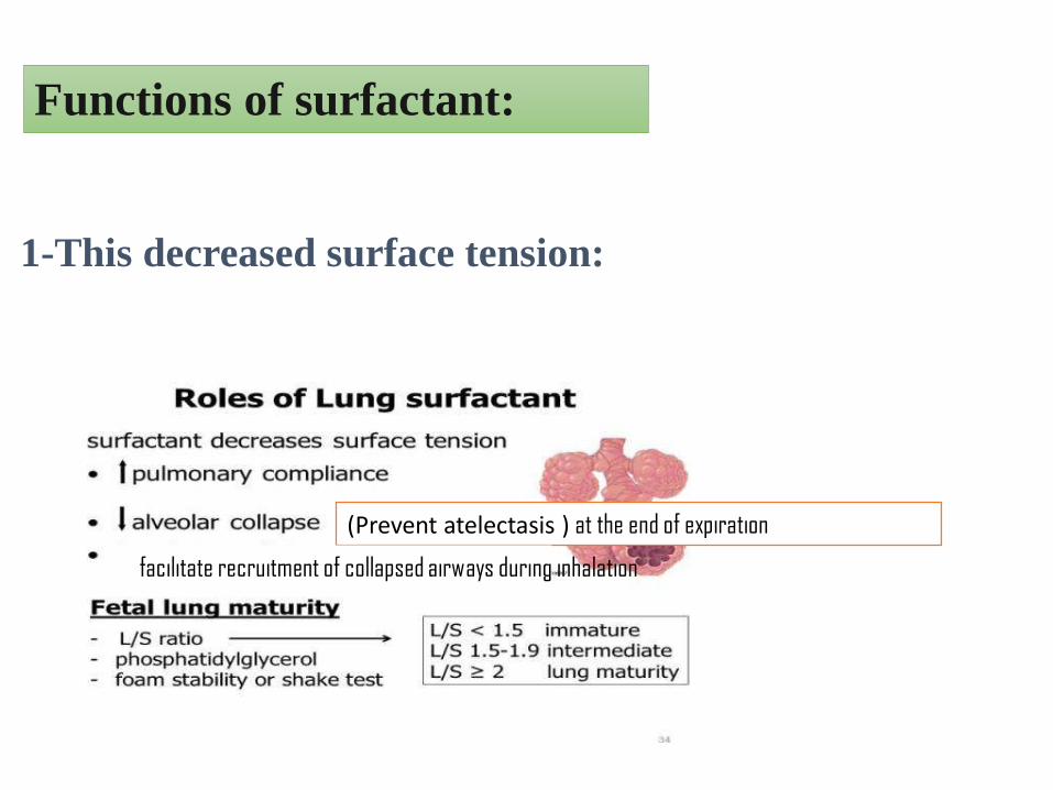
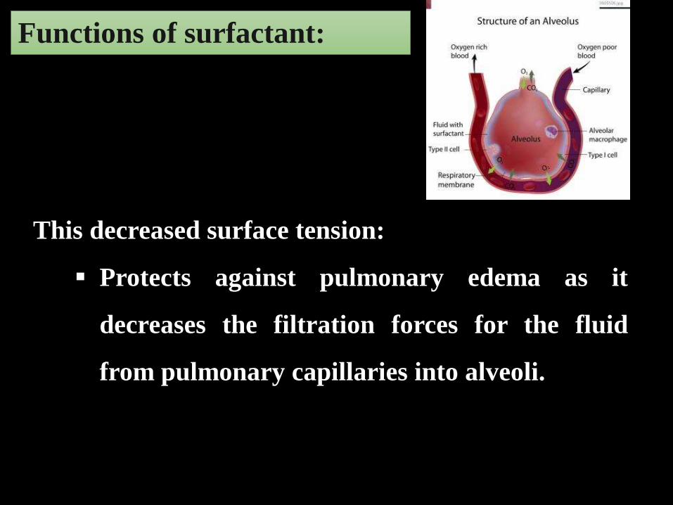
Functions of surfactant:
1-This decreased surface tension:
(Prevent atelectasis ) at the end of expiration
facilitate recruitment of collapsed airways during inhalation
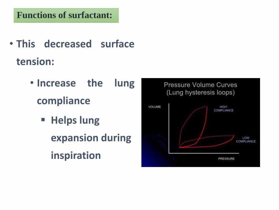
Functions of surfactant:
• This decreased surface
tension:
• Increase the lung
compliance
Helps lung
expansion during
inspiration
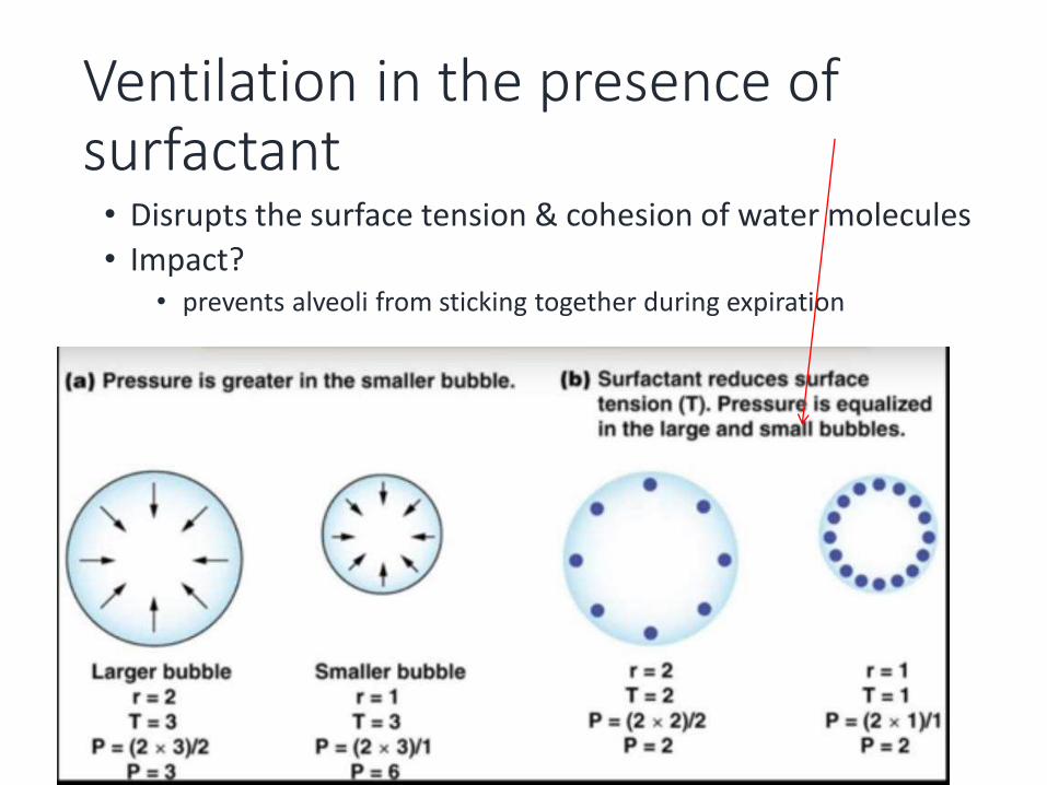
Ventilation in the presence of surfactant• Disrupts the surface tension & cohesion of water molecules
• Impact?• prevents alveoli from sticking together during expiration
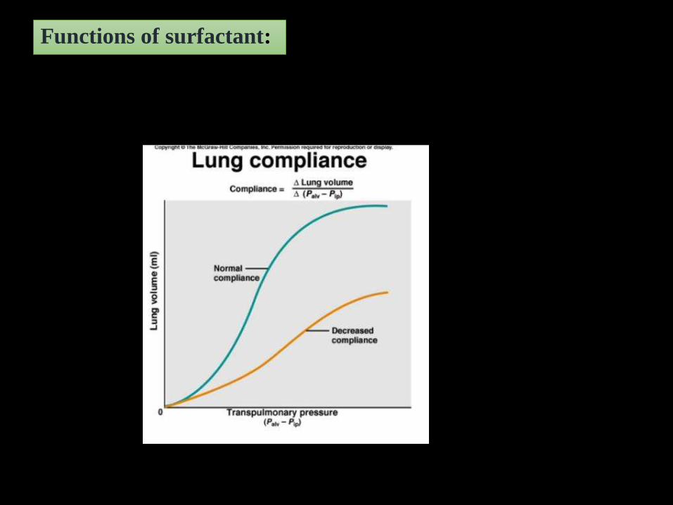
Functions of surfactant:
Functions of surfactant:
This decreased surface tension:
Protects against pulmonary edema as it
decreases the filtration forces for the fluid
from pulmonary capillaries into alveoli.
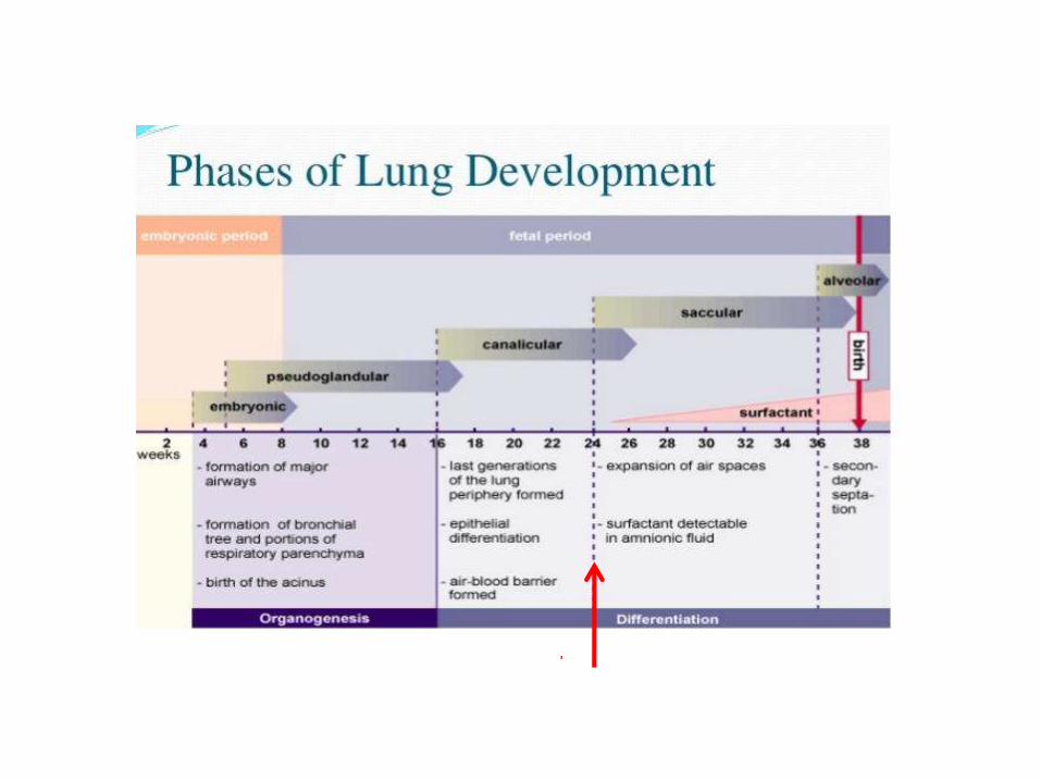
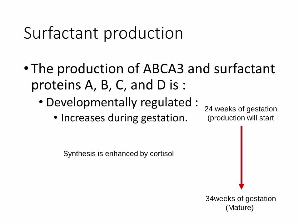
Surfactant production
• The production of ABCA3 and surfactant proteins A, B, C, and D is :• Developmentally regulated :
• Increases during gestation.24 weeks of gestation
(production will start
34weeks of gestation
(Mature)
Synthesis is enhanced by cortisol
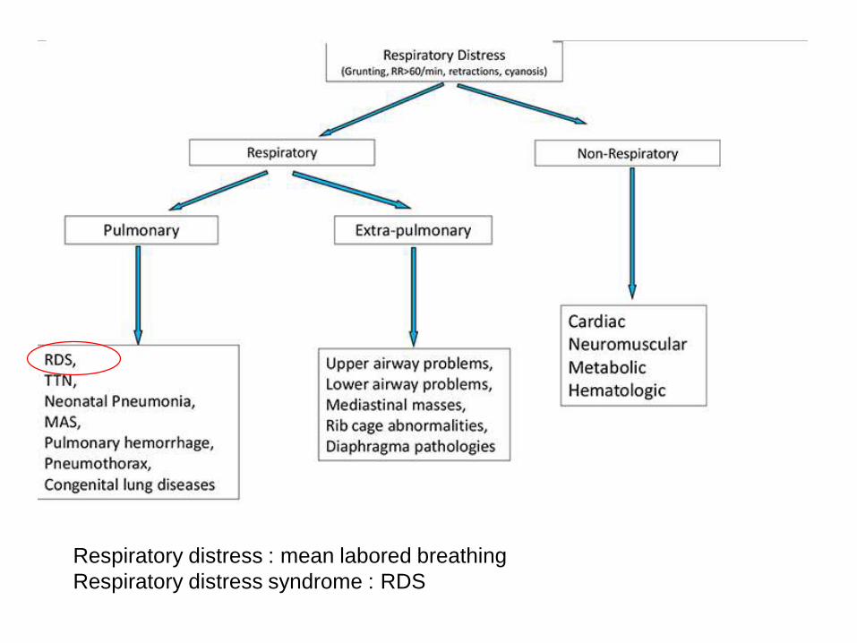
Respiratory distress : mean labored breathing
Respiratory distress syndrome : RDS
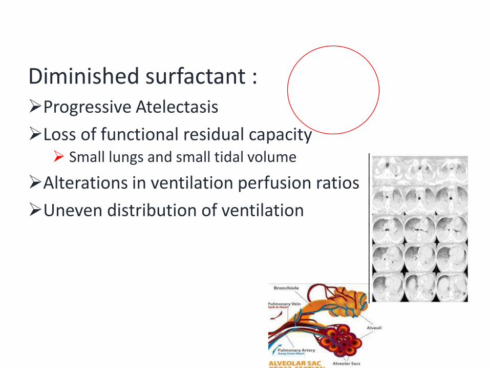
Diminished surfactant :Progressive Atelectasis
Loss of functional residual capacity Small lungs and small tidal volume
Alterations in ventilation perfusion ratios
Uneven distribution of ventilation
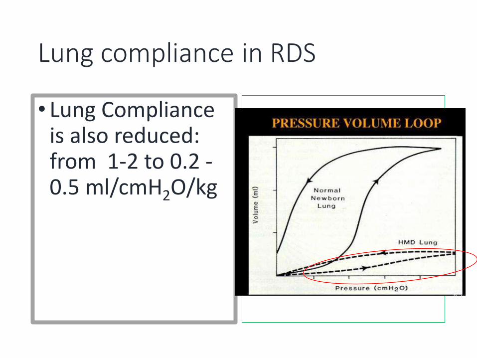
Lung compliance in RDS
• Lung Compliance is also reduced: from 1-2 to 0.2 -0.5 ml/cmH2O/kg

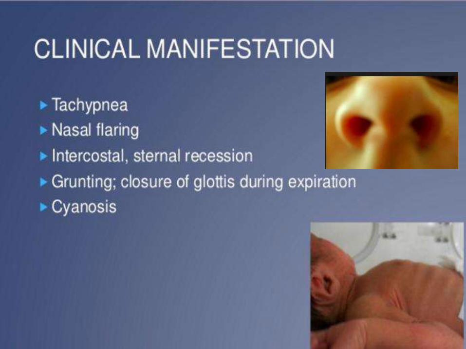
RDS: clinical picture
• At admission of the baby he has
• Cyanosis• Pulse Oximeter 75% ( normal
> 95%)
Blood gas:
• PaO2 = 45% mmHg (normal 80-108)
• Ph= 7.2 (normal 7.35-7.45)
• CO2 = 65 mmHg (normal 35-45)
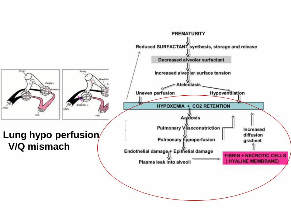
Lung hypo perfusion
V/Q mismach
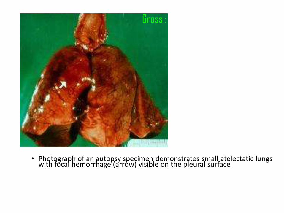
• Photograph of an autopsy specimen demonstrates small atelectatic lungs with focal hemorrhage (arrow) visible on the pleural surface.
Gross : Lung firm, red, liverlike
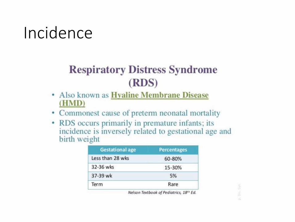
Incidence
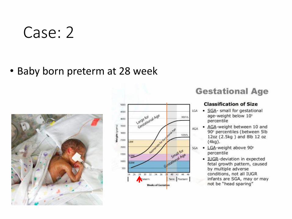
Case: 2
• Baby born preterm at 28 week
Prevention
• Prevent preterm
• Identify patients at risk
• Antenatal steroid
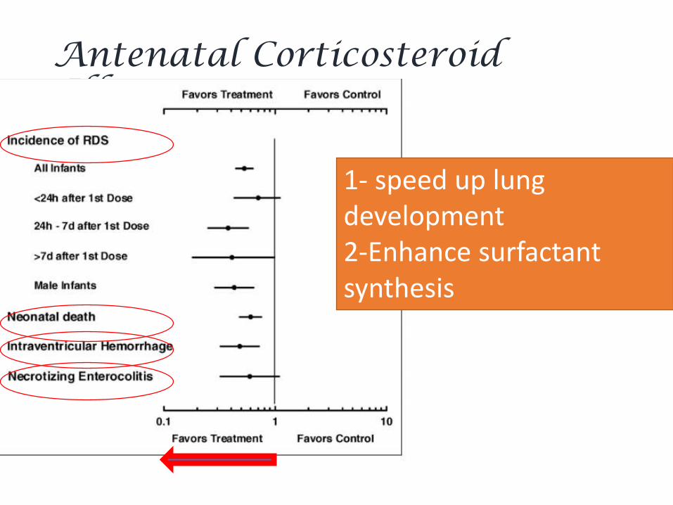
Antenatal Corticosteroid Effects
1- speed up lung development2-Enhance surfactant synthesis
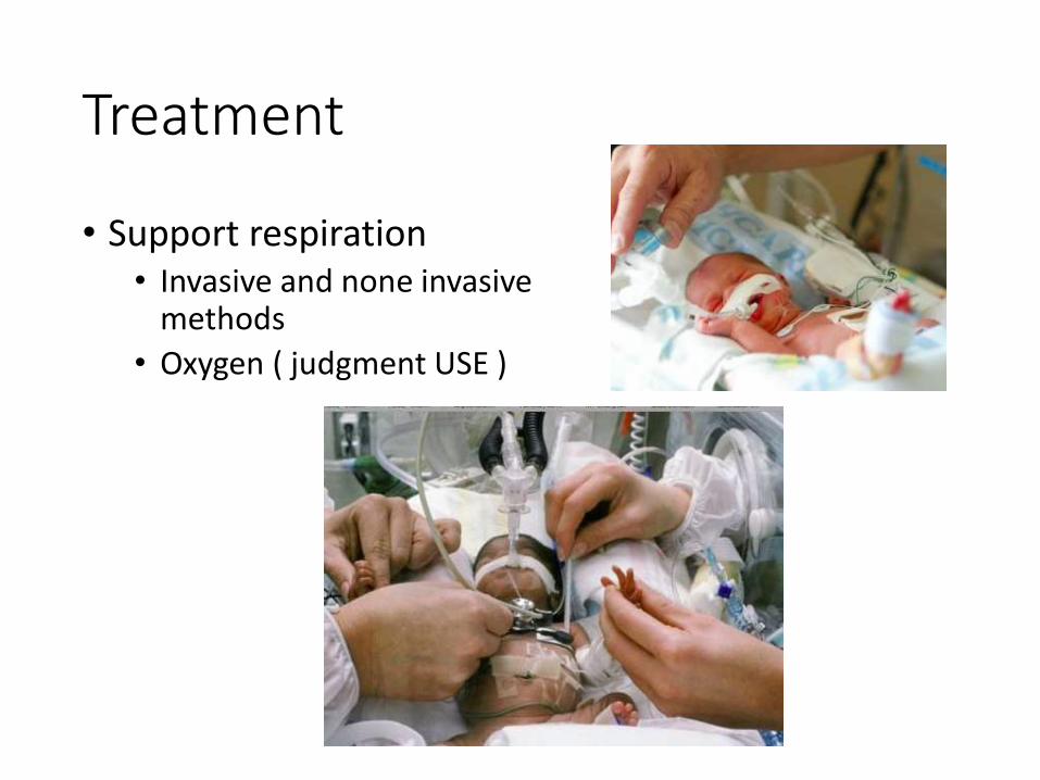
Treatment
• Support respiration• Invasive and none invasive
methods
• Oxygen ( judgment USE )
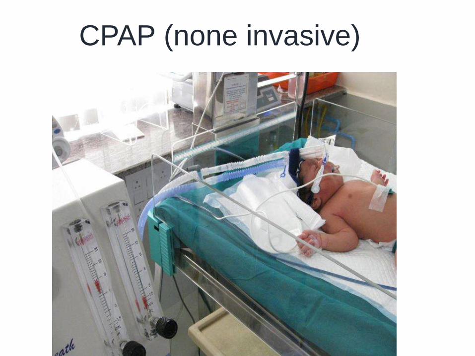
CPAP (none invasive)
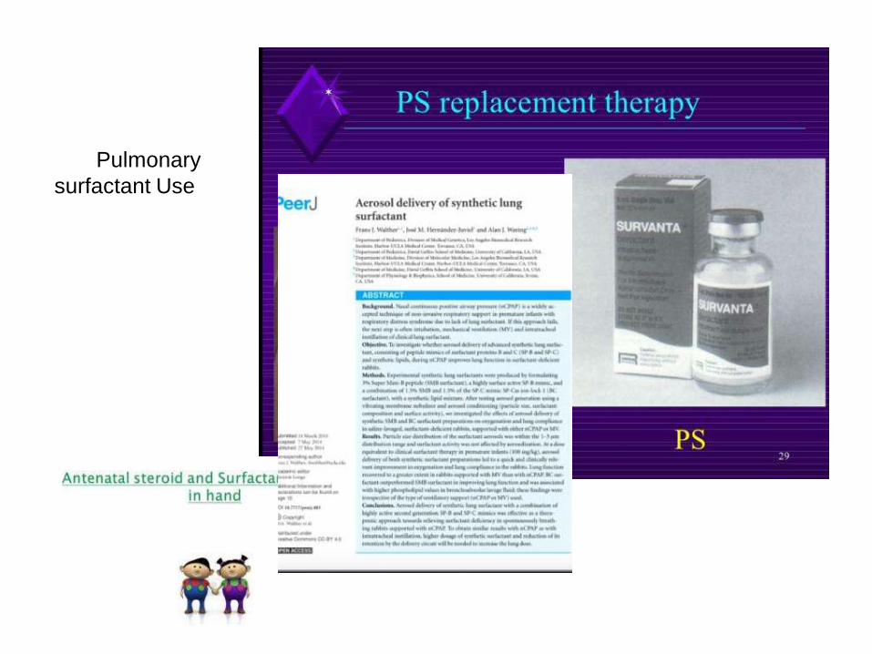
Pulmonary
surfactant Use
Thank YOU