respiratory failure zhihua gao zhejiang university
TRANSCRIPT

Respiratory Failure
Zhihua Gao
Zhejiang University
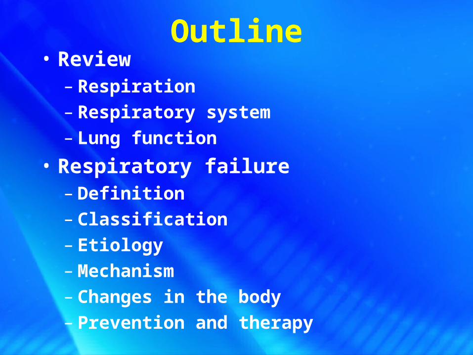
Outline• Review
– Respiration – Respiratory system– Lung function
• Respiratory failure– Definition– Classification– Etiology– Mechanism– Changes in the body– Prevention and therapy
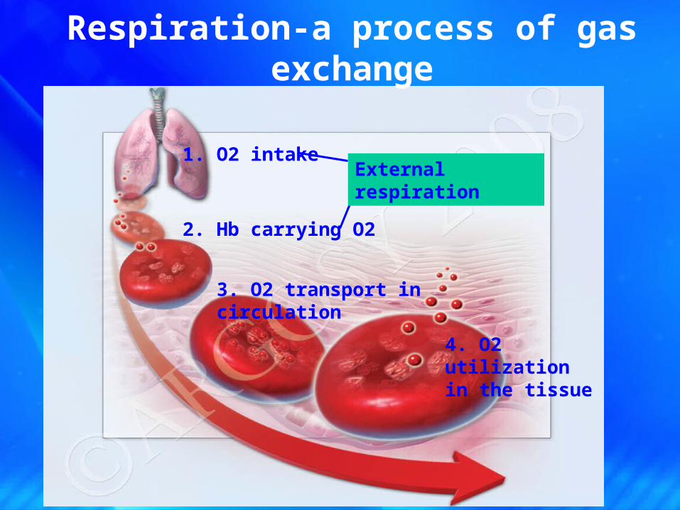
1. O2 intake
2. Hb carrying O2
3. O2 transport in circulation
4. O2 utilization in the tissue
Respiration-a process of gas exchange
External respiration

Respiratory System• Consists of three parts:
• Pumpimg part • respiratory muscles • respiratory control system
• Conductive part • the complete trachea system
• Gas exchange part• Alveoli in the lung
GAS EXCHANGE
VENTILATION

Lung function• Ventilation
– O2 inspiration &CO2 expiration
• Gas exchange– Oxygenation of the blood
respiratory function

6
– Non-respiratory function• Defense• Filtration• Metabolism Endothelial cell Pulmonary surfactant (PS, 肺泡表面活性物
质 ) Metabolism of arachidonic acid ---prostaglandin and leukotrienes APUD cell (amine precursor uptake and decarboxylation
cell) ---VIP 血管活性肠肽 ,P,CCK 胆囊收缩素 ,
somatostatin 生长抑素
Lung function
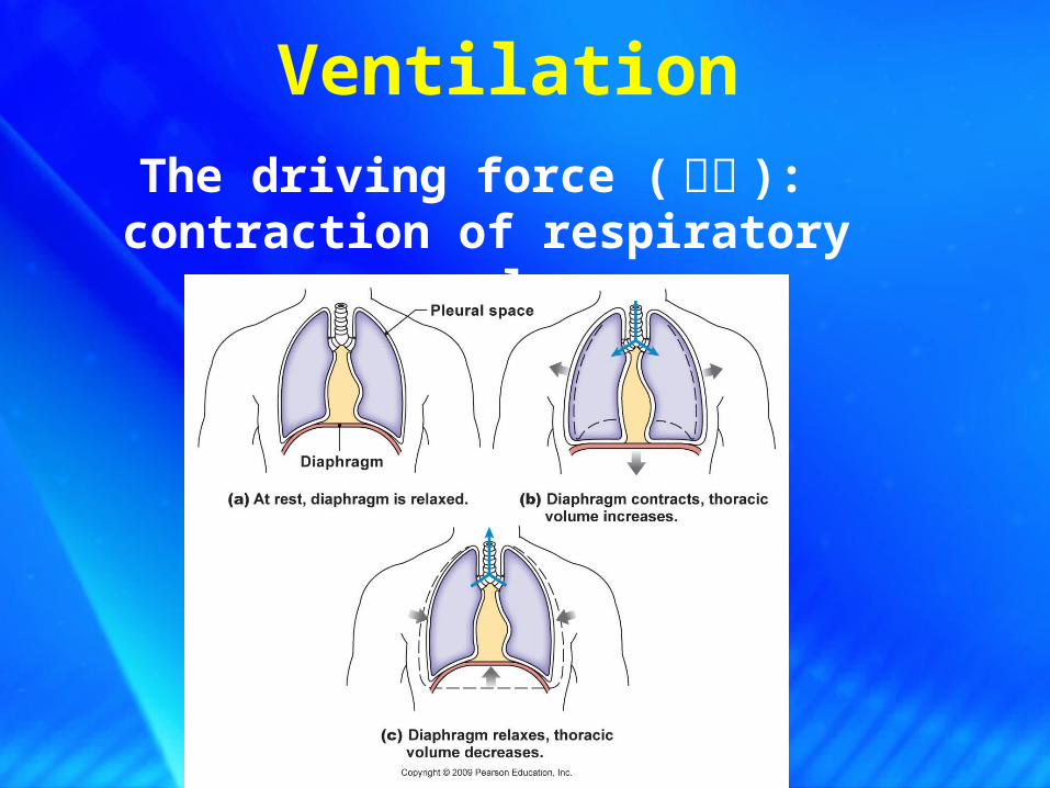
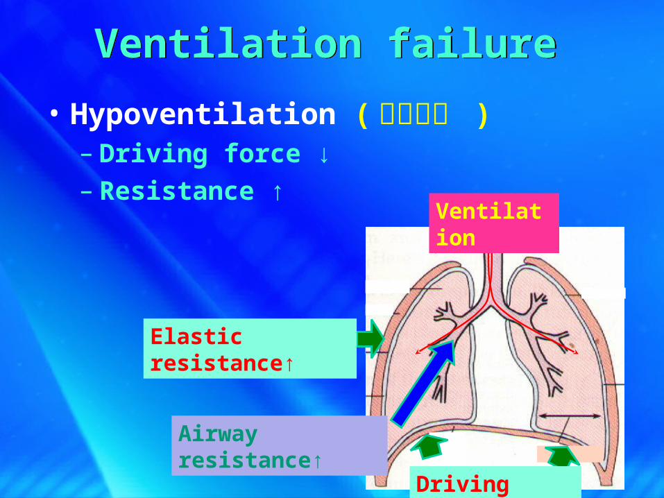
VentilationThe driving force ( 动力 ):
contraction of respiratory muscles

The resistance :
elastic and airway resistance
Ventilation
Airway resistance
Ventilation顺应性 =1/ 弹性阻力 Pulmonary surfactant (PS, 肺泡表面活性物质 )
Elastic resistance

9
Airway resistance
Influence factors:
airway diameter, length and shape, rate of air flow
气道内径,长度,形态,气流速度,形式等大气道阻力:直径 >2mm ,有软骨环支撑,不易塌陷, 80%
小气道阻力:直径 <2mm ,无软骨环支撑,易扭曲闭合, 20%
R=8Lηπr4
气道阻力与气道长度成正比,与气道半径的四次方成反比
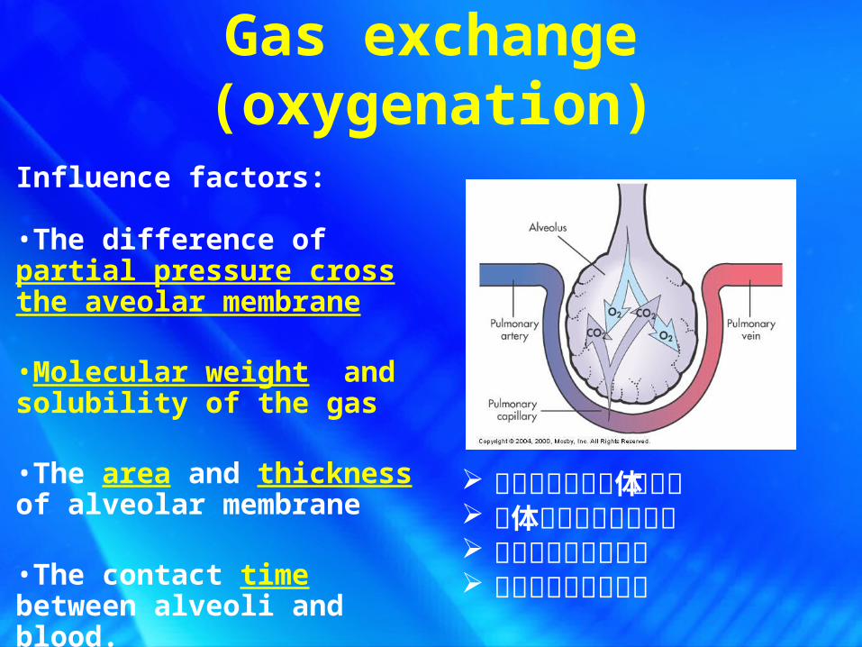
Gas exchange (oxygenation)
Influence factors:
•The difference of partial pressure cross the aveolar membrane
•Molecular weight and solubility of the gas
•The area and thickness of alveolar membrane
•The contact time between alveoli and blood.
肺泡膜两侧的气体分压差 气体的分子量与溶解度 肺泡膜的面积与厚度 血液与肺泡接触时间
11
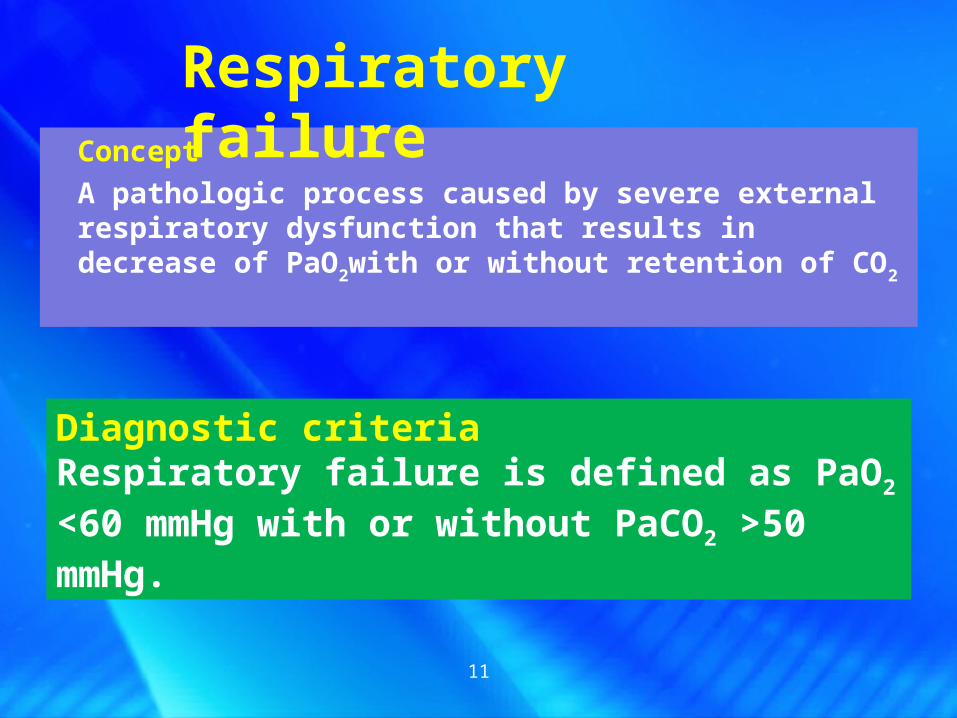
Concept
A pathologic process caused by severe external respiratory dysfunction that results in decrease of PaO2with or without retention of CO2
Respiratory failure
Diagnostic criteriaRespiratory failure is defined as PaO2 <60 mmHg with or without PaCO2 >50 mmHg.
12
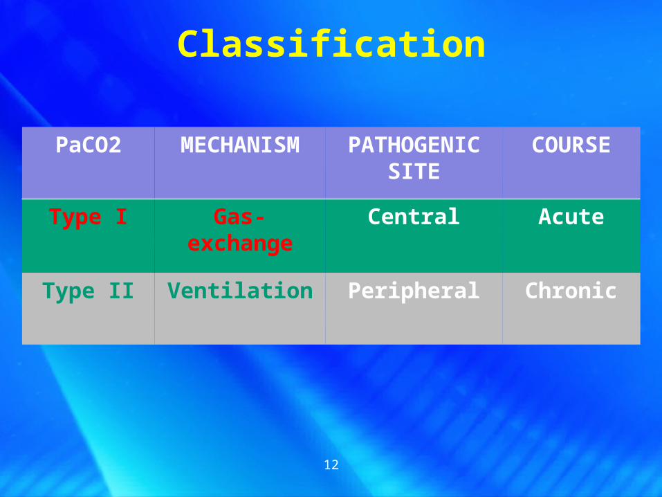
Classification
PaCO2 MECHANISM PATHOGENIC SITE
COURSE
Type I Gas-exchange Central Acute
Type II Ventilation Peripheral Chronic
Classification
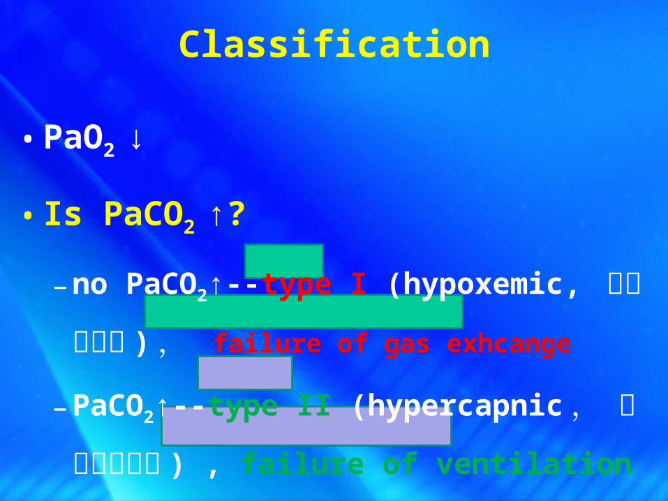
• PaO2 ↓
• Is PaCO2 ↑?
– no PaCO2↑--type I (hypoxemic, 低氧血症
型 ) , failure of gas exhcange
– PaCO2↑--type II (hypercapnic , 高碳酸血
症型 ) , failure of ventilation

Respiratory System• Consists of three parts:
• Pump • respiratory muscles • respiratory control system
• Conductive part • the complete trachea system
• Gas exchange part• Alveoli in the lung
GAS EXCHANGE
VENTILATION
Respiratory failure
External respiration dysfunction

• Ventilation failure ( 通气障碍 )– Restrictive hypoventilation ( 限制性通气不足 )– Obstructive hypoventilation ( 阻塞通气不足 )
• Gas-exchange failure ( 换气障碍 )– Diffusion disorder ( 弥散障碍 )
– VA/Q mismatch ( 通气 / 血流比值失调 )
– Increased anatomical shunt ( 解剖分流增加 )
Etiology and PathophysiologyEtiology and Pathophysiology
Dysfunction of external respiration
Airway resistance↑
Ventilation
Elastic resistance↑
Ventilation failure Ventilation failure
• Hypoventilation ( 通气不足 )– Driving force ↓– Resistance ↑
Driving force ↓

Ventilation failure Ventilation failure
• Hypoventilation ( 通气不足 )– Driving force ↓– Resistance ↑
• Restrictive hypoventilation ( 限制性通气不足 )– Driving force ↓or elastic resistance ↑– Limited alveolar distension ( 肺泡扩张受限 )
• Obstructive hypoventilation ( 阻塞通气不足 )– Airway resistance ↑ due to obstruction

18
Restrictive hypoventilation ( 限制性通气不足 )
Caused by diseases that affect either the
distensibility ( 扩展性 ) of the lungs or chest wall.
Inspiration-an active process—mostly affected in
restrictive hypoventilation
Expiration-an passive process

19
Paralysis of respiratory muscle Disorders of central or peripheral nerve
Inhibition of respiratory center
Intrinsic diseases of respiratory muscle
Decreased compliance of chest wall Severe chest deformity
Multiple rib fracture
Pleura ( 胸膜 ) fibrosis;
Decreased compliance of lung Disorders of lung (diffuse fibrosis, edema);
Lack of alveolar surfactant (as seen in ARDS)
Pneumothorax 气胸 or hydrothorax 胸腔积水
Restrictive hypoventilation ( 限制性通气不足 )

Obstructive hypoventilation ( 阻塞性通气不足 )
Airway obstruction/narrowing→Airway resistance ↑ →obstructive hypoventilation
Central airway obstruction
Peripheral airway obstruction
• 管壁收缩或增厚 :– 支气管哮喘、慢支→支气管痉挛– 炎症→支气管粘膜下充血、水肿、纤维增生
• 管腔阻塞:– 支气管哮喘、慢支→粘液↑– 纤毛损伤、肿瘤、异物
• 管壁受压:肿瘤、肿大淋巴结• 肺组织对小气道管壁的牵拉作用减弱

outside the thorax →inspiratory dyspnea 吸气困难阻塞位于胸外:声带麻痹、喉炎、喉头水肿,喉癌,白喉
• in the airway inside →expiratory dyspnea 呼气困难• 阻塞位于胸内:气管,大支气管的狭窄和阻塞如气管肿瘤,气管异物,气
管外肿物压迫(甲状腺,纵隔肿瘤)
Obstructive hypoventilation ( 阻塞性通气不足 )
Central airway obstruction-obstruction above the rachea crotch ( 气管分叉以上的阻塞 )

Extrathoracic obstruction Intrathoracic
obstruction
Expiratory dyspnea
Inspiratory dyspnea
Central airway obstruction
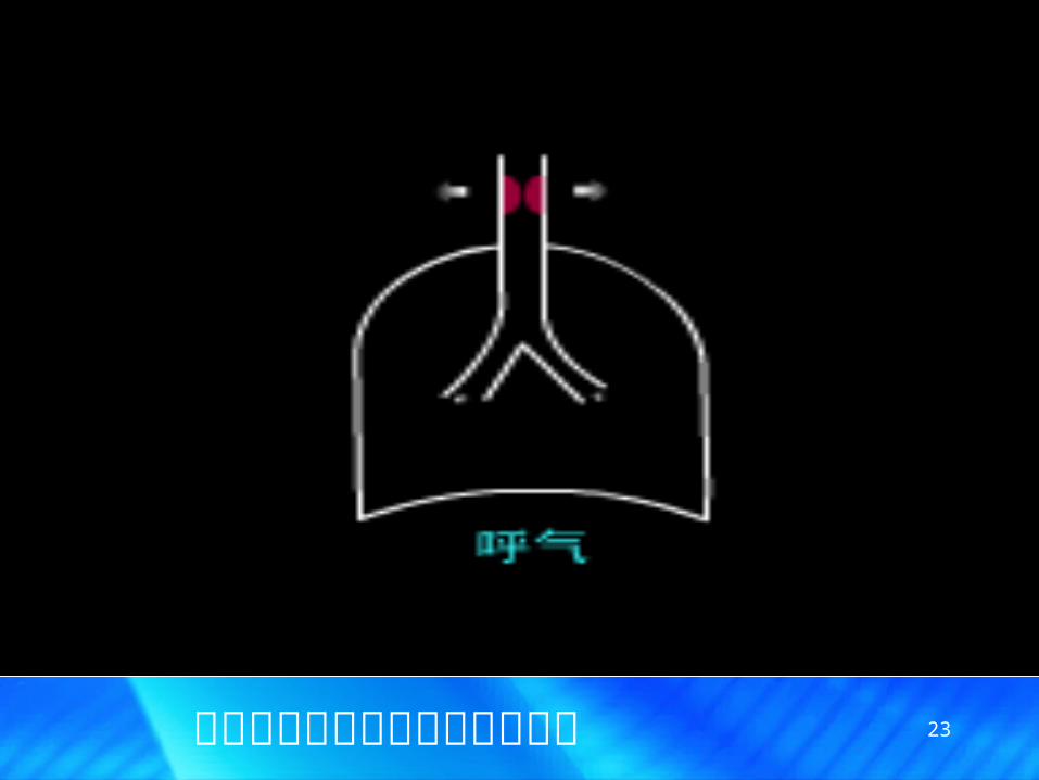
23中央气道胸外段阻塞无呼气困难
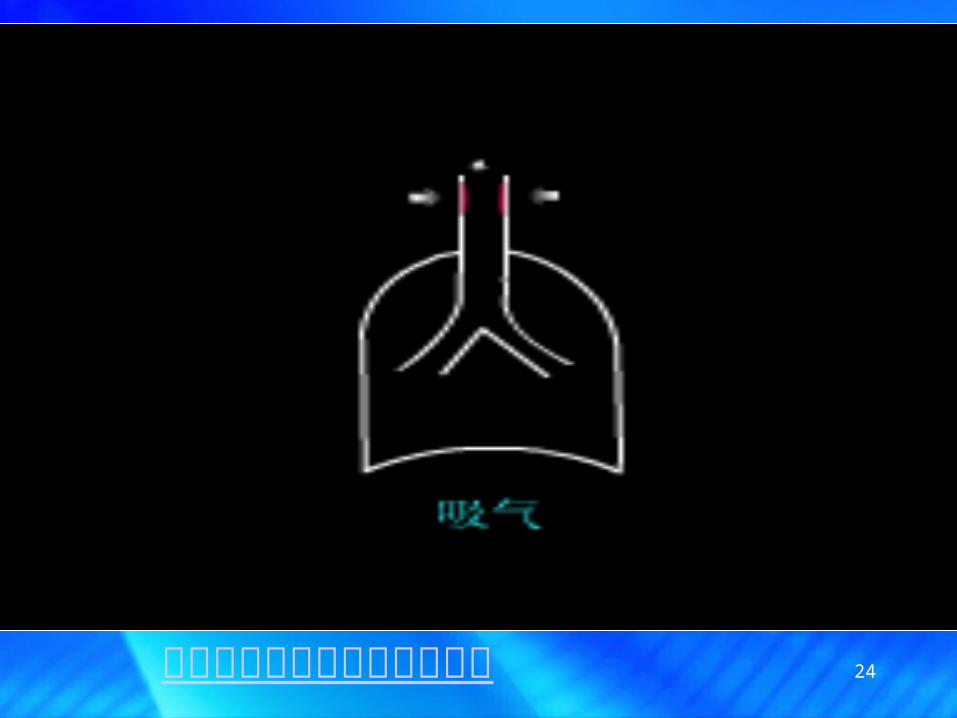
24中央气道胸外段阻塞吸气困难

25中央气道胸内段阻塞呼气困难
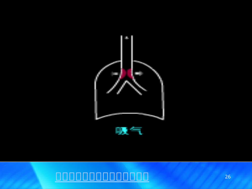
26中央气道胸内段阻塞无吸气困难
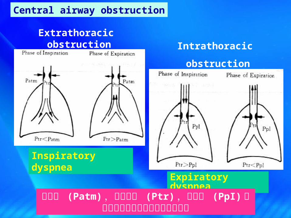
Extrathoracic obstruction Intrathoracic
obstruction
Expiratory dyspnea
Inspiratory dyspnea
Central airway obstruction
大气压 (Patm) ,气管内压 (Ptr) ,胸内压 (PpI)三者之间的关系决定呼吸困难的形式

28
The peripheral airway is usual referred to as the smaller airways (diameter<2 mm).
直径 <2mm ,无软骨环支撑,易扭曲闭合
specific chemical mediators such as histamine, prostaglandins, leukotrients, released during inflammatory and allergic responses
Abnormal neural regulation of airway smooth muscle tone
Edema of mucosa and secretions in the lumen all contribute to the narrowing of airway.
Peripheral airway obstruction
Expiratory dyspnea (呼气性呼吸困难 )
29
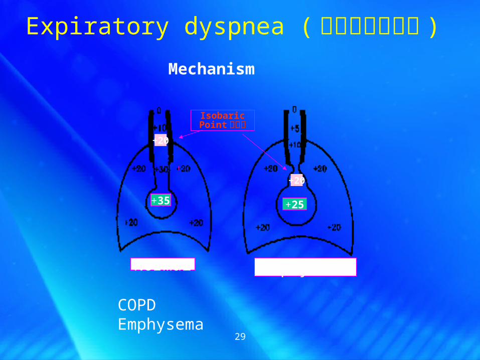
+25+35
IsobaricPoint 等压点
+20
+20
Normal Emphysema
Expiratory dyspnea (呼气性呼吸困难 )
COPDEmphysema
Mechanism
30
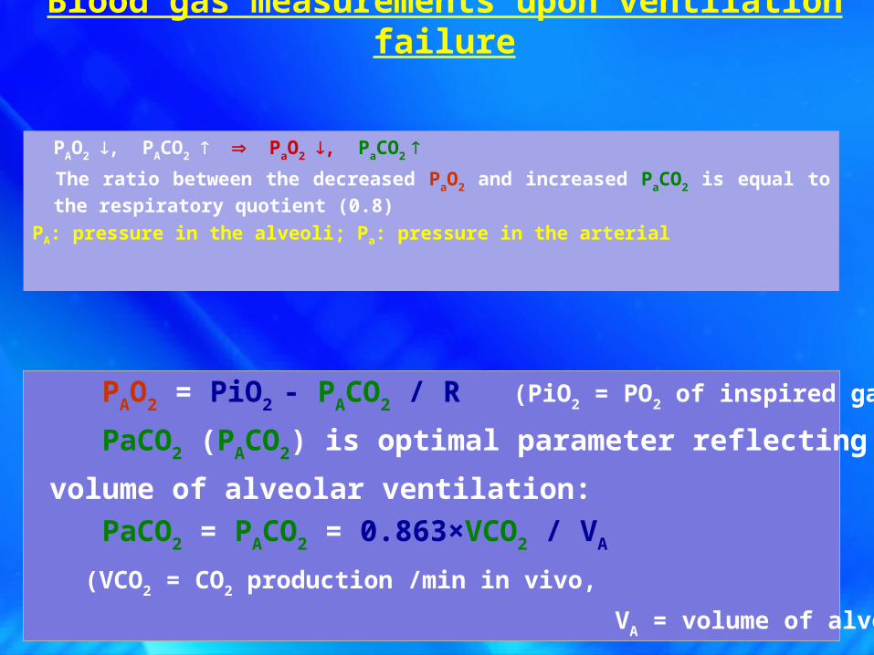
PAO2 , PACO2 PaO2 , PaCO2
The ratio between the decreased PaO2 and increased PaCO2 is equal to the
respiratory quotient (0.8)
PA: pressure in the alveoli; Pa: pressure in the arterial
PAO2 = PiO2 - PACO2 / R (PiO2 = PO2 of inspired gas)
PaCO2 (PACO2) is optimal parameter reflecting the total
volume of alveolar ventilation:
PaCO2 = PACO2 = 0.863×VCO2 / VA
(VCO2 = CO2 production /min in vivo,
VA = volume of alveolar ventilation /min)
Blood gas measurements upon ventilation failure

HypoventilationHypoventilationPPAAOO22↓↓ PaOPaO22↓↓
PPAACOCO22↑↑ PaCOPaCO22↑↑
Hypoventilation→ Type II respiratory failure
Hypoventilation d PaCO2↑
d PaO2↓ = R
Blood gas measurements upon ventilation failure
d: differences

• Ventilation failure ( 通气障碍 )– Restrictive hypoventilation ( 限制性通气不足 )– Obstructive hypoventilation ( 阻塞通气不足 )
• Gas-exchange failure ( 换气障碍 )– Diffusion disorder ( 弥散障碍 )
– VA/Q mismatch ( 通气 / 血流比值失调 )
– Increased anatomical shunt ( 解剖分流增加 )
Etiology and PathophysiologyEtiology and Pathophysiology
Dysfunction of external respiration
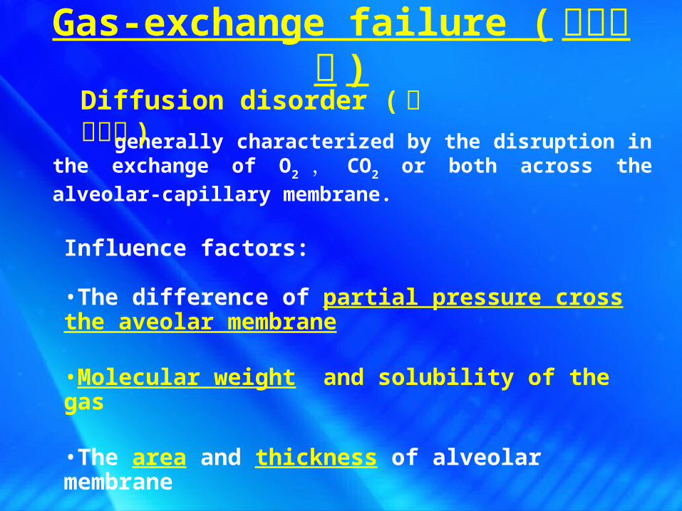
Gas-exchange failure ( 换气障碍 )
generally characterized by the disruption in the exchange of O2 , CO2 or both across the alveolar-capillary
membrane.
Diffusion disorder ( 弥散障碍 )
Influence factors:
•The difference of partial pressure cross the aveolar membrane
•Molecular weight and solubility of the gas
•The area and thickness of alveolar membrane
•The contact time between alveoli and blood

34
Reduction of the alveolar membrane surface area
(normal: 80 m2 , only 35-40 m2 is involved at rest) Atelectasis ( 肺不张 ), emphysema ( 肺气肿 ), pneumonectomy (肺
叶切除)
Increased thickness of alveolar membrane (normal:
1~5µm) Pulmonary edema, fibrosis, hyaline membrane ( 透 明 膜 形
成) formation, pulmonary capillary extension
Shortened diffusion time (normal 0.75s)
Usually only need 0.25s, the PaO2 can increase to PAO2
Increased cardiac output increases , faster blood flow
Causes of diffusion disorder
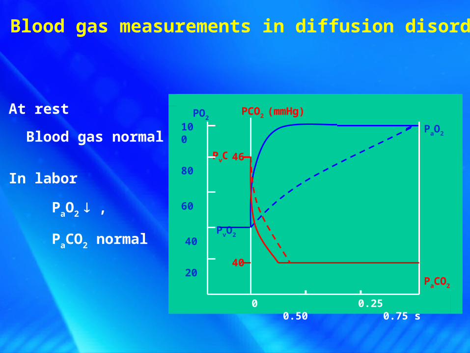
At rest
Blood gas normal
In labor
PaO2 ,
PaCO2 normal
Blood gas measurements in diffusion disorder
PvO2
PaO2
PaCO2
PvCO2
46
40
PO2 PCO2 (mmHg)
100
80
60
40
20
0 0.25 0.50 0.75 s

• Ventilation failure ( 通气障碍 )– Restrictive hypoventilation ( 限制性通气不足 )– Obstructive hypoventilation ( 阻塞通气不足 )
• Gas-exchange failure ( 换气障碍 )– Diffusion disorder ( 弥散障碍 )
– VA/Q mismatch ( 通气 / 血流比值失调 )
– Increased anatomical shunt ( 解剖分流增加 )
Etiology and PathophysiologyEtiology and Pathophysiology
Dysfunction of external respiration

37
= =正常 VA
Q
4L
5L 0.8
VA/Q mismatch ( 通气 / 血流比值失调 )
the most common mechanism of respiratory failure caused by pulmonary diseases.
differences ranged 3.0~0.6 from the top to the bottom of the lung

Hypoventilation in some alveoli →VA / Q↓
While in chronic bronchitis and obstructive emphysema, it is markedly increased up to 30-50 %.
Similar to A-V shunt (functional shunt)
Functional shunt ( venous admixture) ( 功能性分流 )
Normally, only account for ~3 % of total pulmonary blood flow
Reduced ventilation with normal blood flow
VA/Q mismatch ( 通气 / 血流比值失调 )
Chronic bronchitis, asthma, COPD
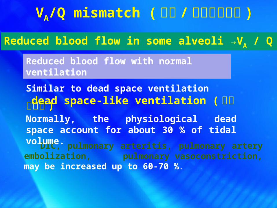
DIC, pulmonary arteritis, pulmonary artery embolization, pulmonary vasoconstriction, may be increased up to 60-70 %.
Reduced blood flow in some alveoli →VA / Q
Similar to dead space ventilation dead space-like ventilation ( 死腔样通气 )Normally, the physiological dead space account for about 30 % of tidal volume.
Reduced blood flow with normal ventilation
VA/Q mismatch ( 通气 / 血流比值失调 )
40
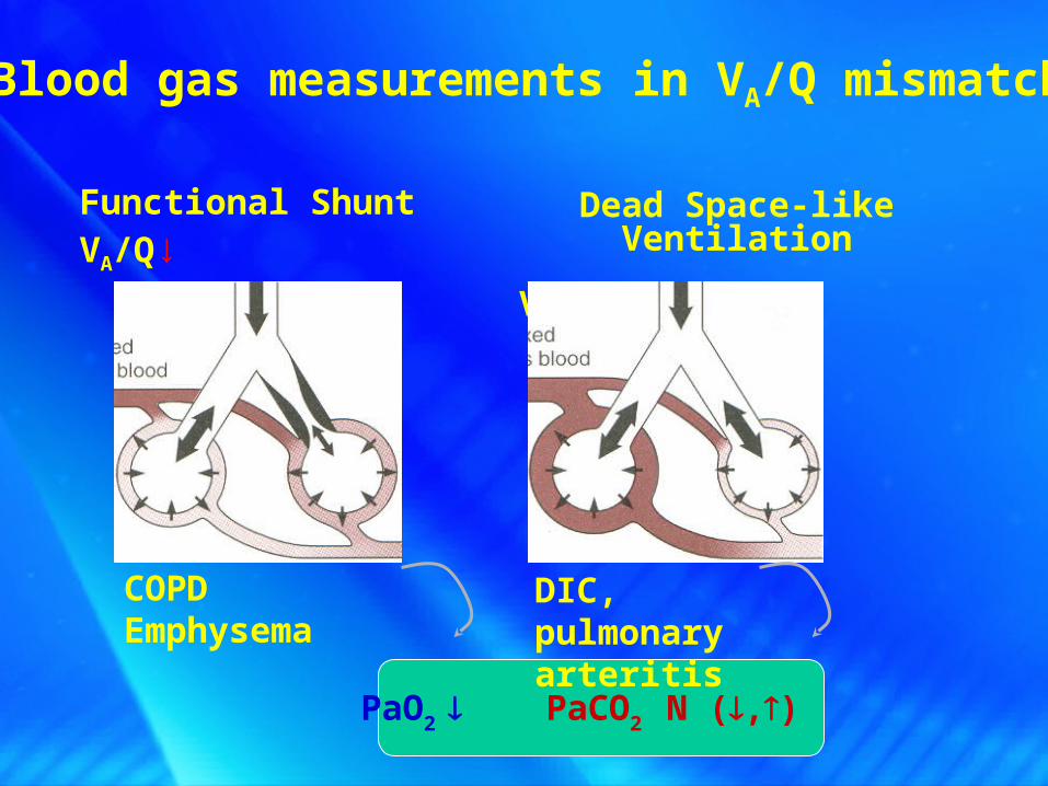
Blood gas measurements in VA/Q mismatch
Functional ShuntVA/Q↓
Dead Space-like Ventilation
VA/Q↑
PaO2 PaCO2 N (,)
COPDEmphysema
DIC, pulmonary arteritis

41
Diseased Normal Total
V/Q <0.8 >0.8 ≈0.8
PaO2
CaO2
PaCO2 N
CaCO2 N
Functional Shunt
Blood gas measurements in QA/V mismatch

42
Dead Space-like Ventilation
Diseased Normal Total
V/Q >0.8 <0.8 ≈0.8
PaO2
CaO2
PaCO2 N
CaCO2 N
Blood gas measurements in QA/V mismatch
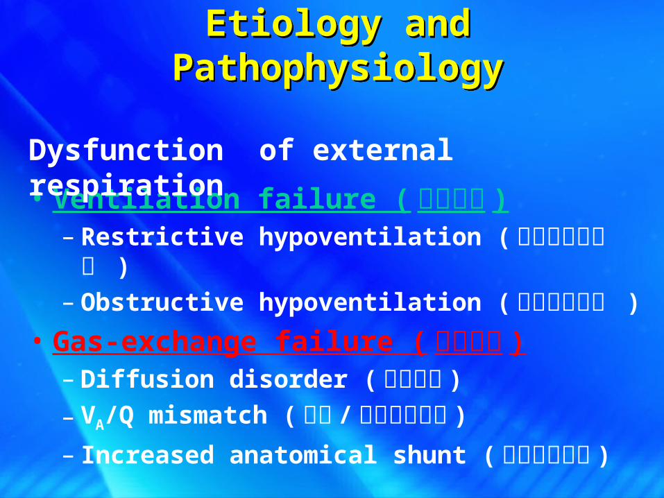
• Ventilation failure ( 通气障碍 )– Restrictive hypoventilation ( 限制性通气不足 )– Obstructive hypoventilation ( 阻塞通气不足 )
• Gas-exchange failure ( 换气障碍 )– Diffusion disorder ( 弥散障碍 )
– VA/Q mismatch ( 通气 / 血流比值失调 )
– Increased anatomical shunt ( 解剖分流增加 )
Etiology and PathophysiologyEtiology and Pathophysiology
Dysfunction of external respiration
44
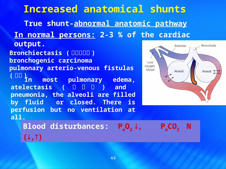
Increased anatomical shuntsTrue shunt-abnormal anatomic pathway
In normal persons: 2-3 % of the cardiac output.
Bronchiectasis ( 支气管扩张 ) bronchogenic carcinomapulmonary arterio-venous fistulas ( 瘘管 )
In most pulmonary edema, atelectasis ( 肺不张 ) and pneumonia, the alveoli are filled by fluid or closed. There is perfusion but no ventilation at all.
Blood disturbances: PaO2 , PaCO2 N (,)
45
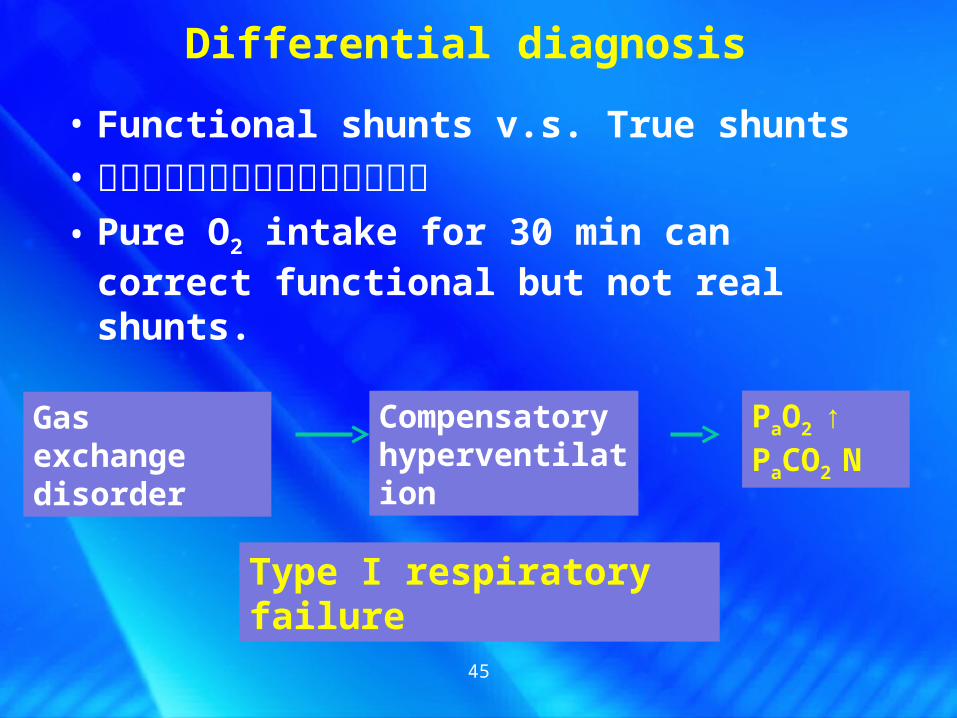
• Functional shunts v.s. True shunts
• 功能性分流与真性分流的鉴别诊断• Pure O2 intake for 30 min can correct
functional but not real shunts.
Differential diagnosis
Gas exchange disorder
Compensatory hyperventilation
PaO2 ↑PaCO2 N
Type I respiratory failure

46
致病因子致病因子
激活中性粒细胞激活中性粒细胞 // 单核巨噬细胞单核巨噬细胞 // 血小板血小板 // 内皮细胞内皮细胞
释放体液介质释放体液介质
肺泡肺泡 -- 毛细血管膜损伤毛细血管膜损伤和通透性增高和通透性增高
微血栓形成 微血栓形成
ARDS 病理改变:肺出血、水肿、肺不张、微血栓、肺泡透明膜形成
Acute Respiratory Distress Syndrome ( ARDS) 急性呼吸窘迫综合征

47
• ARDS 引起呼衰的机制
肺水肿肺水肿透明膜形成透明膜形成
肺不张肺不张支气管阻塞支气管阻塞支气管痉挛支气管痉挛
微血栓形成微血栓形成肺血管收缩肺血管收缩
弥散障碍弥散障碍
功能性分流功能性分流
死腔样通气死腔样通气
PaO2↓PaCO2
N 或↓
Ⅰ型呼衰VA/Q mismatch
48
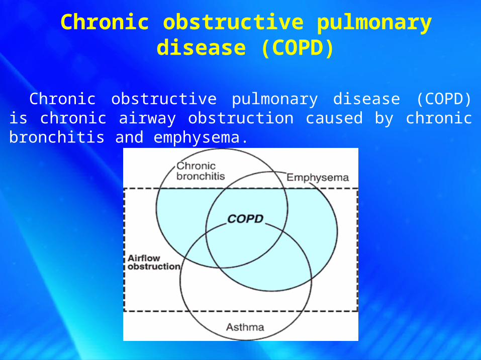
Chronic obstructive pulmonary disease (COPD)
Chronic obstructive pulmonary disease (COPD) is chronic airway obstruction caused by chronic bronchitis and emphysema.

49
?
?
?
?
COPD Pathogenesis:
Ventilatory inadequacy
Ventilation-perfusion mismatching
Diffusion disorder
Ventilatory inadequacy
Obstructive
Restrictive
Congestion,swelling, spasm, blockage
of bronchi wall Upward shift of Isobaric point
Decreased alveolar surfactantRespiratory muscle failure
Decreased alveolar surface area
Low ventilation in part of alveoli
Low blood flow in part of alveoli

50
Changes in the body
Alteration of blood gas
Acid-base and electrolyte disturbances
Disorders of vital organ systems
51
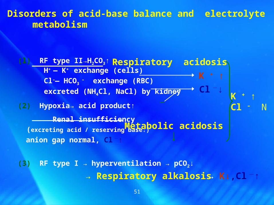
Disorders of acid-base balance and electrolyte metabolism
(l) RF type II→H2CO3↑ → H+ — K+ exchange (cells)
Cl–— HCO3 – exchange (RBC)
excreted (NH4Cl, NaCl) by kidney
(2) Hypoxia→ acid product↑ Renal insufficiency (excreting acid / reserving base↓) anion gap normal, Cl -↑
(3) RF type I → hyperventilation → pCO2↓
K + ↑Cl – N
→ Respiratory alkalosis → K↓,Cl -↑
Metabolic acidosis
Respiratory acidosis
K + ↑
Cl -↓

52
Alterations in respiratory system
Alterations caused by hypoxia and hypercapnia
PCR RC Total effect
PaO <60
<30
PaCO >50,<80
>80
(PCR: peripheral chemical receptor; RC: respiratory center)
- - + +
+ + < - - -+ > - +
O2 therapy for chronic Type II RF: low concentration, low flow rate, continuous O2 supply, maintain PaO2 ~60 mmHg

53
Alterations in circulatory system
(1) Mild PaO2 , PaCO2 excitation (reflex): Cardiac Output Blood redistribution (peripheral constriction, coronary & cerebral artery dilation) (2) Severe PaO2 , PaCO2 inhibition (direct): BP Cardiac contractility , arrhythmia(3) Chronic lung disease
right-heart failure
Pulmonary Heart Disease (PHD)

54
Hypoxia, Hypercapnia, Overload Disorder of electrolyte metabolism
Myocardial injury
Resistance↑
PaO2 , PaCO2→constriction of pulmonary arteriolePrimary disease →thickening and sclerosis of vessel wall, Narrowing of vessel lumen
Persistent hypoxia→ RBC↑, blood volume↑, viscosity↑
P
H
D
→ Pulmonary artery hypertension

55
Alterations in central nervous system
Respiratory failure
dysfunction of brain
Pulmonary encephalopathy
肺性脑病

56
Dilation, permeability↑ →vasogenic brain edema
ATP→ Na-K-ATPase →cytoxic brain edema
Cerebral blood vessels
Cerebral cells
Glutamate decarboxylase↑ →GABA↑→CNS inhibition
Phospholipase↑→release lisozyme→ injury of brain cells
IntracranialPressure↑
Hypoxia
Acidosis
Pulmonary encephalopathy
Respiratory failure
Pulmonary encephalopathy
57
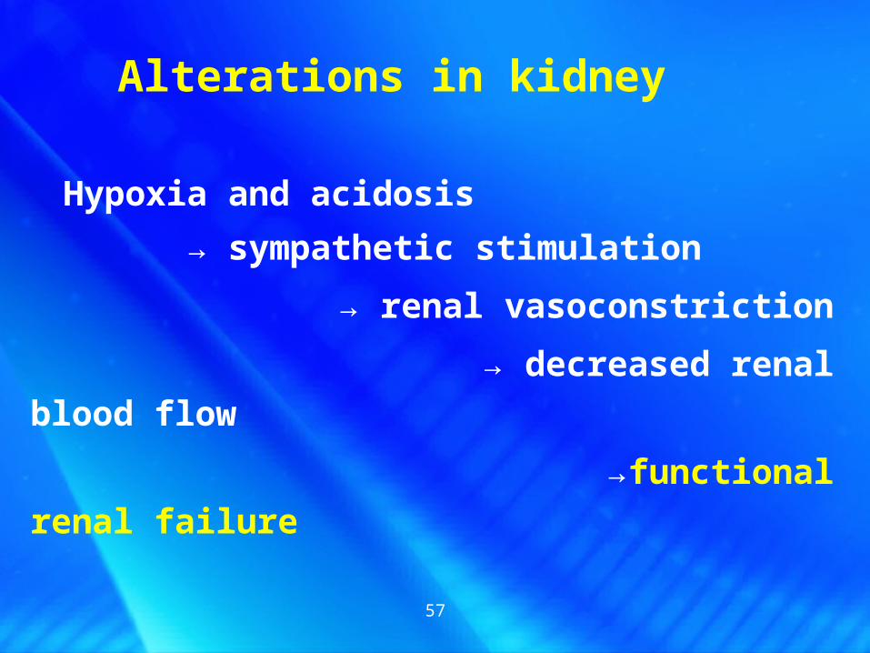
Hypoxia and acidosis
→ sympathetic stimulation
→ renal vasoconstriction
→ decreased renal blood flow
→functional renal failure
Alterations in kidney

58
Alterations in gastrointestine
Severe hypoxia → vasoconstriction in stomach wall → mucosa damage → loss of barrier function
mucosa ulcer bleeding and necrosis
Severe retention of CO2 → active carbonic anhydrase → increased secretion of HCl
59
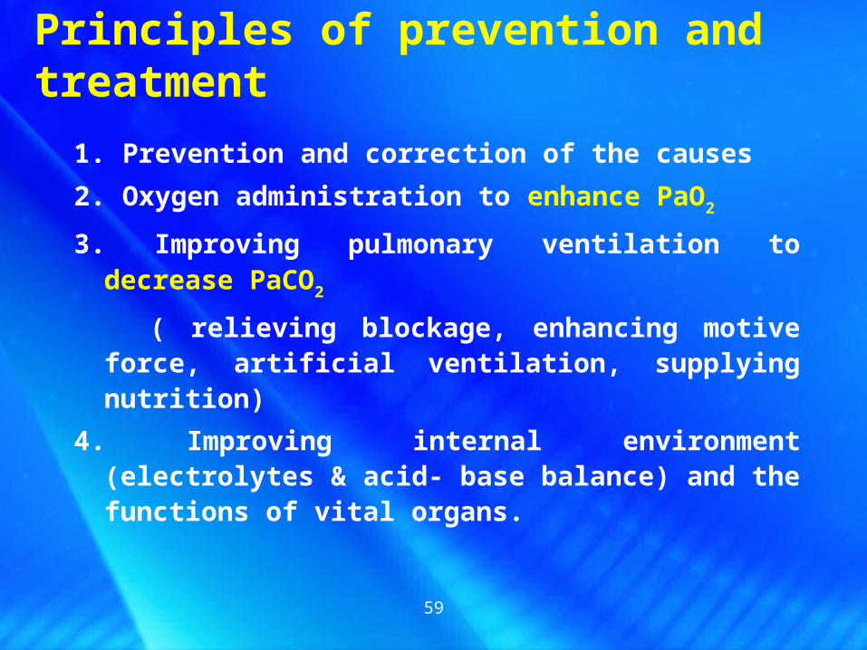
Principles of prevention and treatment
1. Prevention and correction of the causes
2. Oxygen administration to enhance PaO2
3. Improving pulmonary ventilation to decrease PaCO2
( relieving blockage, enhancing motive force, artificial ventilation, supplying nutrition)
4. Improving internal environment (electrolytes & acid- base balance) and the functions of vital organs.

60
Respiratory failure is a pathological process in which
the external respiratory dysfunction leads to an
abnormal decrease of PaO2 (60mmHg or less) with or
without retention of PaCO2 (50mmHg or more)
SummaryDefinition

• Ventilation failure ( 通气障碍 )– Restrictive hypoventilation ( 限制性通气不足 )– Obstructive hypoventilation ( 阻塞通气不足 )
• Gas-exchange failure ( 换气障碍 )– Diffusion disorder ( 弥散障碍 )
– VA/Q mismatch ( 通气 / 血流比值失调 )
– Increased anatomical shunt ( 解剖分流增加 )
SummaryEtiology and Pathophysiology
SummaryEtiology and Pathophysiology
Dysfunction of external respiration

学习目标与要求掌握:呼吸衰竭定义与分类病因与发病机制通气不足与换气障碍引起呼吸衰竭的机制限制性通气不足与阻塞性通气不足原理与病因中枢性与外周性阻塞导致呼吸困难的机制弥散障碍致呼吸衰竭机制通气 / 血流比值失常致呼吸衰竭机制功能性分流,死腔样通气,真性分流特征I 型与 II 型呼吸衰竭血气分析特征ARDS 与 COPD 致呼吸衰竭的主要发病机制慢性 II 型呼吸衰竭氧疗注意事项肺源性心脏病与肺性脑病的发病机制

Thanks!Have a good summer holiday!

64
Driving force↓
Respiratory center respiratory muscle
Resistance ↑ Elastic resistance of chest wall or lung
Airway Resistance for gas flow
respiratory amplitude
respiratory velocity
Restrictive hypoventilation
Obstructive hypoventilation
Inadequate driving force or excessive elastic resistance may restrict respiratory amplitude
Increased resistance of air way may lower respiratory velocity