respiratory medicine, clinical cases uncovered emma baker, dilys lai
TRANSCRIPT

RespiratoryMedicine
CLINICAL CASES UNCOVERED

This book is dedicated to Belinda Brewer and her thirst for lifelong learning

RespiratoryMedicineCLINICAL CASES UNCOVERED
Emma H. BakerPhD, FRCPReader in Clinical Pharmacology and Consultant Physician
St George’s, University of London
London, UK
Dilys LaiMD, MRCPConsultant in Respiratory and General Medicine
Chelsea and Westminster Hospital
London, UK
A John Wiley & Sons, Ltd., Publication

This edition fi rst published 2008, © 2008 by E. H. Baker and D. Lai
Blackwell Publishing was acquired by John Wiley & Sons in February 2007. Blackwell’s publishing program has been merged with Wiley’s global Scientifi c, Technical and Medical business to form Wiley-Blackwell.
Registered offi ce: John Wiley & Sons Ltd, The Atrium, Southern Gate, Chichester, West Sussex, PO19 8SQ, UK
Editorial offi ces: 9600 Garsington Road, Oxford, OX4 2DQ, UK The Atrium, Southern Gate, Chichester, West Sussex, PO19 8SQ, UK 111 River Street, Hoboken, NJ 07030-5774, USA
For details of our global editorial offi ces, for customer services and for information about how to apply for permission to reuse the copyright material in this book please see our website at www.wiley.com/wiley-blackwell
The right of the author to be identifi ed as the author of this work has been asserted in accordance with the Copyright, Designs and Patents Act 1988.
All rights reserved. No part of this publication may be reproduced, stored in a retrieval system, or transmitted, in any form or by any means, electronic, mechanical, photocopying, recording or otherwise, except as permitted by the UK Copyright, Designs and Patents Act 1988, without the prior permission of the publisher.
Wiley also publishes its books in a variety of electronic formats. Some content that appears in print may not be available in electronic books.
Designations used by companies to distinguish their products are often claimed as trademarks. All brand names and product names used in this book are trade names, service marks, trademarks or registered trademarks of their respective owners. The publisher is not associated with any product or vendor mentioned in this book. This publication is designed to provide accurate and authoritative information in regard to the subject matter covered. It is sold on the understanding that the publisher is not engaged in rendering professional services. If professional advice or other expert assistance is required, the services of a competent professional should be sought.
Library of Congress Cataloguing-in-Publication Data
Baker, Emma.Respiratory medicine : clinical cases uncovered / Emma Baker, Dilys Lai.p. ; cm.Includes index.ISBN 978-1-4051-5895-4 (alk. paper)1. Respiratory organs–Diseases–Case studies. 2. Respiratory organs–Diseases–Examinations, questions, etc. I. Lai, Dilys. II. Title.[DNLM: 1. Respiratory Tract Diseases–Case Reports. 2. Respiratory Tract Diseases–Problems and Exercise. 3. Signs and Symptoms, Respiratory–Case Reports. 4. Signs and Symptoms, Respiratory–Problems and Exercises.WF 18.2 B167r 2008]RC732.B34 2008616.2–dc222007050635
ISBN: 978-1-4051-5895-4
A catalogue record for this book is available from the British Library
Set in 9/12pt Minion by SNP Best-set Typesetter Ltd., Hong Kong
Printed in Singapore by COS Printers Pte Ltd
1 2008

Contents
Preface, vii
Acknowledgements, viii
How to use this book, ix
Part 1 Basics, 1
Basic science, 1
Approach to the patient, 17
Part 2 Cases, 41
Case 1 A 64-year-old man with respiratory arrest, 41
Case 2 A 19-year-old man with chest tightness, 46
Case 3 A 31-year-old woman with poorly-controlled asthma, 51
Case 4 A 22-year-old man with life-threatening shortness of breath, 60
Case 5 A 65-year-old man with worsening shortness of breath, 68
Case 6 A worried 56-year-old man, 75
Case 7 An 81-year-old man with acute shortness of breath, 80
Case 8 A 73-year-old woman with confusion, 87
Case 9 A 78-year-old man with ankle swelling, 92
Case 10 A 72-year-old man with a hoarse voice, 98
Case 11 A 56-year-old woman with an abnormal chest X-ray, 106
Case 12 A 68-year-old man with shortness of breath and chest pain, 112
Case 13 A 16-year-old boy with right-sided chest pain, 117
Case 14 A 62-year-old woman with breathlessness, 124
Case 15 A 24-year-old woman with pleuritic chest pain, 129
Case 16 An elderly woman who collapsed suddenly, 139
Case 17 A 45-year-old man with chest pain and breathlessness, 148
v

vi Contents
Case 18 A 72-year-old man who becomes breathless during inpatient stay on a medical
ward, 156
Case 19 A 58-year-old woman with breathlessness following surgery, 162
Case 20 A 16-year-old boy with a sore throat, 169
Case 21 A 45-year-old woman with a chronic cough, 174
Case 22 A 17-year-old girl with cystic fi brosis, 179
Case 23 A 25-year-old woman with cough and weight loss, 186
Case 24 A 44-year-old man commencing treatment for tuberculosis, 196
Case 25 A 32-year-old man who absconds from directly observed therapy for
tuberculosis, 201
Case 26 A 32-year-old woman with a skin rash and abnormal chest X-ray, 207
Case 27 A 68-year-old woman with worsening breathlessness on exertion, 213
Case 28 A 35-year-old man with chronic shortness of breath, 220
Case 29 An overweight 52-year-old man who snores, 226
Part 3 Self-assessment, 232
MCQs, 232
EMQs, 237
SAQs, 243
Answers, 245
Index of cases by diagnosis, 251
Index, 253

Preface
Medicine is a mass of facts and complex science, which
can be dull and indigestible. It is brought to life by
patients with their individual stories, clinical mysteries
and their need for help. Medicine is probably best learnt
through clinical experience backed up by information
and refl ection. However in modern medical practice
there is often too much to do and too little time for
consideration.
This book is one of the fi rst in a novel series that allows
the reader to experience a virtual clinical attachment
with enough time for refl ection and teaching. Patients are
described as they really present to hospital or general
practice. The clinical reasoning required to explain symp-
toms and signs, order and interpret investigations and
start management is demonstrated as the case develops.
Relevant science is described in context to support under-
standing and patients are managed according to current
guidelines. This format will allow the reader to develop
knowledge and skills that are immediately useful for
patient management, rather than to accumulate facts that
need a lot more work to translate them into practice.
Twenty nine patients with respiratory disease are pre-
sented in the book, providing experience of the full
breadth of adult respiratory medicine. We have chosen
these cases as they are common problems that will be
encountered at some point by all doctors in clinical prac-
tice, no matter what fi eld they end up in. Many questions
used to explore the cases are real questions posed by
students during our teaching sessions and the answers are
the end product of many attempts to answer these ques-
tions satisfactorily. As experienced examiners we have
ensured that material covered in this book is suffi cient
for students to pass respiratory sections of undergraduate
or general postgraduate exams. This book will also be
a useful aid to practice for junior doctors and allied
health professionals looking after patients with respira-
tory disease and as basic revision for doctors entering
specialist respiratory training.
We have written the text book that we would have liked
to use to learn respiratory medicine. If we were starting
again we would begin with the patients, attempting to
answer the questions posed throughout the cases alone
or in groups before reading the written answers and rel-
evant science. During relevant clinical attachments we
would use the book to update clinical skills and practice
OSCE checklists. In preparation for exams we would
revise case summaries and key points and practice the self
assessment questions. We hope you will enjoy using it and
that it will help you understand respiratory medicine for
the benefi t of patients.
Emma H. Baker
Dilys Lai
2008
vii

Acknowledgements
The authors gratefully acknowledge the help of the
following:
Dr Belinda Brewer for invaluable comments on the man-
uscript; Dr Cathy Corbishley for generous donation of
pathology slides from her personal collection; Mr Harry
Young for his wonderful contribution to student educa-
tion as a patient and for modelling for this book; Mark
Evenden and staff of media services at St George’s, Uni-
versity of London for endless production of fantastic
illustrations; Sue Adie for tireless administrative support;
and Dr Adrian Draper, Dr Yee Ean Ong, Dr Deirdre
McGrath, Dr Laurie Hanna, Dr Sisa Grubnic, Dr Claire
Wells, Dr Stephen Thomas, nursing staff on Marnham
ward, Reg Ramai and Paula Mclean for provision of clin-
ical material and other assistance.
viii

How to use this book
Clinical Cases Uncovered (CCU) books are carefully
designed to help supplement your clinical experience and
assist with refreshing your memory when revising. Each
book is divided into three sections: Part 1, Basics; Part 2,
Cases; and Part 3, Self-Assessment.
Part 1 gives a quick reminder of the basic science,
history and examination, and key diagnoses in the area.
Part 2 contains many of the clinical presentations you
would expect to see on the wards or crop up in exams,
with questions and answers leading you through each
case. New information, such as test results, is revealed as
events unfold and each case concludes with a handy case
summary explaining the key points. Part 3 allows you to
test your learning with several question styles (MCQs,
EMQs and SAQs), each with a strong clinical focus.
Whether reading individually or working as part of a
group, we hope you will enjoy using your CCU book. If
you have any recommendations on how we could improve
the series, please do let us know by contacting us at:
DisclaimerCCU patients are designed to refl ect real life, with their
own reports of symptoms and concerns. Please note that
all names used are entirely fi ctitious and any similarity to
patients, alive or dead, is coincidental.
ix


PA
RT
1:
BA
SIC
S
Basic science
Anatomy and physiologyThe primary function of the lungs is to take up oxygen
from the air into the blood and excrete carbon dioxide.
To achieve this, the following are required:
• Respiratory centre in the brain to control the process
of breathing
• Respiratory muscles, chest wall and pleura to allow
infl ation and defl ation of the lungs
• Airways to transmit air into and out of the lung
• Alveoli and capillaries to act as an interface for gas
exchange
• Pulmonary circulation to carry deoxygenated blood
into, and oxygenated blood out of, the lungs
• Host defence systems to protect the lung from inhaled
foreign particles (e.g. organisms, allergens, pollutants)
and maintain function
Control of breathingThe act of breathing requires the anatomical structures
shown in Fig. 1. The following sequence of events occurs:
• The respiratory centre in the brainstem generates a
signal to breathe
• This signal is transmitted via the:� spinal cord� anterior horn cells� motor nerves (phrenic and intercostal nerves)� neuromuscular junctions
• The signal stimulates contraction of respiratory
muscles, which lowers intrathoracic pressure and sucks
air into the lungs
• When the signal stops, the respiratory muscles relax
and elastic recoil of the lungs and weight of the chest wall
force air out of the lungs
• The next signal is generated from the respiratory centre
and the cycle starts again
The respiratory centreThe respiratory centre is made up of groups of neurons
in the medulla oblongata and pons of the brainstem.
The dorsal group of neurons in the respiratory centre is
responsible for normal quiet breathing. These neurons
automatically generate repetitive inspiratory signals.
Each signal begins weakly and builds up over about
2 s, stimulating a smooth contraction of the respiratory
muscles. After inspiration the signal ceases for about
3 s, allowing relaxation of the respiratory muscles and
expiration before the inspiratory signal begins again.
These signals continue from birth to death unless the
respiratory centre is damaged. As each cycle takes
about 5 s the normal respiratory rate is around 12
breaths/min.
The chemosensitive area in the brainstem detects
changes in carbon dioxide concentrations and adjusts
respiratory rate as follows:
• As CO2 concentrations rise, H+ ions are generated by
interaction with water:
Henderson–Hasselbach equation
H+ + HCO3− ƨ H2CO3
ƨ H2O + CO2
• H+ ions stimulate chemosensitive neurons, which
signal to other areas of the respiratory centre to increase
the respiratory rate
• Increased ventilation blows off CO2, lowering blood
CO2 concentrations back to normal
• H+ ion concentrations in the brain decrease and this
signals to the respiratory centre to slow the respiratory
rate
Peripheral chemoreceptors are present in carotid and
aortic bodies, where they are ideally placed to monitor
arterial oxygen concentrations. When oxygen concentra-
tions are normal, signalling from peripheral chemo-
receptors is suppressed. However, when blood oxygen
concentrations fall below normal, chemoreceptors send
strong signals to the respiratory centre via the glossopha-
ryngeal and vagal nerves, which drive an increase in
respiratory rate. Signalling by peripheral chemoreceptors
is also stimulated by an increase in H+ ions, for example
in renal failure or acidosis. Patients with acidosis appear
1

2 Part 1: Basics
PA
RT
1:
BA
SIC
S
breathless with deep sighing respiration (Kussmaul’s res-
piration), which has been described as ‘air hunger’.
Hierarchy of respiratory stimuliThe strongest drive to breathe is the repetitive inspiratory
signal generated by the dorsal neurons:
• This signal is unstoppable, unless the respiratory centre
is damaged
• However, ventilation driven by this signal may not be
suffi cient to maintain normal blood PaO2 and PaCO2
• Addition of other stimuli to this signal increases respi-
ratory rate and depth as required
The next strongest drive to breathe is an increase in
PaCO2, detected by the chemosensitive area:
• This process is extremely sensitive. For example, a
small increase in CO2 of 0.13 kPa results in a 2–4 L/min
increase in ventilation
• In patients with chronic hypercapnia, the chemosensi-
tive area becomes less sensitive to changes in PaCO2. As
this central respiratory drive falls, the weaker drive from
peripheral chemoreceptors takes over
The peripheral chemoreceptors generate the weakest
respiratory stimulus. They are important in patients who
have lost the chemosensitive drive to breathe because of
chronic hypercapnia, or in patients with severe hypoxia
or acidosis.
Respiratory muscles, chest wall and pleuraRespiratory musclesInspiratory muscles are:
• Diaphragm (quiet breathing)
• Intercostal muscles
• Neck muscles (accessory)
In inspiration, intrathoracic pressure is lowered by down-
wards movement of the diaphragm and upwards/outwards
movement of the ribs, as a result of contraction of the inter-
costal muscles. This sucks air into the lungs. Neck and
shoulder girdle muscles are recruited to help lower intratho-
racic pressure when more vigorous inspiration is required
(e.g. after exercise and during respiratory distress).
Expiration involves:
• Relaxation of inspiratory muscles
• Elastic recoil of lungs
• Abdominal muscles (accessory)
On expiration, the respiratory muscles relax and elastic
recoil of the lungs and chest wall raise intrathoracic pres-
sure. This process forces air out of the lungs. The abdom-
inal muscles are accessory respiratory muscles that can be
contracted to enhance expiration.
Chest wall and pleuraThe chest wall comprises ribs and muscles, including
internal and external intercostal, subcostal and Trans-
versus thoracis muscles. The chest wall and diaphragm
surround the thoracic cavity, which is lined by a mem-
brane called the parietal pleura. The lungs are also covered
by a pleural membrane called the visceral pleura, which
is continuous with the parietal layer at the lung hila (Fig.
2). Parietal and visceral pleura are separated by a poten-
tial space, which contains a thin layer of fl uid that allows
the lungs to slide smoothly against the chest wall during
inspiration and expiration. There is a negative pressure
in the pleural space, thought to be generated by pumping
of fl uid out of the space by lymphatics. This negative
pressure opposes the elastic recoil of the lungs, keeping
them expanded and ensuring that they infl ate and defl ate
with movement of the chest wall and diaphragm.
AirwaysThe airways comprise nose, pharynx, larynx, trachea,
bronchi and bronchioles (Fig. 3).
Upper airwaysNoseThe nasal cavity is divided into right and left sides by the
nasal septum. As air enters the nose it passes over the
turbinates, which are fl eshy structures with a large blood
Anterior horn cell
Muscles
Chest wall
Lungs
Spinal cord
Respiratory centrein brainstem
Nerves
Neuromuscularjunctions
Pleura
Figure 1 Structures required for the act of breathing.

Basic Science 3
PA
RT
1:
BA
SIC
S
supply that warm and humidify the air. High in the nasal
cavity, air comes into contact with the olfactory nerve
endings, generating the sensations of smell and taste. The
nasal cavity is lined by ciliated epithelium with an over-
lying layer of mucus. This traps inhaled foreign particles,
which can then be removed from the nose by mucociliary
transport to the pharynx or by sneezing. The sinuses and
lacrimal ducts drain into the nose and can become
blocked in patients with nasal disease.
PharynxThis is a muscular tube that extends behind the nasal and
oral cavities and ends at the top of the oesophagus. Its
purpose is to transmit air to the lower respiratory tract
and food to the oesophagus. It contains lymphoid tissue
(tonsils and adenoids) and mucus glands. The eustachian
tubes enter the pharynx from the middle ear and can
become blocked by swollen lymphoid tissue.
LarynxThis organ produces voice and protects the airways from
aspiration of food and secretions. It is made up of carti-
lages (Box 1) held together by ligaments and moved by
muscles. Inside the laryngeal cavity, connective tissue,
muscles and ligaments make up the vocal folds or cords
which are used to produce sound. At the top of the larynx
Chest wall
Lungs Intrapleural pressureopposes elastic recoiland maintains lungexpansion
Potential space containingthin layer of fluid
Parietal pleura
Visceralpleura
Figure 2 Anatomy of the pleura.
Turbinates innasal cavity
Pharynx
Epiglottis
Larynx
Upperairway
Trachea
Nose
Main bronchus
Segmentalbronchi
Lowerairway
~25 divisionsending interminal
bronchioles
Figure 3 Anatomy of the airways.

4 Part 1: Basics
PA
RT
1:
BA
SIC
S
is the epiglottis, which is a leaf-shaped fl ap of tissue that
is brought down to cover the laryngeal opening and
prevent aspiration during swallowing.
Lower airwaysThe trachea is about 10–12 cm in length and divides at
the carina into left and right main bronchi. These divide
into lobar, then segmental and subsegmental bronchi.
There are around 25 divisions between carina and alveoli.
Large airwaysThe trachea and fi rst seven divisions of the bronchi
have:
• Cartilage and smooth muscle in walls
• Submucosal mucus secreting glands
• Ciliated epithelium containing goblet cells
• Endocrine cells
Small airwaysThe next 16–18 divisions of bronchioles have:
• No cartilage and progressive thinning of muscle layer
• Single layer of ciliated cells and very few goblet cells
• Granulated Clara cells producing surfactant-like
substance
Flow of air through the airwaysMovement of air into and out of the lung depends on
differences in air pressure between mouth and alveoli.
• On breathing in, contraction of respiratory muscles
lowers intrathoracic pressure below atmospheric pres-
sure, pulling air into the alveoli
• On breathing out, elastic recoil of the lung and chest
wall raises intrathoracic pressure above atmospheric
pressure, forcing air out of the alveoli
The rate at which air moves into and out of the lungs
in response to these changes in pressure depends on the
rate at which air can fl ow along the airways. This in turn
depends on airway resistance and can be described by
Ohm’s law:
Flow rate =Difference in pressure
between mouth and alveoli
Reesistance to airflow in airways
Poiseuille’s law describes the components of resistance
to fl ow in more detail:
Resistance8 viscosity of air airway length
r4= × ×
π
This is important as it shows that the radius of the
tube is a very powerful determinant of airfl ow resistance.
To test this, try breathing out through a tube with a
large radius, such as the cardboard insert from kitchen
roll. Flow will be rapid through this wide tube and you
will be able to empty your lungs easily. Then try breath-
ing out through a tube with a small radius, such as a
drinking straw. Flow will be very slow through this
narrow tube and it will take a long time to empty your
lungs.
The radius of the lumen of airways in the respiratory
tract depends on (Fig. 4):
• Smooth muscle tone
• Thickness of the epithelium and submucosa
• Luminal contents
• Pressure inside and outside the airways
Airfl ow obstructionDisease causing airfl ow obstruction includes:
• Obstructive sleep apnoea (upper airways)
• Asthma (lower airways)
• Chronic obstructive pulmonary disease (COPD; lower
airways)
In obstructive sleep apnoea, pressure outside the
airway from neck soft tissues compresses the upper
airway causing partial collapse with snoring, or complete
collapse with cessation of breathing (apnoea). In asthma
and COPD, smooth muscle spasm, mucosal oedema and
increased airway secretions and sputum plugs obstruct
the airway (Fig. 4).
Alveoli and capillariesAlveoliAlveoli are small air-fi lled sacs at the end of the smallest
airways (terminal bronchioles). There are over 300
Box 1 Clinical reasons to know the anatomy of laryngeal cartilages
• Two laryngeal cartilages can be felt easily in the neck.
The ‘Adam’s apple’ is the laryngeal cartilage. Below that
is a gap, then a smaller cartilage called the cricoid
cartilage
• In clinical medicine the distance between the cricoid
cartilage and the sternal notch (cricosternal distance) is
used to assess lung hyperinfl ation (see p. 26)
• The cricothyroid membrane between the laryngeal and
cricoid cartilages can be pierced (cricothyrotomy) in an
emergency to allow a patient with severe upper airways
obstruction to breathe

Basic Science 5
PA
RT
1:
BA
SIC
S
million in two lungs. The walls of the sacs include type I
and II pneumocytes.
Type I pneumocytesThese cells form a single epithelial cell layer. They have
extremely thin cytoplasm, which facilitates gas exchange
and make up 90–95% of the surface area of the alveolar
epithelium.
Type II pneumocytesThese cells make surfactant which lines the alveolar
lumen. Surfactant reduces surface tension, preventing
alveolar collapse and reducing the amount of work
required for breathing.
CapillariesEach single alveolus is surrounded by a dense network of
around 1000 capillaries. The wall of the pulmonary cap-
illaries comprises a single layer of endothelial cells, which
facilitates gas exchange.
Alveolar–capillary membraneThe massive numbers of alveoli and capillaries in the lung
result in around 50–100 m2 of contact between these
structures in a normal adult. The enormous alveolar–
capillary interface is ideal for gas exchange. The barrier
that respiratory gases must cross between air in the
alveoli and red blood cells in the capillaries is called the
alveolar–capillary membrane (Fig. 5). It has a thickness
of approximately 0.6 µm. The alveoli, capillaries and
intervening connective tissue are known collectively as
the lung parenchyma.
Gas transportO2 and CO2 move across the alveolar–capillary mem-
brane by diffusion which depends on:
• The difference in partial pressure of each gas on either
side of the membrane
Relaxation ofsmooth muscle
Airway lumen radius
Internal airway pressuredoes not resist externalcompression
Smooth muscleconstriction
Reduction in airwaylumen radius
Secretions in airwaylumen
Swelling of epitheliumand submucosa
(b) Obstructive airways disease
Normal thin epitheliumand submucosa
Clear airway lumen
Internal airway pressureresists externalcompression
(a) Healthy airway
Figure 4 Factors determining the radius of the airway lumen.
CapillaryAlveolus
Air
Blood
Interstitialspace
Basementmembrane
Epithelialcell
Fluid andsurfactant
Basementmembrane
Endothelialcell
Alveolar–capillary membraneFigure 5 Components of the alveolar–capillary
membrane.
6 Part 1: Basics
PA
RT
1:
BA
SIC
S
• The ability of the gas to cross the membrane (diffusion
coeffi cient)
• Membrane properties:� thickness� surface area
Oxygen transportThe partial pressure of O2 (PaO2) in atmospheric air is
21 kPa. When air is inhaled it mixes with air already in
the lungs, so that the PaO2 of air is around 13 kPa by the
time it gets to the alveoli.
O2 diffuses across the alveolar–capillary membrane
from higher partial pressures in the air to lower partial
pressures in the blood. When the lungs are functioning
effectively they can elevate PaO2 in arterial blood to
similar levels to PaO2 in alveolar air. Thus, normal arterial
PaO2 is 10–13.1 kPa.
The diffusion coeffi cient for O2 is 20 times lower than
that of CO2, meaning that it crosses the membrane 20
times more slowly than CO2.
In the blood, most of the O2 taken up by the lungs is
bound to haemoglobin and a small amount is dissolved
in plasma.
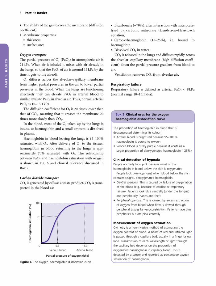
Haemoglobin in blood leaving the lungs is 95–100%
saturated with O2. After delivery of O2 to the tissues,
haemoglobin in blood returning to the lungs is app-
roximately 70% saturated with O2. The relationship
between PaO2 and haemoglobin saturation with oxygen
is shown in Fig. 6 and clinical relevance discussed in
Box 2.
Carbon dioxide transportCO2 is generated by cells as a waste product. CO2 is trans-
ported in the blood as:
• Bicarbonate (∼70%), after interaction with water, cata-
lysed by carbonic anhydrase (Henderson–Hasselbach
equation)
• Carboxyhaemoglobin (15–25%), i.e. bound to
haemoglobin
• Dissolved CO2 in water
CO2 is released in the lungs and diffuses rapidly across
the alveolar–capillary membrane (high diffusion coeffi -
cient) down the partial pressure gradient from blood to
air.
Ventilation removes CO2 from alveolar air.
Respiratory failureRespiratory failure is defi ned as arterial PaO2 < 8 kPa
(normal range 10–13.1 kPa).
Box 2 Clinical uses for the oxygen haemoglobin dissociation curve
The proportion of haemoglobin in blood that is
deoxygenated determines its colour:
• Arterial blood is bright red because 95–100%
haemoglobin is bound to oxygen
• Venous blood is dusky purple because it contains a
larger proportion of deoxygenated haemoglobin (∼25%)
Clinical detection of hypoxiaPeople normally look pink because most of the
haemoglobin in blood below the skin is oxygenated
People look blue (cyanosis) when blood below the skin
contains >5 g/dL deoxygenated haemoglobin.
• Central cyanosis. This is caused by failure of oxygenation
of the blood (e.g. because of cardiac or respiratory
failure). Patients look blue centrally (under the tongue)
and peripherally (hands and feet)
• Peripheral cyanosis. This is caused by excess extraction
of oxygen from blood when fl ow is slowed through
peripheral tissues by vasoconstriction. Patients have blue
peripheries but are pink centrally
Measurement of oxygen saturationOximetry is a non-invasive method of estimating the
oxygen content of blood. A beam of red and infrared light
is passed through a capillary bed, usually in a fi nger or ear
lobe. Transmission of each wavelength of light through
the capillary bed depends on the proportion of
oxygenated haemoglobin in capillary blood. This is
detected by a sensor and reported as percentage oxygen
saturation of haemoglobin.Partial pressure of oxygen (kPa)
Hae
mo
glo
bin
sat
ura
tio
n (
%)
Arterial bloodVenous blood
0 5.3 13.3
100
75
Figure 6 The oxygen–haemoglobin dissociation curve.

Basic Science 7
PA
RT
1:
BA
SIC
S
Type I respiratory failure (Box 3)
• Defi ned as PaO2 < 8 kPa without hypercapnia (PCO2
low or normal)
• Caused by any disease that impairs gas transport across
the alveolar–capillary membrane by:� reducing the surface area of the alveolar–capillary
interface (loss of alveolar ventilation, capillary diffu-
sion or both)� increasing the thickness of the alveolar–capillary
membrane
• As the diffusion coeffi cient for O2 is 20 times lower
than for CO2, mild or moderate impairment of gas trans-
port across the alveolar–capillary membrane will cause
hypoxia without altering blood PaCO2 (type I respiratory
failure). Severe impairment of gas transport across the
alveolar–capillary membrane will impair CO2 transport
as well as O2 transport and cause type II respiratory
failure (see below)
Type II respiratory failure (Fig. 7)
• Defi ned as PaO2 < 8 kPa and hypercapnia
• Caused by:� impaired ventilation, preventing exhalation of CO2
� any severe lung disease that causes suffi cient impair-
ment of gas transport across the alveolar–capillary
Box 3 Some causes of type I respiratory failure
Reduction in surface area of the alveolar–capillary interfaceReduced alveolar ventilation• Obstructive airways disease – reduced airfl ow to alveoli,
e.g.� asthma� COPD (chronic bronchitis)
• COPD (emphysema) – destruction of alveoli
• Pneumonia – pus replaces air in the alveoli
• Lung cancer – replacement of lung parenchyma with
tumour or loss of air-fi lled alveoli because of associated
lung collapse
Reduced capillary perfusion• Pulmonary emboli – pulmonary arteries are obstructed
by clots
• COPD (emphysema) – destruction of pulmonary
capillaries
Increased thickness of the alveolar–capillary membrane• Interstitial lung disease – increased connective tissue is
deposited in the interstitial space
• Pulmonary oedema – the alveolar–capillary membrane is
thickened by oedema fl uid
Cervical cord (C3, 4, 5), e.g.Spinal trauma
TumourSyringomyelia
Muscles, e.g.Muscular dystrophy
Severe lungdisease, e.g.
EndstageCOPD
Respiratory centre in brainstem, e.g.Drugs/alcohol
TraumaRaised intracranial pressure
Nerve, e.g.Guillain–Barré syndromeMotor neuron disease
Neuromuscularjunctions, e.g.Myasthenia gravis
Chest wall, e.g.Obesity hypoventilation
KyphosisFlail chest
Figure 7 Causes of type II
respiratory failure. Conditions
causing neurological, muscular,
chest wall or severe lung disease
all can cause type II respiratory
failure by impairing alveolar
ventilation.
8 Part 1: Basics
PA
RT
1:
BA
SIC
S
membrane to reduce both CO2 and O2 transport (e.g.
advanced COPD, status asthmaticus, massive pulmo-
nary embolus)
KEY POINT
Any lung disease that impairs oxygen transport can lead to
type I respiratory failure. When the lung disease becomes
more severe and carbon dioxide transport is also impaired,
this leads to type II respiratory failure. For example, type I
respiratory failure may be caused by exacerbations of mild
to moderate COPD or by moderate to severe asthma
exacerbations, but severe COPD or life-threatening asthma
may cause type II respiratory failure.
Effects of respiratory failureRespiratory failure may be life-threatening as both
hypoxia (Box 4) and hypercapnia (Box 5) have serious
detrimental effects on cells, tissues and organs.
Some cells are unable to tolerate hypoxia – e.g. neurons
are damaged irreversibly after 4–6 min of hypoxia. Other
cells (e.g. skeletal muscle cells) survive more prolonged
hypoxia.
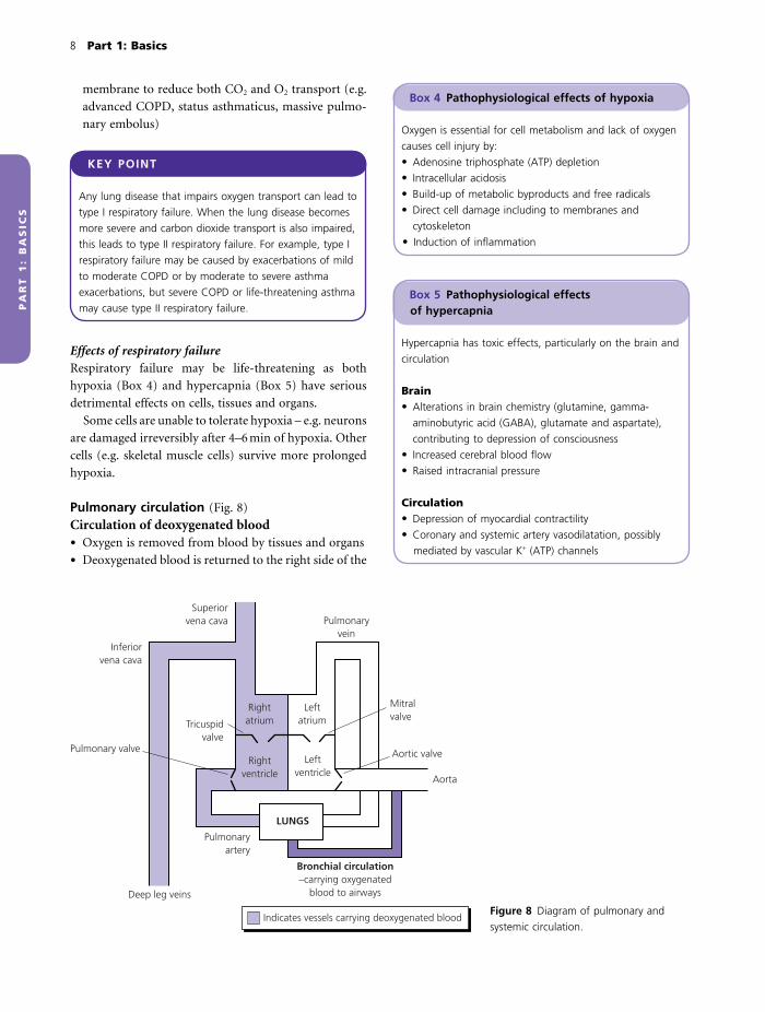
Pulmonary circulation (Fig. 8)
Circulation of deoxygenated blood• Oxygen is removed from blood by tissues and organs
• Deoxygenated blood is returned to the right side of the
Box 5 Pathophysiological effects of hypercapnia
Hypercapnia has toxic effects, particularly on the brain and
circulation
Brain• Alterations in brain chemistry (glutamine, gamma-
aminobutyric acid (GABA), glutamate and aspartate),
contributing to depression of consciousness
• Increased cerebral blood fl ow
• Raised intracranial pressure
Circulation• Depression of myocardial contractility
• Coronary and systemic artery vasodilatation, possibly
mediated by vascular K+ (ATP) channels
Box 4 Pathophysiological effects of hypoxia
Oxygen is essential for cell metabolism and lack of oxygen
causes cell injury by:
• Adenosine triphosphate (ATP) depletion
• Intracellular acidosis
• Build-up of metabolic byproducts and free radicals
• Direct cell damage including to membranes and
cytoskeleton
• Induction of infl ammation
Pulmonaryvein
Superiorvena cava
Inferiorvena cava
Tricuspidvalve
Rightatrium
Rightventricle
Leftventricle
Leftatrium
Mitralvalve
Aorta
Indicates vessels carrying deoxygenated blood
Bronchial circulation–carrying oxygenated
blood to airways
Pulmonaryartery
LUNGS
Deep leg veins
Aortic valvePulmonary valve
Figure 8 Diagram of pulmonary and
systemic circulation.

Basic Science 9
PA
RT
1:
BA
SIC
S
heart by systemic veins, which feed into the superior and
inferior vena cava
• The superior and inferior vena cava drain into the right
atrium. Deoxygenated blood is pumped out of the
right atrium through the tricuspid valve into the right
ventricle
• Contraction of the right ventricle pumps deoxygen-
ated blood through the pulmonary valve into the pulmo-
nary artery
• Features of pulmonary arteries:� pulmonary arteries have thinner walls, less smooth
muscle and greater internal diameters than systemic
arteries� systolic and diastolic blood pressures in the main
pulmonary arteries (∼25/8 mmHg) are much lower
than in systemic arteries (120/80 mmHg)� pulmonary arterial pressure is increased by hypoxia,
hypercapnia and acidosis as well as by stimulation by
the nervous system and infl ammatory mediators
• The pulmonary arteries divide many times and these
branches carry deoxygenated blood down into the net-
work of 280 billion capillaries, where it is oxygenated
Circulation of oxygenated blood• Blood oxygenated at the alveolar–capillary interface
enters the pulmonary veins
• The pulmonary veins drain into the left atrium
• Blood in the left atrium is pumped through the mitral
valve to the left ventricle
• Oxygenated blood in the left ventricle is pumped
through the aortic valve into the aorta and circulates in
the systemic arteries to carry oxygen to tissues and organs
Supply of oxygen to pulmonary tissuesThe bronchial circulation is a second pulmonary blood
supply which carries oxygenated blood to lung structures
including trachea, bronchi down to the level of the ter-
minal bronchioles, hilar lymph nodes and visceral pleura.
Bronchial arteries arise from the aorta or from intercostal
arteries and bronchial veins drain into the pulmonary
vein. The lung parenchyma does not have a second blood
supply as it receives oxygen directly from air in the
alveoli.
Pulmonary defences (Fig. 9)
Inspired air may be contaminated with foreign particles,
such as allergens, organisms and pollutants, with poten-
tial to injure the lung. Adults inhale around 10,000 L air
each day, putting the lung at considerable risk of expo-
sure to these damaging particles. The lung has an elabo-
rate system of defences to trap and clear inhaled particles
from the lung and additional mechanisms to deal with
pathogens that evade removal from the lung.
Trapping of inhaled particles• Large particles (>10–15 µmol) are trapped by nasal
hairs
• Smaller particles (2–10 µmol) are trapped in mucus
lining the upper and lower airways
• Tiny particles reach the alveoli, where they are depos-
ited in alveolar fl uid
Removal of trapped particles from the lungSneeze and coughReceptors in the respiratory tract detect the presence of
particles by mechanical or chemical means. This can
trigger bronchospasm to prevent deeper penetration of
particles into the lung. Stimulation of receptors also trig-
gers a sneeze or cough refl ex, generating explosive airfl ow
that expels particles from upper and lower airways,
respectively.
Mucociliary escalatorThe upper and lower airways are lined with ciliated epi-
thelium. Overlying the epithelium is a thin layer of liquid,
called airway surface liquid, which has two components
(Fig. 9). The watery liquid nearest the epithelial cell
surface is called the ‘sol’ layer, and has a depth equivalent
to the length of the cilia. This layer allows the cilia to
extend fully and beat freely. Overlying the ‘sol’ layer is the
gel layer, which contains mucus. This is the layer that
traps inhaled particles. The beating of the cilia propels
the ‘gel’ layer and trapped particles out of the airway into
the pharynx, where mucus is expectorated or swallowed.
Alveolar macrophagesInhaled particles that reach the alveoli are engulfed by
alveolar macrophages. Potential outcomes of this are:
• Destruction of particles by macrophage lysosomes
• Removal of macrophages by the mucociliary escalator
• Clearance of macrophages by the lymphatic system
• Persistence of macrophages containing particles in the
lung
Epithelial barrierAirway epithelial cells are interspersed by tight junctions,
forming a barrier that resists invasion of organisms or
other particles into the body.

10 Part 1: Basics
PA
RT
1:
BA
SIC
S
Immune systemDetection of foreign particles, such as organisms or aller-
gens, in the lung triggers both innate and adaptive
immune responses.
Innate immunity• This is a non-specifi c front line immune system which
broadly recognizes that a particle is foreign, but does not
identify the particle
• When foreign particles are detected, innate immunity
is activated rapidly and provides immediate defence
• The main action of the innate immune system is to
dispose of foreign particles:� phagocytes such as neutrophils and macrophages
engulf and destroy particles� lysozyme, lactoferrin, defensins and complement in
airway surface liquid kill bacteria� surfactant molecules in airway surface liquid opso-
nize bacteria and particles, making them more suscep-
tible to being engulfed by phagocytes
• The innate immune response changes little with age or
exposure to infection and has no memory
Adaptive immunityThe adaptive immune system generates a specifi c response
to individual pathogens that is much slower than the
innate immune response (days rather than minutes–
hours). The adaptive response destroys pathogens that
evade the innate immune response and has a ‘memory’
that strengthens subsequent responses to the same
infection.
Antigen-presenting cells. Antigen-presenting cells, such as
dendritic cells, provide a link between innate and adap-
tive immune systems. Dendritic cells that have engulfed
pathogens migrate to the lymph nodes. During migra-
tion they process antigens from the pathogen and
present them on their cell surface in association with a
major histocompatibility complex (MHC), normally
used for self-recognition. In the lymph nodes the
MHC–antigen complex is recognized by and activates
T lymphocytes.
CD4+ cells (T helper cells). The role of CD4+ T lympho-
cytes is to orchestrate the immune response. These cells
do not directly destroy pathogens. Activated CD4+ T
lymphocytes generate cytokines and stimulate infl ux of
phagocytes and cytotoxic cells. CD4+ T lymphocytes can
trigger two main responses to activation, mediated by
different cytokines and leading to two different immune
responses:
• Th1 response. Cytotoxic T cells are activated, activating
cell-mediated immunity which is most effective against
intracellular pathogens. CD8 cells (cytotoxic T cells)
destroy infected cells by inducing apoptosis and releas-
ing cytotoxic molecules, which lyse cells by punching
holes in their membranes. Dead cells are cleared by
phagocytes.
• Th2 response. B lymphocytes are activated by free
antigen in combination with T helper (Th) cells. This
stimulates them to differentiate into plasma cells that
produce antibodies (IgM, IgG, IgA, IgE). Antibodies con-
tribute to the immune response by binding to antigen,
triggering phagocytosis and activating the complement
cascade. They can also be directly cytotoxic. This response
is called humoral immunity and is most effective against
extracellular pathogens.
Immune memory. Some T and B cells activated during
the adaptive immune response will become memory
DISTAL LUNGSneezing, coughing
Mucociliary escalator
‘Gel’ layerAir
Cilia within ‘sol’layer of airwaysurface liquid
Innate and adaptive immune systems
InterstitiumTight junctionEpithelial
barrier
Epithelialcell
PHARYNX
Airwaysurface
liquid
Trapped foreignparticle
Figure 9 Defence of the lung against
inhaled foreign particles.

Basic Science 11
PA
RT
1:
BA
SIC
S
cells. If infection with the specifi c pathogen occurs again,
memory cells allow the body to mount a strong response
against infection. Immune memory occurs naturally after
infection and can be induced artifi cially by vaccination.
Pathological processes affecting the lungInfl ammationInfl ammation is a process stimulated by tissue injury
(Box 6), which has the physiological aims of eliminating
the trigger and initiating healing and repair.
Acute infl ammationInitial exposure to an infl ammatory trigger initiates an
acute response including:
• Vascular changes. Capillary vasodilatation and in -
creased permeability
• Release of chemical mediators
• Infl ammatory infi ltrate (innate and adaptive immune
cells)
These responses account for the classic features of
acute infl ammation:
• Rubor – redness
• Calor – heat
• Dolor – pain
• Tumor – swelling
• Laesio functae – loss of function
The clinical features of lung infl ammation depend on
the part of the lung involved (Box 7). The Latin word for
infl ammation is itis, and addition of this term to an ana-
tomical name implies infl ammation of that part of the
body. For example, rhinitis means nasal infl ammation
and bronchitis means bronchial infl ammation.
Outcomes of acute infl ammationAcute infl ammation brings phagocytes and lymphocytes
to the site of tissue injury. These cells act to remove the
infl ammatory trigger and clear damaged or infected cells.
The outcome of these processes may be:
• Resolution. Elimination of the trigger allows normal
tissue architecture and function to be restored
• Scarring. After the trigger has been eliminated, dama-
ged tissue is replaced with fi brosis. This reduces the
Box 6 Some triggers of infl ammation in the lung
Infection• Bacteria (e.g. Streptococcus pneumoniae)
• Viruses (e.g. infl uenza A)
• Fungi (e.g. Pneumocystis jirovecii, causes Pneumocystis
pneumonia)
Allergens• Plant (e.g. pollen)
• Animal (e.g. cat dander)
• Microorganisms (e.g. fungal spores, house dust mite)
Cigarette smokingPollutants• Traffi c fumes
• Smoke inhalation
Inorganic material• Asbestos
• Silica
• Coal dust
Drugs• Amiodarone
• Methotrexate
Box 7 Clinical features of acute infl ammation in the lung
Upper airways (e.g. rhinitis, pharyngitis, laryngitis)
• Tumor – blocked nose, hoarse voice
• Dolor/rubor – sore/red throat
• Loss of function – diffi culty breathing/swallowing/
speaking
• Increased secretion – runny nose
Lower airways (e.g. bronchitis/ asthma)
• Tumor – airways obstruction, wheeze
• Dolor – cough
• Loss of function – respiratory failure
• Increased secretion – sputum production
Lung parenchyma (e.g. pneumonia/ infarction)
• Tumor – reduced air entry
• Loss of function – respiratory failure
• Increased secretion – sputum production
Pleura (pleurisy)
• Dolor – pleuritic chest pain
• Loss of function – diffi culty taking a deep breath
• Increased capillary leak – pleural effusion

12 Part 1: Basics
PA
RT
1:
BA
SIC
S
function of the affected organ to an extent depending on
the amount of scarring
• Persistence. The trigger is not eliminated and drives
chronic infl ammation, leading to progressive tissue
damage, scarring and loss of function. Chronic infl am-
mation includes:� mononuclear cell infi ltrate (macrophages, lympho-
cytes, plasma cells)� tissue destruction� production of granulation tissue containing fi bro-
blasts and small blood vessels� fi brosis
• Systemic spread. One function of infl ammation is
to contain the infl ammatory trigger to a local site. If
this fails the problem may spread to the rest of the
body
Respiratory examples of these processes are given in
Box 8.
HypersensitivityHypersensitivity is a heightened or excessive immune
response to an antigen, that is pathological and has clin-
ical consequences.
Type I hypersensitivity (‘immediate’ or ‘anaphylactic’)Respiratory diseases caused by type I hypersensitivity
reactions include asthma and allergic rhinitis, where the
reaction causes mucosal oedema, increased volume of
secretions and smooth muscle contraction, leading to
airfl ow obstruction.
Exposure to an external allergen, such as pollen, animal
dander or house dust mite, triggers production of immu-
noglobulin E (IgE) which becomes bound to mast cells.
Subsequent exposure to the allergen causes bound IgE on
mast cell surfaces to form cross-links, which results in
degranulation of mast cells and release of:
• Histamine
• Leukotrienes
• Prostaglandins
These infl ammatory mediators:
• Increase capillary permeability, causing mucosal
oedema
• Attract infl ammatory cells, including eosinophils,
neutrophils and macrophages
• Cause bronchoconstriction
An early phase reaction occurs within 5–10 min of
allergen exposure. Infl ux of infl ammatory cells mediates
a late phase response, which is maximal 4–6 h later.
Type III hypersensitivity (‘immune complex’)Respiratory disease caused by type III hypersensitivity
includes extrinsic allergic alveolitis, where the immune
reaction causes alveolar oedema and impairs gas
exchange.
Antigen exposure leads to production of free circulat-
ing IgG antibodies. On re-exposure the antigen binds to
these IgG antibodies, forming immune complexes. These
complexes can cause some mast cell degranulation and
release of infl ammatory mediators, although much less
than in type I reactions. The complexes are also deposited
in target tissues where they stimulate an acute infl amma-
Box 8 Respiratory examples of outcomes of acute infl ammation
Resolution• Acute rhinitis (viral cold) and bronchitis recover
completely when infection resolves
• Acute anaphylaxis or asthma recover completely on
removal of allergen
Scarring• Bronchiectasis can be caused by single episode of
whooping cough, measles or pneumonia
• Acute lung injury (e.g. caused by aspiration, inhalation
of toxic gases/smoke, trauma) may resolve with scarring
Persistence• Respiratory disease caused by chronic exposure to
infl ammatory triggers leading to progressive disease
includes:� COPD – caused by tobacco smoking� chronic asthma – e.g. caused by chronic allergen
exposure� pulmonary fi brosis – e.g. caused by ongoing
connective tissue disease, chronic drug use (e.g.
amiodarone) or unknown trigger� pleural thickening brought about by persistence of
asbestos fi bres in the lung
Systemic spreadDiseases that spread from the respiratory tract to the rest
of the body include:
• Infections such as pneumonia, which may be
complicated by septicaemia
• Primary pulmonary tuberculosis, which can spread by
blood and lymphatics to any site in the body, including
lymph nodes, bone and kidneys

Basic Science 13
PA
RT
1:
BA
SIC
S
tory response, with complement activation, neutrophil
infi ltration and lysosymal enzyme release with tissue
destruction.
Type III reactions come on over 4–12 h following
antigen exposure. Susceptibility to type III reactions is
determined in part by genetic factors and also may be
altered by environmental factors such as infection
or cigarette smoking in conjunction with antigen
exposure.
Type IV hypersensitivity (‘delayed’)Respiratory disease caused by type IV hypersensitivity
includes pulmonary tuberculosis, where the immune
reaction causes progressive tissue destruction. This reac-
tion is mediated by antigen-specifi c T cells and anti-
bodies are not involved. The antigen driving the reaction
is picked up by an antigen-presenting cell such as a tissue
dendritic cell.
The antigen-presenting cell binds to type I or II MHC.
Patrolling T lymphocytes (CD4 helper or CD8 cytotoxic
cells) fi nd the antigen in this context and become acti-
vated, releasing cytokines and chemokines. This attracts
infl ammatory cells and an infi ltrate comprising mainly
T cells and macrophages develops. These infi ltrates may
develop into granulomas.
Other types of hypersensitivity (II, V and VI) have
been described but are not major contributors to lung
disease.
InfectionInfection occurs when an organism enters a host, repli-
cates and causes damage to the host during the process.
The respiratory system is particularly vulnerable to infec-
tion as it is exposed to large volumes of inspired air which
may contain microorganisms. Respiratory infection can
be caused by a vast array of viruses, bacteria and fungi,
which can come from human, animal or environmental
sources (Box 9).
Evasion of respiratory defences by infecting organismsAn organism must overcome a series of immune hurdles
(Fig. 9) before it can infect the respiratory tract.
Mucociliary clearanceThis process traps inhaled organisms and clears them out
of the respiratory tract. Some organisms evade this
defence by adhering to the underlying epithelium, for
example using molecules called adhesins or ligands that
recognize and stick to receptors on host cell surfaces.
Pseudomonas aeruginosa has alternative strategies to
promote adherence. These bacteria produce alginate, a
thick substance that clogs up the mucociliary escalator,
and pyocyanin, a compound that disrupts the beating of
cilia.
Epithelial barrierEpithelial cells interspersed with tight junctions form an
intact barrier that resists invasion of organisms. Once
adherence has occurred, organisms either can invade epi-
thelial cells or disrupt the epithelial barrier and pass
between the cells.
Innate and adaptive immunityOrganisms have a range of strategies to evade innate and
adaptive immune responses. For example, Streptococcus
Box 9 Sources of respiratory infection
HumansMost respiratory infection is spread between humans.
Most organisms in the respiratory tract cause infection in
their host, then are replicated and passed on in respiratory
droplets, e.g.
• Common cold (over 100 different viruses)
• Infl uenza
• Tuberculosis
Some organisms are ‘carried’ in the upper respiratory
tract, but do not cause infection until they are passed on
to a susceptible individual or until their host’s immune
defences are weakened (e.g. Staphylococcus aureus)
Birds and animalsSome microorganisms primarily infect birds or animals, but
can infect humans who come into close contact with
infected creatures, e.g.
• Coxiella burnetti (from cattle/sheep)
• Chlamydia psittaci (from parrots)
• Infl uenza (from birds)
EnvironmentSome organisms normally live in water, soil or other places
in the environment, but can infect the respiratory tract
when they are aerosolized (e.g. as water droplets, dust),
e.g.
• Legionella pneumophila
• Pseudomonas aeruginosa
• Aspergillus fumigatus

14 Part 1: Basics
PA
RT
1:
BA
SIC
S
pneumoniae and Haemophilus infl uenzae surround them-
selves with a glycocalyx to inhibit phagocytosis. Staphy-
lococcus aureus and Streptococcus pneumoniae secrete
a molecule, leucocidin, that kills leucocytes and
macrophages.
Clinical impact of respiratory infectionThe clinical features of respiratory infection are deter-
mined by properties of the organism, the strength of the
host immune response and the interaction between
them.
Organism virulenceVirulence or pathogenicity of an organism refers to its
ability to cause disease.
Highly virulent organisms are capable of causing
infection in anyone, irrespective of the strength of the
immune response. Respiratory examples include:
• Viruses causing the common cold, which cause infec-
tion in 95% of people if they come into contact with the
respiratory tract
• Streptococcus pneumoniae, which causes pneumonia in
healthy people with normal immune systems
Organisms with low virulence do not cause respiratory
infection in healthy individuals. However, if the immune
response becomes suppressed they may cause opportu-
nistic infection. Organisms causing opportunistic respi-
ratory infection include:
• Pseudomonas aeruginosa
• Pneumocystis jirovecii
• Aspergillus fumigatus
• Mycobacterium tuberculosis – TB infection is contained
in a primary focus, but may reactivate to cause post-
primary infection if the host becomes immunosup-
pressed in later life
ImmunosuppressionOpportunistic respiratory infections occur in patients
with disruption of any aspect of the respiratory immune
system.
• Loss of cough refl ex leads to aspiration and respira-
tory infection, particularly with anaerobic organisms,
e.g.� neurological disease� loss of consciousness
• Impaired mucociliary clearance leads to infection with
Gram-negative bacteria such as Pseudomonas aeruginosa,
e.g.
� damage to ciliated epithelium (e.g. bronchiectasis,
COPD)� increased viscosity of airway surface liquid (e.g.
cystic fi brosis)
• Disruption of the epithelial barrier allows infec-
tion with Staphylococcus aureus and Gram-negative bac-
teria such as coliforms and Pseudomonas aeruginosa,
e.g.� Staphylococcus aureus pneumonia following epithe-
lial damage from infl uenza� secondary to cigarette smoking
• Impaired innate or adaptive immune responses:� human immunodefi ciency virus (HIV) infection
causes generalized suppression of the adaptive
immune response allowing opportunistic infec-
tion (e.g. with M. tuberculosis and Pneumocystis
jirovecii)� patients at the extremes of age have immature or
failing immune systems and are predisposed to respira-
tory infection, particularly pneumonia� development of diabetes mellitus can impair im -
munity, leading to reactivation of M. tuberculosis in
infected patients and predisposing patients to other
respiratory infections� immunosuppressive drugs (e.g. cytotoxic chemo-
therapy, disease-modifying drugs for arthritis, pred-
nisolone) increase the risk of all respiratory infections,
including fungal infections with Aspergillus
Excessive host immune responseThe immune response to infection can directly cause
disease. For example, the cell-mediated immune response
to infection with M. tuberculosis (type IV hypersensitiv-
ity) causes granuloma formation and destruction of lung
tissue.
Site of infectionThe clinical features of respiratory infection are also
determined by the site of infection in the respiratory tract
(Fig. 10) and whether the infection remains localized in
the lung or spreads elsewhere in the body. Examples of
effects of respiratory infection beyond the respiratory
tract include:
• Viral infection:� systemic response to infection (e.g. fatigue, lethargy,
myalgia)� involvement of other organs (e.g. Epstein–Barr virus
infection may involve the liver and spleen)
Basic Science 15
PA
RT
1:
BA
SIC
S
• Bacterial infection:� bacteria may enter the blood stream (bacteraemia).
If they cause adverse systemic effects including
hypotension and multiorgan failure, this is called
septicaemia� bacteria may spread from the lung to other tissues
and organs where they cause infection. A good example
is M. tuberculosis which spreads via blood and lym-
phatic system and causes infection elsewhere (e.g. in
lymph nodes, bone and the kidney)
NeoplasiaNeoplasia or cancer is caused by a population of cells that
become genetically altered so that they evade normal
growth controls. Cancer cells:
• Grow and divide excessively
• Invade adjacent tissues and structures
• Spread to distant sites via blood and lymphatic
systems
• Cause disease and death by destroying normal tissue
and preventing normal function
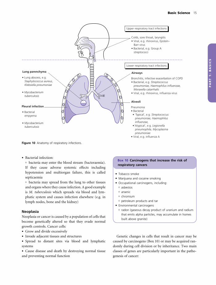
Upper respiratory tract infections
Colds, sore throat, laryngitis• Viral, e.g. rhinovirus, Epstein– Barr virus• Bacterial, e.g. Group A streptococci
Lower respiratory tract infections
Airways
Bronchitis, infective exacerbation of COPD• Bacterial, e.g. Streptococcus pneumoniae, Haemophilus influenzae, Moraxella catarrhalis• Viral, e.g. rhinovirus, influenza virus
Lung parenchyma
• Lung abscess, e.g. Staphylococcus aureus, Klebsiella pneumoniae
• Mycobacterium tuberculosis
Pleural infection
• Bacterial empyema
• Mycobacterium tuberculosis
Alveoli
Pneumonia• Bacterial • ’Typical’, e.g. Streptococcus pneumoniae, Haemophilus influenzae, •’Atypical’, e.g. Legionella pneumophila, Mycoplasma pneumoniae• Viral, e.g. influenza A
Box 10 Carcinogens that increase the risk of respiratory cancers
• Tobacco smoke
• Marijuana and cocaine smoking
• Occupational carcinogens, including:� asbestos� arsenic� chromium� petroleum products and tar
• Environmental carcinogens� radon (gaseous decay product of uranium and radium
that emits alpha particles, may accumulate in homes
built above granite)
Figure 10 Anatomy of respiratory infections.
Genetic changes in cells that result in cancer may be
caused by carcinogens (Box 10) or may be acquired ran-
domly during cell division or by inheritance. Two main
classes of genes are particularly important in the patho-
genesis of cancer:

16 Part 1: Basics
PA
RT
1:
BA
SIC
S
• Oncogenes. Genetic damage to normal genes (proto-
oncogenes) activates oncogenes, increasing cell growth
and division and altering differentiation. These new
properties may allow cells to become cancerous
• Tumour suppressor genes. These genes are normally
responsible for the control of cell division, DNA repair
and programmed cell death. If they are damaged, loss of
function can disrupt these processes, leading to cancer.
Cancers of the respiratory system are listed in Box 11.
The sites of the primary cancers and their metastases
determine the clinical features of the disease.
Box 11 Cancers of the respiratory system
• Nasopharyngeal carcinoma
• Mouth and tongue cancers
• Laryngeal carcinoma
• Bronchogenic carcinoma
• Bronchoalveolar cell carcinoma
• Mesothelioma
PA
RT
1:
BA
SIC
S
Approach to the patient
HistoryMost diagnoses are made from the patient’s history and
this should be taken carefully and thoroughly. At the start
of the consultation you should always introduce yourself,
giving your name and position, and ask for consent to
take a history.
KEY POINT
Always introduce yourself and ask for consent for what
you are going to do whenever working with patients.
Presenting complaintThe consultation should start with an open question to
determine the presenting complaint. This question could
be ‘What is the main problem with your health at the
moment?’ or ‘I understand that you are having problems
with your chest, can you tell me about this in your own
words.’ You should then take a history of the presenting
complaint to establish its cause and determine how it is
affecting the patient’s life.
Patients with respiratory disease most commonly
present with breathlessness or cough.
BreathlessnessBreathlessness or shortness of breath describes the sensa-
tion of not being able to get enough air. The term dys-
pnoea means diffi culty in breathing.
Breathlessness can be quantifi ed using the Medical
Research Council (MRC) dyspnoea score (Box 12).
Repeat measurements are used to determine whether the
patient is stable, improving or deteriorating.
An approach to taking a history from a patient pre-
senting with breathlessness is shown in Fig. 11. The fi rst
step is to determine whether the breathlessness originates
from respiratory, cardiac or other disease (Box 13). This
allows the consultation to be structured towards a more
specifi c differential diagnosis.
Respiratory and cardiac causes of breathlessness can be
distinguished by asking about exacerbating factors, asso-
ciated symptoms and risk factors.
Exacerbating factors• Respiratory disease:
� breathlessness caused by bronchospasm may be
exacerbated by change in temperature, exercise or a
smoky environment
• Cardiac disease:� breathlessness caused by pulmonary oedema is
worse when lying down (orthopnoea)
Associated symptoms• Respiratory disease:
� pleuritic chest pain� cough� sputum� haemoptysis� wheeze
• Cardiac disease:� central chest pain/angina� palpitations� ankle oedema� other vascular disease (myocardial infarction, stroke,
peripheral vascular disease)
Risk factors• Respiratory disease:
� smoking history, childhood illness, occupational
exposure, home environment, family history, infec-
tious contacts (see p. 19)
• Cardiac disease:� hypertension, diabetes, smoking, hypercholesterol-
aemia, obesity, lack of exercise, male gender, ethnic
group, family history
Respiratory disease causing breathlessnessOnce respiratory disease has been identifi ed as the likely
17

18 Part 1: Basics
PA
RT
1:
BA
SIC
S
cause of breathlessness, the next step is to construct a
more specifi c differential diagnosis (Box 14). Demo-
graphic information, including age and gender (Box 15),
may help to narrow down the differential diagnosis.
By this stage in the history-taking process you should
be beginning to consider a differential diagnosis. Further
enquiries as to the onset of breathlessness and respiratory
risk factors may help you to narrow this down further.
Onset of breathlessness• Sudden onset may indicate a sudden pathological
process, e.g.� pulmonary embolus� pneumothorax� inhalation of a foreign body occluding an airway
• Onset over a few hours could indicate:� worsening infl ammation in asthma� accumulation of fl uid in the lungs in pulmonary
oedema
• Onset over days or weeks could be caused by:� accumulation of pleural effusion
Differentiate between specific causes
Use investigation to confirm most likely diagnosis and excludedifferentials and unexpected causes (e.g. anaemia, hyperthyroidism)
Presenting complaint–breathlessness
Othercauses
Respiratorycauses
Consider
Differentiate between main groups of causes
Cardiovascularcauses
Exacerbating and relieving factorsAssociated features
Risk factors
Identify likely organ systeminvolved
Consider specific differential diagnosise.g. respiratory
Pulmonaryfibrosis
Pleuraleffusion
Pulmonaryembolus
AsthmaCOPD
Further history andexamination
Figure 11 Mind map of a consultation with a breathless
patient.
Box 13 Causes of breathlessness
• Respiratory, e.g.� obstructive airways disease, infection, cancer,
pneumothorax, pleural effusion, thromboembolic
disease, restrictive lung disease
• Cardiac, e.g.� Heart failure, arrhythmias, congenital heart disease
• Anaemia
• Renal failure� Kussmaul’s respiration – acidosis
• Hyperthyroidism
• Deconditioning (lack of physical fi tness)
• Anxiety
Box 12 MRC dyspnoea score
1 Gets breathless with strenuous exercise
2 Gets short of breath when hurrying on the level or
walking up a slight hill
3 Walks slower than people of the same age on the level
because of breathlessness, or has to stop for breath
when walking at own pace on the level
4 Stops for breath after walking about 100 yards or after
a few minutes on the level
5 Too breathless to leave the house or breathless when
dressing or undressing
Box 14 Respiratory causes of breathlessness
• Obstructive airways disease� asthma� COPD
• Lung cancer
• Pleural disease� pneumothorax� pleural effusion
• Pulmonary embolus
• Infection� pneumonia� tuberculosis� bronchiectasis
• Interstitial lung disease� pulmonary fi brosis� alveolitis

Approach to the Patient 19
PA
RT
1:
BA
SIC
S
� growth of lung cancer with partial or complete
airway occlusion
• Gradual onset over months or years could represent:� chronic obstructive pulmonary disease (COPD)� lung fi brosis� non-respiratory causes (e.g. anaemia, hyperthy-
roidism)
Risk factors for respiratory diseaseA signifi cant history of risk may point to a specifi c respi-
ratory diagnosis. You should ask about:
• Childhood respiratory illness:� childhood infections such as whooping cough,
measles or recurrent chest infections can cause
bronchiectasis� asthma usually occurs fi rst in childhood. Patients
may ‘grow out’ of childhood symptoms only for asthma
symptoms to recur many years later
• Tobacco smoking. Pack year smoking load should be
calculated (Box 16). A 20-pack year history is widely
accepted as signifi cant risk for:
� COPD� mouth, throat and lung cancer
• Family history. Diseases that run in families include:� asthma and atopy� emphysema (α1-antitrypsin defi ciency)� thromboembolic disease
• Occupational and home environment:� exposure to asbestos increases risk of lung fi brosis,
pleural cancer (mesothelioma) and lung cancer� asthma can be provoked by dusty or damp accom-
modation or can be ‘occupational’ asthma (e.g. caused
by exposure to fl our in a bread-making factory)
• Exposure to animals and birds:� household pets (e.g. cats or dogs) may be a source
of allergen (allergy-provoking factor) for asthma� regular contact with birds is a risk factor for extrin-
sic allergic alveolitis
• Infectious contacts:� a diagnosis of tuberculosis (TB) is much more likely
if the patient has had contact with TB
• Immunosuppression� HIV, immunosuppressant drugs or diabetes mellitus
can increase the risk of opportunistic lung infections
Effect of breathlessness on the patient’s lifeYou should establish:
• The effect of breathlessness on activities of daily living
(e.g. washing, dressing, cooking, eating)
• The available support network (e.g. family and
carers)
• The patient’s ability to take treatment
This information will help you decide on an investiga-
tion and treatment strategy with the patient and may also
be used to initiate extra social support.
CoughThe British Thoracic Society defi nes cough as ‘a forced
expulsive manoeuvre, usually against a closed glottis and
which is associated with a characteristic sound’. Cough
has the important function of preventing foreign matter
entering the lungs and is part of lung defence against
infection (Box 17).
Box 15 Causes of breathlessness in different patient groups
AgeOlder patients are more likely to have degenerative or
neoplastic disease, e.g.
• Heart failure
• COPD
• Lung cancer
• Anaemia (e.g. resulting from gastrointestinal bleeding)
Young adults are less likely to present with breathlessness,
but if they do it is often because of allergy, trauma or
congenital abnormalities:
• Asthma
• Pneumothorax
• Pulmonary embolus
• Congenital heart disease
GenderMales are more likely to:
• Have a spontaneous pneumothorax
• Smoke (although prevalence in women is increasing)
and therefore develop COPD and lung cancer
• Have heart disease at a younger age
Females are more likely to:
• Have connective tissue disease (e.g. rheumatoid arthritis,
systemic lupus erythematosus)
Box 16 Calculation of pack year smoking history
Pack years smoked = (cigarettes per day smoked/20) ×
number of years smoked

20 Part 1: Basics
PA
RT
1:
BA
SIC
S
Taking a history from a patient with coughThe aims of history-taking are to establish the cause of
the cough (Fig. 12) and the effect the cough is having
on the patient’s life.
Duration of the coughThe fi rst step is to establish whether the cough is acute
or chronic. British Thoracic Society defi nitions are:
• Acute cough:� <3 weeks� usually caused by viral infection
• Chronic cough:� >8 weeks� common causes are shown in Box 18
There is a grey area between 3 and 8 weeks where
cough may be caused by recovering acute illness or devel-
oping chronic illness.
If the cough is chronic, the next step is to establish whether
it is caused by pulmonary, nasal or oesophageal disease
or drugs by asking about relevant associated features.
Pulmonary disease. Patients with lung disease may have
associated symptoms of breathlessness or pleuritic chest
pain. Sputum production, especially if purulent or blood-
stained, points to a pulmonary cause.
Nasal or sinus disease. Patients with cough should be
asked about symptoms of nasal (rhinitis) and sinus
(sinusitis) infl ammation, which may cause or merely be
associated with a chronic cough. Symptoms of rhinitis
Rhinosinusitis
Upper respiratory tract• Pharyngitis• Laryngitis• Tracheitis• BronchitisLower respiratory tract• Asthma• Bronchiectasis• COPD• Fibrosis• Lung cancer
Postnasal drip
Inflammation e.g.
Aspiration e.g.FoodAcid
Increased sensitivity of cough reflexAcid reflux stimulating vagal nerve
Figure 12 Causes of cough.
Box 17 The cough refl ex
1 The cough refl ex is initiated by stimulation of cough
receptors in any of:� upper and lower respiratory tract epithelium� pericardium� diaphragm� oesophagus and stomach
2 An afferent impulse is sent from the receptors via the
vagal nerve to the ‘cough centre’ in the medulla of the
brain
3 The higher cortical centres are involved in the cough
refl ex, which to some extent is under voluntary control
4 Efferent fi bres relay impulses to trigger:� inhalation of around 2.5L air� closure of the glottis and vocal cords� forceful contraction of abdominal and intercostal
muscles, generating a very high intrathoracic pressure� sudden opening of the glottis and cords, allowing air
to explode out of the lungs at speeds of up to 100
miles/h� compression of the upper airways so that the air
passes through slit-shaped airways
5 The rapidly moving air carries foreign matter out of the
respiratory tract
Box 18 Common causes of chronic cough
Pulmonary• Asthma
• Smoking
• Any pulmonary disease
Nasal• Rhinitis/sinusitis with postnasal drip
Oesophageal• Gastro-oesophageal refl ux
Drugs• ACE inhibitors

Approach to the Patient 21
PA
RT
1:
BA
SIC
S
include nasal blockage, sneezing and runny nose (rhinor-
rhoea). Sinus symptoms may include pain in the face and
teeth or headaches. Patients may feel catarrh or slime
running down the back of their throat (postnasal drip)
and there may be a small amount of clear or purulent
sputum. Rising from bed or other changes in head posi-
tion may trigger cough by causing sinus drainage and
increased postnasal drip.
Gastro-oesophageal refl ux. If a cough is caused by refl ux
it may occur at times when the lower oesophageal sphinc-
ter is open, including after a meal or when talking or
singing. Patients may have symptoms of choking during
meals or of heartburn and acid or food refl uxing into
their mouths.
Other causes. It is essential to take a full drug history and
ask about smoking habits.
Cough caused by lung diseaseIf the initial history indicates that the patient has lung
disease as the cause of their cough, a detailed history
establishing characteristics of the cough may point to a
specifi c diagnosis.
Character of the cough• A normal cough is explosive and percussive in charac-
ter (Box 17)
• A whooping sound is usually caused by Bordetella per-
tussis infection (whooping cough)
• A bovine cough, which is breathy and has lost the
percussive explosive character of a normal cough, is
heard in patients with vocal cord paralysis. This may be
caused by a lung cancer or enlarged lymph nodes imping-
ing on the recurrent laryngeal nerves
• A wheezy cough may indicate turbulent airfl ow through
narrowed airways as in the bronchoconstriction of
asthma or COPD
TimingThe timing of when the cough occurs may be a clue to
the underlying diagnosis:
• Nocturnal cough. This is classically caused by asthma
which is worse at night. A cough occurring on lying fl at
may be also caused by pulmonary oedema or gastro-
oesophageal refl ux
• Seasonal cough in spring and summer may be caused
by atopic asthma provoked by pollen. Cough occurring
during the winter months may be caused by COPD or
chronic bronchitis, exacerbated by seasonal respiratory
tract infections
• Cough on eating or drinking may indicate an inade-
quate swallow with aspiration of food into the respiratory
tract (e.g. following a stroke)
• Cough on contact with allergens (e.g. household pets)
indicates asthma
• Cough after exercise is usually indicative of asthma
Sputum• A ‘dry’ cough indicates a lack of sputum production
(e.g. seen in pulmonary fi brosis and as a side-effect of
angiotensin-converting enzyme [ACE] inhibitors)
• A ‘fruity’ or ‘rattly’ cough indicates secretions which
may or may not be coughed up (expectorated). Produc-
tion of sputum usually indicates an infective or infl am-
matory process
If the cough is productive of sputum, further questions
should be asked about sputum colour, consistency and
volume:
• Colour:� clear or white phlegm (mucoid or mucus-like) is
common in patients with COPD whose airways are
chronically infl amed but not infected� yellow or green sputum (purulent) is caused by the
presence of neutrophils or eosinophils which release
myeloperoxidase, an enzyme that is green. Purulent spu -
tum usually indicates infection, but may also be seen in
uninfected patients with asthma caused by eosinophils
• Consistency:� fi rm plugs of sputum may be coughed up by patients
with exacerbations of asthma
• Volume:� the daily volume of sputum expectorated can
easily be quantifi ed by the patient in terms of house-
hold measurements (e.g. teaspoonful/tablespoonful/
teacupful)� expectoration of large daily volumes of sputum is
characteristic of bronchiectasis� a cough productive of sputum on most days over at
least a 3-month period for 2 or more consecutive years
in a patient without other explanations for a cough may
be termed ‘chronic bronchitis’. Chronic bronchitis is
usually seen in people who smoke and is part of COPD
HaemoptysisBlood coughed up from the lungs (haemoptysis) can vary
in amount from small streaks of blood to massive bleed-
ing and should ring alarm bells as:

22 Part 1: Basics
PA
RT
1:
BA
SIC
S
• Haemoptysis may indicate an underlying lung tumour
or other life-threatening diagnosis (Box 19)
• The haemoptysis itself may be life-threatening:� large amounts of blood in the airway may asphyxiate
the patient� torrential haemoptysis may cause hypovolaemia and
shock
Haemoptysis should always be thoroughly investigated
to identify or exclude serious underlying disease.
!RED FLAG
• A serious cause underlying a cough should particularly
be considered in patients who have:� haemoptysis� weight loss� night sweats
• Patients with these symptoms require full investigation
Other associated features• Sore throat and nasal congestion may indicate an
upper respiratory tract infection
• Fevers are more indicative of a lower respiratory tract
infection. Rigors and sweats may indicate pneumonia,
whereas persistent night sweats may indicate TB
• Weight loss is a worrying or ‘red fl ag’ symptom and
may be associated with:
� malignancy� TB� other severe infection or lung abscess
• Wheezing occurs in asthma, COPD and other causes
of airway obstruction
• Chest pain:� pleuritic pain (sharp, localized and worse on deep
inspiration and coughing) is most commonly caused
by pneumonia, pneumothorax, pulmonary embolus or
viral pleurisy� pain occurring following vigorous coughing may be
secondary to muscle strain or a fractured rib
ExaminationPreparationYou should obtain informed consent before starting the
examination and ensure that you consider the patient’s
wishes, comfort and feelings at all times. Examination of
the respiratory system ideally requires positioning of the
patient in the centre of a bed or couch, sitting up with
their upper body at an angle of 45° to their legs. The chest
should be exposed before examination and a blanket used
to maintain patient dignity until it is time to examine the
chest directly. Male doctors or students should consider
use of an escort when examining female patients.
RoutineA thorough and professional respiratory examination is
best carried out methodically in the following order.
• General inspection from the end of the bed
• Hands and arms
• Face
• Neck
• Chest front
• Chest back
• Lower limbs
At the end of the examination the fi ndings should be
presented in the same order so nothing is forgotten. This
routine is outlined as an Objective Structured Clinical
Examination (OSCE) checklist in Box 20.
General observationGeneral observations should initially be made from the
end of the bed. In an exam situation this should take
around 30 s and is valuable time that can be used to take
a few deep breaths and look for diagnostic clues.
Around the patientIf available you should look at:
Box 19 Causes of haemoptysis
Common and/or potentially life-threatening• Lung cancer
• Tuberculosis
• Pulmonary embolus
• Pneumonia
• Bronchiectasis
• Heart failure (sputum usually pink)
Massive haemoptysis may occur because of erosion of
an airway blood vessel in patients with:
• Bronchiectasis
• Mycetoma (fungal ball)
Less common• Idiopathic pulmonary haemosiderosis
• Goodpasture’s syndrome
• Trauma
• Blood disorders
• Benign lung tumours

Approach to the Patient 23
PA
RT
1:
BA
SIC
S
• Sputum (in bedside pot) to determine amount pro-
duced and colour
• Medication (e.g. inhalers, nebulizers or oxygen)
• Other equipment and devices (e.g. intravenous
cannula, urinary catheter, peak fl ow meter)
Patient affectYou should determine whether the patient is:
• Alert, drowsy, confused or cooperative
• In any pain
Breathing• Measure the respiratory rate:
� count the number of breaths taken over 15 s and
multiply by 4 to arrive at respirations per minute. This
is often best done when taking the patient’s pulse,
as respiratory rate is under voluntary control and
may change if the patient is conscious that you are
counting� comment on whether this is normal (normal range
12–16 breaths/min)
• Look for diffi culty in breathing or use of accessory
muscles. This may be seen as contraction and promi-
nence of the sternocleidomastoid muscles
• Describe the breathing pattern. Abnormalities
include:� Kussmaul’s respiration – deep sighing breaths seen
in metabolic acidosis� Cheyne–Stokes respiration – alternate deep rapid
breaths and shallow slow breaths with periods of
apnoea occurring in cycles. This is caused by damage
to the respiratory centre in the brainstem (e.g. caused
by stroke or poor cardiac output in heart failure). It is
enhanced by narcotic use
Box 20 OSCE checklist for respiratory examination (10 min station)
PreliminaryIntroduces self (name and role) 1 mark
Explains procedure to patient 1 mark
Obtains consent to proceed 1 mark
Cleans hands 1 mark
Checks patient is at 45° angle and is comfortable 1 mark
General examinationObserves patient from end of bed 1 mark
Measures respiratory rate correctly 1 mark
Comments correctly on:
Presence/absence of respiratory equipment
around bed 1 mark
Use/no use of accessory muscles 1 mark
Shape of chest wall 1 mark
Presence or absence of thoracic scars 1 mark
Presence or absence of intercostal indrawing 1 mark
HandsExamines patient correctly for presence or absence of:
Nicotine staining 1 mark
Finger clubbing 1 mark
Cyanosis 1 mark
Asterixis 1 mark
Comments that would like to take the blood pressure 1 mark
Checks eyes for pallor 1 mark
Comments correctly on presence or absence of
central cyanosis 1 mark
Checks JVP correctly 1 mark
Examines the neck for lymph nodes 1 mark
Determines the position of the trachea 1 mark
Correctly measures the cricosternal distance 1 mark
Comments on presence or absence of tracheal tug 1 mark
ChestChecks expansion of upper and lower lung zones
using correct technique 2 marks
Checks expansion front and back 1 mark
Comments appropriately on fi ndings 1 mark
Percusses over both clavicles (0 if fails to warn
patient) 1 mark
Percusses front and back of chest using correct
technique 1 mark
Compares percussion note on right and left sides
at each level 1 mark
Percusses over lateral chest bilaterally 1 mark
Comments appropriately on fi ndings 1 mark
Auscultates over apices and lateral chest 2 marks
Over front and back of chest compares sides 2 marks
Comments correctly on fi ndings 1 mark
Checks for vocal resonance or fremitus 1 mark
Comments correctly on fi ndings 1 mark
Summarizes examination fi ndings correctly 2 marks
Suggests a differential diagnosis 2 marks
Total 44 marks

24 Part 1: Basics
PA
RT
1:
BA
SIC
S
Chest wall• Look for deformities including:
� barrel-shaped chest (increased anteroposterior
diameter) – caused by chronic hyperinfl ation in COPD
patients� kyphosis and scoliosis (increased forward and lateral
spinal curvature, respectively) – can impair chest wall
movement and cause type II respiratory failure� funnel-shaped chest (pectus excavatum) – may be
congenital� pigeon chest (pectus carinatum) – may occur if the
patient had rickets as a child
• Look for unilateral thoracotomy scars indicating previ-
ous lung or heart surgery
• Look for intercostal indrawing – the muscle between
the ribs is sucked inward as the patient breathes in. This
is seen in patients with severe COPD
Breath sounds• Listen for stridor. This is an inspiratory and/or
ex piratory sound caused by partial obstruction of
the upper respiratory tract (e.g. caused by foreign
body, tumour, oedema, laryngospasm or external
compression)
• It is a medical emergency if the airway is compromised
Hands and armsDuring respiratory examination four main signs should
be looked for in the hands and arms, as follow.
Nicotine stainingIndicates ongoing cigarette smoking.
Finger clubbing (Fig. 13)
Finger clubbing is swelling of the soft tissue of the termi-
nal phalanx. Characteristic features include:
• Loss of nail angle
• Increased bogginess of the nail bed
• Increased side-to-side and lengthwise curvature of the
nail
• A drumstick appearance – where the distal ends of the
fi ngers have a greater diameter than the proximal parts
of the digits
Respiratory diseases causing fi nger clubbing are given
in Box 21. These conditions appear to cause clubbing by
triggering increased blood fl ow to the fi ngertips, causing
oedema followed by expansion of the connective tissue.
Clubbing may also be caused by cardiac and gastrointes-
tinal disease, may be idiopathic or may run in families.
(a)
(b)
Figure 13 Finger clubbing. (a) Loss of nail angle characteristic
of fi nger clubbing. (b) Shamroth’s sign. When two nails are
put back to back as shown you can normally see a diamond
of daylight between them. In this picture the patient has
fi nger clubbing so there is increased tissue in the nail bed with
loss of angle and the diamond is lost.
Box 21 Respiratory diseases causing fi nger clubbing
• Pulmonary fi brosis
• Chronic pulmonary infection� bronchiectasis, cystic fi brosis� empyema� lung abscess� tuberculosis
• Cancer� bronchogenic carcinoma� mesothelioma� pulmonary metastases

Approach to the Patient 25
PA
RT
1:
BA
SIC
S
Evidence of hypoxiaA blue discoloration of the nails (cyanosis) may be seen
and is caused by increased deoxygenated haemoglobin in
the blood in the fi ngers. It may be a result of central or
peripheral causes.
Evidence of carbon dioxide retention• Asterixis. This is looked for by asking the patient to
extend their arms and hyperextend both hands at the
wrist in a policeman ‘stop’ position. If CO2 retention is
present there may be a coarse low-frequency fl apping
tremor. This asterixis is caused by cerebral toxicity and
may also be seen in patients with uraemia or hepatic
encephalopathy. You should ask the patient to keep their
hands in this position for 1 min to ensure asterixis is
absent
• A bounding radial pulse and warm hands may indicate
vasodilatation secondary to CO2 retention
On the way up to the head, the upper arm should remind
you to either take the blood pressure or in an exam to
comment that you would like to take it.
FaceEyesCheck for pallor of the conjunctivae, as an indication of
anaemia as a cause of breathlessness.
MouthAsk the patient to touch the tip of their tongue to the roof
of their mouth and look for cyanosis on the under surface
of the tongue. Cyanosis indicates a mean capillary con-
centration of deoxygenated haemoglobin of around 5 g/dL.
• Central cyanosis occurs when the blood in the systemic
circulation is not oxygenated completely because of
cardiac or respiratory failure. Both the under surface of
the tongue and the fi ngers will appear blue
• Peripheral cyanosis occurs when blood fl ow in the
peripheries (fi ngers, toes, lips) is slow, allowing more
oxygen than usual to be extracted from the peripheral
blood. This can occur because of vasoconstriction on a
cold day or because of vascular disease such as Raynaud’s
syndrome. Only the fi ngers will appear blue and the
under surface of the tongue will be pink
NeckJugular venous pressureMake sure that the patient is sitting with their upper body
at a 45° angle. Ask them to rest their head back on the
pillow to relax the neck muscles and turn their head to
the left. Look for a pulsation along the course of the right
internal jugular vein from between the sternal and cla-
vicular heads of the right sternocleidomastoid up behind
the angle of the jaw to the earlobe.
If a pulsation is seen, it is venous in origin if:
• The pulsation has two visible impulses per heart
beat
• It is not palpable
• It is obliterated by compression of the vein below the
pulse with a fi nger
• Pressure on the abdomen causes the pulsation to rise
in the neck
Then estimate the level of the jugular venous pressure
(JVP) by measuring the vertical height of the pulse (in
cm) above the sternal angle.
• A raised JVP (>3 cm) may indicate right heart failure,
which can be secondary to pulmonary disease (cor
pulmonale)
• Engorged non-pulsating neck veins may indicate supe-
rior vena caval obstruction (e.g. caused by intrathoracic
tumour or lymph node compression). This is usually
accompanied by oedema of the head and neck
LymphadenopathyCervical lymph nodes are most easily felt with the patient
sitting forward and are usually palpated when the back
of the chest is examined. Submental, submandibular,
parotid, mastoid, occipital, anterior and posterior trian-
gle node groups and supraclavicular nodes should be
palpated. If enlarged lymph nodes are detected in the
neck, nodes in the axillae and groins should also be
palpated.
Trachea• Position. Place your index and ring fi ngers on the cla-
vicular heads and push your middle fi nger forward to feel
the dome of the trachea. This should be immediately in
front of your fi nger. Deviation to either side indicates
displacement of the mediastinum (e.g. pulled towards
pulmonary collapse or pushed away by tension pneumo-
thorax or large pleural effusion)
• Tug. Ask the patient to take a deep breath in while
palpating the trachea. A tracheal tug occurs if the trachea
is felt to move downwards on inspiration. This is a sign
of severe COPD, but its cause is unclear. It may be caused
by downward displacement of the mediastinum because
of high intrathoracic pressures generated during inspira-
tion or by upwards movement of a barrel-shaped chest

26 Part 1: Basics
PA
RT
1:
BA
SIC
S
• Cricosternal distance. You should be able to fi t three
fi ngers between the cricoid cartilage (Box 1) and the
notch at the top of the sternum. A reduction of this ‘cri-
costernal distance’ indicates hyperinfl ation of the lungs
ChestThe front of the chest should be examined using the fol-
lowing routine, which should then be repeated at the
back of the chest with the patient sitting forward.
ExpansionExpansion of the upper zones of the lungs (Fig. 14a) is
assessed by placing your hands in the patient’s axillae,
bringing your thumbs together and asking the patient to
breathe in. The degree of expansion is assessed by observ-
ing how far your thumbs are separated on inspiration.
This manoeuvre should be repeated for the lower zones
of the lung (Fig. 14.b) and at the back of the chest. You
should look for:
• Symmetry – do both lungs expand the same amount?
• Amount of expansion – is there any reduction in
expansion?
PercussionYou should check the percussion note of upper, mid and
lower lung zones systematically, comparing right and left
sides. Percuss the apices by tapping the clavicles directly
(Fig. 15a), but warn the patient before you do this that it
may be slightly uncomfortable. Then percuss downwards
through the zones (Fig. 15b), making sure that you
compare the note on left and right sides at each level. Also
percuss laterally over the lungs (Fig. 15c). Repeat percus-
sion at the back of the chest. You should look for:
• Symmetry – is the percussion note the same on each
side at all sites?
• Altered percussion note – is there dullness or hyper-
resonance?
AuscultationAsk the patient to take deep breaths in and out through
their mouth and listen to the breath sounds over the lung
apices with the bell of the stethoscope (used as it fi ts into
the supraclavicular area better than the diaphragm).
Then change to the diaphragm of the stethoscope and
auscultate all zones of the lungs. Once again compare
sides at each level and end laterally. Check the front and
back of the chest. You should listen for:
• Breath sounds – are they present, reduced or absent?
• Characteristics of breath sounds:� vesicular breath sounds (normal) are heard longer
on inspiration than expiration and there is a short
quiet gap between them� bronchial breathing. These breath sounds are
harsher. Inspiration and expiration are more equal in
length and there is no gap between them
• Added sounds?� wheeze is caused by airfl ow obstruction� coarse crackles or crepitations are caused by secre-
tions in large airways� fi ne inspiratory crepitations (like rustling a crisp packet)
are caused by fl uid or fi brosis in or around the alveoli
Vocal resonanceAsk the patient to say ‘99’ repeatedly and listen to the
sound this makes transmitted from the larynx to the
chest wall. You should listen over the upper, mid and
lower zones of the chest, front and back for:
(a)
(b)
Figure 14 Examination of lung expansion. (a) Upper chest.
(b) Lower chest.

Approach to the Patient 27
PA
RT
1:
BA
SIC
S
• Increased resonance – caused by fl uid within the alve-
olar spaces (e.g. pus from pneumonia) which increases
transmission of sound from the larynx
• Decreased resonance – caused by fl uid or air in the
pleural space (effusion or pneumothorax) which muffl es
transmission of sound from the larynx
Alternatively, transmission of sound can be sensed by
touching your hand against the chest wall (vocal fremi-
tus). It is not usually necessary to perform both vocal
resonance and fremitus and you should choose which
one you do depending on how easily you are able to
detect abnormalities with each technique.
LegsTo complete respiratory examination you should check
for
• Peripheral oedema, which may indicate right heart
failure secondary to lung disease
• Unilateral leg swelling, which may indicate a deep vein
thrombosis
Inspect the legs visually. Check for pitting oedema
by pressing over the leg, removing your fi nger and
looking for a dent. Pitting oedema may be immediately
obvious, but can only be excluded if pressure is applied
over the leg in one place for 1 min without causing
denting.
On completion of the examination, thank the patient and
help them back into their clothing if necessary.
Interpretation of examination fi ndingsOnce you have completed your examination decide
whether the fi ndings are symmetrical, affecting both
lungs equally, or asymmetrical, affecting one lung more
than the other (Tables 1 & 2).
Putting it all togetherSome aims of taking a history and performing an exam-
ination are to:
• Make a provisional diagnosis
• Determine an investigation strategy
• Develop a treatment plan
• Form a therapeutic relationship with the patient
• Ascertain the patient’s wishes regarding treatment
• Find out about social circumstances and comorbidities
that may determine response to treatment
• Involve next of kin or other family and friends as
necessary
• Establish the disease prognosis
Once the history and examination fi ndings have been
recorded, this information should be analysed and the
deliberations documented as:
• Differential diagnosis. In straightforward cases a single
diagnosis or a few possible diagnoses (differential diag-
nosis) may be apparent from the history and examina-
tion. This should be written in the notes under this
heading
(a)
(b)
(c)
Figure 15 Percussion of the chest. (a) Lung apices. (b) Front
of chest. (c) Lateral chest wall.

28 Part 1: Basics
PA
RT
1:
BA
SIC
S
• Problem list. In more complicated patients (e.g. those
with multiple illnesses and diffi cult social circumstances)
there may not be a single diagnosis and a problem list
should be written. This should include the new present-
ing problem, comorbidities and social complications. It
can be helpful to document the patient’s problems under
this heading system by system so that no problems are
missed out
Follow-upPatients are often booked to return after a consultation
or told to come back if they remain unwell. The primary
Table 1 Examination fi ndings for diseases affecting both lungs.
Asthma COPD Pulmonary fi brosis Pulmonary oedema
Trachea Central Central Central Central
Reduced cricosternal distance
Tracheal tug
Expansion Reduced bilaterally Reduced bilaterally Reduced bilaterally Normal or reduced
Percussion Normal Normal Normal Normal unless
associated effusions
Auscultation Bilateral expiratory
wheeze
Reduced air entry
May be bilateral expiratory
wheeze
Bibasal fi ne inspiratory
crepitations. Fixed, do
not clear on coughing
Bilateral inspiratory
crepitations, may clear
on coughing
Vocal resonance Normal Normal Normal Normal
Other features Nicotine-stained fi ngers,
intercostal indrawing, use of
accessory muscles
Finger clubbing,
associated connective
tissue disease
Peripheral oedema,
hepatomegaly if right
heart failure
Table 2 Examination fi ndings for diseases affecting one lung more than the other.
Consolidation (pneumonia)
Collapse, e.g. tumour obstructing bronchus Pleural effusion Pneumothorax
Trachea Not displaced Displaced towards
affected side
Small – not displaced Normal – not displaced
Large – displaced away
from affected side
Tension – displaced
away from affected side
Expansion Reduced on affected
side
Reduced on affected
side
Reduced on affected
side
Reduced on affected
side
Percussion Dull on affected side Dull on affected side Stony dull on affected
side
Hyper-resonant on
affected side
Auscultation Bronchial breathing
Crackles
Reduced or absent air
entry
Reduced or absent air
entry
Reduced or absent air
entry
Vocal resonance Increased Reduced Reduced or absent Reduced or absent
Associated features Green/blood-stained/
rusty sputum, pyrexia
Associated features of
malignancy, cachexia

Approach to the Patient 29
PA
RT
1:
BA
SIC
S
purpose of follow-up is to review investigation results
and ascertain response to treatment. In this situation it is
helpful to record your thoughts as an ‘Assessment’, which
both records the likely diagnosis and comments on
change (e.g. whether the patient has improved or dete-
riorated). This is followed in the notes by a plan, detailing
further investigations or treatment required.
KEY POINT
The focal point of patient notes should be the differential
diagnosis, problem list or assessment. This directs patient
care and helps communication between health
professionals and with patients.
InvestigationsMost diagnoses are made by taking a careful history,
backed up by thorough examination. Roles of investiga-
tion therefore include:
• Confi rmation of the clinical diagnosis
• Exclusion of possible differential diagnoses
• Identifi cation of additional diagnoses (comorbidities)
or unexpected diagnoses
• Assessment of fi tness for treatment
• Monitoring of treatment response and adverse
effects
All investigations have some potential to cause
harm. Therefore, the decision to perform a test is made
by weighing up the benefi ts and risks of the test and
discussing these with the patient to ascertain their
wishes. Excessive investigation should be avoided as this
will delay investigations for other patients and waste
resources that could have been used elsewhere for patient
care.
Approach to investigationIt is usual practice to perform low-risk non-invasive tests
fi rst, then to use the results of these to direct further
investigations as necessary.
Initial investigationIn patients with possible respiratory disease initial inves-
tigation usually includes:
• Chest radiograph (X-ray) to look for structural lung
disease
• Peak fl ow recording and/or spirometry to look for
airfl ow obstruction
• Oxygen saturations to assess gas exchange
• Blood tests (venous) including:
� haemoglobin to look for anaemia� C-reactive protein (CRP) and white cell count to
look for infection or infl ammation� eosinophil count to look for allergy
Other investigations that may be useful at this stage
include:
• electrocardiogram (ECG) to look for right heart
abnormalities secondary to lung disease or an alternative
cardiac diagnosis to explain symptoms
• Urea and electrolytes (U&E), bicarbonate, thyroid
function tests, glucose to look for alternative causes of
breathlessness
• Arterial blood gases to assess oxygenation, CO2 excre-
tion and pH
Chest X-rayProcedure. A chest X-ray is normally performed with the
patient standing and facing away from the X-ray tube,
with breath held in deep inspiration. The X-ray beam
thus passes through the patient’s back (posterior) fi rst,
before passing through their front (anterior) to reach the
recording plate. This ‘PA’ fi lm is ideal because:
• A PA fi lm does not exaggerate heart size
• Standing and full inspiration make sure the lungs
are fully expanded and make abnormalities easier to
detect
If a patient is unwell they may have their X-ray sitting
or lying with the plate at their back and the beam passing
from front to back. This is called an AP fi lm and exag-
gerates the heart size.
Risk. The patient receives a radiation dose of 0.1 mSv,
equivalent to 10 days of background radiation. In theory,
any radiation dose carries a slight increased risk of cancer,
but the risk from a chest X-ray is negligible.
Interpretation
• Routine. You should report chest X-rays using a set
routine so that nothing is missed. This routine is outlined
as an OSCE checklist in Box 22
• Quality of X-ray. You should comment on whether the
fi lm is:� PA or AP. If it is AP you should mention that it will
be diffi cult to interpret heart size� the correct way round. The left/right marker should
be on correct side. The correct position of the left
marker is shown in Fig. 16� rotated. The ends of the clavicles (indicated as ‘A’ in
Fig. 16) should be equal distances from the spinous

30 Part 1: Basics
PA
RT
1:
BA
SIC
S
processes (B). If they are not the fi lm is rotated, which
will distort the appearance of thoracic structures
• Features of a normal chest X-ray (Fig. 16). The appear-
ance of a structure on a chest X-ray depends on its density
and the extent to which it allows passage of X-rays. The
least dense substance is air, which does not absorb X-rays
and appears black on the X-ray. The densest substance is
calcium, which absorbs X-rays well and appears white on
the X-ray. In between are fat, which appears dark grey,
and water, which appears light grey. Appearance of indi-
vidual structures is also determined by their thickness,
shape and relationship to adjacent structures (letters in
brackets refer to Fig. 16):� the trachea contains air and appears black. It
should be central and not deviated to left or
right (C)� the heart contains tissue and fl uid and appears very
light grey. Its diameter should be less than half that of
the total cardiothoracic diameter on a PA fi lm. The
heart borders (D, E) should be clearly outlined by the
air in the adjacent lungs. Fuzziness may indicate fl uid
in the airspaces in adjacent lung� the left lung hilum (F) should be higher than the
right hilum (G). The hila normally contain pulmonary
vessels, main airways, lymphatics and nodes. Lymph-
adenopathy or pulmonary arterial hypertension may
cause hilar enlargement� the lung fi elds should appear black and clearly delin-
eated by adjacent structures. Lung markings compris-
ing vessels (seen as small white streaks) should be seen
extending all the way out to the chest wall� the right diaphragm should be higher than the
left as it is pushed up by the liver. The top of the
right diaphragm should be in the 5th intercostal
space (G) anteriorly in the midclavicular line (H). Up
to six ribs should be seen anteriorly above the dia-
phragm (numbered). More than this indicates lung
hyperinfl ation� the costophrenic angles (J) between the rib cage and
diaphragm, and the cardiophrenic angles (K), between
the heart and the diaphragm, should be sharp. Blunt-
ing may indicate the presence of fl uid (effusion) or
fat� it is also helpful to look under the diaphragms at the
gastric air bubble (L). Free gas under the domes of the
diaphragm may indicate perforation of abdominal
viscera� breast shadows should be present in female
patients
Box 22 Example OSCE checklist for chest X-ray interpretation
Checks origin and quality of X-rayChecks patient’s name 1 mark
Checks date of X-ray 1 mark
Checks side marker 1 mark
Checks whether PA/AP 1 mark
Checks rotation 1 mark
Comments on (1 mark) and correctly
describes (1 mark) major abnormality 2 marks
Correctly comments onTracheal position 1 mark
Left heart border 1 mark
Right heart border 1 mark
Heart size 1 mark
Lung hila 1 mark
Right lung fi eld 1 mark
Left lung fi eld 1 mark
Height of diaphragms 1 mark
Clarity of diaphragms 1 mark
Costophrenic and cardiophrenic angles 2 marks for both
Adequate inspiratory effort/hyperinfl ated 1 mark
Breast shadows 1 mark
Bones 1 mark
SummaryCorrectly identifi es abnormalities 2 marks
Suggests differential diagnosis 2 marks
Total 25 marks
A
12
3
4G
F
5
H6 G
D E
JJ
K KL
C
B
A
Figure 16 Normal PA chest X-ray.

Approach to the Patient 31
PA
RT
1:
BA
SIC
S
� you should also look for abnormalities in the bones
such as rib fractures (a break in the straight border of
the rib) or black areas that might indicate metastases
The normal features of the chest X-ray should be kept
in mind when looking at abnormal X-rays with the cases
throughout the book.
Basic lung functionPeak expiratory fl ow rate recordingProcedure. Peak expiratory fl ow rate (PEFR) can be mea-
sured in most situations using a peak fl ow meter (Fig.
17). The patient stands or sits upright and inhales as
deeply as possible. They then seal their lips around the
disposable mouthpiece and make a short sharp expira-
tion, as though attempting to ‘blow out a candle’. The
marker on the meter is displaced by the expiration and
the distance achieved along the scale is noted and relates
to a fl ow rate in litres per minute. This is carried out three
times and the greatest fl ow rate noted (Box 23, p. 35).
Risk. There is a risk of cross-infection which can be
countered by giving patients individual peak fl ow meters
or by using disposable mouthpieces and cleaning the
equipment between patients.
Interpretation. As fl ow through a tube is proportional to
the radius of the tube to the power of 4 (fl ow ∝ r4), peak
fl ow rate gives a crude estimate of airway calibre. Inter-
pretation requires comparison of this with either the
patient’s known best values or with predicted values from
normal range charts depending on gender, age and height.
Peak fl ow can then be expressed as an absolute value in
litres per minute and as a percentage of predicted or best.
Reduction in peak fl ow indicates airfl ow obstruction
resulting from asthma or COPD. The main use of peak
fl ow recording is as serial measurements to look for or
monitor variable airfl ow obstruction in asthma. It is not
usually used to monitor COPD.
SpirometryProcedure. To perform spirometry the patient is asked to
stand or sit upright, to take a deep breath in and to blow
out into the spirometer for as hard and as long as they
can (up to 12 s). This differs from peak fl ow recording
where a short sharp blow is required. The forced mano-
euvre is used to standardize patient effort and fl ow of air
out of the lungs.
Spirometry is recorded as a plot of volume of air blown
out of the lungs in litres (y axis) over a period of time in
seconds (x axis; Fig. 18). The slope of the line indicates
fl ow (liters per second). Three measurements are derived
from the spirometry curve:
• FEV1 – forced expiratory volume in 1 s
• FVC – forced vital capacity, the total volume of air that
is forcibly exhaled
Scale in litres per minute
Flow indicator
Mouthpiece
Figure 17 Peak fl ow meter.
(a) Normal
FEV1
FVC
(b) Restrictive lung disease
(c) Obstructive lung disease
1 2 3 40
1
2
3
4
5 6 7 8 9 10
Time (s)
Volu
me
(L)
Figure 18 Spirometry recording.

32 Part 1: Basics
PA
RT
1:
BA
SIC
S
• FEV1/FVC ratio – this gives some measure of slope
of the line and indicates whether there is airfl ow
obstruction
Risk. There is a risk of cross-infection between patients
with respiratory disease which can be countered by using
disposable mouthpieces and regular cleaning of equip-
ment. Patients may fi nd this procedure makes them
cough or feel breathless.
Interpretation. Normal lung function varies between
individuals depending on age, gender and height. Stan-
dard tables are available to predict an individual’s FEV1
and FVC from these values. Values are normal if they are
80–120% of the predicted value. FEV1 is also expressed
as a percentage of FVC. If the airways are normal, then
at least 70% of the total air in the lungs should have been
blown out in the fi rst second (i.e. FEV1 should be ≥70%
of FVC) (Fig. 18a).
FEV1 and FVC can be reduced both by obstructive
lung disease (e.g. asthma, COPD) and restrictive lung
disease (e.g. lung fi brosis, sarcoidosis). An important
difference between these groups of conditions is that
obstructive lung disease reduces airfl ow, whereas res-
trictive lung disease does not affect the airways. The
ratio of FEV1/FVC can be used to determine airfl ow and
distinguish between obstructive and restrictive lung
disease:
• In obstructive lung disease, the proportion of the air
in the lungs that can leave in 1 s is reduced, hence there
is fl attening of the slope of the spirometric curve and
FEV1 is <70% of FVC (Fig. 18c)
• In restrictive lung disease, the proportion of air in the
lungs that can leave in 1 s is normal, the slope of the line
is normal and FEV1 is ≥70% of FVC (Fig. 18b)
Arterial blood gasesProcedure. Arterial blood is usually sampled from the
radial or brachial artery into a heparinized syringe, to
prevent blood clotting after sampling. The needle is
inserted vertically through the arterial wall and the pres-
sure of blood in the vessel forces blood into the syringe.
The procedure can be painful and local anaesthesia can
be used. However, this may obscure landmarks and make
sampling more diffi cult. Once arterial blood has been
obtained, blood gas analysis should be performed imme-
diately or the sample should be kept on ice until tested
to avoid changes in partial pressure of gases in the
sample.
Risk. The main risks are of occlusion of the artery caused
by spasm of the vessel wall or haematoma around the
artery, which can cause distal ischaemia. Before sampling,
the blood supply of the hand should be tested to ensure
that it is receiving blood from both the radial and ulnar
arteries:
• The patient is asked to make a fi st to squeeze blood out
of their hand
• The doctor presses on both sides of the wrist to com-
press radial and ulnar arteries
• The patient releases the fi st. Their hand looks
white
• The doctor releases the ulnar artery, but not the radial
artery. Blood supply to the hand from the ulnar artery is
proven if the hand goes pink
• The manoeuvre can be repeated, this time releasing the
radial artery and not the ulnar artery to check its contri-
bution to the hand blood supply
• Arterial blood gas sampling from the radial artery is
safest if the hand has a dual blood supply from radial and
ulnar arteries
After sampling, the puncture site should be compressed
fi rmly for several minutes to ensure adequate haemosta-
sis. This will take longer than haemostasis over a vene-
puncture site because the arterial blood is under higher
pressure.
Interpretation. Values should be compared with normal
ranges which will vary depending on the analyser used.
Normal ranges used in this book are given below in
brackets.
Start by looking at the PaO2 (normal range 10–
13.1 kPa). A reduction in PaO2 indicates some problem
with alveolar ventilation, pulmonary capillary perfusion
or the matching of these processes, reducing gas exchange.
PaO2 <8 kPa is defi ned as respiratory failure.
Next, look at PaCO2 (normal range 4.9–6.1 kPa). CO2
excretion depends on its gradient from blood to alveoli,
hence is determined by alveolar ventilation. A fall in CO2
indicates increased alveolar ventilation; a rise indicates
reduced ventilation. Respiratory failure can be classifi ed
further according to CO2 levels:
• Type I respiratory failure – PaO2 <8 kPa, PaCO2 reduced
or normal (Box 3)
• Type II respiratory failure – PaO2 <8 kPa, PaCO2 ele-
vated (Fig. 7)
Determine from pH values (normal range 7.35–7.45)
whether there is acidosis (low pH) or alkalosis (high pH).
If there is acid–base disturbance you then need to decide

Approach to the Patient 33
PA
RT
1:
BA
SIC
S
whether this has a respiratory or metabolic cause using
the following questions:
• Is PaO2 abnormal? If yes this indicates a respiratory
abnormality
• What is the relationship between pH and PCO2?� in respiratory acid–base disturbance, changes in
PaCO2 drive changes in pH according to the Hender-
son–Hasselbach equation
H+ + HCO3− ƨ H2CO3
ƨ H2O + CO2
A rise in PaCO2 drives the equation to the left, increas-
ing production of H+ ions and causing a fall in pH
(respiratory acidosis). A fall in PaCO2 drives the equa-
tion to the right, decreasing production of H+ ions and
causing a rise in pH (respiratory alkalosis). Note that
in respiratory acid–base disturbances PaCO2 and pH
changes occur in opposite directions� in metabolic acid–base disturbance, changes in pH
drive changes in ventilation. A fall in pH (acidosis)
stimulates an increase in respiratory rate with a fall in
PaCO2. A rise in pH stimulates a fall in respiratory rate
with a rise in PaCO2. Note that in metabolic acid–base
disturbances PaCO2 and pH changes occur in the same
direction.
• Finally, look at the bicarbonate (normal range 22–
28 mmol/L). In respiratory acidosis, this may be normal
(acute) or raised (chronic) as renal compensation for
acidosis. In metabolic acidosis (e.g. caused by renal
failure, diabetic ketoacidosis or aspirin overdose), the
bicarbonate is used up as a buffer to the excess acid and
levels are low.
This routine should be used to interpret abnormal blood
gases given with cases throughout the book.
Specialist respiratory investigationsThoracic imagingCT scanningProcedure. Computerized tomography (CT) involves
acquisition of multiple cross-sectional radiographic
images that are reconstructed by a computer to give 3D
images. The patient usually lies on their back (supine)
and is asked to hold their breath to avoid any movement.
The scanner then takes multiple images, either separately
or in a continuous spiral.
• A standard thoracic CT scan images the thorax in
10 mm slices continually down the chest. This does not
miss any part of the lung and is good for investigation of
lung nodules and tumours for example. However, these
thick slices do not obtain detailed images of lung
parenchyma
• A high-resolution CT scan takes more detailed pictures
of the lung in slices that are 1 mm thick. This scan is not
continuous as images are taken at 10 mm intervals to
minimize radiation dose. It is useful for imaging the lung
parenchyma to see emphysema or fi brosis, but may miss
small cancers
• CT pulmonary angiogram uses contrast to outline the
pulmonary arteries and is used to look for pulmonary
emboli
• CT scanning can also be used to guide biopsies of lung
lesions
Risks. Radiation exposure from a thoracic CT scan is
equivalent to 3 years of background radiation. The use of
CT scanning should therefore be considered carefully
particularly in young women, in whom breast exposure
to radiation may be hazardous or who may be pregnant,
and if repeated scanning is planned. The benefi ts of an
accurate diagnosis should be weighed against the poten-
tial risks of CT scanning.
Other thoracic imaging includes• Ultrasound – to evaluate and aspirate pleural effusions
and guide sampling from nodes and soft tissue
• Ventilation–perfusion scanning – to investigate patients
for pulmonary emboli
• Magnetic resonance imaging – sometimes used to clarify
CT fi ndings
• Positron emission tomography – used to assess meta-
bolic activity of structural abnormalities (e.g. possible
lung cancer) seen by other imaging
The use of these techniques is described where relevant
in the cases.
Sampling for pathological diagnosisA defi nitive diagnosis often requires sampling of lung
secretions, fl uid or tissue which is then sent to both
microbiology and pathology laboratories for diagnostic
analysis.
SputumPatients may expectorate sputum spontaneously or after
inhaling nebulized saline (induced sputum).
BronchoscopyProcedure. Fibreoptic bronchoscopy is performed in
patients with or without sedation. The patient is propped

34 Part 1: Basics
PA
RT
1:
BA
SIC
S
in a sitting position and receives a lidocaine spray into
their oropharynx to reduce their cough and gag refl exes.
A thin endoscope is then inserted via the nose or mouth
into the large airways, allowing inspection of the trachea,
main and segmental bronchi.
Rigid bronchoscopy is performed under general anaes-
thesia and involves insertion of a much larger tube into
the trachea. This is usually used where intervention is
required to treat disease such as tumours obstructing the
large airways or narrowing (stricture) of the trachea,
although some centres can now perform such procedures
using fi breoptic bronchoscopy.
Patients must give written informed consent before
any type of bronchoscopy is performed and should be
warned about the risk of complications.
Sampling that can be performed through the broncho-
scope includes:
• Bronchial aspirate. Direct collection of bronchial secre-
tions by suction into a sample pot
• Bronchial washings. Normal saline (10 mL) is syringed
into the airways and removed into a sample pot. This
takes samples from the large airways
• Bronchoalveolar lavage. The bronchoscope is pushed
down in a chosen airway until it is wedged as far as it can
go. A large volume (180–240 mL) of normal saline is
syringed into the lung and removed by suction into a
sample pot. This samples secretions from the distal lung
• Bronchial brushings. A fi ne brush can be used to collect
cells from the epithelial surface
• Endobronchial biopsy. Forceps are used to take tissue
samples directly from a large airway wall
• Transbronchial biopsy. Biopsy forceps are passed into
the lung as deep as possible and biopsies are taken. This
aims to take samples of the lung parenchyma
• Transbronchial nodal aspiration. A fi ne needle is inserted
through the large airway wall into underlying enlarged
lymph nodes and samples are aspirated into the needle.
The site of the nodes is identifi ed either by prior imaging
or by ultrasound down the bronchoscope (endobron-
chial ultrasound)
Risks. Complications of fi breoptic bronchoscopy
include:
• Bleeding
• Infection
• Respiratory compromise
• Pneumothorax
• Death (∼0.01%)
Percutaneous procedures (Fig. 19)
Fine needle aspiration. A fi ne needle is inserted into the
lesion, removed and the contents of the needle expelled
onto a microscope slide for cytological or microbiological
examination. This is particularly useful for sampling
from cervical lymph nodes. The procedure causes minor
discomfort and minimal risk of complications.
Biopsy. A long needle is inserted into the lesion, usually
under radiological guidance, and a core of tissue removed
for histological and microbiological examination. This is
performed under local anaesthetic and may be compli-
cated by bleeding or pneumothorax.
Other sampling• Percutaneous aspiration of effusion or pleural biopsy –
performed with or without radiological guidance to
investigate pleural disease
• Video-assisted thoracoscopy – insertion of an endoscope
through the chest wall to sample pleura and lung
parenchyma
• Open lung biopsy – sampling of lung performed at
open surgery
Percutaneous biopsyneedle
Fine needle foraspiration Figure 19 Sampling needles for
percutaneous procedures.

Approach to the Patient 35
PA
RT
1:
BA
SIC
S
The use of these techniques is described where relevant
in the cases.
Other lung function testingMore detailed information about lung function can be
obtained by measuring lung volumes and gas transfer.
Lung volumesLung volumes are measured by asking the patient to
breathe in and out of a known volume of air containing
a known concentration of helium. Helium is not absorbed
by the lung, so distributes throughout and is diluted by
the air in the lung. The difference between the original
and fi nal helium concentrations allows the volume of air
in the lung to be calculated. Values obtained include:
• Total lung capacity. Take a deep breath in until the
lungs are full. The total volume of air inside them is now
the total lung capacity, which may be:� increased in patients with emphysema who have
reduced elastic recoil of the lung� reduced in patients with pulmonary fi brosis
• Residual volume. Breathe out as far as possible until the
lungs appear empty. There is still some air left inside as
the lungs are prevented from collapsing by negative intra-
pleural pressure. This is the residual volume which:� may be increased in patients with airfl ow obstruc-
tion (e.g. asthma, emphysema)
• Vital capacity. This is the volume of air exhaled (i.e. the
difference between total lung capacity and residual
volume). The vital capacity is reduced in both restrictive
and obstructive lung disease
Gas transferThe ease of diffusion of gases across the alveolar capillary
membrane is tested using carbon monoxide. The patient
breathes out then inhales a single breath of air containing
0.28% carbon monoxide. Their next exhalation is col-
lected and the residual carbon monoxide concentration
is measured. After correction of values for lung volumes
measured by helium, initial and fi nal carbon monoxide
concentrations are compared. The difference indicates
how much carbon monoxide was taken up and is
expressed as a transfer factor.
Diseases of the lung parenchyma that interfere with
transfer of gas across the alveolar–capillary membrane
cause reduction in the transfer factor (e.g. emphysema,
pulmonary fi brosis).
TreatmentGeneral treatment principles are outlined in this section.
Specifi c treatments for individual respiratory diseases are
discussed under the relevant cases.
Approach to treatment of respiratory emergenciesLife-threatening emergencies affecting the respiratory
system include:
Box 23 Example OSCE checklist for performing peak fl ow measurement
This is a rather brief procedure for a 5 min OSCE station
and so is likely to be combined, e.g. with checking inhaler
technique or giving advice
Introduces self (name and role) 1 mark
Outlines procedure 1 mark
Obtains consent 1 mark
DemonstrationShows patient peak fl ow meter 1 mark
Explains principles of action 1 mark
Tells patient to make good seal with
mouth around mouthpiece 1 mark
Explains that they should blow as hard as
they can 1 mark
Asks patient not to put hand over fl ow
indicator 1 mark
Demonstrates peak fl ow measurement
correctly 1 mark
MeasurementChanges mouthpiece 1 mark
Checks patient is standing or sitting
upright 1 mark
Checks peak fl ow indicator is at zero 1 mark
Asks patient to perform manoeuvre 1 mark
Reads peak fl ow correctly 1 mark
Gives feedback to patient 1 mark
Asks them to repeat measurement twice
more 1 mark
Thanks patient 1 mark
InterpretationGives value as best measurement of the
three performed 1 mark
Uses normal range charts to estimate
value as percentage of predicted 1 mark
Interprets measurement 1 mark
Simulated patient markClear explanation up to 2 marks
Total 22 marks

36 Part 1: Basics
PA
RT
1:
BA
SIC
S
• Respiratory arrest
• Life-threatening asthma
• Severe type II respiratory failure
• Massive pulmonary embolus
• Tension pneumothorax
• Anaphylaxis
In these situations the usual consultation order is
reversed and the patient receives life-saving treatment
before an attempt is made to make a diagnosis (Box
24).
The fi rst three steps should always be basic life
support:
A Ensure the airway is patent and clear it if not
B Check if the patient is breathing and support if
not
C Check circulation (pulse and blood pressure) and
support if absent/inadequate
Once basic life support has been performed further
assessment will determine action as described in the indi-
vidual cases. In many cases patients will need respiratory
support as well as disease-specifi c measures.
Respiratory supportPatients with respiratory failure have hypoxia with or
without hypercapnia. These blood gas abnormalities
cause cell and tissue damage (Boxes 4 & 5) and must be
corrected.
HypoxiaHypoxia is treated by increasing the partial pressure of
inspired oxygen. The patient wears nasal cannulae or a
face mask (Fig. 20) which allows them to breathe a
mixture of air (21% oxygen) and 100% oxygen (e.g. from
a cylinder or piped supply). The fi nal concentration of
oxygen in inspired gas depends on the fl ow rate of the
oxygen and the type of device used.
Nasal cannulae. These are convenient to wear and the
patient receives continuous oxygen even when talking
and eating. However, the oxygen concentration delivered
is unpredictable (25–39% at oxygen fl ows 1–4 L/min)
and insuffi cient for many patients.
Venturi mask. The attachments (A) shown in Fig. 21 are
connected to the mask (E) and to the oxygen supply (D).
Entry of oxygen into the attachment at the fl ow rate
shown (C) draws air in through the side holes (B) and
air and oxygen are mixed to a fi nal fi xed inspired oxygen
concentration (C). These devices are particularly useful
Box 24 Priorities in managing the emergency patient
1 Save life and minimize potential disability
2 Relieve distress
3 Make a diagnosis
4 Disease-specifi c treatment
(a) (b) (c)
Figure 20 Devices used for oxygen delivery. (a) Nasal cannulae. (b) Venturi mask. (c) Oxygen mask and reservoir bag.

Approach to the Patient 37
PA
RT
1:
BA
SIC
S
in COPD where a fi xed low inspired oxygen concentra-
tion (24–28%) is required (see Case 8).
Oxygen mask with reservoir bag. This device delivers high
inspired oxygen concentrations (45–90% at oxygen fl ows
3–15 L/min). When the patient breathes out, oxygen fi lls
the reservoir bag; when they breathe in, oxygen from the
bag and from the supply source are inhaled with very
little air, achieving high inspired oxygen concentrations.
A valve prevents exhaled air from entering the bag.
HypercapniaHypercapnia is treated by increasing ventilation by
(Box 25):
• Invasive ventilation, delivered on an intensive care
unit
A
C
B
D D D
E
Figure 21 Venturi mask and
attachments. (A) Attachments,
(B) aperture allowing air entry,
(C) required oxygen fl ow rate to
achieve stated concentration of
oxygen in inspired air, (D) attachment
of tubing carrying 100% oxygen,
(E) attachments (A) connect to mask
here.
Box 25 Ventilation – defi nitions
Invasive versus non-invasiveInvasive. The patient is sedated and ventilated mechanically
via an artifi cial airway such as endotracheal tube or
tracheostomy
Non-invasive. The patient is awake and ventilated by face or
nasal mask without artifi cial airway
Positive versus negative pressure ventilatorsPositive pressure ventilators deliver gases into the airways
using pressures above atmospheric to support the work of
breathing
Negative pressure ventilators encase the chest and abdomen
and generate negative extrathoracic pressure, ‘sucking’ the
chest wall out and gases into the chest. Iron lungs used for
polio in the 20th century were negative pressure ventilators
CPAP versus BiPAPNon-invasive positive pressure ventilation is delivered by
devices that generate:
Continuous positive airway pressure (CPAP) – at the same
set level above atmospheric pressure throughout inhalation
and exhalation. Used in the treatment of patients with heart
failure and obstructive sleep apnoea
Bilevel positive airway pressure (BiPAP) – pressure is higher
during inhalation and lower (but still above atmospheric
pressure) during exhalation. Used in patients with type II
respiratory failure caused by COPD
Spontaneous versus timed modes for bilevel ventilations (BiPAP)In spontaneous mode, the ventilator detects the fl ow rate of
the patient’s breathing and changes the pressure to support
their breathing cycle. High fl ow triggers high pressures to
support inhalation and lower fl ow triggers lower pressures
to allow exhalation
In timed mode, the ventilator alternates between high and
low pressures at set time intervals irrespective of the
patient’s breathing cycle

38 Part 1: Basics
PA
RT
1:
BA
SIC
S
• Non-invasive ventilation, delivered on an intensive
care or high dependency unit or on an acute ward
Approach to treatment of respiratory diseaseRespiratory medicine is a diverse specialty and patients
present at all ages with a wide range of acute and chronic
conditions. Treatment must therefore be tailored to the
individual and can involve:
• Treatment of a single acute episode (e.g. upper or lower
respiratory tract infection)
• Chronic disease management (e.g. asthma, COPD,
cystic fi brosis)
• Treatment of terminal disease (e.g. lung cancer, pulmo-
nary fi brosis)
Treatment options for patients with respiratory disease
are (Box 26)
• Non-pharmacological
• Medical
• Invasive intervention/surgical
In practice, most patients are treated using a combina-
tion of approaches. For example, a patient with COPD
may be treated with smoking cessation, physiotherapy
and bronchodilators, whereas a patient with lung cancer
may require psychological support, radiotherapy and
bronchoscopic intervention. Close working of the multi-
disciplinary team is essential for effective treatment of
respiratory disease (Box 27).
Principles of antibiotic treatmentMany respiratory diseases are caused by bacteria and
require treatment with antibiotics. Factors infl uencing
the choice of antibiotics include:
• Organism factors:� likely organisms (best guess)� local antibiotic resistance
• Patient factors:� allergies and side-effects� ability to take/absorb oral medication� immune competence or suppression
Box 26 Examples of treatment options for patients with respiratory disease
Non-pharmacological, e.g.
• Smoking cessation
• Physiotherapy
• Allergen avoidance
• Psychological support
Medical treatment, e.g.
• Bronchodilators for airfl ow obstruction (asthma, COPD)
• Immunosuppression for infl ammatory disease (asthma,
alveolitis)
• Antimicrobial drugs for infection
• Anticoagulation for pulmonary emboli
• Chemotherapy/radiotherapy for cancer
• Non-invasive ventilation for type II respiratory failure
Invasive intervention/surgery, e.g.
• Invasive ventilation
• Bronchoscopic intervention for large airways obstruction
• Pleural drainage of pneumothorax or effusion
• Video-assisted thoracoscopic procedures (e.g.
pleurodesis)
• Lobectomy/pneumonectomy for lung cancer
• Lung transplant for end-stage respiratory failure
Box 27 Multidisciplinary team involved in care of patients with respiratory disease
Doctors• Respiratory physicians
• General practitioners
• Intensive care doctors
• Cardiothoracic surgeons
• Infectious disease specialists
• Medical and clinical oncologists
• Microbiologists
• Pathologists
Nurses• Specialist respiratory nurses in primary and secondary
care:� asthma/COPD� TB� lung cancer
• Palliative care nurses
Physiotherapists• Inpatient and outpatient
Other important team members• Dietitians
• Pharmacists
• Occupational therapists
• Social workers
• Religious leaders
• Counsellors

Approach to the Patient 39
PA
RT
1:
BA
SIC
S
• Antibiotic factors:� broad or narrow spectrum� bacteriocidal or bacteriostatic� formulations available (enteral or parenteral)
Figures 22 and 23 show the spectrum of Gram-positive
and Gram-negative bacteria causing respiratory infec-
tion, indicating the respiratory diseases caused by and the
antibiotics effective against each organism. This chart is
referred to throughout the book in consideration of anti-
biotic therapy for patients in relevant cases.
Prescribing antibioticsAntibiotics are useful and effective drugs in the treatment
of respiratory infection. However, they should be used
with care as injudicious use may induce development
of antibiotic-resistant organisms and cause side-effects.
Good practice when prescribing antibiotics includes:
• Ensure antibiotics are really required
• Choose antibiotic(s) with as narrow a spectrum of
action as possible to avoid killing commensal organisms
(e.g. organisms in the gastrointestinal tract)
• Check the patient is not allergic to the chosen
antibiotic(s) before prescribing
• State the duration of the course of treatment and
ensure a stop date is given
AMINOGLYCOSIDES
Some newer QUINOLONES e.g moxifloxacin
(SecondGeneration best for
Staphylococci)Antibioticspectrum
(Dotted linesindicate less
activity)
TETRACYCLINES
VANCOMYCIN VANCOMYCIN
METRONIDAZOLE
CEPHALOSPORINS
Organisms Staphylococci Enterococci Streptococci Corynebacterium
Aerobic Anaerobic
BacilliCocci
Bacillus Listeria ClostridiaC.difficile
Pseudomembranouscolitis
AnthraxDiphtheriaURT
PneumoniaSystemic
URTHospitalAcquiredSystemic
Disease
FLUCLOXACILLIN PENICILLIN
MACROLIDES
Figure 22 Spectrum of action of antibiotics against Gram-positive organisms causing respiratory infection. Dotted lines indicate
less activity. URT, upper respiratory tract infection.
• Ask the patient to complete the course of treatment
• If in doubt discuss choice with local microbiologist
Route of administration of antibioticsIf the patient can take oral medication and absorb it reli-
ably, antibiotics should be given by mouth. Intravenous
drugs are more expensive than oral drugs and injections
can introduce infection. Intravenous antibiotics should
be reserved for patients:
• Who are severely ill
• Who need high doses of antibiotics
• Who are vomiting or unable to swallow
• If the antibiotic is only available in injectable form (e.g.
aminoglycosides)
If intravenous antibiotics are used initially, patients
should be reviewed regularly and switched back to oral
treatment:
• When temperature has been <38°C for 48 h or more
• Oral food/fl uids are tolerated
• There is no unexplained tachycardia
• There is no contraindication to oral therapy (see above)
Palliative carePalliative care is treatment intended to alleviate symp-
toms without improving or preventing progression of the

40 Part 1: Basics
PA
RT
1:
BA
SIC
S
underlying disease. Palliative care may be provided for
patients at home by a community team or at a hospice
or hospital on an outpatient or inpatient basis. Major symp-
toms experienced by a patient with end-stage lung disease
include breathlessness, cough, anxiety, depression and
insomnia. Management of these includes the following.
Breathlessness• Non-pharmacological approaches:
� information and relaxation techniques to manage
anxiety� physiotherapy to maintain mobility and avoid
deconditioning� nutrition to maintain optimum body weight� alternative therapies such as acupuncture
• Pharmacological approaches:� oxygen� opiates
Cough• Physiotherapy may help to clear secretions
• Opiates are also cough suppressants
Anxiety, depression and insomnia• Non-pharmacological approaches:
� psychological support and counselling� practical help (e.g. with fi nances)
• Pharmacological approaches:� benzodiazepines (e.g. sublingual lorazepam)� opiates� tricyclic antidepressants
Doctrine of double effectOpiates and benzodiazepines can relieve symptoms in
patients with end-stage lung disease. However, a side-
effect of these drugs is respiratory depression, which
could have the unintended result of hastening death. In
general, it is considered ethically acceptable to administer
these drugs as long as:
• The main intention is to do good (relieve symptoms)
• The adverse effect (hastening of death) is unintended
• The benefi ts of the good effect are considered to out-
weigh the risks of the adverse effect
• The good effect (symptom relief) is not caused by the
adverse effect (death)
Bacilli
Haemo-philus
Pseudo-monas
Borde-tella
Legio-nella
Epiglot-titis
OtitisEx-COPD
LRTISepsis
Whoop-ing
cough
Pneum-onia
TicarcillinAzlocillinAmpicillin
MACROLIDES
QUINOLONES
METRONIDAZOLE
TRIMETHOPRIM
CEPHALOSPORINS (especially third generation)
TETRACYCLINES
AMINOGLYCOSIDES
Gut
Cocci
E. coli Proteus Klebsiella Serratia Entero-bacter
Bacter-oides
Septicaemia
AMINO-PENICILLINSPENICILLIN
Pneumonia
Nesseria
Meningitidisgonorrhoeae
Moraxellacatarrhalis
Disease
Antibioticspectrum
Ex-COPD
Figure 23 Spectrum of action of antibiotics against Gram-negative organisms causing respiratory infection. Ex-COPD,
exacerbations of chronic obstructive pulmonary disease; LRTI, lower respiratory tract infection.

PA
RT
2:
CA
SE
S
A 64-year-old man with respiratory arrest
Case 1
You are on the ward when you hear a shout and run over
to see what has happened. A man has collapsed and is lying
on the fl oor. His eyes are shut and his lips look blue.
What action should you take?You are fi rst on the scene so shout for help then assess
the patient. Check it is safe to approach, then gently shake
him by the shoulders, shouting in his ear ‘Are you alright?’
If he does not respond, you should next:
1 Check his Airway is clear of obstruction
2 Check whether he is Breathing by looking for chest
movements, listening over his mouth and feeling for
breath on your cheek (10 s)
3 Check his Circulation by feeling for a carotid pulse
(10 s)
You fi nd that he is not breathing but he has a weak pulse
with a rate of 120 beats/min.
How do you interpret this information?This is a respiratory arrest (for causes see Box 28).
Untreated he will become increasingly hypoxic, leading
to cardiac arrest and brain death.
What should you do now?Open his airwayThis should be performed as part of the initial assess-
ment. Turn the patient onto his back, put your hand on
his forehead and gently tilt his head back, then lift his
chin with your fi ngertips. Look inside the mouth and
remove foreign bodies, vomit or secretions with fi ngers,
forceps or suction.
Support his breathingThe priority is to get oxygen into the patient, so you
should do whatever you are trained to do that can be
done most quickly.
Initial ventilation. Either:
• Perform mouth-to-mouth or mouth-to-mask ventila-
tion using expired air from your lungs (∼16% oxygen),
or
• Use a bag, valve and face mask to ventilate the patient
(Fig. 24). Use supplementary oxygen if possible, but air
is better than nothing (21% oxygen).
The lungs should be ventilated 10 times per minute.
Check the pulse every 10 breaths. If you cannot feel the
pulse or are not sure if there is a pulse then full cardio-
pulmonary resuscitation with chest compressions should
commence.
The patient’s airway is clear and he is given mouth-to-mask
ventilation until the resuscitation trolley arrives. As he is still
not breathing spontaneously, a Guedal airway is inserted
and he is ventilated using a bag, value and face mask and
high fl ow oxygen. His oxygen saturations are 93% and on
listening to his chest he has air entry bilaterally with no
added sounds. A cardiac monitor is attached and shows a
sinus tachycardia at 112 beats/min. His blood pressure is
100/60 mmHg. You are able to review his wristband and
notes. His name is David Robson, he is 64 and was admitted
for investigation of a transient ischaemic attack. He has
hypertension, but no other signifi cant past medical history.
What does this new information tell you about the cause of his respiratory arrest?1 Upper airway obstruction? This was not the cause of Mr
Robson’s arrest as his airway was clear and he can now
be ventilated successfully
2 Underlying pulmonary disease? This seems unlikely as
he has no signifi cant past medical history. An acute event
such as massive pulmonary embolus or pulmonary
oedema could have caused severe hypoxia and respira-
tory arrest
3 Neuromuscular cause? He has hypertension and a
recent transient ischaemic attack and could have suffered
41

42 Part 2: Cases
PA
RT
2:
CA
SE
S
a stroke or intracranial haemorrhage that affected his
respiratory centre directly (brainstem) or indirectly
(raised intracranial pressure)
4 Drugs? This seems less likely, although a drug history
is not given
What are your options for managing his airway?The airway is normally kept patent by muscle tone, which
prevents obstruction by the tongue, and by the cough
refl ex, which prevents inhalation of particulate matter
such as food and vomit. Mr Robson is unconscious and
unable to protect his own airway. Your options are as
follows (Figs 25 & 26):
1 Nasopharyngeal airway. This airway is inserted through
one nostril into the nasopharynx and top of the orophar-
ynx. The safety pin shown in Fig. 26 is used to stop the
airway slipping further into the nose and becoming dif-
fi cult to remove. Nasal airways are used to keep the airway
patent when oral airways cannot be used, but do not
protect the patient from aspiration
2 Oropharyngeal (Guedal) airway. This airway is inserted
via the mouth into the oropharynx and keeps the upper
airway clear by keeping the tongue out of the way. It
comes in a range of sizes and is easy to use with brief
training. However, as it sits above the larynx it does not
prevent aspiration of food into the lungs
Box 28 Causes of respiratory arrest
Upper airwayPhysical obstruction of the airway:
• Laryngeal oedema – anaphylaxis, infection, trauma
• Laryngeal spasm – post intubation
• Inhaled foreign body – (e.g. meat, chewing gum)
• Obstruction by tongue, palate, false teeth
PulmonarySevere disease causing hypoxia, which affects the
brainstem causing respiratory arrest, e.g.
• Asthma
• COPD
• Massive pulmonary embolus
• Pneumonia
• Pulmonary oedema
NeuromuscularLoss of respiratory drive or neuromuscular function:
• Raised intracranial pressure
• Cervical cord injury
• Neurological (e.g. Guillain–Barré syndrome)
DrugsCentral suppression of respiratory drive:
• Alcohol
• Opiates
• Benzodiazepines
• Anaesthetics
C
A
B Figure 24 Bag, valve and mask for
manual ventilation. (A) Attachment of
bag to mask. The mask can be
removed and the bag attached to an
endotracheal tube. (B) One-way valve
to prevent exhaled air from entering
the bag. (C) Attachment for oxygen
tubing.

Case 1 43
PA
RT
2:
CA
SE
S
3 Laryngeal mask. This consists of a small infl atable mask
and connecting tube which is inserted blindly into the
pharynx and forms a low pressure seal around the larynx.
The laryngeal mask is easier and quicker to place than the
endotracheal tube and provides some protection against
aspiration, although this is not as good as that provided
by an endotracheal tube
4 Endotracheal tube. This tube is inserted through the
larynx into the trachea. Once in place, a cuff around
the tube can be infl ated within the trachea to prevent
aspiration. This is the best option to ‘secure the airway’.
However, insertion of entotracheal tubes requires special-
ized training. Problems with endotracheal tubes include
incorrect placement, obstruction, cuff leak and tracheal
damage.
The anaesthetist arrives and takes over the care of Mr Robson’s
airway and ventilation. She inserts an endotracheal tube and
ventilates him via this tube using a bag, valve and face mask
(Fig. 24) with entrained oxygen. After 10 min she stops
ventilating him for a short period to see if he breathes
spontaneously.
Why did the anaesthetist stop the ventilation?If a patient is being ventilated rapidly this will increase
carbon dioxide excretion. Carbon dioxide is an essential
(a)
(b)(c)
(d)
Figure 25 Depth and routes of insertion of equipment
used to secure the airway. (a) Nasopharyngeal airway;
(b) oropharyngeal airway (Guedal); (c) laryngeal mask;
(d) cuffed endotracheal tube.
A B C D
Figure 26 Equipment used to secure
the airway. (A) Nasopharyngeal
airway; (B) oropharyngeal airway
(Guedal); (C) laryngeal mask;
(D) cuffed endotracheal tube.

44 Part 2: Cases
PA
RT
2:
CA
SE
S
respiratory stimulant; hence the anaesthetist stops the
ventilation to see if the increase in respiratory drive due
to carbon dioxide accumulation stimulates spontaneous
breathing.
Mr Robson does not breathe spontaneously. Resuscitation
has now been continuing for 30 min.
What should his management be now?1 Continued life support. Ventilation should continue
until further decisions are made about his manage-
ment. Ventilation by hand using a bag and mask can
continue on the ward, during patient transfer and during
investigations. However, if ventilation is to be prolonged
the patient should be moved to an intensive care unit,
where a mechanical ventilator can be used to provide
intermittent positive pressure ventilation (Box 25,
p. 37).
2 Diagnosis and prognosis. Now that life support is secure
and Mr Robson is temporarily stable it is essential to
establish why he has had a respiratory arrest and deter-
mine whether he is likely to recover. This will allow the
clinical team to decide on further treatment. If he is likely
to recover he will require admission to intensive care for
continued life support while treatment is instituted. If he
is unlikely to recover then intensive care admission may
not be appropriate.
Our previous analysis suggested that the most likely
cause of his respiratory arrest was neurological. Hence,
the most appropriate next investigation would be a com-
puted tomography (CT) scan of his brain.
Mr Robson’s CT brain scan showed a massive intracranial
haemorrhage with evidence of raised intracranial
pressure.
Who decides what to do next?This type of brain haemorrhage carries a poor prognosis,
with a high likelihood of in-hospital death or severe dis-
ability if the patient survives. However, as Mr Robson is
previously fi t and relatively young he has some chance of
recovery. In this situation, where Mr Robson is unable to
say what he would like regarding treatment, decisions can
be diffi cult.
In England, Wales and Northern Ireland, when a
patient is unable to consent to treatment, doctors have
the authority to act in the patient’s best interest. These
interests generally are determined by:
1 Likely outcome of any treatment
2 The patient’s known or ascertainable wishes
3 The patient’s human rights (Human Rights Act 1998),
particularly rights to:� life� freedom from inhuman or degrading treatment� respect for privacy and family life� right to hold opinions and receive information� freedom from discrimination
Doctors are therefore generally advised to involve
people close to the patient in the discussion about treat-
ment as they may know the patient’s previous wishes.
However, although their views may be taken into account,
relatives cannot insist on treatment or non-treatment for
a patient.
Outcome. The clinical team managed to contact Mr
Robson’s wife and daughter, who were extremely upset
to hear of Mr Robson’s sudden collapse. On arrival at the
hospital they were adamant that Mr Robson would not have
wanted to survive if there was any chance of disability. The
doctors felt, in view of this and the grave prognosis, that it
was in Mr Robson’s interest that he be kept comfortable.
The endotracheal tube was removed and he breathed
spontaneously for 2 h, giving his family suffi cient time to say
goodbye before he died.
ReferencesFor more information about basic and advanced life
support, see the Resuscitation Council UK website http://
www.resus.org.uk/
CASE REVIEW
Mr Robson had a sudden respiratory arrest during
hospital inpatient stay following a transient ischaemic
attack. Basic life support was commenced immediately
with mouth-to-mask ventilation then continued using a
bag, value and face mask and high fl ow oxygen. The
anaesthetist performed endotracheal intubation to secure
his airway and continued manual ventilation until a CT
scan demonstrated a massive intracranial haemorrhage
with raised intracranial pressure. Mr Robson was assessed
as having an extremely poor prognosis and after
discussion with his family he received no further
treatment and died peacefully.

Case 1 45
PA
RT
2:
CA
SE
S
KEY POINTS
• When approaching a person who has collapsed you
should:� call for help� check it is safe to approach� see if the person is responsive
• If they are unresponsive you should:� Airway – check for obstruction and clear if necessary� Breathing – check for breathing and support if not� Circulation – check for pulse and commence cardiac
massage if absent
• A respiratory arrest has occurred where the person has a
cardiac output but is not breathing
• If untreated, a respiratory arrest will cause hypoxic cardiac
arrest and brain death
• Ventilation should be started immediately by the most
rapidly available means
• As equipment arrives the airway should be protected to
assist ventilation and prevent aspiration
• Once resuscitation is established the cause of respiratory
arrest should be identifi ed and is likely to be one of:� upper airway obstruction� hypoxia� neuromuscular disease� drugs
• Further management of respiratory arrest will depend on
the underlying cause and patient progress

PA
RT
2:
CA
SE
S
A 19-year-old man with chest tightness
Case 2
Giorgio Valente is a 19-year-old man who presents to his GP
complaining of tightness in his chest and slight shortness of
breath over the past 6 weeks. He particularly notices these
sensations when he is running and on entering a smoky
room. He also has a cough that wakes him up at night,
although he is not producing any phlegm. He would like to
start training for the marathon and asks his GP if these
symptoms are just caused by lack of fi tness.
How should the GP answer his question?It is possible that his symptoms represent a lack of fi tness.
However, the combination of exercise- and irritant-
induced chest tightness and night cough could be symp-
toms of asthma. His symptoms have come on relatively
recently (6 weeks ago) so could also have been caused by
a viral upper respiratory tract infection.
What questions should the GP ask to determine the likelihood of this being asthma?• Pattern of symptoms. Are symptoms intermittent and
variable, as is characteristic of asthma?
• Triggers. Do known asthma triggers make the symp-
toms worse? (Box 29)
• Past history. Has he had symptoms of asthma before?
Adults with apparent new asthma often had symptoms
of asthma in childhood that they seemed to grow out
of
• History of atopy. Does he have a history of rhinitis or
eczema that indicates he is atopic with a predisposition
to asthma?
• Family history of asthma or atopy. Are family members
affected? These conditions are more common in people
with affected relatives
On further questioning, Mr Valente remembers having a
blue inhaler at school. He had eczema as a child and
hayfever when doing school exams. He is a non-smoker and
does not have any pets. His mother has asthma which is
well controlled. Examination is normal apart from bilateral
expiratory wheeze over his lung fi elds.
How does this new information affect your assessment?Wheeze is indicative of airfl ow obstruction (Box 30).
Although there are other causes of airfl ow obstruction,
asthma is the most likely diagnosis in Mr Valente’s case
as:
• He has a signifi cant past and family history of asthma
and atopy
• As a young non-smoker he is very unlikely to have
chronic obstructive pulmonary disease (COPD) or heart
failure
What causes asthma?Asthma is a chronic infl ammatory condition of the
airways. The exact causes of asthma are still unclear;
however, genetic and environmental factors both appear
to play a part. A general summary of the processes leading
to airways infl ammation in asthma is shown in Fig. 27.
Early allergen exposure in susceptible individuals leads
to generation of:
• Allergen-specifi c T cells (Th2 type)
• Allergen-specifi c immunoglobulin E (IgE), produced
by B cells
Allergen-specifi c T cells move to the lung. Allergen-
specifi c IgE coats mast cells and dendritic cells in the lung
causing the person to become sensitized to that allergen.
On subsequent exposure allergen binds to:
• Sensitized mast cells – which degranulate and release
infl ammatory mediators, causing an immediate immune
response (early phase reaction)
• Sensitized dendritic cells – which activate allergen-
specifi c T cells to release infl ammatory mediators and
attract eosinophils. This perpetuates the immune re -
sponse (late phase reaction). Ongoing infl ammation
leads to airway remodelling
46

Case 2 47
PA
RT
2:
CA
SE
S
Is asthma always caused by an allergic reaction?• Asthma in children and adolescents is likely to be
‘extrinsic’ asthma, which is asthma associated with atopy
and driven by allergen exposure. These patients have sys-
temic IgE production with increased circulating IgE and
a positive response to allergy testing. In this group asthma
that persists into adulthood remains atopic
• Asthma arising de novo in adulthood is less frequently
associated with atopy and may be termed ‘intrinsic’ or
Box 29 Asthma triggers
Factors that trigger asthma symptoms by increasing airway infl ammation• Allergens, e.g.
� outdoors – grass and fungal pollens� indoors – house dust mites, pets� food – eggs, milk� occupational – animals, organic dusts (e.g. fl our), textile
dusts (e.g. cotton, hemp)
• Occupational irritants (e.g. chemical dusts and vapours)
• Other:� upper respiratory tract infections, mainly viral� drugs (e.g. aspirin, other non-steroidal anti-infl ammatory
drugs)
Factors that trigger symptoms by irritating infl amed airways• Cold air
• Exercise
• Smoky atmospheres or strong perfumes
• Pollution
Other triggers• Hormonal (e.g. premenstrual exacerbations)
• Emotion
• Drugs (e.g. β-blockers)
B cells
Allergen-specific IgE
T cells
RESPIRATORYTRACT
Allergen-specificTh2 cells
Dendriticcells
Mast cells
Degranulate
Releasehistamine,
prostaglandins,tryptase
Repeat allergenexposure
Eosinophils
AIRWAYREMODELLING
LATE PHASEREACTION
EARLY PHASEREACTION
Leukotrienes
Eosinophiliccationic protein
TGF-β
IL-5IL-3IL-4
Allergen-specificTh2 cells
Initial allergen exposureRESPIRATORYTRACT
Dendritic cellstake up allergen
Dendritic cells
Antigen presentation
LYMPH NODES
Figure 27 Pathogenesis of asthma.

48 Part 2: Cases
PA
RT
2:
CA
SE
S
late-onset asthma. Many individuals with late-onset
asthma appear to develop the condition following upper
respiratory tract infections, usually viral in origin. These
asthma patients do not have systemic IgE production, but
may produce IgE locally at the bronchial mucosa, trigger-
ing similar pathological processes
How does the airways infl ammation seen in asthma lead to airfl ow obstruction? (Fig. 4, p. 5)
Early phase reactionMast cell degranulation releases histamine, prostaglan-
dins and leukotrienes which produce:
• Vasodilatation and increased vascular permeability,
causing mucosal oedema
• Increased production of bronchial secretions
• Smooth muscle contraction, causing bronchospasm
These processes narrow the airway lumen immediately
after allergen exposure.
Late phase reactionActivated T cells attract eosinophils which release tissue-
damaging proteins and leukotrienes, which perpetuate
mucosal oedema, hypersecretion and bronchospasm.
Chronic asthmaChronic infl ammation results in:
• Airway epithelial damage
• Increased numbers of mucus-secreting goblet cells
• Basement membrane thickening
• Smooth muscle hyperplasia and hypertrophy
These processes can cause more permanent airfl ow
obstruction in chronic asthma.
Why do patients with asthma wheeze?Air fl owing through narrowed airways makes a high-
pitched noise, heard as a wheeze. The wheeze is ‘poly-
phonic’, which means it includes notes at different pitches,
caused by narrowing of airways of different diameters.
Why is the wheeze worse on expiration?During expiration the intrathoracic pressure is increased
and compresses the airways. This does not impede airfl ow
in normal lungs, but in asthmatic patients the infl amed
airways are narrowed further and airfl ow obstruction is
increased. Hence, wheeze is usually heard in expiration.
As airfl ow is greater on inspiration than expiration in
asthma, air can become trapped in the lungs, causing
hyperinfl ation.
Why is asthma worse at night?Airfl ow narrowing and symptoms in asthma are charac-
teristically worse at night and early morning and better
later in the day. Patients with unstable asthma often com-
plain of nocturnal cough. The causes for this ‘diurnal
variation’ probably include:
• Change in body temperature
• Change in intrinsic hormone levels (corticosteroids,
catecholamines)
• Impaired mucociliary clearance during sleep
• Possible oesophageal refl ux
• Diurnal variation in bronchomotor tone
• Increase exposure to allergens, particularly house dust
mite in bedding
How could the GP investigate Mr Valente to make a diagnosis of asthma?Asthma is a clinical diagnosis. There are no investigations
that confi rm the diagnosis; however, tests to look for
atopy and variable airfl ow obstruction may help to
support the clinical diagnosis.
Mr Valente has the following investigation results:
Normal range
Hb 14.6 g/dL (13–18 g/dL)
WCC 6.8 × 10⁹/L (4–11 × 109/L)
Platelets 230 × 109/L (150–400 × 109/L)
Neutrophils 5.2 × 10⁹/L (3.5–7.5 × 10⁹/L)
Eosinophils 1.0 × 10⁹/L (0.0–0.4 × 10⁹/L)
Serum IgE 220 kU/L (0–8 kU/L)
Home peak fl ow monitoring (Fig. 28)
Box 30 Wheeze
Wheeze is expiratory noise caused by airway narrowing
High pitched polyphonic (‘many sounds’) wheezeCaused by disease affecting multiple small airways of
different diameters, e.g.
• Asthma
• COPD
• Heart failure (airways compressed by interstitial oedema)
Low pitched monophonic (‘one sound’) wheezeCaused by disease blocking a single large airway, e.g.
• Trachea blocked by stricture, swelling, tumour (stridor)
• Bronchus blocked by tumour, foreign body

Case 2 49
PA
RT
2:
CA
SE
S
How do you interpret his investigations?Mr Valente has a raised IgE and eosinophil count. These
fi ndings confi rm that he is atopic, but are not diagnostic
of asthma because:
• Not all people who are atopic develop asthma:� in the UK 40–50% children have symptoms of atopy
but up to 20% develop asthma
• Not all people with asthma are atopic:� patients with ‘instrinsic’ or late-onset asthma are
usually not atopic
However, his home peak fl ow monitoring is strongly
indicative of asthma as there is evidence of variable
airfl ow obstruction.
How is variable airfl ow obstruction assessed from peak fl ow measurements?‘Percentage’ variability is determined by:
• Subtracting the lowest from the highest peak fl ow mea-
surement
• Dividing the difference by the highest measurement
• Multiplying the result by 100
That is:
Percentage variability
highest measurement lowest measurem
=− eent
highest measurement×100
In Mr Valente’s case the highest measurement is 480 L/min
and the lowest measurement is 360 L/min. The difference
between the highest and lowest measurement is 120 L/min.
Percentage variability = 120/480 × 100 = 25%
This variability is considered to indicate asthma if it:
• Exceeds 60 L/min
• Exceeds 20% of the highest peak fl ow value
• Occurs on 3 days in any 2 weeks of a monitoring
period
The GP makes a fi rm diagnosis of asthma based on Mr
Valente’s clinical features, the presence of atopy and
evidence of variable airfl ow obstruction on peak fl ow
monitoring. He sends Mr Valente for skin prick testing at the
local hospital and commences him on beclometasone 100 mg
2 puffs twice daily and inhaled salbutamol for symptoms as
required.
What is the purpose of skin prick testing?Skin prick testing is performed to determine whether the
patient has an allergic response to common allergens
including house dust mite, cat dander and grass pollen.
This determines whether the patient is atopic and identi-
fi es allergens that could be avoided to reduce symptoms.
How is skin prick testing performed?Drops of aqueous extracts of relevant allergens, negative
saline and positive histamine controls are placed on the
skin and are each pricked gently into the epidermis with
a fresh needle. The test results are read 15 min later. His-
tamine causes capillary permeability and leak in the epi-
dermis, causing a swelling called a weal. This should be
seen with the positive control, or other tests are unreli-
able. If the patient is allergic to an injected allergen, an
early phase response will occur, with degranulation of
mast cells and histamine release. The presence of a weal
for any allergen that is 3 mm larger than the negative
control is considered a positive reaction to that allergen.
Mr Valente’s skin prick testing demonstrates a positive
allergic response to house dust and grass pollen.
Monwk 2
a
b
c
de
f
800
700
650
600
550
500
450
400
350
300
250
200
150
100
am pm am pm am pm am pm am pm am pm am pm
Tues Wed Thurs Fri Sat Sun
Figure 28 Home peak fl ow diary.

50 Part 2: Cases
PA
RT
2:
CA
SE
S
If Mr Valente avoids house dust and grass pollen will his symptoms improve?A reduction in allergen exposure is worthwhile if possible
as this can reduce the severity of allergic disease.
How can allergen exposure be reduced?This depends on the allergen to be avoided and can be
very diffi cult. Mr Valente is allergic to grass pollen and house
dust mite and some tips for avoiding these are as follows.
Grass pollenExposure to this can be limited in pollen season by:
• Driving with windows shut
• Avoiding walking in the countryside
• Keeping bedroom windows shut at night
• Taking holidays by the coast
Pollen forecasts now given with the weather forecast
can help allergy patients to reduce exposure.
House dust miteHouse dust mite faecal particles contain the allergen and
are abundant throughout the house, particularly in soft
furnishings. This allergen is particularly diffi cult to avoid
but measures that can help if applied rigorously
include:
• Removing soft furnishings – wooden fl oors and
window blinds instead of carpets and curtains
• Complete coverage of mattress and pillows with suit-
able barrier
• Removal of soft toys from bed
• Wash bedding at high temperatures
PetsRemoval of household pets if an allergy is identifi ed may
improve symptoms, but remains controversial.
Outcome. Mr Valente did his best to implement allergen
avoidance measures. He moved to a fl at with wooden fl oors
and no curtains and purchased a mattress cover. He used his
steroid inhaler regularly twice a day and soon found that he
only needed to use salbutamol once or twice a week. His
peak fl ow diary no longer showed morning dipping and his
chest tightness disappeared. He was able to continue his
marathon training.
using a peak fl ow diary. Skin prick testing demonstrated
allergy to house dust mite and grass pollen. Mr Valente’s
symptoms improved with low dose inhaled steroids and
allergen avoidance measures and he was able to train for
the marathon.
CASE REVIEW
Mr Valente presented to his GP with chest tightness and
shortness of breath. Characteristic symptom patterns and
expiratory wheeze on respiratory examination were
suffi cient to make a clinical diagnosis of asthma. This was
confi rmed by demonstration of variable airfl ow obstruction
KEY POINTS
• Asthma is characterized by intermittent and variable
airfl ow obstruction
• Asthma is a clinical diagnosis
• Clinical features pointing to a diagnosis of asthma include
chest tightness, shortness of breath or cough that is
worse:� on exercise� with a change in temperature� in smoky or irritant atmospheres� at night
• Atopy is a tendency to develop an allergic condition,
including asthma, eczema and hayfever
• Atopy is not the same as asthma. Not all people with
atopy develop asthma, not all people with asthma are
atopic (‘intrinsic’ asthma)
• Asthmatic patients undergo investigation to:� confi rm the presence of variable airfl ow obstruction� determine whether they are atopic and, if so, which
allergens they react to
• After investigation treatment is:� non-pharmacological – allergen avoidance if
appropriate� pharmacological – inhaled steroids to reduce airway
infl ammation and bronchodilators to reduce symptoms
• Patients with asthma can live an active life if their
asthma is adequately treated

PA
RT
2:
CA
SE
S
A 31-year-old woman with poorly-controlled asthma
Case 3
Frances Johnson is a 31-year-old woman who presents to
her GP with mild shortness of breath and a non-productive
cough, worse at night. Her symptoms deteriorated slightly
with a cold several weeks previously and have not improved.
She also complains of itchy eyes, sneezing and persistent
blockage of her nose.
She was diagnosed with asthma at the age of 12 and
given a blue and a brown inhaler. She remembers
undergoing skin prick testing, which was positive for house
dust mite allergy. Her asthma was not a problem until the
last few years when she restarted inhaled salbutamol. She
now needs this 3–4 times per day to help her breathing. She
does not measure her peak fl ow rates as she has lost her peak
fl ow meter but remembers her best was around 400 L/min.
Summarize Miss Johnson’s problems• She has a past history of asthma as a child which has
recurred as an adult
• Her symptoms appear to have worsened following a
respiratory tract infection
• Her asthma symptoms are not controlled with ‘as
needed’ use of a bronchodilator (salbutamol)
• Her persistent upper respiratory tract symptoms (itchy
eyes, sneezing, nasal blockage) could indicate allergic
rhinitis
How should you approach assessment of her asthma?The key issues to be determined in this consultation
are:
• Is she having an acute exacerbation of asthma severe
enough to require oral steroid treatment or hospital
referral?
• If not, how should her chronic asthma be managed?
What other information do you need to complete this assessment?Criteria defi ning moderate, acute severe and life-
threatening asthma exacerbations are given in Box 31.
Pulse rate, respiratory rate and peak fl ow measurement
are required to complete the assessment.
On examination:
She is speaking in full sentences
Respiratory rate is 20 breaths/min
Pulse rate is 90 beats/min
Her peak expiratory fl ow rate (PEFR) is 320 L/min (best
400 L/min)
She has a mild expiratory wheeze to hear on auscultation of
her chest
How does this new information alter your assessment?She has no features of an acute severe asthma attack. As
her peak fl ow is 80% of her previous best and her symp-
toms are slightly worse than usual but not increasing, she
does not meet the criteria for a moderate asthma exacer-
bation. She can be treated as having poorly controlled
chronic asthma.
What are the aims of asthma treatment?The aims of treatment are to:
• Control symptoms
• Minimize exacerbations
• Achieve best possible lung function
• Minimize side-effects and inconvenience of treatment
Asthma control is assessed against the standards given
in Box 32.
What treatment will best achieve control of her asthma symptoms?Asthma is a chronic infl ammatory process of the airways
(Fig. 4, p. 5 & Fig. 27, p. 47). Symptoms are caused by a
combination of bronchospasm, mucosal oedema and
mucus hypersecretion, causing airfl ow obstruction. The
mainstay of treatment for asthma is corticosteroids,
which reduce airway infl ammation (Box 33) producing
symptom control, preventing exacerbations and main-
taining lung function.
51

52 Part 2: Cases
PA
RT
2:
CA
SE
S
How are corticosteroids given to asthmatic patients?The administration of corticosteroids in asthma requires
careful balancing of risks and benefi ts. In chronic asthma,
inhaled steroids are usually suffi cient to control asthma
infl ammation with minimal side-effects. In acute asthma,
oral or intravenous corticosteroids are given as high treat-
ment doses are required to control the asthma exacerbation
and the benefi ts of treatment outweigh the risk of side-
Box 31 Levels of severity of acute asthma exacerbations in adults (British Thoracic Society Guidelines)
Moderate asthma exacerbation• No features of acute severe asthma
• Increasing symptoms
• PEFR 50–75% predicted or best
Acute severe asthmaAny one of:
• Cannot complete sentences in one breath
• Tachypnoea (≥25 breaths/min)
• Tachycardia (≥110 beats/min)
• PEFR 33–50% of predicted or best
Life-threatening asthmaAny one of:
• Silent chest
• Cyanosis
• Feeble respiratory effort
• Bradycardia, dysrrhythmia or hypotension
• Exhaustion, confusion or coma
• PEFR <33% of predicted or best
• Arterial oxygen saturation <92%, PaO2 <8 kPa
Near-fatal asthma• Raised PaCO2
Box 32 Features of well-controlled asthma
The British Thoracic Society Asthma Guidelines defi ne
good asthma control as:
• Minimal symptoms during day and night
• Minimal need for reliever medication
• No exacerbations
• No limitation of physical activity
• Normal lung function (in practical terms FEV1 and/or
PEFR >80% predicted or best)
Box 33 Pharmacology of corticosteroids
Mechanism of actionInhibit infl ammation by:
• Inhibiting infl ux of infl ammatory cells
• Inhibiting release of infl ammatory mediators from
infl ammatory cells
• Reducing microvascular leakage and oedema
Some of these actions are mediated by stimulating
production of lipocortin which inhibits leukotriene and
prostaglandin synthesis.
UsesIn respiratory medicine corticosteroids are used for diseases
including asthma, COPD and interstitial lung diseases:
• In higher doses to treat infl ammation
• In lower doses to prevent recurrence of infl ammation
Pharmacokinetics• Steroids can be given topically by inhaler (e.g.
beclometasone, budesonide, fl uticasone), which
minimizes side-effects and maximizes activity in airways
infl ammation
• Higher doses given intravenously (hydrocortisone) or
orally (prednisolone) may be required to treat severe
infl ammation, but should be given for as short a time as
possible to minimize side-effects
Side-effectsInhaled steroidsLocal effects include sore mouth, oral thrush, hoarse voice
Systemic steroids• Immune suppression – increased susceptibility to
infection, especially organisms normally killed by cell-
mediated immunity (e.g. Candida, TB, protozoa)
• Metabolic effects – e.g. osteoporosis, diabetes,
hypertension, cushingoid features (moon face, buffalo
hump), thin skin, easy bruising, suppress growth in children
• Adrenal suppression – hypoadrenalism if stop treatment
quickly
KEY POINT
Treatment of symptomatic asthma must include the use of
corticosteroids to control airway infl ammation in all but
the mildest cases.

Case 3 53
PA
RT
2:
CA
SE
S
effects over the short treatment period required. The phar-
macology of corticosteroids is discussed further in Box 33.
What corticosteroids should Miss Johnson receive?Treatment should follow standard stepped guidelines for
the management of chronic asthma (Box 34). Key fea-
tures of this approach are:
• The aim is to abolish symptoms and optimize peak
fl ow as soon as possible
• The patient should be started on treatment on the stepped
guideline at the level most likely to achieve this aim
• Once control of asthma is achieved treatment can be
stepped down if control is good or stepped up for increas-
ing symptoms
Miss Johnson’s symptoms are not controlled by using
‘as required’ bronchodilators (step 1). A reasonable next
step would therefore be to add in ‘a regular standard dose
inhaled corticosteroid’ to her treatment (step 2).
Which inhaled steroid should the GP choose?The main options are:
• Beclometasone
• Budesonide
• Fluticasone
ChoiceIn general, all of these drugs have similar effi cacy and
side-effects. Selection therefore comes down to patient
choice of delivery device and cost. A reasonable fi rst choice
would be either beclometasone (metered dose inhaler;
Fig. 29a) or budesonide (dry powder inhaler; Fig. 29d).
DoseBeclometasone and budesonide have similar potency,
therefore are used at a similar dose of 100–400 µg twice
daily. However, fl uticasone is more potent and provides
equal clinical activity to beclometasone and budesonide
at half the dosage. If used at step 2 it should therefore be
given at 50–200 µg twice daily.
Miss Johnson’s GP prescribes beclometasone 200 mg one
puff morning and night and salbutamol 100 mg to use two
puffs as required. The GP demonstrates use of the metered
dose inhaler (MDI) and explains the difference between a
reliever (bronchodilator) and a preventer (steroid) inhaler.
What is a metered dose inhaler and how is it used?These inhalers comprise small canisters which contain
the drug and a pressurized propellant gas. When the
nozzle is compressed, a puff of drug is expelled from the
canister. The diffi cult part for the patient is to coordinate
a deep breath in with the puff of drug to ensure the
medication reaches the lung. Patients should be advised
to use their MDI as follows:
Box 34 Stepped care for chronic asthma in adults
Step 1. Occasional relief bronchodilatorsShort-acting β2-agonist as required (up to once daily)
Asthma poorly controlled
Asthma well controlled
Step 2. Regular inhaled preventer therapyShort-acting β2-agonist as required
Regular standard dose inhaled steroid
Asthma poorly controlled
Asthma well controlled
Step 3. Inhaled corticosteroids plus long-acting inhaled b2-agonistShort-acting β2-agonist as required
Regular standard dose inhaled steroid
Regular long-acting β2-agonist
If still not well controlled:
Increase dose of inhaled steroid to upper end of standard dose
Then try one of:
• Leukotriene receptor antagonist
• Modifi ed-release oral theophylline
• Modifi ed release oral β2-agonist
Asthma poorly controlled
Asthma well controlled
Step 4. High-dose inhaled corticosteroids and regular bronchodilatorsShort-acting β2 agonist as required
Regular high dose inhaled steroid
Regular long-acting β2-agonist
and
6-week sequential trial of one or more of:
• Leukotriene receptor antagonist
• Modifi ed-release oral theophylline
• Modifi ed release oral β2-agonist
Asthma poorly controlled
Asthma well controlled
Step 5. Regular corticosteroid tabletsShort-acting β2-agonist as required
Regular high-dose inhaled steroid
One or more long-acting bronchodilators
Regular prednisolone tablets as single daily dose

54 Part 2: Cases
PA
RT
2:
CA
SE
S
• Remove the cap from the mouthpiece
• Shake the inhaler several times to mix the drug
• Breathe all the way out
• Put the inhaler in your mouth and seal your lips around
the opening
• Simultaneously breathe in at a moderate and steady
rate and depress the canister to release the aerosolized
drug, optimizing drug delivery to the lungs
• Hold your breath for 10 s
• Breathe out, then repeat
How would you explain the difference between a reliever and a preventer inhaler to Miss Johnson?It is important to explain that asthma symptoms are caused
by swelling in the tubes carrying air into the lung. These
(e)(d)
(c)
Inhaler
Mouthpiece
(b)(a)
Figure 29 Different types of inhaler
used in the treatment of asthma.
(a) Metered dose inhaler (see text).
(b) Autohaler. Similar to metered dose
inhaler but release of drug initiated
by inhalation and not by pressing
canister. Useful for patients with
arthritis or other condition that
prevents them from pressing the
nozzle, or for patients who have
diffi culty coordinating inspiration and
inhaler actuation. (c) Volumatic spacer
device. The mouthpiece of the
metered dose inhaler is inserted into
the volumatic as shown. A puff of
medication is released into the
volumatic and the patient inhales
through the device in their own time.
Use of a volumatic reduces the need
for coordination of inhalation and
inhaler actuation. It also results in
drug deposition deeper in the airways.
(d) Turbohaler. This device contains
drug in dry powder form which is
released by turning the inhaler base.
The patient then inhales the drug in
their own time. This requires less
coordination than the metered dose
inhaler and can be easier for patients
to use. Potential disadvantages are
airway irritation by the powder.
(e) Accuhaler. This is an alternative dry
powder inhaler.
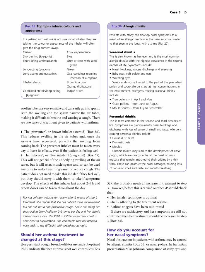
Case 3 55
PA
RT
2:
CA
SE
S
swollen tubes are very sensitive and can easily go into spasm.
Both the swelling and the spasm narrow the air tubes,
making it diffi cult to breathe and causing a cough. There
are two types of treatment given to patients with asthma:
1 The ‘preventer’, or brown inhaler (steroid) (Box 35).
This reduces swelling in the air tubes and, once the
airways have recovered, prevents the swelling from
coming back. The preventer inhaler must be taken every
day to have its effects, even if the patient is feeling well
2 The ‘reliever’, or blue inhaler (β2-agonist) (Box 35).
This will not get rid of the underlying swelling of the air
tubes, but it will relax muscle spasm and so can be used
any time to make breathing easier or reduce cough. The
patient does not need to take this inhaler if they feel well,
but they should carry it with them to take if symptoms
develop. The effects of this inhaler last about 2–4 h and
repeat doses can be taken throughout the day
Frances Johnson returns for review after 2 weeks of step 2
treatment. She reports that she has noticed some improvement
but she still has a non-productive cough. She is still using her
short-acting bronchodilator 2–3 times per day and her steroid
inhaler twice a day. Her PEFR is 350 L/min and her chest is
now clear to auscultation. She comments that her blocked
nose adds to her diffi culty with breathing at night.
Should her asthma treatment be changed at this stage?Her persistent cough, bronchodilator use and suboptimal
PEFR indicate that her asthma is not well controlled (Box
32). She probably needs an increase in treatment to step
3. However, before this is carried out the GP should check
that:
• Her inhaler technique is optimal
• She is adhering to the treatment regime
• Asthma triggers have been minimized
If these are satisfactory and her symptoms are still not
controlled then her treatment should be increased to step
3 (Box 34).
How do you account for her nasal symptoms?Nasal obstruction in patients with asthma may be caused
by allergic rhinitis (Box 36) or nasal polyps. In her initial
presentation Miss Johnson complained of itchy eyes and
Box 35 Top tips – inhaler colours and appearance
If a patient with asthma is not sure what inhalers they are
taking, the colour or appearance of the inhaler will often
give the drug content away.
Inhaler Colour/appearance
Short-acting β2-agonist Blue
Short-acting antimuscarinic Grey or clear with some
green
Long-acting β2-agonist Green
Long-acting antimuscarinic Oval container requiring
insertion of a capsule
Inhaled steroid Brown/maroon
Orange (fl uticasone)
Combined steroid/long-acting Purple or red
β2-agonist
Box 36 Allergic rhinitis
Patients with atopy can develop nasal symptoms as a
result of an allergic reaction in the nasal mucosa, similar
to that seen in the lungs with asthma (Fig. 27).
Seasonal rhinitisThis is also known as hayfever and is the most common
allergic disease with the highest prevalence in the second
decade of life. Symptoms include:
• Nasal blockage, watery discharge and sneezing
• Itchy eyes, soft palate and ears
• Watering eyes
Seasonal rhinitis is limited to the part of the year when
pollen and spore allergens are at high concentrations in
the environment. Allergens causing seasonal rhinitis
include:
• Tree pollens – in April and May
• Grass pollens – from June to August
• Mould spores – from July to September
Perennial rhinitisThis is most common in the second and third decades of
life. Symptoms are predominantly nasal blockage and
discharge with loss of sense of smell and taste. Allergens
causing perennial rhinitis include:
• House dust mites
• Domestic pets
• Moulds
Chronic rhinitis may lead to the development of nasal
polyps, which are overgrowths of the nasal or sinus
mucosa that remain attached to their origins by a thin
stalk. These can obstruct the nasal passages, causing loss
of sense of smell and taste and mouth breathing.

56 Part 2: Cases
PA
RT
2:
CA
SE
S
sneezing, making allergic rhinitis the likely diagnosis.
Allergic rhinitis can be differentiated from infective rhi-
nitis (the common cold) by persistence of symptoms for
several weeks.
How would you confi rm your provisional diagnosis of allergic rhinitis?The diagnosis of allergic rhinitis is based on a detailed
history of symptoms, atopy and nasal reactions to aller-
gen exposure and removal. Nasal inspection should be
performed to look for polyps. Investigations are not
usually indicated but skin prick testing can be used to
confi rm allergic reactions to specifi c allergens that may
guide allergen avoidance strategies.
How would you treat her rhinitis?There is some evidence that treatment of rhinitis may
improve asthma outcomes, as well as relieving nasal
symptoms. Miss Johnson’s GP should therefore consider
treating her rhinitis in parallel with her asthma treat-
ment. Treatment options for allergic rhinitis include anti-
histamines, decongestants and corticosteroids (Box 37).
The GP fi nds that Miss Johnson’s asthma trigger factors
have been addressed satisfactorily. She has no household
pets, she does not smoke and she lives in a house with
wooden fl oors and window blinds. She has mattress and
pillow covers to reduce house dust mite exposure. Her
inhaler technique is satisfactory and she is adamant that she
is taking the prescribed treatment. Her GP escalates her
treatment to step 3 by adding in salmeterol at a dosage of
50 mg twice a day. The inhaled steroid dosage is left at
400 mg/day. Miss Johnson is also prescribed mometasone
nasal spray 50 mg (1 spray) to each nostril daily and cetirizine
10 mg in the morning for her rhinitis.
Are long-acting b2-agonists effective and safe in asthma?A large number of clinical trials have investigated the use
of long-acting β2-agonists in the treatment of patients
with asthma and these have recently been summarized in
a systematic Cochrane review (Cochrane Database Sys-
tematic Review 2007 Jan 24; 1: CD001385). The review
found that there were benefi ts for patients of using long-
acting β2-agonists (Box 38):
• Improved lung function
• Signifi cantly fewer symptoms
• Less use of rescue medications
• Higher quality of life scores
However, the review also found that there were risks
of treatment. The review estimated that for every 1250
patients with asthma treated with long-acting β2-agonists
Box 37 Pharmacology of drugs used to treat allergic rhinitis
Drugs in each of these classes are available over-the-
counter for self-medication.
AntihistaminesFor example, chlorphenamine (Piriton), cetirizine:
• Mechanism of action. Block histamine receptors,
preventing the early phase reaction of the allergic
response (Fig. 27)
• Uses. Control symptoms in mild allergic rhinitis
• Pharmacokinetics. Can be given orally or topically
• Side-effects. Chlorphenamine causes drowsiness.
Second-generation drugs (e.g. cetirizine) do not cross
the blood–brain barrier and are therefore non-sedating
DecongestantsFor example, ephedrine hydrochloride
• Mechanism of action. Sympathomimetics cause
constriction of mucosal blood vessels, reducing oedema
of the nasal mucosa and relieving nasal obstruction
• Uses. Short term to relieve congestion and allow
penetration of nasal steroid
• Pharmacokinetics. Can be given orally or topically
• Side-effects. Once the use of decongestants is stopped
there is secondary vasodilatation causing rebound nasal
congestion and recurrence of symptoms. This is
particularly problematic if decongestants are used for
more than 7 days. Therefore, they should only be given
as a short course
CorticosteroidsFor example, beclometasone, mometasone
• Mechanism of action. Anti-infl ammatory (Box 33)
• Uses. Treatment and prevention of allergic rhinitis
• Pharmacokinetics. Used topically in allergic rhinitis as
nasal drops or spray
• Side-effects:� worsening of untreated nasal infection� dryness, irritation and bleeding of nasal mucosa� raised intraocular pressure� headache

Case 3 57
PA
RT
2:
CA
SE
S
there was one extra asthma-related death. This was
particularly seen in patients not taking inhaled
corticosteroids.
Why might long-acting b2-agonists increase the risk of asthma-related death?The mechanisms are not fully understood. It is possible
that long-acting β2-agonists mask asthma symptoms and
delay patients from seeking medical care, which may
result in a more severe or life-threatening asthma attack.
Alternatively long-acting β2-agonists may reduce the sen-
sitivity of β2-receptors, making short-acting β2-agonists
less effective during an acute attack.
What are the implications of these fi ndings for patient care?Long-acting β2-agonists should not be used in patients
with asthma without inhaled corticosteroids. However,
they appear to have clinical benefi t when used with
inhaled corticosteroids as per stepped guidelines.
KEY POINT
Long-acting β2-agonists should not be prescribed to
patients with asthma without inhaled corticosteroids.
Miss Johnson returns for review 2 weeks later and has
improved again. Her cough is much better and she has
used her short-acting bronchodilator inhaler once in the
last week. Her PEFR is now 380 L/min and her nasal
symptoms are improving. Her GP refers her to the
respiratory nurse for a self-management plan. Miss
Johnson tells the nurse that she is not happy about taking
inhaled steroids as she is worried she may put on weight
and asks how serious it would be if she stopped taking
them.
What is an asthma self-management plan?This is a personalized action plan usually compiled by a
respiratory nurse in consultation with the patient. It gives
the patient instructions on how to increase treatment if
their symptoms worsen or if the PEFR drops below a level
identifi ed as a percentage of their best PEFR. Similarly, if
the patient’s symptoms have been controlled and stable
for suitable time period (e.g. 3 months), instructions are
given about how to step down treatment. The patient is
also given a reserve course of antibiotics and steroids so
that prompt treatment can be started for an infective
exacerbation. The management plan empowers the
patient to manage their asthma themselves with the
support of the respiratory nurse, who is usually con-
tactable at short notice.
Box 38 Pharmacology of b2-adrenoceptor agonists
Mechanism of actionAct at β2-adrenoceptors to stimulate bronchial smooth
muscle relaxation through activation of adenyl cyclase
UsesRelief of symptoms from airfl ow obstruction (asthma and
COPD):
• Short-acting β2-agonists (salbutamol, terbutaline) are
used for acute relief of bronchospasm or to prevent
bronchospasm (e.g. before exercise or going out into
cold air)
• Long-acting β2-agonists (salmeterol, formoterol) are
used to maintain bronchodilatation and prevent
symptoms over a 12-h period (e.g. overnight)
PharmacokineticsDuration of action:
• Short-acting β2-agonists (e.g. salbutamol, terbutaline)
act immediately and have a biological half-life of 2–3 h
• Long-acting β2-agonists (e.g. salmeterol, formoterol)
remain at the receptor longer and have a longer
duration of action of up to 15 h. For example,
salmeterol has a long lipophilic tail which binds to an
exoreceptor near the β2-adrenoceptors in airway smooth
muscle
Route of administration:
• Inhaled directly into the lungs – maximizes effi cacy and
minimizes side-effects
• Short-acting β2-agonists can also be given:� by nebulizer – higher dose and less coordination and
respiratory effort required to take therapy� orally – if inhalers cannot be used (e.g. small children)� intravenously – emergency use where severe airfl ow
obstruction prevents inhalation
Side-effectsTremor, tachycardia, anxiety, hypokalaemia.

58 Part 2: Cases
PA
RT
2:
CA
SE
S
What therapeutic options could have been considered if Miss Johnson’s asthma had not improved with step 3 treatment?The next treatment options (Box 34) would have been to
optimize step 3 treatment by increasing the dose of
inhaled steroids and, if asthma was still not well con-
trolled, to try a leukotriene antagonist (Box 39), theoph-
ylline (Box 40) or an oral β2-agonist. If asthma still was
not well controlled, step 4 options include high-dose
inhaled corticosteroids and step 5 involves commence-
ment of regular oral prednisolone.
How should the nurse answer Miss Johnson’s question?The nurse should explain that it is essential that Miss
Johnson’s asthma is adequately treated as if not:
• She may have an acute exacerbation which could be
life-threatening
• Chronic infl ammation can lead to airway remodelling
with permanent narrowing and disabling breathlessness
in the long term
The nurse can reassure Miss Johnson that very little of
the inhaled steroids will be absorbed and she is extremely
unlikely to put on weight. However, she should advise her
that inhaled steroids can cause oral thrush and a hoarse
voice. Miss Johnson can minimize the risk of these effects
by rinsing her mouth and gargling with water after taking
the inhaler. Miss Johnson can be reassured that it may be
possible to reduce her inhaled steroids in the future if her
symptoms are controlled.
Outcome. Miss Johnson took her inhaled steroids and
salmeterol regularly for the next 3 months. Once her
symptoms were well controlled she stopped the salmeterol
and 6 weeks later reduced inhaled steroids from 400 to
200 mg/day. Six months later she was stable and opted to
stop inhaled steroids altogether. She remained aware, using
her self-management plan, that she would need to restart
treatment and seek advice should her symptoms recur.
Box 39 Pharmacology of leukotriene receptor antagonists (montelukast, zafi rlukast)
Mechanism of actionThe cysteinyl-leukotrienes (LTC4 and LTD4) are
infl ammatory mediators released by eosinophils and other
infl ammatory cells during the pathogenesis of asthma.
They stimulate bronchoconstriction, mucus hypersecretion
and mucosal oedema. Montelukast and zafi rlukast block
leukotriene receptors, inhibiting these infl ammatory effects.
UsesStep 3 or 4 treatment to control chronic asthma. Appear
to be particularly effective in exercise- and aspirin-induced
asthma and patients with associated rhinitis and/or nasal
polyps.
PharmacokineticsUnlike most other asthma drugs they are taken orally once
per day which may aid adherence to treatment.
Side-effectsThese include gastrointestinal disturbances, headache,
thirst and insomnia.
Box 40 Pharmacology of xanthines (theophylline, aminophylline)
Mechanism of actionXanthines inhibit phosphodiesterase enzymes which
metabolize secondary messengers inside airway smooth
muscle cells. Phosphodiesterase enzyme inhibition causes
a build-up of cyclic adenosine monophosphate (cAMP) in
airway smooth muscle cells and bronchodilatation.
UsesBronchodilatation in patients with severe acute asthma or
stable COPD.
Pharmacokinetics• Xanthines have a narrow therapeutic range; their lowest
effective plasma concentration (10 µg/mL) is close to
their toxic concentration (20 µg/mL)
• Xanthines are metabolized in the liver by cytochrome
p450 enzymes. If drugs that inhibit these enzymes are
co-prescribed with xanthines, xanthine plasma levels
may rise from therapeutic to toxic concentrations. Liver
enzyme inhibitors include:� erythromycin� ciprofl oxacin� sodium valproate
• Plasma levels of xanthines should be monitored
routinely and the dose reduced if liver enzyme inhibitors
are co-administered
Side-effectsMore likely at toxic plasma concentrations:
• Nausea and vomiting
• Arrhythmias and convulsions
Case 3 59
PA
RT
2:
CA
SE
S
long-acting β2-agonist) to control her symptoms. She also
receives nasal steroids and antihistamines for rhinitis and
sees the respiratory nurse for a self-management plan.
After her symptoms have been controlled for some months
she is able to step her treatment down, but remains aware
of the need to restart treatment and seek help early if her
symptoms recur.
CASE REVIEW
Frances Johnson presents to her GP with symptoms of
asthma recurring from childhood following a disease-free
period in her twenties. Her GP fi nds no evidence of a
moderate or severe exacerbation and decides to treat her
as chronic poorly controlled asthma according to standard
stepped asthma care guidelines. Miss Johnson requires
escalation of asthma treatment to step 2 (addition of
standard dose inhaled steroids), then step 3 (addition of
KEY POINTS
• The aims of asthma treatment are to control symptoms,
minimize exacerbations and optimize lung function
• Features of poorly controlled asthma include:� daytime or nocturnal symptoms� regular use of reliever medication� falling PEFR� recent exacerbations
• In a patient presenting with poorly controlled asthma, the
fi rst step should be to determine whether this is a
moderate or severe exacerbation requiring acute
treatment
• If the poor asthma control is chronic, control should be
improved using the stepped guidelines for chronic asthma
management
• The mainstay of asthma treatment is corticosteroids• In chronic asthma, corticosteroids are given as inhalers to
optimize drug delivery to the affected airways and
minimize side-effects
• After taking inhaled steroids patients should be advised
to rinse their mouth and gargle to reduce the risk of oral
thrush or a hoarse voice
• The stepped guidelines should be used both to increase
treatment in patients with poorly controlled asthma and
to reduce treatment in patients with well-controlled
asthma. Treatment should be reviewed and adjusted
approximately every 3 months
• Long-acting β2-agonists can be used in the treatment of
asthma, but should only be used with inhaled steroids as
long-acting β2-agonists are associated with an increased
risk of asthma-related deaths if used alone
• Patients with asthma commonly have associated allergic
rhinitis, which should be treated as this may improve
symptoms and assist with asthma control
• Patients with asthma should see a nurse with appropriate
expertise to develop a self-management plan and for
support in management of their condition

PA
RT
2:
CA
SE
S
A 22-year-old man with life-threatening shortness of breath
Case 4
Chris Carter is a 22-year-old man who has been brought
into accident and emergency by his girlfriend. He is unable
to speak because of shortness of breath which came on
suddenly 2 days ago. His observations are:
• Respiratory rate 30/min
• Pulse 120 beats/min
• Blood pressure 110/70 mmHg
• Temperature 36.7°C
• Oxygen saturations 92% on room air
• Glasgow Coma Score 15
What should you do fi rst?In an emergency always start by checking airway, breath-
ing and circulation (ABC). On observation, Mr Carter
has a patent airway, is breathing and has an adequate
cardiac output.
What other assessment can you make from his initial observations?Mr Carter is tachycardic, probably as part of a stress
response to his acute illness, but is maintaining an ade-
quate blood pressure. He is in respiratory distress but
managing to raise his respiratory rate to maintain oxygen
saturations at 92%. He is not compromised from a
neurological point of view as he has a normal Glasgow
Coma Score.
What are the priorities for his management?In an emergency situation it is vital that you take mea-
sures to save life and minimize potential disability even
before you make a full assessment (Box 24, p. 36). Mr
Carter is potentially at risk of cell injury and death from
his hypoxia, which will have greatest impact on his brain
and his heart. Immediate measures for Mr Carter should
therefore include:
• High concentration oxygen therapy, 15 L through a
mask with a reservoir bag
• Insertion of an intravenous line to secure access for
drug and fl uid delivery
• Continuous monitoring:� oxygen saturation� cardiac rhythm� pulse� blood pressure
He then needs more detailed assessment to identify
and treat the underlying cause of his hypoxia.
What is your differential diagnosis for his acute shortness of breath?As he is a young man the most likely diagnoses are:
• Acute asthma attack
• Pneumothorax
• Inhaled foreign body
• Allergic reaction (anaphylaxis)
• Lung trauma
How would you assess him to distinguish between these causes?Although Mr Carter is unable to speak he is conscious
and his girlfriend is with him. You should therefore
briefl y (1–2 min) establish whether he has a past history
of asthma, allergy or lung disease and ask about trauma
or foreign body inhalation. He should then be examined
quickly to look for clinical signs that could point to an
underlying diagnosis.
Mr Carter has a past history of asthma but no known
allergies. There is no history of trauma or foreign body
inhalation. On examination he is holding the sides of the
trolley, leaning forward and gasping for breath. Heart
sounds are faint but normal and there are no signs of
cardiac failure. On respiratory examination his trachea is
midline, expansion is equal on both sides but appears
reduced, percussion is resonant and he has widespread
expiratory polyphonic wheeze throughout both lung
fi elds.
60
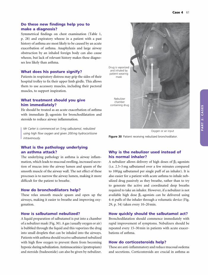
Case 4 61
PA
RT
2:
CA
SE
S
Do these new fi ndings help you to make a diagnosis?Symmetrical fi ndings on chest examination (Table 1,
p. 28) and expiratory wheeze in a patient with a past
history of asthma are most likely to be caused by an acute
exacerbation of asthma. Anaphylaxis and large airway
obstruction by an inhaled foreign body can also cause
wheeze, but lack of relevant history makes these diagno-
ses less likely than asthma.
What does his posture signify?Patients in respiratory distress may grip the sides of their
hospital trolley to fi x their upper limb girdle. This allows
them to use accessory muscles, including their pectoral
muscles, to support inspiration.
What treatment should you give him immediately?He should be treated as an acute exacerbation of asthma
with immediate β2-agonists for bronchodilatation and
steroids to reduce airway infl ammation.
Mr Carter is commenced on 5 mg salbutamol, nebulized
using high fl ow oxygen and given 200 mg hydrocortisone
intravenously.
What is the pathology underlying an asthma attack?The underlying pathology in asthma is airway infl am-
mation, which leads to mucosal swelling, increased secre-
tion of mucus into the airway lumen and spasm of the
smooth muscle of the airway wall. The net effect of these
processes is to narrow the airway lumen, making it more
diffi cult for the patient to breathe.
How do bronchodilators help?These relax smooth muscle spasm and open up the
airways, making it easier to breathe and improving oxy-
genation.
How is salbutamol nebulized?A liquid preparation of salbutamol is put into a chamber
of a nebulizer mask (Fig. 30). A gas (usually oxygen or air)
is bubbled through the liquid and this vaporizes the drug
into small droplets that can be inhaled into the airways.
Patients with asthma should receive salbutamol nebulized
with high fl ow oxygen to prevent them from becoming
hypoxic during nebulization. Antimuscarinics (ipratropium)
and steroids (budesonide) can also be given by nebulizer.
Why is the nebulizer used instead of his normal inhaler?A nebulizer allows delivery of high doses of β2-agonists
(i.e. 2.5–5 mg salbutamol over a few minutes compared
to 100 µg salbutamol per single puff of an inhaler). It is
also easier for a patient with acute asthma to inhale neb-
ulized drug passively as they breathe, rather than to try
to generate the active and coordinated deep breaths
required to take an inhaler. However, if a nebulizer is not
available high dose β2-agonists can be delivered using
4–6 puffs of the inhaler through a volumatic device (Fig.
29, p. 54) taken every 10–20 min.
How quickly should the salbutamol act?Bronchodilatation should commence immediately with
rapid improvement of symptoms. Nebulizers should be
repeated every 15–30 min in patients with acute exacer-
bations of asthma.
How do corticosteroids help?These are anti-infl ammatory and reduce mucosal oedema
and secretions. Corticosteroids are crucial in asthma as
Figure 30 Patient receiving nebulized bronchodilator.
Drug is vaporizedand inhaled bypatient wearing
mask
Nebulizerchamber
containing drug
Oxygen or air input

62 Part 2: Cases
PA
RT
2:
CA
SE
S
• There is no evidence of pneumothorax or pneumonia
on his chest X-ray. Evidence of more than six ribs ante-
riorly seen above fl attened diaphragms indicates that his
lungs are hyperinfl ated
• He has a mildly elevated white cell count. This could
indicate underlying infection or be part of the infl amma-
tory process of asthma. His CRP is normal, making infec-
tion less likely
• The ECG reassures you that there is no cardiovascular
problem, either as a cause or consequence of his breath-
lessness. Tachycardia is a normal response to hypoxia and
the physiological stress of an asthma attack
His arterial blood gases on air show:
Normal range
pH 7.58 (7.35–7.45)
PaO2 8.5 kPa (10–13.1 kPa)
PaCO2 4.1 kPa (4.9–6.1 kPa)
HCO3− 23 mmol/L (22–28 mmol/L)
How do you interpret his arterial blood gases?• PaO2 is low, indicating reduction of alveolar ventilation
secondary to airfl ow obstruction. He is hypoxic but is not
in respiratory failure, which is defi ned as PaO2 <8 kPa
• PaCO2 is low, indicating increased ventilation to com-
pensate for hypoxia
• pH is raised (alkalosis). When PaCO2 is reduced [1],
this drives the Henderson–Hasselbach equation to the
right [2], reducing hydrogen ion concentration [3] and
they treat the underlying infl ammation and the patient
is unlikely to recover without steroids.
KEY POINT
Asthma is an infl ammatory condition and steroids are
crucial for the treatment of the underlying pathology.
How quickly do you expect the steroids to act?Corticosteroids exert most of their actions by binding to
receptors and altering gene transcription. This process
can take some time and corticosteroids are said not to
have any effect for at least half an hour after the drug is
given. However, studies have shown that the earlier cor-
ticosteroids are given in an asthma attack the better the
outcome. The fi rst dose of corticosteroids is usually given
intravenously. This may be helpful in breathless patients
who have diffi culty swallowing and makes sure the patient
receives the drug. However, oral steroids are just as effec-
tive in acute asthma as intravenous steroids.
Which investigations would you perform next (and why)?• Peak expiratory fl ow rate (PEFR) to determine severity
of asthma exacerbation
• Arterial blood gas to assess pH and PaCO2 levels
• Chest X-ray to exclude other causes of breathlessness
such as pneumothorax and underlying consolidation
• Full blood count and C-reactive protein (CRP) to look
for evidence of infection
• Electrocardiogram (ECG) – to exclude cardiac disease
as a cause or consequence of his condition
Initial investigation results are as follows:
Normal range
PEFR 210 L/min (Predicted 590 L/min)
Chest X-ray see Fig. 31
Blood tests:
Haemoglobin 14 g/dL (12.5–17 g/dL)
White cells 12 × 109/L (4.0–11.0 × 10⁹/L)
Platelets 220 × 109/L (120–400 × 109/L)
CRP 2 mg/L (<4 mg/L)
ECG Sinus tachycardia
How do you interpret these results?• His peak fl ow is 35% predicted, which is a feature of
acute severe asthma (Box 31, p. 52)
Figure 31 Chest X-ray.

Case 4 63
PA
RT
2:
CA
SE
S
causing an alkalosis. As the respiratory problem is
the primary abnormality, this is called a respiratory
alkalosis
2�
3 |� H+ + HCO3
− ¨Æ H2CO3 ¨Æ H2O + CO2
|� 1
How do you decide how severe his asthma attack is?Mr Carter meets the criteria for a severe asthma attack
(Box 31, p. 52). His peak fl ow is <50% predicted (35%),
he has a respiratory rate of >25/min (30/min), is tachy-
cardic (120 beats/min) and is unable to complete full
sentences. He does not have any features of life-threaten-
ing asthma. It is important to make this assessment as
this determines how and where he should be treated.
Patients with severe attacks should usually be admitted
to hospital for intensive therapy. Patients with life-
threatening or near-fatal asthma will need intensive care
monitoring.
After Mr Carter has received three 5 mg salbutamol
nebulizers and 200 mg hydrocortisone he is reviewed.
His observations now are:
• Respiratory rate 32/min
• Pulse 132 beats/min
• Blood pressure 124/82 mmHg
• Oxygen saturations 96% on high inhaled oxygen
concentrations
• PEFR 170 L/min
How do you assess his progress?Mr Carter has shown no signs of improvement on treat-
ment. He remains tachycardic and tachypnoeic. If any-
thing his asthma has deteriorated as his PEFR has fallen
from 210 L/min (35% predicted) to 170 L/min (28%
predicted) and his exacerbation is now classifi ed as life-
threatening (Box 31, p. 52). His increasing tachycardia
could be a side-effect of salbutamol.
As he has not responded to initial treatment, what would you do next?He needs additional bronchodilator treatment and refer-
ral to intensive care should be considered. He should
continue with repeated nebulized salbutamol 2.5–5 mg
every 15–30 min or continuously if possible. Other bron-
chodilators can be added to this treatment.
Nebulized ipratropium bromide (Box 41)
This should be given at the dose of 0.5 mg every 4–6 h
and should produce signifi cantly greater bronchodilata-
tion than salbutamol alone. It is only used in acute severe
or life-threatening asthma or patients who are not
responding to β2-agonists.
Intravenous magnesium sulphateA single infusion of 1.2–2 g should be given over 20 min.
Clinical trials have shown an improvement in PEFR and
FEV1 measurements in patients with acute severe asthma
exacerbations treated with intravenous magnesium sul-
phate compared to placebo (Box 42).
Other options include the use of intravenous β2-
agonists or intravenous theophylline. However, there is
less evidence to support the use of these treatments.
Box 41 Pharmacology of antimuscarinic bronchodilators
Mechanism of action• Cholinergic nerves maintain resting tone in the bronchi
and stimulate mucus secretion. Antimuscarinic drugs
block these effects, resulting in bronchodilatation and
reduced mucus secretion
Uses• Acute severe or life-threatening asthma with salbutamol
to increase bronchodilatation. Not part of stepped
guidelines for chronic asthma
• Chronic obstructive pulmonary disease (COPD).
Antimuscarinic drugs are particularly useful in COPD
patients who have increased resting bronchial tone and
increased mucus secretion
Pharmacokinetics• Duration of action
� short-acting (e.g. ipratropium bromide) causes
bronchodilatation for up to 5 h, therefore given four
times daily� long-acting (e.g. tiotropium bromide) causes
bronchodilatation for 24 h, therefore given once daily
• Route of administration� Inhaler (all) or nebulizer (short-acting only)
Main side-effects• Dry mouth, urinary retention, constipation

64 Part 2: Cases
PA
RT
2:
CA
SE
S
Mr Carter receives nebulized 0.5 mg ipratropium and
intravenous 2 g magnesium sulphate and continues
with nebulized salbutamol. He remains tachypnoeic and
his blood gases are repeated on high oxygen
concentrations:
Normal range
pH 7.36 (7.35–7.45)
PaO2 11.7 kPa (10–13.1 kPa)
PaCO2 6.3 kPa (4.9–6.1 kPa)
HCO3− 26 mmol/L (22–28 mmol/L)
How have his blood gases changed?The most concerning change is an increase in PaCO2
levels and fall in pH. At fi rst sight his PaO2 appears
normal, but in fact he is requiring high inspired oxygen
concentrations to maintain oxygenation.
What is the signifi cance of the new fi ndings?A rising PaCO2 now puts him into the category of near-
fatal asthma. He requires urgent intensive care input with
possible ventilatory support.
What might have caused this deterioration?• Mr Carter may be tiring with decreased respiratory
effort leading to a PaCO2 rise
• Patients with asthma can develop a secondary pneu-
mothorax during exacerbation and this should always be
considered if there is a sudden deterioration or failure to
improve
Mr Carter is transferred to the intensive care unit for
monitoring, treatment and consideration of invasive
ventilation. A chest X-ray shows no evidence of
pneumothorax. His condition is optimized with continuous
nebulized salbutamol and intravenous theophylline. Over the
next 30 min there is a gradual improvement in Mr Carter’s
condition until he is able to say a few words. He is
considered not to require invasive ventilation. After 48 h his
condition has improved suffi ciently for him to continue his
recovery on a general medical ward.
How should his asthma be monitored on the general ward?Ongoing assessment is important to evaluate response to
treatment and recovery. Observations should include
respiratory rate and O2 saturations.
Peak fl ow chartPeak expiratory fl ow rate should be measured morning
and evening, before and after nebulizers. During an
asthma attack PEFR will be reduced, lower in the morning
than in the evening and increased by nebulizer treatment
(bronchodilator reversibility). A rise in peak fl ow to
normal values with disappearance of morning dipping
and reduction in bronchodilator reversibility indicates
recovery.
As his asthma improves, how should his treatment be modifi ed?Corticosteroids• British Thoracic Society guidelines state that patients
with acute asthma should receive 40–50 mg oral pred-
nisolone for at least 5 days or until recovery
Box 42 Pharmacology of intravenous magnesium
Mechanism of actionMagnesium sulphate causes bronchodilatation. The
mechanism is unclear but may include:
• Relaxation of bronchial smooth muscle by altering
intracellular calcium concentrations
• Reduction of neurotransmitter release at motor nerve
terminals, reducing excitability of smooth muscle
membranes
• Facilitating activation of adenyl cyclase by
β2-agonists
• Anti-infl ammatory effects, reducing production of
superoxide by neutrophils
Uses• Single infusion for bronchodilatation in acute severe or
life-threatening asthma exacerbations
Pharmacokinetics• Administered intravenously. Causes diarrhoea if given
orally
Main side-effects• Transient fl ushing, lightheadedness, lethargy, nausea or
burning at the IV site
• Hypermagnesaemia could cause muscle weakness and
respiratory failure if excessive doses are given

Case 4 65
PA
RT
2:
CA
SE
S
• He should commence inhaled steroids as soon as pos-
sible during admission. The main reason for this is to
ensure that he is taking inhaled steroids on discharge
to form part of his chronic asthma management plan
to prevent further exacerbations. Patients who have
had an exacerbation in the past 2 years requiring steroids
or nebulized bronchodilator should be taking inhaled
steroids as per the stepped asthma guidelines (see
Case 3).
BronchodilatatorsHe should have nebulized bronchodilators until:
• His symptoms have resolved
• PEFR morning dipping has decreased to less than 20%
of PEFR
Bronchodilators should then be switched to inhaled
therapy.
Suggested treatment regime on dischargeA reasonable approach to treatment on discharge after an
asthma exacerbation would be to give patients:
• A steroid inhaler at standard or high dose twice daily
• A long-acting β2-agonist twice daily
• A short-acting β2-agonist inhaler initially to use regularly
(e.g. 4 times per day) and then to use when needed (p.r.n.)
Patients with asthma will thus leave hospital following
an exacerbation on step 3 or 4 treatment (depending on
inhaled steroid dose) of the asthma guidelines, with a
view to stepping down treatment in outpatients when
their asthma is stable.
When is it safe to discharge Mr Carter from hospital?A reasonable approach is to allow patients to go home
when:
• They are able to mobilize around the ward without
shortness of breath
• They are not breathless or troubled by severe cough at
night
• They have been stable for 24 h after switching from
nebulized to inhaled bronchodilators
• There is no signifi cant morning dipping (<20% of
maximum) on PEFR chart and values have risen to >75%
best or predicted values
Mr Carter’s peak fl ow chart is shown in Fig. 32.
1 Date:Day:Time:
70080604020
60080604020
50080604020
40080604020
30080604020
20080604020
100806040200
INHALERS
POSTNEBS
PEA
K F
LOW
CH
ART
6 12 18 226 12 18 226 12 18 226 12 18 226 12 18 226 12 18 226 12 18 22
2 3 4 5 6
Figure 32 Inpatient peak fl ow chart.

66 Part 2: Cases
PA
RT
2:
CA
SE
S
How do you interpret this chart?Initially, Mr Carter’s PEFR recordings are low, there is
morning dipping and a pre-nebulizer reading is lower
than the post-nebulizer reading. As he improves, his peak
fl ow rises and morning dipping is reduced. By day 4, his
morning dipping is less than 20% and his peak fl ow is
530 L/min (90% predicted best of 590 L/min). He is
therefore switched to inhaled therapy and remains stable
on this for a further 24 h, at which stage he is ready for
discharge.
What should be done before he leaves hospital to reduce the chance of further exacerbations?Mr Carter should receive education about his asthma and
how to prevent further attacks, preferably from a special-
ist respiratory nurse or other trained staff. This education
should include:
• Correct inhaler technique
• Making and recording home peak fl ow
measurements
• Explanation of the purpose of different inhalers pre-
scribed and the importance of taking them
• Self-management plan, including advice about early
management of future exacerbations and when to seek
help
Some of this will need to be carried out or continued
as an outpatient and so an appointment should be given
for follow-up in the respiratory clinic.
Why is education and follow-up so important?Asthma can kill. There are around 1400 deaths annually
in the UK from asthma according to national statistics.
Factors associated with death include:
• Severe asthma
• Inadequate treatment or monitoring of asthma
• Adverse behavioural or psychosocial features leading
to inadequate asthma management
Most severe acute asthma exacerbations develop over
6 h or more, so there should be time for patients to receive
appropriate help. Prompt action at the onset of an exac-
erbation may prevent hospitalization and death. Educa-
tion and follow-up can be used to advise patients when
and how to seek timely help.
Education should also address asthma triggers (Box 29,
p. 47) and measures that can be taken to avoid exacerba-
tions. Patients with severe asthma or recent hospital
admission should be advised to have vaccination against
infl uenza.
ReferenceFurther advice and information for patients with asthma
is provided by Asthma UK and can be found at www.
asthma.org.uk/index.html
Outcome. Mr Carter is seen by the respiratory nurse
specialist. His inhaler technique is corrected and he receives
a self-management plan and a number to contact if he has
questions. He is discharged taking:
40 mg prednisolone orally for 5 more days
250 mg beclometasone per dose, 2 puffs twice daily
25 mg salmeterol per dose, 2 puffs twice daily
Salbutamol metered dose inhaler (100 mg per dose), 2 puffs
as required
In outpatients 4 weeks later his symptoms have
resolved. His PEFR is 580 L/min. His treatment is gradually
stepped down over several months until he is maintained
on 200 mg beclometasone twice daily and salbutamol
p.r.n.
intubation and mechanical ventilation by optimizing
medical treatment. Mr Carter is monitored on intensive
care for 48 h, and then transferred to the ward to complete
his recovery. Prior to discharge, asthma triggers, inhaler
technique and self-management issues are addressed by the
respiratory nurse specialist to reduce the risk of exacerbation
recurrence and he is followed-up in the chest clinic.
CASE REVIEW
Mr Carter presents to accident and emergency with a
severe asthma exacerbation. He does not respond to initial
treatment with nebulized salbutamol and intravenous
hydrocortisone, so ipratropium nebulizers and intravenous
magnesium are given. He tires and his PaCO2 starts to rise,
which puts him into the category of ‘near-fatal asthma’. The
intensive care team are called and manage to avoid

Case 4 67
PA
RT
2:
CA
SE
S
KEY POINTS
• Asthma kills around 1400 people annually in the UK
• Recognition and prompt treatment of asthma
exacerbations is essential to reduce asthma-related deaths
• At presentation, clinical features and basic investigations
should be used to determine the severity of the asthma
exacerbation
• Corticosteroids must be given to patients with asthma
exacerbations as soon as possible to treat underlying
infl ammation
• 2.5–5 mg nebulized salbutamol every 15–30 min should be
used to improve symptoms and respiratory function in
severe asthma while the anti-infl ammatory effects of
corticosteroids are taking effect
• If patients respond poorly to nebulized salbutamol other
bronchodilators recommended for use are:� nebulized ipratropium� intravenous magnesium
• Intravenous theophylline or salbutamol can also be tried by
respiratory clinicians
• Failure to respond or deterioration in a patient with
acute asthma can be caused by:� intractable disease� tiring/exhaustion� secondary pneumothorax
• Intensive care should be considered early for patients
with life-threatening or near-fatal asthma
• Recovery from asthma exacerbation can be judged by
improvement in symptoms and peak fl ow recordings
• Patients can usually be discharged safely when they have
minimal symptoms and PEFR >75% best or predicted
with <20% morning dipping after 24 h on inhalers
• Prior to discharge, patients should receive education
from trained staff to reduce the risk of further
exacerbations
• Patients should be followed-up by a respiratory specialist
after hospitalization for an asthma exacerbation

PA
RT
2:
CA
SE
S
A 65-year-old man with worsening shortness of breath
Case 5
Mr Newman is a 65-year-old man who attends the chest
clinic with the following letter from his general practitioner:
Dear Doctor,
Thank you for seeing this 65-year-old man who complains
of worsening shortness of breath. He fi rst presented to our
practice 2 years ago with mild shortness of breath on
exercise and was started on inhaled salbutamol. This helped
initially but he is now more breathless. I would be grateful
for your assistance in establishing the cause of his
breathlessness and advising on further treatment as
appropriate.
Yours faithfully,
Dr Clarkson
How will the specialist approach the consultation?Mr Newman has been referred to hospital for a specialist
opinion. Dr Clarkson’s letter is helpful as it clearly states
why the patient has been referred and what is required of
the specialist. Thus, the focus of the consultation will
be the diagnosis and treatment of breathlessness. Mr
Newman may have other health concerns which can be
raised, but if unrelated to his breathlessness may be
referred back to his GP.
How would you approach taking Mr Newman’s history?An approach to a consultation with a breathless patient
is outlined in Part 1B. You should start with an open
question, quantify his shortness of breath, then attempt
to determine if the cause of his shortness of breath is
respiratory, cardiac or from another source (Fig. 11, p. 18),
before constructing a more specifi c differential diagnosis.
Mr Newman describes his problem in his own words. ‘I’m an
active man but my breathing won’t keep up with me. I now
walk more slowly than my wife if we go out together and
have to stop to get my breath if I am carrying shopping or
going uphill’.
How would you quantify his breathlessness?Breathlessness can be quantifi ed using the MRC dys-
pnoea score (Box 12, p. 18). From the information given
Mr Newman appears to have a score of 3 as he has to walk
more slowly than his wife.
On further questioning Mr Newman states that his breathing
is comfortable at rest, he sleeps on one pillow and does not
wake up breathless at night. His breathlessness is worse
following a cold or during humid weather. He has had a
persistent cough for the past 2 years and brings up clear or
grey sputum on most mornings. He has never coughed up
blood, had chest pain or experienced palpitations. His
weight is stable.
He is a current cigarette smoker and has smoked 20
cigarettes daily since the age of 19. He works as a builder,
although he recently moved into the offi ce to do lighter
work because of his breathlessness. His job has always been
dusty and he probably was exposed to asbestos in the past.
His father died of emphysema.
Do you think his breathlessness is caused by respiratory or cardiac disease?His history is very suggestive of respiratory disease.
Particular features that point to a respiratory diagnosis
are:
• Chronic productive cough consistent with chronic
bronchitis
• Symptoms exacerbated by respiratory infection and
change in the weather
• Extensive respiratory risk factors including cigarette
smoking (46 pack years; Box 16, p. 19), dust and asbestos
exposure and family history
Lack of orthopnoea, chest pain or palpitations
make cardiac disease less likely. However, smoking
is a risk factor for cardiac as well as respiratory
disease.
68

Case 5 69
PA
RT
2:
CA
SE
S
How does this direct the consultation?It would now be reasonable to consider a respiratory dif-
ferential diagnosis and direct questions towards this.
However, cardiac disease should remain at the back of
your mind and be reconsidered if respiratory lines of
enquiry draw a blank.
Give a respiratory differential diagnosis for Mr Newman• New symptoms in older patients are usually caused by
degenerative or neoplastic disease (Box 15, p. 19), so
chronic obstructive pulmonary disease (COPD) and lung
cancer are strong possibilities. Pulmonary fi brosis should
also be considered
• Mr Newman’s breathlessness has come on gradually
over 2 years. This is more consistent with degenerative dis-
ease such as COPD or pulmonary fi brosis, rather than lung
cancer which usually causes more rapid deterioration
• Mr Newman has not noticed haemoptysis, weight loss
or other systemic features. This does not exclude lung
cancer but makes it less likely
• The diagnosis is therefore most likely to be COPD.
Other diagnoses still to be excluded include lung cancer,
lung fi brosis, cardiac disease and anaemia
If Mr Newman has COPD, how can this diagnosis be confi rmed?The defi nition of COPD is given in Box 43. Mr Newman
has a history of tobacco smoking and symptoms of
chronic bronchitis, both of which are strongly indicative
of COPD. A fi rm diagnosis of COPD requires objective
evidence of airfl ow obstruction. Examination and inves-
tigation are therefore required to confi rm the diagnosis
and exclude the main differentials and unexpected causes
of breathlessness.
On examination Mr Newman has:
No fi nger clubbing, no lymphadenopathy
Nicotine-stained fi ngers
Pulse 84 beats/min regular, jugular venous pressure (JVP) not
elevated, blood pressure 110/70 mmHg
Heart sounds normal, nil added, no pitting ankle oedema
Trachea – midline, cricosternal distance two fi nger breadths
Expansion – reduced bilaterally
Percussion – resonant bilaterally, loss of cardiac dullness
Breath sounds – vesicular with bilateral expiratory wheeze
How do you interpret his examination fi ndings?• He has symmetrical lung disease (Table 1, p. 28) with
hyperinfl ation (reduced cricosternal distance, loss of
cardiac dullness) and expiratory wheeze. These fi ndings
are consistent with obstructive airways disease
• Nicotine-stained fi ngers indicate active cigarette smoking
• Absence of fi nger clubbing and lymphadenopathy is
reassuring, as these can be features of malignant disease
• He has no clinical evidence of heart failure or
arrhythmias
How would you investigate Mr Newman?Initially, the diagnosis of COPD should be confi rmed
with spirometry and other diagnoses excluded with a
chest X-ray and full blood count.
Mr Newman’s spirometry is compared with predicted values
in a graph in Fig. 33.
From the graph what are Mr Newman’s spirometry measurements?• FEV1 – he has exhaled 1.3 L after 1 s
• FVC – he exhales 3.4 L by the end of the test
• FEV1/FVC ratio is (1.3/3.0) × 100 = 43%
How do Mr Newman’s actual measurements compare with his predicted values?• His measured FEV1 (1.3 L) is 41% of his predicted
value (3.2 L), i.e. is reduced
Box 43 Defi nition of chronic obstructive pulmonary disease (COPD)
A disease characterized by airfl ow obstruction that is:
• Usually caused (in the UK) by tobacco smoking
• Not fully reversible
• Does not change markedly over several months
• Is progressive
COPD includes diseases previously defi ned as chronic
bronchitis and emphysema:
• Chronic bronchitis. This term refers to the clinical
symptoms of a daily cough productive of sputum for 3
months in 2 consecutive years
• Emphysema. This term refers to a pathological process
seen on histology or by computed tomography (CT)
scanning. There is an abnormal increase in airspace size
because of irreversible expansion of the alveoli or
destruction of alveolar walls

70 Part 2: Cases
PA
RT
2:
CA
SE
S
• His measured FVC (3.0 L) is 73% of his predicted value
(4.1 L), i.e. is reduced
• FEV1/FVC ratio is 43%, i.e. is less than 70%. The reduc-
tion in FEV1 is therefore a result of airways obstruction
(see p. 31)
Is airways obstruction always a result of COPD?Airways obstruction is usually caused by COPD or
asthma. Less commonly, airways obstruction is caused by
other pathologies of large or small airways (Box 44).
How can you distinguish between COPD and asthma?Airfl ow obstruction caused by COPD is characteristically
not fully reversible and does not change markedly over
several months. By contrast, airfl ow obstruction caused
by asthma is usually fully reversible, unless chronic scar-
ring (remodelling) has occurred, and is variable. COPD
and asthma can be distinguished by:
• Clinical features. A useful table for distinguishing
between COPD and asthma is given in national clinical
guidelines and reproduced in Table 3
• Spirometry. If there is doubt about the diagnosis,
reversibility testing can be performed. Spirometry is mea-
sured before and after inhalation of a short-acting bron-
chodilator (e.g. salbutamol). Signifi cant reversibility is
usually taken as an improvement in FEV1 of ≥15% base-
line value or 400 mL (NICE guidelines) and indicates
asthma rather than COPD
Mr Newman is a smoker with symptoms developing in his
sixties who has a chronic productive cough and progressive
breathlessness. He clearly has COPD and does not need
reversibility testing.
How does COPD cause airfl ow obstruction?COPD is a combination of airway infl ammation
(bronchitis) and loss of alveoli and lung parenchyma
Figure 33 Mr Newman’s spirometry. Line A (bold) represents
Mr Newman’s spirometry recorded in clinic. Line B (dotted) is
his predicted spirometric curve for comparison.
A
B
FEV1B
FEV1A
FVC B
FVC A
1 2 3 40
1
2
3
4
5 6 7 8 9 10
Time (s)
Volu
me
(L)
Box 44 Causes of airways obstruction other than asthma or COPD
Large airways – trachea or main bronchus• Stricture
• Tumour
• Compression
Small airways• Obliterative bronchiolitis
• Pulmonary oedema, compressing small airways
Table 3 Clinical features differentiating COPD and asthma. [From COPD National Clinical Guidelines. Thorax 2004;
59 (Suppl I): 33.]
COPD Asthma
Smoker or ex-smoker Nearly all Possibly
Symptoms under age 35 Rare Often
Chronic productive cough Common Uncommon
Breathlessness Persistent and progressive Variable
Night-time waking with breathlessness or wheeze Uncommon Common
Signifi cant diurnal or day-to-day variability of symptoms Uncommon Common

Case 5 71
PA
RT
2:
CA
SE
S(emphysema) that causes airfl ow obstruction by the
mechanisms discussed in Box 45.
How severe is Mr Newman’s airfl ow obstruction?Severity of COPD is classifi ed according to the degree of
reduction in FEV1 as a percentage of the predicted value
(Box 46).
Mr Newman’s FEV1 (1.3 L) is 41% of his predicted value
(3.2 L). Thus, he has moderate airfl ow obstruction.
Mr Newman’s chest X-ray is shown in Fig. 37.
His haemoglobin (Hb) was 14.2 g/dL (normal range 13–
18 g/dL).
Oxygen saturations were 96%.
How do you interpret these results?On the chest X-ray the top of the liver should be in
the 5th intercostal space in the midclavicular line
and 5–6 ribs should be visible anteriorly. On Mr
Newman’s chest X-ray, 9 anterior ribs are visible
(numbered) and the diaphragms are fl attened (arrow),
Box 45 Pathophysiology of airfl ow obstruction in COPD
BronchitisIn COPD the airways are infl amed and scarred and the
respiratory epithelium is partly replaced by mucus-producing
goblet cells. A combination of thickened airway walls,
increased luminal secretions and smooth muscle spasm
narrows the airway lumen, reducing airfl ow (Fig. 4, p. 5).
EmphysemaThe lung parenchyma is destroyed, partly because of
digestion of the lung by proteolytic enzymes released by
neutrophils. Figure 34 shows a lung resected from a patient
with emphysema at postmortem. The lung has black staining
caused by carbon from cigarettes and air pollution and
contains multiple small holes resulting from loss of lung
parenchyma. Figure 35 compares slices from high-resolution
CT scans from a patient with normal lungs and a patient
with emphysema. The emphysematous lung has multiple
black holes where there should be heterogeneous grey lung
parenchyma. The very large black holes are called bullae.
Emphysematous destruction of the lung parenchyma
causes loss of lung elasticity and elastic recoil, which leads to
airfl ow obstruction (Fig. 36).
Role of elastic recoil in normal respiration• In normal lungs, the elastic recoil acts to collapse the lung
and is opposed by negative intrapleural pressure which
maintains lung expansion
• At rest, when intrapleural pressure (−5 kPa) equals elastic
recoil (+5 kPa), no air moves into or out of the lungs
(Fig. 36a)
• When the respiratory muscles contract, the intrathoracic
pressure becomes more negative, overcoming elastic recoil
and sucking air into the lungs
• When the respiratory muscles relax, the chest wall and
diaphragm return to their resting positions, making the
intrathoracic pressure more positive and pushing air out of
the lungs
• Positive intrathoracic pressure has different effects on
alveoli and airways:� the pressure forces air out of the ‘spherical’ alveoli
into the bronchioles� the pressure compresses the cylindrical airways, causing
them to collapse� during normal expiration the pressure in the airway
lumen generated by air moving from the alveoli is
greater than the pressure outside the airways and
compression is prevented. The airways remain open
throughout expiration� if expiration is forced the intrathoracic pressure
becomes more positive (Fig. 36b) so that external
pressure exceeds luminal pressure and the airways
partially collapse. This can be demonstrated by
comparing what happens if you breathe out quietly and
when you force your breath out. On gentle expiration,
your breathing will sound quiet. On forced expiration,
you will hear wheezing caused by compression of your
large airways
Loss of elastic recoil causes airfl ow obstruction• Elastic recoil contributes to positive intrathoracic pressure
required to force air out of the alveoli and maintain
pressure in the airway lumen. As elastic recoil is reduced
in emphysema, airway luminal pressure is reduced (Fig.
36c) and falls below external pressure during normal
expiration, resulting in airway collapse
• Patients with emphysema thus fi nd it harder to breathe
out than breathe in and air gets trapped in the alveoli,
causing hyperinfl ation
• Emphysematous patients often purse their lips to narrow
the exit from their airways. This increases the pressure in
the airway lumen, preventing airways collapse and making
it easier (although slower) to breathe out

72 Part 2: Cases
PA
RT
2:
CA
SE
S
How should his COPD be managed?The major aims of COPD management are:
• To minimize progression, thus preventing or delaying
disability and prolonging life
• To relieve symptoms
A
B
Figure 34 Pathology of emphysema. (A) Carbon stained lung
from cigarette smoking. (B) Multiple small ‘holes’ in the lung
caused by loss of lung parenchyma.
Normal lungparenchyma
(a)
Emphysema –appears as
black holes inthe lung
parenchyma
Bulla
(b)
indicating hyperinfl ated lungs. There is no evidence of
lung cancer.
Mr Newman’s Hb is normal, ruling out anaemia as a
cause of his breathlessness.
Oximetry was normal, indicating that his COPD is not
causing hypoxia.
Figure 35 Computed tomography
(CT) scans showing radiology of
emphysema. (a) Normal lung.
(b) Emphysema.
Box 46 Assessment of severity of airfl ow obstruction
Normal FEV1 can be predicted for an individual based on
their age, height and gender from tables of normal values
for healthy people. FEV1 should be 80–120% of predicted
values.
FEV1 % predicted
Mild airfl ow obstruction 50–80%
Moderate airfl ow obstruction 30–49%
Severe airfl ow obstruction <30%
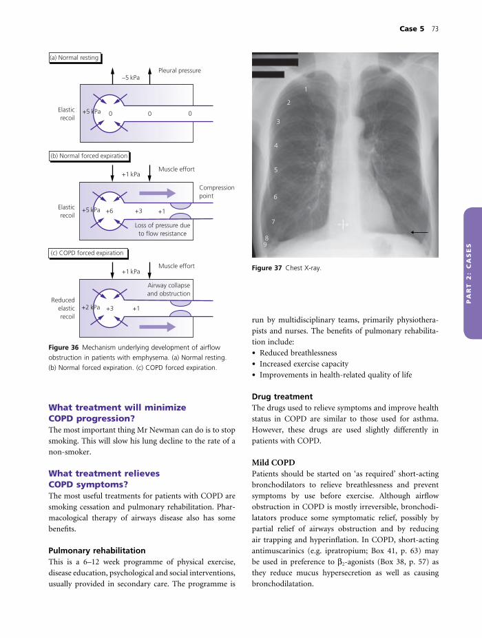
Case 5 73
PA
RT
2:
CA
SE
S
What treatment will minimize COPD progression?The most important thing Mr Newman can do is to stop
smoking. This will slow his lung decline to the rate of a
non-smoker.
What treatment relieves COPD symptoms?The most useful treatments for patients with COPD are
smoking cessation and pulmonary rehabilitation. Phar-
macological therapy of airways disease also has some
benefi ts.
Pulmonary rehabilitationThis is a 6–12 week programme of physical exercise,
disease education, psychological and social interventions,
usually provided in secondary care. The programme is
run by multidisciplinary teams, primarily physiothera-
pists and nurses. The benefi ts of pulmonary rehabilita-
tion include:
• Reduced breathlessness
• Increased exercise capacity
• Improvements in health-related quality of life
Drug treatmentThe drugs used to relieve symptoms and improve health
status in COPD are similar to those used for asthma.
However, these drugs are used slightly differently in
patients with COPD.
Mild COPDPatients should be started on ‘as required’ short-acting
bronchodilators to relieve breathlessness and prevent
symptoms by use before exercise. Although airfl ow
obstruction in COPD is mostly irreversible, bronchodi-
latators produce some symptomatic relief, possibly by
partial relief of airways obstruction and by reducing
air trapping and hyperinfl ation. In COPD, short-acting
antimuscarinics (e.g. ipratropium; Box 41, p. 63) may
be used in preference to β2-agonists (Box 38, p. 57) as
they reduce mucus hypersecretion as well as causing
bronchodilatation.
(a) Normal resting
(b) Normal forced expiration
(c) COPD forced expiration
–5 kPa
+1 kPa
+1 kPa
+5 kPa
+5 kPa
+2 kPa
Pleural pressure
Muscle effort
Muscle effort
Airway collapseand obstruction
Reducedelasticrecoil
Compressionpoint
Loss of pressure dueto flow resistance
0 00
+1+6 +3
+1+3
Elasticrecoil
Elasticrecoil
Figure 36 Mechanism underlying development of airfl ow
obstruction in patients with emphysema. (a) Normal resting.
(b) Normal forced expiration. (c) COPD forced expiration.
1
2
3
4
5
6
7
89
Figure 37 Chest X-ray.

74 Part 2: Cases
PA
RT
2:
CA
SE
S
Moderate and severe COPD• If ‘as required’ short-acting bronchodilators do not
control COPD symptoms, long-acting bronchodilators
are added to treatment. Options are:� long-acting β2-agonists (LABA; e.g. salmeterol, efor-
moterol; Box 38)� long-acting antimuscarinics (e.g. tiotropium; Box
41) – if this is started ipratropium should be stopped
In patients with asthma, use of LABA alone is associ-
ated with increased risk of asthma-related death. It is not
clear whether COPD patients using LABA alone are also
at increased risk of death, but until further information
becomes available LABA are usually prescribed with
inhaled steroids.
• Inhaled corticosteroids (Box 33, p. 52) are the main-
stay of treatment for asthma patients but only have a
small effect in COPD. In COPD, high dose inhaled ste-
roids slightly improve FEV1 by about 0.07 L and reduce
exacerbation rate slightly. Thus, they are reserved for
COPD patients with FEV1 ≤50% predicted who are
having two or more exacerbations per year
• Xanthines (e.g. theophylline; Box 40, p. 58) have
been shown in clinical trials in COPD patients to increase
lung function, improve blood gases and possibly reduce
exacerbations, although it is less clear whether they
improve patient symptoms. A therapeutic trial may be
worthwhile in patients with COPD symptoms unrespon-
sive to inhalers
• Nebulizers. Patients with severe symptoms despite
optimal inhaled therapy (e.g. combination of LABA,
long-acting antimuscarinic, high dose inhaled steroid
and as required short-acting β2-agonist) may benefi t
from higher dose regular nebulized short-acting
β2-agonist/antimuscarinic
Outcome. Mr Newman made a concerted effort to stop
smoking using nicotine replacement therapy with the
support of his local practice nurse. He underwent
pulmonary rehabilitation and by the end of the programme
was able to walk further. He joined a ‘breathe easy’ support
group at his local gym to keep up his exercise regime. He
was already taking inhaled salbutamol, so 50 mg salmeterol
b.d. and 500 mg fl uticasone b.d. were added to his
treatment. He noticed partial reduction in his symptoms,
which were improved further when he started 18 mg
tiotropium o.d.
obstruction of moderate severity on spirometry. Mr
Newman is treated with smoking cessation and pulmonary
rehabilitation. Inhaled long-acting bronchodilators and
steroids are used to improve his symptoms and reduce the
risk of exacerbations.
CASE REVIEW
Mr Newman presents to the chest clinic with a 2 year
history of worsening shortness of breath on exercise. A
chronic cough, extensive smoking history and lack of
cardiac features point to a diagnosis of COPD. The diag-
nosis of COPD is confi rmed by demonstration of airfl ow
KEY POINTS
• COPD is defi ned as progressive airfl ow obstruction that is
not fully reversible
• COPD is usually caused by tobacco smoking
• Common COPD symptoms include progressive shortness of
breath on exertion and a chronic productive cough
• Common examination fi ndings indicating COPD include:� face – purse lip breathing� hands – nicotine-stained fi ngers� hyperinfl ation – reduced cricosternal distance, loss of
cardiac dullness� increased respiratory effort – use of accessory muscles,
intercostal indrawing
� airfl ow obstruction – expiratory wheeze on
auscultation
• The diagnosis of COPD is confi rmed by spirometry if:� FEV1 <80% predicted value� FEV1 <70% FVC� the changes are consistent and irreversible
• The most effective treatments for COPD are smoking
cessation and pulmonary rehabilitation
• Inhaled bronchodilators and steroids can be used to
improve symptoms and reduce exacerbation rate

PA
RT
2:
CA
SE
S
A worried 56-year-old manCase 6
Mr McPherson is a 56-year-old man who presents to his
GP with a cough, productive of yellow sputum and slight
shortness of breath. He is concerned that this is the fourth
time he has had these symptoms this year. He denies
coughing up blood or weight loss.
He works as a sales representative and travels long
distances each week. He has smoked 40 cigarettes per day
since age 18. He has three grandchildren aged 1, 2 and 4
whom he sees most weekends. Mr McPherson comments
that his father died of lung cancer aged 61 years and
wonders whether this could be causing his own symptoms.
What is the differential diagnosis for Mr McPherson’s current symptoms?His symptoms are suggestive of bronchitis (infl amed
bronchi). This could be caused by:
• Chronic obstructive pulmonary disease (COPD)
• Recurrent upper respiratory tract infections. Young
children get frequent respiratory infections as they are
exposed to many different organisms and have naïve
immune systems – they often pass these on to their
carers
• Lung cancer can cause recurrent chest infections if
the tumour obstructs the bronchi, causing distal
infection
How would you answer Mr McPherson’s question?• Currently, there are several possible explanations for
his symptoms, including chest infections or chronic
bronchitis caused by smoking
• Lung cancer does occur in people who smoke heavily
and there is some increased risk of lung cancer in people
who have a close relative with the disease
• Lung cancer does not seem to be the most likely diag-
nosis in Mr McPherson’s case as he does not have other
suggestive symptoms such as haemoptysis or weight loss.
However, his doctor will arrange some tests for him to
make sure he does not have cancer
What tests would you arrange?• Chest X-ray to look for:
� lung shadows, suggestive of lung cancer� consolidation/lung collapse, suggestive of central
airways obstruction by tumour
• Spirometry to look for evidence of COPD
• Sputum culture to look for respiratory infection
• As a heavy smoker in a sedentary occupation Mr
McPherson is at risk of cardiovascular as well as respira-
tory complications of smoking. This is a good opportu-
nity for a general health screen, checking full blood count,
renal function, cholesterol and glucose
How many ‘pack years’ has Mr McPherson smoked?Mr McPherson has smoked 40 cigarettes per day from age
18 to 56 which equates to 76 pack years (Box 16, p. 19).
Does this help you with your diagnosis?The risk of lung cancer is proportional to the total life-
time consumption of cigarettes:
• In people who do not smoke, the lifetime risk of lung
cancer is less than 1%
• In lifelong heavy smokers, the cumulative lifetime risk
of lung cancer may be as much as 30%
• One study showed that there was a 70% increase in risk
of lung cancer for every 10 pack years smoked
As a heavy smoker, Mr McPherson is at risk of lung cancer.
There is also a strong ‘dose–response’ relationship
between cigarettes smoked and the risk and severity of
and mortality from COPD. In the UK, where cigarette
smoking is the main cause of COPD, it is unusual to have
COPD without having smoked around 20 pack years.
Mr McPherson is therefore also at risk of COPD.
What other diseases are caused by tobacco smoking?• Worldwide, tobacco kills 1 in 10 people
• In the UK, tobacco causes 120,000 deaths per year (330
per day)
75

76 Part 2: Cases
PA
RT
2:
CA
SE
S
• Smokers die 3–12 years earlier and have more health
problems than non-smokers
• Diseases caused by tobacco are shown in Box 47
What else should be done as part of the consultation?This is an ideal opportunity to advise Mr McPherson to
quit smoking. Brief advice from a health care practitioner
alone causes 2% smokers receiving it to stop smoking for
at least 6 months in addition to those who would have
stopped smoking anyway. If this does not seem many,
consider that around 25% of adults smoke and there are
many millions of health care encounters each year. Thou-
sands of people would stop smoking if we all remem-
bered to advise smoking cessation at every consultation
(Box 48).
Dr Morrison used the 5 As model to perform a brief quit
smoking intervention for Mr McPherson.
Ask. She recorded Mr McPherson’s 76 pack year smoking
history in his notes.
Advise. She reassured Mr McPherson that he probably
did not have lung cancer but suggested that his symptoms
Box 47 Detrimental effects of tobacco
Tobacco use (usually smoking) increases the risk of many
health problems including the following.
Respiratory• COPD
• Lung cancer
• Laryngeal cancer
Cardiovascular• Ischaemic heart disease
• Cerebrovascular disease (stroke)
• Peripheral vascular disease
• Thromboembolic disease
Gastrointestinal• Peptic ulcers (more common and heal less well)
• Cancers of:� mouth (esp. tobacco chewers)� oesophagus� stomach� pancreas
Genitourinary• Decreased fertility in men and women
• Carcinoma of cervix
• Bladder cancer
Musculoskeletal• Osteoporosis, fractures
Skin• Ageing, wrinkles
• Delayed ulcer healing
Pregnancy• Low birth weight babies
• Miscarriages and placental abruptions
Passive smoking• Spouses of smokers have an increased risk of lung
cancer and deaths from myocardial infarction
• Children of smokers have increased risk of:� sudden infant death syndrome� asthma� respiratory tract/ear infections
Box 48 The ‘5 As’ model for discussing smoking cessation
Ask• Ask all patients whether they smoke
• Record smoking history in their notes
Advise• Advise them to stop smoking in a clear, strong but non-
judgmental manner
Assess• Find out if they are willing to quit
• Discuss factors that prevent them from quitting (e.g.
habit, enjoyment)
• Discuss previous quit attempts and why these might
have failed
Assist• Personalize assistance for individual patients including:
� self-help strategies� support from family and friends� referral for quit smoking support, counselling (many
GP surgeries provide this)� nicotine replacement or other drug therapy
Arrange• Follow-up and support

Case 6 77
PA
RT
2:
CA
SE
S
were a warning to him to stop smoking and strongly
advised him to do so.
Assess. Mr McPherson felt that he had not previously
stopped smoking because smoking helped him concentrate
when driving long distances for work. However, this illness
had given him a fright and he was now keen to stop.
Assist. Dr Morrison gave Mr McPherson a leafl et that had
basic advice on how to stop smoking and a contact number
which he could call to access quit smoking services. They
discussed possible changes he might make to his job to
reduce driving and the use of chewing gum instead of
cigarettes to aid concentration. As he was such a heavy
smoker she prescribed him 15 mg nicotine patches to be
worn during the day and taken off at night.
Arrange. Dr Morrison arranged to see Mr McPherson in 2
weeks to see how he was getting on with his smoking and
to give him the results of his investigations.
What basic tips might help someone to stop smoking?• Set a quit date and stick to it
• Prepare by:� telling family and friends and asking for support� getting nicotine replacement or other treatment
• On the day and after:� throw away left over cigarettes and ashtrays� change your routine to avoid times and places you
used to smoke� ask people not to smoke around you� never buy, hold or light cigarettes for others or have
even one puff� do something active when the urge to smoke hits� keep going, getting used to being without cigarettes
will take a while
What quit smoking services are available?Many GP surgeries provide smoking cessation clinics.
Patients can ring the NHS stop smoking helpline
0800 169 0169. This number gives them access to:
• Information packs
• Registration for the ‘Together programme’, where par-
ticipants choose to receive motivation and support by
phone, text or email
• Telephone support from specialist smoking cessation
advisers
• Phone numbers for their local smoking cessation ser-
vices, through which they can receive face-to-face support
either in groups or on a one-to-one basis
Why did Dr Morrison prescribe nicotine?Tobacco smoke contains nicotine, which is addictive and
drives the smoking habit (Box 49). Nicotine addiction
has a chemical basis and withdrawal creates unpleasant
symptoms (Box 50). Nicotine replacement reduces with-
drawal symptoms and helps smokers quit:
• Nicotine replacement leads to 5–12% of patients
receiving it to stop smoking for at least 6 months in addi-
tion to those who would have stopped anyway
• Nicotine plus behavioural support from quit smoking
services is even better (13–19% extra stop)
• Very heavy smokers such as Mr McPherson are unlikely
to stop smoking without nicotine replacement or other
pharmacological support
Box 49 Constituents of tobacco smoke
Tobacco smoke contains over 4000 compounds, 60 of
which are carcinogenic. It comprises two phases.
Gas phaseThis includes carbon monoxide, acetaldehyde, ammonia,
nitrosamines and hydrogen cyanide.
Particulate phaseAn aerosol of tar and nicotine which is well absorbed in
the lungs:
• Nicotine is an addictive psychoactive stimulant, which
drives the smoking habit and has some cardiovascular
effects
• Tar is a carcinogenic mix of polycyclic aromatic
hydrocarbons and some radioactive elements including
polonium. These components of tobacco smoke are the
main cause of smoking-related disease
Box 50 Nicotine
Nicotine addictionNicotine appears to activate dopaminergic pathways in
the mesocorticolimbic system that are involved in positive
brain reward which drives addictive disorders. Other
neurotransmitters including glutamate and serotonin may
be involved in learning and sensitization to drug use.
Withdrawal symptoms0–7 days Dizziness, tiredness, sleep
disturbance
2–4 weeks or longer Irritability, poor concentration,
strong cravings, anxiety
Months Weight gain 2–3 kg
78 Part 2: Cases
PA
RT
2:
CA
SE
S
Why did she prescribe nicotine in patch form?Nicotine is available in six forms:
1 Transdermal patch
2 Chewing gum
3 Lozenge
4 Inhalator
5 Nasal spray
6 Sublingual tablet
There was no difference in quit smoking success or
withdrawal discomfort in studies where the different
forms were compared. Choice is therefore based largely
on common sense and patient preference:
• Transdermal patches release nicotine throughout the
day and are good for background irritability and crav-
ings. Patients take them off at night to avoid nightmares
• All the other forms give the patient something to do
(i.e. chew, sniff, puff or suck nicotine) when they crave a
cigarette which may help them beat the urge. They also
give an acute dose of nicotine which can replicate or
exceed the physical ‘buzz’ of having a cigarette
• Different nicotine therapies can be used in combina-
tion (e.g. nicotine patches reduce background withdrawal
and other forms control acute cravings)
Mr McPherson attends for follow-up 2 weeks later. He has
enrolled for group support through the local smoking
cessation programme and has set a quit smoking date for 1
week’s time. In the meantime he has cut down to 10
cigarettes per day with the help of nicotine patches, but has
noticed redness and blistering when he takes the patches off.
His investigation results are as follows:
Chest X-ray – hyperinfl ated lung fi elds, no other
abnormality.
Spirometry:
• FEV1 1.8 L (55% predicted)
• FVC 3.04 L (76% predicted)
• FEV1/FVC 59%
Cholesterol and glucose were normal
How do you interpret his investigations?• His chest X-ray shows no evidence of lung cancer
• His spirometry shows mild airfl ow obstruction, which
in combination with his hyperinfl ated chest X-ray and
smoking history would be consistent with a diagnosis of
COPD
• COPD could account for his persistent productive
cough and slight breathlessness
What has happened to his skin?Skin reactions can occur as a side-effect of the nicotine
patches and if severe require discontinuation of treatment.
Does nicotine replacement therapy have any other side-effects?Nicotine treatment can cause nausea, dizziness and fl u-
like symptoms. It also stimulates the sympathetic nervous
system and has been reported to cause chest pain and
blood pressure changes. It is thus contraindicated in
severe heart disease and immediately after a stroke.
However, even in these situations, using nicotine at low
doses may be preferable to continuing to smoke.
Are there any alternatives to nicotine replacement?• Bupropion (amfebutamone) is an antidepressant
which, when given with intensive behavioural support,
caused 9% of patients receiving it to stop smoking for at
least 6 months in addition to those who would have
stopped anyway
• It is given initially for 4 weeks, with a repeat prescrip-
tion if the quit attempt is continuing
• Common side-effects are dry mouth and insomnia
KEY POINT
Bupropion can trigger seizures and is contraindicated in
patients with a history of or predisposition to seizures.
Does he need any treatment for his COPD?Stopping smoking is the single most important interven-
tion for his COPD. It slows the decline in FEV1, delaying
progression of symptoms and prolonging survival. His
symptoms may resolve when he stops smoking. If not he
may benefi t from bronchodilatators (see Case 5).
Outcome. Mr McPherson was delighted that he did not have
lung cancer. The fright was suffi cient to motivate him to stop
smoking with the assistance of group support and bupropion.
During his fi rst few weeks as an ex-smoker he found his cough
was worse, but this slowly improved and his mucus production
reduced. He found that an ipratropium inhaler reduced his
breathlessness and he increased his exercise in an attempt to
‘get fi t’. Twelve months later he remained an ex-smoker.

Case 6 79
PA
RT
2:
CA
SE
S
KEY POINTS
• Around 25% of adults in the UK smoke tobacco
• Lifetime smoking load is calculated as pack years, where 1
pack year is equivalent to having smoked 20 cigarettes per
day for 1 year
• Tobacco smoking increases the risk of lung disease,
particularly COPD and lung cancer. There is a strong dose–
response relationship between lifetime smoking load and
the risk of these diseases
• Brief advice from a health practitioner causes 2% smokers
receiving it to stop smoking for at least 6 months in
addition to those who would have stopped smoking
anyway
• Brief quit smoking intervention by health professionals
should include:� ask� advise� assess� assist� arrange
• Interventions that increase the chance of a successful
quit smoking attempt include:� support and counselling� nicotine replacement, to reduce withdrawal symptoms� bupropion
patches, but developed skin hypersensitivity to the patches.
He was able to stop smoking completely using bupropion
and group support and remained a non-smoker 12 months
later.
CASE REVIEW
Mr McPherson attended his GP with recurrent respiratory
symptoms, concerned that he might have lung cancer. He
had a 76 pack year smoking history and agreed a smoking
cessation plan with his GP. He initially managed to cut
down from 40 to 10 cigarettes per day using nicotine

PA
RT
2:
CA
SE
S
An 81-year-old man with acute shortness of breath
Case 7
Mr Ronald Harrison is an 81-year-old man who lives in
sheltered accommodation. This morning his warden found
him to be acutely short of breath and summoned his GP,
who sent him into hospital. On arrival, emergency
assessment was:
• Airway – patent
• Breathing – respiratory rate 26 breaths/min, oxygen
saturations 89%
• Circulation – pulse 104 beats/min, blood pressure
126/76 mmHg
How do you evaluate these results?He is hypoxic with an increased respiratory rate and
tachycardia.
What should be done to stabilize him before further assessment?Hypoxia is the abnormality that could cause death or
disability and therefore should be corrected as soon as
possible. It is not yet known whether he has type I or II
respiratory failure and whether he is at risk of carbon
dioxide retention. A reasonable fi rst approach therefore
would be to:
• Give 28% oxygen by mask
• Monitor oxygen saturations, aiming for >92%
• Check blood gases to assess PaCO2 and pH as soon as
possible
He should also have a cannula inserted to secure
intravenous access and cardiac monitoring to exclude
arrhythmias.
Mr Harrison is commenced on 28% oxygen and his oxygen
saturations improve to 93%. A cardiac monitor shows sinus
rhythm. He gives a brief history in broken sentences.
He is normally able to walk a few hundred yards to collect
his paper, stopping once or twice because of breathlessness.
He has not been out for the last 3–4 days because of
worsening shortness of breath. He has slept in a chair for 3
nights because he was too breathless to lie down.
He usually has a daily cough, productive of grey sputum.
His sputum is currently a dark green colour.
He has smoked 20 cigarettes per day from his teens, but
has cut down to 5 per day in the last year after advice from
his doctor. In the last few years he has had frequent chest
infections in the winter, requiring antibiotics 3–4 times each
year.
What is the most likely diagnosis?This appears to be an acute exacerbation of chronic
obstructive pulmonary disease (COPD).
What features in the history point to this diagnosis?• He is a life-long smoker, which puts him at risk of
COPD
• He had chronic shortness of breath with limited exer-
cise tolerance before this episode, indicating underlying
chronic respiratory disease
• A persistent daily productive cough indicates underly-
ing chronic bronchitis
• Worsening breathlessness with a change in sputum
colour are features of exacerbation of underlying COPD
(Box 51)
What is the differential diagnosis?This includes:
• Pneumonia
• Bronchogenic carcinoma
• Heart failure
Why should these diagnoses be considered?• Green sputum indicates infection, which could be in
the airways (acute exacerbation of COPD) or alveoli
(pneumonia) (Fig. 10, p. 15; Table 4)
• Mr Harrison has an extensive smoking history and so
other smoking-related diseases including lung cancer and
heart disease should be considered:
80

Case 7 81
PA
RT
2:
CA
SE
S
� his symptoms are too acute to be caused directly by
lung cancer, although an underlying tumour could
obstruct the large airways leading to infection� heart failure is possible, although this does not cause
green sputum
On examination Mr Harrison looks short of breath sitting
upright and is using accessory muscles of respiration. His
temperature is 37.6°C. His fi ngers are nicotine-stained, he
has no asterixis and he has peripheral, but not central
cyanosis while wearing the oxygen mask. His trachea is
central, the cricosternal distance is one fi nger breadth and
there is a tracheal tug. He has poor chest expansion with
intercostal indrawing. There is symmetrical hyper-resonance
on chest percussion with loss of cardiac dullness. He has
bilateral expiratory wheeze and normal vocal resonance.
How do you interpret these examination fi ndings?• Use of accessory muscles during inspiration (neck and
shoulder girdle muscles) and expiration (abdominal
muscles) indicates respiratory distress
• His mild pyrexia would fi t with a diagnosis of
infection
• Nicotine-stained fi ngers are a sign of current smoking
(also look for yellowed hair if grey/white and smell of
smoke)
• Lack of central cyanosis indicates that he is not severely
hypoxic and absence of asterixis (coarse fl ap of out-
Box 51 Defi nition of acute exacerbation of COPD
A sustained worsening of symptoms from the stable state,
e.g:
• Increased cough, breathlessness, sputum production
• Change in sputum colour
which is:
• More than usual day-to-day variations
• Acute in onset
• May require a change in treatment
Table 4 Differences between infective exacerbations of chronic obstructive pulmonary disease (COPD) and pneumonia.
COPD exacerbations Pneumonia
Underlying COPD Always Sometimes
Site of infection (Fig. 10) Airways Alveoli
Chest X-ray changes Clear lung fi elds Consolidation, may be lobar
Systemic upset Mild Varies from mild to severe including septic shock
Respiratory failure Destabilization of compromised lungs,
often type II respiratory failure
Effect of consolidation, often type I respiratory
failure
Mortality of hospitalized patients About 15% 22–30% in severe cases
Assessed by CURB-65 score (see Case 17)
Most common organisms H. infl uenzae S. pneumoniae
M. catarrhalis H. infl uenzae
S. pneumonia Viruses
Viruses Atypical organisms
First choice antibiotic Usually can be given orally Given intravenously if severely ill
Aminopenicillin or macrolide or
tetracycline
Aminopenicillin + macrolide if severe (atypical
organism likely)
Oral steroids Yes No

82 Part 2: Cases
PA
RT
2:
CA
SE
S
stretched hands) suggests that there is no clinical evi-
dence of CO2 retention
• Reduced cricosternal distance, hyper-resonance and
loss of cardiac dullness all indicate hyperinfl ation of the
lungs, consistent with COPD
• Expiratory wheeze indicates airways obstruction
These fi ndings would fi t with the clinical diagnosis of
acute exacerbation of COPD.
Do the examination fi ndings exclude any of the other possible diagnoses?• Lack of crepitations and bronchial breathing makes
pneumonia less likely
• There are no positive features of lung cancer (clubbing,
lymphadenopathy)
• There are no specifi c features of heart failure, although
pulmonary oedema can cause wheeze by compressing
small airways
Mr Harrison appears to have a respiratory infection. Is it important to make an exact diagnosis?There are a number of key differences between infective
exacerbations of COPD and pneumonia (Table 4), which
lead to differences in treatment for patients with these
con ditions. Therefore, it is important to get the diagnosis
right.
What investigations would you recommend to clinch the diagnosis and why?• A chest X-ray. In clinical practice, pneumonia is usually
diagnosed if there is consolidation on the chest X-ray and
is less likely if the chest X-ray is clear. This test will also
help to rule out lung cancer and heart failure
• An electrocardiogram (ECG) to look for ischaemic or
right heart disease associated with COPD
• Full blood count to look for associated haemoglobin
abnormalities and changes in white cell count indicative
of infection
• Plasma biochemistry to allow detection of electrolyte
abnormalities and underlying disease
• C-reactive protein (CRP) as an indicator of acute
infl ammation and infection
• Blood cultures to look for infection as he is pyrexial
• Sputum should be sent for culture and sensitivity
testing as he has green sputum
• Arterial blood gases, particularly to look for type II
respiratory failure and a metabolic acidosis
• Brain natriuretic peptide (BNP) may be useful if heart
failure is suspected. A low value makes heart failure
unlikely, although elevated concentrations could indicate
ischaemic heart disease, right heart failure secondary to
COPD or critical illness in general
His chest X-ray showed hyperinfl ation but his lung fi elds
were clear.
Blood tests were normal apart from:
Normal range
CRP 30 mg/L (<4 mg/L)
White cells 16 × 109/L (4–11 × 109/L)
Neutrophils 14 × 109/L (3.5–7.5 × 10⁹/L)
Arterial blood gases on 28% oxygen:
pH 7.39 (7.35–7.45)
PaO2 9.1 kPa (10–13.1 kPa)
PaCO2 5.2 kPa (4.9–6.1 kPa)
HCO3− 24 mmol/L (22–28 mmol/L)
How do you interpret his results?• Chest X-ray. Mr Harrison’s fi lm shows hyperinfl ated
lungs consistent with COPD. His lung fi elds are clear,
therefore he does not have any evidence of pneumonia,
cancer or cardiac failure
• CRP. Mr Harrison has a mild elevation of his CRP,
which is much less than that seen in pneumonia. This is
consistent with infection but could indicate other infl am-
mation (Box 52). The elevated white cell count would
also fi t with infection (Box 53)
• Arterial blood gases. Oxygen therapy has resulted in
satisfactory oxygenation (PaO2 >8 kPa) without elevation
of PaCO2
Box 52 Causes of elevated C-reactive protein
CRP is an acute phase protein, which means its plasma
concentrations increase by at least 25% during
infl ammatory conditions such as infection or autoimmune
disease.
Values >100 mg/L• Bacterial infection (80–85% cases)
Values >10 mg/LClinically signifi cant infl ammation, e.g. caused by:
• Infection
• Infl ammatory conditions, such as rheumatoid arthritis

Case 7 83
PA
RT
2:
CA
SE
S
What do you think is the most likely diagnosis now?The clinical features and test results all point to an acute
exacerbation of COPD. There is no evidence for any of
the other diagnosis from the differential list.
What caused his COPD exacerbation?Some causes of COPD exacerbations are shown in Box
54. Sputum purulence has been shown to be associated
with infection, so a bacterial or viral cause should be
suspected in Mr Harrison’s case.
How should his exacerbation be managed?He requires hospital admission as:
• He is not currently able to cope at home
• He requires acute oxygen therapy to maintain his
oxygen saturations >90%
His exacerbation should be treated with:
• Nebulized short-acting bronchodilators. These relieve
breathlessness by bronchodilatation
• Oral corticosteroids. These have an anti-infl ammatory
action. They modestly reduce the duration of exacerba-
tion symptoms and reduce hospital stay by 1–2 days
Should he be given antibiotics?Antibiotics are useful in exacerbations triggered by bacte-
rial infections. A bacterial trigger is most likely in patients
who have purulent (green/yellow) sputum, although viral
infections can also cause purulent sputum. Mr Harrison
has purulent sputum and so should receive antibiotics
in addition to his nebulized bronchodilators and
steroids.
Which antibiotic would you choose?The bacteria most likely to cause a COPD exacerbation
are:
• Haemophilus infl uenzae
• Moraxella catarrhalis
• Streptococcus pneumoniae
The antibiotic chosen by ‘best guess’ principles there-
fore needs to have a broad spectrum of action as it
must cover Gram-positive cocci as well as Gram-negative
cocci and bacilli. From the antibiotic chart (Figs 22 & 23,
pp. 39 and 40) suitable choices include an aminopenicil-
lin (e.g. amoxicillin), a macrolide (e.g. erythromycin) or
a tetracycline (e.g. doxycycline). However, the majority of
M. catarrhalis and some Haemophilus strains produce
β-lactamases, making them penicillin resistant. If
aminopenicillins are used they should be combined
with clavulinic acid (e.g. co-amoxiclav), which has a
structure similar to the β-lactam ring in penicillin. When
clavulinic acid and amoxicillin are given together, the
β-lactamase is used up by cleaving the β-lactam ring of
the clavulinic acid, preserving the amoxicillin to kill the
bacteria.
Mr Harrison is commenced on 2.5 mg nebulized salbutamol
4-hourly and 250 µg ipratropium 6-hourly. He remains on
28% oxygen. He is prescribed 625 mg co-amoxiclav 8-hourly
and 30 mg/day prednisolone by mouth. He receives his fi rst
dose of antibiotics immediately while the nurses are waiting
for his prednisolone to arrive from pharmacy. Mr Harrison
calls for help 45 min later. He is feeling more breathless and
wheezy and has developed a red rash. His blood pressure is
90/40 mmHg.
Summarize Mr Harrison’s new problems• Worsening airfl ow obstruction
• New hypotension
• Rash
How can these be explained?These would be consistent with anaphylaxis (Box 55), an
allergic reaction which has most probably been caused by
antibiotics.
Box 53 Common causes of neutrophilia
• Acute infection
• Chronic infl ammation, particularly during exacerbation
• Physiological stress (adrenaline)
• Drugs (glucocorticoids, β-agonists)
• Marrow stimulation or invasion
Box 54 Some causes of acute COPD exacerbations
• Infection� viral� bacterial
• Air pollution
• Smoking
• Allergens

84 Part 2: Cases
PA
RT
2:
CA
SE
S
What should be done to treat Mr Harrison?This is an emergency and requires urgent treatment.
Give:
• Intramuscular adrenaline (0.5–1 mL 1 in 1000)
• Intravenous antihistamine (chlorphenamine 10 mg IV)
• Intravenous hydrocortisone (100 mg)
How do these drugs work?• Adrenaline causes vasoconstriction and bronchodila-
tation, reversing the effects of histamine (Box 55)
• Chlorphenamine blocks H1 receptors and reduces the
effects of histamine
• Hydrocortisone reduces vascular permeability, oedema
and hypotension and suppresses further antibody–
antigen reactions. Its actions require at least 30 min to
take effect
Mr Harrison received adrenaline, chlorphenamine and
hydrocortisone, intravenous 0.9% sodium chloride to elevate
his blood pressure and a further salbutamol nebulizer. His
breathlessness had settled 30 min later and his blood
pressure was 116/84 mmHg. On questioning he admitted he
had had a rash with antibiotics before. This had not been
noted in his admission clerking.
KEY POINT
Always make a point of asking each patient about drug
allergies yourself before writing a prescription. Only rely on
records or relatives if the patient is unconscious.
How will you treat his COPD exacerbation now?He must not receive any further drugs from the penicillin
class. Of patients who are allergic to penicillins, 10% are
also allergic to cephalosporins because of structural sim-
ilarities between drugs. Cephalosporins can be used with
caution if there is no alternative. However, in Mr
Harrison’s case better choices would be a macrolide,
tetracycline or trimethoprim.
What advice should Mr Harrison receive about future treatment?He should be advised that he is allergic to penicillin and
should not receive this in the future. Ideally, this advice
should be both verbal and in writing as a warning card,
letter or worn as an alert bracelet. He should tell any
health care professional he sees in the future about this
allergy.
Additionally, his penicillin allergy should be recorded
prominently in the hospital notes, on his drug
chart during inpatient stay and his GP should be
informed.
Over the next few days Mr Harrison was treated with
500 mg erythromycin q.d.s., nebulizers and steroids. He
gradually improved and was able to mobilize around the
bed and walk to the bathroom. His medication was changed
to inhalers and community respiratory nurse input and home
help were arranged to support him on discharge. Before
going home he asks whether there is anything that can be
done to make sure he does not end up in hospital with the
same condition again.
How would you answer Mr Harrison’s question?Measures that could reduce future hospitalization for
COPD exacerbations include:
• Vaccination. Patients with COPD should be vaccina-
ted against Streptococcus pneumoniae and infl uenza
(Box 56) because respiratory infection with these organ-
isms may be life-threatening in patients with chronic
respiratory disease. Mr Harrison should be advised to
contact his GP for pneumonia vaccination when he has
recovered from his acute illness and fl u vaccination in the
autumn
• Outpatient review of COPD. This should be performed
after discharge to confi rm the diagnosis, optimize therapy,
promote smoking cessation and arrange pulmonary
rehabilitation
Box 55 Mechanism of anaphylaxis (Fig. 27, p. 47)
• Initial allergen exposure causes formation of
immunoglobulin E (IgE) antibodies which become fi xed
to mast cells and leucocytes
• On further exposure, allergen interacts with cell-bound
antibody, activating cells to release histamine and other
active mediators
• Histamine stimulates H1 receptors, causing smooth
muscle contraction (bronchoconstriction), vasodilatation
(hypotension) and increased capillary permeability (tissue
oedema [e.g. larynx], skin rash)

Case 7 85
PA
RT
2:
CA
SE
S
• Inhaled steroids. High doses of inhaled steroids appear
to reduce exacerbation rates in patients who have more
severe COPD and have two or more exacerbations
per year. Mr Harrison would be suitable for such
treatment
• Early treatment of exacerbation symptoms. Mr Harrison
could have antibiotics and steroids at home with written
instructions on how to recognize an early exacerbation
and when to start treatment. Prompt exacerbation treat-
ment can speed recovery and prevent admission. Respira-
Box 56 Pharmacology of respiratory vaccines
Pneumococcal vaccineThe pneumococcal vaccine used for patients with respiratory
disease contains purifi ed polysaccharide from 23 capsular
types of pneumococci, which stimulates production of
antibodies.
UsePatients with chronic respiratory disease should receive a
single dose and do not need a booster as:
• Repeat vaccination is associated with adverse reactions
• Pneumococcus is antigenically stable, therefore antibodies
will be long-lasting
ContraindicationsIt should not be given to patients with acute illness or who
have had a previous allergic reaction to a similar vaccine.
Infl uenza vaccineInfl uenza viruses A and B constantly change their antigenic
structure, hence new vaccines against new viral strains must
be developed each year to generate appropriate immunity.
UsesPatients with chronic respiratory disease should be
vaccinated yearly in October/November so that they have
developed immunity in time for the ‘fl u season’ which
reaches a peak in January.
ContraindicationsChicken eggs are used in vaccine production; hence patients
allergic to eggs should not receive the fl u jab. Other
contraindications are the same as for pneumococcal
vaccination.
tory nurse support at home can also help reduce
re-admission
Outcome. Mr Harrison was discharged home after 5 days to
complete antibiotic and steroid treatment at home. He was
visited 2 weeks later by the respiratory nurse, who found
him to be less breathless. Over the next 4 weeks he
increased his walking distance until he was able to buy his
own paper. He then attended outpatients for review of his
COPD.
develops an anaphylactic reaction to co-amoxiclav. This is
treated with adrenaline, hydrocortisone and antihista-
mines. His antibiotic treatment is changed to erythromycin
and he makes an uneventful recovery. On discharge, advice
to prevent re-admission includes pneumococcal and infl u-
enza vaccination, smoking cessation, optimization of
COPD treatment and early treatment of exacerbations.
CASE REVIEW
Mr Harrison is a life-long smoker who presents with
acute shortness of breath and a cough newly productive of
dark green sputum. A clinical diagnosis of an acute exac-
erbation of COPD is made. Investigations support this
diagnosis and do not show any evidence of pneumonia. Mr
Harrison is admitted to hospital for oxygen therapy, nebu-
lized bronchodilators, corticosteroids and antibiotics, but

86 Part 2: Cases
PA
RT
2:
CA
SE
S
KEY POINTS
• An acute exacerbation of COPD is defi ned as ‘sustained
worsening of symptoms from the stable state that is more
than usual day-to-day variations, acute in onset and may
require a change in treatment’
• Exacerbations are usually caused by viral or bacterial
infection, but can be non-infective
• New purulent (green or yellow) sputum indicates an
infective cause
• COPD patients may develop:� infective exacerbations of COPD, where infection is in
the airways� pneumonia, where infection is in the alveoli
• Infective exacerbations of COPD should be differentiated
from pneumonia by chest X-ray as causes, treatment and
prognosis are different.
• Management of COPD exacerbations includes:� respiratory support� nebulized bronchodilators� systemic corticosteroids� antibiotics in patients with purulent sputum
• Future exacerbations requiring hospitalization can be
reduced by:
� infl uenza and pneumococcal vaccination� smoking cessation and optimization of COPD
treatment� inhaled corticosteroids� early treatment of exacerbations at home� respiratory nurse support
• Anaphylaxis is a type I hypersensitivity reaction to an
allergen that can be life-threatening because of:� laryngeal oedema� bronchoconstriction� vasodilatation and hypotension
• Clinicians should always check for a history of drug
allergy before prescribing any drug, particularly
antibiotics, to patients to reduce the risk of drug-induced
anaphylaxis
• Anaphylaxis should be treated immediately with
intramuscular adrenaline, intravenous antihistamine and
hydrocortisone
• Once the patient has recovered they should be given
verbal and written information about their allergy and
warned to avoid the causative allergen in the future
PA
RT
2:
CA
SE
S
A 73-year-old woman with confusionCase 8
A 73-year-old woman, Mrs Briggs, is brought into accident
and emergency. Her daughter is with her and explains that
Mrs Briggs, who lives alone, has been increasingly confused
over the past few days.
The following observations were performed on arrival:
• Pulse 108 beats/min
• Blood pressure 120/84 mmHg
• Respiratory rate 12 breaths/min
• Oxygen saturations 78% on air
What should you do fi rst?In an emergency always start with the ABC. Mrs Briggs’
observations indicate that she is breathing and her circu-
lation is intact. However, you should look at the patient
for yourself as information may be inaccurate or her
clinical condition may have changed.
How do you interpret her admission observations?She is tachycardic, but is maintaining reasonable blood
pressure. Although she has a ‘normal’ respiratory rate
(normal range 12–18 breaths/min), she is markedly hypoxic.
What are the priorities for her management?In an emergency situation it is vital that you take mea-
sures to save life and minimize potential disability even
before you make a full assessment (Box 24, p. 36). Mrs
Briggs is clearly in danger from her hypoxia, which can
cause cell injury and death (Box 4, p. 8). Immediate mea-
sures for Mrs Briggs should therefore include:
• Oxygen therapy
• Insertion of an intravenous line
• Continuous monitoring:� oxygen saturation� cardiac rhythm� pulse� blood pressure� respiratory rate
She then needs more detailed assessment to identify and
treat the underlying cause of her hypoxia.
On inspection Mrs Briggs’ lips and tongue look blue, her
hands feel warm and she has a bounding pulse. When her
arms and wrists are extended there is a rhythmic fl apping of
her hands.
Mrs Briggs’ arterial blood gases on air are:
Normal range
pH 7.24 (7.35–7.45)
PaO2 5.6 kPa (10–13.1 kPa)
PaCO2 8.8 kPa (4.9–6.1 kPa)
Bicarbonate 28 mmol/L (22–28 mmol/L)
How do you interpret her arterial blood gases?• PaO2 is low, indicating a problem with alveolar ventila-
tion and/or capillary perfusion affecting gas exchange.
Because PaO2 is below 8 kPa, Mrs Briggs is in respiratory
failure
• PaCO2 is elevated, indicating hypoventilation.
Mrs Briggs is in type II respiratory failure (Fig. 7,
p. 7)
• pH is low (acidosis). When PaCO2 is elevated [1], this
drives the Henderson–Hasselbach equation to the left [2],
increasing hydrogen concentration [3] and causing an
acidosis. As the respiratory problem is the primary
abnormality, this is called a respiratory acidosis
2�
3 |�
H+ + HCO3− ¨Æ H2CO3
¨Æ H2O + CO2 |
� 1
• HCO3− is normal. In type II respiratory failure,
normal bicarbonate concentrations indicate an acute
problem. In patients with chronic CO2 retention, there
is renal retention of bicarbonate with correction of
acidosis (compensation) and a rise in bicarbonate
levels.
87

88 Part 2: Cases
PA
RT
2:
CA
SE
S
Can her examination fi ndings be explained by her abnormal blood gases?• The blue colour of her lips and tongue is central cya-
nosis caused by hypoxia
• The bounding pulse and fl apping tremor can be
accounted for by neurological and circulatory effects of
hypercapnia (Box 5, p. 8)
• Her confusion could be caused by both hypoxia (Box
4, p. 8) and hypercapnia (Box 5)
Give a differential diagnosis for type II respiratory failureType II respiratory failure is caused by alveolar hypoven-
tilation that is severe enough to impair both oxygenation
of the blood and CO2 excretion. The most common cause
of type II respiratory failure is severe COPD. However,
any severe lung, neurological, muscular or chest wall
disease that impairs breathing can cause type II respira-
tory failure (Fig. 7).
What else would you look for when examining Mrs Briggs to establish the cause of type II respiratory failure?The main differential is between severe lung disease and
neurological, muscular or chest wall disease:
• Severe lung disease. You should perform a respiratory
examination to look for signs of COPD (e.g. expiratory
wheeze, lung hyperinfl ation) or other severe lung disease
• Neurological, muscular or chest wall disease. Reduction
in consciousness suppressing respiration can be assessed
by the Glasgow Coma Scale. A neurological examination
should be carried out to assess spinal cord and motor
nerve function. Visual inspection of the chest will reveal
severe deformities (e.g. kyphosis), obesity or rib fractures
Mrs Briggs is slumped on the trolley but is able to cooperate
with simple commands during examination. Her Glasgow
Coma Score is 14. Neurological examination is otherwise
unremarkable.
On chest examination she is thin with intercostal
indrawing, a tracheal tug and reduced cricosternal distance.
She has no obvious chest wall deformity. She has loss of
cardiac dullness and downward displacement of her liver.
She has quiet breath sounds with expiratory wheeze.
How do you interpret her examination fi ndings?• On respiratory examination she has classic signs of
severe COPD with lung hyperinfl ation and airfl ow
obstruction
• Her Glasgow Coma Score of 14 indicates a mild reduc-
tion in consciousness. Lack of other neurological or chest
wall abnormalities indicates that this is most likely a con-
sequence rather than a cause of her type II respiratory
failure
The most likely cause of her type II respiratory failure
is therefore severe COPD, probably worsened by an
exacerbation.
Mrs Briggs is given 60% oxygen by the admitting team
while other investigations are ordered. When they return
her Glasgow Coma Score is 10. Repeat arterial blood gases
are:
Normal range
pH 7.18 (7.35–7.45)
PaO2 6.8 kPa (10–13.1 kPa)
PaCO2 10.2 kPa (4.9–6.1 kPa)
Bicarbonate 28 mmol/L (22–28 mmol/L)
What has happened?Mrs Briggs’ Glasgow Coma Score has fallen from 14 to
10, indicating neurological deterioration. The most likely
cause of this is worsening hypercapnia and acidosis.
Why does she have worsening hypercapnia and acidosis?Ventilation is normally regulated through the chemosen-
sitive area in the respiratory centre to maintain PaCO2
within a tight range. Traditional teaching states that in
patients with severe COPD, chronic CO2 retention causes
this chemosensitive area to become desensitized to
changes in CO2, which thus no longer controls breathing.
These patients rely on the weaker stimulus of hypoxia for
ventilatory drive. When they are given oxygen therapy
that improves hypoxia they are no longer stimulated to
breathe at an adequate rate and this hypoventilation
worsens hypercapnia with toxic effects.
How could this have been prevented?Oxygen therapy should be started cautiously in patients
with a possible diagnosis of COPD who are at risk of CO2
retention. One approach is to commence oxygen at 24–
28% using a Venturi mask (Fig. 21, p. 37), then to adjust
inhaled oxygen concentrations to correct hypoxia with
careful monitoring of blood gases. The aim should be to
maintain PaO2 >8 kPa or oxygen saturations >90%. If CO2
retention develops or worsens with oxygen treatment,
ventilatory support may be required. It is important to
emphasize that even though oxygen treatment is diffi cult
in these patients, correction of hypoxia is essential.

Case 8 89
PA
RT
2:
CA
SE
S
How should Mrs Briggs be treated now?Mrs Briggs’ most life-threatening problem is her hypoxia
and this must be corrected. However, 60% oxygen therapy
has worsened her condition by suppressing her hypoxic
drive to breathe, increasing PaCO2 and acidosis with toxic
effects. Mrs Briggs now needs either non-invasive or
invasive ventilation (Box 25, p. 37) to maintain her respi-
ratory rate while she receives oxygen, allowing simultane-
ous correction of hypoxia, hypercapnia and respiratory
acidosis.
Mrs Briggs is started on bi-level positive airway pressure
(BiPAP) non-invasive ventilation (Fig. 38). The ward sister
makes sure she is propped up comfortably at an angle of
30–45°, then puts on a face mask, which is connected by
tubing to the ventilator. The ventilator is set to deliver 12 cm
H2O pressure during inspiration and 4 cm H2O during
expiration in spontaneous mode (Box 25). Supplemental
oxygen is delivered through the face mask. The inspiratory
pressure and inhaled oxygen concentrations are increased
until her oxygen saturations are >90%. Her arterial gases are
rechecked after 1 h to measure her PaCO2 and pH. She also
receives bronchodilators, antibiotics and steroid treatment
for her COPD exacerbation.
What does non-invasive ventilation feel like?It has been likened to trying to breathe while your head
is out of the window of a moving car. Patients often fi nd
non-invasive ventilation uncomfortable and diffi cult to
tolerate.
How does BiPAP work?Positive inspiratory pressure (10–20 cm H2O):
• Increases alveolar ventilation
• Reduces the work of inhalation, reducing oxygen
demand
Positive expiratory pressure (4–8 cm H2O):
• Prevents airway collapse during exhalation, which pro-
longs expiration, prevents hyperinfl ation and increases
elimination of CO2
• Reduces the amount of work required to start the next
inhalation, reducing oxygen demand
• Opens up small airways, increasing the distribution of
inspired air and improving ventilation
The advantages of BiPAP are that high inspiratory
pressures (10–20 cm H2O) can be generated to optimize
benefi cial effects, while expiratory pressures are kept
relatively low to allow and support exhalation. Continu-
ous positive airway pressure (CPAP) at pressures up to
12.5 cm H2O in both inspiration and exhalation is used
in patients without underlying lung disease with acute
heart failure or obstructive sleep apnoea, who are capable
of breathing out against these pressures.
Why non-invasive ventilation and not invasive ventilation?• Non-invasive ventilation is an effective treatment for
type II respiratory failure in patients with acute exacerba-
tions of COPD, reducing length of hospital stay and
death rate
• Patients receiving non-invasive ventilation are not
intubated or receiving invasive monitoring, therefore
they experience less complications, particularly hospital-
acquired pneumonia and other infections, than patients
receiving invasive ventilation
• If patients do not improve with non-invasive ventila-
tion, invasive ventilation remains an option
D
c
B
A
Figure 38 Patient receiving bi-level positive airway pressure
(BiPAP). (A) Straps ensuring tight fi t of mask; (B) tight fi tting
face mask; (C) tubing; (D) machine generating BiPAP.

90 Part 2: Cases
PA
RT
2:
CA
SE
S
Are there any patients who should not have non-invasive ventilation?• COPD patients who do best with non-invasive ventila-
tion are those who have moderately severe exacerbations.
Patients with mild exacerbations recover without non-
invasive ventilation and tolerate it poorly. Patients who
are severely unwell with imminent respiratory arrest will
need intubation and invasive ventilation
• Severely impaired consciousness is a contraindication
to non-invasive ventilation. These patients cannot protect
their airways or clear secretions and are at risk of
aspiration
Are there any complications of non-invasive ventilation?Non-invasive ventilation can cause:
• Aspiration in patients with reduced consciousness
• Pneumothorax
• Gastrointestinal distension and perforation
Non-invasive ventilation treatment fails in 10–20%
patients with respiratory failure caused by COPD
exacerbations.
What else can be done if non-invasive ventilation fails?If non-invasive ventilation fails to correct respiratory
failure, a decision must be made as to whether invasive
ventilation on an intensive care unit should be started.
This is a diffi cult decision as in this situation the patient
may well not survive without invasive ventilation, but
could potentially be in an even worse situation with
invasive ventilation. Possible problems of escalating
treatment to full intensive care could include loss of
an opportunity for a dignifi ed and comfortable death
supported by family or a prolonged intensive care stay
with diffi culty weaning the patient from the ventilator
and subsequent greatly impaired quality of life. In any
patient admitted with respiratory failure a decision
should be made as soon as possible by a senior clinician
as to what is the ‘ceiling of therapy’, i.e. the most invasive
treatment the patient will receive. The main options
are:
• Palliative care only
• Active medical treatment without ventilatory support
• Non-invasive ventilation
• Invasive ventilation and full intensive care
This decision is usually based on the patient’s premor-
bid state, the patient’s wishes, input from family and after
discussion between admitting medical teams and the
intensive care unit. The decision should be made early so
that, if non-invasive ventilation fails, appropriate man-
agement can be implemented. However, it should also be
reviewed regularly and could be altered if the patient’s
condition or wishes change.
Outcome. Mrs Briggs tolerates non-invasive ventilation and
at 1 h her blood gases show improvement of her PaCO2 to
7.4 kPa and pH to 7.30. She remains on non-invasive
ventilation for 12 h and arterial gases show a sustained
improvement in PaCO2 and pH. Over the next 2 days she is
weaned off BiPAP, initially by using it intermittently during
the day then reducing to night only before stopping
non-invasive ventilation completely. She continues
bronchodilator, steroid and antibiotic therapy and mobilizes
around the ward. Prior to discharge her arterial gases are
rechecked and are:
Normal range
pH 7.39 (7.35–7.45)
PaO2 8.4 kPa (10–13.1 kPa)
PaCO2 5.7 kPa (4.9–6.1 kPa)
Bicarbonate 28 mmol/L (22–28 mmol/L)
She thus does not require long-term oxygen or home
ventilatory support and is discharged home on medical
therapy with respiratory nurse support.
BiPAP. Inspiratory pressure and inhaled oxygen are
increased until her oxygen saturations are >90%. She also
receives bronchodilators, antibiotics and steroids for an
exacerbation of COPD. BiPAP corrects her hypercapnia
and acidosis and she is weaned off ventilation. After several
days she is well enough to be discharged home on medical
therapy with respiratory nurse support.
CASE REVIEW
Mrs Briggs is brought to accident and emergency because
she has become confused and diffi cult to rouse. On exam-
ination she is hypoxic with expiratory wheeze and right
heart failure. Blood gases confi rm that she is in type II
respiratory failure with respiratory acidosis. The admitting
team erroneously give her 60% oxygen which worsens her
hypercapnia and acidosis by suppressing her hypoxic drive
to breathe. She is started on non-invasive ventilation with

Case 8 91
PA
RT
2:
CA
SE
S
KEY POINTS
• Type II respiratory failure is defi ned as PaO2 <8 kPa with
hypercapnia
• Hypoxia causes cell damage and death
• Hypercapnia also has toxic effects, particularly on the brain
and circulation
• Clinical features of hypercapnia include:� bounding pulse� fl apping tremor� confusion
• Severe COPD and COPD exacerbations are the most
common causes of type II respiratory failure. Other causes
include neurological, muscular and chest wall disease
• In COPD patients with type II respiratory failure, respiratory
drive is often altered:� chronic CO2 retention reduces the sensitivity of the
chemosensitive area in the brainstem and central control
of breathing is lost� respiratory rate is maintained by hypoxic stimulation of
peripheral chemoreceptors� injudicious correction of hypoxia with high
concentrations of inhaled oxygen thus can suppress
ventilation, worsening hypercapnia and causing acidosis
• Oxygen should be administered cautiously to COPD
patients at risk of worsening CO2 retention:� treatment should aim to improve PaO2 to >8 kPa or
oxygen saturations to >90%
� patients should be started on oxygen at 24–28%� blood gases should be monitored to ensure that there
is no increase in CO2 or fall in pH before further
increases in oxygen therapy
• COPD patients with CO2 retention and respiratory
acidosis usually require ventilatory support to correct
hypoxia, hypercapnia and acid–base balance. Options
are:� non-invasive ventilation (BiPAP)� invasive ventilation on an intensive care unit
• The underlying cause of type II respiratory failure should
be treated and the patient weaned off ventilation as
their underlying condition improves
• On admission a clear management plan should be
decided for patients with respiratory failure to establish
whether the ‘ceiling of therapy’ should be:� palliative care only� active medical treatment without ventilatory support� non-invasive ventilation� invasive ventilation and full intensive care
• Decisions regarding the ‘ceiling of therapy’ should be
made by a senior clinician based on the patient’s wishes
and premorbid state and taking into account the
opinions of the patient’s family and clinicians from the
intensive care unit

PA
RT
2:
CA
SE
S
A 78-year-old man with ankle swellingCase 9
Mr Shamin is a 78-year-old man referred to the clinic by the
community respiratory nurse specialist for urgent medical
review. Over the past 4 weeks he has noticed increasing
ankle swelling and is now unable to put his shoes on. He
was diagnosed with chronic obstructive pulmonary disease
(COPD) 3 years previously. He has a 75 pack year smoking
history and still smokes 5–6 cigarettes per day. He is
breathless dressing or getting in and out of the bath and
rarely leaves the house.
His medications are:
100 mg salbutamol inhaler, 2 puffs when required
18 mg tiotropium bromide inhaler, 1 puff daily
25 mg salmeterol, 2 puffs twice daily
500 mg fl uticasone, 1 puff twice daily
What causes ankle swelling?Ankle swelling (oedema) is usually caused by excess salt
and water in the interstitium (Box 57). The ankles are the
fi rst place to swell in a mobile person as gravity causes
interstitial fl uid to accumulate at the lowest point. In
a bed-bound patient oedema may present as sacral
swelling.
How is the volume of interstitial fl uid normally controlled?There are four main forces controlling movement of fl uid
between blood and interstitium:
• Hydrostatic capillary pressure and interstitial osmotic
pressure drive movement of fl uid from capillaries to
interstitium
• Osmotic pressure of plasma proteins and interstitial
hydrostatic pressure drive movement of fl uid into the
capillaries
At the arterial end of the capillaries, the net effect of
these pressures is to drive fl uid out of capillaries into the
interstitium. At the venous end of the capillaries, the net
effect is to draw fl uid back into the capillaries, where it is
returned by veins to the right side of the heart. Excess
water in the interstitium is cleared by the lymphatics.
How does oedema arise?Disruption of any of the normal physiological pro-
cesses controlling interstitial fl uid volume can lead to
oedema.
Increased capillary hydrostatic pressureThis is caused by downstream reduction in capillary
drainage, e.g.
• Deep vein thrombosis
• Right heart failure
or an increase in sodium and water retention, e.g.
• Renal impairment
Reduced capillary oncotic pressureThis is caused by a reduction in plasma proteins, par-
ticularly albumin, e.g.
• Malnutrition
• Acute illness
• Liver failure
• Nephrotic syndrome
Increased leakiness of the capillary wallsThis is caused by infl ammation and allows leak of pro-
teins and fl uid into the intravascular space, e.g.
• Infection
Obstruction of lymphaticsThis prevents clearance of fl uid from the extravascular
space and is caused by, e.g.
• Cancer
• Radiotherapy
What are the most likely causes of ankle swelling in a patient with COPD?• Right heart failure secondary to lung disease
• Other heart disease caused by smoking
• Deep vein thrombosis resulting from immobility
• Hypoalbuminaemia brought about by malnutrition
92

Case 9 93
PA
RT
2:
CA
SE
S
Examination fi ndings. Mr Shamin is short of breath getting
onto the examination couch.
Body mass index 19 kg/m2
Cyanosed, oxygen saturations 87%
Pulse 104 beats/min regular, jugular venous pressure (JVP)
+6 cm, blood pressure 112/68 mmHg
Bilateral pitting ankle oedema to his knees
Chest auscultation – poor air entry and expiratory wheeze
bilaterally
Abdominal examination – liver palpable 2 cm below the
right costal margin
How do you interpret these examination fi ndings?• His body mass index is reduced (normal range 20–
25 kg/m2)
• He is hypoxic
• He has clinical evidence of right heart failure (raised
JVP, ankle swelling)
• Chest examination is consistent with his diagnosis of
COPD
How do you explain his palpable liver?• The liver could be congested and enlarged because of
right heart failure
• His liver could be a normal size but pushed down
(displaced) by hyperinfl ated lungs
Can you tell between causes of a palpable liver on clinical examination?The top of the liver should normally be percussed at the
5th intercostal space, midclavicular line on the right side
of the chest. The bottom of the liver should not be pal-
pable below the right costal margin.
• Enlarged liver. The top of the liver is percussed at
the normal point but the liver is palpable in the
abdomen
• Displaced liver. The top of the liver is percussed below
the normal point and the liver is palpable 2–4 fi nger
breadths below the costal margin
It is possible in a patient with COPD that the liver is
both displaced and enlarged. This may be diffi cult to
detect clinically and may require liver function tests and
ultrasound to clarify the situation.
What is the differential diagnosis for Mr Shamin’s clinical deterioration?• Worsening COPD with hypoxia and right heart
failure� exacerbation of COPD over past 4 weeks� progression of COPD with ongoing smoking
• Ischaemic heart disease
• Superimposed lung disease (e.g. cancer, pneumonia)
• Other disease including hepatic or renal impairment
What tests would you arrange immediately?• Spirometry to reassess lung function
• Chest X-ray to look for pneumonia or lung cancer
• Electrocardiogram (ECG) to look for right heart
strain
• Arterial blood gases to confi rm hypoxia and determine
whether there is CO2 retention
• Blood tests:� C-reactive protein, white cell count to look for
infection� haemoglobin and haematocrit to look for anaemia
or polycythaemia� renal function, albumin and liver function to look
for other causes of oedema
Mr Shamin’s results are available 2 h later and show the
following results.
Spirometry:
• Today – FEV1 0.6 L (22% predicted), FVC 1.8 L (64%
predicted), FEV1/FVC % 33%
• 1 year ago – FEV1 1.0 L (37% predicted), FVC 2.0 L (71%
predicted), FEV1/FVC % 50%
Chest X-ray: hyperinfl ated lung fi elds, prominent pulmonary
arteries
ECG: P pulmonale in lead II (Fig. 39)
Box 57 Body fl uid compartments
Total body fl uid volume is around 42 L. Fluid is
compartmentalized as:
• Extracellular fl uid (outside the cells, approximately one-
third of body fl uid)� circulating fl uid (e.g. plasma, lymph)� interstitial fl uid (among and between the cells, but
not inside cells)
• Intracellular fl uid (inside the cells, approximately two-
thirds of body fl uid)

94 Part 2: Cases
PA
RT
2:
CA
SE
S
Arterial blood gases on air:
Normal range
pH 7.39 (7.35–7.45)
PaO2 6.9 kPa (10–13.1 kPa)
PaCO2 5.7 kPa (4.9–6.1 kPa)
Bicarbonate 28 mmol/L (22–28 mmol/L)
Blood tests:
Na 138 mmol/L (135–145 mmol/L)
K 3.8 mmol/L (3.5–5.0 mmol/L)
Urea 6 mmol/L (2.5–8 mmol/L)
Creatinine 62 µmol/L (60–110 µmol/L)
C-reactive protein 5 mg/L (<8 mg/L)
Albumin 36 g/L (34–48 g/L)
Gamma glutamyl
transferase 73 U/L (11–50 U/L)
Haemoglobin 18.6 g/dL (13–18 g/dL)
Packed cell volume 0.6 L/L (0.42–0.53 L/L)
White cells 7.4 × 10⁹/L (4–11 × 109/L)
Platelets 290 × 109/L (150–400 × 109/L)
D-dimer Negative
How do you interpret these investigations?• Spirometry shows severe airfl ow obstruction, which
has deteriorated from a year ago. It is not possible to tell
whether the deterioration is acute caused by an exacerba-
tion or chronic as a result of disease progression
• The chest X-ray shows no evidence of other respiratory
disease, such as lung cancer or pneumonia. However, he
has prominent pulmonary arteries which could indicate
pulmonary hypertension
• His ECG shows P pulmonale which indicates right
heart hypertrophy and strain (Figs 39 & 40)
• Arterial blood gases show type I respiratory failure
• Blood tests show:� normal renal function and albumin� no evidence of infection� gamma glutamyl transferase is slightly raised, con-
sistent with hepatic congestion or other non-specifi c
pathology� haemoglobin and packed cell volume are elevated
(polycythaemia)
Do these fi ndings allow refi nement of the differential diagnosis?There is no evidence of respiratory disease other than
COPD, or of primary cardiac disease, hypoalbuminae-
mia, hepatic or renal impairment.
The likely diagnosis is therefore:
• Deteriorating COPD complicated by right heart dys-
function and failure
(a) Diagram of normal p wave (c) Diagram of p pulmonale
Right atriumRight atrium Left atrium
Left atrium
(d) ECG of p pulmonale(b) ECG of normal p wave
Figure 39 P pulmonale. The right atrium contains the
sinoatrial node which initiates depolarization. If the atria are
normal the right atrium therefore depolarizes slightly before
the left atrium (a). A normal p wave (b) should be ≤2 small
squares (2 mm) high. In patients with cor pulmonale there is
right atrial dilatation and/or hypertrophy. Depolarization of the
right atrium is delayed and occurs at the same time as the left
atrium (c), making the p wave narrower and taller (>2 mm) (d).
Pulmonaryhypertension caused
by
1. Pulmonaryartery constriction
• Hypoxia• Acidosis
Right heartstrain and
hypertrophy
Right heart failureRaised JVP
HepatomegalyPeripheral oedema
2. Increasedblood viscosity• Polycythaemia
3. Reduction inpulmonary vascular bed
Rightatrium
Rightventricle
Leftventricle
Leftatrium
LUNGS
Figure 40 Cor pulmonale and right heart failure. Changes in
the pulmonary circulation secondary to COPD cause
pulmonary hypertension, leading to right heart strain and
failure.

Case 9 95
PA
RT
2:
CA
SE
S
How does COPD lead to right heart dysfunction and failure?COPD causes changes in the pulmonary circulation,
leading to increased blood pressure in the pulmonary
arteries (pulmonary hypertension; Fig. 40). Mechanisms
underlying this secondary pulmonary hypertension
include:
• Loss of pulmonary arterioles and capillaries as part of
the pathology of emphysema
• Pulmonary arterial vasoconstriction caused by hypoxia,
secondary to COPD
• Increased viscosity of blood in the pulmonary circula-
tion caused by polycythaemia, secondary to hypoxia
As the pressure in the pulmonary arteries rises, the
right heart has to pump harder to force blood out of the
right ventricle. The right ventricular muscle becomes
thickened (hypertrophy) and problems extend to the
right atrium, which can become dilated. Worsening
hypoxia or secondary arrhythmias may cause ‘decompen-
sation’ of right heart function, with systemic venous con-
gestion, ankle oedema and hepatomegaly.
The clinical syndrome of right heart failure secondary
to lung disease, associated with peripheral oedema is
known as ‘cor pulmonale’. Cor pulmonale is most com-
monly caused by COPD, but can also be caused by other
chronic lung diseases that cause lung destruction and
hypoxia (e.g. pulmonary fi brosis).
How could you assess his pulmonary arteries and right heart further?• Two-dimensional echocardiogram. This studies cardiac
structure and function and can detect right heart hyper-
trophy and strain
• Doppler echocardiography. This studies blood fl ow and
can estimate pulmonary artery pressures from the veloc-
ity of blood regurgitating back through the tricuspid
valve
• Right heart catheterization. A catheter inserted into the
right heart and pulmonary artery via a large vein (inter-
nal jugular or femoral) can directly measure pressures in
the right heart and pulmonary artery
Why does hypoxia cause polycythaemia?Red blood cell production (erythropoiesis) is stimulated
by erythropoietin, a hormone made by the kidney.
Chronic hypoxia increases erythropoietin levels, over-
stimulating the bone marrow and causing excessive red
blood cell production. Polycythaemia increases blood
viscosity, contributing to pulmonary hypertension and
increasing the risk of systemic vascular events such as
stroke.
Mr Shamin does have polycythaemia, as shown by his
elevated haemoglobin and packed cell volume.
How would you treat Mr Shamin?The fi rst step is to write a problem list. Summarizing his
problems will allow each one to be addressed.
Summarize Mr Shamin’s medical problems• COPD with worsening airfl ow obstruction
• Continues to smoke
• Hypoxia
• Right heart failure with symptomatic ankle swelling
• Polycythaemia
• Low body mass index
How should these problems be managed?COPD with worsening airfl ow obstructionThis is the cause of all his other problems. Improvement
in his COPD could improve oxygenation and reduce
right heart failure. He is already on short- and long-
acting bronchodilators and inhaled steroids. Reasonable
options include:
• Check and optimize inhaler technique, advise use of
volumatic
• Give short-acting bronchodilators as regular home
nebulizers
• Treat him as if he is having an exacerbation with a
short course of oral prednisolone
Ongoing smokingHe should receive strong advice and support to stop
smoking because:
• Smoking will cause progression of his COPD
• If he needs home oxygen, smoking is dangerous as
oxygen speeds burning and is a fi re hazard. There are a
number of reports in the literature of patients receiving
severe burns from smoking while on oxygen. He must be
advised of this before starting treatment
HypoxiaPatients with COPD who have chronic hypoxia (PaO2
<8 kPa) and develop signs of cor pulmonale have a 5-year
survival of less than 50%. Oxygen treatment for at least

96 Part 2: Cases
PA
RT
2:
CA
SE
S
15 h/day can improve survival in this group of patients
and in all COPD patients with PaO2 <7.3 kPa:
• He should undergo treatment to improve COPD if
possible, then should have his blood gases rechecked in
at least 3 weeks to see if he is still hypoxic
• If his PaO2 remains <8 kPa he should start on long-
term oxygen therapy (Box 58)
Right heart failureThis often improves with treatment of hypoxia, either by
optimizing COPD therapy or starting home oxygen
treatment:
• Oedema can be controlled by diuretic treatment (e.g.
20–40 mg/day furosemide [frusemide]). However, watch
out for the common side-effect of hypokalaemia which
may precipitate cardiac arrhythmias.
PolycythaemiaTreatment of his polycythaemia may improve his cor pul-
monale by reducing pulmonary artery pressure and pul-
monary vascular resistance:
• Venesection. In the short term, removal of red blood
cells to reduce his packed cell volume to <0.5 L/L could
improve his cor pulmonale
• Oxygen. In the long term, improvement of oxygenation
and reduction of exposure to carbon monoxide (smoking)
could prevent recurrence of polycythaemia
Low body mass indexPatients with low body mass index (BMI) are more likely
to die than heavier patients. He should receive dietary
advice and supplements as necessary to encourage him
to put on weight. Smoking cessation may also help.
How does the multidisciplinary team interact in management of COPD patients?Primary and secondary care professionals (Box 27, p. 38)
work together in the care of COPD patients and their
roles may overlap. The patient should be at the centre of
the team and should be as involved in the management
as possible. For example, patients may have a self-
management plan and be able to start antibiotics and
steroids for an exacerbation themselves. Roles include the
following.
Doctor• Diagnoses disease, comorbidities and complications
• Provides education and information
• Prescribes therapy
Specialist respiratory nurse• Supports and educates patients with new diagnoses
• Optimizes treatment, inhaler technique, nebulizer
trials, oxygen assessment
• Manages exacerbations
• Provides home support
Box 58 Long-term oxygen therapy
IndicationsLong-term oxygen therapy (>15 h/day) improves survival in
COPD patients who have persistent:
• PaO2 <7.3 kPa; or
• PaO2 <8 kPa with evidence of right heart failure or
polycythaemia
It is not given to improve breathlessness.
SupplyOxygen can be provided as:
• Cylinders
• Liquid oxygen
• Purifi ed from room air by electrically driven
concentrators
Concentrators are the cheapest and easiest way to
provide oxygen where it is needed continuously. Patients
may have back-up cylinders in case of power cuts.
StartingRespiratory drive in patients with severe COPD may
depend on hypoxia. Therefore, oxygen therapy should be
tested with an oxygen trial to make sure CO2 retention
does not develop before the patient is started on oxygen
at home. Arterial blood gases are tested before and 1 h
after oxygen therapy at 1 L/min. The oxygen fl ow rate is
adjusted upwards until a rate (usually 1–3 L/min) is
identifi ed that improves PaO2 to >8 kPa without signifi cant
increase in PaCO2.
What is it like?Patients have to spend at least 15 h/day on oxygen. This
can be made easier by:
• Sleeping with oxygen on
• Using nasal prongs to allow oxygen treatment while
chatting and eating
• Having long oxygen tubing to allow them to wear
oxygen while in bed, watching TV or doing household
chores
Some patients fi nd oxygen treatment very restrictive.
Nasal oxygen tubing can rub their ears and oxygen
treatment itself may dry their nasal membranes and cause
nose bleeds.

Case 9 97
PA
RT
2:
CA
SE
S
PhysiotherapyProvides:
• Pulmonary rehabilitation programmes
• Education and support
• Clearance of respiratory secretions
Dietitian• Supports maintenance of normal BMI and good
nutrition
Palliative care, including hospice• Symptom control (e.g. management of chronic
breathlessness)
• End of life issues
Social work• Financial support, benefi ts, carer support
Occupational therapy• Arrange support with activities of daily living, includ-
ing shopping, washing and dressing
Outcome. Mr Shamin was started on nebulized
bronchodilators and 30 mg/day prednisolone to improve his
COPD. His ankle swelling was treated with co-amilofruse
(furosemide combined with a potassium-sparing diuretic)
and he underwent venesection. He was strongly advised to
stop smoking and was prescribed nicotine patches to help
him with this.
Three weeks later his ankle swelling was reduced but he
remained hypoxic with PaO2 of 6.9 kPa. After an oxygen trial
he was started on oxygen treatment with fl ow rate 2 L/min
for at least 15 h/day and had a concentrator installed at
home. He was strongly advised not to smoke in the house
because of the fi re hazard.
Three months later he was much improved with no
evidence of right heart failure. His packed cell volume was
satisfactory at 0.48 L/L. He was managing oxygen therapy
and receiving visits from the community respiratory nurse.
He had reduced his cigarettes to 1–2 per day and smoked
these outside, away from oxygen treatment. He was now
felt to be fi t enough to benefi t from pulmonary
rehabilitation and was referred to the programme.
KEY POINTS
• COPD and other severe chronic lung diseases cause
pulmonary hypertension by:� destruction of pulmonary arterioles and capillaries� hypoxia, causing pulmonary arterial vasoconstriction� increased blood viscosity in pulmonary circulation caused
by hypoxia-induced polycythaemia
• Pulmonary hypertension leads to right heart hypertrophy,
dilatation and failure
• Clinical features of right heart failure include:� raised JVP� hepatic enlargement brought about by congestion� peripheral oedema� pleural effusions
• Clinical trials have shown that long-term oxygen therapy
>15 h/day improves survival of COPD patients with:
� PaO2 <7.3 kPa� PaO2 <8 kPa with evidence of right heart failure or
polycythaemia
• In clinical practice, long-term oxygen therapy improves
symptoms of right heart failure and reduces incidence of
polycythaemia
• Patients who require long-term oxygen therapy should
be strongly advised to give up smoking as oxygen speeds
burning and is a fi re hazard. Home oxygen should only
be given to patients who smoke if they are reliably able
to keep smoking and oxygen apart
• Multidisciplinary teamwork by professionals including
doctors, nurses, physiotherapists, dietitians, palliative care
and social workers is essential to provide good care for
patients with end-stage COPD
and venesection for his polycythaemia. He is commenced
on long-term oxygen therapy for at least 15 h/day with the
aim to improve prognosis from his COPD and reduce
symptoms of right heart failure and recurrence of polycy-
thaemia. Three months later he is fi t enough to commence
a pulmonary rehabilitation programme.
CASE REVIEW
Mr Shamin has severe COPD and now presents with a 4-
week history of ankle swelling. Examination reveals right
heart failure and hypoxia and a diagnosis of cor pulmonale
is made. Mr Shamin is also found to have polycythaemia.
He is initially treated with bronchodilators and steroids to
optimize his COPD, diuretics to relieve his ankle swelling
PA
RT
2:
CA
SE
S
A 72-year-old man with a hoarse voiceCase 10
A 72-year-old man, Mr Cole, attends a routine appointment
with his GP. As Mr Cole speaks his doctor notices that his
voice is a hoarse whisper. Mr Cole explains that his voice has
been like this for 3–4 weeks, but he had thought it was
because of a cold. Mr Cole then produces a breathy
cough.
What causes a hoarse voice?Sound is made during speech by changes in air pressure
in the larynx, which cause the vocal cords to snap together.
A hoarse voice can be caused by the following.
Laryngeal or vocal cord lesions• Laryngitis (e.g. viral infection, smoking, acid refl ux)
• Voice overuse
• Inhaled steroids
• Vocal cord nodules
• Laryngeal carcinoma
Damage to nerves that supply the larynx (Box 59)
• Lung cancer
• Thyroid surgery
• Thyroid cancer
• Dissection of the thoracic aorta
What is the most likely cause of Mr Cole’s hoarse voice?Mr Cole’s voice is a hoarse whisper and he has a breathy
cough. These signs indicate that his vocal cords are
not coming together to make a proper sound. This is
likely to be caused by recurrent laryngeal nerve palsy,
preventing movement of one of the vocal cords. However,
lesion of the cords themselves cannot be ruled out at this
stage.
Laryngeal or lung cancer should be considered as the
cause of hoarseness lasting more than 3 weeks in an adult.
Mr Cole’s hoarse voice has been present for 3–4 weeks so
cancer is a possible diagnosis.
What other information does the GP need to assess Mr Cole fully?The GP should establish in the history whether there
is:
• Other information pointing to a vocal cord/laryngeal
lesion:� voice overuse?� use of inhaled medication?
• Other information pointing towards a thoracic
lesion:� respiratory symptoms?� vascular disease?
• History of exposure to risk factors causing laryngeal or
lung cancer:� smoking history – this is the most important cause
of laryngeal as well as lung cancer
The GP should also examine the thyroid gland, cervical
lymph nodes and respiratory system.
Mr Cole has smoked 30 cigarettes per day since the age
of 20 and still enjoys the habit. He has coughed up grey
sputum in the morning on most days for several years and
is short of breath walking uphill.
On examination his lungs are hyperinfl ated, but there are
no other abnormal fi ndings.
How many pack years has he smoked?He has smoked 30 cigarettes per day for 52 years (i.e.
approximately 78 pack years; Box 16, p. 19).
What do his other symptoms and signs indicate?His chronic productive cough is chronic bronchitis, a
component of chronic obstructive pulmonary disease
(COPD) caused by smoking. He has MRC grade 2 short-
ness of breath (Box 12, p. 18) and hyperinfl ation, which
are consistent with COPD. There are no other features of
lung cancer.
98

Case 10 99
PA
RT
2:
CA
SE
S
How does this information help in Mr Cole’s assessment?His smoking history would be suffi cient to cause laryn-
geal or lung cancer. Other negative fi ndings are not suf-
fi cient to exclude a diagnosis of cancer and this needs
further investigation.
What should his GP do now?Department of Health guidelines state that anyone who
has had a hoarse voice for more than 3 weeks should be
referred urgently for chest X-ray and ENT examination,
so Mr Cole should undergo this assessment.
!RED FLAG
Patients with a hoarse voice for >3 weeks should be
referred urgently for chest X-ray and ENT examination to
look for laryngeal or lung cancer.
Mr Cole’s chest X-ray is carried out that day (Fig. 41). He
receives an ENT appointment for the following week.
What does the chest X-ray show?The X-ray shows a mass at the left hilum.
What could be causing this chest X-ray abnormality?The most likely cause is primary lung cancer.
Less likely causes include:
• Secondary tumour deposit in the lung (Box 60; Fig.
42)
• Tuberculosis
• Lymphoma
• Consolidation caused by pneumonia
Could the chest X-ray fi ndings account for Mr Cole’s hoarse voice?The X-ray shows a hilar mass, which is probably lung
cancer. This could be involving the recurrent laryngeal
nerve, causing vocal cord paralysis and hoarseness. Mr
Cole’s hilar mass is on the left side. The left recurrent
laryngeal nerve is more susceptible than the right to
damage from chest lesions as it runs a longer course
deeper into the chest (Box 59).
What are the priorities for Mr Cole’s management now?He needs a diagnostic work-up to:
• Determine the cause of his left hilar mass
• Determine treatment options for his left hilar lesion
• Establish whether the left hilar mass is suffi cient to
account for his hoarseness or whether alternative diagno-
ses should be sought
• His ENT appointment could be cancelled
Box 59 Innervation of the larynx
• The 10th cranial nerve (vagus nerve) arises in the
medulla of the brain
• Left and right recurrent laryngeal nerves are motor
branches of the vagus nerve, which supply the vocal
cords
• Anatomy:� left recurrent laryngeal nerve – this arises from the
left vagus nerve as it passes in front of the thoracic
aortic arch, then passes below and behind the arch� right recurrent laryngeal nerve – this arises from the
right vagus nerve as it passes in front of the right
subclavian artery, then loops round and behind the
right subclavian artery� both nerves then ascend in grooves between the
trachea and oesophagus on their respective sides
behind the thyroid gland to reach the larynx
• Damage� the left recurrent laryngeal nerve is susceptible to
damage from chest and aortic lesions, as it loops
down into the chest� both recurrent laryngeal nerves are susceptible to
damage from thyroid surgery or disease
Figure 41 Mr Cole’s chest X-ray.

100 Part 2: Cases
PA
RT
2:
CA
SE
S
Why did the chest physician arrange a bronchoscopy?The aim of the bronchoscopy is to attempt to visualize
the lesion and take biopsies from it to obtain a tissue
diagnosis which may guide treatment (see Part 1,
Approach to the Patient, p. 33). As Mr Cole’s lesion is
central (at the lung hilum) it may be in a large airway
where it will be accessible to biopsy. In patients with
peripheral lesions arising from smaller airways inacces-
sible to the bronchoscope, cancer cells may be obtained
by bronchial washings. Transbronchial needle aspiration
can also be used to take samples from enlarged lymph
nodes through the airway wall.
What information is needed before the bronchoscopy can be perfomed?The main adverse events of bronchoscopy and biopsy are
hypoxia and bleeding. Patients particularly at risk of
complications include those with severely impaired lung
function or bleeding disorders. These patients should be
identifi ed before the procedure and alternative tests
arranged if the risks of procedure outweigh potential
benefi ts. Prebronchoscopy assessment should include:
• Clinical assessment
• Oxygen saturations
• Platelet count
• Clotting function
What is the purpose of the CT scan?In patients with possible lung cancer CT scanning can be
used to:
• Characterize the lung lesion
• Stage the cancer, assessing local and metastatic spread
of the tumour
• Direct bronchoscopy to the best area in the lung for
sampling
• Identify enlarged lymph nodes for transbronchial
needle aspiration
• Take a percutaneous CT guided biopsy from lesions
inaccessible at bronchoscopy
At bronchoscopy, Mr Cole’s left vocal cord was found to be
fi xed in a paramedian position and no laryngeal lesions were
seen. A lesion with the appearances of lung cancer was
seen in the left main bronchus (Fig. 43). The lesion was
biopsied and brushings and washings were performed.
Bronchial biopsies revealed squamous cell carcinoma as
the cause of his lung lesion.
His CT scan showed mediastinal lymphadenopathy and his
tumour was staged at T2 N2 M0.
Box 60 Secondary tumours in the lung
Haematogenous spreadCancer cells enter tumour veins, are carried through the
right side of the heart and are deposited in the lung
parenchyma (Fig. 42). Tumours that metastasize in this
way include:
• Renal
• Gastrointestinal tract
• Breast
• Cervix
• Ovary or testicle
Lymphatic spreadTumour cells deposited in mediastinal nodes can spread
along lymphatics into the lung. As the lymphatics become
stuffed with tumour the patient becomes more and more
short of breath (lymphangitis carcinomatosis). Cancers that
spread in this way are usually adenocarcinomas and
include:
• Stomach
• Colon
• Breast
Lymphangitis carcinomatosis is also a recognized
complication of primary lung cancer.
Figure 42 Chest X-ray showing cannon ball metastases in the
lung from renal cell carcinoma.
Mr Cole is seen urgently by a chest physician who arranges
a bronchoscopy and a contrast-enhanced computed
tomography (CT) scan of Mr Cole’s chest and
abdomen.
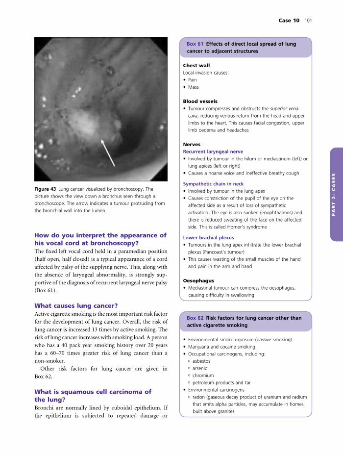
Case 10 101
PA
RT
2:
CA
SE
S
How do you interpret the appearance of his vocal cord at bronchoscopy?The fi xed left vocal cord held in a paramedian position
(half open, half closed) is a typical appearance of a cord
affected by palsy of the supplying nerve. This, along with
the absence of laryngeal abnormality, is strongly sup-
portive of the diagnosis of recurrent laryngeal nerve palsy
(Box 61).
What causes lung cancer?Active cigarette smoking is the most important risk factor
for the development of lung cancer. Overall, the risk of
lung cancer is increased 13 times by active smoking. The
risk of lung cancer increases with smoking load. A person
who has a 40 pack year smoking history over 20 years
has a 60–70 times greater risk of lung cancer than a
non-smoker.
Other risk factors for lung cancer are given in
Box 62.
What is squamous cell carcinoma of the lung?Bronchi are normally lined by cuboidal epithelium. If
the epithelium is subjected to repeated damage or
Figure 43 Lung cancer visualized by bronchoscopy. The
picture shows the view down a bronchus seen through a
bronchoscope. The arrow indicates a tumour protruding from
the bronchial wall into the lumen.
Box 61 Effects of direct local spread of lung cancer to adjacent structures
Chest wallLocal invasion causes:
• Pain
• Mass
Blood vessels• Tumour compresses and obstructs the superior vena
cava, reducing venous return from the head and upper
limbs to the heart. This causes facial congestion, upper
limb oedema and headaches
NervesRecurrent laryngeal nerve• Involved by tumour in the hilum or mediastinum (left) or
lung apices (left or right)
• Causes a hoarse voice and ineffective breathy cough
Sympathetic chain in neck• Involved by tumour in the lung apex
• Causes constriction of the pupil of the eye on the
affected side as a result of loss of sympathetic
activation. The eye is also sunken (enophthalmos) and
there is reduced sweating of the face on the affected
side. This is called Horner’s syndrome
Lower brachial plexus• Tumours in the lung apex infi ltrate the lower brachial
plexus (Pancoast’s tumour)
• This causes wasting of the small muscles of the hand
and pain in the arm and hand
Oesophagus• Mediastinal tumour can compress the oesophagus,
causing diffi culty in swallowing
Box 62 Risk factors for lung cancer other than active cigarette smoking
• Environmental smoke exposure (passive smoking)
• Marijuana and cocaine smoking
• Occupational carcinogens, including:� asbestos� arsenic� chromium� petroleum products and tar
• Environmental carcinogens� radon (gaseous decay product of uranium and radium
that emits alpha particles, may accumulate in homes
built above granite)

102 Part 2: Cases
PA
RT
2:
CA
SE
S
irritation (e.g. by cigarette smoking) it may change
its form to squamous epithelium to withstand this
(metaplasia). Early malignant changes (dysplasia) may
arise during this process which may progress to cancer
(neoplasia).
What other pathological types of lung cancer are there and how are they classifi ed?Lung cancer is generally classifi ed by histological fi ndings
(Box 63), which allows planning and standardization of
treatment. The main distinction is between non-small
cell and small cell lung cancers as these respond differ-
ently to treatment:
• Non-small cell cancer:� responds poorly to chemotherapy� surgery can be curative for patients with localized
disease
• Small cell lung cancer:� grows fast, therefore initially is responsive to
chemotherapy� metastasizes early, therefore surgery has no role in
the management of this condition
What does tumour staging mean and why is it done?Tumour staging is an assessment of tumour spread and
metastasis. It allows accurate prediction of response to
therapy and prognosis and is used in combination with
histological fi ndings to decide on appropriate treatment
for individual patients.
Mr Cole’s tumour was staged at T2 N2 M0. What does this mean?The TNM system is used for staging non-small cell lung
cancer:
• ‘T’ is a measure of local invasion by the tumour
and ranges from 0 (no evidence of primary tumour)
to 4 (local spread of tumour to mediastinum or
pleura)
• ‘N’ is a measure of spread of tumour to lymph nodes
and ranges from 0 (no lymph node spread) to 3 (spread
to more distant nodes)
• ‘M’ is a measure of distant tumour metastasis and
ranges from 0 (no metastasis) to 1 (distant metastasis is
present)
The more the tumour has spread or metastasized, the
worse the prognosis and the lower the chance of curative
treatment. In Mr Cole’s case his T2 N2 M0 tumour is
associated with a 23% 5-year survival rate.
How is non-small cell lung cancer treated?Surgical resection of localized tumour gives the best
chance of cure.
Suitability for surgery is determined by:
• Tumour resectability (lack of local spread or
metastases)
• Physiological fi tness of patient to survive major
surgery
Surgical options include:
• Lobectomy or pneumonectomy
• Limited resection (segment or lung wedge containing
tumour) if patients are physiologically compromised
• Mediastinal nodes are also removed for treatment and
disease mapping
The decision to operate should be made by a thoracic
surgeon with expertise in oncology as part of the multi-
disciplinary team. Unfortunately, 80% of patients with
non-small cell lung cancer present with disease that is too
advanced for surgery.
Radiotherapy can be used as:
• An adjunct to surgery in patients with potentially
curable disease
• Radical therapy in patients with potentially curable
disease who are considered inoperable
• Palliative therapy in patients with local or metastatic
complications of their lung tumour
Box 63 Histological classifi cation of lung cancer
The frequency of tumour types in the UK as a proportion
of all lung cancer cases is given in brackets (information
from Cancer Research UK statistics, November 2004).
Non-small cell lung cancer• Squamous cell (35–45%)
� derived from bronchial epithelial cells
• Adenocarcinoma (15%)� derived from glandular tissue in the airway submucosa
• Large cell carcinoma (10%)
• Carcinoid (1%)
• Bronchoalveolar cell carcinoma (rare)
Small cell lung cancer (~20%)• Derived from endocrine cells in the lung

Case 10 103
PA
RT
2:
CA
SE
S
The clinical team considered that Mr Cole’s tumour was
probably too advanced for surgical treatment. However, to
confi rm this they arranged a positron emission tomography
(PET) scan to examine his mediastinal lymph nodes. While
waiting for the scan Mr Cole was admitted as an emergency
complaining of pain in his lower back and diffi culty passing
urine.
How is a PET scan performed?• A radiolabelled sugar (fl uorodeoxyglucose [FDG]) is
injected into the patient’s veins
• There is a delay of around 1 h while FDG is taken up
by the tissues
• The scan is performed to identify areas of high FDG
uptake, then correlated with CT fi ndings to relate anatomy
to metabolic function
• Tumours grow and metabolize rapidly, therefore they
take up more FDG than normal adult tissues
• Lymph node enlarged by tumour may be seen on the
PET scan as areas of high isotope uptake, whereas lymph
nodes enlarged for other reasons may not be detected by
PET scanning
Why was Mr Cole asked to undergo a PET scan?Mr Cole appears to have node involvement on his CT
scan which stages his tumour at an advanced stage. PET
scanning will help establish whether his lymphadeno-
pathy is caused by cancer or other causes. If his PET
scan is negative he could still be a candidate for surgical
treatment.
What should you think of as the cause of Mr Cole’s new symptoms?You should immediately think of spinal cord compres-
sion in a patient with back pain and a change in bladder
or bowel function. In Mr Cole’s case the most important
possible cause is spinal metastases from his lung cancer
causing cord compression through effects of tumour
bulk or by causing vertebral collapse.
What should the doctors do next?Spinal cord compression is a clinical emergency and
requires urgent diagnosis and treatment to prevent per-
manent neurological damage. Mr Cole should undergo a
neurological examination, then X-ray and magnetic reso-
nance imaging (MRI) of his spine to establish whether
this has occurred.
!RED FLAG
Acute spinal cord compression is a medical emergency and
requires rapid diagnosis and treatment before permanent
neurological disability occurs.
It should be considered in patients with changes in control
of bladder or bowel or neurological symptoms in their legs.
On examination Mr Cole had normal tone in his legs. Power
was 5/5 in all muscle groups, coordination and sensation
was normal and plantars were downgoing. He had a
palpable bladder on abdominal examination. Rectal
examination revealed normal anal tone, a smooth enlarged
prostate and impacted faeces.
Does he have spinal cord compression?Neurological examination is reassuring as there is no evi-
dence of increased tone, weakness, upgoing plantars or
sensory loss in his legs. Examination fi ndings provide the
alternative explanation of constipation and prostatic
hypertrophy for his urinary retention. His back pain
still requires investigation, although now not as an
emergency.
What tests would be useful in investigating his back pain?• Plain X-rays
• Bone scan
• CT or MRI of the spine
Serum calcium should be measured to exclude hyper-
calcaemia, which can be caused by bone destruction
by metastases or by secretion of parathyroid hor-
mone related peptide or parathyroid hormone by the
tumour.
Mr Cole’s spinal X-rays and CT scan are shown in Fig. 44.
His serum calcium is normal.
Have the X-rays identifi ed a cause for his back pain?He has a compression fracture of L2 which looks abnor-
mal on CT scanning and contains metastasis (Box 64)
from his primary lung cancer.
How should Mr Cole be treated?As he has spinal metastases curative surgical resection
of his lung tumour is not an option. His PET scan is
cancelled.

104 Part 2: Cases
PA
RT
2:
CA
SE
S
He should receive palliative and supportive care
including:
• Radiotherapy for painful metastases
• Analgesia
• Nutritional support
• Psychological and spiritual support
• Financial and practical help
• Legal and other advice
Is this the fi rst time in Mr Cole’s treatment that he has been considered as a person and not just ‘a case of lung cancer’?Patients with suspected lung cancer go through a rapid
series of tests to establish the diagnosis, stage the tumour
and decide on treatment. The reason for this haste is to
offer as many people as possible treatment with some
chance of cure or prolonged survival before their disease
has progressed too far. Mr Cole went from discussing a
routine problem with his GP to an urgent chest X-ray,
respiratory referral, bronchoscopy, diagnosis and staging
CT in around 2 weeks. A key part of Mr Cole’s manage-
ment is that he should be informed and contribute to the
decision-making about his care. At each step of his inves-
tigation doctors and other professionals should discuss
possible diagnoses, explain procedures and discuss
L2
L3
(a)
(c)
(b)
Figure 44 Plain X-rays and computed
tomography (CT) scan of lumbar spine
showing metastases. (a) Plain X-ray of
lumbar spine showing compression
fracture of L2. L3 is normal. (b) CT
appearance of L2 showing
abnormality in the vertebral body and
fracture resulting from metastasis.
(c) Normal CT appearances of L3
vertebral body for comparison.
Box 64 Metastatic complications of lung cancer
Bone• Causes pain, hypercalcaemia, pathological fractures,
compression of adjacent neurological tissue
Liver• Causes hepatomegaly, obstructive jaundice
Brain• Causes headache, fi ts, confusion, raised intracranial
pressure, focal neurological defi cit (e.g. hemiplegia,
hemianopia, depending on site of metastasis)
Adrenals• May cause hypoadrenalism

Case 10 105
PA
RT
2:
CA
SE
S
benefi ts and risks of investigations and treatments with
him. Mr Cole should be invited to involve a family
member or other close person in his consultations if he
wishes and be offered written information, support and
symptom relief at all stages.
Outcome. Mr Cole received radiotherapy to his back with
good pain relief. He was discharged home with outpatient
support from his local hospice. Over the next 8 months he
deteriorated with marked loss of weight and increasing
shortness of breath. He died at home of a respiratory
infection supported by his wife and Macmillan nurses.
KEY POINT
Lung cancer is diffi cult to treat but relatively easy to
prevent.
It is the responsibility of all doctors to advise people not
to smoke.
KEY POINTS
• Patients with a hoarse voice for more than 3 weeks should
be referred urgently for chest X-ray and ENT examination
to look for lung or laryngeal cancer
• Causes of a mass seen on chest X-ray include:� primary lung cancer� secondary tumour deposit in the lung� tuberculosis� lymphoma
• Hilar masses are more likely to involve the recurrent
laryngeal nerve on the left than on the right because of
the different anatomy of the nerve on the two sides
• Further investigations of a mass seen on chest X-ray
include CT scanning and bronchoscopy to characterize the
lesion, obtain tissue for histology and stage possible
cancers
• Lung cancer is predominantly caused by tobacco
smoking
• Histological types of primary lung cancer are non-small
cell (squamous cell, adenocarcinoma, large cell, carcinoid,
bronchoalveolar cell) or small cell
• Tissue diagnosis of lung cancer is important as it guides
treatment
• Treatment for non-small cell lung cancer includes:� curative resection for localized lung cancer� radiotherapy� chemotherapy, although the response is poor
• Complications of lung cancer include:� local spread� distant metastases (bone, liver, brain, adrenals)� paraneoplastic phenomena (see Case 11)
cinoma. Staging CT shows metastasis to mediastinal lymph
nodes. He subsequently develops back pain and an MRI
demonstrates spinal metastases. He receives palliative
radiotherapy to his back with good relief and dies at home
with support from Macmillan nurses and his family.
CASE REVIEW
Mr Cole is noticed to have a hoarse voice on a routine visit
to his GP. An urgent chest X-ray shows a left hilar mass and
he is referred to secondary care for urgent investigation. At
bronchoscopy, left vocal cord palsy and tumour in the left
main bronchus are seen. Biopsy shows squamous cell car-

PA
RT
2:
CA
SE
S
A 56-year-old woman with an abnormal chest X-ray
Case 11
Mrs Gibson, a 56-year-old woman, sees her GP complaining
of cough and weight loss and is sent for a chest X-ray (Fig.
45). The radiologist reporting the fi lm fi nds an abnormality
and contacts the respiratory team who arrange to see her
that week in a rapid access clinic.
What does her chest X-ray show?The most notable abnormality is a large mass in the
midzone of her left lung. There is some associated
pleural fl uid or reaction obscuring the left costophrenic
angle.
What is the differential diagnosis for this chest X-ray abnormality?This appearance is most likely caused by cancer which is
either:
• Primary lung cancer; or
• Secondary metastasis
Lung abcess, tuberculosis or pneumonia are less likely
possibilities.
What is a rapid access clinic?The UK National Cancer Plan has recommended a series
of waiting time targets to prevent treatment delays in
patients with lung cancer. These targets include a
maximum of 14 days from GP referral to hospital
appointment and 62 days from GP referral to fi rst treat-
ment. To facilitate this many hospitals have set up specifi c
clinics to ensure patients with possible lung cancer have
rapid access to services.
Mrs Gibson attends the clinic with her husband. She is
extremely concerned about her abnormal chest X-ray and
immediately asks the doctor whether she has cancer.
How should the doctor respond?There is no easy response to such a question, particularly
at this stage. The doctor already knows that cancer is the
most likely diagnosis; however, it has not yet been proven
and non-cancer diagnoses are possible. He has just met
Mrs Gibson and has had no opportunity to establish a
doctor–patient relationship with her or predict how she
may react. His response to her question may affect their
future interaction and could facilitate or damage future
communication.
A reasonable approach could include some or all of the
following:
• Asking Mrs Gibson what she has been told already and
what her suspicions are
• Following this up by admitting that cancer is a possible
diagnosis, but there are other differentials
• Explaining that the purpose of the clinic is to fi nd out
what is wrong with her, but this will require questions,
examination and investigations
• Reassuring her that the assessment will be performed
as quickly as possible and she will be informed of the
results
• Establishing how much she would like to be told as
information becomes available
Mrs Gibson tells her doctor that she knows she has a
shadow on her chest X-ray. She has jumped to the
conclusion that it is lung cancer as her father died of this
condition. She agrees to further assessment, on the
condition that she is fully informed of all results and
involved in any decisions about her treatment.
What features in her history would point to a diagnosis of lung cancer?Mrs Gibson presented to her GP with cough and weight
loss. Further characterization of these and enquiry about
associated symptoms are important to support the
diagnosis.
Most patients with lung cancer present with respira-
tory symptoms, generally coming on over weeks. It is
helpful to establish:
• Whether Mrs Gibson’s cough is recent or has changed
at all. A long-standing cough over months or years indi-
106

Case 11 107
PA
RT
2:
CA
SE
S
cates another diagnosis (e.g. chronic obstructive pulmo-
nary disease [COPD])
• Whether she has associated respiratory symptoms:� chest pain – chest wall involvement� haemoptysis – bleeding from the tumour� chest infection – tumours may partially obstruct
airways, impeding mucociliary clearance and causing
recurrent or intractable chest infection� shortness of breath – this may be caused by large
airway obstruction or development of associated
pleural effusion� malaise, weight loss and anorexia occur in almost all
patients with lung cancer at some point in their illness
Risk factors for lung cancer should also be established.
Mrs Gibson’s cough started around 4 weeks ago and is non-
productive. Over that time she has lost around 1 stone in
weight. As a child both her parents smoked heavily and she
started smoking aged 12. She currently smokes around 40
cigarettes per day.
On examination Mrs Gibson looked cachectic. She had a
palpable lymph node in the left supraclavicular fossa. There
were no abnormal fi ndings on chest examination. Her liver
was palpable at 3 cm below the costal margin.
How many pack years has Mrs Gibson smoked?She has smoked 2 packets of cigarettes per day for most
of 44 years (i.e. approximately 88 pack years; Box 16,
p. 19).
How do you explain the enlarged lymph node in her left supraclavicular fossa?Supraclavicular lymphadenopathy, especially in patients
over 40 years, is most commonly caused by malignancy.
The route of lymphatic drainage to the supraclavicular
nodes is outlined in Box 65. An enlarged left supracla-
vicular lymph node is often caused by metastatic stomach
cancer and is known as Virchow’s node. However, in Mrs
Gibson’s case the combination of left supraclavicular
lymphadenopathy and an abnormal chest X-ray is most
likely to be because of a primary lung malignancy (Box
66).
Why might her liver be enlarged?This could be explained by metastases from primary lung
or abdominal/pelvic malignancy.
What investigations should be performed to make a diagnosis?As cancer is likely to be the cause of Mrs Gibson’s illness
it is important to obtain tissue to make a histological
diagnosis. This could be performed using a number of
different approaches.
Figure 45 Chest X-ray.
Box 65 Lymphatic drainage of the lung
Lymphatic drainage of the lung occurs via:
• The superfi cial lymphatic plexus, which is deep to the
visceral pleura and drains around the periphery of the
lungs to bronchopulmonary nodes in the hila
• The deep lymphatic plexus, which follows vessels and
airways to the hila and also drains into the hilar
(bronchopulmonary) nodes
Efferent lymph channels carry lymph from the
bronchopulmonary nodes to (sequentially):
• Tracheobronchial nodes at the bifurcation of the trachea
(carina)
• Paratracheal nodes (alongside the trachea)
• Mediastinal lymph trunks
The right mediastinal lymph trunk joins right subclavian
(from the right upper limb) and right jugular (from the
right side of the head and neck) trunks and all three have
a common opening into the origin of the right
brachiocephalic vein via right supraclavicular lymph nodes.
The left mediastinal lymph trunk, left subclavian (from
the left upper limb) and jugular (left head and neck)
trunks usually join the thoracic duct (from the lower limbs
and abdomen), which drains via supraclavicular lymph
nodes into great veins at the root of the neck.

108 Part 2: Cases
PA
RT
2:
CA
SE
S
Fine needle aspiration of supraclavicular nodesA fi ne needle is inserted into the node and negative pres-
sure applied using a syringe to suck cells out of the node.
The sample is smeared on a slide and examined cyto-
logically. This is said to be no more painful than having
blood taken by venepuncture, does not usually require
anaesthetic and has few if any complications.
Liver ultrasound and biopsyIf radiology demonstrates that a mass is causing her
hepatomegaly, percutaneous liver biopsy could be used
to obtain tissue to make a diagnosis. Local anaesthetic is
used to numb the overlying skin and ultrasound guides
the biopsy needle into the abnormal part of the liver. The
main complication is haemorrhage.
Fibreoptic bronchoscopyIf the tumour arises from a large airway this procedure
will allow direct visualization of the tumour, which can
be biopsied using forceps inserted along the broncho-
scope. The main complication is haemorrhage. If the
tumour is peripheral it will not be seen, but bronchial
washings can be taken from the affected segment and
examined cytologically for cancer cells.
CT guided biopsyPeripheral lung tumours not accessible by bronchoscopy
can be sampled by percutaneous transthoracic needle
biopsy (i.e. a biopsy needle inserted through the chest
wall into the tumour under CT guidance). Local anaes-
thetic is required and the procedure may be complicated
by pneumothorax or haemorrhage.
Which of these investigations would be best for Mrs Gibson?Fine needle aspiration of her palpable supraclavicular
node is a very low risk procedure that has a good chance
of making a positive diagnosis. The other techniques all
carry risks of complications, with a real (although very
low) risk of mortality. An initial approach would be to
sample her supraclavicular node and arrange a contrast
enhanced CT scan of her chest and abdomen to assess
and stage her tumour and examine her liver. If the lymph
node aspirate was unhelpful, further investigations could
be planned based on the CT result. These investigations
should be carried out as quickly as possible so as not to
delay the diagnosis.
Mrs Gibson underwent fi ne needle aspiration of her left
supraclavicular node. Cytological examination revealed small
cell lung cancer. Her CT scan showed a large mass in her left
hemithorax with mediastinal lymphadenopathy. Her liver was
pushed down by hyperinfl ation of her lungs but not enlarged
and no metastases were seen. Her adrenal glands looked normal.
What is small cell lung cancer?Small cell lung cancer arises from endocrine cells in the
lung which are members of the APUD system (amine
precursor uptake and decarboxylation). Small cell
tumours comprise 20–30% of lung cancers and are
rapidly growing and highly malignant. These tumours
have usually metastasized, either overtly or occultly, at the
time of presentation. As the tumour cells have an endo-
crine origin they often secrete hormones which have
systemic effects.
Why did the radiologist comment on her adrenal glands?Bronchogenic carcinoma often metastasizes to the adrenal
glands, therefore examination of these is an important
part of tumour staging.
Mrs Gibson’s adrenal glands and liver look normal on CT scan
so she has no overt evidence of distant tumour metastasis.
What is the importance of staging a patient’s lung cancer?Tumour staging is helpful for planning treatment and can
give an indication of prognosis.
Box 66 Differential diagnosis of supraclavicular lymphadenopathy
Malignancy (most common, particularly in patients
over 40)
Right sided:
• Lung
• Mediastinum
• Oesophagus
• Lymphoma
Left sided:
• Abdominal and pelvic malignancies (e.g. stomach,
pancreas, ovaries, prostate)
• Lung (left side of thorax)
• Lymphoma
Other• Tuberculosis

Case 11 109
PA
RT
2:
CA
SE
S
Mrs Gibson has limited disease (Box 67) which involves the
left hemithorax, mediastinum and supraclavicular nodes but
has not extended beyond these. As she is female and has
good performance status she is in the best prognostic
group.
What is the prognosis of small cell lung cancer?This is an aggressive disease with a terrible prognosis. At
presentation median survival is:
• Limited disease. 12 weeks
• Extensive disease. 4 weeks
With best treatment:
• Survival is increased four- to fi vefold.
• 10% of patients will remain disease-free for 2 years
from start of therapy
• 5–10% of patients will survive for 5 years
Investigation and start of treatment must there-
fore be as rapid as possible to give patients with small
cell lung cancer the best chance of disease-free
survival.
Mrs Gibson returns 1 week after her fi rst appointment. Her
husband has become concerned as she is irritable,
nauseated and confused. He feels this is brought about by
more than anxiety about her diagnosis.
What might account for Mrs Gibson’s new confusion?Possible causes include:
• Cerebral metastases from her small cell lung cancer
• Hormone excretion by the tumour causing hypona-
traemia and confusion
• Secondary respiratory infection
• Hypoxia or hypercapnia secondary to lung disease
• Hypercalcaemia secondary to malignancy or bony
metastases
What investigations would you order?• CT brain scan
• Plasma biochemistry, calcium, full blood count and
infl ammatory markers
• Pulse oximetry, with arterial blood gases if abnormal
Mrs Gibson underwent these tests on the same day. Her
results were as follows:
CT head. Normal
Blood tests:
Normal range
Sodium 108 mmol/L (135–145 mmol/L)
Potassium 4.2 mmol/L (3.5–5.0 mmol/L)
Urea 5.6 mmol/L (2.5–8 mmol/L)
Creatinine 94 mmol/L (60–110 mmol/L)
Calcium 2.42 mmol/L (2.2–2.6 mmol/L)
Liver function Normal
CRP 2 mg/L (<8 mg/L)
Serum osmolarity 250 mosm/kg (280–296 mosm/kg)
Urine osmolarity 600 mosm/kg (350–1000 mosm/kg)
Full blood count Normal
Oxygen saturations 98%
How do you interpret these results?• She is hyponatraemic, which is suffi cient to cause her
symptoms
• There is no evidence of cerebral metastases or
hypercalcaemia
• Infl ammatory markers and oxygen saturations are
normal
Why is she hyponatraemic?• Her serum osmolarity is very low, indicating excess
water in the blood
• In this situation, feedback to the posterior pituitary
should turn off secretion of antidiuretic hormone (ADH),
allowing water to be excreted in the urine. This should
be seen as a low urine osmolarity
• Mrs Gibson has normal urine osmolarity, which
indicates that she has failed to switch off ADH
appropriately
• It is likely that Mrs Gibson has inappropriate ADH
secretion from her small cell lung cancer that is not sus-
ceptible to normal regulation
Box 67 Staging of small cell lung cancer and prognostic indicators
Staging• Limited disease. Disease limited to the hemithorax of
origin, mediastinum and supraclavicular nodes
• Extensive disease. Widespread tumour beyond the
extent of limited disease
Prognostic factors• Better prognosis. Good performance status, female
gender, limited disease
• Poor prognosis. Poor performance status, liver or central
nervous system (CNS) involvement

110 Part 2: Cases
PA
RT
2:
CA
SE
S
This is a paraneoplastic complication of her lung tumour.
Paraneoplastic syndromes are caused by factors produced
by cancer cells that act at a distance from the primary
cancer site or metastases. Other paraneoplastic phenom-
ena associated with lung tumours are given in Box 68.
How should her hyponatraemia be treated?Treatment of her small cell tumour to reduce cancer bulk
may reduce ADH secretion and improve her hyponatrae-
mia. She is not currently fi t for this and needs restriction
of water and fl uid intake, monitoring and possibly dem-
eclocycline, a drug that reduces renal sensitivity to ADH
and allows water excretion.
In view of her confusion and urgent need to commence
cancer treatment, Mrs Gibson is admitted to hospital
to stabilize her condition. Over the next few days her
serum sodium normalizes with water restriction and
demeclocycline. As her confusion clears she is told the
diagnosis. After a long discussion with the oncologists she
and her husband decide that she should have chemotherapy
and radiotherapy as advised by the specialist. She prepares
for treatment and also sees the palliative care team.
Why is she being given chemotherapy?Small cell cancer is responsive to chemotherapy as it is
rapidly growing. It also metastasizes early and so systemic
treatment is required as local treatments such as surgery
have little chance of curing or controlling her disease.
Why will she also receive radiotherapy?In patients with limited stage small cell carcinoma, com-
bination chemotherapy and radiotherapy of tumour have
been shown to produce a modest improvement in prog-
nosis in comparison to chemotherapy alone.
Does referral to the palliative care team at this stage indicate that there is no hope of cure?The palliative care team can help with symptom control
in patients with cancer, even during active treatment. A
rapport built up at this stage may help later in the illness
if it becomes necessary to withdraw active treatment.
Patients with cancer and their families often need support
with coming to terms with the diagnosis and dealing with
end of life issues.
Mrs Gibson starts her fi rst cycle of chemotherapy 2 weeks
after her initial referral. She tolerates the treatment well
apart from some diarrhoea and fatigue. The doctors monitor
her blood count and during treatment she loses her hair.
Why does her chemotherapy cause diarrhoea and hair loss?Chemotherapy kills cancer cells and normal cells that
divide rapidly. In adults, most cells are non- or slowly
dividing. Hair and intestinal cells do divide and so are
most commonly affected, hence hair loss and diarrhoea.
Blood cells are also rapidly dividing and a common side-
effect of chemotherapy is bone marrow suppression
which can increase susceptibility to infection. Her blood
count is monitored to detect this.
Outcome. Mrs Gibson completes four cycles of
chemotherapy and has thoracic radiation. However, 4
months later she is readmitted to hospital with worsening
shortness of breath. She is found to have a large pleural
effusion and liver metastases. At this point she refuses
further chemotherapy and is transferred to the local hospice
for palliative care. She dies comfortably 2 weeks later.
Box 68 Paraneoplastic phenomena associated with lung cancer
Cachexia• Weight loss and wasting
Musculoskeletal and skin• Finger clubbing
• Hypertrophic pulmonary osteoarthropathy with pain,
tenderness and swelling over affected bones
• Acanthosis nigricans (localized hyperpigmentation)
Endocrine• Inappropriate ADH secretion
• Ectopic adrenocorticotrophic hormone secretion
• Hypercalcaemia caused by secretion of parathyroid
hormone-like substance
• Gynaecomastia resulting from secretion of human
chorionic gonadotrophin
Neurological• Eaton–Lambert syndrome (antibodies to voltage-gated
calcium channels causing proximal muscle weakness)
• Peripheral neuropathies
• Subacute cerebellar degeneration
• Cortical degeneration
• Polymyositis

Case 11 111
PA
RT
2:
CA
SE
S
KEY POINTS
• Small cell lung cancers are rapidly growing and highly
malignant, hence have usually metastasized at time of
presentation and carry a poor prognosis
• Small cell lung cancer arises from endocrine cells in the
lung which are members of the APUD system. Therefore,
they may secrete hormones that have a systemic effect,
e.g.� antidiuretic hormone� adrenocorticotrophic hormone� parathyroid hormone-like hormone
• Small cell lung cancer is responsive to chemotherapy,
although is unlikely to be cured
• The multidisciplinary team caring for patients with lung
cancer includes:� doctors� nurses – hospital palliative care nurses and community
Macmillan nurses� dietitians� occupational therapists� physiotherapists� social workers� religious leaders� counsellors
awaiting chemotherapy she is admitted with confusion,
caused by hyponatraemia secondary to inappropriate ADH
secretion. Once her condition is stabilized, her cancer is
treated with four cycles of chemotherapy and thoracic
radiotherapy. Despite this she re-presents 4 months later
with a pleural effusion and liver metastases. She is treated
palliatively and dies comfortably in the local hospice.
CASE REVIEW
Mrs Gibson presents to her GP with cough and weight loss.
A chest X-ray shows a large mass in the midzone of her left
lung and she is referred to the rapid access lung cancer
clinic. Examination reveals an enlarged left supraclavicular
lymph node and fi ne needle aspiration of this node gives
a histological diagnosis of small cell lung cancer. On
staging, she is found to have limited disease, but while
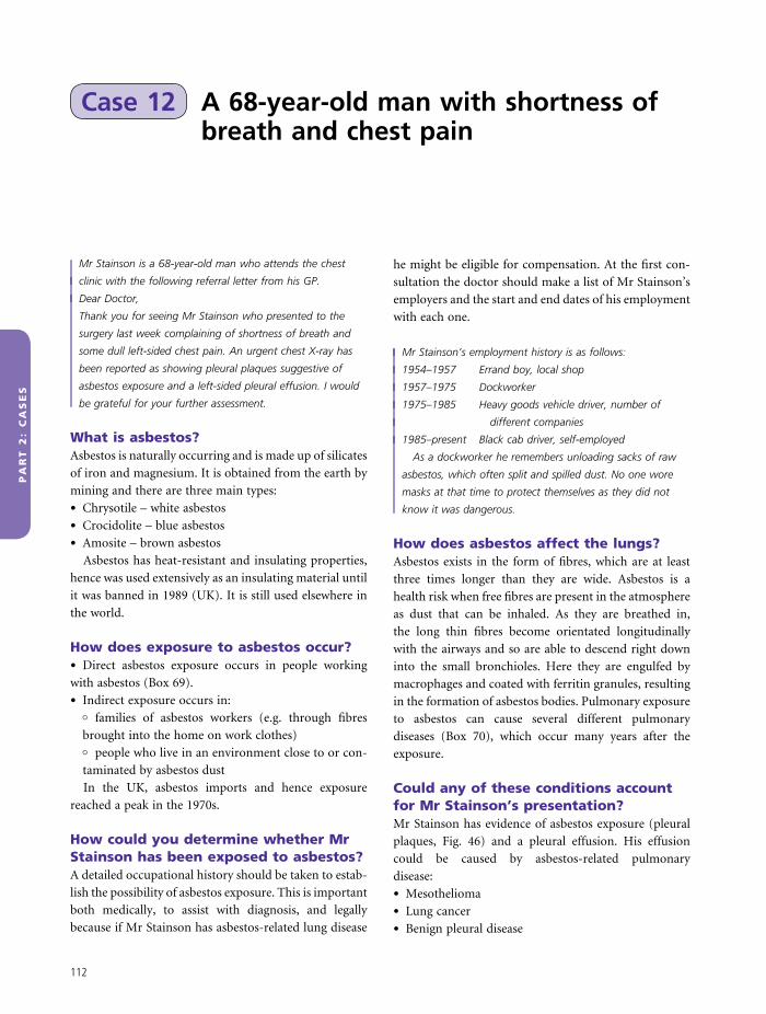
PA
RT
2:
CA
SE
S
A 68-year-old man with shortness of breath and chest pain
Case 12
Mr Stainson is a 68-year-old man who attends the chest
clinic with the following referral letter from his GP.
Dear Doctor,
Thank you for seeing Mr Stainson who presented to the
surgery last week complaining of shortness of breath and
some dull left-sided chest pain. An urgent chest X-ray has
been reported as showing pleural plaques suggestive of
asbestos exposure and a left-sided pleural effusion. I would
be grateful for your further assessment.
What is asbestos?Asbestos is naturally occurring and is made up of silicates
of iron and magnesium. It is obtained from the earth by
mining and there are three main types:
• Chrysotile – white asbestos
• Crocidolite – blue asbestos
• Amosite – brown asbestos
Asbestos has heat-resistant and insulating properties,
hence was used extensively as an insulating material until
it was banned in 1989 (UK). It is still used elsewhere in
the world.
How does exposure to asbestos occur?• Direct asbestos exposure occurs in people working
with asbestos (Box 69).
• Indirect exposure occurs in:� families of asbestos workers (e.g. through fi bres
brought into the home on work clothes)� people who live in an environment close to or con-
taminated by asbestos dust
In the UK, asbestos imports and hence exposure
reached a peak in the 1970s.
How could you determine whether Mr Stainson has been exposed to asbestos?A detailed occupational history should be taken to estab-
lish the possibility of asbestos exposure. This is important
both medically, to assist with diagnosis, and legally
because if Mr Stainson has asbestos-related lung disease
he might be eligible for compensation. At the fi rst con-
sultation the doctor should make a list of Mr Stainson’s
employers and the start and end dates of his employment
with each one.
Mr Stainson’s employment history is as follows:
1954–1957 Errand boy, local shop
1957–1975 Dockworker
1975–1985 Heavy goods vehicle driver, number of
different companies
1985–present Black cab driver, self-employed
As a dockworker he remembers unloading sacks of raw
asbestos, which often split and spilled dust. No one wore
masks at that time to protect themselves as they did not
know it was dangerous.
How does asbestos affect the lungs?Asbestos exists in the form of fi bres, which are at least
three times longer than they are wide. Asbestos is a
health risk when free fi bres are present in the atmosphere
as dust that can be inhaled. As they are breathed in,
the long thin fi bres become orientated longitudinally
with the airways and so are able to descend right down
into the small bronchioles. Here they are engulfed by
macrophages and coated with ferritin granules, resulting
in the formation of asbestos bodies. Pulmonary exposure
to asbestos can cause several different pulmonary
diseases (Box 70), which occur many years after the
exposure.
Could any of these conditions account for Mr Stainson’s presentation?Mr Stainson has evidence of asbestos exposure (pleural
plaques, Fig. 46) and a pleural effusion. His effusion
could be caused by asbestos-related pulmonary
disease:
• Mesothelioma
• Lung cancer
• Benign pleural disease
112
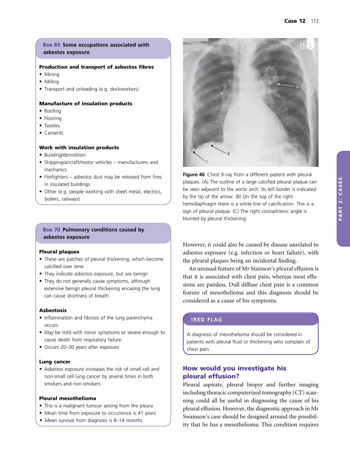
Case 12 113
PA
RT
2:
CA
SE
S
However, it could also be caused by disease unrelated to
asbestos exposure (e.g. infection or heart failure), with
the pleural plaques being an incidental fi nding.
An unusual feature of Mr Stainson’s pleural effusion is
that it is associated with chest pain, whereas most effu-
sions are painless. Dull diffuse chest pain is a common
feature of mesothelioma and this diagnosis should be
considered as a cause of his symptoms.
!RED FLAG
A diagnosis of mesothelioma should be considered in
patients with pleural fl uid or thickening who complain of
chest pain.
How would you investigate his pleural effusion?Pleural aspirate, pleural biopsy and further imaging
including thoracic computerized tomography (CT) scan-
ning could all be useful in diagnosing the cause of his
pleural effusion. However, the diagnostic approach in Mr
Swainson’s case should be designed around the possibil-
ity that he has a mesothelioma. This condition requires
Box 69 Some occupations associated with asbestos exposure
Production and transport of asbestos fi bres• Mining
• Milling
• Transport and unloading (e.g. dockworkers)
Manufacture of insulation products• Roofi ng
• Flooring
• Textiles
• Cements
Work with insulation products• Building/demolition
• Shipping/aircraft/motor vehicles – manufacturers and
mechanics
• Firefi ghters – asbestos dust may be released from fi res
in insulated buildings
• Other (e.g. people working with sheet metal, electrics,
boilers, railways)
Box 70 Pulmonary conditions caused by asbestos exposure
Pleural plaques• These are patches of pleural thickening, which become
calcifi ed over time
• They indicate asbestos exposure, but are benign
• They do not generally cause symptoms, although
extensive benign pleural thickening encasing the lung
can cause shortness of breath
Asbestosis• Infl ammation and fi brosis of the lung parenchyma
occurs
• May be mild with minor symptoms or severe enough to
cause death from respiratory failure
• Occurs 20–30 years after exposure
Lung cancer• Asbestos exposure increases the risk of small cell and
non-small cell lung cancer by several times in both
smokers and non-smokers
Pleural mesothelioma• This is a malignant tumour arising from the pleura
• Mean time from exposure to occurrence is 41 years
• Mean survival from diagnosis is 8–14 months
c
B
A
Figure 46 Chest X-ray from a different patient with pleural
plaques. (A) The outline of a large calcifi ed pleural plaque can
be seen adjacent to the aortic arch. Its left border is indicated
by the tip of the arrow. (B) On the top of the right
hemidiaphragm there is a white line of calcifi cation. This is a
sign of pleural plaque. (C) The right costophrenic angle is
blunted by pleural thickening.

114 Part 2: Cases
PA
RT
2:
CA
SE
S
special care as any pleural intervention (e.g. aspirate,
biopsy) can seed mesothelioma cells through the chest
wall, spreading the tumour. The number of invasive
pleural procedures should thus be minimized in patients
investigated for mesothelioma and if the diagnosis is sub-
sequently proven, patients should have radiotherapy to
biopsy tracks to prevent tumour spread.
Mr Swainson’s chest physician explained to him that there
were a number of possible causes for his condition, but that
disease caused by asbestos exposure was one possibility. Mr
Swainson agreed to undergo CT scanning to assist with
further diagnosis. At CT scanning irregular pleural thickening
around the left lung was seen (Fig. 47) and was reported as
showing some invasion of the chest wall. Pleural fl uid was
aspirated and a biopsy was performed of the irregular area
under CT guidance.
Pleural fl uid:
pH 7.4
Protein 40 g/L
Glucose 4 mmol/L
Cytology Negative
How do you interpret his results?• On CT scanning irregular pleural thickening with
chest wall invasion indicates a malignant process. The
differential diagnosis is:� local invasion by mesothelioma� pleural and chest wall spread from a primary lung
tumour� invasive infection such as tuberculosis
• Pleural fl uid shows:� an exudate (protein >30 g/L) – this would be consis-
tent with malignant pleural disease or tuberculosis
� the glucose, pH and cytology are unremarkable –
which does not help us to distinguish between possible
causes. Negative cytology does not exclude malignant
disease
Why was the biopsy performed under CT guidance?Mesothelioma tends to affect patches of the pleura and
therefore biopsy without radiological guidance may miss
the disease and may need to be repeated. CT guidance
increases the chance of making a positive diagnosis and
should reduce the number of pleural interventions
required.
Mr Stainson’s pleural biopsy showed malignant
mesothelioma. He attends clinic with his wife to fi nd out
the results of his investigations. The respiratory consultant
breaks the news to him that he has a malignant pleural
tumour. He is stunned by the news, but after a few
moments he asks how long he has to live and whether
there is any treatment that could offer him chance of a
cure.
How should the consultant answer Mr Stainson’s questions?• Patients with mesothelioma usually survive about a
year (8–14 months), although it can be longer or
shorter
• There are no treatments available that have been
proven in randomized controlled trials to cure
mesothelioma
• There is some evidence that triple therapy (radical
surgery to remove pleura and lung, chemotherapy and
radiotherapy) can improve survival for patients with less
malignant cell types. However, radical surgery carries a
high mortality
• There are clinical trials currently ongoing that are
looking for better treatments for mesothelioma. Mr
Stainson could be considered for these if he wishes (www.
mesothelioma.uk.com)
• Otherwise, the best treatment available is symptom
control
Mr Stainson and his wife go away to discuss their situation
and return to clinic 1 week later. They have decided to
opt for symptom control to maximize the quality of Mr
Stainson’s remaining months, but Mr Stainson would like to
be referred to a specialist centre to participate in a clinical
trial.
Figure 47 CT scan showing irregular pleural thickening
around the left lung (arrow).

Case 12 115
PA
RT
2:
CA
SE
S
How could Mr Stainson’s symptoms be controlled?Pleural effusionTreatment should aim to drain the effusion and prevent
recurrence using one of the following:
• Video-assisted thoracoscopy (VATS procedure)
• Open surgery
• Closed chest drain
Recurrent effusions are prevented by sticking the vis-
ceral and parietal pleura together (pleurodesis):
• Medically – talc is put into the pleural space
• Surgically – part of the visceral pleura is scraped
off
Both methods cause infl ammation and adhesion of the
pleural layers.
Radiotherapy• Radiotherapy to the mesothelioma may reduce chest
pain
• Radiotherapy to tracks made by invasive pro-
cedures will prevent growth of the tumour along the
tracks
Box 71 WHO analgesic ladder
Patients in pain should receive regular analgesia with
additional drugs available for breakthrough pain.
Step 1Regular simple analgesia (e.g. paracetamol)
If pain persists
Step 2Add mild opioid analgesia (e.g. codeine)
If pain persists
Step 3Use stronger opiate (e.g. morphine)
If pain persists
Increase dose of opiate until pain relieved
Adjuvant drugs such as non-steroidal anti-infl ammatory
drugs, steroids or drugs used for nerve pain can be added
to any step.
Other measures• Pain control – the World Health Organization (WHO)
analgesic ladder (Box 71) should be used when prescrib-
ing drugs to achieve pain control
• Support:� Mr Stainson would benefi t from referral to palliative
care services for physical, psychological and emotional
support� he could also be put in contact with asbestos support
groups (www.mesothelioma.uk.com) to help him
deal with practical issues and come to terms with his
condition
Is Mr Stainson entitled to fi nancial compensation?Yes – he should be advised to pursue one or both of the
following.
Government benefi tPatients are eligible if:
• They have a diagnosis of mesothelioma, lung cancer or
asbestosis
• They have a history of occupational exposure to
asbestos
• They were not self-employed when exposed
Patients have to complete application forms success-
fully to obtain these.
Common law compensationPatients may get compensation from the employer or
their insurance company. This is trickier to obtain as:
• The action must be started within 3 years of knowledge
of the diagnosis
• Claiming requires the assistance of an expert solicitor
• The claimant must prove that:� injuries were caused by asbestos exposure� the employer was negligent or did not comply with
health and safety requirements
Outcome. Mr Stainson was referred to a mesothelioma
specialist, but unfortunately was not considered to be
suitable for any current clinical trials. He underwent VATS
pleurodesis and radiotherapy for pain control. He remained
comfortable for 6 months, but presented again with
worsening breathlessness. At this time he was found to have
extensive mesothelial tumour encasing the lung, pericardium
and superior vena cava. His symptoms were palliated with
opiates and he died at home 2 weeks later.

116 Part 2: Cases
PA
RT
2:
CA
SE
S
KEY POINTS
• Asbestos exposure occurs in a wide range of occupational
and domestic environments
• Pulmonary disease caused by asbestos usually presents
decades after exposure and includes:� pleural plaques� asbestosis (interstitial fi brosis)� lung cancer� pleural mesothelioma
• A diagnosis of mesothelioma should be considered in
patients with pleural fl uid or thickening who complain of
chest pain
• The diagnosis of mesothelioma is made by histological
examination of a sample of affected pleura obtained
under CT guidance or by VATS
• Mesothelioma has a poor prognosis (8–14 months
survival) and treatment is usually symptomatic including:� drainage of pleural fl uid and pleurodesis� radiotherapy� pain control� palliative support
• Patients with asbestos-related illness may be entitled to
fi nancial compensation from government or employer
fl uid and biopsy and a diagnosis of malignant mesothelioma
is made. Mr Stainson is treated symptomatically with VATS
pleurodesis and radiotherapy, but dies 6 months later from
progressive disease.
CASE REVIEW
Mr Stainson presents with shortness of breath and dull
right-sided chest pain. Chest X-ray shows pleural plaques
and a left-sided pleural effusion. His history reveals
extensive asbestos exposure starting 40 years ago. He
undergoes thoracic CT scanning with aspiration of pleural
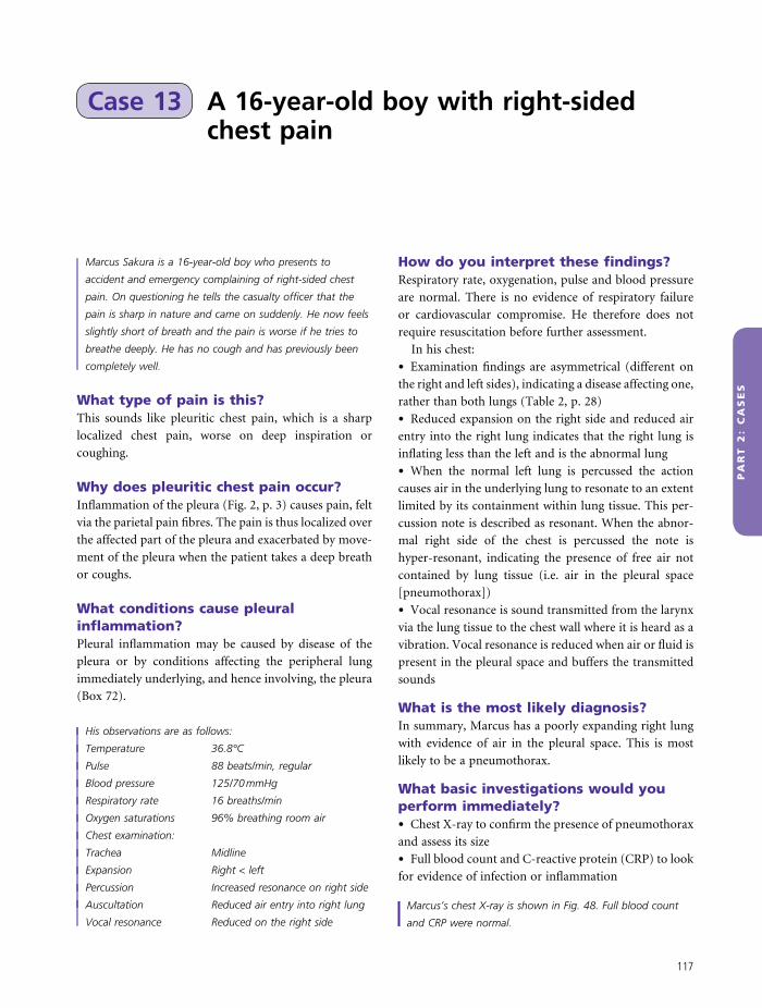
PA
RT
2:
CA
SE
S
A 16-year-old boy with right-sided chest pain
Case 13
Marcus Sakura is a 16-year-old boy who presents to
accident and emergency complaining of right-sided chest
pain. On questioning he tells the casualty offi cer that the
pain is sharp in nature and came on suddenly. He now feels
slightly short of breath and the pain is worse if he tries to
breathe deeply. He has no cough and has previously been
completely well.
What type of pain is this?This sounds like pleuritic chest pain, which is a sharp
localized chest pain, worse on deep inspiration or
coughing.
Why does pleuritic chest pain occur?Infl ammation of the pleura (Fig. 2, p. 3) causes pain, felt
via the parietal pain fi bres. The pain is thus localized over
the affected part of the pleura and exacerbated by move-
ment of the pleura when the patient takes a deep breath
or coughs.
What conditions cause pleural infl ammation?Pleural infl ammation may be caused by disease of the
pleura or by conditions affecting the peripheral lung
immediately underlying, and hence involving, the pleura
(Box 72).
His observations are as follows:
Temperature 36.8°C
Pulse 88 beats/min, regular
Blood pressure 125/70 mmHg
Respiratory rate 16 breaths/min
Oxygen saturations 96% breathing room air
Chest examination:
Trachea Midline
Expansion Right < left
Percussion Increased resonance on right side
Auscultation Reduced air entry into right lung
Vocal resonance Reduced on the right side
How do you interpret these fi ndings?Respiratory rate, oxygenation, pulse and blood pressure
are normal. There is no evidence of respiratory failure
or cardiovascular compromise. He therefore does not
require resuscitation before further assessment.
In his chest:
• Examination fi ndings are asymmetrical (different on
the right and left sides), indicating a disease affecting one,
rather than both lungs (Table 2, p. 28)
• Reduced expansion on the right side and reduced air
entry into the right lung indicates that the right lung is
infl ating less than the left and is the abnormal lung
• When the normal left lung is percussed the action
causes air in the underlying lung to resonate to an extent
limited by its containment within lung tissue. This per-
cussion note is described as resonant. When the abnor-
mal right side of the chest is percussed the note is
hyper-resonant, indicating the presence of free air not
contained by lung tissue (i.e. air in the pleural space
[pneumothorax])
• Vocal resonance is sound transmitted from the larynx
via the lung tissue to the chest wall where it is heard as a
vibration. Vocal resonance is reduced when air or fl uid is
present in the pleural space and buffers the transmitted
sounds
What is the most likely diagnosis?In summary, Marcus has a poorly expanding right lung
with evidence of air in the pleural space. This is most
likely to be a pneumothorax.
What basic investigations would you perform immediately?• Chest X-ray to confi rm the presence of pneumothorax
and assess its size
• Full blood count and C-reactive protein (CRP) to look
for evidence of infection or infl ammation
Marcus’s chest X-ray is shown in Fig. 48. Full blood count
and CRP were normal.
117

118 Part 2: Cases
PA
RT
2:
CA
SE
S
What does his chest X-ray show?If you look carefully you can see that the vascular mark-
ings of the normal left lung extend all the way from the
mediastinum to the chest wall. On the right side, however,
there is a small ball of collapsed lung around the hilum
with a clear edge, which is separated from the chest wall
by a large black rim of air. This is illustrated more clearly
in Fig. 49. The X-ray confi rms that Marcus has had a
pneumothorax.
What is normally in the pleural space?There is normally a thin layer of fl uid between the vis-
ceral and parietal pleura. The surface tension of this fl uid
couples the pleural layers together and creates a negative
pressure between them. In a normal chest there is nothing
else between the pleural layers, thus the pleural space is
a ‘potential’, rather than an actual space.
What is the function of the negative intrapleural pressure?The negative intrapleural pressure keeps the lungs
expanded and opposes their tendancy to collapse as a
result of elastic recoil.
How does air get into the pleural space?Pneumothoraces are caused when a hole in either visceral
or parietal pleura occurs that allows air into the pleural
space. Causes of pneumothorax are given in Box 73.
What happens to the lung when air enters the pleural space?• When a hole in the pleura occurs, air enters the pleural
space and the intrapleural pressure becomes less negative
Figure 48 Chest X-ray.
Box 72 Causes of pleuritic chest pain
Pleural disease• Pneumothorax
• Pleural malignancy (e.g. mesothelioma)
• Pleural infl ammation in connective tissue disease, e.g.� rheumatoid arthritis� systemic lupus erythematosis
• Pleural infection� viral (e.g. Coxsackie B)� bacterial (empyema)
Conditions affecting the peripheral lung• Pulmonary embolus
• Pneumonia
Figure 49 Pathogenesis of pneumothorax. The darker line
indicates the position of the parietal pleura lining the chest
wall. The lighter line indicates the position of the visceral
pleura covering the surface of the lung. On the left side, the
two pleural layers are stuck together by the negative
intrapleural pressure generated by continual reabsorption of
the intervening fl uid which keeps the lung expanded. On the
right side, there is a hole in the visceral pleura (A), allowing air
to leak into the pleural space (B). As intrapleural pressure now
equals atmospheric pressure there is nothing to oppose elastic
recoil of the lung, which has caused the lung to collapse into
a small ball of tissue at the hilum (C).

Case 13 119
PA
RT
2:
CA
SE
S
• As the intrapleural pressure becomes less negative it is
less able to oppose the elastic recoil of the lung. The lung
therefore starts to collapse
• The degree of lung collapse depends on the amount
of air entering the intrapleural space, which in turn
depends on the size of the hole and whether the hole
closes spontaneously:� if the hole closes as the lung collapses, the intra-
pleural pressure will remain somewhat negative,
preventing the lung from collapsing completely
(Fig. 50)� if the hole is large or remains open until intra-
pleural pressure equals atmospheric pressure, the lung
will collapse completely into a small ball around the
hilum (Figs 48 & 49). This has occurred in Marcus’s
case.
What is the most likely cause of Marcus’s pneumothorax?He was previously well without underlying lung disease
and does not give a history of trauma. This is therefore
likely to be a spontaneous primary pneumothorax.
How should his pneumothorax be treated?The aim of treatment is to remove the air from the pleural
cavity to allow his lung to re-expand (Box 74). If this is
not carried out he may develop infection in and chronic
damage to his collapsed lung, with severe consequences
for his health.
Marcus is admitted to hospital and started on high fl ow
oxygen at 10 L/min. His doctor advises him that his
pneumothorax should be aspirated to allow his lung to
expand.
Box 73 Causes of pneumothorax
Traumatic pneumothoraxEntry of air into pleural space is through a wound in chest
wall and parietal pleura (wound may also penetrate
visceral pleura), e.g.
• Rib fractures
• Stab wound
Spontaneous pneumothoraxEntry of air into pleural space is through a hole in lung
and visceral pleura, which often occurs because of rupture
of a pleural bleb or bulla (small bubbles of lung with thin
covering of pleura).
• Primary:� occurs in people without underlying lung disease. This
is more common in tall thin young men
• Secondary to underlying lung disease, e.g.� chronic obstructive pulmonary disease (COPD)� more rarely, asthma, bronchogenic carcinoma, lung
abscess
Iatrogenic• Central line insertion
• Transbronchial biopsy
Figure 50 Partial (small) pneumothorax. The arrow indicates
the edge of the right lung. There is a small rim of air seen in
the pleural space at the right apex in a patient with underlying
lung disease.
Box 74 Treatment options for pneumothorax
Air can be removed from the pleural space and the lung
allowed to re-expand by:
• Spontaneous reabsorption without intervention
• Simple aspiration (thoracocentesis)
• Chest drain (thoracostomy)
• Surgical intervention, e.g.� open chest surgery (thoracotomy) and removal of
pleura (pleurectomy)� video-assisted thoracoscopy and pleurodesis

120 Part 2: Cases
PA
RT
2:
CA
SE
S
Why was he started on high fl ow oxygen?Air in his pleural cavity is made up of approximately 79%
nitrogen and 21% oxygen. Inhalation of high fl ow oxygen
is thought to reduce the partial pressure of nitrogen in
the pulmonary capillaries, increasing the gradient for
reabsorption of gases from pleura to blood. Whatever the
mechanism, while Marcus is breathing high fl ow oxygen
he will be reabsorbing his pneumothorax four times
more quickly than if he was breathing air.
Why has the doctor told him his pneumothorax should be aspirated?The British Thoracic Society (BTS) has issued guidelines
which are very helpful in deciding how to treat patients with
spontaneous pneumothorax (http://www.brit-thoracic.
org.uk/c2/uploads/PleuralDiseaseSpontaneous.pdf). The
doctor has decided that Marcus has a large pneumotho-
rax (Box 75) with complete collapse of the lung. This
pneumothorax will take many weeks to resolve spontane-
ously and so intervention to remove the air from the
pleural space is indicated to aid recovery and prevent
complications. The BTS guidelines state that where inter-
vention is required aspiration should always be tried
fi rst.
How do you aspirate a pneumothorax?You need a large syringe (50 mL), a three-way tap and a
venfl on. The venfl on is inserted through the chest wall
into the pleural space under local anaesthetic using
aseptic technique. The site of venfl on insertion is the safe
triangle area of the chest, so called as it is an area where
a needle can usually be inserted without hitting blood
vessels, breast or muscle (Fig. 51). Once in place the
venfl on needle is removed, leaving the plastic cannula
which is connected to the syringe via the three-way tap.
When the tap is turned in one direction it allows air to
be sucked out of the pleural space into the syringe. The
tap is then turned so it is closed to the patient, but open
to the atmosphere. This allows air to be expelled into the
atmosphere without taking the syringe off the venfl on or
allowing air to leak from the atmosphere back into the
pleural space.
Box 75 British Thoracic Society classifi cation of pneumothoraces by size
Small• <2 cm visible rim of air on chest X-ray between lung
margin and chest wall (Fig. 50)
• Occupy <50% of the volume of the hemithorax (half
chest)
• Resolve spontaneously in around 3 weeks (1.25–1.8%
hemithorax volume reabsorbed per day)
• Hole causing pneumothorax has often closed
spontaneously
• Insertion of a sharp needle into the pleural space may
not be advisable as lung is close to chest wall
• Usually treated by observation
Large• ≥2 cm visible rim of air on chest X-ray between lung
margin and chest wall
• Occupy ≥50% of the volume of the hemithorax
• Takes many weeks to resolve spontaneously (1.25–1.8%
hemithorax volume reabsorbed per day)
• Hole causing pneumothorax may still be open
• Usually treated by aspiration initially
• Chest drain or other intervention required if aspiration
unsuccessful
B
A
C
Figure 51 The safe triangle. The safe triangle is an area of
the chest wall away from the usual course of the internal
mammary artery and avoiding breast and muscle. The walls of
the triangle are (A) the posterior border of pectoralis major,
(B) the anterior wall of latissimus dorsi, (C) a line drawn at the
horizontal level of the nipple. The tip (apex) of the triangle is
in the axilla.

Case 13 121
PA
RT
2:
CA
SE
S
As the doctor prepares a trolley with equipment to aspirate
the pneumothorax he is called back to the patient who has
suddenly deteriorated. Marcus is distressed with rapid
laboured breathing.
Marcus’s observations are now as follows:
Temperature 36.8°C
Pulse 120 beats/min
Blood pressure 105/55 mmHg
Respiratory rate 30 breaths/min
Oxygen saturations 78% on high fl ow oxygen
His trachea is deviated to the left
What has happened?It is very likely that he has developed a tension
pneumothorax.
• The hole in his lung is acting as a one-way valve,
opening on inspiration and allowing air into the pleural
space but closing on expiration and trapping air in
the pleural space. Thus, the volume of intrapleural air
is expanding with every breath and the right-sided
intrapleural pressure has increased above atmospheric
pressure
• The expanding right-sided pneumothorax has reached
the point of ‘tension’ as it is now causing respiratory and
haemodynamic compromise:� the right pneumothorax is pushing Marcus’s medi-
astinum to the left (detected as tracheal deviation)� this compresses major vessels in the mediastinum,
reducing venous return and cardiac output, resulting
in a fall in blood pressure with compensatory
tachycardia� reduced ventilation (right lung collapse and com-
pression of the left lung) and impaired pulmonary
perfusion (mediastinal compression) cause hypoxia
with compensatory tachypnoea
What do you need to do?This is a medical emergency and cardiorespiratory arrest
and death will occur unless the trapped air is removed.
A long cannula should be inserted immediately into
the pleural space via the second intercostal space, midcla-
vicular line to decompress the pneumothorax. The
cannula should be left in place until a formal intercostal
chest drain has been inserted.
Is it safe to do this without a chest X-ray fi rst?The diagnosis of tension pneumothorax is primarily a
clinical diagnosis, based on either detection of or suspi-
cion of a pneumothorax combined with cardiorespira-
tory compromise. There is often no time for a chest X-ray.
Immediate needle decompression on clinical suspicion is
thus considered best practice and recommended in the
BTS guidelines. It should be remembered that needle
decompression can have some complications including
pneumothorax or bleeding.
The doctor inserts a cannula into Marcus’s chest via the
second intercostal space, midclavicular line. There is a hiss of
air and Marcus rapidly improves. Over the next 15 min his
observations return to normal. An intercostal chest drain is
inserted and is attached to an underwater seal, which
bubbles for 24 h, then stops bubbling.
How is the intercostal drain inserted?• The patient should give informed consent for the
procedure
• The procedure can be painful, therefore premedication
with opiates or benzodiazepines should be offered, taking
care with the dosage as respiratory depression is a side-
effect of these agents. Local anaesthetic is also used
• Aseptic technique is used to avoid causing infection
• The drain should be inserted between two ribs within
the safe triangle (Fig. 51). Remember that the intercostal
nerves and vessels run on the underside of the ribs so that
needles and drains should be inserted over the top of a
rib
• An incision is made in the skin, a dilator or forceps are
used to make a track through the intercostal tissue, the
pleura is perforated and a drain is fed into the pleural
space
• The drain is held in place by sutures and transparent
dressings as appropriate
• The free end of the drain is connected to an underwa-
ter seal (Fig. 52)
What is the purpose of the underwater seal?Once the chest drain is inserted it is attached to a long
tube and the open end is sited under water. When the
patient breathes out, intrathoracic pressure increases
above atmospheric pressure, forcing air out down the
tube and bubbling out through the water. When the
patient breathes in, intrathoracic pressure decreases, but
water in the drain prevents air from re-entering the chest.
As long as the underwater seal is below the site of entry
of the drain into the chest, water is unable to rise up the
drain suffi ciently to enter the pleural space.

122 Part 2: Cases
PA
RT
2:
CA
SE
S
KEY POINT
The underwater seal of a chest drain should always be
kept below the site of entry of the drain into the chest to
prevent water entering the pleural space.
What does bubbling of the drain for the fi rst 24 h indicate?When the drain is inserted the air in the pneumothorax
is expelled, causing bubbles to come out of the end of the
drain under the water. This should stop when the lung is
re-expanded. Persistent bubbling indicates that the hole
causing the pneumothorax has not sealed and air is still
leaking from the lung into the pleural space.
How does the pleural hole seal?Pleural infl ammation leading to repair or scarring is
probably responsible for the hole sealing.
Why might the drain have stopped bubbling after 24 h?Possible reasons include:
1 The hole has sealed, air is no longer leaking into the
pleural space and all residual air has been removed
Air
Parietal pleura
Chest drainVisceralpleura
Underwater seal
Figure 52 Diagram of a chest drain with underwater seal.
The chest drain is inserted into the pleural space. The other
end is placed under 3–4 cm water. This allows air to leave the
pleural space and the lung to re-expand. The water prevents
air from re-entering the pleural space and does not pass along
the tube into the chest unless the seal is raised above the level
of entry of the drain into the chest.
2 The drain has dislodged and is no longer in the intra-
pleural space
3 The drain has become blocked
How can you fi nd out which is the case?If the drain is swinging (the water level rises in the
drain with inspiration when intrathoracic pressure is
reduced and falls with expiration when intrathoracic
pressure increases) this indicates that it is in the right
place and not blocked. Therefore, the fi rst option is more
likely.
A drain that is neither bubbling nor swinging is likely
to be blocked or displaced.
These observations are best confi rmed by a chest
X-ray.
On observation of Marcus’s drain a small amount of water
from the drain bottle is seen to be ‘swinging’ up and down
in the tube with respiration, indicating patency of the drain.
His repeat chest X-ray shows that his lung has completely
re-expanded and the drain is in the same position as before.
What should be done now?His chest drain can be removed. If his lung remains re-
expanded on repeat chest X-ray he can go home with
follow-up in the chest clinic.
Prior to leaving the hospital Marcus tells you he is due to
travel abroad with his father on a diving trip and wants to
know the likelihood of recurrence of the pneumothorax.
What do you tell him?• He should not fl y until at least 1 week after resolution
of his pneumothorax (current BTS guidelines)
• He can never dive without prior preventative surgery
• The chance of a recurrent pneumothorax on the same
side is up to 50%
• He should not smoke cigarettes as this will increase his
chance of pneumothorax recurrence
• He should seek medical help if his symptoms recur so
that a recurrent pneumothorax can be dealt with
promptly
Why are fl ying and diving dangerous in patients who have had a pneumothorax?• Boyle’s law states that the volume of a gas is inversely
proportional to the pressure to which it is exposed

Case 13 123
PA
RT
2:
CA
SE
S
• Atmospheric pressure falls during fl ying and increases
during deep sea diving
• Patients who have had one pneumothorax have a high
risk of recurrence, probably because of other blebs and
bullae on the surface of their lungs
• Changes in atmospheric pressure from fl ying or diving
could cause expansion and rupture of these lesions and
recurrence of the pneumothorax:� if a pneumothorax occurs when diving at depth
this is particularly dangerous as it will expand and
become a tension pneumothorax as the diver
ascends
� if a patient with a pneumothorax fl ies before it is
fully recovered it can expand at high altitude, resulting
in a tension pneumothorax
Outcome. Marcus returned to outpatients 6 weeks later with
his father. His lung remained re-expanded and he had had
no further problems. He had cancelled his diving holiday
but remained concerned about the possibility of recurrent
pneumothorax. Three months later he suffered a recurrence
on the same side and was referred to the cardiothoracic
surgeons who performed video-assisted thoracoscopy (VATS)
and surgical pleurodesis on the right side.
KEY POINTS
• Pneumothoraces most commonly occur spontaneously in
tall thin young men (primary) or patients with underlying
lung disease (secondary)
• Entry of air into the pleural space through a pleural hole
makes the intrapleural pressure less negative and the lung
collapses partially or completely because of elastic recoil
• If the lung remains collapsed it can become infected and
scarred, therefore treatment is to remove pleural air,
allowing lung re-expansion
• Small pneumothoraces (<2 cm visible rim of air on chest X-
ray) can be allowed to resolve spontaneously in patients
with primary, mildly symptomatic pneumothorax and
asymptomatic secondary pneumothorax, and will do so
more quickly if the patient breathes high fl ow oxygen
• Large pneumothoraces (>2 cm visible rim of air on chest X-
ray) should be aspirated fi rst. If they recur the patient
requires a chest drain
• A tension pneumothorax is caused by a hole in the lung
that allows air into but not out of the pleural space. It
compresses the mediastinum causing cardiovascular and
respiratory collapse
• A tension pneumothorax is an emergency and should be
treated urgently by insertion of a cannula into the pleural
space via the second intercostal space, midclavicular line
without waiting for a chest X-ray
• On discharge, patients with spontaneous pneumothorax
should be advised:� they have up to 50% chance of recurrence� they should not fl y until at least 1 week after
resolution of pneumothorax� they should never go deep sea diving
by blind insertion of a cannula. A chest drain was inserted
and his lung re-expanded. On discharge he was advised
not to fl y or dive. Three months later his pneumothorax
recurred and he underwent VATS pleurodesis to prevent
further recurrence.
CASE REVIEW
Marcus Sakura presented to hospital with pleuritic chest
pain, which was found to be caused by a large right
spontaneous pneumothorax. As the medical team prepared
to treat him by aspirating the pleural air he deteriorated
and required urgent treatment for a tension pneumothorax

PA
RT
2:
CA
SE
S
A 62-year-old woman with breathlessness
Case 14
Mrs Barbara Smith is a 62-year-old woman who is referred
to the chest clinic by her GP with a 6-week history of
increasing shortness of breath, which is now problematic at
rest. Her history is otherwise unremarkable. Respiratory
examination shows:
Trachea – midline
Expansion – slightly reduced on the left
Percussion – normal on right, stony dull on left side to
midzone
Auscultation – decreased air entry on the left side
Vocal resonance – decreased on the left side to the midzone
How do you interpret her examination fi ndings?• Her abnormal chest examination fi ndings are asym-
metrical, indicating lung consolidation, collapse, pleural
effusion or pneumothorax (Table 2, p. 28)
• The left side is abnormal with reduced air entry and
expansion. The dull percussion note narrows the diagno-
sis to:� an abnormality in the lung – consolidation� an abnormality around the lung – pleural effusion
• The stony dull nature of the percussion note indicates
that she has a pleural effusion:� effusion – fl uid is around the lung in the pleural
space and muffl es the percussion note completely� consolidation – air in the alveoli and small airways is
replaced by infl ammatory exudates. The percussion
note is partly muffl ed by this, but less completely than
by a pleural effusion
• The other examination fi ndings are consistent with the
presence of a pleural effusion:� the effusion is occupying part of her left hemithorax,
reducing air entry into the left lung which is detected
as reduced expansion and air entry� vocal resonance is assessed by listening to sounds
transmitted from the larynx through the peripheral
lung and chest wall. Mrs Smith has reduced vocal res-
onance over the left lung because pleural fl uid around
the lung is absorbing and preventing transmission of
the sound
In summary, her clinical fi ndings indicate that she has
a pleural effusion.
Could a pleural effusion account for her presenting symptoms?Yes. Dyspnoea is the most common symptom of a pleural
effusion and usually indicates a large effusion (>500 mL).
Pleuritic chest pain may also be a presenting complaint
but becomes less intense as the effusion increases in size.
What test would you carry out initially to confi rm your clinical suspicions?The most useful initial investigation would be a chest
X-ray.
Mrs Smith’s chest X-ray is shown in Fig. 53. As she is so
symptomatic she is admitted to hospital for rapid
investigation and treatment.
What does her chest X-ray show?Her left lung is obscured by a white shadowing. The
upper border of the shadowing is clearly delineated,
concave and highest laterally (meniscus). This is a classic
appearance of a pleural effusion.
How do pleural effusions develop?There is normally a thin layer of fl uid between the vis-
ceral and parietal pleural layers (Fig. 2, p. 3) which holds
the layers together by surface tension and allows them to
slide smoothly over each other. Mechanisms keeping
pleural fl uid at the correct volume for normal function
include:
• Normal pleural structure, acting as a barrier to uncon-
trolled fl uid and solute movement
• Balanced hydrostatic and oncotic pressures, preventing
excess movement of water into the pleural space
• Removal of excess pleural fl uid by lymphatics
124

Case 14 125
PA
RT
2:
CA
SE
S
A pleural effusion can be caused by any pathology that
disrupts these normal processes (Box 76).
How would you investigate her pleural effusion?A wide range of diseases cause pleural effusion. Investiga-
tion therefore needs to be planned carefully to make the
diagnosis as quickly as possible, while minimizing inva-
sive procedures.
The fi rst step is to fi nd out whether her pleural effusion
is a transudate or an exudate. The reason for this is that
these different effusion types have very different causes
(Box 76) and treatments. As she has a unilateral pleural
effusion she requires aspiration and analysis of the fl uid
to distinguish effusion types. Bilateral effusions are likely
to be transudates and do not need aspirating unless they
have atypical features or do not respond to treatment of
the underlying disease.
How is pleural aspiration performed?The site of the effusion is identifi ed by percussion and
the aspiration is performed at the bedside. If the effusion
is small, ultrasound may be used to mark the effusion for
bedside aspiration or the radiologist may aspirate fl uid
under ultrasound guidance. Aseptic technique is used
and local anaesthetic is infi ltrated if considered necessary.
A fi ne bore (21 G) needle is inserted into the infusion
and 50 mL pleural fl uid is aspirated into a large syringe.
The appearance of the aspirated fl uid should be noted
(Table 5).
Mrs Smith’s pleural aspirate revealed blood-stained fl uid.
What is the signifi cance of the appearance of her pleural fl uid?Her pleural aspirate is blood-stained, which could
be a result of slight trauma during the procedure.
Figure 53 Chest X-ray.
Box 76 Causes of pleural effusion
Pleural diseaseInfl amed or diseased pleura allows fl uid and solutes to leak from the blood and interstitium into the pleural space. These effusions usually have high protein content (>30 g/L) and are called exudates. Causes include:• Infection:
� pneumonia� tuberculosis
• Cancer:� pleural – mesothelioma� spread or metastasis from other cancers (e.g.
bronchus, breast, gastrointestinal)• Infl ammation:
� pulmonary embolus with pleural involvement� connective tissue disease (e.g. rheumatoid arthritis,
systemic lupus erythematosus)� post-myocardial infarction syndrome� sarcoidosis� pancreatitis
Altered balance between hydrostatic and oncotic pressuresThese effusions usually have low protein content (<30 g/L) and are called transudates.• If intravascular hydrostatic pressure increases, water is
forced out into the pleural space. Causes include:� cardiac failure� constrictive pericarditis
• If oncotic pressure (pull of water by proteins) in the blood decreases, water leaks out into the pleural space. Causes include:� nephrotic syndrome� liver failure
• Other transudates:� Meigs’ syndrome associated with ovarian tumours� hypothyroidism
Failure of lymphatic drainage• Trauma• Malignancy

126 Part 2: Cases
PA
RT
2:
CA
SE
S
Other causes of a blood-stained pleural effusion
include:
• Malignancy
• Pulmonary infarction resulting from pulmonary
embolus
• Trauma (haemothorax)
• Post-cardiac injury syndrome
What should be done with her pleural fl uid?The fl uid should be divided and sent to three different
laboratories to request the following tests.
BiochemistrySend 10–20 mL in a sterile pot requesting analysis of:
• Protein
• Lactate dehydrogenase (LDH)
• Glucose
• Amylase – if oesophageal rupture or pancreatitis
suspected
A small sample of fl uid should be collected in a
heparinized tube and analysed in a blood gas analyser
for:
• pH
MicrobiologySend 10–20 mL in sterile pot for:
• Gram stain of pleural sediment for bacteria
• Alcohol and acid-fast bacilli (AAFB) stain if tubercu-
losis (TB) suspected
• TB culture
Also send 10–20 mL in blood culture bottles for:
• Culture and sensitivity for aerobic and anaerobic
bacteria
CytologySend 20 mL of pleural fl uid in sterile pot. Get this to the
laboratory as soon as possible as it is best analysed fresh
for:
• Differential cell count
• Malignant cells
The initial results of analysis of Mrs Smith’s pleural fl uid
were:
Biochemistry:
Protein 42 g/L
LDH 540 U/L
pH 7.45 (normal 7.60)
Glucose 2.9 mmol (normal >3.3 mmol)
Microbiology:
Gram stain Scanty lymphocytes seen, no bacteria
AAFB Negative
Cytology Awaited
Is Mrs Smith’s effusion an exudate or a transudate?Mrs Smith’s pleural fl uid protein concentration is high
(>30 g/L) so this is an exudate. Protein concentrations are
high in exudative effusions because protein leaks from
the blood into the pleural space across diseased pleura.
How does the serum protein concentration affect the pleural protein concentration?If the serum protein concentration is low there is less
protein to exude into the pleural space; an exudate may
have a protein concentration of 25–35 g/L. It can thus be
confused with a transudate and additional tests are
required for pleural effusions with borderline protein
concentrations to tell if they are transudates or exudates
(Box 77).
Do Mrs Smith’s other pleural fl uid results give further useful information?Lactic dehydrogenaseNormal serum LDH concentrations are around 240–
460 U/L. Mrs Smith’s pleural fl uid LDH is 540 U/L which
is more than two-thirds of normal serum upper limits,
confi rming her effusion is an exudate (Box 77).
Table 5 Appearance of pleural fl uid.
Appearance Possible pathology
Pale yellow (straw-coloured) Non-specifi c
Pus Empyema
• infected pleural effusion
White (milky) Chylothorax
• lymph in the pleural space
Brown Long-standing bloody effusion
Ruptured amoebic liver abscess
Yellow/green Rheumatoid pleurisy
Colour of enteral tube feed/
central venous catheter
infusate
Feeding tube/central venous
catheter migration

Case 14 127
PA
RT
2:
CA
SE
S
Very high levels of LDH (>1000 IU/L) may be found
in:
• Empyema
• Rheumatoid pleurisy
• Malignancy (sometimes)
pH and glucoseIf infl ammatory or malignant cells are present in the
effusion they use glucose and cause accumulation of
hydrogen ions. Hence, effusions resulting from infection
(empyema, TB), infl ammation (rheumatoid arthritis)
and malignancy tend to have low pH and glucose.
Mrs Smith’s pleural fl uid is slightly acidotic and has a
reduced glucose concentration, indicating an infl amma-
tory or malignant cause for her effusion.
MicrobiologyThere are no neutrophils or bacteria seen on Gram stain,
making empyema unlikely. TB microscopy is negative.
!RED FLAG
• If pleural glucose concentrations and pH are falling and
LDH concentrations are rising, this is strongly indicative
of pleural infection
• If the patient has pleural infection, a pleural fl uid pH of
<7.2 indicates the need for a chest drain
What is your differential diagnosis?In summary, Mrs Smith has a blood-stained exudate with
no evidence of infection but low pH and glucose concen-
tration. The most likely diagnosis is a malignant pleural
effusion, although other causes of an exudate are still
possible.
Box 77 Use of Light’s criteria to distinguish between an exudate and a transudate
If the protein level is borderline (25–35 g/L), Light’s criteria
should be applied. The effusion is an exudate if one or
more of the following criteria are met:
• Pleural fl uid protein/serum protein >0.5
• Pleural fl uid LDH/Serum LDH >0.6
• Pleural fl uid LDH >two-thirds upper limits of normal
serum LDH
What should the next steps in her investigation be?She now requires a contrast-enhanced CT scan of her
chest to look for underlying pleural or lung lesions and
further sampling or analysis of pleural fl uid or tissue to
diagnose the cause of her pleural effusion.
What methods of pleural sampling can be used?Percutaneous pleural biopsy• Blind biopsy, using an Abram’s needle inserted through
the chest wall:� most useful for tuberculosis, makes diagnosis in 75%
cases if sent for histology (granulomas) and TB culture� about 50% of cases of malignant pleural effusion are
diagnosed on cytology of the pleural aspirate. Biopsy
slightly increases the chance of a positive diagnosis of
malignant pleural effusion
• CT guided biopsy. Required for focal pleural disease
(e.g. mesothelioma)
Thoracoscopy• If other techniques have failed to make a diagnosis,
direct inspection of the pleura with a thoracoscope and
biopsy of abnormal areas may be diagnostic.
Mrs Smith undergoes a contrast-enhanced CT scan of her
thorax. Her scan shows normal lung fi elds, but there is a
suspicion of an opacity in the left breast which was not
detected on clinical examination. A mammogram confi rms a
left breast spiculated lesion, which is suspicious for a
carcinoma of the breast. Mrs Smith’s pleural fl uid cytology
results were reported as showing the presence of malignant
breast cells. The pleural aspirate therefore made the
diagnosis and she did not require pleural biopsy.
Mrs Smith was referred to the oncologists who staged her
disease as stage IV metastatic breast carcinoma. She was
advised that palliative chemotherapy and/or hormonal
treatment could prolong her survival, although treatment
would not be curative. Before discharge Mrs Smith was
reviewed by the respiratory team for a decision regarding
defi nitive management of her pleural effusion.
How should her large pleural effusion be treated?Mrs Smith is very symptomatic from her large pleural
effusion. She requires insertion of a chest drain to remove
the fl uid and relieve her symptoms. However, as the
underlying cause of her pleural effusion has not been

128 Part 2: Cases
PA
RT
2:
CA
SE
S
treated, the effusion is likely to recur once the chest drain
is removed.
How can recurrent pleural effusion be prevented?Medical pleurodesisThis is done by:
• Draining the pleural effusion to dryness
• Instillation of a sclerosing agent such as talc or doxy-
cycline into the pleural space
• This causes aseptic infl ammation with dense adhesions
which stick the parietal and visceral pleura together,
obliterating the pleural space and preventing reaccumu-
lation of fl uid
• This technique can be performed via a percutaneous
chest drain or at thoracoscopy
nant breast cells in the fl uid. Her pleural effusion was
drained to relieve her symptoms and, as her disease was
incurable, medical pleurodesis was performed to prevent
re-accumulation of pleural fl uid and recurrence of her
symptoms.
CASE REVIEW
Mrs Smith presented with a 6-week history of breathless-
ness and was found to have a large pleural effusion
on examination. A pleural aspirate showed that her effu-
sion was an exudate and a diagnosis of malignant pleural
effusion was made by cytological identifi cation of malig-
KEY POINTS
• A stony dull percussion note at the lung bases on
examination indicates a pleural effusion
• On chest X-ray an effusion appears as white shadowing at
the lung base. The upper border of the shadow is concave
and highest laterally (meniscus)
• Pleural effusions may be exudates resulting from pleural
disease or transudates caused by cardiac disease or
hypoproteinaemia
• The fi rst step in managing an effusion is to fi nd out
whether it is an exudate or transudate. This is done by
aspirating pleural fl uid and measuring the protein content
(exudate >30 g/L protein, transudate <30 g/L)
• Aspirated pleural fl uid should also be sent for further
biochemical, microbiological and cytological analysis
• If a fi rm diagnosis is not made by pleural aspirate the
next steps are:� CT scan of the thorax to look for pleural or lung
lesions� sampling of pleural tissue
• Once the diagnosis is made treatment should aim to:� relieve symptoms by draining the effusion� prevent re-accumulation of fl uid by treating underlying
cause or by pleurodesis
Surgical pleurodesis (pleurectomy)This involves surgical removal of pleura at thoracoscopy
or open surgery, with subsequent scarring and loss of the
pleural space.
Outcome. Mrs Smith has a chest drain inserted and obtains
considerable relief from drainage of 2 L fl uid. Once her
effusion has drained completely, talc is inserted into her
pleural space via the drain. She experiences some discomfort
after the procedure, relieved by paracetamol. The chest
drain is removed and she is discharged from hospital. She
remains under the care of the oncologists and has no re-
accumulation of her pleural fl uid during her palliative
chemotherapy.

PA
RT
2:
CA
SE
S
A 24-year-old woman with pleuritic chest pain
Case 15
A 24-year-old woman, Marie Lefevre, presents herself to the
accident and emergency department complaining of sharp
left-sided chest pain. She tells the triage nurse that it hurts
to take a deep breath or cough.
What type of pain is this?This is pleuritic chest pain (see Case 13).
Give a differential diagnosis for her presentation• Pneumonia
• Pulmonary embolus
• Pneumothorax
• Pleural infl ammation resulting from viral infection or
connective tissue disease
Which of these conditions are most likely to occur in her because she is a young woman?• Women are more likely than men to get pulmonary
emboli as the oral contraceptive pill, pregnancy and
hormone replacement therapy are all risk factors for this
condition. Women are also more likely to develop con-
nective tissue disease, whereas men are more at risk of
pneumothorax
• Mesothelioma is a pleural malignancy that develops
20–40 years after asbestos exposure. At 24 years old she
is extremely unlikely to have this disease
What features in the history may help to distinguish between these diagnoses?• A cough productive of purulent sputum or blood may
indicate underlying lung disease (i.e. pneumonia or pul-
monary embolus)
• Fever, malaise and myalgia are suggestive of infection.
Rigors (uncontrollable shaking) can occur in bacterial
pneumonia
• Joint pains or skin rash could indicate connective
tissue disease
• Calf pain could be caused by deep vein thrombosis as
a source of pulmonary embolus
• Pulmonary emboli and pneumothoraces may
be recurrent, so a previous history of disease is
helpful
• You should ask about risk factors for the different con-
ditions. Patients on immunosuppressive drugs or with a
history of being immunocompromised (e.g. blood disor-
ders, HIV) are at risk of pneumonia. Patients with under-
lying lung disease or who are tall and thin are at risk of
pneumothorax. Risk factors for pulmonary embolus are
given in Box 78
Miss Lefevre stated that the pain had been present for 24 h.
In addition, she had coughed up a small amount of bright
red blood and found it painful to cough. She did not feel
feverish; however, she had noticed some right calf
discomfort which she had attributed to a ‘pulled muscle’.
Three days ago she had travelled to London from the South
of France by coach.
On further questioning, she has smoked 20 cigarettes
per day since the age of 21, she does not take the oral
contraceptive pill but there is no potential for her to be
pregnant. Her mother had a deep vein thrombosis during
pregnancy 25 years previously.
What is the most likely diagnosis and why?Deep vein thrombosis complicated by pulmonary
embolus.
The clinical features of pleuritic pain and haemo-
ptysis without fever are most likely to be caused by
a pulmonary embolus. Right calf discomfort could
indicate a deep vein thrombosis as a source of the
embolus. She has several risk factors for thromboem-
bolic disease including immobility (long distance coach
travel), cigarette smoking and a positive family history
(Box 78).
129
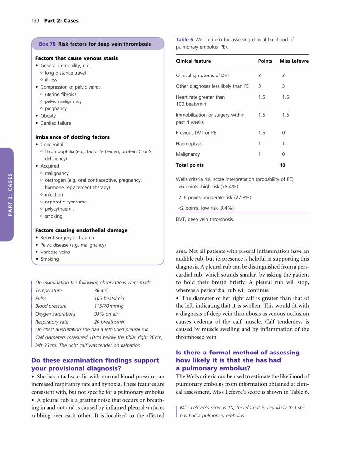
130 Part 2: Cases
PA
RT
2:
CA
SE
S
On examination the following observations were made:
Temperature 36.4ºC
Pulse 105 beats/min
Blood pressure 115/70 mmHg
Oxygen saturations 93% on air
Respiratory rate 20 breaths/min
On chest auscultation she had a left-sided pleural rub
Calf diameters measured 10 cm below the tibia: right 36 cm,
left 33 cm. The right calf was tender on palpation
Do these examination fi ndings support your provisional diagnosis?• She has a tachycardia with normal blood pressure, an
increased respiratory rate and hypoxia. These features are
consistent with, but not specifi c for a pulmonary embolus
• A pleural rub is a grating noise that occurs on breath-
ing in and out and is caused by infl amed pleural surfaces
rubbing over each other. It is localized to the affected
Table 6 Wells criteria for assessing clinical likelihood of
pulmonary embolus (PE).
Clinical feature Points Miss Lefevre
Clinical symptoms of DVT 3 3
Other diagnoses less likely than PE 3 3
Heart rate greater than 1.5 1.5
100 beats/min
Immobilization or surgery within 1.5 1.5
past 4 weeks
Previous DVT or PE 1.5 0
Haemoptysis 1 1
Malignancy 1 0
Total points 10
Wells criteria risk score interpretation (probability of PE):
>6 points: high risk (78.4%)
2–6 points: moderate risk (27.8%)
<2 points: low risk (3.4%)
DVT, deep vein thrombosis.
area. Not all patients with pleural infl ammation have an
audible rub, but its presence is helpful in supporting this
diagnosis. A pleural rub can be distinguished from a peri-
cardial rub, which sounds similar, by asking the patient
to hold their breath briefl y. A pleural rub will stop,
whereas a pericardial rub will continue
• The diameter of her right calf is greater than that of
the left, indicating that it is swollen. This would fi t with
a diagnosis of deep vein thrombosis as venous occlusion
causes oedema of the calf muscle. Calf tenderness is
caused by muscle swelling and by infl ammation of the
thrombosed vein
Is there a formal method of assessing how likely it is that she has had a pulmonary embolus?The Wells criteria can be used to estimate the likelihood of
pulmonary embolus from information obtained at clini-
cal assessment. Miss Lefevre’s score is shown in Table 6.
Miss Lefevre’s score is 10, therefore it is very likely that she
has had a pulmonary embolus.
Box 78 Risk factors for deep vein thrombosis
Factors that cause venous stasis• General immobility, e.g.
� long distance travel� illness
• Compression of pelvic veins:� uterine fi broids� pelvic malignancy� pregnancy
• Obesity
• Cardiac failure
Imbalance of clotting factors• Congenital:
� thrombophilia (e.g. factor V Leiden, protein C or S
defi ciency)
• Acquired� malignancy� oestrogen (e.g. oral contraceptive, pregnancy,
hormone replacement therapy)� infection� nephrotic syndrome� polycythaemia� smoking
Factors causing endothelial damage• Recent surgery or trauma
• Pelvic disease (e.g. malignancy)
• Varicose veins
• Smoking

Case 15 131
PA
RT
2:
CA
SE
S
What is the purpose of clinical risk assessment?In Miss Lefevre’s case it is very likely that she has had a
pulmonary embolus, so she should have the appropriate
investigations and treatment. However, these tests and
therapies themselves have potential risks for the patient.
The clinical risk score is therefore useful in helping clini-
cians decide on a course of action for individual patients.
For example, in patients with a low score an alternative
diagnosis may be sought.
What basic investigations would you perform at the initial assessment?The patient should have a chest X-ray and electrocardio-
gram (ECG) to exclude other diagnoses such as pneumo-
nia, pneumothorax and myocardial disease. Arterial gases
will be helpful to assess the need for oxygen and sup-
portive treatment as well as in supporting the diagnosis.
Investigation results:
Chest X-ray – unremarkable
ECG – sinus tachycardia
Arterial blood gases on air:
Normal range
pH 7.52 (7.35–7.45)
PO2 9.2 kPa (10–13.1 kPa)
PCO2 3.9 kPa (4.9–6.1 kPa)
HCO3− 22 mmol/L (22–28 mmol/L)
How do you interpret her results?The chest X-ray and ECG show no features consistent
with alternative diagnoses.
Arterial gases:
• Mild hypoxia – respiratory impairment
• Hypocapnia – hyperventilation secondary to hypoxia
• Alkalosis secondary to hypocapnia driving the Hen-
derson–Hasselbach equation to the right
2�
3 |� H+ + HCO3
− ¨Æ H2CO3 ¨Æ H2O + CO2
|� 1
Further investigation also showed:
• D-dimer level 0.84 mg/L (normal range <0.3 mg/L)
What are D-dimers?In the normal control of haemostasis, procoagulant, anti-
coagulant and fi brinolytic factors are balanced to prevent
intravascular thrombosis. Plasmin stimulates breakdown
of fi brin (Fig. 54) and cross-linked fi brin degradation
products are called d-dimers. Where thrombosis is acti-
vated anywhere in the body, fi brinolysis is also activated
and d-dimer levels become elevated.
Are elevated D-dimer levels in Miss Lefevre diagnostic of pulmonary embolus?No. d-dimers are elevated in many clinical conditions
(e.g. following trauma or surgery, as a result of myocar-
dial infarction, renal impairment, pregnancy, stroke).
Therefore, a positive d-dimer test is not informative and
should not guide further investigation and treatment.
Why do a D-dimer test then?If d-dimers are normal then this is a useful result as pul-
monary embolus is unlikely. Negative (normal) d-dimers
have >90% negative predictive value for pulmonary
embolus (i.e. >90% of people with a negative d-dimer
result do not have pulmonary embolus). However, the
d-dimer test may give false negative results.
When should you do a D-dimer test in clinical practice?Measuring d-dimer levels is controversial and if used
indiscriminately can be more of a hindrance than a help
in making the diagnosis and can lead to unnecessary
investigation. A reasonable approach is to measure d-
dimers only in patients with suspected pulmonary
embolus who are assessed as being at low or moderate
clinical risk of pulmonary embolus using Wells criteria
(Table 6). In these patients a negative result can exclude a
pulmonary embolus and prevent unnecessary investiga-
tion and treatment, whereas a positive result is supportive
of the need for defi nitive tests for pulmonary embolus. If
a patient is judged as being at high risk of pulmonary
embolus, d-dimer measurement is unnecessary. If pul-
monary embolus is not suspected clinically, d-dimers
should not be measured.
KEY POINTS
• Elevated D-dimers are not useful in making a positive
diagnosis of pulmonary embolus
• However, normal (negative) D-dimers correctly exclude
>90% of pulmonary emboli
• D-dimer results should be taken in clinical context and
not used alone

132 Part 2: Cases
PA
RT
2:
CA
SE
S
How do you assess Miss Lefevre now?She has a high clinical probability of pulmonary embolus
and deep vein thrombosis and requires urgent treatment,
followed by investigations to confi rm the diagnosis.
How does deep vein thrombosis occur?Deep leg veins are large veins that lie deep in the muscles
of the leg and carry most of the deoxygenated, nutrition-
ally depleted blood from the legs back up towards the
heart. Movement of blood within these veins depends on
compression by leg muscles and a system of valves that
direct the blood out of the leg. Blood does not normally
clot in the veins.
Thrombosis of blood in the deep veins can be pre-
cipitated by any of a triad of factors described by Virchow
over 150 years ago (Box 78).
• Venous stasis. This promotes clotting as it allows more
time for blood to clot, stops small clots from being
washed away and increases blood viscosity
• Imbalance of clotting factors. In the blood, natural pro-
coagulant, anticoagulant and fi brinolytic factors are bal-
anced to prevent clots forming within the blood vessels.
An imbalance of clotting factors (e.g. from hereditary
abnormalities) can predispose to clots
• Endothelial damage. This triggers the coagulation
cascade and forms a focus for clots
How does clot form in deep veins?Clotting is initiated by one of Virchow’s factors, which
triggers the coagulation cascade and aggregation of plate-
lets, fi brin and large numbers of red blood cells (Fig. 54).
The process of thrombosis triggers fi brinolysis with
breakdown of formed clot. During thrombosis and fi bri-
nolysis the thrombus is fragile and may break away from
the vein wall, becoming an embolus. Alternative fates
for thrombi are complete removal by fibrinolysis,
organization, recanalization or calcifi cation.
Why do venous thrombi embolize to the lungs?Free thrombus in the deep leg veins will embolize via the
pelvic veins and inferior vena cava into the right side of
Endothelial damage
Plateletadhesion
Platelet aggregation Stasis
THROMBUS
FATE
Extension
Fibrindegradation
products
FibrinolysisPlasmin
Factor X Factor Xa
ThrombinProthrombin
XIII
FibrinFibrinogen
XIIIa
Organization ofthrombus
Recanalization
Calcification
Fibrin cross-linked
Plasminogen
Build-up ofplatelet
aggregates
Large numbersof red bloodcells, whiteblood cells
Activation ofcoagulation
cascade
Hypercoagulablestate
Figure 54 Outline of processes
underlying thrombus formation and
resolution in the deep veins.

Case 15 133
PA
RT
2:
CA
SE
S
the heart, then into the pulmonary artery (Fig. 55). The
embolus will come to rest at a point in the pulmonary
arterial circulation where its diameter exceeds that of
these vessels. Thus, a large embolus may impact in the
main pulmonary arteries, whereas a small embolus may
pass into smaller arteries supplying segments or subseg-
ments of the lung.
Has Miss Lefevre had a large (massive) or small (non-massive) pulmonary embolus?Miss Lefevre’s symptoms are pleuritic chest pain and hae-
moptysis. These symptoms are caused by a small (correct
term, non-massive) embolus that has impacted in a sub-
segmental or smaller artery, causing infarction of a small
part of the lung (haemoptysis) and a patch of pleura
(pleuritic pain). A large (correct term, massive) pulmo-
nary embolus impacted in the main pulmonary arteries
will block blood fl ow through the heart, causing shock
and collapse. A massive pulmonary embolus does not
immediately cause lung infarction because of dual blood
supply to the lungs from the systemic circulation (bron-
chial arteries) and pulmonary arteries.
What treatment should she receive?Supportive therapyShe should have oxygen to correct hypoxia and analgesia
for her chest and calf pain.
Therapy for thromboembolic diseaseShe requires immediate anticoagulation with low molec-
ular weight (LMW) heparin (Box 79; Fig. 56).
What effect will anticoagulation with low molecular weight heparin have?Heparin will inhibit further extension of the deep vein
thrombus. This will allow existing leg thrombus to
become organized by natural processes, reducing the risk
of further embolus.
Superiorvena cava
Aorta
Rightatrium
Rightventricle
Leftventricle
Leftatrium
Inferiorvena cava
Pulmonaryvein
Pulmonaryartery
LUNGS
Deep leg veins
CLOT
Figure 55 Circuit diagram of the heart and major vessels.
Arrows indicate the route of embolism of the clot from the
deep leg veins into the inferior vena cava, then through the
right side of the heart and pulmonary artery to reach the
lungs.
Box 79 Pharmacology of low molecular weight heparin
Mechanism of actionHeparin catalyses inhibition of factor Xa and thrombin by
the natural anticoagulant antithrombin III (Fig. 56), hence
inhibiting the coagulation cascade.
Uses• Prevention or treatment of deep vein thrombosis and
pulmonary embolus
• Early treatment of unstable angina and myocardial
infarction
• Prevention of thrombosis in cannulae, dialysis circuits, etc.
Pharmacokinetics• LMW heparin is poorly absorbed, hence is given by
subcutaneous injection
• It has a plasma half-life of 4 h, but a longer duration of
action and can be given once or twice daily
• Dose–response of weight-adjusted dose is predictable,
hence dosage depends on patient weight. No
monitoring of anticoagulant effect is required
• It is renally excreted and should be used with caution in
patients with renal impairment
Major side-effects• Bleeding
• Hypersensitivity – heparin is from animals and can be
antigenic, causing allergic reactions
• Thrombocytopaenia – this is a serious complication of
heparin use, platelet counts should be monitored during
treatment
• Osteoporosis may occur with prolonged heparin
treatment
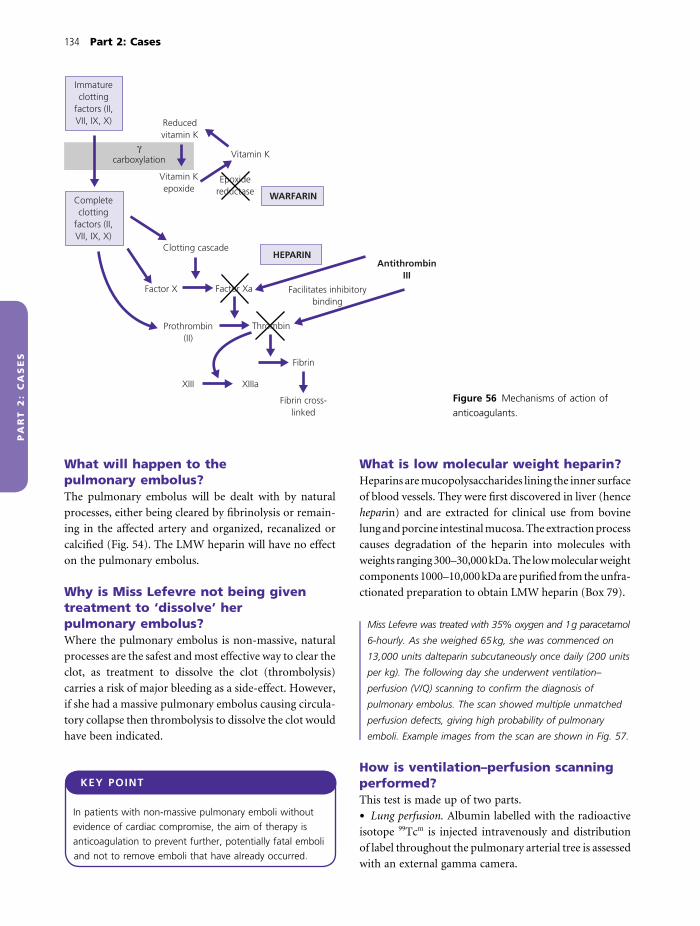
134 Part 2: Cases
PA
RT
2:
CA
SE
S
What will happen to the pulmonary embolus?The pulmonary embolus will be dealt with by natural
processes, either being cleared by fi brinolysis or remain-
ing in the affected artery and organized, recanalized or
calcifi ed (Fig. 54). The LMW heparin will have no effect
on the pulmonary embolus.
Why is Miss Lefevre not being given treatment to ‘dissolve’ her pulmonary embolus?Where the pulmonary embolus is non-massive, natural
processes are the safest and most effective way to clear the
clot, as treatment to dissolve the clot (thrombolysis)
carries a risk of major bleeding as a side-effect. However,
if she had a massive pulmonary embolus causing circula-
tory collapse then thrombolysis to dissolve the clot would
have been indicated.
What is low molecular weight heparin?Heparins are mucopolysaccharides lining the inner surface
of blood vessels. They were fi rst discovered in liver (hence
heparin) and are extracted for clinical use from bovine
lung and porcine intestinal mucosa. The extraction process
causes degradation of the heparin into molecules with
weights ranging 300–30,000 kDa. The low molecular weight
components 1000–10,000 kDa are purifi ed from the unfra-
ctionated preparation to obtain LMW heparin (Box 79).
Miss Lefevre was treated with 35% oxygen and 1 g paracetamol
6-hourly. As she weighed 65 kg, she was commenced on
13,000 units dalteparin subcutaneously once daily (200 units
per kg). The following day she underwent ventilation–
perfusion (V/Q) scanning to confi rm the diagnosis of
pulmonary embolus. The scan showed multiple unmatched
perfusion defects, giving high probability of pulmonary
emboli. Example images from the scan are shown in Fig. 57.
How is ventilation–perfusion scanning performed?This test is made up of two parts.
• Lung perfusion. Albumin labelled with the radioactive
isotope 99Tcm is injected intravenously and distribution
of label throughout the pulmonary arterial tree is assessed
with an external gamma camera.
KEY POINT
In patients with non-massive pulmonary emboli without
evidence of cardiac compromise, the aim of therapy is
anticoagulation to prevent further, potentially fatal emboli
and not to remove emboli that have already occurred.
Reducedvitamin K
Vitamin K
Vitamin Kepoxide
γcarboxylation
Immatureclotting
factors (II,VII, IX, X)
Epoxidereductase WARFARIN
Clotting cascadeHEPARIN
AntithrombinIII
Facilitates inhibitorybinding
Thrombin
Fibrin
Fibrin cross-linked
XIIIaXIII
Prothrombin(II)
Factor X Factor Xa
Completeclotting
factors (II,VII, IX, X)
Figure 56 Mechanisms of action of
anticoagulants.

Case 15 135
PA
RT
2:
CA
SE
S
• Lung ventilation. The patient inhales nebulized 99Tcm
diethylene triamine-pentacetic acid (DTPA) and distri-
bution of label in the airways and alveoli is assessed with
an external gamma camera.
If pulmonary emboli are present there will be areas of
lung that are ventilated but not perfused (ventilation–
perfusion mismatching; Fig. 57). The V/Q scan is inter-
preted by a radiologist who looks for areas of V/Q
mismatching and reports their fi ndings as ‘high’, ‘inter-
mediate’ or ‘low probability’ of pulmonary embolus.
Are there any other tests that can be used to diagnose pulmonary embolus?• Pulmonary angiogram. In this test a catheter is inserted
via the femoral or brachial vein into the right side of the
heart, then through into the pulmonary arteries. Radio-
graphic contrast is injected into the pulmonary arteries
and the pulmonary arteries are visualized by repeated
X-rays to look for clot (Fig. 58)
• Computed tomography pulmonary angiogram (CTPA).
In this technique an X-ray beam moves around the chest
as the patient lies on a table, taking multiple pictures
which are collated in a series of 2D images on a monitor.
To obtain a CTPA, images are taken after intravenous
injection of contrast
What are the risks to the patient of these tests?• Radiation exposure. CT involves the greatest radiation
exposure (2–3 times the annual exposure to background
radiation), which itself is associated with a 150–300 in
1,000,000 increased annual chance of death. Radiation ex-
posure is less with V/Q scanning and least with angiography
(a)
(b)
Figure 57 Ventilation–perfusion lung scan. (a) The ventilation
scan shows the airspaces of the lung outlined clearly in black.
(b) The perfusion scan shows areas without contrast
(unperfused) in left and right lungs.
Figure 58 Pulmonary angiogram from a different patient.

136 Part 2: Cases
PA
RT
2:
CA
SE
S
• Allergy. Radiographic contrast (CT and non-CT angi-
ography) contains iodine which may induce allergy and
anaphylaxis if patients are sensitive to this
• Dangers of intervention. Pulmonary angiography
requires insertion of a catheter through the right side of
the heart into the pulmonary arteries. This has the risk
of introducing infection and damaging myocardium,
valves or vessels
How do you decide which test to use?Hospitals usually have a policy to use either V/Q scanning
or CTPA as fi rst line and only exceptionally use pulmo-
nary angiography. These tests are roughly comparable in
their ability to diagnose pulmonary emboli and avail-
ability generally depends on resource allocation by indi-
vidual hospitals. Specifi c uses are:
• V/Q scan – patients with iodine allergy
• CTPA – patients with structural lung disease that will
affect the V/Q scan
How could you confi rm the diagnosis of deep vein thrombosis?Duplex ultrasound of the veinsThis non-invasive rapid technique is a combination of:
• Standard ultrasound. Sound waves are refl ected off
the vessel walls, allowing visualization of the vascular
structures
• Doppler ultrasound. Bursts of sound waves are refl ected
off moving red blood cells, allowing detection of blood
fl ow
Clots may be visualized directly and as alteration in
venous blood fl ow, although not all are detected.
VenogramThese are rarely performed these days but can be used if
duplex ultrasound is not helpful. A foot vein is cannu-
lated and radiographic dye is injected to show the leg
veins. Clot appears as fi lling defects in the delineated
veins. Complications include contrast allergy and phlebi-
tis (infl amed veins).
Miss Lefevre had a positive duplex of her right leg, confi rming
a right calf deep vein thrombosis. LMW heparin was continued
while she commenced oral anticoagulation with warfarin. By
day 4 of her inpatient stay she was well and pain-free and
her international normalized ratio (INR) was 1.6. She was
discharged with follow-up in the anticoagulant and
respiratory clinics, remaining on self-administered dalteparin
until her INR was >2.0.
Why was her heparin (dalteparin) treatment changed to warfarin?Standard practice is to anticoagulate patients for 3–6
months after a pulmonary embolus to minimize the risk
of recurrence. Over a 3–6 month period tablets are pref-
erable to (and cheaper than) daily injections. Also, heparin
given for a prolonged period can cause osteoporosis.
Why was she not given warfarin instead of heparin in the fi rst place?After her pulmonary embolus she required immediate
anticoagulation to prevent further clot. As warfarin
inhibits synthesis of clotting factors (Fig. 56; Box 80) and
Box 80 Pharmacology of warfarin
Mechanism of actionIn the fi nal stages of synthesis of clotting factors II, VII, IX
and X, the factors undergo gamma carboxylation, a
process that oxidizes vitamin K. Vitamin K is recycled for
further clotting factor synthesis by reduction catalysed by
epoxide reductase. Warfarin inhibits clotting factor
synthesis by inhibiting regeneration of reduced vitamin K.
Uses• Prevention of recurrence of pulmonary emboli or deep
vein thrombosis
• Prevention of intracardiac thrombosis in atrial fi brillation,
mechanical heart valves
Pharmacokinetics• Warfarin is metabolized by the liver, which terminates
its action
• It has a narrow therapeutic range (i.e. the plasma
concentration at which warfarin prevents clotting is
close to the concentration at which major bleeding side-
effects occur)
• Drug interactions are common in patients taking warfarin:� drugs that induce activity of liver enzymes increase
warfarin metabolism and increase the risk of clotting
(e.g. phenytoin, rifampicin and regular alcohol use)� drugs that inhibit activity of liver enzymes reduce
warfarin metabolism and increase the risk of bleeding
(e.g. erythromycin, ciprofl oxacin, sodium valproate)
Side-effects• Bleeding
• Skin necrosis
• Teratogenesis (fetal abnormalities when given to
pregnant women)

Case 15 137
PA
RT
2:
CA
SE
S
existing clotting factors have to be used up, it can take
3–4 days after starting warfarin before anticoagulation is
achieved. Heparin causes immediate anticoagulation as it
promotes the natural anticoagulant effect of antithrom-
bin III (Fig. 56), therefore it is used to bridge the gap
while full warfarin anticoagulation is achieved.
What is the international normalized ratio?Patients taking warfarin have blood sampled regularly for
measurement of the prothrombin time, which is used to
calculate the INR. During sampling, calcium is removed
from the blood, then the sample is spun to remove blood
cells. The resulting plasma is recalcifi ed in the presence
of a reagent with tissue factor activity which activates the
clotting cascade. The time to production of a clot (pro-
thrombin time) is measured, normally 11–15 s.
The INR is calculated by dividing the prothrombin
time of the patient taking warfarin by the prothrombin
time of a control subject. An INR of 1.6 indicates that
Miss Lefevre’s blood was clotting 1.6 times more slowly
than a person not taking warfarin. In general, INR values
of 2.0–4.0 are considered to indicate adequate anticoagu-
lation, hence she was able to stop her heparin when her
INR was >2.0.
Does she need further investigation?• Thrombophilia screen. As Miss Lefevre is young and has
a family history of thrombosis she may have an underly-
ing genetic coagulation abnormality (Box 81) and should
be tested for this after stopping warfarin as anticoagula-
tion will affect the results
• Pelvic ultrasound. Miss Lefevre probably developed
her thrombus because of immobility from a long coach
journey. Patients without detectable risk factors (particu-
larly women) should undergo pelvic ultrasound to look
for predisposing masses or malignancy
Outcome. Miss Lefevre stopped her warfarin after 6 months
and remained completely well. Her thrombophilia screen did
not detect a heritable clotting abnormality. She was
therefore given advice to help avoid recurrence of her
thromboembolic disease (Box 82), encouraged to stop
smoking and discharged from follow-up.
KEY POINT
Before prescribing other drugs with warfarin, always check
for potential interactions in Appendix 1 of the British
National Formulary and adjust prescribing accordingly.
Box 81 Overview of hereditary thrombophilia
Patients with hereditary thrombophilia have an increased
tendency to clot. The most common abnormalities are
caused by defi ciencies in the natural anticoagulant system.
Factor V Leiden is a genetic variant of factor V which is
resistant to natural inactivation by activated protein C.
Proteins C, S and antithrombin inactivate clotting as
part of the natural anticoagulant system. Genetic variants
causing protein defi ciencies result in anticoagulant defects
and increased clotting tendencies.
Box 82 Prevention of deep vein thrombosis during prolonged travel
The aim is to maintain venous fl ow and minimize
hypercoagulability.
Maintain venous fl ow• Leg exercises, short walks, journey breaks if possible,
avoid immobility
• Use travel compression stockings
Reduce hypercoagulability• Keep hydrated
• Single dose prophylactic heparin with medical advice
How long should she remain on warfarin for?• Standard practice is for patients to take warfarin for 6
months following a deep vein thrombosis or pulmonary
embolus to minimize the risk of recurrence
• Warfarin treatment should continue for longer than 6
months if the risk factor for clots (e.g. untreated uterine
fi broids, persistent immobility, hereditary thrombo-
philia) is still present
• Warfarin treatment is often life-long in patients who
have a recurrence of their deep vein thrombosis or pul-
monary embolus
• Patients with deep vein thrombosis may stop warfarin
earlier than 6 months if the risk factor has been
removed

138 Part 2: Cases
PA
RT
2:
CA
SE
S
KEY POINTS
• Deep vein thrombosis can be precipitated by any of
‘Virchow’s triad’ of factors:� venous stasis� imbalance of clotting factors� endothelial damage
• Thrombi in the deep veins can break free, becoming
emboli that travel to the lungs – pulmonary emboli
• Pulmonary emboli impact in the pulmonary circulation at
the point where the diameter of the embolus exceeds that
of the vessels
• Smaller pulmonary emboli impact in peripheral pulmonary
arteries where they may cause infarction of the supplied
lung and overlying pleura with symptoms of pleuritic chest
pain and haemoptysis
• Pulmonary emboli can be diffi cult to diagnose:� at presentation, clinical features are used to estimate the
likelihood of pulmonary emboli (Wells criteria)� D-dimer measurement can be used to rule out
pulmonary emboli in patients with low or moderate
likelihood of emboli
� defi nitive investigations including V/Q scanning and
CTPA are often not available out of hours except in
severe emergencies, so patients at moderate or high
risk of emboli are usually treated until diagnosis can
be confi rmed
• Patients with suspected or proven deep vein thrombosis
and/or pulmonary embolus are initially treated with
subcutaneous LMW heparin to prevent further
pulmonary embolus
• Existing thrombus or embolus is usually cleared or
organized by intrinsic (natural) mechanisms
• Once the diagnosis has been confi rmed, patients receive
oral warfarin for 3–6 months to prevent recurrence
• Investigations including pelvic ultrasound scanning and
hereditary thrombophilia testing should be performed to
look for an underlying cause of thromboembolic disease
• Patients with recurrent thromboembolic disease should
receive life-long anticoagulation
and a V/Q scan showing high probability of a pulmonary
embolus. She receives anticoagulation with warfarin for 6
months. Her thromboembolic disease is attributed to her
long distance journey and cigarette smoking and a heritable
clotting abnormality is excluded. She is advised to stop
smoking and take precautions when immobile for long
periods to reduce the risk of recurrence.
CASE REVIEW
Marie Lefevre presents with left-sided pleuritic chest pain
and haemoptysis following a long distance coach journey.
She has also noticed some right calf discomfort. A clinical
diagnosis of deep vein thrombosis and pulmonary embolus
is made and she is treated with LMW heparin. The
following day these diagnoses are confi rmed by a duplex
ultrasound demonstrating right calf deep vein thrombosis

PA
RT
2:
CA
SE
S
An elderly woman who collapsed suddenly
Case 16
An elderly woman is rushed into accident and emergency
by ambulance having collapsed at home. Her husband,
Mr Khan, told the ambulance crew she had been well
previously with no relevant past medical history and that
this had happened without warning.
On arrival at accident and emergency:
Her airway is patent and she is breathing spontaneously
with respiratory rate 20 breaths/min
Oxygen saturations are 74% on 35% oxygen
Her pulse is 140 beats/min and blood pressure 60/36 mmHg
She has a Glasgow Coma Score of 11/15 and has an
intravenous cannula in her right arm inserted by the
ambulance crew
What is the fi rst priority in her management?Mrs Khan needs urgent resuscitation. Her life-threaten-
ing problems are hypoxia and hypotension, which cause
tissue damage. Her brain is particularly at risk as it can
only survive anoxia for 4–5 min before permanent
damage occurs. Her reduced Glasgow Coma Score may
indicate this is already occurring.
How should she be resuscitated initially?HypoxiaShe should immediately receive higher concentrations of
inhaled oxygen via a mask with a reservoir bag (15 L/min
will deliver an oxygen concentration of 75–90%). Blood
gas tests should be performed as soon as possible to look
for CO2 retention which may be contributing to her
impaired consciousness. If high fl ow oxygen does not
correct her hypoxia or is contraindicated by CO2 reten-
tion she will require ventilatory support.
HypotensionInitially, she should be given intravenous fl uid to attempt
to raise her blood pressure. A crystalloid such as isotonic
saline or a colloid solution of macromolecules such as
dextrans or gelatin, which stay in the circulation longer,
should be used.
After an increase in oxygen therapy to 15 L/min via mask
and reservoir bag and 1 L isotonic saline, Mrs Khan’s
observations were as follows:
Oxygen saturations 86%
Pulse 125 beats/min and blood pressure 90/60 mmHg
Glasgow Coma Score 14
How do you interpret these new observations?There is some improvement after her initial resuscitation,
which has created time to identify and treat the underly-
ing cause of her collapse.
What mechanisms might explain her sudden collapse?She has hypoxia, hypotension and confusion. Any
of these could have been the primary abnormality that
caused her clinical presentation:
• Hypoxia. This could have developed rapidly as a result
of any of the conditions that cause respiratory arrest (Box
28, p. 42). If hypoxia is severe, cardiac and brain function
are impaired, leading to hypotension and confusion
• Hypotension. Low blood pressure can be caused by
hypovolaemia (e.g. haemorrhage), cardiac impairment
(e.g. post myocardial infarction), infection (septic shock)
or anaphylaxis. Reduced cerebral and pulmonary perfu-
sion could cause secondary confusion and hypoxia
• Confusion. A primary brain problem, such as haemor-
rhage or stroke could impair consciousness. This could
lead to hypoventilation and hypoxia
How would you approach her assessment?The next step is to establish which of these mechanisms
has caused her collapse and whether her primary disease
is cardiovascular, respiratory or neurological. This
139
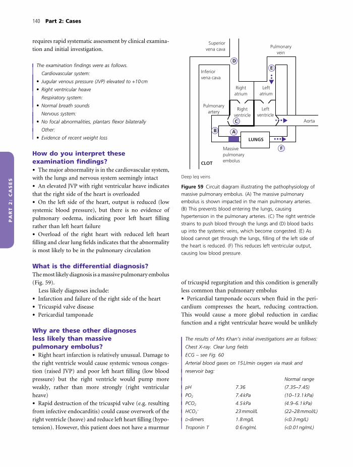
140 Part 2: Cases
PA
RT
2:
CA
SE
S
requires rapid systematic assessment by clinical examina-
tion and initial investigation.
The examination fi ndings were as follows.
Cardiovascular system:
• Jugular venous pressure (JVP) elevated to +10 cm
• Right ventricular heave
Respiratory system:
• Normal breath sounds
Nervous system:
• No focal abnormalities, plantars fl exor bilaterally
Other:
• Evidence of recent weight loss
How do you interpret these examination fi ndings?• The major abnormality is in the cardiovascular system,
with the lungs and nervous system seemingly intact
• An elevated JVP with right ventricular heave indicates
that the right side of the heart is overloaded
• On the left side of the heart, output is reduced (low
systemic blood pressure), but there is no evidence of
pulmonary oedema, indicating poor left heart fi lling
rather than left heart failure
• Overload of the right heart with reduced left heart
fi lling and clear lung fi elds indicates that the abnormality
is most likely to be in the pulmonary circulation
What is the differential diagnosis?The most likely diagnosis is a massive pulmonary embolus
(Fig. 59).
Less likely diagnoses include:
• Infarction and failure of the right side of the heart
• Tricuspid valve disease
• Pericardial tamponade
Why are these other diagnoses less likely than massive pulmonary embolus?• Right heart infarction is relatively unusual. Damage to
the right ventricle would cause systemic venous conges-
tion (raised JVP) and poor left heart fi lling (low blood
pressure) but the right ventricle would pump more
weakly, rather than more strongly (right ventricular
heave)
• Rapid destruction of the tricuspid valve (e.g. resulting
from infective endocarditis) could cause overwork of the
right ventricle (heave) and reduce left heart fi lling (hypo-
tension). However, this patient does not have a murmur
of tricuspid regurgitation and this condition is generally
less common than pulmonary embolus
• Pericardial tamponade occurs when fl uid in the peri-
cardium compresses the heart, reducing contraction.
This would cause a more global reduction in cardiac
function and a right ventricular heave would be unlikely
The results of Mrs Khan’s initial investigations are as follows:
Chest X-ray. Clear lung fi elds
ECG – see Fig. 60
Arterial blood gases on 15 L/min oxygen via mask and
reservoir bag:
Normal range
pH 7.36 (7.35–7.45)
PO2 7.4 kPa (10–13.1 kPa)
PCO2 4.5 kPa (4.9–6.1 kPa)
HCO3− 23 mmol/L (22–28 mmol/L)
D-dimers 1.8 mg/L (<0.3 mg/L)
Troponin T 0.6 ng/mL (<0.01 ng/mL)
Pulmonaryvein
Superiorvena cava
Inferiorvena cava
Rightatrium
Rightventricle
Leftventricle
Leftatrium
Aorta
B A
C
D
E
FMassivepulmonaryembolus
Pulmonaryartery
CLOT
LUNGS
Deep leg veins
Figure 59 Circuit diagram illustrating the pathophysiology of
massive pulmonary embolus. (A) The massive pulmonary
embolus is shown impacted in the main pulmonary arteries.
(B) This prevents blood entering the lungs, causing
hypertension in the pulmonary arteries. (C) The right ventricle
strains to push blood through the lungs and (D) blood backs
up into the systemic veins, which become congested. (E) As
blood cannot get through the lungs, fi lling of the left side of
the heart is reduced. (F) This reduces left ventricular output,
causing low blood pressure.

Case 16 141
PA
RT
2:
CA
SE
S
Can these investigations be interpreted to make a fi rm diagnosis?• The normal chest X-ray and abnormal ECG support
the deduction that the abnormality is somewhere in the
heart or circulation rather than in the lungs
• The ECG shows features of right heart strain (Fig. 60)
consistent with the clinical fi ndings of right ventricular
heart strain and raised JVP
• Arterial gases show type I respiratory failure, which has
been poorly corrected by high fl ow oxygen therapy. This
is consistent with a massive pulmonary embolus because
increasing oxygen concentrations in the alveoli will have
little impact if there is insuffi cient perfusion of pulmo-
nary capillaries for oxygen uptake into the blood.
However, any other disease causing severe ventilation–
perfusion (V/Q) mismatch could account for these results
• Elevated d-dimers indicate breakdown of clot some-
where in the body, but are raised in a wide range of
clinical conditions and are unhelpful in making a diag-
nosis. It could be argued in this situation that testing
them is simply a waste of money
• Troponin T is specifi cally released from cardiac
myocytes and so indicates myocardial damage. In this
patient raised troponin T could indicate a primary cardiac
event (e.g. right ventricular infarction). However, massive
pulmonary embolus causes secondary cardiac ischaemia
by reducing coronary perfusion with elevation of tropo-
nin T
C2
C3
aVR
aVL
aVF
(b)
I
II
III
I
II
III
AUR
AUL
AUF
(a)
U3
U1
U2
U4
U5
U6
C1
C5
C6
C4
Figure 60 Electrocardiograms. ECG (a) was taken during the
acute admission and is shown compared with ECG (b), found
in Mrs Khan’s old notes from a previous hospital visit. The
notable differences are: 1. The heart rate is faster (∼150 beats/
min) in (a) than in (b) (∼100 beats/min). 2. (a) shows new big
R waves (fi rst upward defl ection) in leads V1 and V2, which
are the leads that look at the right ventricle and new S waves
in leads V5 and V6. As the QRS complexes are also wider
(2.5–3 small squares) this is new right bundle branch block.
These ECG abnormalities are features of right heart strain. 3.
In ECG (a) there is a new S wave in lead I and Q wave in lead
III. These abnormalities which can occur with an inverted T
wave in lead III are the classic SIQIIITIII of acute pulmonary
embolus. In practice this is not often seen.

142 Part 2: Cases
PA
RT
2:
CA
SE
S
Thus, the initial investigations give information that is
supportive, but not specifi c for a diagnosis of massive
pulmonary embolus so further investigations are required
to make a fi rm diagnosis and guide therapy.
How long should it have taken to arrive at this point?The results of clinical examination, chest X-ray, ECG and
blood gases can all be available in about 20 min, suffi cient
to guide decision-making. Blood test results take longer,
depending on the hospital laboratory. However, the clini-
cians should not wait for these to make a decision regard-
ing further care.
What tests could be used to make a fi rm diagnosis of massive pulmonary embolus?EchocardiogramThis can confi rm the diagnosis by:
• Visualizing thrombus in the right heart or main pul-
monary artery
• Demonstrating right ventricular strain
• Excluding cardiac diagnoses (infarction, valve disease)
Computed tomography pulmonary angiographyComputed tomography pulmonary angiography (CTPA)
is most useful for direct visualization of the thrombus in
the pulmonary arteries.
How do you decide which of these tests to use?Both tests will give diagnostic information. The consid-
erations are:
• Which test is available most quickly?
• Is the patient stable enough to undergo testing?
For example, it may be safer to bring a portable echo-
cardiography machine to the patient if an operator is
available, than move the patient to the CT scanning
department away from resuscitation facilities. The most
suitable test will therefore vary depending on local
resources.
Mrs Khan was judged stable enough to undergo further
investigation and, as echocardiography was not available, an
urgent CTPA was arranged. The medical registrar
accompanied her to the radiology department with the
resuscitation trolley in case of rapid deterioration.
However, Mrs Khan tolerated the scan, which is shown
in Figs 61 and 62. A diagnosis of massive pulmonary
embolus was confi rmed and she was transferred to
intensive care for consideration of urgent thrombolysis.
A right pleural effusion was also seen on the scan,
presumed secondary to her massive pulmonary embolus.
As Mrs Khan remained drowsy and unwell the medical
team spoke briefl y to her husband to determine whether
she had any contraindications to this treatment
(Box 83).
Figure 61 Computed tomography pulmonary angiogram
(CTPA). This is a single slice taken from a CTPA at the level of
the aortic arch. The patient is supine, so their front is at the
top of the picture. Ribs and vertebrae appear white, soft tissue
is grey and lung fi elds are black. The vessels show white as
they contain radiographic contrast injected intravenously. The
arrow indicates a point in the left pulmonary artery where it
narrows suddenly and a grey area can be seen in the contrast
(shown in more detail in Fig. 62). Clot is occluding the artery
and preventing perfusion of the vessel beyond. There is also a
right-sided pleural effusion which could be secondary to
pulmonary emboli in this patient.

Case 16 143
PA
RT
2:
CA
SE
S
hypotension and prevent further damage by reperfusing
the pulmonary arteries and increasing left heart fi lling.
How could this be achieved by thrombolysis?Thrombolytic drugs are infused into an arm vein and
circulate to the pulmonary artery. They activate plas-
minogen to plasmin, a naturally occurring thrombolytic
molecule (Fig. 63), which dissolves thrombus and recan-
alizes the main pulmonary artery.
Do clinical trials show that thrombolysis is benefi cial in massive pulmonary embolus?Although it seems intuitive that removing pulmonary
thrombus would be a good thing, clinical trials showed
that thrombolysis compared to anticoagulation with
heparin produced:
• No signifi cant reduction in mortality
• Short-term improvement in right ventricular
function
• Possible longer term lower pulmonary arterial pres-
sures (one study)
(a)
(b)
Figure 62 Comparison of pulmonary
arteries imaged using computed
tomography pulmonary angiogram
(CTPA). (a) Normal. (b) Pulmonary
embolus. In the normal artery there
is (A) unoccluded vessel allowing
contrast to (B) pass distally into
(C) tributaries. In the abnormal artery
(D) embolus can be seen as a grey
protrusion into the white (contrast)
arterial lumen (E). There is little run
off from the occluded artery into the
distal pulmonary circulation.
Box 83 Contraindications to thrombolysis
Absolute contraindications• Previous history of intracranial haemorrhage
• Active intracranial neoplasm
• Recent (<2 months) intracranial surgery or trauma
• Current or recent (<6 months) internal bleeding
Relative contraindications• Other coagulation disorder with increased bleeding risk
• Uncontrolled severe hypertension (blood pressure
>200 mmHg systolic or >110 mmHg diastolic)
• Non-haemorrhagic stroke (<2 months)
• Surgery (≤10 days)
• Low platelet count (<100 × 109/L)
How could thrombolysis help Mrs Khan?Mrs Khan’s massive pulmonary embolus is causing severe
hypoxia and hypotension and she already has signs of end
organ damage including confusion (brain) and raised
troponin T (heart). In theory, removal of the pulmonary
embolus by thrombolysis would correct her hypoxia and

144 Part 2: Cases
PA
RT
2:
CA
SE
S
However, clinical trials are diffi cult to perform and inter-
pret in this group of patients because of small numbers
and problems conducting trials in an emergency situa-
tion. Most clinicians therefore accept that thrombolysis
can be life-saving in selected patients with major pulmo-
nary embolus and would use it in this situation if the
benefi ts appear to outweigh the risks of major haemor-
rhage (Box 83).
Which drugs are used for thrombolysis? (Box 84)
Some options include the following.
StreptokinaseThis thrombolytic drug, derived from streptococci, has
been available for clinical use for many years. As an ‘old’
drug it has several advantages:
• Its manufacture is no longer restricted by patent, so it
is relatively cheap
• It has been widely studied in the clinical situation
However, as a bacterial product it is antigenic and can
cause allergic reactions. It also has to be given as an infusion
over 24 h, which may be too slow in this patient group.
Recombinant tissue plasminogen activatorThis agent occurs naturally in tissues including endothe-
lial cells. Pharmacological recombinant tissue plasmino-
gen activator (rTPA) has also been widely studied in
patients with pulmonary emboli. rTPA is more expensive
than streptokinase, but can be given more quickly (≤2 h)
and does not induce allergic reactions.
TenecteplaseThis is a newer agent which can be given rapidly as a
bolus. It is not yet licensed for use in patients with pul-
monary emboli, but is currently being tested in clinical
trials.
Fibrinolysis
Fibrinogen
Thrombin
Factor Xa
Activatedcoagulation
cascade
Fibrin
Fibrindegradation
products
Plasmin
Activate
Plasminogen
Fibrin cross-linked
FIBRINOLYTIC DRUGS
Thrombus
XIII XIIIa
Factor X
Prothrombin
Figure 63 Mechanism of action of thrombolytic drugs.
Fibrinolytic drugs activate plasminogen to plasmin. This
degrades fi brin, hence breaks up thrombi.
Box 84 Pharmacology of thrombolytic drugs
Mechanism of action• Streptokinase binds to plasminogen, forming an active
enzyme that activates plasmin. It is not well absorbed by
fi brin thrombi, so acts generally in the circulation, which
may increase risk of bleeding
• rTPA binds to fi brin, increasing its affi nity for
plasminogen and enhancing plasminogen activation. As
rTPA binds to fi brin it is less likely to produce a general
coagulation disturbance than streptokinase
UsesRemoval of thrombus in emergency situations including:
• Massive pulmonary embolus
• Myocardial infarction
• Acute stroke
• Arterial embolus
Pharmacokinetics• Drugs given intravenously and have plasma half-life of
30 (rTPA) to 90 min (streptokinase)
Major side-effects• Major bleeding, particularly intracranial (∼1%)
• Allergy, hypotension (streptokinase)

Case 16 145
PA
RT
2:
CA
SE
S
Is thrombolysis dangerous?The major side-effect of thrombolytic drugs is bleeding,
particularly from the gastrointestinal tract, into the
retroperitoneal space and into the brain. Up to 3% of
patients with massive pulmonary embolus who receive
thrombolysis have a brain haemorrhage.
Patients with massive pulmonary embolus may be
more at risk of bleeding from thrombolysis than patients
with myocardial infarction because other disease, includ-
ing malignancy and recent surgery, may underlie the
development of their thromboembolism.
Before thrombolysis is given, the risk of bleeding
should be assessed using a checklist of contraindications
(Box 83). Thrombolysis should not be given to patients
who have any of the absolute contraindications. The risks
and benefi ts of thrombolysis for patients with relative
contraindications need to be weighed up by the treating
clinician.
Are there any alternatives to thrombolysis?Thrombus can also be removed from the pulmonary
artery using:
• Catheter embolectomy. The thrombus is physically
broken up by a catheter inserted directly into the pulmo-
nary artery, with fragments either retrieved by suction or
allowed to pass into the distal pulmonary arteries where
they do less damage
• Surgical embolectomy. The patient is put on to cardio-
pulmonary bypass and the thrombus is removed directly
at operation
These options may be considered in patients with
massive pulmonary embolus when thrombolysis either
fails or is contraindicated.
Mr Khan told the clinical team that Mrs Khan did not have
any contraindications for thrombolysis. However, Mr Khan
was concerned that thrombolysis was dangerous and stated
that he would rather she did not receive it.
What are the ethical issues surrounding consent to treatment?As a basic principle, patients have a right to autonomy
(i.e. the right to make their own choices as to how their
lives will be lived).
Health professionals must therefore:
• Respect their patients’ autonomy
• Provide suffi cient information to support patients in
making an autonomous decision
• Obtain informed consent before performing any diag-
nostic or therapeutic intervention
Patients should be assumed to be able to make an
autonomous choice (i.e. give or withhold consent).
However, some patients, including those who are uncon-
scious, confused or mentally ill, may lack capacity to
make an autonomous choice. This should be tested using
the Re C test (Box 85).
If an individual in England lacks capacity, the law
states that no one can consent to treatment on their
behalf (this law is different in other countries). However,
in this situation the health professional has the duty of
benefi cence (i.e. is morally required to do good and may
take decisions about medical treatment in the patient’s
best interests). In an emergency situation, health profes-
sionals should do no more than ‘what is immediately
necessary to save life or avoid signifi cant deterioration in
the patient’s health’ (General Medical Council [GMC];
Box 86).
Who should decide what treatment Mrs Khan receives?Mrs Khan lacks capacity to decide on her treatment as
she is drowsy and unwell and unable to retain and weigh
up information regarding her thrombolysis.
Under English law, Mr Khan does not have the right
to decide on her treatment. The clinical team caring for
her must therefore act in her best interests.
Box 86 Useful websites for further reading about ethics of informed consent
General Medical Council. Seeking patients’ consent: the
ethical considerations. http://www.gmc-uk.org/guidance/
current/library/consent.asp
UK Clinical Ethics Network. http://www.ethox.org.uk/
Ethics/econsent.htm
Box 85 Re C test
The ‘Re C’ test refers to a legal case (regarding patient
‘C’).
Can the individual:
• Understand and retain information
• Believe it and
• Weigh it up to arrive at a choice
If they are unable to do any one of these, they are
considered to lack capacity or be incompetent to make
autonomous choices.
146 Part 2: Cases
PA
RT
2:
CA
SE
S
How are Mrs Khan’s ‘best interests’ established?These are defi ned in the UK by the GMC (Box 86). The
following should be taken into account:
• What investigations and treatment are clinically
indicated?
• Does she have any previously stated preferences?� advance directive?� third party report (e.g. from husband)� indicated by cultural or other beliefs
• Which option least restricts her future choices?
The clinical team discussed the situation further with Mr
Khan. He stated that Mrs Khan had no formal advance
directive, but had always said she would like to keep going
with her life as long as possible. She had agreed to
treatments including surgery in the past, even when told she
might not recover. The outcome of this discussion was that
Mr Khan and the clinical team agreed that Mrs Khan would
probably want treatment and in her case the chance of
clinical benefi t was felt to be greater than the risk of
bleeding. A decision was made to go ahead and Mrs Khan
received thrombolysis with rTPA, followed by anticoagulation
with unfractionated heparin to prevent further
thromboembolus as per treatment guidelines.
Observations before thrombolysis:
Oxygen saturations 84% on 15 L oxygen
Pulse 132 beats/min
Blood pressure 82/56 mmHg
Observations 2 hours after thrombolysis:
Oxygen saturations 96% on 15 L oxygen
Pulse 106 beats/min
Blood pressure 132/74 mmHg
Mrs Khan experienced bleeding from her gums, but no
other complications.
Was the thrombolysis effective?Increasing oxygenation and blood pressure and improve-
ment in her tachycardia indicate that the pulmonary
artery thrombus is no longer obstructing pulmonary
arterial blood fl ow and left heart fi lling.
Why did she receive unfractionated, rather than low molecular weight heparin?Unfractionated heparin comprises molecules of molecu-
lar weight 300–30,000 kDa and has different properties
from low molecular weight heparin (LMW), which is
purifi ed to contain only molecules 1000–10,000 kDa:
• Half-life. Unfractionated heparin binds to a wide range
of proteins and is thus rapidly deactivated and cleared
from the plasma. It has a short plasma half-life of 30–
60 min and thus anticoagulation wears off more quickly
than for LMW heparin which is renally excreted, has a
plasma half life of ∼4 h and a longer effect duration
• Reversibility. The anticoagulant effect of unfraction-
ated heparin can be reversed by protamine, which binds
the heparin and neutralizes it. LMW heparin cannot be
fully neutralized by heparin
Unstable patients with massive pulmonary embolus
are therefore anticoagulated with unfractionated heparin
as it can be stopped and reversed rapidly if the patient
deteriorates.
In most other clinical situations LMW is preferred to
unfractionated heparin because LMW heparin is easier
to use, does not require monitoring with regular blood
tests and can be self-administered by the patient at home.
Mrs Khan made a good recovery from her massive
pulmonary embolus. On day 2 she was transferred out of
intensive care to a medical ward. She mobilized and on day
3 was commenced on warfarin. An echocardiogram showed
no residual right heart strain and an ultrasound of her pelvis
was normal. No underlying cause for her embolus was
identifi ed. When her INR was 3.0 she was discharged home
with follow-up appointments for the anticoagulation and
respiratory clinics.
Are there any long-term complications of pulmonary emboli?Pulmonary emboli can be fatal and are responsible for
around 60 deaths per million people in the UK annually.
Luckily, Mrs Khan survived. It is likely that her embolus
has resolved either completely or with a small residual
defi cit. However, a small proportion of patients develop
pulmonary hypertension, although the mechanisms
underlying this are not fully understood.
Should Mrs Khan undergo any further tests?Further tests can be helpful in deciding how long Mrs
Khan remains on warfarin and checking for complica-
tions of pulmonary emboli. Useful tests include:
• Lung function tests. Reduced gas transfer may indicate
a persistent perfusion defect preventing normal oxygen-
ation of blood by the lungs
• Echocardiogram. This checks for residual right heart
strain or pulmonary hypertension

Case 16 147
PA
RT
2:
CA
SE
S
• Ventilation–perfusion scan. This demonstrates persis-
tent perfusion defects. It is useful to perform this when
she is well to identify residual defects, which can then be
distinguished from new defects if she presents with recur-
rent pulmonary embolus
If all of these tests are normal 6 months after her pul-
monary embolus then it may be possible for her to stop
warfarin if there are no residual risk factors for thrombus.
If she has persistent pulmonary hypertension or gas
transfer abnormalities then she may need further inves-
tigation and prolonged anticoagulation.
Mrs Khan will also be seen in the anticoagulation clinic
for monitoring of her warfarin therapy by INR and
thrombophilia testing (see Case 15).
Outcome. At 6 months Mrs Khan was well and was able to
walk to the shops without breathlessness. She had a normal
gas transfer and echocardiogram. However, as her massive
pulmonary embolus remained unexplained, she elected on
discussion with the haematologist to remain on warfarin to
prevent recurrence.
KEY POINTS
• ‘Massive’ pulmonary emboli impact in the main pulmonary
arteries causing:� acute right heart strain as the right heart attempts to
force blood past the clot� reduced left heart fi lling and output causing low
systemic blood pressure and shock
• Where massive pulmonary embolus is suspected, urgent
CTPA or echocardiogram should be performed to make a
fi rm diagnosis
• Confi rmed massive pulmonary embolus can be treated
with thrombolysis to remove the clot. Clinical trials have
shown that this produces short-term improvement in right
ventricular function and possible longer term lower
pulmonary arterial pressures
• Thrombolysis is associated with signifi cant risk of
major haemorrhage, including intracranial
haemorrhage
• Patients should be screened carefully for risk of
haemorrhagic complications before decisions about
thrombolysis are made
• Thrombolysis should be followed by treatment with
unfractionated heparin and patients should be started on
warfarin when stable
• Patients who do not receive thrombolysis should still be
anticoagulated, usually with LMW heparin
• Patients with massive pulmonary emboli should receive 6
months of anticoagulation
• The decision to continue or stop anticoagulation depends
on many factors including:� underlying cause of thromboembolic disease (safer to
stop if the cause has been removed)� presence or absence of pulmonary hypertension
complicating the embolus (continue anticoagulation if
present)
unfractionated heparin. Two hours later her oxygenation,
pulse and blood pressure had improved considerably and
Mrs Khan went on to make a good recovery. No underlying
cause for her pulmonary embolus was identifi ed. She was
discharged home on warfarin and elected to remain on this
treatment for life to prevent recurrence.
CASE REVIEW
Mrs Khan collapsed at home and was hypoxic and hypo-
tensive on arrival in accident and emergency. Examination
and investigations showed right heart strain and a provi-
sional diagnosis of massive pulmonary embolus was con-
fi rmed by an urgent CTPA. Mrs Khan was treated with
rTPA to break down the clot (thrombolysis) followed by

PA
RT
2:
CA
SE
S
A 45-year-old man with chest pain and breathlessness
Case 17
Mr Sivakumaran is a 45-year-old man who presents to
accident and emergency with worsening chest pain and
shortness of breath over the past 24 h. The pain is in the left
side of his chest and is particularly bad when he coughs or
takes a deep breath. He is not usually short of breath,
although he does not take much exercise, but is now
breathless when climbing the stairs.
What type of pain is this?Pleuritic chest pain (see Case 13).
What conditions are most likely to be the cause of his symptoms?Three important common causes of pleuritic chest pain
are:
• Pneumonia
• Pulmonary embolus
• Pneumothorax
For other causes see Box 72, p. 118.
What features will you ask for when taking a history to help you distinguish between these diagnoses?PneumoniaPatients may complain of fevers, shivers and aches or
develop uncontrollable shaking (rigors). They may have
a cough with or without purulent (yellow/green) sputum,
which may contain blood (haemoptysis).
Pulmonary embolusPatients may have a cough and haemoptysis, but do not
produce purulent sputum. They may have pain or swell-
ing in a calf (deep vein thrombosis) and give a history of
immobility, trauma, pregnancy or oestrogen treatment
(oral contraceptive, hormone replacement).
PneumothoraxPain is usually of sudden onset, cough is a less frequent
feature and there is no sputum or haemoptysis. Patients
are more often male (6 : 1 male : female) and may be
young, tall and thin, older with underlying lung disease
or a victim of trauma.
Mr Sivakumaran’s cough has been present for the last 24 h
and is now productive of a small amount of green sputum.
He has no risk factors for pulmonary embolus but has
smoked 10 cigarettes per day since he was 15. He has not
taken any treatment for his condition.
His examination fi ndings are:
Temperature 38.4°C
Pulse 102 beats/min
Blood pressure 110/80 mmHg
Respiratory rate 32 breaths/min
Oxygen saturations 90% on air
Chest:
Trachea – midline
Expansion – reduced on the left
Percussion note – dull left base
Auscultation – bronchial breathing and crepitations over left
base
How do you interpret his examination fi ndings?His elevated temperature suggests infection and tachycar-
dia is likely to be caused by fever. As a general rule, the pulse
increases by 20 beats/min per degree rise in temperature.
Chest fi ndings are consistent with airspace consolida-
tion, brought about by pneumonia (Box 87). Alveoli in
the infected lobe of the lung become fi lled with fl uid and
cells instead of air (Fig. 64) and the lobe becomes solid.
In the affected lobe expansion is reduced, percussion note
is dull and normal vesicular breath sounds are reduced.
Harsher ‘bronchial’ breath sounds are transmitted to the
chest wall from the large airways across the consolidated
lung and crackles are caused when swollen lung tissue
and fl uid-fi lled alveoli are infl ated on inhalation.
What is the most likely diagnosis?Lobar pneumonia.
148

Case 17 149
PA
RT
2:
CA
SE
S
• Full blood count and C-reactive protein (CRP) to
confi rm infection
• Blood and sputum cultures to look for a causative
organism
• Blood gases for further assessment of oxygenation and
ventilation
His test results are as follows.
Chest X-ray (Fig. 65):
Normal range
Na 140 mmol/L (135–145 mmol/L)
K 3.9 mmol/L (3.5–5.0 mmol/L)
Urea 11 mmol/L (2.5–8 mmol/L)
Creatinine 89 mmol/L (60–110 mmol/L)
CRP 230 mg/L (<8 mg/L)
Haemoglobin 14 g/dL (13–18 g/dL)
White cells 19.2 × 10⁹/L (4–11 × 109/L)
Neutrophils 14.8 × 10⁹/L (2.0–7.5 × 10⁹/L)
Platelets 380 × 109/L (150–400 × 109/L)
pH 7.50 (7.35–7.45)
PaO2 7.8 kPa (10–13.1 kPa)
PaCO2 3.9 kPa (4.9–6.1 kPa)
What does his chest X-ray show?The obvious abnormality is confl uent, fairly homoge-
neous (uniform) white shadowing in the lower half of the
left lung fi eld. This is airspace shadowing which occurs
in consolidation because the air in the alveoli has been
replaced by infl ammatory exudates (Fig. 64). The left
(a)
Alveolarlumen
Alveolarlumen
(b)
Figure 64 Pathology of pneumonia. (a) Normal air fi lled
alveoli. (b) Alveoli in acute pneumonia fi lled with exudate and
infl ammatory cells.
Box 87 Pathology of pneumonia
Pneumonia is alveolar infl ammation caused by infection. It
can affect alveoli uniformly from one or more lobes of the
lung (lobar pneumonia) or spread from the terminal
bronchioles to the alveoli in a more patchy distribution
(bronchopneumonia).
Lobar pneumonia classically has four pathological phases,
although these may be less distinct now antibiotics are
widely used.
Phase 1. Congestion1–2 days. Pulmonary capillaries vasodilate and serous fl uid
exudes out of the capillaries into the alveoli. The patient is
feverish and develops shortness of breath and cough.
Phase 2. Red hepatization2–4 days. This literally means that the lungs look like ‘red
liver’. The affected lobe of the lung is solid as the alveoli are
full of red blood cells and fi brin strands instead of air. As
there is no gas exchange in this lobe the patient becomes
breathless and hypoxic. They may also cough up the red
blood cells as blood-stained or rusty sputum.
Phase 3. Grey hepatization4–8 days. The affected parts of the lungs remain solid but
now look like ‘grey liver’. The alveoli are full of neutrophils
and dense fi brous strands. The patient coughs up purulent
sputum and remains breathless.
Phase 4. ResolutionBeginning day 8–10. Monocytes clear the infl ammatory
debris and normal air-fi lled lung architecture is restored.
Pleuritic chest pain indicates involvement of the visceral and
parietal pleura overlying the infected lung lobe (pleurisy).
Fluid may exude through the infl amed pleura into the
pleural space; hence pneumonia may be complicated by a
pleural effusion.
What initial investigations will you request?• Chest X-ray to confi rm consolidation
• Renal function, electrolytes and glucose to assess illness
severity

150 Part 2: Cases
PA
RT
2:
CA
SE
S
hemidiaphragm is obscured by the shadowing, indicating
that the pathology is in the left lower lobe of the lung
(Fig. 66).
This confi rms a diagnosis of left lower lobe
pneumonia.
How do you know this is consolidation and not a pleural effusion?A chest X-ray of a pleural effusion is shown for com-
parison in Fig. 67. The main differences are:
• The top of the effusion (Fig. 67) is a meniscus (curve)
indicating a fl uid level in the pleural space. The top of the
consolidation (Fig. 65) is fl uffy, indicating fl uid (pus) in
the alveoli
• Shadowing of the effusion is a dense white, completely
obscuring the lung and costophrenic angle. Shadowing
of the consolidation is a less dense light grey and the
structure of the underlying lung can still be made outFigure 65 Chest X-ray.
Left upper lobe consolidation
Looks like aveil over theleft lung,more denseat the top
Obliquefissure
Horizontalfissure
Right middle lobe consolidation
Right upper lobe consolidation
Obscuresthe rightheartborder
Horizontalfissure
Obliquefissure
Right lower lobe consolidation Left lower lobe consolidation
Lower lobeconsolidationon either sideobscures thediaphragm
DiaphragmDiaphragm
ObliquefissureOblique
fissureFigure 66 X-ray abnormalities in lobar
pneumonia.

Case 17 151
PA
RT
2:
CA
SE
S• You may see air bronchograms in consolidation (Fig.
68). These occur because pus eliminates air from the
alveoli, making them look grey–white, but not from the
airways, which remain air-fi lled and black (see arrow in
Fig. 68).
How severe is his pneumonia?The severity of pneumonia is judged using the CURB-65
score (Box 88). This 5-point score is based on core adverse
prognostic factors that have been shown to be associated
with an increased risk of death from pneumonia. Mr
Sivakumaran scores:
New confusion 0
Urea >7 mmol/L 1
Respiratory rate ≥30 breaths/min 1
Low blood pressure 0
Age ≥65 years 0
His CURB-65 score is 2. His hypoxia is also a concern.
He has an increased risk of death and requires hospital
admission.
What organisms cause pneumonia?The most common cause of community-acquired pneu-
monia is Streptococcus pneumoniae. Pneumonia can also
be caused by a variety of other bacteria and viruses (Fig.
69). The cause is never identifi ed in at least 25% of
patients.
What is ‘atypical’ pneumonia?‘Atypical’ pneumonia is an old term used to describe
pneumonias where symptoms were more systemic
(headache, malaise, diarrhoea) than pulmonary (cough,
sputum production, shortness of breath). ‘Atypical’ pneu-
monia was most often caused by pathogens such as Myco-
plasma, Chlamydia and Legionella (Table 7), hence the
term ‘atypical’ pathogens is applied to these organisms.
Currently, it is recognized that symptoms do not reliably
Figure 67 Chest X-ray showing pleural effusion.
Figure 68 Chest X-ray showing air bronchograms (arrows).
Box 88 CURB-65 Score
One point each for:
New Confusion – mini mental test score <8
Urea >7 mmol/L
Respiratory rate ≥30 breaths/min
Blood pressure <90 mmHg systolic and/or ≤60 mmHg
diastolic
Age ≥65 years
The British Thoracic Society guidelines (http://www.brit-
thoracic.org.uk/) state that:
• Patients with a CURB-65 score of 3 or more are at high
risk of death and must be hospitalized and treated as
having severe pneumonia
• Patients with a CURB-65 score of 2 are at increased risk
of death and probably need at least a short hospital stay
• Patients with a CURB-65 score of 0 or 1 are at low risk
of death and may be suitable for home treatment
Other worrying features in patients with pneumonia:
• Underlying disease
• Hypoxia – sats <92% or PaO2 <8 kPa
• Bilateral or multilobe involvement

152 Part 2: Cases
PA
RT
2:
CA
SE
S
Table 7 Pneumonia caused by ‘atypical’ pathogens.
Pathogen Clinical features associated with pathogen Investigations other than serology
Legionella pneumophila Pathogen lives in water (e.g. in hotel showers, cooling towers) Urinary legionella antigen
Most common in male smokers
Associated gastrointestinal and neurological symptoms
Causes severe pneumonia and multisystem involvement
Mycoplasma pneumoniae Outbreaks in young people living in institutions Cold agglutinins (IgM antibodies
that attach to red cells at low
temperatures causing them to stick
together)
Prodromal headache and malaise
Hyponatraemia
Less severe pneumonia
Immunological extrapulmonary complications, e.g.
• Myocarditis/pericarditis
• Haemolytic anaemia/thrombocytopaenia
• Neurological abnormalities
Chlamydia spp. C. pneumoniae
Milder illness, headache
C. psittaci
Infection from affected birds, especially parrots
May be a severe illness
Coxiella burnetii (Q fever) Zoonosis (infection spread by animals via dust,
unpasteurized milk, etc.)
Acute fl u-like illness
Chronic infection – systemic involvement of heart, eyes,
testes and bone
Legionella pneumophilaChlamydia pneumoniaeOther ChlamydiaMycoplasma pneumoniaeCoxiella burnetii
Sometimesreferrd to
as ‘atypical’organisms
Others
Influenza A
No organism
Streptococcuspneumoniae
Haemophilus influenzae
Moraxella catarrhalis
Figure 69 Microbiological causes of
community acquired pneumonia in the UK.
[Figure drawn from data in Lim et al. Study of
community acquired pneumonia aetiology
(SCAPA) in adults admitted to hospital:
implications for management guidelines.
Thorax 2001; 56: 296–301.]

Case 17 153
PA
RT
2:
CA
SE
S
predict the infecting organisms and that bacteria in the
atypical group commonly cause pneumonia (Fig. 69).
However, the term ‘atypical’ has stuck as it is useful to think
of these organisms in a separate group to S. pneumoniae
as they require different investigation and treatment.
What microbiological tests can be carried out to identify the organism?Blood and sputum culturesThese tests rely on the presence of living organisms in
samples that can be grown, identifi ed and tested for anti-
biotic sensitivity. Samples are most likely to give a useful
result if:
• The patient has not had antibiotics before testing
• The patient has severe pneumonia
• Proper samples are taken – sputum should be purulent
(yellow–green) and not clear (saliva)
Urinary antigen testingAntigens from S. pneumoniae and L. pneumophila are
excreted in the urine during active infection. Commercial
kits are available which work in a manner similar to a
pregnancy test. Urine is dropped on to a test booklet
impregnated with antibodies and appearance of coloured
lines indicates a positive test. These tests are useful as the
results are rapidly available and they remain positive even
after antibiotics, although antibiotic sensitivity testing is
not possible. Their use is recommended in patients with
severe pneumonia.
SerologyAtypical bacteria and viruses that cause pneumonia do
not grow in laboratory culture. Infection is detected by
testing for antibodies in blood from affected patients.
Antibody titres are compared in acute and convalescent
samples taken 7–10 days apart, with an increase in titres
indicating infection. Results are usually available too late
to infl uence treatment, but serology is useful for epide-
miological surveillance and in patients in whom a diag-
nosis remains elusive.
Blood cultures are taken from Mr Sivakumaran. He is unable
to produce any sputum so urine is sent for pneumococcal
antigen testing. He is started on 35% oxygen by mask and
his oxygen saturations rise to 94% (>92% is considered
satisfactory). His raised urea indicates that he may be
hypovolaemic and he is given 1 L intravenous normal saline
over 6 h. Paracetamol 1 g is given for symptomatic relief of
pain and pyrexia. He is started on antibiotic therapy.
How do you decide which antibiotic to give him?The principles of antibiotic therapy are outlined in Part
1B. You should make a best guess as to the likely causative
organism(s) and choose antibiotics effective against those
organisms (Figs 22 & 23, p. 39 and 40). Other factors
including local antibiotic resistance and patient allergies
should be taken into account when making the fi nal
decision.
Box 89 Pharmacology of penicillins
Mechanism of actionBacteria make cell walls by cross-linking peptidoglycans
using a transpeptidase enzyme. Penicillins comprise a β-
lactam ring which binds this transpeptidase, inhibiting cell
wall production and killing the bacteria.
Uses in respiratory diseasePenicillins can be classifi ed into four groups:
1 Standard penicillin (e.g. benzyl penicillin, phenoxymethyl
penicillin) is used to treat streptococcal infection such as
pneumonia and sore throat
2 Antistaphylococcal penicillin (e.g. fl ucloxacillin) is used
to treat staphylococcal pneumonia (e.g. following
infl uenza or acquired in hospital – unless methicillin or
fl ucloxacillin-resistant)
3 Aminopenicillins (e.g. ampicillin, amoxicillin) are broad
spectrum with activity against Gram-negative as well as
Gram-positive organisms. Used to treat community-
acquired respiratory tract infections (e.g. infective
exacerbation of COPD, community-acquired pneumonia)
4 Antipseudomonal penicillins (e.g. piperacillin, azlocillin)
are like aminopenicillins, but their spectrum of action is
even broader. They are used to treat pseudomonal
infection (e.g. in patients with cystic fi brosis or with
hospital-acquired pneumonia)
Pharmacokinetics• They are very water soluble; hence many of them have
to be given intravenously as they are poorly absorted
• They are rapidly excreted by the kidney, hence usually
have to be given at least 3–4 times daily
Main side-effects• Hypersensitivity
• Gastrointestinal upset
• Clostridium diffi cile diarrhoea (aminopenicillins)
• Rash in patients with Epstein–Barr virus infection
(aminopenicillins)
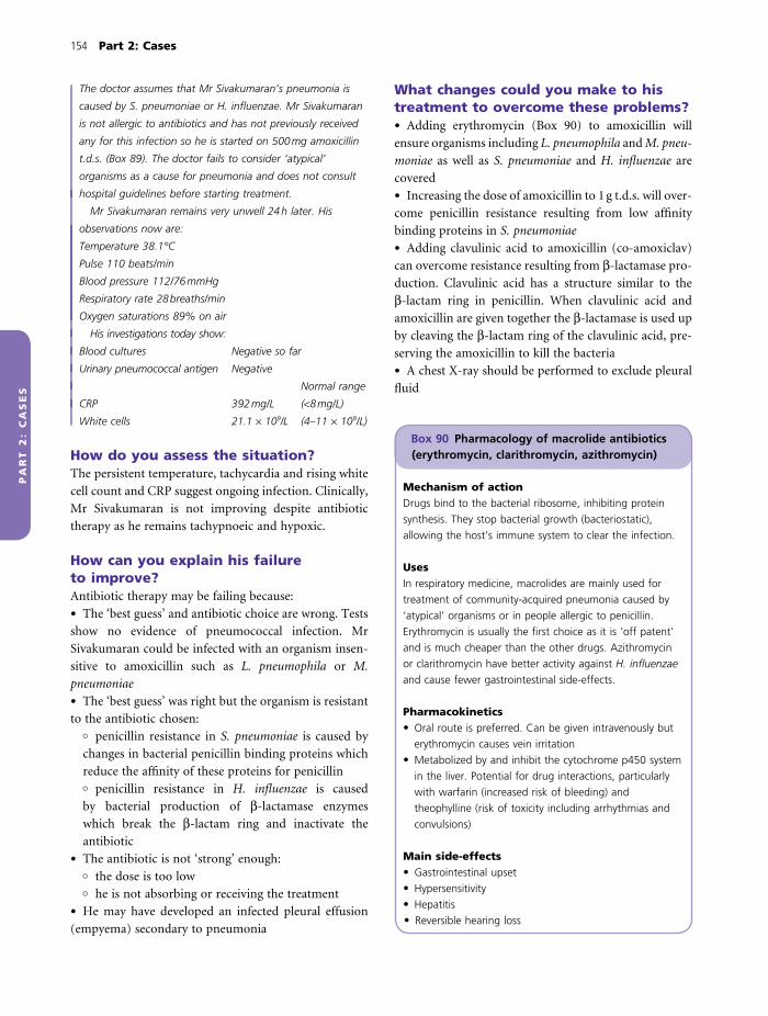
154 Part 2: Cases
PA
RT
2:
CA
SE
S
The doctor assumes that Mr Sivakumaran’s pneumonia is
caused by S. pneumoniae or H. infl uenzae. Mr Sivakumaran
is not allergic to antibiotics and has not previously received
any for this infection so he is started on 500 mg amoxicillin
t.d.s. (Box 89). The doctor fails to consider ‘atypical’
organisms as a cause for pneumonia and does not consult
hospital guidelines before starting treatment.
Mr Sivakumaran remains very unwell 24 h later. His
observations now are:
Temperature 38.1°C
Pulse 110 beats/min
Blood pressure 112/76 mmHg
Respiratory rate 28 breaths/min
Oxygen saturations 89% on air
His investigations today show:
Blood cultures Negative so far
Urinary pneumococcal antigen Negative
Normal range
CRP 392 mg/L (<8 mg/L)
White cells 21.1 × 10⁹/L (4–11 × 109/L)
How do you assess the situation?The persistent temperature, tachycardia and rising white
cell count and CRP suggest ongoing infection. Clinically,
Mr Sivakumaran is not improving despite antibiotic
therapy as he remains tachypnoeic and hypoxic.
How can you explain his failure to improve?Antibiotic therapy may be failing because:
• The ‘best guess’ and antibiotic choice are wrong. Tests
show no evidence of pneumococcal infection. Mr
Sivakumaran could be infected with an organism insen-
sitive to amoxicillin such as L. pneumophila or M.
pneumoniae
• The ‘best guess’ was right but the organism is resistant
to the antibiotic chosen:� penicillin resistance in S. pneumoniae is caused by
changes in bacterial penicillin binding proteins which
reduce the affi nity of these proteins for penicillin� penicillin resistance in H. infl uenzae is caused
by bacterial production of β-lactamase enzymes
which break the β-lactam ring and inactivate the
antibiotic
• The antibiotic is not ‘strong’ enough:� the dose is too low� he is not absorbing or receiving the treatment
• He may have developed an infected pleural effusion
(empyema) secondary to pneumonia
What changes could you make to his treatment to overcome these problems?• Adding erythromycin (Box 90) to amoxicillin will
ensure organisms including L. pneumophila and M. pneu-
moniae as well as S. pneumoniae and H. infl uenzae are
covered
• Increasing the dose of amoxicillin to 1 g t.d.s. will over-
come penicillin resistance resulting from low affi nity
binding proteins in S. pneumoniae
• Adding clavulinic acid to amoxicillin (co-amoxiclav)
can overcome resistance resulting from β-lactamase pro-
duction. Clavulinic acid has a structure similar to the
β-lactam ring in penicillin. When clavulinic acid and
amoxicillin are given together the β-lactamase is used up
by cleaving the β-lactam ring of the clavulinic acid, pre-
serving the amoxicillin to kill the bacteria
• A chest X-ray should be performed to exclude pleural
fl uid
Box 90 Pharmacology of macrolide antibiotics (erythromycin, clarithromycin, azithromycin)
Mechanism of actionDrugs bind to the bacterial ribosome, inhibiting protein
synthesis. They stop bacterial growth (bacteriostatic),
allowing the host’s immune system to clear the infection.
UsesIn respiratory medicine, macrolides are mainly used for
treatment of community-acquired pneumonia caused by
‘atypical’ organisms or in people allergic to penicillin.
Erythromycin is usually the fi rst choice as it is ‘off patent’
and is much cheaper than the other drugs. Azithromycin
or clarithromycin have better activity against H. infl uenzae
and cause fewer gastrointestinal side-effects.
Pharmacokinetics• Oral route is preferred. Can be given intravenously but
erythromycin causes vein irritation
• Metabolized by and inhibit the cytochrome p450 system
in the liver. Potential for drug interactions, particularly
with warfarin (increased risk of bleeding) and
theophylline (risk of toxicity including arrhythmias and
convulsions)
Main side-effects• Gastrointestinal upset
• Hypersensitivity
• Hepatitis
• Reversible hearing loss

Case 17 155
PA
RT
2:
CA
SE
S
KEY POINT
Community-acquired pneumonia severe enough to require
hospital admission is usually treated with a combination of
penicillin (e.g. amoxicillin) and macrolide (e.g. erythromycin)
to cover S. pneumoniae and ‘atypical’ organisms that are
the most common causes of this condition.
Erythromycin 500 mg orally q.d.s. was added to Mr
Sivakumaran’s treatment and amoxicillin increased to 1 g t.d.s.
intravenously. A chest X-ray showed no pleural fl uid. Over
the next 48 h Mr Sivakumaran’s temperature and tachycardia
settled and he started to improve. On further testing Mr
Sivakumaran’s urine was positive for Legionella antigen. By
day 6 Mr Sivakumaran was much better with oxygen
saturations of 96% on air and was mobile on the ward. He
was discharged home to complete 14 days of treatment and
to be followed up as an outpatient 6 weeks later.
Why does he need outpatient follow-up?Follow-up is booked to ensure that he has recovered from
his pneumonia. At his appointment he will have a repeat
chest X-ray to monitor resolution of his consolidation.
Persistent consolidation could indicate an obstructing
lesion in the airways (e.g. lung cancer) and may require
further investigation. Follow-up will also provide the
opportunity for further counselling regarding smoking
cessation.
Outcome. At follow-up Mr Sivakumaran is better but not
completely back to normal. His chest X-ray is now clear. He
has not smoked since discharge and is using nicotine
patches to support smoking cessation. He is advised that
tiredness can persist for up to 3 months following
pneumonia and is discharged.
KEY POINTS
• Community-acquired pneumonia is caused by bacterial or
viral pathogens, most commonly Streptococcus pneumoniae
• ‘Atypical’ pathogens causing pneumonia (Legionella
pneumophila, Mycoplasma pneumoniae and Chlamydia
spp.) are ‘atypical’ because they:� cause more systemic symptoms than other bacteria� require serological testing rather than culture to confi rm
the diagnosis� do not respond to penicillins and require macrolide or
tetracycline treatment
• Legionella pneumophila should particularly be considered
as a cause in patients with severe pneumonia, although it
can cause pneumonia of any severity
• Infections causing pneumonia trigger an infl ammatory
reaction, causing airspaces within affected alveoli to
become replaced with infl ammatory exudate
• Patients with pneumonia have symptoms of cough,
haemoptysis, pleuritic chest pain, fever and shortness of
breath
• Clinical signs of lobar pneumonia include dullness to
percussion, crepitations and bronchial breathing over the
affected area of lung
• The diagnosis of pneumonia requires demonstration of
consolidation, usually on chest X-ray
• Severity of pneumonia is assessed by CURB-65 score and
determines the site of treatment (home, hospital ward or
intensive care)
• Community-acquired pneumonia severe enough to
require hospital admission should be treated with a
combination of penicillin and macrolide antibiotics to
cover usual and ‘atypical’ pathogens
• Patients should return for an outpatient chest X-ray 6
weeks after discharge to ensure resolution of
consolidation
• If pneumonia has not resolved at review, an underlying
cause such as obstruction of an airway by lung cancer
should be considered
mycin is added to his treatment to cover ‘atypical’ patho-
gens. A diagnosis of Legionella pneumonia is subsequently
made by urinary antigen testing. With this treatment he
makes a good recovery and his chest X-ray abnormality has
resolved when he is reviewed 6 weeks later in the out patient
clinic.
CASE REVIEW
Mr Sivakumaran is admitted as an emergency with pleu-
ritic chest pain and is found to have left lower lobe pneu-
monia. His CURB-65 score is 2 and he is judged to be at
increased risk of death from his pneumonia. He is errone-
ously initially treated with 500 mg amoxicillin t.d.s. but
becomes increasingly unwell over the next 24 h. Erythro-

PA
RT
2:
CA
SE
S
A 72-year-old man who becomes breathless during inpatient stay on a medical ward
Case 18
Mr Virgil Simpson is a 72-year-old man from Jamaica, who
was admitted 1 week ago with a dense right hemiplegia and
expressive aphasia. A computed tomography (CT) brain scan
demonstrated an infarction (stroke) caused by left middle
cerebral artery thrombosis. He has been sleepy and unable
to get out of bed since admission. Today the ward staff
notice that he is breathless and call for a medical review.
His observations are:
Respiratory rate 28 breaths/min
Oxygen saturations 91% on air
Pulse 105 beats/min
Blood pressure 158/104 mmHg
Give a differential diagnosis for his breathlessness?The most likely diagnoses are:
• Pneumonia
• Pulmonary embolus
• Cardiac disease
Why are these the most likely causes of his breathlessness?PneumoniaHis stroke may have impaired his lung defences against
infection by several mechanisms (Fig. 9, p. 10):
• He is sleepy and may not be protecting his airway with
an adequate cough refl ex
• His stroke may have impaired his swallowing, which,
in combination with an inadequate cough refl ex, could
have resulted in aspiration of oral or refl uxed gastric
contents into his lungs
• He is unable to care for himself and maintain oral
hygiene. Bacterial overgrowth in the mouth increases the
likelihood that aspiration of oral secretions will result in
lung infection
• He is immobile, which may be reducing his lung ven-
tilation and clearance of lung secretions. Poorly cleared
‘stagnant’ secretions can block the airways causing mini
lung collapses (atelectasis) and can become infected
• Patients with acute illness also have reduced activity
of innate and acquired immune defences against
infection
Pulmonary embolus• He is immobile with reduced blood fl ow in his leg
veins and this stasis could have caused deep vein throm-
bosis (DVT) and pulmonary embolus. Medical patients
who are immobile should all receive prophylactic
heparin and/or anti-embolus stockings to prevent this
complication.
Cardiac diseaseCardiac disease may cause breathlessness because of pul-
monary oedema, arrhythmias, ischaemia and infarction.
In hospital patients, overenthusiastic intravenous fl uid
replacement should also be considered as a cause of pul-
monary oedema.
What clinical signs would you look for to distinguish between your diagnoses?As Mr Simpson has expressive aphasia and is unable to
answer questions, the diagnosis depends on examination
fi ndings and investigation. Possible fi ndings are as
follows.
Pneumonia• Pyrexia
• Purulent respiratory secretions
• Lung crepitations over the affected area
• Bronchial breathing
• Impaired gag refl ex or unsafe swallow (Fig. 70)
Pulmonary embolusPulmonary emboli can be diffi cult to detect so you should
have a ‘high index of suspicion’ whenever the diagnosis
is even slightly possible. Look for:
156

Case 18 157
PA
RT
2:
CA
SE
S
• Tachycardia, low blood pressure, right ventricular
heave and raised jugular venous pressure (JVP) (large
central embolus)
• Pleural rub (small peripheral embolus)
• Calf tenderness and swelling (DVT) – this may be
clinically apparent as a source of emboli in some patients
but absence of a DVT does not rule out pulmonary
embolus
Cardiac diseaseCheck pulse rate and rhythm for arrhythmias and look
for signs of left heart failure:
• Fine bibasal crepitations
and associated right heart failure:
• Raised JVP
• Hepatomegaly
• Peripheral oedema – do not forget to check for sacral
oedema as this is the fi rst affected site in bed-bound
patients
Mr Simpson is diffi cult to examine as he is a large
gentleman and requires two staff to sit him forward. His
breathing sounds rattly and he has:
Temperature 38.6°C
Pulse 96 beats/min regular
JVP not elevated, looks dehydrated
Heart sounds normal
No oedema, calf swelling or tenderness
Breath sounds vesicular with widespread coarse crepitations,
worse on the right
His swallow is felt to be unsafe when tested. A provisional
diagnosis of aspiration pneumonia is made.
What would be your immediate management?• Sit him up to optimize ventilation
• Oxygen to increase saturations >92%
• Nil by mouth to prevent further aspiration
• IV access and IV fl uids to replace fl uid losses and main-
tain hydration while he is nil by mouth
• Suction to help him clear respiratory secretions by
nurses or physiotherapist
What investigations would you carry out?• Culture of sputum obtained by suction
• Blood cultures
• Blood tests as below.
Plasma biochemistry:
Normal range
Na 143 mmol/L (135–145 mmol/L)
K 3.9 mmol/L (3.5–5.0 mmol/L)
Urea 18 mmol/L (2.5–8 mmol/L)
Creatinine 112 mmol/L (60–110 mmol/L)
CRP 196 mg/L (<8 mg/L)
Full blood count:
Haemoglobin 15.2 g/dL (13–18 g/dL)
White cells 21.4 × 10⁹/L (4–11 × 109/L)
Neutrophils 18.2 × 10⁹/L (2.0–7.5 × 10⁹/L)
Platelets 274 × 109/L (150–400 × 109/L)
Arterial blood gases on air:
pH 7.34 (7.35–7.45)
PaO2 7.4 kPa (10–13.1 kPa)
PaCO2 6.9 kPa (4.9–6.1 kPa)
HCO3− 26 mmol/L (22–28 mmol/L)
Chest X-ray:
See Fig. 71
ECG:
Sinus tachycardia
Signs ofaspiration*
Allow to eat and drink normallyand observe
No problems
No problems
No problems
Give patient half a cup of waterto drink
Signs of aspiration*
Give patient 2 more teaspoonfulsof water to swallow
Make sure the patient is sittingup
Give patient 1 teaspoonful ofwater to swallow
Water comes out of mouthFails to swallowSigns of aspiration*
Nil by mouthFormal swallowassessment and
treatment
Figure 70 A simple clinical assessment of swallow. * Signs of
aspiration include coughing, choking, breathlessness or a ‘wet
voice’ after swallowing. This simple clinical assessment should
be performed immediately in any patient where swallowing
diffi culties are suspected and can be carried out by any health
professional with brief training. If in doubt hospitalized
patients should be kept nil by mouth and given intravenous
fl uids until specialist assessment by a speech and language
therapist can be arranged.

158 Part 2: Cases
PA
RT
2:
CA
SE
S
How do you interpret these investigations?• His raised white cell count (mostly neutrophils) and
elevated C-reactive protein (CRP) support the clinical
diagnosis of infection
• The raised urea and relatively normal creatinine could
be accounted for by dehydration, although he may have
some underlying renal impairment
• His blood gases show hypoxia on air. As the PaO2 is
<8 kPa this meets the defi nition for respiratory failure.
His PaCO2 is slightly elevated, indicating hypoventilation,
which would fi t with his immobility and stroke. This has
caused a slight respiratory acidosis
• He has right midzone airspace shadowing consistent
with consolidation and pneumonia
• His electrocardiogram (ECG) shows no evidence of a
cardiac cause for his breathlessness
Is it signifi cant that the pneumonia is in his right lung?The right main bronchus is more vertical than the left
main bronchus and so aspiration is more often into the
right than the left lung.
Which organisms are most likely to be causing his aspiration pneumonia?This is most commonly caused by:
• Anaerobes from the mouth (e.g. Bacteroides, Fusobac-
terium and Peptococcus spp.)
• Gram-negative bacteria (e.g. Klebsiella and Pseudomo-
nas spp.)
However, Mr Simpson has been in hospital for 1 week and
so his pneumonia could also have been caused by organ-
isms that are commonly picked up in hospital, including:
• Pseudomonas aeruginosa
• Staphylococcus aureus (including methicillin-resistant
strains)
Hospital-acquired pneumonia is also called ‘nosoco-
mial pneumonia’.
How does hospital-acquired pneumonia differ from community-acquired pneumonia?In the community the most common pneumonia patho-
gens are Streptococcus pneumoniae, Haemophilus infl uen-
zae and ‘atypical’ pathogens (see Case 17). These are very
different from the organisms causing hospital-acquired
pneumonia (see above). This has important implications
when choosing antibiotic treatment. Some reasons for
these differences are given in Table 8.
What antibiotic treatment would you recommend for Mr Simpson?A rational choice of antibiotic should be based on the
most likely causative organism. In Mr Simpson’s case the
antibiotic regime must cover Gram-negative organisms
including pseudomonas and anaerobes (see Figs 22 & 23,
pp. 39 and 40). A possible choice would be a third gen-
eration cephalosporin such as cefotaxime (Box 91) with
metronidazole to cover anaerobes (Box 92).
How should the antibiotics be given?Seriously ill patients should be given antibiotics intrave-
nously to ensure therapeutic plasma concentrations.
Other considerations are:
• Mr Simpson is nil by mouth because of his swallowing
problems, hence he cannot take oral medication
• Third generation cephalosporins can only be given
parenterally as oral preparations are not absorbed
Mr Simpson was started on 1 g cefotaxime IV and 500 mg
metronidazole IV t.d.s. He was made nil by mouth to
prevent further aspiration and received intravenous
rehydration. His oxygen saturations improved to 95% on
35% humidifi ed oxygen and he had regular physiotherapy
with suction to clear respiratory secretions. A nasogastric
tube was inserted for enteral feeding and hydration.
Why was the oxygen humidifi ed?Oxygen is humidifi ed by bubbling through sterile water.
This appears to maintain hydration and ease clearance
Figure 71 Chest X-ray.

Case 18 159
PA
RT
2:
CA
SE
S
of respiratory secretions. Patients on oxygen therapy
may also fi nd oxygen treatment more comfortable if
humidifi ed as it causes less drying of the mucous
membranes.
Mr Simpson initially appeared to improve. However, after
72 h he was unwell, pyrexial and had had two episodes of
diarrhoea. His blood results were:
Normal range
Na 143 mmol/L (135–145 mmol/L)
K 3.2 mmol/L (3.5–5.0 mmol/L)
Urea 9 mmol/L (2.5–8 mmol/L)
Creatinine 118 mmol/L (60–110 mmol/L)
CRP 255 mg/L (<8 mg/L)
White cells 25.6 × 10⁹/L (4–11 × 109/L)
Neutrophils 22.0 × 10⁹/L (2.0–7.5 × 10⁹/L)
Table 8 Some reasons for differences in pathogens causing hospital- and community-acquired pneumonias.
Community-acquired pneumonia Hospital-acquired pneumonia
Patient factorsPre-morbid state Usually well before infection Immunocompromised by acute illness or comorbidity
Exposure Mostly in contact with well people Close to other people with infection and illness
Looked after by staff who may be colonized with
pathogens and have poor hand hygiene
Organism factorsAntibiotic resistance Organisms live in community where
antibiotic use is relatively low and
resistance is uncommon
Organisms live in hospitals where antibiotic use is high
and resistance is common
Persistence in environment Pathogens fragile, need human host
and are poorly persistent in
environment
Pathogens persistent in hospital environment (e.g.
Staphylococcus aureus, nasal and skin colonization,
Pseudomonas aeruginosa in water). These organisms
form biofi lms (i.e. bacterial communities) which can
survive when times are hard
Box 91 Pharmacology of cephalosporins
Mechanism of actionCephalosporins come from a fungus called Cephalosporium.
They are similar to penicillins as they have a β-lactam ring,
impair bacterial cell wall synthesis and are bacteriocidal.
UsesCephalosporins are grouped together in generations
according to their spectrum of action. In respiratory medicine
they have the following main uses:
• First generation (e.g. cefaclor [oral]) – treatment of
uncomplicated respiratory tract infections in the
community
• Second generation (e.g. cefuroxime [oral or IV]) –
treatment of community-acquired pneumonia
• Third generation (e.g. cefotaxime [IV]) for severe
community-acquired and hospital-acquired pneumonias.
Ceftazidime (IV) is particularly useful against Pseudomonas
PharmacokineticsCephalosporins are renally excreted and dose reduction may
be required in renal impairment.
Main side-effects• Hypersensitivity. Note that 10% of patients who are
allergic to penicillin will also be allergic to cephalosporins
(and vice versa) because the two groups of antibiotics
have structural similarities
• Diarrhoea and antibiotic-associated colitis

160 Part 2: Cases
PA
RT
2:
CA
SE
S
What do these blood results show?Most notably his CRP and white cell count have risen
rather than fallen in response to antibiotics. His serum
potassium has also fallen, probably because of the
diarrhoea.
How can you explain his failure to improve?His pneumonia may not be resolving because:
• Best guess antibiotic therapy was wrong and the organ-
ism is untreated
• Underlying cause (e.g. aspiration, immunosuppres-
sion) persists
His symptoms may now be caused by his diarrhoea,
which could be a result of:
• Colonic infection with toxin-producing C. diffi cile sec-
ondary to antibiotic treatment
• Initiation of hyperosmolar enteral feeding
How will you investigate him to distinguish between these possibilities?• Send stool sample to microbiology to look for C.
diffi cile toxin
• Repeat chest X-ray to look for worsening
consolidation
Mr Simpson’s oxygen saturations were 95% on 24% oxygen.
His repeat chest X-ray showed some improvement in
consolidation. His original sputum and blood cultures were
negative. His stool sample was positive for C. diffi cile toxin.
How do you interpret this new information?His improving oxygenation and chest X-ray suggest that
his pneumonia is responding to antibiotics. His cultures
did not identify bacteria resistant to the antibiotics used.
His stool sample indicates that his deterioration is caused
by C. diffi cile diarrhoea.
How does Clostridium diffi cile diarrhoea occur?Patients may become infected with a toxin-producing
strain of C. diffi cile while in hospital. Clostridium diffi cile
persists in the hospital environment in spore form and
infection is spread by the faeco-oral route, facilitated by
poor hand hygiene by staff, patients and visitors. Clos-
tridium diffi cile is most likely to cause diarrhoea if an
infected patient also receives a broad-spectrum antibiotic
(cefotaxime in Mr Simpson’s case) which kills normal
bowel fl ora. This allows overgrowth of C. diffi cile and
secretion of toxins A and B, which cause colonic mucosal
injury and infl ammation. Patients often have profuse
debilitating diarrhoea that can impair recovery and
increase mortality.
How should his diarrhoea be treated?The antibiotic charts (Figs 22 & 23, pp. 39 and 40) indi-
cate that metronidazole and vancomycin are effective
against C. diffi cile. He is already on metronidazole so
vancomycin would be the most sensible choice here. Van-
comycin should be given down Mr Simpson’s nasogastric
tube to make sure it is in the gut, which is the site of
infection. Treatment is for 10–14 days, although patients
may still relapse after this time.
Why did he get C. diffi cile infection if he was already taking metronidazole?• The strain of C. diffi cile he acquired may have been
metronidazole-resistant
• Metronidazole given IV may not have reached suffi -
cient concentrations in the gut lumen to prevent C.
diffi cile overgrowth
Box 92 Pharmacology of metronidazole
Mechanism of actionThis drug is taken up by anaerobic organisms and
activated by reduction of a nitro group. It binds to DNA,
prevents nucleic acid formation and stops bacterial
growth.
UsesIn respiratory medicine metronidazole is mainly used for
the treatment of aspiration pneumonia or lung abscesses.
Pharmacokinetics• Can be given orally, rectally or intravenously
• Inhibits alcohol and aldehyde dehydrogenase in the
liver. Patients should avoid taking alcohol with
metronidazole as acetaldehyde will accumulate in the
body, causing fl ushing, headache, tachycardia, nausea,
vomiting and collapse. Patients do not need to avoid
alcohol with other classes of antibiotics
Main side-effects• Hypersensitivity
• Gastrointestinal upset
• Rarely, peripheral neuropathy and seizures
Case 18 161
PA
RT
2:
CA
SE
S
Could anything have been done to prevent this C. diffi cile infection?• Good hygiene and infection control to prevent the
spread of hospital-acquired pathogens
• Limiting use of broad-spectrum antibiotics to prevent
C. diffi cile overgrowth in the bowel� consider amoxicillin/macrolides/tetracyclines instead
of broad-spectrum cephalosporins� use shortest course of treatment possible
KEY POINT
It is essential that everyone (staff, students, visitors and
patients) on a hospital ward maintains excellent hand
hygiene to prevent the spread of infection. Thorough
handwashing is the most effective method of
decontamination, although alcohol gels are also widely
used. One approach is:
• Always wash hands after touching a patient
• Always apply alcohol gel before touching a patient
Outcome. Mr Simpson’s treatment was changed to 625 mg
co-amoxiclav t.d.s. and 125 mg vancomycin q.d.s. via
nasogastric tube. Infection control measures were instituted
to prevent spread of infection from Mr Simpson to other
patients, including obligatory wearing of gloves and aprons
by staff when caring for him. He received chest
physiotherapy to help him clear chest secretions. Over the
next week his diarrhoea settled and his pneumonia
improved. His swallow was reviewed by the speech and
language therapist and he was allowed to take a small
amount of thickened fl uids by mouth. Once he was free of
infection and off antibiotics he was transferred to a
rehabilitation ward.
KEY POINTS
• Hospitalized patients often have compromised pulmonary
defence, caused by� impaired cough refl ex or swallow� reduced mucociliary clearance because of
hypoventilation� impaired innate or acquired immunity
• Pneumonia secondary to aspiration is most commonly
caused by � anaerobes from the mouth� Gram-negative bacteria
• Aspiration pneumonia should therefore be treated with
antibiotics that cover:� anaerobes – metronidazole� Gram-negative organisms – amoxicillin, cephalosporin,
macrolide
• Hospital-acquired (nosocomial) pneumonia may be caused by
bacteria that persist in the hospital environment, particularly:� Pseudomonas aeruginosa� Staphylococcus aureus (including methicillin-resistant
strains)
• Clostridium diffi cile diarrhoea is caused by:� bowel colonization with a toxin-producing strain of C.
diffi cile� broad-spectrum antibiotics (particularly cephalosporins)
which kill normal gut fl ora and allow C. diffi cile overgrowth
• The risk of C. diffi cile diarrhoea in hospital patients should
be reduced by:� excellent hygiene to reduce transmission� minimizing use of cephalosporins and broad-spectrum
antibiotics
of C. diffi cile-related diarrhoea. This was treated with oral
vancomycin. As his pneumonia and diarrhoea resolved,
oral intake was restarted under supervision of the speech
and language therapist and he was transferred to a reha-
bilitation ward for further treatment of his stroke.
CASE REVIEW
Mr Simpson became short of breath 1 week after suffering
a stroke resulting from aspiration pneumonia, secondary
to an unsafe swallow. His oral intake was stopped and he
received intravenous antibiotics, fl uid and chest physio-
therapy. After initial improvement he deteriorated because

PA
RT
2:
CA
SE
S
A 58-year-old woman with breathlessness following surgery
Case 19
Mrs Elinor Aaronson is a 58-year-old woman who
underwent an open cholecystectomy 48 h ago and has now
become short of breath.
What is the fi rst thing you should do on arriving at her bedside?You should perform an ‘ABC’ assessment. Does she have
a patent airway, is she breathing and is her circulation
intact?
Mrs Aaronson is fully responsive and has the following
observations:
Pulse 110 beats/min
Blood pressure 115/65 mmHg
Respiratory rate 24 breaths/min
Oxygen saturations 93% on air
How do you interpret her observations?She has an increased respiratory rate with mild hypoxia.
She has a tachycardia but normal blood pressure and is
not haemodynamically compromised. Her observations
indicate that she is unwell but there is time to make a
full assessment of her condition before commencing
treatment.
What causes of breathlessness should you consider in her case?The most notable feature of her case is that she has
recently had upper abdominal surgery. It is therefore
important to consider whether she might have developed
a postoperative complication accounting for her breath-
lessness (Box 93).
What will you do next to narrow down the differential diagnosis?You should take a focused history including symptoms,
risk factors and past medical history to determine whether
her breathlessness is respiratory or cardiac in origin, then
explore possible diagnoses in more detail (Fig. 11, p. 18).
Mrs Aaronson has had diffi culty breathing for the last 24 h.
Her shortness of breath occurs in all positions, is not worse
on lying fl at, but is associated with a troublesome cough.
She is experiencing pain from her abdominal wound,
particularly on taking a deep breath or coughing. She is not
producing sputum and has not coughed up any blood. She
has no chest pain, palpitations or ankle swelling.
In the past she has had chest infections requiring
antibiotics from her GP about twice per year for the last 5
years and has a chronic productive cough in the winter. She
has never been admitted to hospital and does not normally
take any medication. She started smoking when she was 15
years of age and smoked 10 cigarettes per day until she
gave up 3 years ago. She does not to her knowledge have
high blood pressure or cholesterol and is not diabetic. In her
family her father had emphysema and her son has asthma.
Is her shortness of breath more likely to be caused by cardiac or respiratory disease?Her history points to a respiratory rather than a cardiac
cause of her postoperative breathlessness because:
• Associated symptoms are respiratory (cough) and
there are no other cardiac symptoms (no chest pain, pal-
pitations or oedema)
• Lying fl at does not exacerbate her breathlessness (this
is most often a feature of pulmonary oedema)
• She is a smoker, which can cause both heart and lung
disease, however:� she has no family history of cardiac disease and has
no other cardiac risk factors� her past medical history indicates previous recurrent
respiratory illness
Which respiratory causes should be considered?Mrs Aaronson complains of abdominal pain that is pre-
venting her from coughing and taking a deep breath. This
predisposes her to (Box 94):
162

Case 19 163
PA
RT
2:
CA
SE
S
• Atelectasis
• Infection
She appears to have underlying lung disease, so could
also have:
• Exacerbation of underlying chronic lung disease
At this stage, further information from examination
and investigations is still required to exclude other
diagnoses.
What underlying lung disease may she have?Her chronic productive cough and regular need for anti-
biotics could be consistent with chronic bronchitis. Mrs
Aaronson has smoked around 10 cigarettes per day for 40
years (i.e. ∼20 pack years). It is likely that she has underly-
ing chronic obstructive pulmonary disease (COPD).
On examination she has:
Temperature 37.9°C
Dry mouth and decreased skin turgor
Pale conjunctivae
Respiratory system:
Decreased air entry throughout both lung fi elds
Inspiratory and expiratory crackles at both lung bases with
some expiratory wheeze
Abdominal examination:
Soft abdomen, but tender around the wound site which
looks clean
Box 93 Postoperative complications causing breathlessness
Respiratory complications• Atelectasis
• Infection, including bronchitis and pneumonia
• Bronchospasm
• Ventilatory failure
• Exacerbation of underlying chronic lung disease
Cardiac/vascular complications• Ischaemia/infarction
• Arrhythmia
• Pulmonary oedema (heart failure/fl uid overload)
• Pulmonary embolus
Other• Postoperative anaemia
• Fluid overload
Box 94 Mechanisms underlying development of respiratory postoperative complications
Factors affecting the respiratory system postoperatively
include:
• Pain from the surgical wound inhibiting deep breathing
and coughing
• Muscle dysfunction preventing effective ventilation
caused by:� surgical damage� prolonged neuromuscular blockade
• Drugs that reduce the respiratory drive:� opiates� anaesthetic drugs
These factors reduce tidal volume and deep sighing
breaths, limiting lung expansion. The cough is less
effective and there is reduced mucociliary clearance. This
causes:
• Atelectasis – obstruction of small airways with distal
collapse (Figs 72 & 73) causing hypoxia.
• Infection – stagnation of respiratory secretions which
become infected
• Ventilatory failure – weakness of respiratory muscles
leading to inadequate ventilation
Obstruction of theairway, e.g. by foreignbody, tumour,secretions
Air-filled lung, lobe orsegment
Supplying airway
Gases in air are absorbedinto blood and not replaced
Obstructed lung collapses in on itself,pulling adjacent structures towards it
Figure 72 Pathophysiology of pulmonary collapse and
atelectasis.

164 Part 2: Cases
PA
RT
2:
CA
SE
S
Audible bowel sounds
Urinary catheter in situ and the bag contains 150 mL
concentrated urine which she has passed over the last 4 h.
Urinalysis shows blood +, nil else.
She is wearing anti-embolism stockings.
How do you interpret these examination fi ndings?Mrs Aaronson has mild pyrexia. This could indicate
infection in her:
• Chest. She has poor air entry consistent with reduced
lung expansion postoperatively. Crepitations could be
caused by retained secretions which could have become
infected (Box 94)
• Abdomen. This is soft and bowel sounds are heard.
There is no evidence of postoperative peritonitis or
wound infection
• Biliary tree. This could have become infected from
surgery. There is no clinical evidence of biliary obstruc-
tion as she is not jaundiced
• Urine. Urinalysis shows no evidence of leucocytes or
nitrates which are present in urine infection
Her dry mucus membranes, reduced skin turgor and
concentrated urine indicate hypovolaemia. Bilateral
inspiratory and expiratory crepitations on chest exami-
nation are most likely to indicate retained secretions
and infection as there is no evidence of heart failure or
fl uid overload. Expiratory wheeze is consistent with
bronchospasm.
What is the purpose of anti-embolism stockings?Deep vein thrombosis and pulmonary emboli may
occur postoperatively as a result of immobility, vascular
trauma and hypercoagulability. Anti-embolism stockings
(a)
(b)
Trachea andmediastinum pulled
towards affected side
Collapsed lowerlobe – opaque asdoes not contain
air
Figure 73 Chest X-rays illustrating
lobar collapse and subsegmental
collapse (atelectasis). (a) Lobar
collapse. (b) Basal atelectasis. Arrows
indicate linear shadows of pulmonary
atelectasis – collapse of very small
segments of lung.

Case 19 165
PA
RT
2:
CA
SE
S
and prophylactic low molecular weight heparin are used
to prevent these complications. Thromboembolic com-
plications usually occur at least 5 days postoperatively
and are unlikely to be causing Mrs Aaronson’s breathless-
ness 48 h after surgery, although early emboli have been
described.
What is your differential diagnosis now?• Atelectasis
• Pulmonary infection
• Underlying COPD
What investigations would you request and why?• Chest X-ray – to visualize atelectasis or pneumonia
• Full blood count and C-reactive protein (CRP) – to look
for infection and postoperative anaemia
• Arterial blood gases – to look for CO2 retention result-
ing from underlying COPD or postoperative ventilatory
failure
• ECG – to exclude cardiac ischaemia or arrhythmias
• Troponin T or I – to look for myocardial damage
• Peak fl ow recording or spirometry – to look for airfl ow
obstruction
• U and Es – to look for renal impairment and electrolyte
imbalance
Her investigation results were as follows:
Chest X-ray – poorly expanded lungs with atelectasis at both
lung bases
ECG – sinus tachycardia, nil else
Troponin T – normal
Peak fl ow – not performed because of abdominal pain
Blood tests (Table 9)
Arterial blood gases on 4 L oxygen:
Normal range
pH 7.49 (7.35–7.45)
PaO2 10.4 kPa (10–13.1 kPa)
PaCO2 4.4 kPa (4.9–6.1 kPa)
HCO3− 22 mmol/L (22–28 mmol/L)
How do you interpret these results?• Chest X-ray – bi-basal atelectasis has occurred postop-
eratively, impairing gas exchange and predisposing her to
infection
• Normal cardiac investigations support the clinical sus-
picion that her breathlessness is respiratory in origin
• Airfl ow obstruction could not be assessed because of
pain
• Her rising neutrophil count and CRP are consistent
with developing infection
• Her haemoglobin has fallen postoperatively and she is
anaemic
• Her rising plasma urea and sodium concentrations
confi rm dehydration
• Mrs Aaronson’s PaCO2 is low, indicating hyperventila-
tion in response to hypoxia rather than ventilatory failure
or CO2 retention. She is maintaining normal PaO2 with
supplemental oxygen
• None of these investigations rule out a pulmonary
embolus. However, her rising infl ammatory markers and
early presentation make infection more likely. She should
remain on prophylactic heparin and the diagnosis should
be reviewed regularly
How do her test results modify your differential diagnosis?• The likely cause of her breathlessness is atelectasis,
probably with some superadded pulmonary infection
Table 9 Mrs Aaronson’s blood test results.
Hb(g/dL)
WCC(¥ 109/L)
Neutrophils(¥ 109/L)
Platelets(¥ 109/L)
Na(mmol/L)
K(mmol/L)
Urea(mmol/L)
Creatinine(mmol/L)
CRP(mg/L)
Normal 12–16 4–11 2–7.5 150–400 135–145 3.5–5 2.5–8 60–110 <8
Preop 13.2 6.2 4.3 230 141 4.1 4 76 2
Postop day 1 9.8 11.5 7.0 410 143 3.6 7 90 26
Postop day 2 10.1 13.0 9.2 460 146 3.6 10 94 55
Hb, haemoglobin; CRP, C-reactive protein; WCC, white cell count.

166 Part 2: Cases
PA
RT
2:
CA
SE
S
• Postoperative anaemia and previously undiagnosed
COPD may also be contributing to her symptoms
How would you manage her breathlessness?Mrs Aaronson needs treatment of her lung atelectasis and
infection, control of abdominal pain to facilitate breath-
ing and coughing, optimization of probable COPD and
improvement in fl uid balance.
AtelectasisAtelectasis can be improved by increasing lung expansion
and clearance of secretions with:
• Physiotherapy to increase deep breathing and
coughing
• Nebulized saline or mucolytics to make secretions
easier to clear
• Continuous positive airway pressure (CPAP). This can
reduce postoperative lung atelectasis by maintaining lung
expansion, but should only be used if other treatment
fails as it has complications including discomfort, gastric
distension and pressure-related lung trauma
InfectionMrs Aaronson’s pulmonary infection has developed
around 72 h after hospital admission. Hospital-acquired
pneumonia occurs most commonly in the fi rst 5 postop-
erative days and is most likely to be caused by multiple
organisms including:
• Gram-negative bacteria, particularly:� Pseudomonas aeruginosa� Haemophilus infl uenzae
• Staphylococcus aureus
• Streptococcus pneumoniae
Organisms acquired in hospital may be multidrug-
resistant. Hospital-acquired pneumonia is therefore
usually treated with broad-spectrum antibiotics that are
effective against drug-resistant bacteria (Box 95).
Control of abdominal painPostoperative pain can be controlled with opiate analge-
sia; however, opiates can cause respiratory depression
and worsen respiratory complications. An alternative
is epidural anaesthesia, which reduces postoperative
pulmonary complications signifi cantly compared to
opiate analgesia. Physical measures including support-
ing the abdominal wound during coughing may also be
effective.
Box 95 Example antibiotic treatment regime for hospital-acquired pneumonia
Early onset infection (came on <5 days from
admission)
• Start with cefuroxime IV or co-amoxiclav IV
• Switch to oral co-amoxiclav if the patient is responding
after 48 h
• Give antibiotics for a total of 7 days
Late onset infection (came on ≥5 days from
admission)
• ceftazidime IV ± gentamicin IV
• The patient should be reassessed for consideration of
ongoing treatment after 7 days
Once sputum culture results are available the treatment
regime may need to be modifi ed depending on the
fi ndings:
Pseudomonas aeruginosa treat as for late onset
infection
Methicillin-resistant S. aureus add IV vancomycin
Legionella pneumophila add erythromycin
Evidence of aspiration add metronidazole
Optimization of COPDShe should be given bronchodilators (e.g. nebulized sal-
butamol and ipratropium). If her COPD fails to improve
or deteriorates, oral prednisolone could be added to
treatment, but this should be done cautiously as cortico-
steroids impair wound healing.
Improve fl uid balanceSystemic dehydration can make respiratory secretions
more diffi cult to clear and cause other important effects
such as renal impairment. Reasons for her dehydration
may include:
• Increased insensible loss because of pyrexia and
tachypnoea
• Inability to regulate her own fl uid intake as she is nil
by mouth following abdominal surgery
Her fl uid requirements should be calculated by
adding:
• Usual daily requirement (2–3 L)
• Estimated fl uid defi cit (estimated by looking for nega-
tive fl uid balance (output > input) on fl uid balance charts
or from clinical evidence of low venous pressure, urine
output and blood pressure)
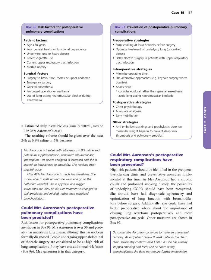
Case 19 167
PA
RT
2:
CA
SE
S
Box 97 Prevention of postoperative pulmonary complications
Preoperative strategies• Stop smoking at least 8 weeks before surgery
• Optimize treatment of underlying lung (or cardiac)
disease
• Delay elective surgery in patients with upper respiratory
tract infection
Intraoperative strategies• Minimize operating time
• Use alternative approaches (e.g. keyhole surgery where
possible)
• Anaesthesia� consider epidural rather than general anaesthesia� avoid long-acting neuromuscular blockade
Postoperative strategies• Chest physiotherapy
• Adequate analgesia
• Early mobilization
Other strategies• Anti-embolism stockings and prophylactic dose low
molecular weight heparin to prevent deep vein
thrombosis and pulmonary embolus
Box 96 Risk factors for postoperative pulmonary complications
Patient factors• Age >50 years
• Poor general health or functional dependence
• Underlying lung or heart disease
• Recent cigarette use
• Current upper respiratory tract infection
• Morbid obesity
Surgical factors• Surgery to brain, face, thorax or upper abdomen
• Emergency surgery
• General anaesthesia
• Prolonged operation/anaesthesia
• Use of long-acting neuromuscular blocker during
anaesthesia
• Estimated daily insensible loss (usually 500 mL, may be
1 L in Mrs Aaronson’s case)
The resulting volume should be given over the next
24 h as 0.9% saline or 5% dextrose.
Mrs Aaronson is treated with intravenous 0.9% saline and
potassium supplementation, nebulized salbutamol and
ipratropium. Her opiate analgesia is increased and she is
started on intravenous co-amoxiclav. She receives chest
physiotherapy.
After 48 h Mrs Aaronson is much less breathless. She
is now able to walk around the ward and go to the
bathroom unaided. She is apyrexial and oxygen
saturations are 96% on air. Her treatment is changed to
oral antibiotics and inhaled, rather than nebulized
bronchodilators.
Could Mrs Aaronson’s postoperative pulmonary complications have been predicted?Risk factors for postoperative pulmonary complications
are shown in Box 96. Mrs Aaronson is over 50 and prob-
ably has underlying lung disease, although this has not been
formally diagnosed. People undergoing upper abdominal
or thoracic surgery are considered to be at high risk of
lung complications if they have one additional risk factor
(Box 96). Mrs Aaronson is in that category.
Could Mrs Aaronson’s postoperative respiratory complications have been prevented?High risk patients should be identifi ed in the preopera-
tive clerking clinic and preventative measures imple-
mented at this time. As Mrs Aaronson had a chronic
cough and prolonged smoking history, the possibility
of underlying COPD should have been recognized.
She should have had diagnostic spirometry and
optimization of lung function with bronchodila-
tors before surgery. Additionally, she could have had
better preoperative advice about the importance of
clearing lung secretions postoperatively and more
postoperative analgesia. Other measures are shown in
Box 97.
Outcome. Mrs Aaronson continues to make an uneventful
recovery. At outpatient review 8 weeks later in the chest
clinic, spirometry confi rms mild COPD. As she has already
stopped smoking and feels well on short-acting
bronchodilators she does not require further intervention.

168 Part 2: Cases
PA
RT
2:
CA
SE
S
KEY POINTS
• Shortness of breath is a common postoperative
complication
• Initial assessment of breathlessness in a postoperative
patient should attempt to distinguish between respiratory,
cardiac or other causes
• Important respiratory postoperative complications are:� atelectasis� infection� bronchospasm� ventilatory failure� exacerbation of underlying lung disease
• Patients at risk of postoperative respiratory complications
can be identifi ed at the preoperative clerking clinic
• Useful preoperative measures to prevent respiratory
complications include:� smoking cessation� optimal treatment of underlying lung disease
• Useful postoperative measures to prevent or treat
postoperative respiratory complications include:� chest physiotherapy� adequate analgesia� early mobilization
• Anti-embolism stockings and low dose low molecular
weight heparin should be used to prevent postoperative
deep vein thrombosis and pulmonary embolus
atelectasis, infection and suggests underlying COPD. She is
treated with physiotherapy, rehydration, nebulized bron-
chodilators and antibiotics. She makes an uneventful
recovery and is found to have mild COPD at follow-up.
CASE REVIEW
Mrs Aaronson becomes short of breath and mildly hypoxic
following upper abdominal surgery. She has been unable
to cough or take a deep breath because of pain from her
abdominal wound. Full assessment identifi es pulmonary

PA
RT
2:
CA
SE
S
A 16-year-old boy with a sore throatCase 20
Matthias Obugu is a 16-year-old boy who presents to his GP
with a 3-day history of a sore throat. He tells Dr Brewster
that it is very painful and diffi cult to swallow and he is
feeling feverish.
What are the main issues the GP should address during the consultation?Sore throat is very common and is usually a minor self-
limiting illness. GPs will see many patients with this
symptom and must sift the few who need investigation,
treatment or even hospital referral from the majority who
will get better on their own.
What causes a sore throat?The usual cause is infection, which is most commonly
viral or bacterial (Box 98). This leads to infl ammation in
the pharynx or tonsils which is painful.
What clinical features would reassure Dr Brewster that Matthias has a self-limiting viral illness?It is very diffi cult to distinguish between viral and bacte-
rial causes of a sore throat using clinical features. However,
other symptoms of a common cold, including runny and
blocked nose, sneezing, cough and recent contact with
people with colds, would point to a viral upper respira-
tory tract infection (Box 99).
Matthias denies cough or nasal symptoms. On examination
his temperature is 38°C and he has a red infl amed pharynx
with large tonsils covered with exudate. He has tender
swollen anterior cervical lymph nodes.
How do you interpret this new information?The clinical fi ndings indicate infection of the pharynx
and tonsillitis. It is still not possible to distinguish clini-
cally between a viral and bacterial cause.
What pathophysiological processes underlie changes found in his throat?Infection of the pharynx causes increased lymph drain-
age from the affected area to the tonsils and anterior
cervical nodes. Antigen-presenting cells carried in the
lymph trigger reactive changes, including activation of B
and T lymphocytes and increased blood fl ow, in the
tonsils and nodes which become enlarged and painful.
The tonsillar exudate comprises infl ammatory cells, par-
ticularly neutrophils, and debris on the surface of the
tonsils.
Dr Brewster is concerned that Matthias may have a bacterial
throat infection. She checks his immunization history,
performs a throat swab and gives him a prescription for
antibiotics.
Why does Dr Brewster check Matthias’s immunization history?Sore throat and upper respiratory tract symptoms can be
caused by infections with potentially serious complica-
tions, including measles, mumps, diphtheria (Box 100)
and rubella. In the UK these infections are rarely seen
because most children are immunized against them.
However, recent reduction in the uptake of vaccination
and increased immigration or travel from countries
without vaccination programmes has led to re-emer-
gence of these conditions.
Dr Brewster is reassured to fi nd that Matthias is fully
vaccinated and has not travelled abroad.
Why did Dr Brewster perform a throat swab?Culture of a throat swab can identify bacteria present in
the throat. The most common bacterial throat infection
is with group A β-haemolytic streptococcus (Box 101).
However, the use of throat swabs is not recommended
169

170 Part 2: Cases
PA
RT
2:
CA
SE
S
routinely in general practice for patients with sore throat
(Box 102) because:
• A positive result does not necessarily indicate infection
as bacteria can be carried in the throat without causing
disease
• A negative result does not necessarily exclude bacterial
infection as the swab may miss the causative pathogen
• Throat swab results come back over 48 h later, so tend
not to alter a GP’s prescribing decisions
• Throat swabs are expensive
Throat swabs may guide therapy in a few patients
where the sore throat is not responding to treatment.
Matthias’s throat swab is unlikely to make much difference
to the management of his condition and could have been
avoided to ensure effi cient use of health care resources.
Should Dr Brewster have given Matthias a prescription for antibiotics?The use of antibiotics for upper respiratory tract infec-
tions (URTI) is a diffi cult issue in general practice. On
the one hand, GPs feel pressure to prescribe to ensure
they do not undertreat serious infections and patients or
parents may demand prescriptions. On the other hand,
most URTIs resolve without antibiotics and prescription
can increase patient reattendance, antibiotic resistance
and health care costs. In the UK, the National Institute
for Clinical Excellence (NICE) has issued guidelines for
Box 98 Causes of a sore throat
Infection• Viruses, including:
� rhinovirus� coronavirus� parainfl uenza virus� respiratory syncitial virus� infl uenza virus� adenovirus� Epstein–Barr virus
• Bacteria:� group A β-haemolytic streptococcus (Streptococcus
pyogenes)� Corynebacterium diphtheriae� Haemophilus infl uenzae� Neisseria gonorrhoea
• Fungi� Candida albicans
Non-infective• Allergy
• Dryness
• Pollution and irritants
• Muscle strain
• Acid refl ux
Box 99 Important facts about the common cold
Impact• Most adults get colds 2–3 times per year and symptoms
resolve over about 1 week
• This may lead to lost work productivity in the general
population
• Colds can have serious health effects by triggering
exacerbations in people with chronic respiratory disease
(e.g. COPD, asthma)
Spread• Cold viruses are highly infectious, requiring only 1–30
viral copies to enter the nose to start an infection
• People with colds are most infectious over the fi rst 3
days when they have the highest concentration of virus
in their nasal secretions
• Spread is by respiratory droplets. Cold virus
contaminates the hands of those who sneeze, use
tissues or touch their noses. Virus is spread directly
through human contact (e.g. handshaking) or indirectly
by contact with furniture or other surfaces. The virus
enters a new respiratory tract when the contaminated
person touches their hands or face and will infect 95%
of people it comes into contact with
Prevention• This is particularly important for patients with chronic
respiratory disease
• There is no vaccine to prevent colds because they are
caused by over 100 different viruses
• To avoid contracting a cold:� avoid those with colds, especially during the fi rst 3
days of their infection� keep a good distance from people coughing and
sneezing to avoid direct contact with respiratory
droplets� wash hands carefully with soap and water after
contact with people with colds, children (a major
reservoir for the cold virus) or potentially
contaminated surfaces� avoid touching face and eyes

Case 20 171
PA
RT
2:
CA
SE
S
prescribing antibiotics in patients with sore throats
(Box 103).
Dr Brewster felt that Matthias’s condition fell within these
guidelines as he has systemic upset with fever. She was also
concerned about his diffi culty swallowing. Hence her
decision to prescribe antibiotics was justifi ed.
Should patients with acute sore throats ever be referred to hospital?Patients may need hospital care if throat swelling is
causing signifi cant airfl ow obstruction or preventing
swallowing. Other indications for referral include a
developing quinsy or underlying immunosuppression.
Box 100 Diphtheria
CauseToxin-producing Corynebacterium diphtheriae
EpidemiologyThis disease is seen rarely in countries with a widespread
vaccination programme. It is endemic with epidemic
outbreaks in USSR, India, South-East Asia and South
America. Immigrants or travellers from these countries
could potentially develop the infection in the UK.
TransmissionTransmission is by respiratory droplet spread by close
contact. Incubation period is 1–7 days
Pathogenesis• The bacterium infects the throat and upper airway
where it releases toxin. This causes necrosis of mucosal
cells and growth of a membrane, which contains fi brin,
epithelial cells, bacteria and neutrophils. This membrane
may cause respiratory obstruction
• There is reactive lymphadenopathy and soft tissue
swelling in the neck, which becomes very enlarged (‘bull
neck’)
• The bacterium does not invade beyond the throat, but
there is systemic release of the toxin which can affect
the heart and nervous system, causing cardiac failure
with shock and paralysis
Treatment• Penicillin to kill the bacteria and stop toxin production
• Antitoxin to neutralize toxin in blood if systemic effects
• Supportive therapy including maintenance of patent
airway, cardiac support, ventilation if required
Box 101 Group A b-haemolytic streptococcus
Group A β-haemolytic streptococci are found in
approximately 30% of people with sore throats.
Infection may be complicated by:Local suppurative complications• Otitis media
• Sinusitis
• Peritonsillar abscess (quinsy)
Toxin production• Scarlet fever (desquamative rash)
• Streptococcal toxic shock syndrome (rare)
Non-suppurative complicationsThese are now very rare in developed countries, but are
still a problem in the developing world:
• Rheumatic fever
• Glomerulonephritis
Treatment• Symptoms of sore throat caused by group A
streptococci resolve spontaneously in 85% people
within 1 week
• If antibiotics are required use penicillin or erythromycin
if penicillin-allergic
• Antibiotics should not be used to prevent the
complications of streptococcal infection as they have
little clinically signifi cant impact in the UK
Box 103 NICE recommendations for antibiotics in patients with sore throat
Patients with sore throat should only receive antibiotics if
there are:
• Features of marked systemic upset secondary to the
acute sore throat
• Unilateral peritonsillitis
• History of rheumatic fever
• Increased risk from acute infection (e.g. diabetes
mellitus or immunodefi ciency)
Box 102 NHS funded Clinical Knowledge Summaries Service
Useful guidelines to aid clinical practice are published by
the NHS as part of the UK National Library for Health.
These can be found at: http://www.prodigy.nhs.uk

172 Part 2: Cases
PA
RT
2:
CA
SE
S
Matthias does not need hospital referral unless his diffi culty
swallowing prevents drinking and causes dehydration.
Dr Brewster gives Matthias 250 mg amoxicillin t.d.s. for 5
days. He is back in her surgery 48 h later complaining of
itching. On examination he has a widespread maculopapular
rash.
Why might this rash have developed?Aminopenicillins (amoxicillin, ampicillin, co-amoxiclav)
should not be used as fi rst line treatment for patients with
a sore throat (Box 89, p. 153). This is because patients
with sore throat caused by infectious mononucleosis
(glandular fever; Box 104) almost always develop a rash
if given these drugs. This unexplained reaction may lead
to patients mistakenly being labelled as penicillin-
allergic. Phenoxymethylpenicillin (or erythromycin if
penicillin-allergic) should be the fi rst line antibiotic for
patients with a sore throat.
It is also possible that his rash may have arisen from a
true penicillin allergy.
How could Dr Brewster confi rm the diagnosis of infectious mononucleosis?• Blood fi lm – patients with infectious mononu-
cleosis have a lymphocytosis with atypical activated T
lymphocytes
• Monospot or Paul–Bunnell tests – for IgM heterophil
antibodies which are able to agglutinate horse (Monospot)
or sheep (Paul–Bunnell) red blood cells, indicating infec-
tion with Epstein–Barr virus.
If Dr Brewster stops Matthias’s antibiotics, what other treatment options are available for sore throat?Sore throat should be treated with paracetamol or
soluble aspirin for symptomatic relief and fl uid intake
should be maintained. Symptoms of URTI will usually
resolve in approximately 1 week, but infectious mono-
nucleosis may take weeks or even months to resolve
completely.
When Matthias is better should he have his tonsils removed?Matthias has had one episode of tonsillitis and is unlikely
to benefi t from tonsillectomy. Guidelines indicate that
patients should have fi ve or more episodes of tonsillitis
per year that prevent normal functioning before they
should be considered for tonsillectomy.
Outcome. Matthias’s Monospot test was positive and he
was diagnosed with glandular fever. His rash resolved on
stopping the antibiotics and his sore throat improved
with rest and paracetamol. He remained tired and listless
for a few weeks but gradually returned to normal
activities.
Box 104 Infectious mononucleosis
CauseEpstein–Barr virus, a member of the herpes family.
TransmissionThe virus is transmitted via respiratory secretions and
saliva.
EpidemiologyEpstein–Barr virus most commonly infects young children
and young adults (15–25 years) who share secretions.
Hence, it is also known as ‘kissing disease’.
Clinical featuresInfection causes fever, sore throat, lymphadenopathy and
hepatosplenomegaly. Systemic symptoms of fatigue,
lethargy and debilitation may persist for months.
Complications, which are relatively rare, include:
• Haemolytic anaemia and thrombocytopaenia
• Neurological involvement
• Cardiac involvement
TreatmentNo specifi c treatment is required as the infection resolves
spontaneously.
CASE REVIEW
Matthias Obugu sees his GP complaining of a sore throat
for the past 3 days. He is pyrexial with enlarged tonsils
and lymphadenopathy. His GP treats him with amoxicil-
lin, causing Matthias to develop a widespread maculo-
papular rash. His GP suspects Epstein–Barr virus
infection and this is confi rmed with a positive Monospot
test. Matthias’ condition improves over several weeks.

Case 20 173
PA
RT
2:
CA
SE
S
KEY POINTS
• Sore throat is most commonly caused by viral or bacterial
infection
• Throat swabs are not generally indicated but may be
useful in a few patients who are not responding to
treatment
• Most sore throats will resolve with symptomatic relief
irrespective of aetiology
• Antibiotics are only indicated if there are:� features of marked systemic upset secondary to the
acute sore throat� unilateral peritonsillitis� history of rheumatic fever� increased risk from acute infection (e.g. diabetes mellitus
or immunodefi ciency)
• First line antibiotics for treatment of sore throat are
phenoxymethylpenicillin or erythromycin for penicillin-
allergic patients
• Aminopenicillins should not be prescribed for patients
with sore throats as they will cause a rash in those with
infectious mononucleosis, which may be mistaken for
penicillin allergy
• Patients with sore throat rarely require hospital referral,
but this may be indicated if they have:� airfl ow obstruction� inability to swallow� quinsy (tonsillar abscess)� immunosuppression

PA
RT
2:
CA
SE
S
A 45-year-old woman with a chronic cough
Case 21
Mrs Li is a 45-year-old woman who attends the chest clinic
complaining of a 6-month history of cough occurring
throughout the day and at night. She has had prolonged
periods of coughing in the past, although these have usually
recovered more quickly.
Mrs Li coughs up phlegm at least 20 times a day which is
yellow or green and has been streaked with blood on
several occasions. She gets an ache in her left side and is
short of breath running for a bus. She has no nasal
obstruction or bleeding, although she has been treated for
sinusitis. She has not noticed heartburn or refl ux. She is on
no medication and has never smoked. She feels tired and
has poor appetite, but has no weight loss or night sweats.
Analyse this information to produce a differential diagnosis for her cough• The fi rst step is to decide whether her cough is acute
or chronic (see Part 1, Approach to the Patient, p. 20).
Mrs Li’s cough is clearly chronic as she has had it for
much longer than 8 weeks
• The next step is to determine whether her chronic
cough is caused by lung disease, postnasal drip, gastro-
oesophageal disease or drugs (Box 18, p. 20). Mrs Li’s
productive cough and shortness of breath point to lung
disease. She has no nasal or gastro-oesophageal symp-
toms and is not taking any medication
• The next step is to narrow down the respiratory
differential diagnosis. Her copious sputum production
indicates bronchiectasis. Haemoptysis is seen in bronchi-
ectasis, but tuberculosis should be considered. Her age,
prolonged history and non-smoker status make other
causes of haemoptysis (lung cancer, pneumonia, pulmo-
nary emboli) less likely (Box 19, p. 22).
What other information would be helpful at this point?She should be asked about causative and risk factors to
distinguish between bronchiectasis and tuberculosis and
the effect of her cough on her life should be established.
Mrs Li was born in the UK, but was hospitalized for several
months aged 5 with severe pneumonia. After that she often
had a cough as a child and fi nds that colds ‘go to her chest’
as an adult. She had the bacille Calmette–Guérin (BCG)
vaccination aged 14. On leaving school she started work in
an offi ce and is still working for the same company. She is
married with two children who are well, lives in a modern
house and has no pets. She has no personal or family
history of asthma, hayfever or eczema and has never had
family or social contacts with tuberculosis. She fi nds that her
cough is embarrassing, causes stress incontinence of urine
and restricts her social life.
How do you interpret this information?• Her severe childhood respiratory infection could have
caused bronchiectasis
• Childhood cough could also indicate asthma, although
she has no personal or family history of atopic disease
(some asthmatics are allergic to the fungus Aspergillus
and develop bronchiectasis of proximal airways known
as allergic bronchopulmonary aspergillosis)
• She has no history of risk factors for tuberculosis
(Box 107, p. 188). She has been vaccinated against
tuberculosis
• Her cough is having considerable impact on her quality
of life
On examination she looks well. She has fi nger clubbing but
no lymphadenopathy. Her oxygen saturations are 98%. She
has coarse crepitations on auscultation at her left lung base.
How do you interpret her examination fi ndings?Mrs Li has fi nger clubbing, which is seen in a number of
respiratory conditions (Box 21, p. 24). Patients with
bronchiectasis commonly develop fi nger clubbing, par-
ticularly if they have extensive disease or chronic infec-
tion. However, fi nger clubbing has been reported in
20–30% of patients with tuberculosis.
174

Case 21 175
PA
RT
2:
CA
SE
S
Crepitations at her left lung base could be consistent
with bronchiectasis or tuberculosis, although tuberculo-
sis more commonly affects the lung apices. The otherwise
normal fi ndings do not exclude other diagnoses.
How would you investigate Mrs Li?Mrs Li’s main differential diagnosis is bronchiectasis or
pulmonary tuberculosis. Investigation should aim to dis-
tinguish between these diagnoses and exclude unexpected
diagnoses such as asthma, lung cancer or lung abscess.
Initial tests should include:
• Chest X-ray to look for structural lung abnormalities
• Sputum microscopy and culture to look for Mycobac-
terium tuberculosis and other pathogens including bacte-
ria and fungi
• Peak fl ow measurements and/or lung function tests to
look for airways obstruction
• Full blood count:� neutrophils – elevation could indicate infection� eosinophils – may be elevated in asthma� white cells – immunosuppression can predispose to
lung infection
• Plasma biochemistry:� glucose – diabetes can predispose patients to lung
infections
KEY POINT
• The lung function department must be informed if there
is any chance that a patient to be tested may have
tuberculosis
• Patients with suspected tuberculosis are tested at the end
of the day prior to disinfection of lung function equipment
to prevent cross-infection via contaminated tubing
Mrs Li’s results:
Chest X-ray – Fig. 74
Sputum microscopy – no acid-fast bacilli
Sputum culture – Haemophilus infl uenzae +++Awaiting results of prolonged culture for mycobacteria
Lung function
FEV1 1.9 L (68% predicted), FVC 3.0 L (92% predicted),
FEV1/FVC 63%
Blood tests unremarkable
How do you interpret these results?Chest X-rayThick-walled airways can be seen on the chest X-ray as
double white lines (tramtracks) behind the heart (arrow).
This is consistent with bronchiectasis. There are no fea-
tures of tuberculosis or other lung pathology.
SputumHer sputum has grown Haemophilus infl uenzae and this
infection may be exacerbating her symptoms.
There were no acid-fast bacilli on sputum microscopy,
but a single negative sputum sample does not rule out
tuberculosis. Pulmonary tuberculosis can be diffi cult to
diagnose, requiring accumulation of evidence from clin-
ical symptoms and signs, Mantoux testing, chest X-ray
and microbiology (see Case 23). Consistent lack of evi-
dence of tuberculosis and positive evidence of bronchi-
ectasis in this patient makes the diagnosis of tuberculosis
decreasingly likely.
Lung functionHer FEV1 is <80% predicted and FEV1/FVC is <70% pre-
dicted. These results are consistent with airfl ow obstruc-
tion. This can occur in patients with bronchiectasis, asthma
or chronic obstructive pulmonary disease (COPD).
There is still some diagnostic doubt. What other investigations could be useful?• A high-resolution computed tomography (HRCT)
scan of her chest is the best way of confi rming a diagno-
sis of bronchiectasis
Figure 74 Chest X-ray.

176 Part 2: Cases
PA
RT
2:
CA
SE
S
• Further tests to exclude tuberculosis would include two
more sputum samples for microscopy and culture and a
Mantoux test
• Evidence of allergy to Aspergillus (IgE [RAST] and skin
prick testing)
One slice from Mrs Li’s HRCT chest is shown in Fig. 75. Her
Mantoux test and further sputum microscopy and culture for
tuberculosis were negative.
What does the HRCT show?Large dilated airways can be seen in the periphery of the
lung (arrows). These have thick walls. This is consistent
with bronchiectasis.
What is bronchiectasis?The term is derived from the Greek ektasis meaning
‘expand’ or ‘dilate’. Bronchiectasis is abnormal and irre-
versible dilatation of the bronchi.
What causes bronchiectasis?Bronchiectasis is caused by factors that impair the
immune defences of the lung (Fig. 76). These causative
factors and the subsequent bronchiectasis may be gener-
alized (e.g. cystic fi brosis, dysfunctional cilia, defects of
innate or acquired immunity) or localized (e.g. inhaled
foreign body, pneumonia).
Mrs Li’s bronchiectasis was most likely caused by her severe
childhood pneumonia, causing localized damage.
How do these factors lead to the pathological fi ndings of bronchiectasis?Reduction in host immunity leads to pulmonary infec-
tion and infl ammation, which heals with scarring. Scar-
ring causes a permanent defi cit in mucociliary clearance,
increasing the risk of further infection and more damage.
This sets up a vicious cycle, with progressive scarring and
thickening of the bronchial walls.
Why do patients with bronchiectasis produce a lot of sputum?Bronchiectatic airways (Fig. 77) are infi ltrated with
neutrophils, which produce an enzyme called elastase.
This stimulates secretion of large volumes of mucus
from submucosal glands. As this mucus is not cleared
effectively by damaged mucociliary apparatus it pools
in the lungs, increasing bacterial colonization and Figure 75 Computed tomography (CT) scan of chest.
Air
Distal lungMucociliary escalator
Pharynx
ABNORMAL MUCUSe.g. Cystic fibrosis
ABNORMAL CILIAe.g. Primary ciliarydyskinesia
EPITHELIAL DAMAGEAND SCARRINGInfection, e.g. pneumonia,measles, pertussis,tuberculosis, HIV
Interstitium
INNATE AND ACQUIRED IMMUNEDEFICIENCYe.g. Immunoglobulin deficieny
Epithelialcell
LUMINAL OBSTRUCTIONe.g. Foreign body, lymph nodes
Figure 76 Some causes of
bronchiectasis. Bronchiectasis can be
initiated by any factor that disrupts
normal lung defence, particularly the
mucociliary escalator. Damage
initiates a vicious cycle of infection,
lung damage and more infection
which causes scarring and dilatation
of the bronchi. Investigation of a
patient with bronchiectasis should
include immunoglobulin levels and
other tests for underlying defects in
lung defence.

Case 21 177
PA
RT
2:
CA
SE
S
chronic infection. Bronchiectatic airways may bleed as
a result of infl ammation or abnormal vessels, causing
haemoptysis.
How is bronchiectasis treated?The aims of treatment are to relieve symptoms and
prevent deterioration of disease by keeping the lungs
clear of respiratory secretions as far as possible and by
treating infection. The underlying cause should be iden-
tifi ed and treated if possible.
Clearance of respiratory secretionsPhysiotherapy is used to keep the lungs clear of respira-
tory secretions. The procedure involves:
1 Loosening of secretions by vibration of the chest (e.g.
by manual percussion)
2 Clearance of loosened secretions by gravitational
drainage, requiring a head down posture
Ideally, patients should do this themselves for 15–30 min
3–4 times per day. In practice, many cannot fi nd the time
or perform the technique correctly.
Pharmacological assistance for sputum clearance
includes:
1 Bronchodilators to treat airfl ow obstruction
2 Good hydration, inhaled saline or mucolytic drugs to
hydrate and thin mucus and make it easier to clear from
the chest
Treatment of infectionPatients with bronchiectasis should receive antibiotics:
• To treat an acute infective exacerbation
(a)
(b)
Normalsized
bronchi
Dilatedpus-filledbronchi
Figure 77 Pathology of bronchiectasis.
(a) Normal lung. (b) Bronchiectasis.

178 Part 2: Cases
PA
RT
2:
CA
SE
S
• Chronically if they have frequent recurrent exacerba-
tions
As patients with bronchiectasis have scarred lungs
and impaired pulmonary defence against infection
they are at risk of infection with unusual organisms
including:
• Bacteria (e.g. Pseudomonas aeruginosa)
• Fungi (e.g. Aspergillus fumigatus)
• Atypical mycobacteria (e.g. Mycobacterium avium
intracellulare)
These need to be identifi ed by sputum examination
and treated appropriately.
Outcome. Mrs Li was informed of her diagnosis of bronchiectasis.
She was treated with 500 mg amoxicillin t.d.s. for 10 days,
inhaled salbutamol and started home physiotherapy. Her GP
was contacted and confi rmed the diagnosis of severe childhood
pneumonia and her bronchiectasis was attributed to this.
She returned to clinic for follow-up 6 weeks later. She
was pleased to report that she was doing her physiotherapy
regularly and had noticed a considerable improvement. Her
sputum was now white following the antibiotics, her cough
was less frequent and her stress incontinence had improved.
She was encouraged to persist with physiotherapy and seek
early antibiotic treatment for future exacerbations.
KEY POINTS
• Bronchiectasis is defi ned as chronic and irreversible
dilatation of the bronchi
• Bronchiectasis occurs in patients with local or generalized
impairment in lung defence against infection
• Loss of host defence triggers infection and infl ammation
that damage the bronchi and impair mucociliary clearance
• Bronchiectasis may present clinically with copious sputum
production, recurrent chest infections or haemoptysis
• Clinical signs of bronchiectasis may be non-specifi c, but
include fi nger clubbing and lung crepitations
• A fi rm diagnosis of bronchiectasis can be made by HRCT
scanning of the chest
• Additional tests should be carried out to look for
ongoing respiratory infection or underlying reduction in
host immunity (e.g. immunoglobin levels)
• The mainstay of bronchiectasis treatment is
physiotherapy to clear pooled lung secretions
• Additional treatment includes antibiotics,
bronchodilators and prompt treatment of
exacerbations
childhood pneumonia. She is treated with antibiotics,
bronchodilators and physiotherapy with considerable
improvement in symptoms and is advised to treat future
exacerbations promptly.
CASE REVIEW
Mrs Li presents with a chronic recurrent cough, productive
of purulent sputum and blood, which is severely impairing
her quality of life. Bronchiectasis is confi rmed by HRCT
scanning and investigations show no evidence of tubercu-
losis. Her bronchiectasis is attributed to scarring following

PA
RT
2:
CA
SE
S
A 17-year-old girl with cystic fi brosisCase 22
Helen Gaskell is a 17-year-old girl who has recently moved
into the area. She was diagnosed with cystic fi brosis aged 3
when she had the following test results:
Genotype – homozygous for ∆F508 mutation in CF
gene
Sweat chloride 108 mmol/L (normal <60 mmol/L)
She has been referred to the cystic fi brosis unit for
ongoing care of her disease.
What is cystic fi brosis?Cystic fi brosis (CF) is an inherited disorder of a chloride
transporter called the cystic fi brosis transmembrane
regulator (CFTR). Transporter mutations cause malfunc-
tion and consequences are altered secretions in many
organs including:
• Lung
• Pancreas
• Gastrointestinal tract
• Biliary tree
• Vas deferens
• Sweat ducts
Patients with cystic fi brosis currently have a life expec-
tancy of around 30 years. Most die of chronic lung disease
including infection and respiratory failure.
Is Helen’s genotype consistent with cystic fi brosis?Cystic fi brosis is an autosomal recessive condition, which
means that mutations in CFTR genes must be inherited
from both father and mother to cause the disease. As 1
in 25 Caucasian people carry the gene, approximately 1
in 2500 Caucasian children have the disease. CF is much
less common but not unknown in people of African or
Asian origin.
Helen is homozygous for the CFTR ∆F508 mutation,
which means she has two copies of the abnormal gene
required to develop the disease. The ∆F508 mutation pre-
vents expression of CFTR on cell surfaces, which means
the protein is unable to function. There are over 1000
mutations of the CFTR gene that can cause cystic fi brosis,
but ∆F508 is the most common.
Why did she have an elevated sweat chloride concentration?Water, Cl− and Na+ are fi ltered out of blood into sweat in
the distal coil of the sweat duct. As fl uid moves proxi-
mally up the normal duct, Cl− is reabsorbed by CFTR and
Na+ is reabsorbed through epithelial sodium channels
(Fig. 78a). Sweat leaving the top of the duct is dilute and
Cl− and Na+ are conserved. In patients with cystic fi brosis,
Cl− reabsorption by CFTR is reduced or absent. Some Na+
reabsorption still occurs, but the negative electric charge
generated by movement of Na+ into the cells and build
up of Cl− in the duct prevents Na+ absorption (Fig. 78b).
Thus, increased Cl− and Na+ are lost in the sweat.
Babies with CF often taste salty if you lick them. A
sweat chloride of >60 mmol/L is strongly suggestive of
CF. As CF patients have diffi culty conserving Cl− and Na+
in their sweat they are at risk of salt and water depletion
on hot days.
Is the information provided suffi cient for a fi rm diagnosis of cystic fi brosis?A diagnosis of CF requires two of:
• Typical clinical features
• Positive sweat test
• Genotype confi rmation
Helen has a clear diagnosis of CF. In some children the
sweat test is only mildly abnormal or unusual CFTR
mutations are not detected and the diagnosis is more
diffi cult.
Why has she been referred to the cystic fi brosis unit?Cystic fi brosis requires intensive management by an
experienced multidisciplinary team including doctors,
nurses, physiotherapists, dietitians and lung function
technicians to reduce complications and maximize life
179

180 Part 2: Cases
PA
RT
2:
CA
SE
S
expectancy. This expertise is concentrated in specialist
centres.
Helen tells the doctor that she is studying for ‘A’ levels and
is well enough to attend school. However, she has a blocked
nose and a chronic cough, currently productive of thick
yellow sputum and feels short of breath if she runs. She
usually has around two chest infections per year, sometimes
requiring treatment with intravenous antibiotics.
How does cystic fi brosis affect the lungs?In CF, ion transport by respiratory epithelial cells is
impaired, reducing the volume of airway surface liquid
(Fig. 79). The cilia of epithelial cells are unable to beat
properly in the low-volume viscous liquid, impairing
mucociliary clearance. This leads to recurrent pulmonary
infection which causes epithelial damage and build-up of
luminal debris from dying cells which further thickens
the mucus. Failing immune defences and increasing
infection create a vicious cycle with progressive lung
destruction, particularly of the small airways.
What types of chest infections are common in cystic fi brosis patients?The spectrum of organisms infecting the lungs in CF
patients is surprisingly limited. The reasons for this are
not fully understood despite much research. Early infec-
tions in children with CF are usually with Staphylococcus
aureus and Haemophilus infl uenzae. As patients get older
their lungs become colonized and chronically infected
with Pseudomonas aeruginosa. As lung damage increases
CF patients may acquire other opportunistic pathogens
including Burkholderia cepacia, Aspergillus fumigatus and
non-tuberculous mycobacteria.
Why has Helen got a blocked nose?Cystic fi brosis affects the fl uid lining the nose and sinuses
as well as the lungs. CF patients therefore commonly have
sinusitis and nasal polyps and Helen should be checked
for these conditions.
Helen also tells the doctor that she fi nds it diffi cult to put on
weight and has intermittent offensive diarrhoea.
How do you explain these additional symptoms?Cystic fi brosis gradually destroys the pancreas (Fig. 80),
reducing release of pancreatic enzymes into the intestine.
Without these enzymes fat is not digested or absorbed.
Undigested fat in the faeces draws in water, causing ste-
atorrhoea which is large volumes of faeces that are pale,
offensive and diffi cult to fl ush away. Malabsorption of fat
makes it diffi cult to put on weight and many CF patients
have nutritional defi ciencies.
How does cystic fi brosis affect the pancreas?The pancreas is made up of acini which secrete digestive
enzymes (Fig. 80). These enzymes pass from the acini
Dilute sweat
Water
Na+
Na+ Na+
Na+
(a)
(b)
Cl–
Cl–
Cl–
Increased sweat Cl–
and Na+
Proximalducts
Filtration
Distal coil
Cell
MutatedCFTR
Water
Water
Na+
Cl–
Proximalducts
Distal coil
Filtration
Cell
CFTR
Figure 78 Effect of CFTR mutations on sweat chloride
secretion. (a) Normal sweat duct. (b) Cystic fi brosis sweat duct.

Case 22 181
PA
RT
2:
CA
SE
S
Normal
Respiratoryepithelial
cell
Respiratoryepithelial
cell
Airwaysurfaceliquid
Airwaysurfaceliquid
Air (lumen)
Air (lumen)
(b)(a)Na+
Na+ Na+
Na+ Na+
Na+
Na+
Na+
K+
K+
Cl–
Cl–(d)(c)
Epithelialsodium
channels
Epithelialsodium
channels
Cystic fibrosis
Blood
Blood
Cilia
Cilia
along pancreatic ducts where HCO3−, Cl− and water,
secreted from epithelial cells, dilute and alkalinize the
enzymes, washing them out into the intestine. Cl− is
secreted into the lumen by CFTR and recycled by a
HCO3−/Cl− exchanger, promoting the secretion of HCO3
−
(Fig. 80a).
In cystic fi brosis, CFTR mutations cause reduction or
absence of Cl− secretion into the pancreatic duct lumen
(Fig. 80b). This results in reduced HCO3− secretion and
reduced movement of water into the lumen. Lack of fl uid
in the pancreatic duct means that pancreatic enzymes
cannot be carried into the intestinal lumen. Plugging of
mucus in the ducts makes the situation worse. The
trapped enzymes digest and destroy the pancreatic
tissue.
Is insulin production by the pancreas affected?Yes. The destruction of the pancreas also destroys islets
of Langerhans which secrete insulin and insulin produc-
tion falls. Diabetes mellitus is a common complication of
CF and increases with age. Around 10% of 10-year-olds
and 30% of 30-year-olds with CF have diabetes.
On examination Helen has a body mass index of 18 kg/m2.
Her oxygen saturations are 98% on air. She has fi nger
clubbing but no lymphadenopathy.
In chest examination she has expiratory wheeze and basal
crepitations.
How do you interpret her examination fi ndings?• She has a reduced body mass index (normal 20–25 kg/m2)
consistent with poor nutrition and malabsorption
• Most patients with CF have fi nger clubbing resulting
from chronic lung infection
• Basal crepitations are consistent with bronchiectasis
that develops in patients with CF
• Expiratory wheeze indicates some airways obstruction
which can occur in CF bronchiectasis and may respond
to bronchodilators
• Her normal oxygen saturations on air indicate that her
respiratory disease is not yet suffi cient to cause respira-
tory failure
What investigations would you recommend to assess her disease further?Her lung disease should be assessed by:
Figure 79 Effect of CFTR mutations on
airway surface liquid. (a) In normal
respiratory epithelium Na+ absorption
through epithelial Na+ channels (ENaC)
and Cl− secretion (mostly from
submucosal glands) are balanced. (b) As
water follows ions, this ion transport
ensures adequate airway surface liquid
(ASL) volumes for normal ciliary function
(see p. 9). (c) In cystic fi brosis, loss of
CFTR causes reduction in Cl− secretion. As
CFTR normally controls ENaC activity, loss
of CFTR releases ENaC from control and
Na+ absorption is increased. (d) As water
follows ions, this abnormal transport
reduces the volume of ASL and ciliary
function is impaired, leading to chronic
infection.

182 Part 2: Cases
PA
RT
2:
CA
SE
S
• Chest X-ray
• Lung function testing
• Sputum culture
She should also have:
• Random blood glucose or oral glucose tolerance test to
look for diabetes mellitus
• Blood tests to assess nutritional status, chronic infl am-
mation and to look for other complications such as liver
and bone disease including:� full blood count� C-reactive protein (CRP)� liver function tests� albumin� calcium and phosphate
Her investigations show:
Chest X-ray – Fig. 81
Spirometry:
Before bronchodilators:
• FEV1 1.7 L (52% predicted), FVC 2.8 L (74% predicted),
FEV1/FVC 61%
After salbutamol:
• FEV1 2.1 L. Change of 24%
Sputum culture
• Pseudomonas aeruginosa
How do you interpret these investigations?• Chest X-ray. There are multiple small white patches on
her chest X-ray, which are airways with thickened walls
surrounded by local infl ammation. At her right lung
base there is loss of the costophrenic angle, consistent
with old pleural infl ammation, probably caused by a lung
infection
• Spirometry. Reduced FEV1 and FEV1/FVC% indicate
airfl ow obstruction caused by bronchiectasis. The fi nding
of 24% reversibility with salbutamol indicates that her
breathlessness could be improved with bronchodilators
• Sputum. She has respiratory infection with P. aeruginosa
which may be driving deterioration of her lung disease
How would you summarize her clinical condition?• Helen has an established diagnosis of CF
• Her condition is complicated by bronchiectasis with
reversible airfl ow obstruction and pseudomonal infection
Figure 80 Effect of CFTR mutations on pancreatic exocrine
secretion. (a) Normal. (b) Cystic fi brosis.
(a)
(b)
Duct cell
Duct cell
Secretorycells
Cl– Cl–
Cl–
CFTR
HCO3–
HCO3–
HCO3–, Cl–, water
Pancreaticsecretions
Duct
Duct
Enzymes Mucins
Enzymes Mucins
Acinus
Acinus
Reduced orabsentpancreaticenzymesecretionPlug
Secretorycells
Enzymaticdestruction
of acini
Figure 81 Chest X-ray.

Case 22 183
PA
RT
2:
CA
SE
S
• Her low body weight and diarrhoea indicate ongoing
inadequate nutrition and pancreatic insuffi ciency with
malabsorption
What treatment would you recommend?The aims of treatment are to optimize function and
prolong survival. A multidisciplinary approach add-
ressing physical, psychological and social needs is required
to allow people with CF to live as normal a life as
possible.
Lung diseaseThe goals of therapy are to clear secretions from the
airways, reduce infective load and improve lung
function.
Airways secretions• Physiotherapy. Helen should have been performing
chest physiotherapy with vibration and postural drainage
several times daily at home ever since diagnosis. She
should undergo physiotherapy review to check her tech-
nique and encourage adherence. She should also be
encouraged to take as much exercise as possible, both for
her lungs and her general health
• Pharmacology. In CF sputum there is a large amount
of DNA released from degenerating neutrophils. This
polymerizes and makes CF sputum more viscous.
Recombinant human DNAase delivered by nebulizer
degrades the concentrated DNA and reduces sputum vis-
cosity, improving lung function and reducing infective
exacerbations
Infective load• Treatment. Antibiotics are used to treat infective exac-
erbations of CF and eradicate Pseudomonas from sputum
if possible. Suitable fi rst line antibiotics that can be given
orally (Figs 22 & 23, pp. 39 and 40) are:� S. aureus – fl ucloxacillin� P. aeruginosa – ciprofl oxacin
However, as CF patients require many courses of anti-
biotics, they acquire resistant organisms and antibiotic
sensitivity must be determined by laboratory testing.
Many second line antibiotics have to be given intrave-
nously and good nursing and pharmacy support is
required to do this at home whenever possible.
• Prevention. Oral and nebulized antibiotics are used to
prevent infection or reduce bacterial load. This strategy
can reduce exacerbations and slow decline in lung
function
Lung function• Clearance of airways secretions and reduction in infec-
tion slow decline in and can improve lung function
• Bronchodilator treatment by inhaler or nebulizer can
be used to treat reversible airfl ow obstruction
NutritionGood nutrition is critical for the health of CF patients as
it maintains muscle, lung and immune system function
and minimizes other complications including osteoporo-
sis. Advice from a dietitian is essential:
• Diet. This should be high in calories and patients may
require nutritional supplements
• Pancreatic replacement. Patients with pancreatic insuf-
fi ciency take pancreatic enzymes by mouth with all meals
and snacks. This helps them to digest and absorb fat and
reduces unpleasant diarrhoea
• Vitamins. Absorption of fat soluble vitamins (A, D, E,
K) is reduced in CF patients and supplements may be
needed
SupportCystic fi brosis has a huge impact on patients and families.
The disease is usually diagnosed in infancy or early child-
hood and parents may have diffi culty coming to terms
with the possibility that their child may not have a normal
life expectancy. Parents are essential for success of the
home treatment regime including physiotherapy, medi-
cation and nutrition. This can be very time-consuming
and family relationships may become strained if parents
do not devote equal time to unaffected children. The
illness and its treatment can impact on all aspects of life,
including schooling, leisure activities, work, holidays and
travel and may cause fi nancial hardship. Teenagers may
fi nd CF particularly diffi cult to handle, being embar-
rassed by their illness and concerned that they will not
be able to have a normal social life. Teenagers may rebel
against their CF by stopping treatment and experiment-
ing with cigarette smoking and this can be potentially
life-threatening. Psychologists, counsellors, social workers
and patient and/or family support groups therefore all
have a role in helping patients and their families to live
as normally as possible with CF (Box 105).
After review by the CF team Helen’s treatment is as follows:
• Chest physiotherapy – at least twice daily
• Salbutamol inhaler 100 mg, 2 puffs as required
• Salmeterol inhaler 25 mg, 2 puffs twice daily
184 Part 2: Cases
PA
RT
2:
CA
SE
S
• Inhaled fl uticasone 250 mg b.d.
• Nebulized colistin (antibiotic)
• Oral ciprofl oxacin for 3 weeks
• High calorie diet and dietary supplements
• Supplementation of fat-soluble vitamins (A, D, E, K)
• Pancreatin, variable dose with meals and snacks
Several months later Helen returns for routine review.
She is feeling well but is concerned because she has read on
the Internet that patients with CF are infertile. She asks you
if she will ever be able to have children and if so whether
they will have CF.
How would you answer her questions?Nearly all men with CF are infertile because the disease
affects the vas deferens, which become blocked very early
in life. Thus, although men with CF can make sperm, the
sperm are unable to reach the ejaculate. Specialized tech-
niques can be used to harvest sperm from the testicles by
needle if male CF patients wish to have children. This
condition does not affect the fallopian tubes, so women
with CF can be fertile, although the chance of pregnancy
may be reduced by low body mass index and chronic
infection.
Helen will be able to have children but pregnancy is
likely to cause deterioration in her health and she should
consider using contraception to prevent this. If she does
become pregnant she will pass a defective CFTR gene on
to her children. However, as the condition is autosomal
recessive her partner would also have to donate a defec-
tive CFTR gene for the child to be affected.
Helen remains under the care of the CF unit. She passes her
‘A’ levels and goes to university. Over the next 10 years she
develops chronic Pseudomonas infection and her lung
function deteriorates. At the age of 27 she develops
respiratory failure, requiring treatment with oxygen and
frequent hospitalization. She is listed for lung
transplantation.
Box 105 Further information about cystic fi brosis for patients and professionals
Useful information is available from the Cystic Fibrosis
Trust: http://www.cftrust.org.uk
Why does cystic fi brosis cause respiratory failure?Infection and infl ammation in CF lungs causes gradual
destruction of the small airways and impairment of gas
exchange.
What other pulmonary complications of cystic fi brosis can occur?• Pneumothorax
• Massive haemoptysis
What types of transplant operations are performed in cystic fi brosis patients?• In the early days of transplantation, CF patients
received heart–lung transplants, where their heart and
both diseased lungs were replaced by donor heart and
lungs. The CF patient’s heart was then donated to a
second recipient if healthy
• Subsequently, lung-only transplants have been per-
formed, where both diseased lungs are removed and
replaced by donor lungs. The patient is either put onto
cardiopulmonary bypass so both lungs can be replaced
at once, or one lung is replaced at a time, avoiding the
need for bypass
• Donor organs are very scarce, so transplants of two
lobes from different living donors have also been used
What is the likelihood of Helen receiving a lung transplant?In the UK, over half of CF patients on the transplant
waiting list die before transplantation.
Does transplantation increase life expectancy for cystic fi brosis patients?Survival after lung transplant is around 70–80% at 1 year
and 30–45% after 5 years. Early deaths are usually because
of overwhelming infection following immunosuppres-
sion. Later deaths result from transplant rejection (e.g.
from bronchiolitis obliterans). Transplantation increases
life expectancy in CF patients with a prognosis of <2 years
and can improve quality of life.
Outcome. Helen was on the transplant list for 18 months
without success. During that time her condition deteriorated
despite optimal care. As her respiratory failure became
terminal she asked to be removed from the transplant list
and to be made comfortable. Morphine and
benzodiazepines were used to relieve her breathlessness.
Two weeks later she died peacefully in her sleep.

Case 22 185
PA
RT
2:
CA
SE
S
KEY POINTS
• Cystic fi brosis is an autosomal recessive inherited condition
caused by mutations in the CFTR gene
• ∆F508 is the most common CFTR mutation causing
disease, but there are over 1000 others
• In the UK, 1 in 25 Caucasians carry CFTR mutations, hence
1 in 2500 Caucasian children have CF
• CFTR mutations alter secretions in many organs. Most of
the morbidity and mortality of the disease occurs because
of lung and pancreatic abnormalities:� in the lung, thickened secretions impair mucociliary
clearance, resulting in recurrent lung infection and
infl ammation, bronchiectasis and respiratory failure� in the pancreas, thickened secretions block the
pancreatic duct, resulting in pancreatic autodigestion.
Reduction in exocrine secretions causes malabsorption
and low body weight. Loss of endocrine tissue reduces
insulin secretion, resulting in diabetes
• The mainstays of CF treatment are:� lung – daily home physiotherapy, prompt treatment of
infection and bronchodilators� pancreas – high calorie diet, vitamin supplements,
replacement of pancreatic enzymes to aid digestion
• People with CF should be managed in a specialist centre
by a multidisciplinary team. Optimal management will
both prolong life and allow support around specialist
issues including:� fertility and pregnancy� unusual complications and infections� lung transplantation� end of life issues in young people
vitamin supplementation and replacement pancreatic
enzymes with food. Despite treatment she develops chronic
pseudomonal infection and her lung function deteriorates
over the next 10 years. She is listed for lung transplantation,
but waits 18 months without success. Her condition dete-
riorates further and she dies comfortably with palliative
care support.
CASE REVIEW
Helen Gaskell was diagnosed with CF aged 3 on the basis
of a genotype homozygous for the ∆F508 mutation and
elevated sweat chloride. She is referred to a new CF unit
aged 17 when she moves areas. On assessment she has
respiratory infection with Pseudomonas aeruginosa, nasal
symptoms, is underweight and has diarrhoea. Helen is
treated with regular home chest physiotherapy, broncho-
dilators and antibiotics. She is given a high calorie diet,

PA
RT
2:
CA
SE
S
A 25-year-old woman with cough and weight loss
Case 23
Mrs Jamila Ahmed is a 25-year-old woman who is referred
to the medical assessment unit by her GP. Mrs Ahmed does
not speak English, but her husband acts as an interpreter.
She gives a 1-month history of a cough productive of clear
sputum which has now become green with streaks of blood.
She also complains of fevers and drenching night sweats
and has lost approximately 10 lb in weight. She has no
signifi cant past medical history, is not on any regular
medication and does not smoke or drink alcohol.
What are your main differential diagnoses at this stage?• Pulmonary tuberculosis (TB). Cough, haemoptysis,
night sweats and weight loss are classic features of TB
• Other lower respiratory tract infection:� pneumonia – this is an important cause of haemop-
tysis, purulent sputum and fevers. A 1-month history
is probably too long for acute bacterial pneumonia.
However, viral infection complicated by bacterial
pneumonia could have a more prolonged course� bronchiectasis – clinical features are consistent with
bronchiectasis, although the normal past medical
history and relatively short (1 month) onset of symp-
toms make this less likely
• Underlying malignancy. Haematological malignancies,
such as lymphoma and leukaemia, cause ‘B’ symptoms
of weight loss and night sweats. These conditions could
cause chest symptoms by suppressing normal immunity
and predisposing to infection
• Underlying HIV infection. This could cause systemic
symptoms of weight loss and predispose to opportunistic
respiratory infection
If you suspect tuberculosis what should you do next?Tuberculosis is caused by the bacterium Mycobacterium
tuberculosis. This can be caught from a person with active
pulmonary TB who has M. tuberculosis aerosolized in
respiratory droplets expelled from their lungs. These
droplets are released when the person coughs, speaks or
spits. Tuberculosis is diffi cult to treat and can be fatal,
therefore infection should be prevented whenever
possible.
If she has TB, it is important to ensure that Mrs Ahmed
does not infect other hospital patients or staff, particu-
larly patients who are immunocompromised. UK guide-
lines (www.nice.org.uk/page.aspx?o=CG033) state that
patients with suspected pulmonary TB admitted to hos-
pital should be isolated in a side room to reduce the risk
of spread. If they leave the room they should wear a
surgical mask. People entering the room should wear a
mask if there is suspicion of drug-resistant TB or if the
patient is generating respiratory droplets (cough, sputum
induction or bronchoscopy).
Mrs Ahmed should be isolated until the diagnosis has
been proved and treatment commenced, or until active
TB has been excluded with three negative sputum samples
examined for M. tuberculosis by microscopy.
!RED FLAG
Have a low threshold for suspecting TB in patients with
pulmonary illness
If you have any suspicion of pulmonary TB, isolate the
patient fi rst and ask questions afterwards.
How infectious is tuberculosis?According to the World Health Organization (WHO), a
person with active pulmonary TB can infect 10–15 people
per year. The risk is greatest in family members or other
people who have close and frequent contact with the
infected person. Health care workers are also at risk of
contracting TB as they can be exposed to the infection
many times throughout their career.
For more information see the WHO TB fact sheet:
www.who.int/mediacentre/factsheets/fs104/en
186
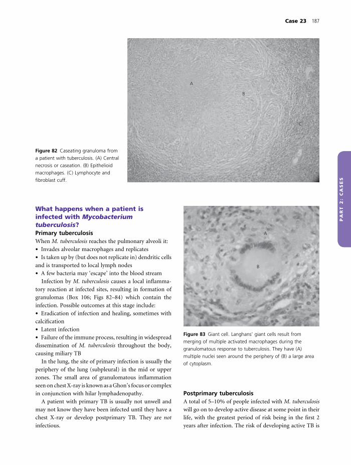
Case 23 187
PA
RT
2:
CA
SE
S
What happens when a patient is infected with Mycobacterium tuberculosis?Primary tuberculosisWhen M. tuberculosis reaches the pulmonary alveoli it:
• Invades alveolar macrophages and replicates
• Is taken up by (but does not replicate in) dendritic cells
and is transported to local lymph nodes
• A few bacteria may ‘escape’ into the blood stream
Infection by M. tuberculosis causes a local infl amma-
tory reaction at infected sites, resulting in formation of
granulomas (Box 106; Figs 82–84) which contain the
infection. Possible outcomes at this stage include:
• Eradication of infection and healing, sometimes with
calcifi cation
• Latent infection
• Failure of the immune process, resulting in widespread
dissemination of M. tuberculosis throughout the body,
causing miliary TB
In the lung, the site of primary infection is usually the
periphery of the lung (subpleural) in the mid or upper
zones. The small area of granulomatous infl ammation
seen on chest X-ray is known as a Ghon’s focus or complex
in conjunction with hilar lymphadenopathy.
A patient with primary TB is usually not unwell and
may not know they have been infected until they have a
chest X-ray or develop postprimary TB. They are not
infectious.
Postprimary tuberculosisA total of 5–10% of people infected with M. tuberculosis
will go on to develop active disease at some point in their
life, with the greatest period of risk being in the fi rst 2
years after infection. The risk of developing active TB is
A
B
C
Figure 82 Caseating granuloma from
a patient with tuberculosis. (A) Central
necrosis or caseation. (B) Epithelioid
macrophages. (C) Lymphocyte and
fi broblast cuff.
B
A
Figure 83 Giant cell. Langhans’ giant cells result from
merging of multiple activated macrophages during the
granulomatous response to tuberculosis. They have (A)
multiple nuclei seen around the periphery of (B) a large area
of cytoplasm.
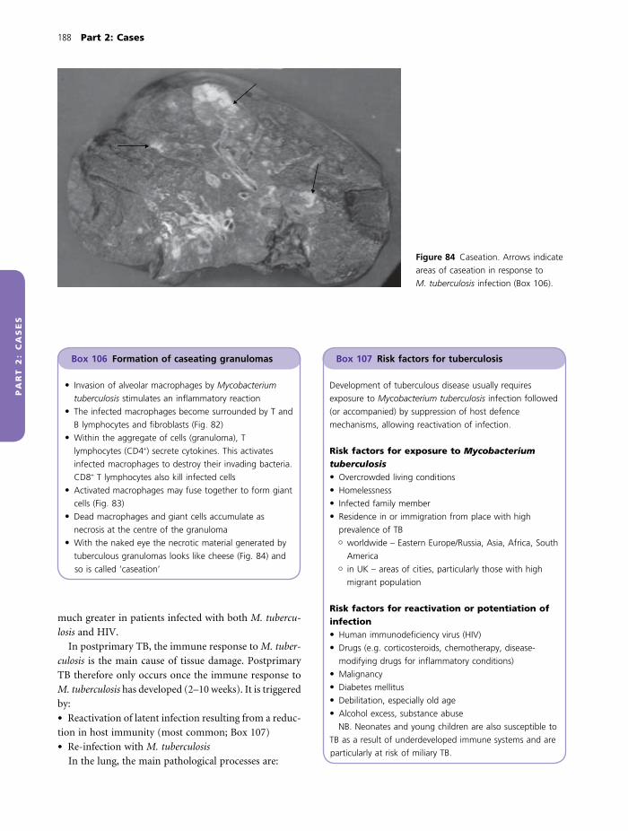
188 Part 2: Cases
PA
RT
2:
CA
SE
S
much greater in patients infected with both M. tubercu-
losis and HIV.
In postprimary TB, the immune response to M. tuber-
culosis is the main cause of tissue damage. Postprimary
TB therefore only occurs once the immune response to
M. tuberculosis has developed (2–10 weeks). It is triggered
by:
• Reactivation of latent infection resulting from a reduc-
tion in host immunity (most common; Box 107)
• Re-infection with M. tuberculosis
In the lung, the main pathological processes are:
Figure 84 Caseation. Arrows indicate
areas of caseation in response to
M. tuberculosis infection (Box 106).
Box 106 Formation of caseating granulomas
• Invasion of alveolar macrophages by Mycobacterium
tuberculosis stimulates an infl ammatory reaction
• The infected macrophages become surrounded by T and
B lymphocytes and fi broblasts (Fig. 82)
• Within the aggregate of cells (granuloma), T
lymphocytes (CD4+) secrete cytokines. This activates
infected macrophages to destroy their invading bacteria.
CD8+ T lymphocytes also kill infected cells
• Activated macrophages may fuse together to form giant
cells (Fig. 83)
• Dead macrophages and giant cells accumulate as
necrosis at the centre of the granuloma
• With the naked eye the necrotic material generated by
tuberculous granulomas looks like cheese (Fig. 84) and
so is called ‘caseation’
Box 107 Risk factors for tuberculosis
Development of tuberculous disease usually requires
exposure to Mycobacterium tuberculosis infection followed
(or accompanied) by suppression of host defence
mechanisms, allowing reactivation of infection.
Risk factors for exposure to Mycobacterium tuberculosis• Overcrowded living conditions
• Homelessness
• Infected family member
• Residence in or immigration from place with high
prevalence of TB� worldwide – Eastern Europe/Russia, Asia, Africa, South
America� in UK – areas of cities, particularly those with high
migrant population
Risk factors for reactivation or potentiation of infection• Human immunodefi ciency virus (HIV)
• Drugs (e.g. corticosteroids, chemotherapy, disease-
modifying drugs for infl ammatory conditions)
• Malignancy
• Diabetes mellitus
• Debilitation, especially old age
• Alcohol excess, substance abuse
NB. Neonates and young children are also susceptible to
TB as a result of underdeveloped immune systems and are
particularly at risk of miliary TB.

Case 23 189
PA
RT
2:
CA
SE
S
• Consolidation or cavitation. This usually occurs in the
lung apices or superior segments of the lower lobes as
these have higher oxygen tension which supports bacte-
rial growth. Processes include:� further formation of granulomas� necrosis and caseation, causing lung cavities� fi brosis
• Pleural infection. This may cause a pleural effusion
and/or thickening
• Bronchiectasis. Obstruction of large airways by
enlarged lymph nodes can cause pulmonary collapse and
bronchiectasis
• Miliary tuberculosis. This affects the whole body. In the
lung it may be seen on chest X-ray as multiple small
nodules throughout both lung fi elds. This form of TB is
particularly serious and needs urgent treatment as it
carries 20% mortality even with urgent treatment
Patients with consolidation, cavitation or bronchiec-
tasis may have cough or haemoptysis. Patients with
extensive parenchymal disease or a large pleural effusion
may be breathless. Patients may also have systemic fea-
tures of disease such as lethargy, fevers, sweats and weight
loss.
Patients with active pulmonary disease may shed M.
tuberculosis into their lung secretions. These patients are
infectious.
KEY POINT
There is a crucial difference between TB infection and
tuberculous disease:
• Tuberculosis infection. The patient has live M.
tuberculosis organisms somewhere in their body but
their immune system is controlling the infection
effectively and they are not unwell or infectious
• Tuberculous disease. This usually occurs when the
immune system becomes weakened and latent infection
is reactivated. This results in tissue destruction and the
patient becomes unwell. If the lung is affected they may
be infectious
Armed with this information, what other features would you ask about in the history to support your diagnosis of pulmonary tuberculosis?You now need to determine whether Mrs Ahmed may
have been exposed to TB and whether she has any risk
factors for reactivation of infection (Box 107).
Mrs Ahmed is originally from Somalia and came to the UK 3
years ago shortly after marrying. She now has a 2-year-old
child. She is not aware of any previous exposure to TB or
possible immunodefi ciency.
What are your concerns when taking a full history from Mrs Ahmed?Patients have a right to confi dentiality. Mrs Ahmed is
unable to speak English and her husband is acting as an
interpreter. There may be information relating to her
condition that she does not wish to share with him. This
is highlighted by the diffi culty of taking a history of pos-
sible HIV exposure in these circumstances.
How might you address these diffi culties?One option is to speak to Mrs Ahmed at a separate time
without her husband present. An independent Somali
interpreter could be used who is either present in person
or on a three-way phone line using a service such as
Language Line.
Mrs Ahmed agrees to be examined. Her temperature is
37.6°C. There is no evidence of lymphadenopathy. On
auscultation there are crepitations over the right upper
zone of her chest. No other abnormalities are
identifi ed.
Does her examination yield any new information?The pyrexia fi ts with a diagnosis of TB or other respira-
tory infection. Right upper zone crepitations would be
consistent with consolidation or other pathology at this
site.
What initial investigations would you perform?The aim of initial investigation is to look for evidence to
support your clinical diagnosis of pulmonary TB and
rule out other diagnoses including pneumonia, bronchi-
ectasis or malignancy.
• Chest X-ray. This would be particularly useful to look
for features of TB or other lung disease
• Full blood count and infl ammatory markers. These
would be carried out to look for:� raised neutrophil count and C-reactive protein
(CRP) consistent with bacterial infection� anaemia and raised infl ammatory markers of chronic
disease such as TB

190 Part 2: Cases
PA
RT
2:
CA
SE
S
� abnormal cell counts consistent with haematological
malignancy or immunosuppression
• Biochemical profi le. To look for abnormalities in renal,
hepatic or bone profi les – sites that might be affected by
TB
Mrs Ahmed’s chest X-ray is shown in Fig. 85.
Her blood test results were as follow:
Normal range
Haemoglobin 9.8 g/dL (12–16 g/dL)
Mean cell volume (MCV) 84 fl (80–96 fl )
White cell count 10.8 × 10⁹/L (4–11 × 109/L)
Neutrophils 8.2 × 10⁹/L (3.5–7.5 × 10⁹/L)
Lymphocytes 0.8 × 10⁹/L (1.5–4 × 10⁹/L)
Platelets 220 × 109/L (150–400 × 109/L)
Erythrocyte sedimentation 57 mm/h (1–20 mm/h)
rate (ESR)
CRP 77 mg/L (<10 mg/L)
Sodium 131 mmol/L (135–145 mmol/L)
Potassium 4.1 mmol/L (3.5–4.8 mmol/L)
Liver/bone/renal profi les Normal
How do you interpret these investigations?Chest X-rayThe striking abnormalities are:
• Consolidation, which is dense white shadowing con-
taining black air bronchograms
• Cavitation of the lung within the consolidation, seen
as a large black hole
• The abnormality is in the right upper lobe, defi ned at
the bottom of the shadow by the horizontal fi ssure
Blood testsAbnormalities identifi ed are:
• Normocytic anaemia which can be caused by chronic
disease such as TB
• Mildly elevated neutrophil count but low lymphocyte
count, which could indicate suppression of cell-mediated
immunity
• Mildly elevated CRP and ESR, indicating underlying
infl ammation
• Hyponatraemia (low sodium). Tuberculosis, pneumo-
nia and lung abscess can cause syndrome of inappro-
priate antidiuretic hormone secretion (SIADH), with
dilution of plasma sodium by retention of excess water
How do these results affect your differential diagnosis?Cavitating upper lobe consolidation on chest X-ray is
highly suspicious for TB.
Other causes of cavitation (Box 108) are less likely as
staphylococcal or klebsiella pneumonia would cause
more systemic upset, lung cancer is unlikely in a 25-year-
old non-smoker and purulent sputum and weight loss
point away from a pulmonary embolus.
C
Figure 85 Chest X-ray. The arrow indicates air bronchograms
within consolidation of the right upper lobe. (C) indicates a
cavity within the consolidated right upper lobe.
Box 108 Causes of cavitating lung disease
A lung cavity is an abnormal hole in the lung tissue where
lung parenchyma has been destroyed. Causes include the
following.
Cancer• Primary lung cancer
• Lung metastases
Infection• Tuberculosis
• Bacterial pneumonias (Staphylococcus spp., Klebsiella
spp.)
Infarction• Pulmonary embolus
Lung cavities can become infected with bacteria (abscess),
or with Aspergillus, which can grow into a fungal ball
called a mycetoma.

Case 23 191
PA
RT
2:
CA
SE
S
What tests could confi rm the diagnosis of pulmonary tuberculosis?Sputum analysisThree morning sputum samples should be sent for:
• Direct microscopy with specifi c staining. The sample can
be stained with Ziehl–Neelsen stain (red) or auramine
(fl uorescent). The bacteria have lipid-rich cell walls,
which require phenol to make them permeable to dyes.
This means that once they are stained the colour cannot
be removed by acidic or alcohol washes. If these acid-fast
bacilli (AFB) are seen on microscopy the sample is des-
ignated ‘smear positive’, which indicates high infectivity
• Culture. All samples are sent for culture:� smear-positive samples are cultured to determine
whether the mycobacterium is tuberculosis or an atyp-
ical form and to determine antibiotic sensitivities to
guide treatment� smear-negative samples may still contain AFB in
numbers too small to be seen. The absence of M. tuber-
culosis needs to be confi rmed by prolonged culture
• Microscopy, culture and sensitivity to detect the pres-
ence of other organisms that could cause pneumonia
with cavitation (Box 108)
Tuberculosis skin testThis is also known as the tuberculin test and detects
immunological evidence of infection with M. tuberculo-
sis. The antigen used is tuberculin, a glycerine extract of
the tubercle bacillus which is prepared as a purifi ed
protein derivative. The test is performed by:
• Intradermal injection of a standard dose of tuberculin
into the forearm away from visible blood vessels using
one of two methods:� Mantoux test (named after the French physician
who developed it). A single needle is used to inject
tuberculin
� Heaf test (named after a British physician). Six tiny
needles in a circle are used to inject tuberculin
• Measurement of the delayed-type hypersensitivity
reaction to tuberculin:� Mantoux test is read 48–72 h after tuberculin
injection� if there has been previous exposure to bacterial
protein, an immune response will be mounted around
the injection site� the reaction is quantifi ed by measuring the diameter
of induration (palpable raised hardened area) across
the forearm (perpendicular to the long axis) in milli-
meters. Absence of induration should be recorded as
‘0 mm’. Erythema (redness) should not be measured.� the tuberculin skin test detects an immune response
to TB antigen. It is therefore positive in those with
active disease, latent infection, treated (cured) TB or
those who have received vaccination against TB.
The results must therefore be interpreted with care
(Table 10).
Mrs Ahmed gives three early morning sputum samples and
they are all smear-negative for AFB on microscopy. She was
not sure whether she had previously had a BCG vaccination,
but on examination she did not have a scar on the upper
arm or lateral thigh region. Her Mantoux test was read at
48 h and measured as having produced induration of 2 mm.
What is BCG vaccination?BCG stands for bacille Calmette–Guérin which is a
live attenuated vaccine against TB. The vaccine was
developed to confer immunity to infection with M.
tuberculosis and prevent tuberculous disease. It is usually
given into the upper arm around the insertion of the
deltoid muscle. However, the vaccine is not universally
effective:
Table 10 Interpreting the tuberculin skin test (Mantoux).
Mantoux induration* <4 mm 4–15 mm >15 mm
Result Negative Positive Strongly positive
Possible interpretations No TB exposure or vaccination Previous vaccination
If no vaccination indicates TB
infection
Likely infection
Lack of immune response despite TB
exposure or vaccination (e.g. HIV)
*Using 10 tuberculin units (0.1 mL 100 TU/mL [i.e. 1 : 1000 concentration]).

192 Part 2: Cases
PA
RT
2:
CA
SE
S
• Not everyone who is vaccinated develops an immune
response to tuberculin
• Those who do develop an immune response to tuber-
culin are not all protected against TB
• Even where there is an effect, the duration is not
known
• There is no evidence it is effective in people >35
years
Vaccination also interferes with the use of tuberculin skin
tests to look for TB infection.
In view of the limitations of BCG vaccination it is
no longer given to everyone in the UK, but is used selec-
tively in:
• Tuberculin-negative new immigrants from high preva-
lence countries aged ≤35 years
• Tuberculin-negative household contacts of pulmonary
TB
• Neonates of ethnic minority groups (e.g. Indian Sub-
continent or Black African)
• Tuberculin-negative, previously unvaccinated health
care workers
• Neonates born in areas of the UK with high TB preva-
lence (>40/100,000 cases per year)
How do you interpret Mrs Ahmed’s test results?So far tests have not confi rmed the diagnosis of TB as
her three sputum samples are smear-negative and her
Mantoux test is negative.
Are these test results surprising?Her history and chest X-ray are strongly indicative
of TB. The results are surprising but TB still remains
the most likely diagnosis. False negative sputum
microscopy can occur if the bacterial load is low or the
sample is taken poorly. A false negative Mantoux test
could indicate underlying immunosuppression (e.g.
HIV).
What other investigations would help to make a diagnosis?• Computed tomography (CT) scan of her chest to char-
acterize the abnormality seen on X-ray and exclude less
likely diagnoses including cancer and pulmonary
embolus
• Sputum induction, i.e. physiotherapy to induce cough-
ing after inhalation of hypertonic saline to obtain samples
from deep in the lung
• Flexible bronchoscopy to sample lower airway secre-
tions if sputum induction is unhelpful in a further
attempt to isolate AFB
• HIV test
Mrs Ahmed’s chest CT confi rms consolidation and cavitation
in the right upper lobe (Fig. 86), consistent with pulmonary
TB. She also has mediastinal lymph node enlargement.
Sputum induction was unsuccessful. Bronchoscopic
appearances were unremarkable, but microscopy of
washings from the right upper lobe identifi ed M.
tuberculosis, which was fully sensitive to antituberculous
treatment when cultured.
After bronchoscopy Mrs Ahmed commences quadruple
antituberculous therapy with rifampicin, isoniazid,
pyrazinamide and ethambutol.
What else must be done now a diagnosis of tuberculosis has been made?• Tuberculosis is a notifi able disease, which means that
there is a statutory requirement to inform the local
‘proper offi cer’, usually the Consultant in Communicable
Disease Control, of any cases that have occurred. This
information is collected by the Health Protection Agency
and allows detection of TB outbreaks and monitoring of
TB management
• People close to Mrs Ahmed may have become infected
with M. tuberculosis. Her contacts must now be identifi ed,
traced, tested and treated as necessary
Mrs Ahmed is counselled for and consents to an HIV test
which is found to be positive.
Figure 86 Chest CT.

Case 23 193
PA
RT
2:
CA
SE
S
What is the signifi cance of a positive HIV test?A positive HIV test indicates that Mrs Ahmed has anti-
bodies to and is infected with the human immunodefi -
ciency virus (HIV).
HIV is a retrovirus that infects immune cells, particu-
larly the CD4 (or T helper) lymphocyte. It replicates in
and damages these cells:
• Directly during the release of virus from the cell
• By inducing apoptosis
• By marking the cell for destruction by other immune cells
As a consequence of HIV the CD4 count is reduced
and this impairs cell-mediated immunity, increasing sus-
ceptibility of the affected person to infection with oppor-
tunistic pathogens and to tumour growth. Mrs Ahmed
may have reactivated latent TB as her cell-mediated
immunity became impaired by HIV.
How might Mrs Ahmed have contracted HIV?HIV infection is very common in sub-Saharan Africa,
although prevalence rates in Somalia, where Mrs Ahmed
comes from, are relatively low. HIV is spread from person
to person by contact with body fl uids. The most common
modes of transmission are:
• Sexual contact
• Parenteral exposure to infected needles or blood products
• Vertical transmission from mother to infant during
delivery or breastfeeding
Why was Mrs Ahmed counselled before undergoing an HIV test?When HIV was fi rst discovered in the 1980s it was
untreatable and the only benefi t in knowing the diagno-
sis was reduced risk of transmission of virus to others. By
contrast, there were many disadvantages to testing includ-
ing concerns around confi dentiality, insurance and legal
issues as well as the negative emotional and social conse-
quences of a positive result.
Testing has become more important since the develop-
ment of highly active antiretroviral therapy (HAART),
which prolongs survival and reduces morbidity from
HIV. However, a positive HIV test can still have consider-
able physical, emotional and social implications and
counselling before testing is still practiced.
How does HIV affect the respiratory system?The effects of HIV on the respiratory system depend on
the stage of the illness (Table 11). Once decline in cell-
mediated immunity has begun the major respiratory
infections associated with HIV are TB and pneumocystis
pneumonia (Box 109).
Outcome. Mrs Ahmed is referred to an infectious diseases
consultant who takes over her care. Her TB is treated fi rst
before she is given HAART for her HIV because of potential
drug interactions. She improves rapidly on antituberculosis
quadruple therapy and is discharged home to continue
treatment. Contact and family testing for TB and HIV are
planned.
Box 109 Pneumocystis pneumonia
CausePneumocystis pneumonia is caused by the fungal organism
Pneumocystis jirovecii (not P. carinii, a similar organism
which affects rats). Despite this it is usually known as
Pneumocystis carinii pneumonia (PCP). Pneumocystis is
widespread in the environment and is not a pathogen to
healthy individuals, but causes lung infection in people
who are immunocompromised.
PathologyInfection causes thickening of the alveoli and alveolar
septae. This causes severe hypoxia which can be fatal if
untreated.
Clinical featuresPatients experience fever, cough and breathlessness. On
examination they may be extremely hypoxic without chest
signs or with inspiratory crepitations and wheezes.
DiagnosisSuccessful diagnosis is more likely if clinicians maintain a
high index of suspicion for PCP in immunocompromised
patients.
A chest X-ray may show perihilar haziness resulting from
alveolar thickening (Fig. 87).
Diagnosis is confi rmed by visualization of the organism
by microscopy of induced sputum or bronchial washings.
TreatmentPCP is treated with co-trimoxazole, a combination of
trimethoprim and sulfamethoxazole antibiotics. Alternative
drugs include pentamidine, trimethoprim-dapsone,
clindamycin, primaquine and atovaquone.
Patients with hypoxia and moderate to severe PCP are
given corticosteroids to reduce alveolar infl ammation and
hasten recovery.

194 Part 2: Cases
Figure 87 Pneumocystis pneumonia.
CASE REVIEW
Mrs Ahmed is referred for urgent assessment of fever,
weight loss and haemoptysis. Her chest X-ray demon-
strates consolidation and cavitation in the right upper
lobe. As TB seems likely she is admitted to a side room
to avoid spread of infection. Initial investigations do not
confi rm TB and her negative tuberculin test raises the
possibility of a false negative result because of coexistent
HIV infection. Subsequently, M. tuberculosis is seen in
bronchial washings and she is started on quadruple anti-
tuberculous treatment. She is also found to be HIV
positive and is referred to the infectious diseases consul-
tant for consideration of HAART.
Table 11 Respiratory effects of HIV infection.
Illness stage Respiratory effect
Acute HIV infectionInitial infection with seroconversion Flu-like illness with pharyngitis, lymphadenopathy and fever
LatencyStage I*
Stable CD4 count and HIV viral load No effect
Stage II* Recurrent upper respiratory tract infections including sinusitis, pharyngitis, bronchitis
Stage III* Pulmonary tuberculosis
Bacterial pneumonia
Stage IV* AIDS
Opportunistic lung infections including candidiasis, pneumocystis pneumonia
Kaposi’s sarcoma
*World Health Organization (WHO) classifi cation.
Progressive decline in
CD4 count and
increase in HIV load�

Case 23 195
PA
RT
2:
CA
SE
S
KEY POINTS
• Tuberculosis should be suspected in all patients presenting
with night sweats, weight loss or haemoptysis
• If a patient is suspected of having active pulmonary TB they
should be cared for in a side room to prevent cross-
infection
• Risk factors for TB include:� previous exposure� impaired immunity, reactivating previous infection
• Initial diagnostic tests for pulmonary TB should include:� chest X-ray� three early morning sputum samples for microscopy and
culture� tuberculin skin testing
• If these are unhelpful other useful tests for pulmonary TB
include:
� induced sputum for microscopy and culture� fi breoptic bronchoscopy with bronchial washings for
microscopy and culture
• Once the diagnosis of pulmonary TB has been made:� the patient should be started on appropriate treatment� the case must be notifi ed to the Health Protection
Agency via the local offi cer� contact tracing should be performed to identify, screen
and treat close contacts as necessary
• Co-infection with HIV and M. tuberculosis is common and
HIV testing should be considered in all patients with TB
• HAART prolongs life and reduces morbidity in patients
with HIV
• The most common pulmonary manifestations of AIDS are
TB and pneumocystis pneumonia

PA
RT
2:
CA
SE
S
A 44-year-old man commencing treatment for tuberculosis
Case 24
Mr Stanislav Bulovski is a 44-year-old man who has posterior
cervical lymphadenopathy. Histological examination of a fi ne
needle aspirate from a lymph node revealed acid-fast bacilli
(Fig. 88). Mycobacterial culture is not yet available. His chest
X-ray showed an old granuloma, consistent with tuberculosis
(TB) infection. Fibreoptic bronchoscopy was unremarkable and
mycobacteria were not seen on bronchial washing microscopy.
He is HIV negative and is not immunosuppressed.
How do you interpret this information?Mr Bulovski has evidence of previous primary TB infec-
tion on chest X-ray. He now has postprimary TB involv-
ing his posterior cervical nodes, but no evidence of
pulmonary TB. This is extrapulmonary TB (Box 110).
If acid-fast bacilli were seen on histology why was the lymph node also sent for culture?Culture allows differentiation of Mycobacterium tubercu-
losis from other members of the Mycobacterium species
(Box 111) and takes 20–56 days. Once the Mycobacterium
has been formally identifi ed, the organism can be tested
for sensitivity to antimycobacterial drugs which may take
a further 4–6 weeks. The process can be speeded up using
polymerase chain reaction (PCR) amplifi cation to detect
mycobacteria and identify gene mutations associated
with antibiotic resistance.
Should the doctors wait for culture results before starting treatment?A presumptive diagnosis of TB can be made from the
clinical features and investigation results and treatment
should be started promptly. Treatment can then be mod-
ifi ed once culture and sensitivity results are available.
Is Mr Bulovski likely to infect his family with tuberculosis?Tuberculosis is usually spread by respiratory droplets and
patients with extrapulmonary TB are rarely infectious.
However, care should be taken with any suppurating
extrapulmonary TB as TB can be spread from these secre-
tions. Mr Bulovski has no evidence of pulmonary TB and
bronchial washings are negative for mycobacteria, so he
is unlikely to infect his family. However, contact tracing
should still be performed as he may have acquired his
infection from another family member.
In clinic the TB specialist explains the diagnosis and need for
prolonged therapy to cure his TB. Mr Bulovski is anxious to
start therapy and confi dent that he will be able to stick to
the treatment regime unaided. In clinic he has blood taken
to test his liver and renal function and is sent for eye tests.
He is then started on:
Rifater (rifampicin, isoniazid and pyrazinamide), 6 tablets
daily
Ethambutol 1050 mg/day
Why is he treated with multiple antituberculous drugs?The aims of treatment are to:
• Eradicate mycobacteria, curing the patient
• Prevent transmission of infection
Metabolic statesMycobacterium tuberculosis exists in several metabolic
states:
• Rapid growth – this form is responsible for active
infection
• Intermittent growth
• Slow growth – this form is responsible for latent
infection
Different drugs are effective against bacteria in differ-
ent metabolic states. At least two different drugs must be
used to kill all bacteria and eradicate infection.
Emergence of resistanceMycobacteria resistant to antituberculosis drugs arise
spontaneously during treatment. If a single drug is used,
196

Case 24 197
PA
RT
2:
CA
SE
S
resistant organisms survive and vulnerable tubercle
bacilli are killed. The use of multiple drugs ensures that
bacilli emerging with resistance to one drug are killed by
other drugs in the regimen, ensuring that all tubercle
bacilli are eradicated.
What is the role of each drug in the treatment regimen?• Isoniazid is bacteriocidal against (kills) rapidly divid-
ing bacteria and makes the patient non-infectious within
days of starting treatment
• Rifampicin is bacteriocidal against rapidly growing
and slowly dividing bacteria (persisters) and can cause
complete eradication of tubercle bacilli (sterilization)
• Pyrazinamide eradicates slowly dividing and intracel-
lular bacteria, also contributing to sterilization
• Ethambutol is not particularly effective at killing
tubercle bacilli; however, it prevents emergence of resis-
tance during therapy
Why are three drugs given together as a combination pill?• To aid adherence to the treatment regime by reducing
number of pills taken
• To ensure that three active drugs are taken together, as
separate use could lead to mycobacterial resistance
Why does he have blood taken for liver and renal function testing?Isoniazid, rifampicin and pyrazinamideThese cause hepatic toxicity (Boxes 112–114) and are
eliminated by the liver. Liver function testing before treat-
ment is essential to:
• Establish a baseline to allow detection of drug-related
hepatotoxicity
• Identify underlying liver disease, necessitating reduc-
tion in dosage of drug used or frequent monitoring of
liver function
EthambutolEthambutol (Box 115) is renally excreted, therefore renal
function should be checked to:
• Identify underlying renal disease, necessitating re -
duction in ethambutol dose or use of an alternative
drug
Figure 88 Acid-fast bacilli. Arrows indicate multiple small
dark rods which are the bacilli.
Box 110 Extrapulmonary tuberculosis
Tuberculosis spreads via the blood stream and/or
lymphatics to any extrapulmonary organ or body system,
most commonly the lymph nodes and bones:
• Bones and joints. TB osteomyelitis or arthritis can occur
• Heart. TB can cause constrictive pericarditis
• Gastrointestinal tract. TB of the small intestine can
cause malabsorption. Peritoneal infection can cause
ascites
• Genitourinary system. There may be a tuberculous mass
in the kidney. TB can cause epididymitis (men) or
salpingitis (women)
• Central nervous system. Tuberculous meningitis or
intracranial tuberculomas
• Other. Infections of the skin, eyes and adrenal glands
are described
Box 111 Mycobacterium species
Mycobacteria are a family of slow growing, acid-fast,
aerobic bacilli, classifi ed into three groups according to
their capacity to cause disease:
1 Mycobacterium tuberculosis complex – cause TB� Mycobacterium tuberculosis� Mycobacterium bovis (cattle and can infect humans)
2 Non-tuberculous mycobacteria. Environmental
pathogens found in soil and water that cause a lung
disease resembling TB in immunocompromised patients
and those with chronic lung disease such as
bronchiectasis:� Mycobacterium avium intracellulare� Mycobacterium kansasii
3 Mycobacterium leprae – causes leprosy
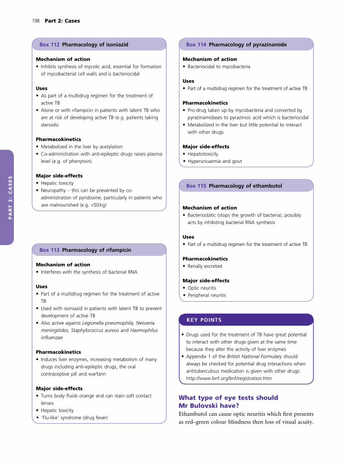
198 Part 2: Cases
PA
RT
2:
CA
SE
S
KEY POINTS
• Drugs used for the treatment of TB have great potential
to interact with other drugs given at the same time
because they alter the activity of liver enzymes
• Appendix 1 of the British National Formulary should
always be checked for potential drug interactions when
antituberculous medication is given with other drugs:
http://www.bnf.org/bnf/registration.htm
What type of eye tests should Mr Bulovski have?Ethambutol can cause optic neuritis which fi rst presents
as red–green colour blindness then loss of visual acuity.
Box 112 Pharmacology of isoniazid
Mechanism of action• Inhibits synthesis of mycolic acid, essential for formation
of mycobacterial cell walls and is bacteriocidal
Uses• As part of a multidrug regimen for the treatment of
active TB
• Alone or with rifampicin in patients with latent TB who
are at risk of developing active TB (e.g. patients taking
steroids)
Pharmacokinetics• Metabolized in the liver by acetylation
• Co-administration with anti-epileptic drugs raises plasma
level (e.g. of phenytoin)
Major side-effects• Hepatic toxicity
• Neuropathy – this can be prevented by co-
administration of pyridoxine, particularly in patients who
are malnourished (e.g. <50 kg)
Box 113 Pharmacology of rifampicin
Mechanism of action• Interferes with the synthesis of bacterial RNA
Uses• Part of a multidrug regimen for the treatment of active
TB
• Used with isoniazid in patients with latent TB to prevent
development of active TB
• Also active against Legionella pneumophila, Neisseria
meningitides, Staphylococcus aureus and Haemophilus
infl uenzae
Pharmacokinetics• Induces liver enzymes, increasing metabolism of many
drugs including anti-epileptic drugs, the oral
contraceptive pill and warfarin
Major side-effects• Turns body fl uids orange and can stain soft contact
lenses
• Hepatic toxicity
• ‘Flu-like’ syndrome (drug fever)
Box 115 Pharmacology of ethambutol
Mechanism of action• Bacteriostatic (stops the growth of bacteria), possibly
acts by inhibiting bacterial RNA synthesis
Uses• Part of a multidrug regimen for the treatment of active TB
Pharmacokinetics• Renally excreted
Major side-effects• Optic neuritis
• Peripheral neuritis
Box 114 Pharmacology of pyrazinamide
Mechanism of action• Bacteriocidal to mycobacteria
Uses• Part of a multidrug regimen for the treatment of active TB
Pharmacokinetics• Pro-drug taken up by mycobacteria and converted by
pyrazinamidases to pyrazinoic acid which is bacteriocidal
• Metabolized in the liver but little potential to interact
with other drugs
Major side-effects• Hepatotoxicity
• Hyperuricaemia and gout

Case 24 199
PA
RT
2:
CA
SE
S
He should therefore have testing of his colour vision
(Ishihara testing) as well as visual acuity testing with a
Snellen chart.
What advice would you give him before sending him home to start treatment?He should look out for any:
• Signs of hepatic toxicity (e.g. fever, malaise, vomiting
or jaundice)
• Change in his vision
• Other unexpected effects
If these occur he should discontinue therapy and seek
immediate medical advice.
He should be given a contact number for emergencies
and a follow-up appointment for 2 weeks’ time.
Mr Bulovski returns for follow-up 2 weeks later. He fi nds he
is able to tolerate his medication and is not aware of any
side-effects apart from nausea. He has no problems with his
eyesight. He asks the TB nurse how long he must stay on
treatment for.
What assessment is required at follow-up during treatment for tuberculosis?It is important to establish:
• Treatment effi cacy. This can be monitored by checking
symptoms and resolution of active disease. In Mr
Bulovski’s case his lymphadenopathy would be expected
to resolve
• Adherence to treatment regime (Box 116):� this is indicated by clinical improvement in TB
symptoms and signs� it can be checked by asking the patient and perform-
ing tablet counts� other checks include examination of urine (should
be orange with rifampicin) or measurement of plasma
drug levels
• Drug side-effects. Patients should be asked about
changes in eyesight and liver function should be
checked
How would you answer his question?• Initial phase. Patients take four drugs until culture and
sensitivities are available, which usually takes about 2
months
• Continuation phase. If sensitivity testing shows no bac-
terial resistance to isoniazid or rifampicin, treatment is
continued with these two drugs alone for a further 4
months and pyrazinamide and ethambutol are stopped
Is the duration of treatment the same for all tuberculosis infections?Treatment duration may be affected by:
• Drug resistance
• Site of TB (e.g. prolonged treatment is required for
CNS disease)
Outcome. Mr Bulovski tolerated antituberculous therapy.
After 2 months lymph node culture grew M. tuberculosis
fully sensitive to therapy. His four-drug regime was reduced
to rifampicin and isoniazid and he completed the course of
treatment without signifi cant toxicity. His family were screened
and were all negative for TB. At the completion of treatment
his lymphadenopathy had resolved and he was discharged.
Box 116 Defi nitions of compliance, concordance and adherence
These terms refer to the prescriber–patient interaction
around the prescribing of medicines.
Compliance implies an old fashioned consultation
model, sometimes referred to as ‘paternalistic’ (fatherly),
where the prescriber tells the patient what to do. The
patient then does what they are told (complies) or does
not (non-compliant).
Concordance indicates an alternative consultation
model. The prescriber and patient both come to the
consultation with valid knowledge, attitude and beliefs.
They discuss treatment options and come to an agreement
(concordance) on the way forward.
Adherence is a measure of whether the patient sticks
to the agreed treatment regimen. These days patients are
‘adherent’ or ‘non-adherent’ to treatment, rather than
‘compliant’ or ‘non-compliant’.
CASE REVIEW
Mr Bulovski presented with postprimary TB affecting his
posterior cervical lymph nodes. Prior to treatment his
liver and renal function were checked and he underwent
eye tests for red–green colour vision and visual acuity. He
was started on rifampicin, isoniazid, pyrazinamide and
ethambutol. He did not have severe side-effects of treat-
ment and was able to complete 2 months on these four
drugs, before reducing treatment to rifampicin and iso-
niazid for a further 4 months. At the end of treatment his
lymphadenopathy had resolved and he was discharged.

200 Part 2: Cases
PA
RT
2:
CA
SE
S
KEY POINTS
• Mycobacterium tuberculosis may spread from the site of
primary infection in the lung to other sites by blood or
lymphatics
• The most common sites for extrapulmonary TB infection
are lymph nodes and bone
• A diagnosis of TB is likely if acid-fast bacilli and caseating
granuloma are seen on microscopy of fi ne needle aspirates
• Culture is required to:� confi rm that acid-fast bacilli are M. tuberculosis and not
another member of the species� test the bacilli for sensitivity to antimycobacterial drugs
• TB culture takes several weeks, therefore treatment should
be started while results are awaited
• In the initial phase (fi rst 2 months) four drugs are used for
treatment (mnemonic RIPE):� rifampicin, isoniazid, pyrazinamide, ethambutol
• In the continuation phase (further 4 months), rifampicin
and isoniazid are continued to complete treatment
• Multiple drugs are required to ensure eradication of
bacteria in different growth phases and to prevent
emergence of resistance
• Major side-effects of treatment are hepatic toxicity
(rifampicin, isoniazid, pyrazinamide), visual disturbance
(ethambutol) and neuropathy (isoniazid, ethambutol)
• Liver and renal function, red–green colour vision and
visual acuity are checked prior to starting treatment
• Patients are monitored for disease response, treatment
adherence and drug side-effects during treatment
• Treatment for TB is usually for 6 months, but may be
given for longer if it is infecting non-pulmonary sites or
is multidrug resistant

PA
RT
2:
CA
SE
S
A 32-year-old man who absconds from directly observed therapy for tuberculosis
Case 25
Mr Ewan Evans, a 32-year-old man, is admitted to hospital
with fevers and a cough productive of brown sputum. His
chest X-ray (Fig. 89) shows right upper lobe shadowing (A)
and hilar lymphadenopathy (B) and his sputum is smear-
positive for acid-fast bacilli. The sample rapidly cultures
Mycobacterium tuberculosis which is fully sensitive to
treatment. He undergoes an HIV test which is negative. His
case is notifi ed and he is commenced on quadruple
antituberculosis therapy with rifampicin, isoniazid,
pyrazinamide and ethambutol.
How can you fi nd out whether he has infected other people with tuberculosis?When a diagnosis of tuberculosis (TB) is made, contact
tracing is performed, usually by nurse specialists in TB,
with the aims to identify people with:
• Active disease who require treatment
• Latent infection, who may require prophylactic treat-
ment to prevent active disease
• No evidence of infection but who may benefi t from
bacille Calmette–Guérin (BCG) vaccination
The infected person is referred to as the index case. The
‘index case’ is most likely to have infected people they are
very close to (e.g. those they share accommodation with).
These people are traced and contacted fi rst and offered
screening. If more than 10% of these close contacts are
positive for TB, the next ‘ripple’ of less frequent contacts
is screened (e.g. people who work with the index case;
Fig. 90). If more than 10% of these contacts are positive,
the tracing and screening is extended further into more
casual contacts. This can be referred to as a ‘stone in the
pond’ or patient-centred model. Tracing and screening
stop at the level of contacts where <10% have been
infected.
How is contact tracing coordinated?Specialist nurses performing contact tracing usually
work as part of an NHS TB service in a local hospital,
primary care or a local Health Protection Unit (HPU;
Fig. 91). Services are organized differently depending on
local needs and expertise throughout the country. The
NHS team notifi es each clinical case to the Consultant
in Communicable Disease Control in the local Health
Protection Unit (HPU). The involvement of the HPU
allows coordination of individual NHS providers (hospi-
tals and primary care) at local, regional or national
level as appropriate to investigate and manage disease
outbreaks.
How are contacts screened?Screening is performed with tuberculin skin testing
(Mantoux) and chest X-ray. The results can be inter-
preted as follows:
• Positive Mantoux and abnormal chest X-ray and/or
clinical features of TB – possible active disease
• Positive Mantoux and normal chest X-ray and/or no
clinical features of TB – possible latent disease or previ-
ous BCG vaccine
• Negative Mantoux and normal clinical assessment –
not infected with TB
Although this appears clear-cut it can be more diffi cult
in practice. Mantoux results can be falsely positive as a
result of BCG vaccination or exposure to environmental
mycobacteria or falsely negative because of immunosup-
pression. New γ-interferon (IFN-γ) immunological tests
aim to be more specifi c for Mycobacterium tuberculosis
infection (Box 117) and have been used to diagnose
latent TB.
How are the results of screening used?• Contacts with possible active disease require further
investigation and are likely to need antituberculous
treatment
• Patients with latent disease have a 10–15% chance of
developing active disease. Treatment with isoniazid for 6
months or isoniazid and rifampicin for 3 months has
been shown to reduce the risk of developing active disease
201

202 Part 2: Cases
PA
RT
2:
CA
SE
S
effi cacy for people over 35 years, therefore vaccination is
usually limited to people who are ≤35 years
Mr Evans is interviewed by the TB specialist nurse. He has a
past history of intravenous drug use, has recently been
homeless and is currently staying in a hostel.
A
B
Figure 89 Chest X-ray.
Occasional
Occupational
Household
Indexcase
10% positive
10% positiveContacts
Contacts
Contacts
Figure 90 ‘Stone in the pond’ model used for contact
tracing.
Health Protection Agency
National
National surveillanceand coordination
Regional
Regional officesRegional directorsof public health
Regional surveillanceand coordination
28 HealthProtection Units
Local directors
Distribution of workdepends on local service
organization
Delivery of clinical careContact tracing and screening
Delivery of clinical careContact tracing and screening
NHS clinical TBservices
Local disease surveillanceLocal coordination of disease response
Notification of tuberculosis
in the future. As hepatotoxicity of drugs increases with
age, the risk of treatment of latent TB begins to outweigh
the benefi ts in older patients, so only those ≤35 years are
usually treated
• Contacts who have not been infected with TB should
be offered the BCG vaccine. There is no evidence of BCG
Figure 91 Organization of services for
management and control of
tuberculosis.

Case 25 203
PA
RT
2:
CA
SE
S
What concerns do his social circumstances raise?• He lives in a hostel where he has come into contact
with a large number of people who may have become
infected. Some of these may be particularly susceptible to
infection because of immunosuppression (e.g. with HIV)
• His lifestyle is unstable and he will not reliably be able
to complete the 6-month course of antituberculous treat-
ment required to cure his disease
What should be done to prevent him from infecting other people?As his social circumstances will not permit some form of
‘home isolation’, he needs to remain in hospital until he
is unlikely to infect others. The minimum treatment time
for fully sensitive acid-fast bacilli in the lungs to be killed
and for the patient to become non-infectious is generally
accepted as 2 weeks. Where it is essential (as in this case)
to ensure the patient is non-infectious, three smear-
negative sputum samples produced on separate occasions
may be required before discharge.
How will his circumstances affect contact tracing?He is likely to have come into contact with many people
when homeless or living in the hostel who will now be
hard to trace. The National Institute for Clinical Excel-
lence (NICE) recommends active case fi nding for TB in
homeless people using chest X-rays taken opportunisti-
cally and in symptomatic patients. Contact tracing is
likely to be diffi cult and may require input from the
Health Protection Agency.
The TB nurses arrange contact tracing at the hostel. They
screen 20 staff and fi nd that six have possible evidence of
M. tuberculosis infection, although none have active disease.
The six staff with possible M. tuberculosis infection undergo
IFN-γ testing, which identifi es four who receive treatment for
latent infection to prevent active disease. On discussion with
the local HPU, the clinical team decide to offer chest X-rays
to all hostel residents, irrespective of contact with Mr Evans.
Two cases of active disease are identifi ed and treated.
Why is it important that Mr Evans receives adequate antituberculosis treatment?Completion of antituberculosis therapy is very important
for two main reasons:
1 Incomplete treatment may allow tubercle bacilli to
mutate and become multidrug-resistant. This is much
more diffi cult to treat than fully sensitive TB
2 Relapse of the disease can lead to a resurgence of infec-
tivity which may become a public health concern
How could you ensure Mr Evans receives the full course of antituberculosis treatment?Mr Evans could receive directly observed therapy (DOT).
DOT is used when the clinical team are concerned that a
patient may have diffi culty adhering to a treatment
regimen and completing the full course of antitubercu-
lous medication. DOT involves a TB nurse specialist
administering the tablets and watching the patient
swallow them in a hospital clinic. Alternatively, a respon-
sible individual outside the hospital, such as a school
teacher, can perform DOT at a location more convenient
to the patient. Hospital visits for DOT can be minimized
by using treatment regimens where tablets are taken three
times per week.
What other ways are there to ensure that patients take their treatment?• Education, regular support and reminders from health
care workers are important
Box 117 γ-Interferon immunological tests for Mycobacterium tuberculosis
Principles• The test determines the immunological response to
‘early secretion antigen target 6’ (ESAT-6) and culture
fi ltrate protein (CFP-10)
• ESAT-6 and CFP-10 are used because they are expressed
by M. tuberculosis and not by most environmental
mycobacteria or the live attenuated bacillus used in the
BCG vaccine
Methods• A sample of whole blood is taken from the patient
• The antigens (ESAT-6 and CFP-10) are added to the
blood
• Immunological response is measured either by release of
IFN-γ from the whole sample (QuantiFERON-TB Gold) or
by counting individual activated T cells (T.SPOT-TB)
• Research has shown that positive responses to these
immunological tests correlate with degree of exposure
to M. tuberculosis and are much less infl uenced by
previous BCG vaccination
204 Part 2: Cases
PA
RT
2:
CA
SE
S
• Medication is given as combination tablets (e.g. Rifater
contains rifampicin, isoniazid and pyrazinamide) to
make treatment adherence easier
• Patients should be seen and monitored regularly:� tablet counts can be performed to assess whether
medication has been taken� rifampicin stains the urine red, which can be
assessed� blood levels of some antituberculosis drugs can be
checked
Mr Evans remains in hospital for the fi rst 2 weeks of
antituberculosis treatment. His cough improves and he
becomes apyrexial. Once three smear-negative sputum
samples are obtained he is discharged from hospital to
receive DOT as an outpatient. After discharge he attends
for treatment twice, then absconds and is lost to
follow-up.
Three months later, the TB nurse specialists are contacted
by staff from a nearby hospital informing them that Mr
Evans has been admitted to one of their negative pressure
rooms. He is unwell with fevers, weight loss and a
productive cough. His chest X-ray shows multilobar
shadowing. He has informed them that he received some
treatment for TB, but stopped this as he had felt better.
They now suspect that he may have drug-resistant TB.
What is drug-resistant tuberculosis?When bacteria multiply they develop mutations that
may confer resistance to one or more drugs used to
treat TB:
• Resistant organisms are defi ned as those that need four
or more times the usual drug concentration to kill them
or inhibit their growth
• Multidrug-resistant TB (MDR-TB) is infection with
M. tuberculosis resistant to isoniazid and rifampicin,
which are the two most important drugs used in the
treatment of TB
• Extreme drug-resistant TB (XDR-TB) is infection with
M. tuberculosis resistant to usual antituberculous treat-
ment and to three out of six available second line drugs
used for MDR-TB
How do patients acquire drug-resistant tuberculosis?• Fully sensitive TB may become drug-resistant if:
� clinicians prescribe inadequate treatment (e.g. do
not use multiple drugs)
� patients do not adhere to the treatment regimen, as
in Mr Evans’ case
• Patients may become infected by a drug-resistant strain
of M. tuberculosis
Why is multidrug-resistant tuberculosis a major concern?Multidrug-resistant TB is diffi cult to treat because some
or all of the drugs usually used in quadruple therapy are
ineffective. This means that patients:
• Remain infectious for longer
• Are more likely to die and less likely to be cured
• Need alternative drugs that:� are much more expensive and more diffi cult to use
than usual therapy� may be associated with greater toxicity than usual
treatment
The problems are magnifi ed further in XDR-TB, which
may be untreatable.
The treatment of MDR-TB is specialized and complex
and should only be undertaken at recognized centres
with experience of managing such patients.
What is a negative pressure room?This is a room where the ventilation system is des-
igned to remove air from the room to the outside of
the building, creating a negative pressure inside the
room. Air is thus drawn into the room. This prevents
infectious particles escaping from the room into the cor-
ridor and reduces or prevents cross-infection. Any patient
with suspected MDR-TB should be isolated in a negative
pressure room to prevent spread of this infection which
is diffi cult to treat. Staff and visitors entering the room
also need to wear face masks that meet health and safety
criteria.
!RED FLAG
Patients with suspected MDR-TB should immediately be
isolated in a negative pressure room until MDR-TB is ruled
out or confi rmed and treated.
Why did the nearby hospital suspect that Mr Evans had multidrug-resistant tuberculosis?Mr Evans had previous inadequate treatment for TB,
which could have resulted in emergence of drug-resistant

Case 25 205
PA
RT
2:
CA
SE
S
organisms. Other factors in the history associated with
an increased risk of drug-resistant disease include:
1 Contact with another patient known to have drug-
resistant disease
2 Birth in foreign country, particularly sub-Saharan
Africa and the Indian Subcontinent
3 HIV infection
4 Residence in London
5 Age 25–44
6 Male gender
How can multidrug-resistant tuberculosis be treated?Treatment should be started before drug sensitivities are
available. A detailed history and information from other
organizations should be sought to fi nd out what drugs
the patient has already been exposed to. The clinicians
should then start treatment with 3–4 alternative antitu-
berculosis drugs that the patient has not received previ-
ously. When the results of drug resistance testing are
known the regimen can be tailored accordingly. Treat-
ment for MDR-TB is usually longer than for fully sensi-
tive TB and less likely to be successful. Surgery may
sometimes have a place in MDR-TB treatment. For
example, if disease is confi ned to one or two lobes, lobec-
tomy may offer a better chance of cure than continued
drug treatment.
Mr Evans is started on treatment with six antituberculosis
drugs (rifampicin, isoniazid, pyrazinamide, amikacin,
clarithromycin and ciprofl oxacin). On sensitivity testing he
has a strain of M. tuberculosis that is resistant to rifampicin
and isoniazid, but sensitive to the other four drugs, which
are continued. He is advised that he must stay in isolation
until his sputum is culture negative. At this point he
attempts to leave the ward.
What are your concerns around him leaving the ward?He has MDR-TB, is infectious and may spread the
disease to others. He thus poses a public health risk
and should not leave the ward until he is no longer
infectious.
Is there any way he can be prevented from leaving?Discussion and reasoning should be tried fi rst. If all else
fails, Section 38 of the Public Health (control of disease)
Act 1984 can be used to detain him. Section 38 authorizes
the ‘detention for a period specifi ed in the order’ of a
person with TB (or other named contagious diseases).
The hospital has to apply for an order to a local magis-
trate. There is a time limit set against the order but it can
be extended as often as is necessary. If a patient leaves
hospital the court may order that he is brought back to
the hospital. The act does not, however, allow treatment
to be enforced upon the patient.
Outcome. Mr Evans is eventually persuaded to remain in
hospital and continue with treatment. After 2 months his
sputum is culture negative for M. tuberculosis and he is
discharged. He remains under close supervision and this time
successfully completes 9 months of treatment, which results
in cure of his TB, although his chest X-ray shows residual
pulmonary scarring.
before completing treatment, but re-presents 3 months
later with MDR-TB. This time he is required to stay in
hospital until his sputum is culture negative for TB as he
is considered to be a public health risk. He is treated with
second line antituberculosis medication for 9 months and
is cured of his MDR-TB.
CASE REVIEW
Mr Evans is admitted to hospital with smear-positive
pulmonary TB and commences quadruple antituberculosis
therapy. He has a past history of intravenous drug use and
homelessness and is living in a hostel. In view of his
unstable social circumstances he remains in hospital until
his sputum is smear-negative for acid-fast bacilli, then is
discharged to receive DOT in the community. He absconds

206 Part 2: Cases
PA
RT
2:
CA
SE
S
KEY POINTS
• Patients with active pulmonary disease are likely to have
infected close contacts
• Contact tracing should be used to identify contacts of the
‘index case’, who should be invited for screening
• The aims of screening are to identify those:� with active tuberculous disease who require treatment� with latent M. tuberculosis infection who require
prophylactic treatment to reduce the risk of developing
active disease� who are not infected with M. tuberculosis but who
require BCG vaccination
• The core components of contact screening are clinical
assessment, Mantoux testing and chest X-ray. IFN-γ testing
may be used to detect latent TB
• Patients with TB must receive a full course of treatment
with multiple drugs to prevent the emergence of drug-
resistant strains of M. tuberculosis
• MDR-TB is defi ned as resistance to rifampicin and
isoniazid
• Patients with MDR-TB are infectious for longer, more
likely to die and less likely to be cured than patients with
fully sensitive TB
• Patients recognized as being at risk of MDR-TB on
admission to hospital should be isolated in a negative
pressure facility to prevent transmission
• MDR-TB should be treated in recognized centres with
experience in treating this condition
• Section 38 of the Public Health Act 1984 can be used to
detain a patient with TB against their wishes if they are
considered to be a risk to public health, but cannot be
used to enforce treatment
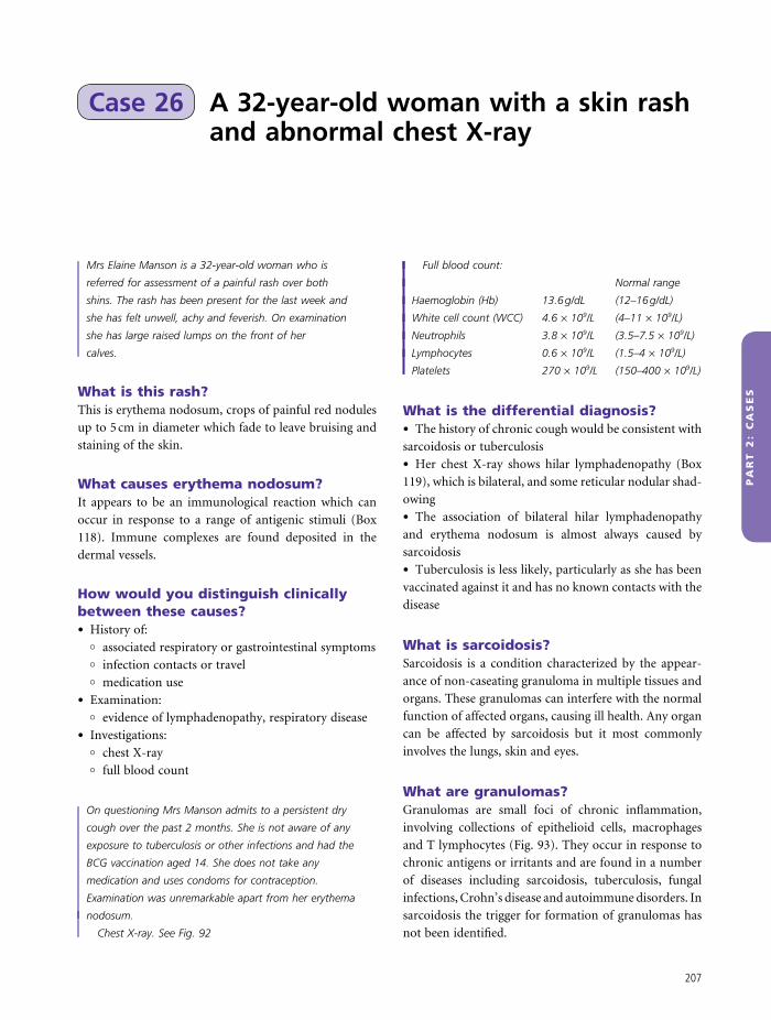
PA
RT
2:
CA
SE
S
A 32-year-old woman with a skin rash and abnormal chest X-ray
Case 26
Mrs Elaine Manson is a 32-year-old woman who is
referred for assessment of a painful rash over both
shins. The rash has been present for the last week and
she has felt unwell, achy and feverish. On examination
she has large raised lumps on the front of her
calves.
What is this rash?This is erythema nodosum, crops of painful red nodules
up to 5 cm in diameter which fade to leave bruising and
staining of the skin.
What causes erythema nodosum?It appears to be an immunological reaction which can
occur in response to a range of antigenic stimuli (Box
118). Immune complexes are found deposited in the
dermal vessels.
How would you distinguish clinically between these causes?• History of:
� associated respiratory or gastrointestinal symptoms� infection contacts or travel� medication use
• Examination:� evidence of lymphadenopathy, respiratory disease
• Investigations:� chest X-ray� full blood count
On questioning Mrs Manson admits to a persistent dry
cough over the past 2 months. She is not aware of any
exposure to tuberculosis or other infections and had the
BCG vaccination aged 14. She does not take any
medication and uses condoms for contraception.
Examination was unremarkable apart from her erythema
nodosum.
Chest X-ray. See Fig. 92
Full blood count:
Normal range
Haemoglobin (Hb) 13.6 g/dL (12–16 g/dL)
White cell count (WCC) 4.6 × 10⁹/L (4–11 × 109/L)
Neutrophils 3.8 × 10⁹/L (3.5–7.5 × 10⁹/L)
Lymphocytes 0.6 × 10⁹/L (1.5–4 × 10⁹/L)
Platelets 270 × 109/L (150–400 × 109/L)
What is the differential diagnosis?• The history of chronic cough would be consistent with
sarcoidosis or tuberculosis
• Her chest X-ray shows hilar lymphadenopathy (Box
119), which is bilateral, and some reticular nodular shad-
owing
• The association of bilateral hilar lymphadenopathy
and erythema nodosum is almost always caused by
sarcoidosis
• Tuberculosis is less likely, particularly as she has been
vaccinated against it and has no known contacts with the
disease
What is sarcoidosis?Sarcoidosis is a condition characterized by the appear-
ance of non-caseating granuloma in multiple tissues and
organs. These granulomas can interfere with the normal
function of affected organs, causing ill health. Any organ
can be affected by sarcoidosis but it most commonly
involves the lungs, skin and eyes.
What are granulomas?Granulomas are small foci of chronic infl ammation,
involving collections of epithelioid cells, macrophages
and T lymphocytes (Fig. 93). They occur in response to
chronic antigens or irritants and are found in a number
of diseases including sarcoidosis, tuberculosis, fungal
infections, Crohn’s disease and autoimmune disorders. In
sarcoidosis the trigger for formation of granulomas has
not been identifi ed.
207

208 Part 2: Cases
PA
RT
2:
CA
SE
S
What is caseation?If necrosis occurs in the centre of the granuloma this
looks soft and cheese-like, so is called caseation (from the
Latin ‘caseus’ for cheese). Caseation is the hallmark of
granulomas caused by tuberculosis. There is little or no
necrosis in the centre of a sarcoid granuloma, so this is
called a non-caseating granuloma.
How do you interpret her full blood count?The major abnormality is a low blood lymphocyte count.
This probably occurs because lymphocytes are concen-
trated in the lungs. It does not appear to cause immuno-
suppression in sarcoidosis patients, as they have no
evidence of increased risk of infection or neoplasms.
Figure 92 Chest X-ray. Arrows indicate the hila, which are
bulky due to lymphadenopathy. There is patchy
‘reticulonodular’ shadowing throughout both lung fi elds due
to parenchymal lung disease. This is seen as multiple small
specks.
Box 118 Causes of erythema nodosum
Infective, e.g.• Streptococcus
• Tuberculosis
• Viral
Infl ammatory• Sarcoidosis
• Infl ammatory bowel disease
Drugs• Sulphonamide antibiotics
• Oral contraceptive pill
Box 119 Causes of hilar lymphadenopathy
• Sarcoidosis (most common cause if bilateral)
• Tuberculosis
• Lymphoma
• Bronchogenic carcinoma
AFigure 93 Non-caseating granuloma.
Compare this non-caseating
granuloma of sarcoidosis to the
caseating granuloma caused by
tuberculosis shown in Fig. 82, p. 187.
The centre of this non-caseating
granuloma is cellular (A), containing
epithelioid cells, macrophages and T
lymphocytes. There is no evidence of
central necrosis, seen as (A)
amorphous material in Fig. 82.

Case 26 209
PA
RT
2:
CA
SE
S
What further investigations would you arrange?The diagnosis is most likely to be sarcoidosis, but
tuberculosis remains a possibility and should be excluded.
Two useful tests can help to distinguish between these
diagnoses:
• Tuberculin skin testing (Mantoux or Heaf). This
should be positive in tuberculosis, but is negative in sar-
coidosis, probably because T lymphocytes are seques-
tered in the lung, interfering with this cell-mediated
immune response
• Fibreoptic bronchoscopy with bronchoalveolar lavage
and biopsies for pathological and microbiological exam-
ination (see Part 1, Approach to the Patient, p. 33)
Mrs Manson’s Mantoux test is negative. At fi breoptic
bronchoscopy her airways looked normal, but
bronchoalveolar lavage, endobronchial and transbronchial
biopsies were performed.
Three days later her pathology results were available.
Bronchoalveolar lavage showed a lymphocytosis and was
smear-negative for acid-fast bacilli. Non-caseating
granulomas, but no acid-fast bacilli, were found in
endobronchial and transbronchial biopsies.
How do you explain the lymphocytosis in her lavage?Increased cell numbers are seen in bronchoalveolar
lavage samples from sarcoidosis patients. There is a
predominance of T lymphocytes (Box 120), particu-
larly CD4 type, and there is an increased CD4 : CD8
ratio.
Why were both endobronchial and transbronchial biopsies taken?Sarcoidosis can involve the mucosa of the large air-
ways, sampled by endobronchial biopsy, or the lung
parenchyma, sampled by transbronchial biopsy. If both
types of biopsy are taken this increases the chance of
making a positive diagnosis of sarcoidosis at fi breoptic
bronchoscopy.
How do you interpret her results?Her results are strongly suggestive of sarcoidosis and
consistently negative for tuberculosis. Her lavage and
biopsies should be cultured for mycobacteria and this
will take around 8 weeks. Negative cultures will pro-
vide further reassurance that she does not have
tuberculosis.
Mrs Manson returns to clinic and the diagnosis of
sarcoidosis is explained to her. Further investigations are
booked and the results are as follows:
Blood tests:
Normal range
CRP 26 mg/L (<8 mg/L)
ESR 42 mm/h (<20 mm/h)
Serum calcium 2.48 mmol/L
Albumin 40 g/L (34–48 g/L)
Corrected calcium 2.48 mmol/L (2.20–2.67 mmol/L)
Serum ACE 126 IU/L (<80 IU/L)
24-h urine
Calcium excretion 3 mmol/day (2.5–7.5 mmol/day)
ECG Normal
Lung function
FEV1 1.9 L (60% predicted), FVC 2.1 L (57% predicted),
FEV1/FVC 90%
Gas transfer 61% predicted
HRCT scan. See Fig. 94
Why did the doctor request all these investigations?Sarcoidosis can involve any tissue or organ (Box 121).
These investigations were performed to look for meta-
bolic or cardiac abnormalities associated with sarcoid.
Infl ammatory markers, serum angiotensin converting
enzyme (ACE) level (see below), lung function tests and
high-resolution computed tomography (HRCT) scan
assess the severity of sarcoidosis and establish baseline
Box 120 T lymphocytes
T lymphocytes arise from stem cells in the bone marrow
and migrate to the thymus where they mature.
Mature T lymphocytes are found in the circulation and
lymphatic tissue, including spleen and lymph nodes. They
survey the body for pathogens and if they fi nd these they
differentiate further.
• T lymphocytes differentiated into the CD4 subtype
become T helper cells that regulate the function of
other immune cells through release of cytokines and
growth factors
• T lymphocytes differentiated into the CD8 subtype
become cytotoxic T cells whose role is to destroy
infected cells or tumour cells
In sarcoidosis, T lymphocytes in the lung are
predominantly CD4 type T helper cells. The reason for this
is unclear.

210 Part 2: Cases
PA
RT
2:
CA
SE
S
values for monitoring of progression or treatment
effi cacy.
How do you interpret her blood, urine and ECG results?• Her infl ammatory markers are mildly raised. This can
be attributed to her sarcoidosis
• Her serum calcium and urinary calcium excretion are
reassuringly normal (Box 121)
• Her serum ACE concentrations are raised. This is
common, but not invariable, in patients with sarcoidosis,
especially if the disease is active as there is some evidence
that ACE is synthesized by lung granulomas. Elevated
serum ACE levels are supportive but not conclusive evi-
dence of sarcoidosis, as serum ACE is also raised in other
conditions (e.g. connective tissue disease). If elevated at
diagnosis, changes in serum ACE can be useful in monitor-
ing disease progression or response to treatment
• Her electrocardiogram (ECG) is normal and there is
no evidence of cardiac sarcoidosis
How do you interpret her lung function?• Her FEV1 is reduced below levels predicted for her age,
gender and height. In general, a reduction in FEV1 can be
caused by either obstructive or restrictive lung disease
• Her FVC is reduced by a similar amount to FEV1 and
her FEV1/FVC ratio is 90%. She thus has a restrictive
defect in her lung function
• Her gas transfer between alveoli and capillaries is also
reduced
• Taken together these results suggest an abnormality in
the lung parenchyma, which is preventing full lung
expansion and causing thickening of the alveolar–
capillary membrane, interfering with gas transfer. This
abnormality could be caused by infi ltration of alveolar
walls and interstitium with chronic infl ammatory cells,
leading to the formation of sarcoid granulomas
Do the HRCT fi ndings fi t with the abnormal lung function results?Her HRCT shows multiple small nodules consistent with
chronic infl ammatory cells in the interstitium. She also
has hilar lymphadenopathy.
Figure 94 High-resolution CT scan.
Box 121 Tissues and organs affected by sarcoidosis
Any tissue can be affected by sarcoidosis. Apart from lung
disease and erythema nodosum manifestations of
sarcoidosis include:
Eyes• Anterior uveitis causes painful red eyes and disturbed
vision. Posterior uveitis causes progressive loss of vision
Metabolism• Abnormal macrophages increase activation of vitamin D,
causing hypercalcaemia. Symptoms of this include
constipation, depression and increased urinary calcium
excretion with kidney stone formation
Bones• Sarcoidosis affecting the bones can cause cysts and
swelling
Skin• Infi ltration of the skin causes chilblain-like lesions called
lupus pernio
Spleen• Splenomegaly may occur with sequestration of blood
cells, thrombocytopenia and anaemia
Bone marrow• Involvement may exacerbate haematological
abnormalities
Heart• Cardiac involvement is rare but may lead to heart block,
arrhythmias and myocardial dysfunction
Nervous system• Involvement is also rare but includes
meningoencephalitis, peripheral neuropathy and cranial
nerve palsies
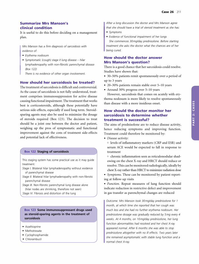
Case 26 211
PA
RT
2:
CA
SE
S
Summarize Mrs Manson’s clinical conditionIt is useful to do this before deciding on a management
plan.
Mrs Manson has a fi rm diagnosis of sarcoidosis with
evidence of:
• Erythema nodosum
• Symptomatic (cough) stage II lung disease – hilar
lymphadenopathy with non-fi brotic parenchymal disease
(Box 122)
There is no evidence of other organ involvement.
How should her sarcoidosis be treated?The treatment of sarcoidosis is diffi cult and controversial.
As the cause of sarcoidosis is not fully understood, treat-
ment comprises immunosuppression for active disease
causing functional impairment. The treatment that works
best is corticosteroids, although these potentially have
serious side-effects, especially if used long term. Steroid-
sparing agents may also be used to minimize the dosage
of steroids required (Box 123). The decision to treat
should be a joint one between the doctor and patient,
weighing up the pros of symptomatic and functional
improvement against the cons of treatment side-effects
and potential lack of effectiveness.
After a long discussion the doctor and Mrs Manson agree
that she should have a trial of steroid treatment as she has:
• Symptoms
• Evidence of functional impairment of her lungs
She commences 30 mg/day prednisolone. Before starting
treatment she asks the doctor what the chances are of her
being cured.
How should the doctor answer Mrs Manson’s question?There is a good chance that her sarcoidosis could resolve.
Studies have shown that:
• 30–50% patients remit spontaneously over a period of
up to 3 years
• 20–30% patients remain stable over 5–10 years
• Around 30% progress over 5–10 years
However, sarcoidosis that comes on acutely with ery-
thema nodosum is more likely to resolve spontaneously
than disease with a more insidious onset.
How should the doctor monitor her sarcoidosis to determine whether treatment is successful?The aims of prednisolone are to reduce disease activity,
hence reducing symptoms and improving function.
Treatment could therefore be monitored by:
• Disease activity:� levels of infl ammatory markers (CRP and ESR) and
serum ACE would be expected to fall in response to
treatment� chronic infl ammation seen as reticulonodular shad-
owing on the chest X-ray and HRCT should reduce or
resolve. This can be monitored radiologically, ideally by
chest X-ray rather than HRCT to minimize radiation dose
• Symptoms. These can be monitored by patient report-
ing at follow-up visits
• Function. Repeat measures of lung function should
indicate reduction in restrictive defect and improvement
in gas transfer as parenchymal changes are reduced
Outcome. Mrs Manson took 30 mg/day prednisolone for 1
month, at which time she reported that her cough was
much less and she had no further erythema nodosum. Her
prednisolone dosage was gradually reduced by 5 mg every 4
weeks. At 4 months, on 10 mg/day prednisolone, her lung
function abnormalities had resolved and her chest X-ray
appeared normal. After 6 months she was able to stop
prednisolone altogether with no ill effects. Two years later
she remained asymptomatic with stable lung function and a
normal chest X-ray.
Box 122 Staging of sarcoidosis
This staging system has some practical use as it may guide
treatment:
Stage I. Bilateral hilar lymphadenopathy without evidence
of parenchymal disease
Stage II. Bilateral hilar lymphadenopathy with non-fi brotic
parenchymal disease
Stage III. Non-fi brotic parenchymal lung disease alone
(hilar nodes are shrinking, therefore not seen)
Stage IV. Fibrosis and distortion of the lung
Box 123 Some immunosuppressant drugs used as steroid-sparing agents in the treatment of sarcoidosis
• Azathioprine
• Methotrexate
• Cyclophosphamide
• Chlorambucil

212 Part 2: Cases
PA
RT
2:
CA
SE
S
KEY POINTS
• Sarcoidosis is a condition characterized by the appearance
of non-caseating granulomas in multiple tissues and
organs. The cause is not fully understood
• Tissues and organs affected by sarcoidosis include:� lungs� skin� eyes� bones� bone marrow and spleen� heart� CNS
• Non-caseating granulomas in affected organs can interfere
with normal function, causing ill health
• Sarcoidosis usually presents with respiratory symptoms,
erythema nodosum, rheumatological or ophthalmological
complaints or hypercalcaemia, or may be asymptomatic
• Sarcoidosis is diagnosed by a combination of:� clinical features� demonstration of non-caseating granulomas in biopsies
of affected tissues� radiological appearance
• Pulmonary sarcoidosis is staged radiologically:� Stage I. Bilateral hilar lymphadenopathy without
evidence of parenchymal disease
� Stage II. Bilateral hilar lymphadenopathy with non-
fi brotic parenchymal disease� Stage III. Non-fi brotic parenchymal lung disease alone
(hilar nodes are shrinking, therefore not seen)� Stage IV. Fibrosis and distortion of the lung
• Sarcoidosis should be treated with immunosuppression if
there is evidence of active disease causing functional
impairment or symptoms
• The treatment of sarcoidosis is glucocorticoids. However,
these have side-effects including:� metabolic effects such as diabetes mellitus,
osteoporosis, weight gain and hypertension� increased susceptibility to infection� adrenal suppression
• Steroid-sparing agents may be used to reduce the dose
of glucocorticoids required
• The prognosis for patients with sarcoidosis is:� 30–50% remit spontaneously over a period of up to 3
years� 20–30% remain stable over 5–10 years� around 30% progress over 5–10 years
that she has stage II sarcoidosis with bilateral hilar
lymphadenopathy and non-fi brotic parenchymal disease.
As she has symptoms and impairment of lung function on
testing she is treated with a reducing course of prednisolone
over 6 months. Her symptoms and chest X-ray abnormalities
resolve and 2 years later she remains asymptomatic with
stable lung function and a normal chest X-ray.
CASE REVIEW
Elaine Manson presents with a painful rash over both shins
and a 2-month history of dry cough. Endobronchial and
transbronchial biopsies show non-caseating granulomas
consistent with sarcoidosis. The major differential diagnosis
for her presentation is tuberculosis, but this is excluded as
her Mantoux test is negative and there is no evidence
of infection with acid-fast bacilli. HRCT scanning shows

PA
RT
2:
CA
SE
S
A 68-year-old woman with worsening breathlessness on exertion
Case 27
Mrs Borg is a 68-year-old woman who presents to her GP
complaining of shortness of breath. This has been coming
on for several months and she is now panting when going
up a slight incline or doing housework. She is not breathless
at rest or at night. She denies other symptoms including
cough, chest pain, palpitations or ankle swelling.
She has no signifi cant past medical history or signifi cant
family history.
She is not on any medication.
She has never smoked, has spent her life bringing up her
family and has no pets.
Summarize the main features of her historyIsolated shortness of breath on exertion in a 68-year-old
woman.
What is your differential diagnosis so far?• Respiratory or cardiac disease. These are the most
common causes of shortness of breath. However, she has
no associated features or risk factors to point to either of
these systems as the source of her breathlessness
• Anaemia. This could account for shortness of breath
with no other cardiorespiratory symptoms
• Other diagnoses. In the absence of cardiac or respiratory
features other diagnoses that should be considered include:� renal failure� thyroid disease� breast disease with pulmonary involvement (pleural
effusion, lymphatic involvement)
The GP examines Mrs Borg.
She has no pallor or cyanosis. She has fi nger clubbing.
Breast, thyroid and lymph node examination are normal.
Pulse 68 beats/min, regular, jugular venous pressure (JVP)
not elevated, blood pressure 112/72 mmHg.
Heart sounds normal, no murmurs, no ankle oedema.
Chest. Trachea midline, expansion and percussion normal,
auscultation – bilateral basal end-inspiratory crepitations.
Abdominal examination is normal and there is no
hepatomegaly.
How do you interpret these fi ndings?Inspiratory crepitations indicate a pathological process
involving the alveoli or airways (Box 124). As Mrs Borg’s
crepitations are bilateral they are most likely caused by
pulmonary oedema or fi brosis.
At this stage, pulmonary fi brosis appears more likely
than oedema as the cause of her crepitations as:
• She has fi nger clubbing, which is seen with fi brosis
• She does not have any other signs of heart failure (her
JVP is normal and she has no peripheral oedema or
hepatomegaly)
There is no evidence of anaemia, thyroid or breast
disease on examination.
What should Mrs Borg’s GP do next?Mrs Borg’s GP should order some basic investigations
to:
• Distinguish between her differential diagnoses
• Determine whether Mrs Borg can be managed in the
community or needs a specialist opinion
• Decide which specialist service Mrs Borg should be
referred to if required (respiratory, cardiac or other)
What investigations should the GP order?• Chest X-ray – to look for the cause of her crepitations
and assess heart size
• ECG – to look for heart disease that might be causing
pulmonary oedema
• Blood tests to look for anaemia, thyroid or renal
disease
Mrs Borg returned 1 week later for the results of her
investigations. Her GP checked the results, which were as
follows.
Chest X-ray report: ‘There is volume loss bilaterally with
areas of reticular shadowing at both bases’ (Fig. 95)
ECG: normal.
Blood tests: normal.
213

214 Part 2: Cases
PA
RT
2:
CA
SE
S
What is reticular shadowing?The word reticular comes from the Latin ‘reticulum’
which is the diminutive of ‘rete’, meaning a net. Reticular
shadowing on a chest X-ray is a term used to describe
‘netlike’ shadowing, caused by multiple fi ne white lines
crossing over each other. These fi ne white lines are caused
by thickening of the interstitium resulting from infl am-
mation or fi brosis.
How does this information affect your differential diagnosis?The chest X-ray fi ndings support the diagnosis of pulmo-
nary fi brosis, which was considered most likely after
examining Mrs Borg. The investigation results rule out
anaemia, renal and thyroid disease and make cardiac
disease less likely.
What should the GP do now?Mrs Borg should be referred for a specialist opinion from
a hospital respiratory physician to:
(a)
(b) (c)
Figure 95 (a) Chest X-ray.
(b) Enlargement of the right lower lung
fi eld shows multiple fi ne white lines
and nodules which as emphasized in
(c) with drawn lines.
Box 124 Causes of crepitations
Crepitations can originate in the alveoli or airways
Alveolar causesAlveolar crepitations are often fi ne, like crumpling an
empty crisp packet
• Fluid in the alveoli:� pulmonary oedema� pneumonia (infl ammatory exudate)
• Infl ammation or fi brosis in or around the alveoli:� pulmonary fi brosis� extrinsic allergic alveolitis
AirwaysCrepitations can be caused by secretions in the airway.
These crepitations are usually coarser and lower pitched:
• Bronchitis
• Bronchiectasis
• Bronchiolitis

Case 27 215
PA
RT
2:
CA
SE
S
• Confi rm the diagnosis
• Look for underlying causes
• Establish the type of condition and prognosis
• Consider treatment options
Mrs Borg was referred for a respiratory opinion. The
consultant agreed with the GP’s examination fi ndings and
arranged a high-resolution CT scan to look for interstitial
lung disease (Fig. 96).
What does the high-resolution CT scan show?Lung tissue normally looks light grey and is ‘homoge-
neous’, which means it looks the same throughout the
lung fi elds. In this CT scan patches of the lung look
normal (A), but other patches have been replaced by
dense white tissue (B) which is fi brosis. The fi brosis con-
tracts the lung, pulling other parts of the lung out of
place. This can be seen as honeycombing (C) and traction
bronchiectasis (D).
What type of disease is this?The terminology of this type of disease is very compli-
cated as it has a number of names:
• It is often referred to as pulmonary fi brosis or fi brosing
alveolitis, particularly in older text books
• It is a type of ‘interstitial lung disease’ or ‘interstitial
pneumonia’, so-called because the pathological changes
appear to start in or involve the interstitium (i.e. the space
in between the cells)
• Another term for this group of lung diseases is ‘diffuse
parenchymal lung diseases’ (DPLD). The lung paren-
chyma refers to the alveoli, capillaries and supporting
tissue. As can be seen from the CT scan, this disease
affects the alveoli and capillaries as well as the lung inter-
stitium, hence the use of this more inclusive term
For the rest of the case this group of diseases will be
referred to as DPLD.
What causes DPLD?This term encompasses over 200 different conditions,
which present with related clinical features and have a
wide range of causes. The simplest classifi cation of
DPLD is:
• DPLD of known cause or association (Box 125)
• DPLD of unknown cause (Box 126)
How should the consultant proceed with Mrs Borg’s assessment?It is important to establish:
• Whether there is an identifi able cause for her DPLD
• How the lung disease is affecting her lung function
KEY POINT
The cause of DPLD is assessed primarily by taking a
meticulous history, including asking about occupational
and environmental exposure in detail, then by
examination.
D
C
A
B
Figure 96 High-resolution CT scan.
(A) Relatively normal area of lung
tissue. (B) The dense white tissue,
particularly seen towards the edge
of the lung, is caused by fi brosis.
(C) Fibrosis replaces normal lung
tissue and causes holes to appear
surrounded by thick white lines. This
is called ‘honeycombing’. An early
example is seen in this scan.
(D) Fibrosis pulls on the walls of the
airways, making them baggy and
dilated. This is called ‘traction
bronchiectasis’ and a good example is
seen in this scan.

216 Part 2: Cases
PA
RT
2:
CA
SE
S
Patients should also have the following investigations:
• Full blood count and eosinophils (hypersensitivity),
erythrocyte sedimentation rate (ESR)
• Renal and hepatic function
• Rheumatoid factor and antinuclear antibodies (con-
nective tissue disease)
Other investigations will depend on history and exam-
ination fi ndings and could include:
• Other autoantibodies if the history or examination are
indicative of connective tissue disease or vasculitis
• Serum precipitins if there is a history of environmental
exposure
• Serum angiotensin converting enzyme (ACE) if there
is any suggestion of sarcoidosis
The effect of DPLD on her lungs should be assessed
by:
• Full lung function testing
• Oxygen saturations, followed by arterial blood gases if
abnormal
Mrs Borg had no features on history, examination or
investigation to indicate an identifi able underlying cause for
her DPLD.
Lung function:
FEV1 1.25 L (58% predicted)
FVC 1.45 L (57% predicted)
FEV1 % FVC 86%
Transfer factor 43% predicted
Residual volume 1.55 L (86% predicted)
Total lung capacity 3.0 L (65% predicted)
Oxygen saturations were 94% on air
How do you explain her lung function results?SpirometryHer FEV1 and FVC are reduced proportionately. This is a
restrictive lung defect and indicates reduced lung expan-
sion. This is the usual fi nding in patients with DPLD
as infl ammation and fi brosis replace some alveoli and
restrict expansion of others. The airways are largely
spared apart from some traction bronchiectasis; hence
there is no evidence of airfl ow obstruction.
Lung volumesThe reduction in lung volumes is also shown as a reduc-
tion in total lung capacity (TLC), which is a measure of
lung volume at maximum inspiration.
The residual volume (air left in the ‘dead space’ of the
lungs on maximum expiration) is unaffected by paren-
chymal changes and is normal.
Gas transferLoss of alveoli and capillaries resulting from fi brosis re -
duces the surface area available for gas transfer. This also
explains the slight hypoxia measured on pulse oximetry.
What is the diagnosis?Mrs Borg has DPLD of unknown cause, also known as
idiopathic interstitial pneumonia.
Are there different types of idiopathic interstitial pneumonia?The most common type is idiopathic pulmonary fi brosis,
previously known as cryptogenic fi brosing alveolitis. The
Box 126 Diffuse parenchymal lung disease of unknown cause
• Idiopathic interstitial pneumonias
• Granulomatous disease, including sarcoidosis
• Other forms
Box 125 Diffuse parenchymal lung disease of known cause or association
Occupation or environmental exposure• Inorganic chemicals, e.g.
� asbestos� coal� silica
• Organic agents:� causes of extrinsic allergic alveolitis (Box 129, p. 222)
Drugs and toxins• Oral drugs, e.g.
� amiodarone� methotrexate
• Illicit drugs, e.g.� heroin� crack cocaine
• Radiation
Associated with systemic disease• Connective tissue disease, e.g.
� systemic sclerosis� rheumatoid arthritis� systemic lupus erythematosus
• Vasculitis
• Cancer

Case 27 217
PA
RT
2:
CA
SE
S
histological fi ndings seen on lung biopsy from patients
with this clinical diagnosis are referred to as usual inter-
stitial pneumonia. Other types are listed in Box 127.
Is it important to tell between these types?The different types of idiopathic interstitial pneumonia
each have different natural history, prognosis and treat-
ment response; therefore it is useful to distinguish
between them.
How can you tell between the types of idiopathic interstitial pneumonia?The different types of idiopathic interstitial pneumonia
are defi ned by clinical and pathological behaviour. Clini-
cians, radiologists and pathologists therefore must work
as a team to combine information from history and
examination, radiology and pathology to make a fi nal
diagnosis. The diagnosis can often be made from clinical
information and using the CT scans. Sometimes a lung
biopsy is required.
How are lung biopsies taken?There are three main methods, all of which can be com-
plicated by haemorrhage, pneumothorax and are associ-
ated with a small risk of death.
Open lung biopsy• A surgical incision is made in the chest wall (thora-
cotomy) and a piece of lung is removed
• A general anaesthetic is required and the complication
(7%) and mortality (<1%) rates are the highest for any
form of lung biopsy
• The diagnostic yield is good
Video assisted thoracoscopic (VATS) lung biopsy• A fi breoptic endoscope and operating instruments are
inserted into the thorax through small holes in the chest wall
and used to take biopsies from the periphery of the lung
• This has similar diagnostic yield but probably less
complications than open lung biopsy
Transbronchial lung biopsy• A biopsy forceps is inserted as deep as possible into the
lungs through a bronchoscope and a biopsy taken of the
airway wall and associated lung parenchyma
• This has the advantages of being a relatively safe out-
patient procedure which does not require a general
anaesthetic. However, it has lower diagnostic yield than
open or VATS lung biopsies
Mrs Borg’s case was reviewed at the multidisciplinary team
meeting. The radiologists felt that her CT fi ndings were
consistent with idiopathic pulmonary fi brosis. On discussion
the team agreed that this was a fi rm diagnosis and there
was no need for a lung biopsy.
What is the natural history of idiopathic pulmonary fi brosis?Idiopathic pulmonary fi brosis (IPF) is a progressive
disease with a poor prognosis. The median survival for a
patient with this diagnosis is less than 3 years.
Is there any treatment available for idiopathic pulmonary fi brosis?Immunosuppression is used in an attempt to stop infl am-
mation of the lung progressing to fi brosis. However, this
treatment is not very effective, possibly because the
fi brosis occurs without prior infl ammation. Additionally,
treatment may have considerable adverse effects. Patients
may receive a treatment trial with:
• Corticosteroids
• A second immunosuppressive agent such as azathio-
prine (Box 128)
Disease is monitored using symptoms, radiology and
lung function. If there is improvement treatment may
continue; if there is no improvement or deterioration
treatment is usually discontinued.
What should the consultant discuss with Mrs Borg when she returns to clinic?It will be a diffi cult consultation for them both. Ethical
principles governing the consultation should include the
following.
Box 127 Classifi cation of idiopathic interstitial pneumonias
• Idiopathic pulmonary fi brosis
• Other interstitial pneumonias, e.g.� desquamative interstitial pneumonia� non-specifi c interstitial pneumonia� respiratory bronchiolitis-associated interstitial lung
disease� cryptogenic organizing pneumonia� lymphoid interstitial pneumonia
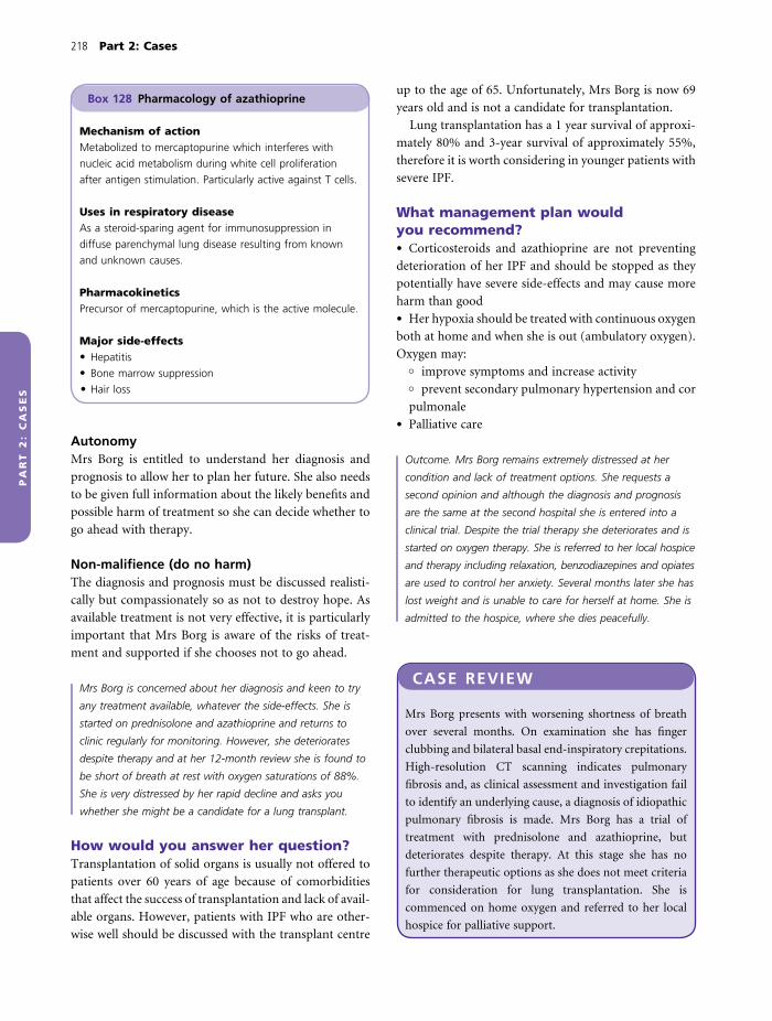
218 Part 2: Cases
PA
RT
2:
CA
SE
S
AutonomyMrs Borg is entitled to understand her diagnosis and
prognosis to allow her to plan her future. She also needs
to be given full information about the likely benefi ts and
possible harm of treatment so she can decide whether to
go ahead with therapy.
Non-malifi ence (do no harm)The diagnosis and prognosis must be discussed realisti-
cally but compassionately so as not to destroy hope. As
available treatment is not very effective, it is particularly
important that Mrs Borg is aware of the risks of treat-
ment and supported if she chooses not to go ahead.
Mrs Borg is concerned about her diagnosis and keen to try
any treatment available, whatever the side-effects. She is
started on prednisolone and azathioprine and returns to
clinic regularly for monitoring. However, she deteriorates
despite therapy and at her 12-month review she is found to
be short of breath at rest with oxygen saturations of 88%.
She is very distressed by her rapid decline and asks you
whether she might be a candidate for a lung transplant.
How would you answer her question?Transplantation of solid organs is usually not offered to
patients over 60 years of age because of comorbidities
that affect the success of transplantation and lack of avail-
able organs. However, patients with IPF who are other-
wise well should be discussed with the transplant centre
Box 128 Pharmacology of azathioprine
Mechanism of actionMetabolized to mercaptopurine which interferes with
nucleic acid metabolism during white cell proliferation
after antigen stimulation. Particularly active against T cells.
Uses in respiratory diseaseAs a steroid-sparing agent for immunosuppression in
diffuse parenchymal lung disease resulting from known
and unknown causes.
PharmacokineticsPrecursor of mercaptopurine, which is the active molecule.
Major side-effects• Hepatitis
• Bone marrow suppression
• Hair loss
up to the age of 65. Unfortunately, Mrs Borg is now 69
years old and is not a candidate for transplantation.
Lung transplantation has a 1 year survival of approxi-
mately 80% and 3-year survival of approximately 55%,
therefore it is worth considering in younger patients with
severe IPF.
What management plan would you recommend?• Corticosteroids and azathioprine are not preventing
deterioration of her IPF and should be stopped as they
potentially have severe side-effects and may cause more
harm than good
• Her hypoxia should be treated with continuous oxygen
both at home and when she is out (ambulatory oxygen).
Oxygen may:� improve symptoms and increase activity� prevent secondary pulmonary hypertension and cor
pulmonale
• Palliative care
Outcome. Mrs Borg remains extremely distressed at her
condition and lack of treatment options. She requests a
second opinion and although the diagnosis and prognosis
are the same at the second hospital she is entered into a
clinical trial. Despite the trial therapy she deteriorates and is
started on oxygen therapy. She is referred to her local hospice
and therapy including relaxation, benzodiazepines and opiates
are used to control her anxiety. Several months later she has
lost weight and is unable to care for herself at home. She is
admitted to the hospice, where she dies peacefully.
CASE REVIEW
Mrs Borg presents with worsening shortness of breath
over several months. On examination she has fi nger
clubbing and bilateral basal end-inspiratory crepitations.
High-resolution CT scanning indicates pulmonary
fi brosis and, as clinical assessment and investigation fail
to identify an underlying cause, a diagnosis of idiopathic
pulmonary fi brosis is made. Mrs Borg has a trial of
treatment with prednisolone and azathioprine, but
deteriorates despite therapy. At this stage she has no
further therapeutic options as she does not meet criteria
for consideration for lung transplantation. She is
commenced on home oxygen and referred to her local
hospice for palliative support.

Case 27 219
PA
RT
2:
CA
SE
S
KEY POINTS
• The term ‘lung parenchyma’ refers to the lung alveoli,
capillaries and supporting tissue
• The term ‘diffuse parenchymal lung diseases’ encompasses
a group of conditions where the pathology (infl ammation
and fi brosis) affects the lung parenchyma
• The cause of DPLD may be unknown (idiopathic pulmonary
fi brosis) or of known cause or association, e.g.� occupational or environmental exposure� drugs or toxins� associated with systemic disease (e.g. connective tissue
disease)
• Patients with DPLD usually present with shortness of
breath or cough and may have fi nger clubbing and end-
inspiratory bibasal crepitations on auscultation of the lung
bases
• Structural changes of DPLD are best identifi ed by a high-
resolution CT scan of the thorax
• Patients with radiological evidence of DPLD require
meticulous review of history and investigation to look for
an underlying cause
• A lung biopsy may be performed to make a histological
diagnosis of DPLD type. The benefi ts of lung biopsy in
determining treatment and prognosis should be weighed
against the risks of haemorrhage, pneumothorax and
small risk of death
• Idiopathic pulmonary fi brosis is a progressive disease
with median survival of less than 3 years from
diagnosis
• Treatment of IPF with immunosuppression (prednisolone
and azathioprine) is not very effective
• Transplantation may be considered for patients with IPF
who are otherwise well up to the age of 65 years
• Patients with end-stage IPF may require home oxygen
and palliative care support
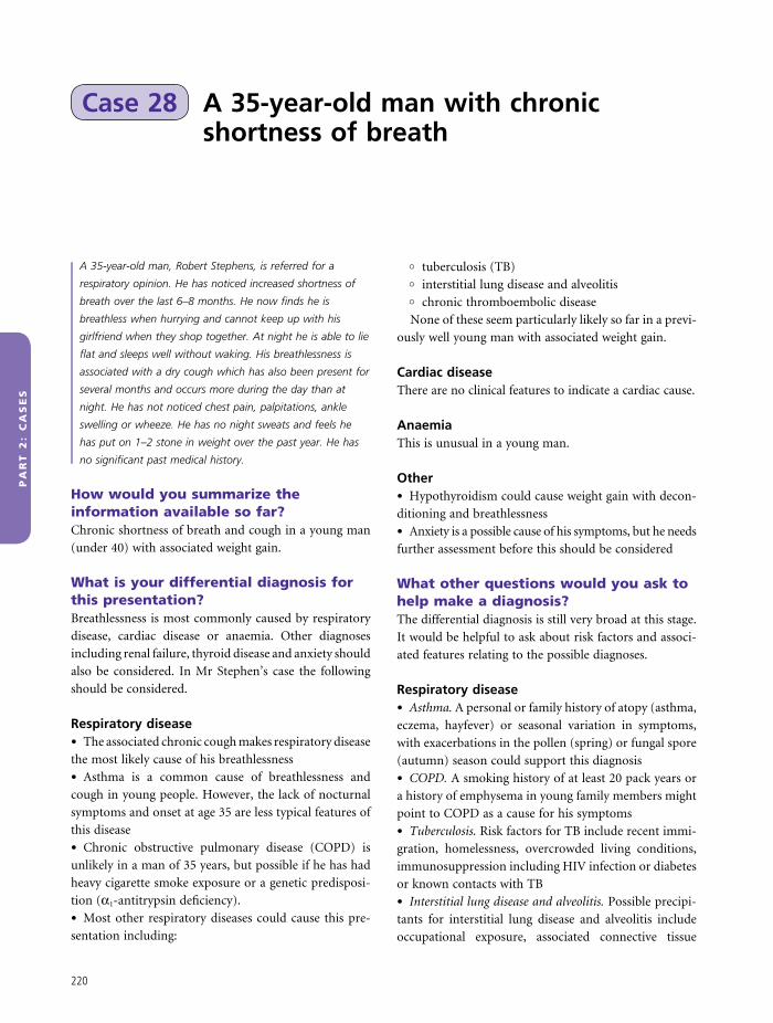
PA
RT
2:
CA
SE
S
A 35-year-old man with chronic shortness of breath
Case 28
A 35-year-old man, Robert Stephens, is referred for a
respiratory opinion. He has noticed increased shortness of
breath over the last 6–8 months. He now fi nds he is
breathless when hurrying and cannot keep up with his
girlfriend when they shop together. At night he is able to lie
fl at and sleeps well without waking. His breathlessness is
associated with a dry cough which has also been present for
several months and occurs more during the day than at
night. He has not noticed chest pain, palpitations, ankle
swelling or wheeze. He has no night sweats and feels he
has put on 1–2 stone in weight over the past year. He has
no signifi cant past medical history.
How would you summarize the information available so far?Chronic shortness of breath and cough in a young man
(under 40) with associated weight gain.
What is your differential diagnosis for this presentation?Breathlessness is most commonly caused by respiratory
disease, cardiac disease or anaemia. Other diagnoses
including renal failure, thyroid disease and anxiety should
also be considered. In Mr Stephen’s case the following
should be considered.
Respiratory disease• The associated chronic cough makes respiratory disease
the most likely cause of his breathlessness
• Asthma is a common cause of breathlessness and
cough in young people. However, the lack of nocturnal
symptoms and onset at age 35 are less typical features of
this disease
• Chronic obstructive pulmonary disease (COPD) is
unlikely in a man of 35 years, but possible if he has had
heavy cigarette smoke exposure or a genetic predisposi-
tion (α1-antitrypsin defi ciency).
• Most other respiratory diseases could cause this pre-
sentation including:
� tuberculosis (TB)� interstitial lung disease and alveolitis� chronic thromboembolic disease
None of these seem particularly likely so far in a previ-
ously well young man with associated weight gain.
Cardiac diseaseThere are no clinical features to indicate a cardiac cause.
AnaemiaThis is unusual in a young man.
Other• Hypothyroidism could cause weight gain with decon-
ditioning and breathlessness
• Anxiety is a possible cause of his symptoms, but he needs
further assessment before this should be considered
What other questions would you ask to help make a diagnosis?The differential diagnosis is still very broad at this stage.
It would be helpful to ask about risk factors and associ-
ated features relating to the possible diagnoses.
Respiratory disease• Asthma. A personal or family history of atopy (asthma,
eczema, hayfever) or seasonal variation in symptoms,
with exacerbations in the pollen (spring) or fungal spore
(autumn) season could support this diagnosis
• COPD. A smoking history of at least 20 pack years or
a history of emphysema in young family members might
point to COPD as a cause for his symptoms
• Tuberculosis. Risk factors for TB include recent immi-
gration, homelessness, overcrowded living conditions,
immunosuppression including HIV infection or diabetes
or known contacts with TB
• Interstitial lung disease and alveolitis. Possible precipi-
tants for interstitial lung disease and alveolitis include
occupational exposure, associated connective tissue
220

Case 28 221
PA
RT
2:
CA
SE
S
disease and exposure to organic antigens from animals,
birds or fungi. You should also ask about associated fea-
tures of sarcoidosis (skin, eye, bone involvement)
• Chronic thromboembolic disease. You should ask about
immobility, travel history and family history of throm-
boembolic disease
Other• Hypothyroidism. Associated symptoms could include
cold intolerance, constipation, altered appearance and
depression.
Mr Stephens has never smoked and has no personal or
family history of respiratory or thromboembolic disease or
atopy. He is otherwise well and has no joint, skin or eye
symptoms. He was born in the UK, has worked as an
accountant since completing his training and is not aware of
any occupational exposure to toxins or allergens or exposure
to TB infection. Twelve months ago he moved into his
girlfriend’s fl at where she keeps a budgerigar.
On examination he looked well. Oxygen saturations were
97% on air and pulse was 84 beats/min, regular. He had a
body mass index of 32 kg/m2. There were no abnormal
examination fi ndings.
How does this information affect the differential diagnosis?• The only positive fi nding in his history is 1 year of
contact with a budgerigar, with exposure starting around
4 months before his symptoms commenced. His symptoms
could be caused by respiratory allergy to this bird, either:� asthma; or� extrinsic allergic alveolitis
• His body mass index is elevated and obesity and decon-
ditioning could also account for his symptoms
• Lack of any smoking history makes COPD very unlikely
• Normal pulse rate and lack of other symptoms make
hypothyroidism less likely, although weight gain could be
brought about by an underactive thyroid gland
• There is no history to support TB or thromboembolic
disease
KEY POINT
Patients often do not mention that they keep birds when
asked about pets. It pays to ask specifi cally and repeatedly
about bird exposure to jog their memories and to ask
family members.
What investigations would you do at this clinic visit?Investigations that could be performed immediately and
would give useful information to guide further manage-
ment are:
• Spirometry to look for obstructive or restrictive lung
disease
• Chest X-ray to look for parenchymal lung disease
• To exclude other diagnoses:� electrocardiogram (ECG) to exclude cardiac disease� full blood count to exclude anaemia� thyroid function tests to rule out hypothyroidism
Investigation results were as follows:
Spirometry: FEV1 2.2 L (62% predicted), FVC 2.8 L (67%
predicted), FEV1/FVC 79%
Chest X-ray report: there is diffuse shadowing throughout
the lungs with a nodular quality
ECG, full blood count and thyroid function tests: normal.
How do you interpret these results?Spirometry shows an equal reduction in both FEV1 and
FVC with an FEV1/FVC ratio >70%. This is consistent
with restrictive lung disease.
The chest X-ray shows some diffuse shadowing, which
needs further imaging by high-resolution CT (HRCT)
scanning.
Other tests reassuringly show no evidence of cardiac
disease, anaemia or hypothyroidism.
How do these results modify your differential diagnosis?Mr Stephens appears to have a restrictive lung disease
(Box 129) to account for his chronic cough and breath-
lessness. As he is a young man and has exposure to bird
antigens the most likely diagnosis is extrinsic allergic
alveolitis.
Asthma is now less likely because he has no evidence of
airfl ow obstruction and he has an abnormal chest X-ray.
Obesity can cause restrictive lung function, but should
not cause an abnormal chest X-ray (Box 129).
What is extrinsic allergic alveolitis?‘Alveolitis’ means infl ammation of the alveoli. The infl am-
mation actually also affects the small airways, hence this
disease is also called ‘hypersensitivity pneumonitis’.
Extrinsic allergic alveolitis (EAA) is caused by inhalation
of organic antigens (Box 130). Susceptible patients
develop a hypersensitivity reaction, leading to alveolar

222 Part 2: Cases
PA
RT
2:
CA
SE
S
Anatomy• Extrinsic allergic alveolitis affects the alveoli and small
airways
• Asthma affects the bronchi and larger bronchioles
• Rhinitis affects the nasal epithelium
Allergens• Extrinsic allergic alveolitis is most commonly caused
by bird proteins, fungal and protozoal allergens. Allergens
must be small enough (approximately 1 µm) to be depos-
ited in the distal lung
• Asthma and rhinitis are caused by:� seasonal allergens – tree and grass pollen (spring),
fungal spores (autumn)� perennial (year round) allergens – animal and bird
proteins, house dust mites
Pathology• Extrinsic allergic alveolitis is caused by a type III hyper-
sensitivity reaction. Inhaled antigens become bound to
circulating IgG (precipitins) to that antigen. The result-
ing immune complexes trigger minor mast cell degranu-
lation and infl ammatory cell infi ltration, initially with
neutrophils then with lymphocytes and macrophages
which form granulomas. The reaction and development
of symptoms takes 4–12 h after antigen exposure
• Asthma and rhinitis are caused by a type I hypersensi-
tivity reaction. Inhaled antigens bind to immunoglobulin
E (IgE) on the surface of mast cells, causing mast cell
degranulation and release of infl ammatory mediators.
Increased vascular permeability with oedema, infl amma-
tory infi ltrate and bronchoconstriction can come on over
5–10 min
Effect on lung function• Extrinsic allergic alveolitis causes alveolar and intersti-
tial infl ammation which limits lung infl ation, causing a
restrictive picture on spirometry. As small airways are
also involved there may be a component of obstruction
• Asthma causes oedema and constriction of the larger
airways, obstructing airfl ow and causing an obstructive
picture on spirometry
How would you investigate Mr Stephens to confi rm the diagnosis of extrinsic allergic alveolitis?Tests should be arranged to look at his lung structure and
function and identify histological evidence of alveolitis
and immunological evidence of hypersensitivity. The
Box 129 Causes of restrictive lung function (reduction in lung volumes)
PulmonaryLung disease in the interstitium or alveoli restricts
expansion of the alveoli, e.g.
• Interstitial lung disease
• Alveolitis
Pleural or chest wallAbnormalities of the pleural or chest wall can constrict the
lungs, preventing expansion, e.g.
• Diffuse bilateral pleural thickening or pleural effusions,
e.g. caused by asbestos (benign pleural thickening or
mesothelioma) or infection (empyema)
• Severe obesity (Pickwick’s syndrome)
Neuromuscular disordersDiseases of the nerves or muscles prevent full lung
expansion during breathing, e.g.
• Motor neurone disease
• Guillain–Barré syndrome
• Myasthenia gravis
• Muscular dystrophy
Box 130 Causes of extrinsic allergic alveolitis
Allergens causing EAA come from organic material or
organisms growing on organic material. Some examples
are:
• Bird fancier’s lung. This is caused by antigens secreted
by birds that are associated with feather dust (bloom) or
found in bird droppings
• Farmer’s lung. This is caused by organisms
(thermoactinomycetes) growing on hay stored in damp
conditions
• Humidifi er fever. This is caused by organisms (protozoa
– Naegleria gruberi, Thermoactinomyces vulgaris)
growing in contaminated water, which is aerosolized by
air conditioners
• Maltworker’s lung. This is caused by mouldy barley
growing Aspergillus fumigatus or A. clavatus
and small airway infl ammation that impairs gas transfer
and causes breathlessness and cough (Fig. 97).
How does extrinsic allergic alveolitis differ from asthma and allergic rhinitis?These three conditions are all allergic diseases of the
respiratory tract. They have a number of important
differences.

Case 28 223
PA
RT
2:
CA
SE
S
doctor arranged the following tests, which are shown
with their results.
Transbronchial biopsy: pieces of lung tissue showing non-
caseating granulomas with an associated prominent
lymphocytic infi ltrate and foamy macrophages consistent
with EAA.
HRCT scan (Fig. 98): demonstrated diffuse ground glass
change and mosaicism.
Gas transfer: 63% of predicted values.
Avian precipitins:
Normal range
Budgerigar serum (IgG abs) 8.1 mg/L (0–40 mg/L)
Budgerigar feathers (IgG abs) 72.1 mg/L (0–40 mg/L)
Budgerigar droppings (IgG abs) 48 mg/L (0–40 mg/L)
How do you interpret these results?• A transbronchial lung biopsy showed infl ammatory
changes characteristic of the type III hypersensi-
tivity reaction that causes EAA. However, this result is
unusually informative as biopsy results are often
non-diagnostic
• The infl ammatory infi ltrate in the alveoli and intersti-
tium looks grey on CT scan images, like ground glass, and
replaces air which normally looks black. Mosaicism
means that there are some grey patches (A) and some
black patches (B) on the images. The black patches are
caused by pockets of air trapped in some alveoli by small
airway infl ammation
• His gas transfer is reduced. This is not surprising
because there is infl ammation of the alveoli and intersti-
tium which impairs diffusion of oxygen between alveoli
and capillaries
• He has elevated avian precipitins (IgG) for budgeri-
gar feathers and droppings. This indicates that he has
been exposed to and has an immunological reaction
to these antigens. In the context of the other investiga-
tion results it is likely that he has subacute EAA
(Box 131) caused by chronic inhalation of budgerigar
antigens
Terminal bronchiole
Alveoli
CapillariesInterstitium
Inhaled antigen
Inhaled antigen
(a) Alveolar capillary unit(b) Inhaled antigen deposited in alveoli and small airways
LymphocytesNeutrophils
Macrophages
LymphocytesNeutrophils
Macrophages
(d) Fibrosis and scarring
Fibrosis
(c) Inflammatory response
Figure 97 Pathogenesis of extrinsic
allergic alveolitis. Inhaled antigens
(b) trigger an infl ammatory response with
cellular infl ammation and oedema in the
small airways, alveoli and interstitium
(c) which impairs gas exchange. If
infl ammation is chronic, fi brosis and
thickening of the alveolar septae and
basement membrane occurs causing
permanent impairment.

224 Part 2: Cases
PA
RT
2:
CA
SE
S
How should Mr Stephen’s subacute extrinsic allergic alveolitis be treated?Antigen avoidanceThis is very important, but can be extremely diffi cult. In
Mr Stephen’s case he would be advised to fi nd a new
home for the budgerigar. However, people may be reluc-
tant to get rid of their (or their partner’s) pets, and even
if they do, bird antigens can persist in the home for a long
time. In other cases of EAA a change of job may be
required.
ImmunosuppressionPrednisolone can suppress the type III hypersensitivity
reaction and speed recovery of symptoms. However,
symptoms may reoccur when steroids are stopped if
antigen exposure continues.
Mr Stephens was advised to get rid of the budgerigar and
take 30 mg prednisolone for 2 weeks, followed by a
reducing dose of steroids to stop after a total of 6 weeks of
treatment. Before agreeing to follow this plan he asks how
much improvement he can expect in his condition.
How would you answer his question?If he is able to avoid bird antigens he is likely to make a
full recovery. This is likely as he has had a relatively short
exposure to the budgerigar, there is no evidence of irre-
versible fi brosis on his investigations and he is young. The
outlook for older patients with more chronic exposure or
fi brosis is worse.
Outcome. Mr Stephens takes the prescribed course of
steroids and feels much better. He is no longer breathless
and his cough disappears. On review in clinic his chest X-ray
is now normal and his gas transfer has increased to 92%
predicted. His girlfriend agrees to get rid of the budgerigar
and they decide to take the opportunity to move from their
fl at to a larger house. With this antigen avoidance strategy
he has no further symptoms.
A
B
Figure 98 High-resolution CT scan.
Box 131 Classifi cation of extrinsic allergic alveolitis
Acute extrinsic allergic alveolitis• Acute episodes occur 4–12 h after exposure to antigen
• Reaction proportional to exposure
• Respiratory and systemic symptoms occur, including
fever and chills
• Symptoms usually resolve on removal of antigen
Subacute extrinsic allergic alveolitis• Onset of symptoms more insidious than the acute form,
probably because of ongoing antigen exposure
Chronic extrinsic allergic alveolitis• Fibrosis of the lung secondary to acute or subacute
EAA

Case 28 225
PA
RT
2:
CA
SE
S
KEY POINTS
• Extrinsic allergic alveolitis is infl ammation of the alveoli and
small airways that occurs as a hypersensitivity reaction to
the inhalation of organic antigens
• Organic antigens causing EAA include:� bird antigens� moulds and other microorganisms (growing on material,
e.g. damp hay, contaminated water, mouldy barley)
• Extrinsic allergic alveolitis may be:� acute with symptoms immediately following exposure� subacute with insidious symptoms probably caused by
ongoing antigen exposure� chronic with fi brosis of the lung secondary to acute or
subacute EAA
• Symptoms of EAA may be respiratory (cough, shortness of
breath) and/or systemic (fever and chills)
• Investigation of EAA may show:� restrictive defect in lung function with reduced gas
transfer� patchy ground glass shadowing on HRCT scanning� lymphocytic infi ltration on pathological examination of
transbronchial lung biopsy or bronchoalveolar lavage� elevated IgG (precipitins) to the causative antigen
• Treatment of EAA requires avoidance of the causative
antigen. This should be permanent if possible to prevent
recurrence of alveolitis and development of irreversible
fi brosis
• A course of prednisolone can be given to speed recovery
of symptoms
HRCT, lymphocytic infi ltrate on transbronchial biopsy
and positive avian precipitins gives a diagnosis of subacute
EAA. He is treated with a course of prednisolone and
advised to get rid of the budgerigar. After moving house
he has no further symptoms.
CASE REVIEW
Robert Stephens presents with a 6–8 month history of
shortness of breath and dry cough. Twelve months ago he
moved into his girlfriend’s fl at where she keeps a budgerigar.
A combination of insidious onset of symptoms, restrictive
lung function abnormality, diffuse ground glass change on

PA
RT
2:
CA
SE
S
An overweight 52-year-old man who snores
Case 29
Mr Callum Campbell is a 52-year-old man who is referred
to the sleep clinic by his GP for investigation of his snoring.
He is a heavy goods vehicle driver and over the last 10
years he has put on around 4 stones in weight. His wife
reports that over a similar time period his snoring has
become louder. She has also noticed that he quite
frequently stops breathing for several seconds during
sleep when he either stirs or she has to shake him
awake.
What do you suspect the diagnosis may be?Obstructive sleep apnoea.
What is obstructive sleep apnoea?The Scottish Intercollegiate Guidelines Network (www.
sign.ac.uk/pdf/sign73.pdf) defi ne obstructive sleep
apnoea/hypopnoea syndrome (OSAHS) as ‘A condition
where the upper airway collapses intermittently and
repeatedly during sleep causing either total obstruction
of the airway lumen with no respiratory fl ow (apnoea),
or partial obstruction with reduction in the cross-
sectional area of the upper airway lumen causing
hypoventilation (hypopnoea).’
How does this syndrome account for his wife’s observations?The following cycle occurs:
• As the person falls into a deep sleep, the muscle tone
of the upper airway decreases leading to upper airway
narrowing:� if the narrowing is complete, the patient stops
breathing� if the narrowing is partial, the patient snores
• Airway narrowing stimulates an increase in inspiratory
effort in an attempt to overcome the narrowing
• This causes transient arousal from deep sleep to
wakefulness or a lighter sleep phase during which the
muscle tone is restored. This arousal may be triggered by
pharyngeal or pleural receptors detecting increased inspi-
ratory effort• The cycle is completed when the patient then falls back
into a deep sleep and muscle tone decreases once again
• The cycle can occur many times throughout the night,
disrupting sleep
• The daytime consequences include sleepiness and
uncreased risk of cardiovascular disease (Box 136, p. 229)
Why do you think his condition has become worse over the past 10 years?Mr Campbell has put on 4 stones in weight because of
his sedentary occupation and it is likely that his collar size
has also increased. Obesity is a major risk factor for
obstructive sleep apnoea. The mechanisms underlying
this are not fully understood; however, it is likely that the
weight of soft tissues in a large neck compresses the
pharynx and causes narrowing of the upper airway.
What features in his history would further support your provisional diagnosis of obstructive sleep apnoea?The main features of obstructive sleep apnoea include:
• History of poor sleep:� restless sleep� unrefreshing sleep� nocturia
• Evidence of upper airways collapse during sleep:� choking episodes during sleep� witnessed apnoeas� snoring
• Daytime problems resulting from poor sleep:� excessive daytime sleepiness� impaired concentration� irritability and personality change� decreased libido
Collateral history from a family member is often
required to obtain this history as patients may not notice
or admit to many of these features.
226

Case 29 227
PA
RT
2:
CA
SE
S
On further questioning Mr Campbell admits that he does
not feel refreshed when waking from sleep and often falls
asleep in the evening when watching television. He has
never fallen asleep at the wheel when driving. His wife
complains that he is restless at night and has become
diffi cult to talk to. He is irritable and forgetful and she feels
he has lost interest in sex. Mr Campbell has a 40 pack year
smoking history and continues to smoke around 20
cigarettes per day. He drinks 2–3 bottles of beer every night
and more at weekends. His Epworth Sleepiness Score (ESS)
is 16/24.
How do you interpret this new information?Mr Campbell has features of disturbed sleep as well as
daytime problems resulting from poor sleep. His use of
cigarettes and alcohol may be exacerbating his condition
(Box 132).
What is the Epworth Sleepiness Score (ESS)?This is a validated scoring system for assessing the level
of daytime sleepiness (Box 133).
How do you interpret Mr Campbell’s Epworth Sleepiness Score?Mr Campbell’s ESS is clearly abnormal at 16, refl ecting
moderate subjective daytime sleepiness. Patients with
abnormal ESS require further assessment and possible
investigation for obstructive sleep apnoea.
What should Mr Campbell be advised regarding his driving at this stage?This is very important as people with daytime sleepiness
are at increased risk of falling asleep at the wheel and can
cause road traffi c accidents. At this stage he should be
informed that he must not drive if he feels sleepy and that
falling asleep at the wheel is a criminal offence that can
potentially lead to a prison sentence. He may require
further advice about his driving if the diagnosis of
obstructive sleep apnoea is confi rmed.
Are there any other causes of daytime sleepiness?Many other factors can cause daytime sleepiness (Box
134) and should be considered in the differential diagno-
sis of obstructive sleep apnoea. It is important to clarify
whether there is adequate sleep ‘opportunity’, as inade-
quate quality or quantity of sleep may cause sleepiness.
Common everyday factors that can interfere with sleep
include:
• Shift work
• Poor sleep environment:
Box 132 Factors predisposing to obstructive sleep apnoea and hypopnoea
• Increasing age
• Male gender
• Obesity
• Sedative drugs
• Smoking (causes nasal and pharyngeal congestion)
• Alcohol consumption (reduces tone in pharyngeal
muscles)
Box 133 Epworth Sleepiness Score
The following questionnaire should be completed
independently by both the patient and their partner to
improve accuracy. Patients are particularly unlikely to
admit to falling asleep in a car as they may be anxious
about being told not to drive.
‘How likely are you to doze off or fall asleep in the
following situations in contrast to just feeling tired? Use
the following scale to choose the most appropriate
number for each situation.’
1 would never doze
2 slight chance of dozing
3 moderate chance of dozing
4 high chance of dozing
Situation1 Sitting and reading
2 Watching TV
3 Sitting inactive in a public place (e.g. a theatre or a
meeting)
4 As a passenger in a car for an hour without a break
5 Lying down to rest in the afternoon when circumstances
permit
6 Sitting and talking to someone
7 Sitting quietly after a lunch without alcohol
8 In a car, while stopped for a few minutes in traffi c
InterpretationThe maximum score is 24 points:
Normal ESS <11
Mild subjective daytime sleepiness ESS 11–14
Moderate subjective daytime sleepiness ESS 15–18
Severe subjective sleepiness ESS >18

228 Part 2: Cases
PA
RT
2:
CA
SE
S
� too noisy� too light� television or other distractions in bedroom� uncomfortable bed
• Poor preparation for sleep:� recent strenuous exercise� recent caffeine or other stimulation
• Stress, anxiety or pain
On examination Mr Campbell has a body mass index (BMI)
of 33 kg/m2, collar size 19 in, normal mandibular size, patent
upper airway and no pharyngeal obstruction. His blood
pressure is 160/95 mmHg and routine examination of all
systems is normal. Spirometry is also normal and there are
no features to suggest other underlying medical conditions.
How do you interpret his examination fi ndings?• Mr Campbell’s BMI is 33 kg/m2, which classifi es him as
obese (Box 135). This is a major risk factor for obstruc-
tive sleep apnoea
• His collar size is 19 in. Patients with obstructive sleep
apnoea often have a neck circumference greater than
17 in (43 cm). The large amount of soft tissue around the
neck appears to compress the upper airway during sleep
• He has no abnormal facial or upper airways features
that might predispose to obstructive sleep apnoea.
Features to look for include:
� an abnormally small mandible which can reduce
airway calibre� nasal obstruction – this can be detected visually
using a speculum or by asking the patient to sniff while
occluding each nostril in turn. When the nose is
blocked the pressures in the pharynx are more negative
during inspiration (compare breathing in when your
nose is pinched to breathing in through a clear nose),
which may increase upper airways collapse� mouth – look for a large tongue (macroglossia) and
‘crowded’ pharynx with large (‘kissing’) tonsils or
uvula that may increase obstruction of the airway
• He is hypertensive. Hypertension is associated with
obstructive sleep apnoea, although the mechanism is not
fully understood (Box 136) (NB a large cuff should be
used when measuring blood pressure in obese patients as
an inappropriately small cuff will lead to overestimation
of blood pressure.)
• No other abnormalities were detected on examination:� respiratory, cardiovascular and neurological exami-
nations should be performed to detect any coexisting
disease as clinically indicated� spirometry should be checked to look for associated
chronic obstructive pulmonary disease (COPD). This
is particularly important as a combination of severe
obstructive sleep apnoea and COPD can lead to
decompensated respiratory failure and right heart
strain
What tests would you perform to determine whether Mr Campbell has obstructive sleep apnoea?He should undergo a sleep study to confi rm the diagno-
sis of obstructive sleep apnoea and to determine the
severity of the condition. Essential components of a sleep
study include detection of:
• Ventilation
• Disruption of ventilation by upper airways obstruction
• Effect of disruption on sleep quality
Box 134 Pathological causes of daytime sleepiness
• Depression
• Hypothyroidism
• Drugs causing sleep disturbance and subsequent
daytime sleepiness:� nicotine replacement� amphetamines� theophyllines� serotonin reuptake inhibitors
• Drugs causing somnolence:� sedatives� β-blockers
• Neurological conditions:� previous encephalitis� previous head injury� parkinsonism
• Narcolepsy
Box 135 Assessment of body mass index
Body mass index (BMI) is calculated as follows:
Body mass index = weight in kg
(height in m)2
Normal range is 20–25 kg/m2. Patients are considered
obese if BMI >30 kg/m2.

Case 29 229
PA
RT
2:
CA
SE
S
Limited sleep studies can be performed at home,
allowing the patient to sleep in their own bed, reducing
costs and increasing the number of studies that can be
performed. These should be suffi cient to diagnose
obstructive sleep apnoea but the information provided
may be too limited to identify other disorders. More
complex sleep studies (polysomnography) provide in-
depth assessment of sleep but require hospital admission,
which is more costly and itself may disrupt the patient’s
sleep.
Mr Campbell has a home sleep study. On the day of the test
he spends some time with the lung function technician and
is trained to set up the equipment and use a pulse oximeter,
nasal probe and chest and abdominal straps. That night he
puts on the equipment and attaches the instruments to a
sleep monitor. After a night’s sleep he returns the sleep
monitor to the hospital for analysis. The results of his sleep
study are shown in Fig. 99.
What does each instrument measure?• The pulse oximeter provides continuous measure-
ments of oxygen saturation and pulse rate
• The nasal probe comprises a thermistor, which records
airfl ow from the nose and a room microphone to record
the level of snoring
• Movement sensors (straps) attached to the patient’s
ribcage or abdomen are used to measure respiratory effort
How can his sleep study results be interpreted?Nasal fl owThis determines how much air he is moving in and out
of his nose. In Fig. 99 at (A) nasal fl ow changes continu-
ously throughout inspiration and expiration, which
shows his airway is not obstructed. At (B) the nasal fl ow
line becomes fl at because there is no movement of air.
This is an apnoea. In analysis of the study a breathing
pause must last for at least 10 s to count as an apnoea.
The breathing pause marked lasts for 20 s until airfl ow
resumes at (C). Hypopnoeas are 10-s events where airfl ow
continues but is reduced by at least 50% from the
Box 136 Cardiovascular risk and obstructive sleep apnoea
Patients with obstructive sleep apnoea commonly have
other cardiovascular risk factors such as hypertension,
insulin resistance and diabetes mellitus. This association
may be partly caused by underlying obesity and lack of
exercise. However, obstructive sleep apnoea appears to be
an independent risk factor for hypertension and possibly
for other cardiovascular disease. Putative mechanisms
underlying the causal association include:
• Chronic sympathetic activation
• Chronic infl ammation
• Endothelial dysfunction
• Hypercoagulability
• Metabolic dysregulation (e.g. activation of the renin–
angiotensin system)
There is increasing evidence that obstructive sleep
apnoea is an independent risk factor for development of
and morbidity and mortality from cardiovascular and
cerebrovascular disease.
8990
20 secondsTime
Nasal Flow (373 mL/s/cm)
Thorax (113mV/cm)
Snore Pr. (99.2µbar/cm)
B
A
D
C
FE
HG
J
I
80 82 83 85 848888 8677 79 81 84 90 83 83 84 84 83 85 85 84 83 85 84 85 86 88 83
76 78 82 83 8190 92
76 80 84 82 8091 84 78
88 8793
97 97 96 94 90 89 8994 95 93 92 92 89 90
93 95 93 90 88 86 8793 96 94 92
89 87 87 8893 96 94 91 90
SpO2 (70–110%)
3:10:50 03:11:00 03:11:10 03:11:20 03:11:30 03:11:40 03:11:50 03:12:00 03:12:10 03:12:20 03:12:30 03:12:50 03:13:0003:13:1003:13:20 03:13:30 03:13:40 03:13:5003:12:40
Pulse (25–125bpm)
Figure 99 Sleep study.

230 Part 2: Cases
PA
RT
2:
CA
SE
S
previous baseline, but are not seen in Mr Campbell’s
sleep study.
Thoracic movementsDuring the apnoea, movement of thoracic muscles con-
tinues (D). This indicates that Mr Campbell has stopped
breathing because his airway is blocked (obstructive sleep
apnoea), rather than because of lack of signal from the
respiratory centre to his respiratory muscles (central
sleep apnoea).
Pulse oximetryOxygen saturations can be seen to fall from 96% to 88%
(E). This desaturation lags behind the apnoea, as does
recovery of oxygenation (F) following restoration of
airfl ow. The pulse oximeter also shows relative bradycar-
dia during the apnoea (G) with an increase in pulse rate
when airfl ow is restored (H). These transient rises in
heart rate are an indirect marker of arousal from sleep.
MicrophoneThis detects the volume of snoring which can be seen to
be loud when there is airfl ow (I) and silenced during
apnoea (J). This indicates that there is continuous partial
airfl ow obstruction during sleep which worsens intermit-
tently to complete apnoea.
How bad is his obstructive sleep apnoea?The severity of obstructive sleep apnoea is determined by
the hourly frequency of apnoeas or hypopnoeas (Table
12) and is known as the apnoea/hypopnoea index (AHI)
or respiratory disturbance index. In this part of his study
Mr Campbell can be seen to have six apnoeas in 3 min.
Over the whole night his AHI is 60 per hour (i.e. he has
severe obstructive sleep apnoea).
What should Mr Campbell now be advised regarding his driving?In the UK, the Driver and Vehicle Licensing Agency
(DVLA) regulations state that driving must cease if the
driver is excessively sleepy (from any cause). Group 1
drivers (ordinary licence holders) with a diagnosis of
sleep apnoea should notify the DVLA but can drive pro-
vided their sleepiness is controlled by treatment. Group
2 drivers (public service and heavy goods vehicles) should
cease driving until it has been confi rmed by a specialist
that their condition is adequately treated. The doctor
should now advise Mr Campbell to stop driving and to
inform the DVLA about his condition. If Mr Campbell
fails to inform the DVLA it is the duty of the doctor to
do so. In this situation the public interest overrides indi-
vidual patient confi dentiality.
How can Mr Campbell retain his heavy goods vehicle licence?He needs to commence treatment for his obstructive
sleep apnoea.
What treatment options are available?All patients should receive advice, education and support
to improve their lifestyle and health and reduce their
weight. While the health benefi ts of these behavioural
interventions are taking effect, the main treatments for
moderate to severe obstructive sleep apnoea are noctur-
nal continuous positive airways pressure (CPAP) and
mandibular advancement devices.
What behavioural advice should Mr Campbell receive?Mr Campbell should be advised to:
• Lose weight. This improves obstructive sleep apnoea
and helps with control of other obesity-related disorders
such as hypertension and diabetes. Weight loss is diffi cult
to achieve but may be assisted by referral to a dietitian
and starting an exercise programme
• Stop smoking. This can improve nasal patency and
reduce pharyngeal congestion
• Avoid alcohol and sedatives in the evenings, which will
improve his pharyngeal tone
• Avoid sleeping on his back. A tennis ball sewn into a
pyjama top can help remind him to do this during the
night
• Elevate the head of his bed. This also can help to main-
tain nasal patency
Table 12 Severity of obstructive sleep apnoea.
SeverityApnoea/hypopnoea rate (per hour)
Treatment strategies
Borderline 5–14 Behavioural
Mild 15–30 Behavioural unless
severe symptoms
Moderate 30–50 CPAP
Severe >50 CPAP
CPAP, continuous positive airway pressure.

Case 29 231
PA
RT
2:
CA
SE
S
How does continuous positive airway pressure work?Continuous positive airway pressure delivered by a nasal
or full face mask splints open the patient’s upper airways,
preventing them from collapsing when the pharyngeal
muscles relax during sleep. This prevents apnoea and
hence breaks the cycle of apnoea and arousal. The patient
is provided with a home machine and trained to put
themselves on CPAP to continue throughout the night
when they go to bed. Expected benefi ts in treatment are
reduction in daytime sleepiness, improved driving per-
formance and reduced risk of accidents, improved mood
and quality of life and reduction in blood pressure.
Despite these benefi ts some patients are unable to accept
the need for CPAP or tolerate the noise or discomfort of
the treatment.
What are mandibular advancement devices?These are dental appliances designed to alter upper
airway patency. They pull the jaw forwards, thus increas-
ing the diameter of the upper airway. They are used to
treat snorers, patients with mild obstructive sleep apnoea
and patients with moderate to severe sleep apnoea who
are unable to tolerate CPAP.
Is surgery an option?Tonsillectomy could be considered for patients with
obstructive sleep apnoea if indicated. Other surgical
treatments remain experimental with no current
convincing evidence for benefi t.
Outcome. Mr Campbell is given a trial of CPAP treatment. He
is reviewed after 6 weeks and reports feeling much less sleepy
during the day with more refreshing sleep at night. He has
joined a gym and managed to lose 4 kg in weight. Three
months later his ESS has fallen to 8/24 and his blood pressure
is 140/85 mmHg. As he needs to retain his HGV licence his
sleep study is repeated on CPAP. His AHI has dropped to 3 per
hour and he has no desaturation events. He is therefore allowed
to drive his truck once more. He continues to use his CPAP
machine at night and remains under review in the sleep clinic.
KEY POINTS
• Obstructive sleep apnoea is caused by intermittent upper
airway collapse during sleep
• This disrupts sleep by causing transient wakefulness
• Detrimental effects of obstructive sleep apnoea include:� daytime sleepiness and change in mood� increased blood pressure� possible increase in risk of cardiovascular and
cerebrovascular disease
• Obstructive sleep apnoea should be suspected in people
who snore or have daytime sleepiness
• The diagnosis is confi rmed by a sleep study which measures:� ventilation� disruption of ventilation by upper airways obstruction� effect of disruption on sleep quality
• Severity of obstructive sleep apnoea can be assessed on
sleep study by the number of recorded apnoeas per
hour
• Obstructive sleep apnoea increases the risk of patients
falling asleep at the wheel and causing road traffi c
accidents:� sleepy patients should be told not to drive� patients with a diagnosis of obstructive sleep apnoea
must inform the DVLA and must not drive until their
condition is controlled
• Obstructive sleep apnoea is treated by:� behavioural intervention� CPAP� mandibular advancement devices
starts treatment with CPAP and gains control of his
obstructive sleep apnoea, experiencing considerable
improvement in his daytime symptoms. Once his condition
is controlled his heavy goods vehicle licence is restored. He
makes lifestyle changes, including increasing exercise and
losing weight, to maintain his clinical improvement.
CASE REVIEW
Mr Campbell is referred to the sleep clinic for investigation
of snoring. His ESS indicates signifi cant daytime sleepiness
and his history suggests airfl ow obstruction occurring
during sleep. He has a home sleep study which shows
frequent apnoeas (60 per hour) and other features
characteristic of obstructive sleep apnoea. He is advised
not to drive and to inform the DVLA of his diagnosis. He

PA
RT
3:
SE
LF
-AS
SE
SS
ME
NT
MCQs
1 A 28-year-old man complains of a 3-month history of a
cough productive of occasional yellow sputum. The cough
occurs predominantly at night and is triggered by exercise
and smoky atmospheres. He has previously had sinusitis
but is otherwise in good health.
Which of the following is the most likely diagnosis?
a. Asthma
b. Bronchiectasis
c. Chronic obstructive pulmonary disease (COPD)
d. Gastro-oesophageal refl ux
e. Postnasal drip
2 A 78-year-old woman develops new shortness of breath
on exercise 3 weeks after a right knee replacement. She is
not breathless at rest and sleeps comfortably using one
pillow at night. She has a cough which causes a shooting
pain in the right side of her chest. She produces clear
sputum which contains occasional specks of blood. Her
right leg remains swollen after surgery.
What is the most likely cause of her breathlessness?
a. Atelectasis
b. Bronchospasm
c. Hospital-acquired pneumonia
d. Pulmonary embolus
e. Pulmonary oedema
3 A 32-year-old man is referred to the chest clinic as his
GP wonders whether he might have bronchiectasis.
Which of the following features in his history would be
most indicative of this diagnosis?
a. Cough every day for 3 months in 2 consecutive
years
b. Expectoration of sputum plugs
c. Problematic night cough
d. Production of a teacupful of sputum per day
e. Weight loss and night sweats
4 A 65-year-old man is found collapsed in the ward. On
examination there is no evidence of stridor, respiratory rate
is 4 breaths/min and oxygen saturations are 82% on air.
His pulse is 120 beats/min and regular, blood pressure (BP)
90/60 mmHg, Glasgow Coma Scale is 3/15 and he has
bilateral pupillary constriction and no focal neurological
defi cit.
What is the most likely diagnosis?
a. Anaphylaxis
b. Myocardial infarction
c. Opiate toxicity
d. Pulmonary embolus
e. Raised intracranial pressure
5 A 37-year-old man is short of breath 4 h after major
abdominal surgery. Observations show: pulse 108 beats/
min regular, jugular venous pressure (JVP) +1 cm, BP
98/66 mmHg. Chest examination reveals trachea midline,
expansion poor bilaterally, percussion resonant right = left,
auscultation vesicular breath sounds with bilateral
expiratory wheeze, no ankle oedema.
What is the most likely cause of his breathlessness?
a. Atelectasis
b. Bronchospasm
c. Hospital-acquired pneumonia
d. Pulmonary embolus
e. Pulmonary oedema
232
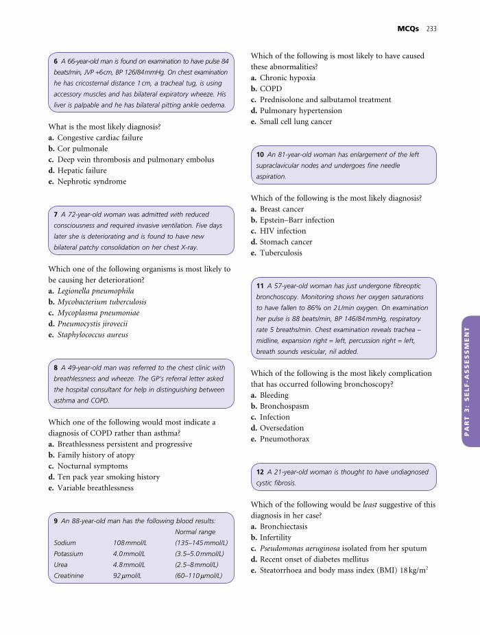
MCQs 233
PA
RT
3:
SE
LF
-AS
SE
SS
ME
NT
6 A 66-year-old man is found on examination to have pulse 84
beats/min, JVP +6 cm, BP 126/84 mmHg. On chest examination
he has cricosternal distance 1 cm, a tracheal tug, is using
accessory muscles and has bilateral expiratory wheeze. His
liver is palpable and he has bilateral pitting ankle oedema.
What is the most likely diagnosis?
a. Congestive cardiac failure
b. Cor pulmonale
c. Deep vein thrombosis and pulmonary embolus
d. Hepatic failure
e. Nephrotic syndrome
7 A 72-year-old woman was admitted with reduced
consciousness and required invasive ventilation. Five days
later she is deteriorating and is found to have new
bilateral patchy consolidation on her chest X-ray.
Which one of the following organisms is most likely to
be causing her deterioration?
a. Legionella pneumophila
b. Mycobacterium tuberculosis
c. Mycoplasma pneumoniae
d. Pneumocystis jirovecii
e. Staphylococcus aureus
8 A 49-year-old man was referred to the chest clinic with
breathlessness and wheeze. The GP’s referral letter asked
the hospital consultant for help in distinguishing between
asthma and COPD.
Which one of the following would most indicate a
diagnosis of COPD rather than asthma?
a. Breathlessness persistent and progressive
b. Family history of atopy
c. Nocturnal symptoms
d. Ten pack year smoking history
e. Variable breathlessness
9 An 88-year-old man has the following blood results:
Normal range
Sodium 108 mmol/L (135–145 mmol/L)
Potassium 4.0 mmol/L (3.5–5.0 mmol/L)
Urea 4.8 mmol/L (2.5–8 mmol/L)
Creatinine 92 mmol/L (60–110 mmol/L)
Which of the following is most likely to have caused
these abnormalities?
a. Chronic hypoxia
b. COPD
c. Prednisolone and salbutamol treatment
d. Pulmonary hypertension
e. Small cell lung cancer
10 An 81-year-old woman has enlargement of the left
supraclavicular nodes and undergoes fi ne needle
aspiration.
Which of the following is the most likely diagnosis?
a. Breast cancer
b. Epstein–Barr infection
c. HIV infection
d. Stomach cancer
e. Tuberculosis
11 A 57-year-old woman has just undergone fi breoptic
bronchoscopy. Monitoring shows her oxygen saturations
to have fallen to 86% on 2 L/min oxygen. On examination
her pulse is 88 beats/min, BP 146/84 mmHg, respiratory
rate 5 breaths/min. Chest examination reveals trachea –
midline, expansion right = left, percussion right = left,
breath sounds vesicular, nil added.
Which of the following is the most likely complication
that has occurred following bronchoscopy?
a. Bleeding
b. Bronchospasm
c. Infection
d. Oversedation
e. Pneumothorax
12 A 21-year-old woman is thought to have undiagnosed
cystic fi brosis.
Which of the following would be least suggestive of this
diagnosis in her case?
a. Bronchiectasis
b. Infertility
c. Pseudomonas aeruginosa isolated from her sputum
d. Recent onset of diabetes mellitus
e. Steatorrhoea and body mass index (BMI) 18 kg/m2

234 Part 3: Self-Assessment
PA
RT
3:
SE
LF
-AS
SE
SS
ME
NT
13 A 23-year-old woman complains of mild chest
tightness and has a provisional diagnosis of asthma.
Which one of the following investigations would be
most likely to confi rm the diagnosis?
a. Antineutrophil cytoplasmic antibodies
b. IgE measurement
c. Peak fl ow diary
d. Skin prick testing
e. Spirometry
14 A 72-year-old man is admitted with a chest X-ray
showing a peripheral lung mass.
Which one of the following investigations is most likely
to yield a histological diagnosis?
a. Endobronchial biopsy
b. Percutaneous computed tomography (CT) guided
lung biopsy
c. Positron emission tomography (PET) scan
d. Staging CT scan
e. Transbronchial needle aspiration of mediastinal
lymph nodes
15 Which one of the following is not a feature of a
normal chest X-ray?
a. The diameter of the heart is <0.5 times the
cardiothoracic diameter
b. The left hemidiaphragm is lower than the right
hemidiaphragm
c. The left main bronchus is more vertical than the
right main bronchus
d. The right hilum is lower than the left hilum
e. The top of the right hemidiaphragm is at the 5th
intercostal space in the midclavicular line
16 A 56-year-old woman is found to have diffuse
parenchymal lung disease.
Which one of the following investigations would be
most likely to reveal an underlying cause for her
condition?
a. Aspergillus precipitins
b. Autoantibody screen
c. Retroviral test
d. Skin prick testing
e. Thrombophilia screen
17 A 73-year-old woman has collapsed on the ward with
a respiratory arrest and has vomited.
Which one of the following pieces of equipment would
be most useful to prevent aspiration?
a. Endotracheal tube
b. Guedel airway
c. Laryngeal mask
d. Mouth guard
e. Nasopharyngeal airway
18 A 61-year-old man has an Epworth Sleepiness Score of
18/24, BP 174/98 mmHg, BMI 37 kg/m2 and is diagnosed
on a home sleep study as having obstructive sleep apnoea.
Which one of the following statements about his
treatment is most true?
a. A mandibular advancement device should be tried
before continuous positive airways pressure (CPAP)
b. As he is starting treatment he does not need to
inform the Driver and Vehicle Licensing Agency
(DVLA) about his condition
c. He should not drive a car until he has lost weight
d. Night sedation will improve sleep quality and reduce
daytime somnolence
e. Nocturnal CPAP is likely to lower his blood pressure
19 A 35-year-old woman commences treatment with
rifampicin, isoniazid, ethambutol and pyrazinamide for
tuberculosis.
Which one of the following methods should be used to
monitor her for side-effects of treatment?
a. Audiometry
b. Full blood count
c. Liver function tests
d. Plasma drug levels
e. Transfer factor

MCQs 235
PA
RT
3:
SE
LF
-AS
SE
SS
ME
NT
20 A 68-year-old man has moderate COPD, causing
shortness of breath on exercise.
Which one of the following treatments will best prevent
progression of his lung disease?
a. Inhaled steroids
b. Pulmonary rehabilitation
c. Salmeterol
d. Smoking cessation
e. Tiotropium
21 A 77-year-old man with known COPD is admitted to
hospital with an exacerbation.
Which one of the following is the most useful indicator
in determining whether antibiotics will be effective in
the treatment of his exacerbation?
a. FEV1 <50% predicted
b. Hyperinfl ated lungs on chest X-ray
c. PaO2 <8 kPa
d. Purulent sputum
e. Respiratory rate >25 breaths/min
22 A 65-year-old woman is discharged from hospital
following an exacerbation of COPD.
Which of the following treatments is least likely to
reduce her risk of hospitalization from future
exacerbations?
a. Early self-management of exacerbations with
antibiotics and steroids
b. Annual infl uenza vaccination
c. Regular long acting β2-agonists and tiotropium
d. Smoking cessation
e. Twice daily inhaled steroids
23 A 38-year-old woman has taken antituberculous
treatment for 1 week and now feels so nauseated that she
refuses to take any more drugs.
Which of the following statements is most correct?
a. Partial treatment of tuberculosis increases the risk of
drug resistance
b. She can be forced to take treatment in the public
interest
c. She has capacity to make this decision and so should
be allowed to make it
d. Treatment should be stopped under the ethical
principle of non-malifi ence (do no harm)
e. Treatment so far is suffi cient to have rendered her
non-infectious
24 A 32-year-old woman is admitted with a pulmonary
embolus.
Which one of the following is an indication for
thrombolysis?
a. Bilateral emboli on computed tomography
pulmonary angiography (CTPA) scan
b. Blood pressure 72/44 mmHg
c. d-dimers 4.9 mg/L (normal <0.3 mg/L)
d. PO2 9.4 kPa (normal 10–13.1 kPa)
e. Recent surgery
25 Which one of the following statements about lung
cancer is most correct?
a. Compression of the left recurrent laryngeal nerve by
mediastinal lymph nodes causes Horner’s syndrome
b. Distant metastases preclude patients from curative
surgery
c. Hypernatraemia is a presenting feature of small cell
lung cancer
d. Secondary tumour deposits in the lung most
commonly originate from prostate
e. Squamous cell lung cancer is more likely than small
cell lung cancer to have metastasized at presentation.
26 Which one of the following respiratory infections is not
correctly paired with its anatomical site?
a. Community acquired pneumonia – alveoli
b. Diphtheria – pharynx
c. Empyema – pleural cavity
d. Infective exacerbation of COPD – lung parenchyma
e. Acute rhinitis – nasal mucosa

236 Part 3: Self-Assessment
PA
RT
3:
SE
LF
-AS
SE
SS
ME
NT
27 Which one of the following respiratory diseases is not
correctly paired with a relevant occupational risk factor?
a. Asthma and working as a baker
b. Lung cancer and working as a publican
c. Mesothelioma and working with isocyanates
d. Extrinsic allergic alveolitis and working in an aviary
e. Pulmonary tuberculosis and working in a hospital
28 A 56-year-old man complains of excessive day time
sleepiness. His wife is concerned that he snores and often
stops breathing when he is asleep.
Which one of the following is most likely to have
caused his condition?
a. BMI 43 kg/m2
b. Diabetes mellitus
c. Hypertension
d. Hypothyroidism
e. Previous tonsillectomy
29 A 39-year-old woman plans to stop smoking. Her risk
of developing which of the following conditions will be
least reduced by smoking cessation?
a. Carcinoma of the bronchus
b. COPD
c. Ischaemic heart disease
d. Laryngeal carcinoma
e. Pulmonary fi brosis
30 A 28-year-old man has nasal obstruction that makes it
diffi cult to breathe. This has been present most days for
the past 15 months. He has a postnasal drip and
intermittent cough.
Exposure to which of the following allergens is most
likely to be causing his symptoms?
a. Grass pollens
b. House dust mite
c. Mould spores
d. Pigeon bloom
e. Tree pollen

PA
RT
3:
SE
LF
-AS
SE
SS
ME
NT
On examination his oxygen saturations are 89%
on air. His chest X-ray shows bilateral midzone
shadowing.
2 Examination
a. Asthma
b. Bronchopneumonia
c. Chronic obstructive pulmonary disease (COPD)
d. Cystic fi brosis
e. Idiopathic pulmonary fi brosis
f. Lobar pneumonia
g. Lung cancer
h. Non-tension pneumothorax
i. Obstructive sleep apnoea
j. Pleural effusion
k. Pulmonary collapse
l. Pulmonary embolus
m. Pulmonary tuberculosis
n. Sarcoidosis
o. Tension pneumothorax
For each of the patients whose examination fi ndings are
described below choose the most likely diagnosis from
the list above. Each diagnosis may be chosen once, more
than once or not at all.
1. A 65-year-old man with fi nger clubbing and
bibasal end-inspiratory crepitations on
auscultation.
2. A 16-year-old man with fi nger clubbing and
widespread coarse inspiratory crepitations on
auscultation.
3. A 75-year-old woman who has a cricosternal
distance of 1 fi nger breadth, intercostal indrawing
on inspiration and poor air entry and expiratory
wheeze on auscultation.
4. A 22-year-old man with decreased expansion,
decreased air entry and hyper-resonance on the
right side of his chest. His trachea is central.
EMQs
1 Microorganisms causing respiratory infection
a. Anaerobic bacteria
b. Chlamydia psittaci
c. Corynebacterium diphtheriae
d. Epstein–Barr virus
e. Haemophilus infl uenzae
f. Infl uenza A virus
g. Legionella pneumophila
h. Moraxella catarrhalis
i. Mycobacterium tuberculosis
j. Mycoplasma pneumoniae
k. Pneumocystis jirovecii
l. Pseudomonas aeruginosa
m. Rhinovirus
n. Streptococcus pneumonia
o. Streptococcus pyogenes
For each of the patients described below choose the
organism most likely to be causing their respiratory
infection from the list above. Each organism may be
chosen once, more than once or not at all.
1. A 22-year-old woman complains to her GP of a sore
throat. On examination she has mild jaundice and
an enlarged liver and spleen.
2. A 17-year-old man with cystic fi brosis has increased
shortness of breath and a cough newly productive of
thick dark green sputum.
3. A 36-year-old man with a 2-day history of shortness
of breath comes to hospital complaining of
haemoptysis. On examination he has crepitations
and bronchial breathing at the right lung base,
which is dull to percussion.
4. A 72-year-old woman with a 2-month history of a
productive cough is sent to hospital by her GP for a
chest X-ray which shows consolidation with
cavitation at the right lung apex.
5. A 29-year-old man with known HIV disease is
admitted with increasing shortness of breath.
237

238 Part 3: Self-Assessment
PA
RT
3:
SE
LF
-AS
SE
SS
ME
NT
5. A 56-year-old woman with decreased expansion,
dull percussion note and decreased air entry on the
right side of her chest. Her trachea is deviated to the
right.
3 History
a. Asthma
b. Bronchopneumonia
c. COPD
d. Cystic fi brosis
e. Idiopathic pulmonary fi brosis
f. Lobar pneumonia
g. Lung cancer
h. Non-tension pneumothorax
i. Obstructive sleep apnoea
j. Pleural effusion
k. Pulmonary collapse
l. Pulmonary embolus
m. Pulmonary tuberculosis
n. Sarcoidosis
o. Tension pneumothorax
For each of the patients whose history is described below
choose the most likely diagnosis from the list above. Each
diagnosis may be chosen once, more than once or not at
all.
1. A 31-year-old woman who has had a productive
cough for 3 months. On further questioning she
admits to weight loss and night sweats. She has
never smoked.
2. A 45-year-old woman complains of a painful rash
on her shins. She also has had a dry cough for the
past 4 months.
3. A 67-year-old man complains of a drooping left
eyelid. His wife has noticed his left pupil is smaller
than his right.
4. A 38-year-old man complains of sudden onset of
pain on the right side of his chest. It is worse on
taking a deep breath. He otherwise feels well, but
has coughed up some clear sputum streaked with
bright red blood.
5. A 27-year-old man complains of waking at night
with a dry cough for the past 4 weeks. He has
recurrent attacks of sneezing and eye irritation.
4 Chest X-ray interpretation
a. Asthma
b. Bronchopneumonia
c. COPD
d. Cystic fi brosis
e. Idiopathic pulmonary fi brosis
f. Lobar pneumonia
g. Lung cancer
h. Non-tension pneumothorax
i. Obstructive sleep apnoea
j. Pleural effusion
k. Pulmonary collapse
l. Pulmonary embolus
m. Sarcoidosis
n. Tension pneumothorax
For each of the patients whose chest X-rays are shown
below choose the most likely diagnosis from the list
above. Each diagnosis may be chosen once, more than
once or not at all.
EMQ 4 X-ray 1.

EMQs 239
PA
RT
3:
SE
LF
-AS
SE
SS
ME
NT
EMQ 4 X-ray 2.
EMQ 4 X-ray 3.
EMQ 4 X-ray 4.
EMQ 4 X-ray 5.

240 Part 3: Self-Assessment
PA
RT
3:
SE
LF
-AS
SE
SS
ME
NT
6 Choice of investigations
a. Avian precipitins
b. Bronchoalveolar lavage
c. Chest X-ray
d. D-dimers
e. Echocardiogram
f. Endobronchial biopsy
g. Fine needle aspirate
h. High-resolution CT scan
i. IgE level
j. Pleural biopsy
k. Skin prick allergy testing
l. Sweat test
m. Transbronchial biopsy
n. Ventilation–perfusion scan
o. Video-assisted thoracoscopy
For each of the patients described below choose the single
investigation that will be most useful in confi rming the
suspected diagnosis from the list above. Each test may be
used once, more than once or not at all.
1. A 28-year-old man who has collapsed and has a
provisional diagnosis of a massive pulmonary
embolus.
2. A 56-year-old man with a pleural effusion suspected
to be caused by Mycobacterium tuberculosis.
3. A 2-year-old girl with failure to thrive thought to be
caused by cystic fi brosis.
4. A 73-year-old woman with a dry cough and reduced
gas transfer thought to be caused by diffuse
parenchymal lung disease.
5. A 17-year-old man with sudden onset shortness of
breath who is thought to have a spontaneous
pneumothorax.
5 Interpretation of investigations
a. Aspiration pneumonia
b. Asthma
c. Bronchiectasis
d. Cardiac failure
e. COPD
f. Empyema
g. HIV-related lung disease
h. Lobar pneumonia
i. Lung cancer
j. Mesothelioma
k. Non-tension pneumothorax
l. Obstructive sleep apnoea
m. Pulmonary embolus
n. Pulmonary fi brosis
o. Pulmonary tuberculosis
For each of the patients whose investigation results are
shown below choose the most likely diagnosis from the
list above. Each diagnosis may be chosen once, more than
once or not at all.
1. A 65-year-old man who has the following
spirometry results:
Before broncho-dilatators (L)
After broncho-dilatators (L)
Predicted (L)
FEV1 1.6 1.7 3.2
FVC 3.2 3.3 4.1
2. A 72-year-old woman who has the following
spirometry results:
FEV1 0.9 L (46% predicted)
FVC 1.0 L (42% predicted)
3. A 46-year-old woman with a ventilation perfusion
scan reported as showing multiple unmatched
perfusion defects.
4. A 72-year-old woman with the following pleural
aspirate results:
Protein 22 g/L
pH 7.58 (normal 7.60)
Glucose 4.7 mmol/L (normal >3.3 mmol/L)
Microbiology – no cells or organisms seen
5. A 25-year-old man with the following pleural
aspirate results:
Protein 48 g/L
pH 7.31 (normal 7.60)
Glucose 1.8 mmol/L (normal >3.3 mmol/L)
Microbiology – neutrophils ++++

EMQs 241
PA
RT
3:
SE
LF
-AS
SE
SS
ME
NT
8 Treatment
a. Adrenaline, chlorphenamine and hydrocortisone
b. Ambulatory home oxygen
c. As required home oxygen by cylinder
d. Bilevel positive airways pressure (BiPAP)
e. Breathe into a paper bag
f. Continuous home oxygen by concentrator
g. Continuous positive airways pressure (CPAP)
h. High-dose inhaled steroids
i. High fl ow oxygen by mask
j. Intravenous magnesium
k. Intubation and ventilation
l. Mandibular advancement device
m. Nebulized salbutamol
n. No treatment
o. Short-term oxygen 28% via nasal cannulae
Normal ranges: pH 7.35–7.45, PaO2 10–13.1 kPa, PaCO2
4.9–6.1 kPa
For each of the patients described below choose the
most appropriate next treatment from the list above.
Each treatment may be used once, more than once or not
at all.
1. A 21-year-old girl with acute shortness of breath
who has the following blood gases on air:
pH 7.62
PaO2 12 kPa
PaCO2 2.2 kPa
2. A 23-year-old man with acute asthma who has the
following blood gases on 60% oxygen:
pH 7.20
PaO2 6.4 kPa
PaCO2 8.5 kPa
3. A 64-year-old man with an Epworth Sleepiness
Score of 19/24 and nocturnal apnoea/hypopnoea
rate of 40 per hour.
4. A 66-year-old woman with stable COPD who has
the following blood gases on air:
pH 7.41
PaO2 6.6 kPa
PaCO2 5.7 kPa
5. A 78-year-old man with an exacerbation of COPD
on 28% oxygen:
pH 7.24
PaO2 5.3 kPa
PaCO2 10.6 kPa
7 Treatment of respiratory infection
a. Amoxicillin and clavulinic acid
b. Benzyl penicillin and fl ucloxacillin
c. Cefuroxime and metronidazole
d. Ciprofl oxacin
e. Erythromycin and amoxicillin
f. Flucloxacillin and cefotaxime
g. Isoniazid and pyridoxine
h. Itraconazole
i. Metronidazole
j. Phenoxymethyl penicillin
k. Pyrazinamide and ethambutol
l. Pyridoxine, rifampicin and ethambutol
m. Rifampicin and isoniazid
n. Rifampicin, isoniazid, pyrazinamide and ethambutol
o. Trimethoprim and sulfamethoxazole
For each of the patients described below choose the most
appropriate fi rst treatment from the list above. Each
treatment may be used once, more than once or not at
all.
1. A 66-year-old man with a mild infective exacerba-
tion of COPD caused by a β-lactamase producing
strain of Haemophilus infl uenzae.
2. A 32-year-old woman with community acquired
pneumonia who is confused, has respiratory rate of
34 breaths/min, urea 9.6 mmol/L and blood pressure
86/44 mmHg.
3. A 92-year-old woman on day 4 of cefotaxime
treatment for respiratory infection who now has
profuse diarrhoea.
4. A 48-year-old man with pulmonary tuberculosis
who has completed 2 months of treatment and now
requires continuation treatment for 4 months.
5. A 17-year-old man with cystic fi brosis with his fi rst
episode of infection with Pseudomonas aeruginosa.
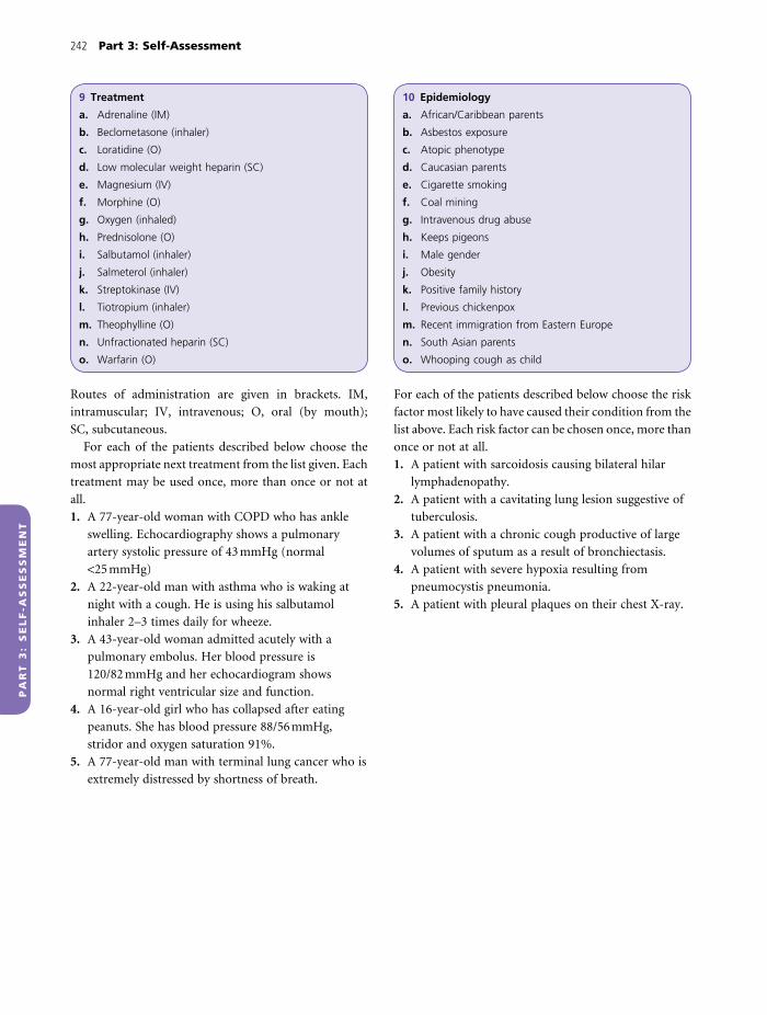
242 Part 3: Self-Assessment
PA
RT
3:
SE
LF
-AS
SE
SS
ME
NT
10 Epidemiology
a. African/Caribbean parents
b. Asbestos exposure
c. Atopic phenotype
d. Caucasian parents
e. Cigarette smoking
f. Coal mining
g. Intravenous drug abuse
h. Keeps pigeons
i. Male gender
j. Obesity
k. Positive family history
l. Previous chickenpox
m. Recent immigration from Eastern Europe
n. South Asian parents
o. Whooping cough as child
For each of the patients described below choose the risk
factor most likely to have caused their condition from the
list above. Each risk factor can be chosen once, more than
once or not at all.
1. A patient with sarcoidosis causing bilateral hilar
lymphadenopathy.
2. A patient with a cavitating lung lesion suggestive of
tuberculosis.
3. A patient with a chronic cough productive of large
volumes of sputum as a result of bronchiectasis.
4. A patient with severe hypoxia resulting from
pneumocystis pneumonia.
5. A patient with pleural plaques on their chest X-ray.
9 Treatment
a. Adrenaline (IM)
b. Beclometasone (inhaler)
c. Loratidine (O)
d. Low molecular weight heparin (SC)
e. Magnesium (IV)
f. Morphine (O)
g. Oxygen (inhaled)
h. Prednisolone (O)
i. Salbutamol (inhaler)
j. Salmeterol (inhaler)
k. Streptokinase (IV)
l. Tiotropium (inhaler)
m. Theophylline (O)
n. Unfractionated heparin (SC)
o. Warfarin (O)
Routes of administration are given in brackets. IM,
intramuscular; IV, intravenous; O, oral (by mouth);
SC, subcutaneous.
For each of the patients described below choose the
most appropriate next treatment from the list given. Each
treatment may be used once, more than once or not at
all.
1. A 77-year-old woman with COPD who has ankle
swelling. Echocardiography shows a pulmonary
artery systolic pressure of 43 mmHg (normal
<25 mmHg)
2. A 22-year-old man with asthma who is waking at
night with a cough. He is using his salbutamol
inhaler 2–3 times daily for wheeze.
3. A 43-year-old woman admitted acutely with a
pulmonary embolus. Her blood pressure is
120/82 mmHg and her echocardiogram shows
normal right ventricular size and function.
4. A 16-year-old girl who has collapsed after eating
peanuts. She has blood pressure 88/56 mmHg,
stridor and oxygen saturation 91%.
5. A 77-year-old man with terminal lung cancer who is
extremely distressed by shortness of breath.

PA
RT
3:
SE
LF
-AS
SE
SS
ME
NT
SAQs
1 A 25-year-old woman with known asthma comes to
accident and emergency complaining of increasing
shortness of breath. On examination she is able to
complete sentences; her respiratory rate is 20 breaths/min,
pulse 96 beats/min, oxygen saturations of 93% on air and
peak fl ow 250 L/min (best 420 L/min). She has a
widespread expiratory wheeze on auscultation.
a. How severe is her asthma attack? Explain your
assessment (2 marks)
b. Outline your immediate management of this patient
(4 marks)
c. Which two investigations would be most useful in
this patient. Explain why (2 marks)
d. Give two measures that could be taken after
discharge to reduce the risk of further exacerbations
(2 marks)
2 A 56-year-old man is admitted through accident and
emergency coughing up blood. He is worried that he may
have lung cancer.
a. State four other causes of haemoptysis (4 marks)
b. Give two risk factors for lung cancer that you would
ask about when taking a history (2 marks)
c. Give two histological types of lung cancer (2 marks)
and for each give one type of treatment that may
cause remission or be curative (2 marks)
3 A 59-year-old man with COPD comes into hospital with
an exacerbation causing respiratory failure. On admission
his blood gases are:
pH 7.26
PaO2 6.7 kPa (10–13.1 kPa)
PaCO2 8.9 kPa (4.9–6.1 kPa)
HCO3− 26 mmol/L (22–28 mmol/L)
a. Give a pathophysiological explanation for each abnor-
mality shown on arterial blood gas testing (3 marks)
b. Describe your immediate management of his
respiratory failure and explain your answer (4 marks).
In outpatients when stable his blood gases are:pH 7.39PaO2 7.0 kPa (10–13.1 kPa)PaCO2 8.1 kPa (4.9–6.1 kPa)HCO3
- 38 mmol/L (22–28 mmol/L)
c. How do you account for the change in pH? (1 mark)
d. Give two possible long-term complications of his
condition based on these results and explain why
each occurs (2 marks)
4 A 53-year-old woman presents to the chest clinic
complaining of a cough which has been present for 3
months. Her cough is embarrassing and restricting her
social activities.
a. How would you classify her cough? (1 mark)
b. Give three causes of cough other than lung disease?
(3 marks)
c. What features in her history would be consistent
with lung disease rather than other causes (b) as the
cause of her cough? (4 marks)
d. Give two investigations you would perform and
explain why you would do each (2 marks)
5 A 48-year-old man is seen in accident and emergency,
complaining of a sharp pain in his right side which is
preventing him from taking a deep breath. He has no
previous history of lung disease and has never smoked.
Examination fi ndings: oxygen saturations 96% on air,
pulse 88 beats/min, blood pressure (BP) 114/76 mmHg,
trachea – midline, expansion left > right, percussion –
resonant right > left, breath sounds vesicular but reduced
on the right, vocal resonance reduced at the right base.
243

244 Part 3: Self-Assessment
PA
RT
3:
SE
LF
-AS
SE
SS
ME
NT
a. What type of pain is he complaining of? (1 mark)
b. Interpret his examination fi ndings (3 marks) and
state the most likely diagnosis (1 mark)
c. Outline the management of this condition
(4 marks)
d. Give two other possible causes of this type of pain
(1 mark)
6 A 76-year-old woman who was living independently is
admitted with community acquired pneumonia. On
examination her Glasgow Coma Score is 14/15, oxygen
saturations are 88% on air, pulse 120 beats/min, BP
80/50 mmHg and respiratory rate 32 breaths/min. Blood
tests show:
Normal range
Sodium 141 mmol/L (135–145 mmol/L)
Potassium 3.7 mmol/L (3.5–5.0 mmol/L)
Urea 10.8 mmol/L (2.5–8 mmol/L)
Creatinine 92 mmol/L (60–110 mmol/L)
a. How severe is her pneumonia? Explain your answer
(3 marks)
b. What factors will you take into consideration
when deciding which antibiotics to prescribe?
(4 marks)
c. Outline the management of her condition in
addition to the use of antibiotics (3 marks)
7 A 26-year-old woman is referred to the medical team by
accident and emergency with a suspected pulmonary
embolus. She presents with a 2-day history of pleuritic
chest pain, worse on inspiration. She is on the oral
contraceptive pill and has sprained her left ankle 2 weeks
ago. Examination fi ndings are: pulse 94 beats/min, jugular
venous pressure (JVP) +1 cm, BP 114/76 mmHg, chest
examination normal, calf diameters left 34 cm, right 32 cm.
D-dimer 0.6 mg/L (normal <0.3 mg/L)
a. How likely is it that she has had a pulmonary
embolus? (2 marks)
b. What are d-dimers? (1 mark) How does her d-dimer
result help in her assessment? (2 marks)
c. Explain how you would confi rm the diagnosis and
treat her pulmonary embolus (5 marks)
8 A 54-year-old woman with a cough has the following
investigation results:
FEV1 1.2 L (48% predicted), FVC 1.47 L (49% predicted),
FEV1/FVC 82%
Gas transfer 54% predicted
Transbronchial biopsy – non-caseating granulomas
a. How do you interpret her lung function results?
(3 marks)
b. What is the most likely diagnosis? (1 mark) Explain
how you made this diagnosis (1 mark)
c. What extrapulmonary abnormalities would you look
for? (4 marks)
d. Outline the treatment of her condition (1 mark)
9 A 42-year-old man, known to be living on the streets, is
admitted with alcohol withdrawal. On examination his
body mass index is 18 kg/m2 and his temperature is
37.8°C. His chest X-ray shows a large cavity in the apex of
the left lung.
a. Give three possible causes for these X-ray fi ndings
(3 marks)
b. What is the most important step that should be
taken in the immediate management of his
respiratory condition? (1 mark)
c. How would you investigate his respiratory condition?
(4 marks)
d. How might his social circumstances affect the
treatment of his respiratory disease? (2 marks)
10 A 27-year-old man sees his GP with a sore throat. He has
no signifi cant past medical history. He has been unwell for
3 days and is unable to eat, although he can take oral fl uids.
On examination his tonsils are enlarged and covered with
a white exudate. He has tender cervical lymphadenopathy.
a. Give two organisms most likely to be causing his
sore throat (2 marks)
b. Would you prescribe antibiotics? (1 mark) Justify
your answer (3 marks)
c. If antibiotics are prescribed, give one that would be
suitable and one that would be contraindicated for
the treatment of his condition (2 marks)
d. Give two complications that may arise following
upper respiratory tract infection (2 marks)

PA
RT
3:
SE
LF
-AS
SE
SS
ME
NT
MCQs Answers
1. a
2. d
3. d
4. c
5. b
6. b
7. e
8. a
9. e
10. d
11. d
12. b
13. c
14. b
15. c
16. b
17. a
18. e
19. c
20. d
245
21. d
22. c
23. a
24. b
25. b
26. d
27. c
28. a
29. e
30. b

PA
RT
3:
SE
LF
-AS
SE
SS
ME
NT
EMQs Answers
11. d
2. l
3. n
4. i
5. k
21. e
2. d
3. c
4. h
5. k
31. m
2. n
3. g
4. l
5. a
41. f
2. k
3. h
4. j
5. g
51. e
2. n
3. m
4. d
5. f
61. e
2. j
3. l
4. h
5. c
71. a
2. e
3. i
246
4. m
5. d
81. e
2. k
3. g
4. f
5. d
91. g
2. b
3. d
4. a
5. f
101. a
2. m
3. o
4. g
5. b

PA
RT
3:
SE
LF
-AS
SE
SS
ME
NT
SAQs Answers
1a. She is having a moderate asthma exacerbation (1
mark) as she has no clinical features of severe asthma
and peak fl ow is >50% predicted (1 mark).
b. (1 mark for each correct answer, maximum 4 marks).
The fi rst part of the management should be to
establish whether the patient needs basic life support
or not (ABC) (1 mark). As this patient does not, the
next step should be general supportive measures
including administration of high fl ow oxygen (1
mark) and specifi c asthma treatment with nebulized
salbutamol (1 mark) and systemic corticosteroids
(1 mark). Other immediate measures would include
securing intravenous access (1 mark) and regular
cardiorespiratory monitoring (1 mark).
c. (1 mark for each correct answer, maximum 2 marks).
• Chest X-ray to establish whether or not she has a
pneumothorax. This is important because it is a
recognized complication of asthma and can be easily
reversed. Chest X-ray will also provide information
about other pathologies that can exacerbate asthma
such as pneumonia (1 mark).
• Infl ammatory blood markers such as white cell
count and C-reactive protein, which may indicate
infection underlying the exacerbation (1 mark).
• Also allow arterial blood gases to quantify hypoxia
and look for hypercapnia or acid–base disturbances
(1 mark).
d. (1 mark per correct answer, maximum 2 marks).
• Regular inhaled steroids
• Self-management plan including regular peak fl ow
monitoring and adjustment of medication
• Allergen avoidance
2a. (1 mark for each correct answer, maximum 4 marks).
Tuberculosis, bronchiectasis, pulmonary embolus,
Goodpasture’s syndrome, pneumonia,
aspergilloma.
b. (1 mark for each correct answer, maximum 2 marks).
Smoking, asbestos exposure (Box 62, p. 101).
c. Non-small cell such as squamous cell,
adenocarcinoma, large cell (1 mark). Curative
treatment could be surgery (1 mark).
Small cell (1 mark). Treatment that may cause
remission is chemotherapy (1 mark).
3a. • He is hypoxic. COPD causes a severe reduction
in alveolar ventilation by obstructing the airways,
preventing air entry, and by alveolar destruction
(emphysema). This can be worse during an
exacerbation. Poor alveolar ventilation results in V/Q
mismatch and reduction in oxygen uptake (1 mark)
• He is hypercapnic. Excretion of CO2 depends on
adequacy of alveolar ventilation. Good ventilation
blows off CO2 and ensures a large diffusion gradient
for CO2 from blood to air. Hypercapnia indicates
alveolar hypoventilation (1 mark) either because of
severe lung damage (in his case) or neuromuscular
disease (Fig. 7, p. 7)
• He is acidotic. The Henderson–Hasselbach equation
describes the relationship between CO2 and H+.
H+ + HCO3− ƨ H2CO3
ƨ H2O + CO2
His acidosis is caused by his hypercapnia which is
driving the equation to the left, increasing
generation of H+ ions (1 mark)
b. • He needs oxygen as without this he is at risk of
hypoxic damage especially to the brain and heart.
However, as he is retaining CO2 he is likely to have
lost his central drive to breathe so too much oxygen
could make things worse (1 mark)
• He therefore should receive oxygen at 24–28%
initially. His blood gases should be checked around
30 min after starting (1 mark)
• If adequate oxygenation cannot be achieved
without an increase in PaCO2 and worsening of
247

248 Part 3: Self-Assessment
PA
RT
3:
SE
LF
-AS
SE
SS
ME
NT
acidosis, he will need non-invasive ventilation/BiPAP
(1 mark)
• Treatment of his COPD exacerbation should be
commenced immediately as improvement in his lung
disease will help his respiratory failure (1 mark).
This should include salbutamol and ipratropium
nebulizers, oral prednisolone and antibiotics
c. Renal retention of bicarbonate has occurred in
response to respiratory acidosis. This has buffered
the acidosis, with a return of pH to normal
(respiratory acidosis with metabolic compensation)
(1 mark).
d. 1 mark per each explained answer. Chronic hypoxia
will cause:
• Cor pulmonale (0.5 mark). Hypoxia causes
pulmonary arterial vasoconstriction, contributing to
pulmonary hypertension and right heart
dysfunction.
• Polycythaemia (0.5 mark) occurs because chronic
hypoxia stimulates erythropoietin production, which
in turn stimulates red blood cell production by the
bone marrow (0.5 mark).
4a. This is a chronic cough as it has lasted longer than 8
weeks (1 mark).
b. Nasal/sinus disease – postnasal drip (1 mark); gastro-
oesophageal refl ux (1 mark); drugs, e.g. ACE
inhibitors (1 mark); impaired swallow with
aspiration (1 mark).
c. (1 mark for each correct answer, maximum 4 marks).
Breathlessness (1 mark), pleuritic chest pain
(1 mark), weight loss/night sweats indicative of
TB/lung cancer (1 mark), night cough and wheeze –
may indicate asthma, signifi cant smoking history
(1 mark), sputum production (although may see
mucus with postnasal drip) (1 mark).
d. (1 mark for each correct answer, maximum 2 marks).
Chest X-ray to look for lung disease, e.g. tuberculosis/
lung cancer (1 mark), spirometry/peak fl ow
recording to look for airfl ow obstruction of asthma/
COPD (1 mark), sputum cultures (if produced) to
look for bacterial infection/acid-fast bacilli (1 mark),
eosinophil count/IgE to look for atopy (1 mark).
5a. Pleuritic chest pain (1 mark).
b. ABC are intact and he is stable from a
cardiorespiratory point of view (normal oxygen
saturations, pulse and blood pressure) (1 mark). Chest
fi ndings are asymmetrical and abnormal on the right
(1 mark) indicating a right-sided pneumothorax (1
mark). The mediastinum is not displaced, so this is
not a tension pneumothorax (1 mark).
c. (1 mark per correct answer, maximum 4 marks). A
chest X-ray should be performed to confi rm the
diagnosis (1 mark). He should be started on high
fl ow oxygen to speed reabsorption of the
pneumothorax (1 mark). If the pneumothorax is
small (<2 cm visible rim of air on chest X-ray) it can
be observed (1 mark). If the pneumothorax is large
(≥2 cm air rim) it should be aspirated (1 mark). If
unsuccessful a chest drain should be inserted
(1 mark).
d. (0.5 mark per correct answer, maximum 1 mark).
Pneumonia, pulmonary embolus, viral pleurisy.
6a. Pneumonia severity is assessed using the CURB-65
score (1 mark). Her CURB-65 score is 5 (Box 88,
p. 151) (1 mark). She thus has severe pneumonia and
is at high risk of death (1 mark).
b. (1 mark per correct answer, maximum 4 marks). Best
guess as to causative organism (1 mark) and likely
microbial antibiotic resistance (1 mark), patient
antibiotic allergy (1 mark), risk of side-effects (e.g.
Clostridium diffi cile diarrhoea) (1 mark), indications
for intravenous therapy (severe illness, inability to
swallow) (1 mark).
c. (1 mark per correct answer, maximum 3 marks). She
requires oxygen to treat hypoxia (1 mark) and IV
fl uids to maintain blood pressure (1 mark). She
should be referred to intensive care (ICU) to
consider admission (1 mark). She needs careful
monitoring (oxygen saturations, respiratory rate,
blood pressure, Glasgow Coma Score, urinary
output, electrolytes) (1 mark) and may need other
supportive care on ICU including ventilation and
inotropic support.
7a. By Wells criteria (Table 6, p. 130) she scores 7.5 (i.e.
swollen left calf –3, other diagnoses less likely –3,
some immobilization from sprained ankle –1.5)
(1 mark), which indicates a high probability of
her having had a pulmonary embolus (PE).
b. d-dimers are cross-linked fi brin degradation
products formed by clot breakdown (1 mark).

SAQs Answers 249
PA
RT
3:
SE
LF
-AS
SE
SS
ME
NT
Elevated d-dimers are unhelpful as they can be
produced by clot anywhere in the body (1 mark) and
so do not add useful information to the clinical risk
assessment (1 mark). A normal d-dimer result would
have been useful in ruling out thromboembolic
disease as it has >90% negative predictive value for
pulmonary embolus.
c. (1 mark per correct answer, maximum 5 marks). She
should be started on low molecular weight heparin
to prevent further clot on the presumption that she
has had a PE (1 mark). Imaging (V/Q scan or CTPA)
should be performed to confi rm the diagnosis (1
mark). Once confi rmed she should be commenced
on warfarin (1 mark). Low molecular weight heparin
can be stopped when she is fully anticoagulated with
warfarin (1 mark). She should stay on warfarin for 6
months (1 mark). She needs investigation for a
predisposing cause (pelvic ultrasound,
thrombophilia screen).
8a. A proportionate reduction in FEV1 and FVC (ratio
>70%) (1 mark) indicates restrictive lung disease (1
mark). She also has reduced gas transfer indicating
probable involvement of the lung parenchyma (1
mark).
b. Non-caseating granulomas are the pathological
hallmark (1 mark) of sarcoidosis (1 mark).
c. (1 mark per correct answer, maximum 4 marks).
Extrapulmonary manifestations of sarcoidosis are
given in Box 121, p. 210.
d. As she has signifi cant impairment of pulmonary
function (0.5 mark) immunosuppression with
prednisolone should be tried (0.5 mark).
9a. Tuberculosis should be considered to be the
diagnosis until proven otherwise (1 mark). Other
causes of cavitating lung disease include primary
(1 mark) and secondary (1 mark) lung cancer,
pulmonary embolus (1 mark) and bacterial
pneumonia (1 mark) (Box 108, p. 190).
b. You should presume he has tuberculosis. Infection
control is a priority and he should be isolated in a
side room to prevent spread to staff and patients
(1 mark).
c. (1 mark per correct answer, maximum 4 marks).
Three sputum samples should be examined
microscopically (1 mark) with Ziehl–Neelsen
staining to look for acid-fast bacilli, then cultured to
look for Mycobacterium tuberculosis (1 mark).
Tuberculin skin test (1 mark) (Mantoux, Heaf) will
detect an immune response to TB antigen. HIV
testing may indicate underlying immunodefi ciency
(1 mark). A thoracic CT scan will help to clarify
nature of the cavity and look for other causes
(1 mark).
d. (1 mark per correct answer, maximum 2 marks). He is
living on the streets and appears to be alcohol-
dependent. This may make it diffi cult for him to
adhere to therapy and attend for follow-up (1 mark).
Additionally, he is unlikely to have regular health
care from his GP (1 mark). His general health and
nutrition appears poor (low body mass index),
which may make it diffi cult for him to heal (1 mark).
Assuming that he has TB he will need directly
observed therapy. Contact tracing will also be
diffi cult.
10a. His infection is most likely to be caused by a virus,
e.g. rhinovirus (1 mark) or group A β-haemolytic
streptococcus (Box 98, p. 170) (1 mark).
b. No (1 mark).
(1 mark for each correct answer, maximum 3 marks).
The infection is more likely to be viral (unresponsive
to antibiotics) than bacterial (1 mark) and these
aetiologies cannot be distinguished using clinical
features (1 mark). Also, sore throats are usually self-
limiting, whatever the organism (1 mark). He does
not meet NICE criteria for prescription of antibiotics
(Box 103, p. 171) (1 mark).
c. Suitable – penicillin or erythromycin (1 mark).
Contraindicated – amoxicillin or ampicillin (cause
rash in patients with Epstein–Barr virus) (1 mark).
d. (1 mark for each correct answer, maximum 2 marks).
Complications of upper respiratory tract infections
depend on the organism and include: common cold –
exacerbation of underlying lung disease, e.g. COPD
(1 mark); group A streptococci (Box 101, p. 171)
(1 mark for any correct complication); diphtheria
(Box 100, p. 171) (1 mark for any correct complication).


Index of cases by diagnosis
Case 1 Respiratory arrest, 41
Case 2 Asthma – diagnosis, 46
Case 3 Asthma – treatment, 51
Case 4 Asthma – acute exacerbation, 60
Case 5 COPD diagnosis, 68
Case 6 Smoking cessation, 75
Case 7 Exacerbation of COPD, 80
Case 8 Type II respiratory failure (COPD), 87
Case 9 Long-term complications of chronic lung
disease, 92
Case 10 Non-small cell lung cancer, 98
Case 11 Small cell lung cancer, 106
Case 12 Mesothelioma, 112
Case 13 Pneumothorax, 117
Case 14 Pleural effusion, 124
Case 15 Non-massive pulmonary embolus, 129
Case 16 Massive pulmonary embolus, 139
Case 17 Community-acquired pneumonia, 148
Case 18 Hospital-acquired pneumonia, 156
Case 19 Postoperative breathlessness, 162
Case 20 Upper respiratory tract infection, 169
Case 21 Bronchiectasis, 174
Case 22 Cystic fi brosis, 179
Case 23 Pulmonary tuberculosis and HIV
infection, 186
Case 24 Tuberculosis – treatment, 196
Case 25 Tuberculosis – public health, 201
Case 26 Sarcoidosis, 207
Case 27 Diffuse parenchymal lung disease (pulmonary
fi brosis), 213
Case 28 Extrinsic allergic alveolitis, 220
Case 29 Obstructive sleep apnoea, 226
251


Index
Note: page numbers in italics refer to fi gures, those in bold refer to tables
abdominal pain, control 166adaptive immunity 10
infections 13–14adherence to treatment 199adrenal glands, bronchogenic carcinoma
metastases 108adrenaline, anaphylaxis 84air bronchograms 151
tuberculosis 190airfl ow obstruction 4, 48
causes 70COPD 70–1, 95elastic recoil loss 71mechanisms 73variable 49
airway 2–4air fl ow 4infl ammation 48, 61lower 4lumen radius 4, 5management 42–3obstruction causes 70opening 41remodelling 58resistance 4secretion clearance in cystic fi brosis 183upper 2–4
Airway, Breathing, Circulation (ABC) 36, 41confusion in elderly woman 87
allergens 12anaphylaxis 84asthma 46, 222extrinsic allergic alveolitis 222nasal obstruction 236peanuts 242radiographic contrast 136rhinitis 222skin prick testing 49
alveolar macrophages 9alveolar ventilation, impaired 7alveolar–capillary interface, surface area
reduction 7alveolar–capillary membrane 5, 6
gas diffusion 35thickness increase 7
alveoli 4–5alveolitis 12
diagnosis 220–1extrinsic allergic 12, 221–5
Ambu bag see bag, valve and maskamikacin 205aminopenicillins, rash 172aminophylline 58amosite 112amoxicillin 154, 155analgesia
abdominal pain 166mesothelioma 115
anaphylaxis 12COPD exacerbation 83–4
angiotensin-converting enzyme (ACE), serum levels in sarcoidosis 209, 210
ankle swelling 92–7antibiotics
administration routes 39allergic reactions 84bronchiectasis 177–8COPD exacerbation 83cystic fi brosis 183, 184pneumonia treatment 153–4, 158,
166prescribing 39regime for penicillin resistance 154respiratory disease treatment 38–9sore throat 170–1spectrum of action 39, 40
anti-embolism stockings 164–5antidiuretic hormone (ADH) 109, 110antigen-presenting cells (APCs) 10, 13antihistamines
allergic rhinitis treatment 56anaphylaxis 84
antimuscarinics 63COPD 73, 74nebulized 61
antituberculous drugs 192, 193, 196, 197
directly observed therapy (DOT) 203drug interactions 198duration of treatment 199hepatic toxicity 197importance of treatment 203refusal to take 205, 235resistance 196–7side effect monitoring 234treatment regimen 199
anxiety, palliative care 40approach to patient 17–40
antibiotic use 38–9breathlessness 17–19examination 22–9
253
follow-up 28–9history taking 27–9investigations 29–35palliative care 39–40presenting complaint 17putting it together 27–9respiratory disease treatment 38respiratory emergencies 35–8
arms, examination 24–5arterial blood gases 32–3
asthma 62–3, 64confusion in elderly woman 87–8COPD 82, 90pneumonia 158pulmonary embolism 140, 141
asbestos exposure 68, 112–16respiratory disease risk 19
asbestosis 113asterixis 25asthma 4, 8, 46–50, 234, 243
acute exacerbation 61airfl ow obstruction 48, 70allergens 222childhood cough 174chronic 48, 53COPD differential diagnosis 70corticosteroid therapy 52–3deaths 66diagnosis 48, 220education 66exacerbations 51, 52, 66
treatment on discharge 65extrinsic 47family history 46hospital discharge 65hypersensitivity reaction 222intrinsic 47–8late-onset 48life-threatening 60–7lung examination 28near-fatal 64pathogenesis 47peak expiratory fl ow rate 31peak fl ow measurement 49poorly controlled 51–9respiratory disease risk 19self-management plan 57, 66stepped care 53symptom control 51, 52treatment 241, 242
aims 51triggers 47, 66

254 Index
atelectasis 164, 165breathlessness following surgery 165–8management 166
atopy 47family history 46
autonomy 145, 218azathioprine 217, 218azithromycin 154
β2-agonists 56, 57bronchodilatation management 61COPD 73, 74inhaled 65
bacille Calmette–Guérin (BCG) vaccination 191–2
tuberculosis contacts 202bag, valve and mask 43, 44basic life support 36
respiratory arrest 41beclometasone 53benzodiazepines, doctrine of double effect
40best interests of patient 44, 145–6bi-level positive airway pressure (BiPAP) 37,
89, 90bicarbonate 6, 33biopsy 34
extrinsic allergic alveolitis 223idiopathic interstitial pneumonia 217liver 108lung cancer 108mesothelioma 114sarcoidosis 209
bird antigens 222, 224blood vessels, lung cancer local spread 101body fl uid compartments 93body mass index, low 96brachial plexus, lung cancer local spread
101brain natriuretic peptide (BNP) 82brainstem
chemosensitive area 1respiratory centre 7
breast cancer, pleural effusion 127–8breath sounds 24breathing 2
air fl ow 4control 1patient examination 23support 41see also Airway, Breathing, Circulation
(ABC)breathlessness 17–19
cardiac disease 156, 157chest pain 148–55chronic 220–5COPD 68–74
acute exacerbation 80–6cough 232following surgery 162–8, 232investigations 165pleural effusion 124–8
asbestos exposure 112–16causes 18, 19consultation 18
effect on patient’s life 19onset 18–19palliative care 40respiratory disease 17–19
pneumonia 156–61pulmonary embolism 156–7stroke patient 156–61treatment 241wheezing 233worsening on exertion 213–19
British Thoracic Society, pneumothorax classifi cation 120
bronchi 4bronchiectasis 174, 175, 232
causes 176diagnosis 175, 176pathology 176risk factors 242sputum production 176–7traction 215treatment 177–8tuberculosis 189
bronchitis, chronic 69, 70–1cough 98
bronchoalveolar lavage, sarcoidosis 209bronchodilatation
asthma acute exacerbation 63–4management 61
bronchodilatorsCOPD 73, 74cystic fi brosis 183nebulized 61, 65
short-acting 83speed of action 61
bronchoscopy 33–4complications 233lung cancer 100, 108sarcoidosis 209tuberculosis 192
budesonide 53nebulized 61
budgerigar exposure 221, 224Burkholderia cepacia, cystic fi brosis chest
infections 180
C-reactive protein (CRP)COPD 82pneumonia 158
capacity, mental 145capillaries 5
hydrostatic/oncotic pressure 92capillary wall leaks 92carbon dioxide
accumulation in ventilation 43–4respiratory rate 1retention 25, 139transport 6see also PaCO2
carboxyhaemoglobin 6carcinogens 15cardiac disease
breathlessness 156, 157see also right heart failure; right heart
infarctioncatheter embolectomy 145
CD4+ T cells 10‘ceiling of therapy’ 90cephalosporins, pharmacology 159cervical lymph nodes
examination 25lymphadenopathy 244
cetirizine 56chemotherapy, lung cancer 110chest
auscultation 26, 27examination 26–7percussion 26, 27tightness 46–50
chest drain, intercostal 121–2chest pain 22, 243–4
asbestos exposure 112–16breathlessness 148–55right-sided 117–23see also pleuritic chest pain
chest wall 2examination 24lung cancer local spread 101
chest X-ray 29–31abnormal 106–11asthma 62chronic breathlessness 221chronic cough 175COPD 82, 93, 94cystic fi brosis 182hilar lymphadenopathy 207, 208hoarseness 99interpretation 238, 239normal 30–1, 234OSCE checklist for interpretation 30pleural effusion 124, 125, 150, 151pneumonia 149–50pneumothorax 117–18pulmonary fi brosis 214sarcoidosis 207, 208tuberculosis 190
Cheyne–Stokes respiration 23Chlamydia, pneumonia 152chlorphenamine 84chronic obstructive pulmonary disease
(COPD) 4, 8, 69acute exacerbation 80–6
causes 83management 83
airway obstruction 70–1ankle swelling 92–7antimuscarinics 63asthma differential diagnosis 70breathlessness 19
following surgery 163–8chest wall examination 24cough 98defi nition 69diagnosis 69, 220drug treatment 73–4exacerbation 93, 235, 243forced expiration 73investigations 82lung examination 28management 72mild 73

Index 255
moderate 74, 235multidisciplinary team interactions 96–7non-invasive ventilation 89–90optimization 166pneumonia differential diagnosis 80, 81progression 93, 94
minimizing 73pulmonary rehabilitation 73, 74respiratory drive 96respiratory failure Type II 88right heart failure 93, 94, 95risk 75severe 74severity 71, 72smoking 80, 92–7, 95, 98
cessation 74, 78symptom relief 73–4tracheal tug 25, 81treatment 241, 242vaccinations 84, 85worsening airfl ow obstruction 95
chrysotile 112ciprofl oxacin 205circulation
deoxygenated blood 8–9oxygenated blood 9see also Airway, Breathing, Circulation
(ABC)clarithromycin 154, 205Clinical Knowledge Summaries Service
(NHS) 171Clostridium diffi cile diarrhoea 160–1clotting, initiation 132clotting factors 132coagulation cascade 132collapse of elderly woman 139–47
assessment 139–40differential diagnosis 140
collapse of man in hospital 232common cold 170compensation, mesothelioma 115compliance with treatment 199computed tomography (CT) 33
asbestos exposure 114lung cancer 100respiratory arrest 44tuberculosis 192see also high-resolution computed
tomography (HRCT)computed tomography (CT)-guided biopsy
lung cancer 108mesothelioma 114
computed tomography pulmonary angiogram (CTPA) 135, 136
pulmonary embolism diagnosis 142, 143concordance with treatment 199confusion 87–91, 139consciousness
Glasgow Coma Scale assessment 88reduced 233
consent to treatment 145continuous positive airway pressure
(CPAP) 37, 89atelectasis management 166obstructive sleep apnoea 231
cor pulmonale 95corticosteroids
airway infl ammation reduction 61allergic rhinitis treatment 56anaphylaxis 84asthma treatment 52–3, 61–2, 64–5nebulized 61oral for COPD exacerbation 83pharmacology 52speed of action 62see also steroids, inhaled
cough 9, 10, 19–22, 75–9, 232asthma 55character 21childhood 174chronic 174–8, 243differential diagnosis 174duration 20–1history taking 20–1hoarseness 98–105lung cancer 106–7nocturnal 48non-caseating granuloma 244palliative care 40timing 21weight loss 186–95
Coxiella burnetii, pneumonia 152crepitations 213, 214cricosternal distance 26, 233
COPD 81, 82crocidolite 112cryptogenic fi brosing alveolitis see pneumonia,
idiopathic interstitialCURB-65 score, pneumonia 151cyanosis 25
confusion in elderly woman 87, 88cystic fi brosis 179–85
chest infections 180counselling 184investigations 181–2lung effects 180lung transplantation 184respiratory failure 184support 183treatment 183undiagnosed 233
cystic fi brosis transmembrane receptor (CFTR) mutations 179, 181
cytotoxic T cells 10
daytime sleepiness 227–8, 236decongestants, allergic rhinitis treatment 56deep vein thrombosis
diagnosis 136embolization to lungs 132–3formation 132investigations 131pleuritic chest pain 129–38prevention 137risk factors 130smoking 129treatment 133–4
dehydration, systemic 166–7depression, palliative care 40detention for treatment 205
diet, cystic fi brosis 183, 184dietitians 97diffuse parenchymal lung disease (DPLD) 215,
216of unknown cause 216
D-dimers 131, 140, 141diphtheria 171diuretics, oedema management 96DNAase, recombinant human 183doctrine of double effect 40Doppler echocardiography 95dorsal group of neurons 1driving
daytime sleepiness 227obstructive sleep apnoea 230
duplex ultrasound, deep vein thrombosis diagnosis 136
dyspnoea 17–19MRC score 17, 18, 68see also breathlessness
echocardiogram for pulmonary embolism 142, 146
eformoterol 74elastic recoil 71electrocardiogram (ECG)
asthma 62COPD 93, 94pulmonary embolism 141two-dimensional 95
embolectomy 145emphysema 69, 71
family history 68pathology 72
endothelial damage, deep vein thrombosis 132
endotracheal tube 43ENT examination, hoarseness 99Epstein–Barr virus (EBV) 172Epworth Sleepiness Score 227, 234, 241erythema nodosum 207–12
causes 208erythromycin 154, 155ethambutol 192, 197, 198
side effects 198–9ethics 145–6, 218extrinsic allergic alveolitis 12, 221–5
classifi cation 224hypersensitivity reaction 222lung biopsy 223pathogenesis 223treatment 224
eye test, ethambutol treatment 198–9eyes, examination 25
factor V Leiden 137fi breoptic bronchoscopy 33–4
complications 233lung cancer 108sarcoidosis 209
fi brinolysis 132fi ne needle aspiration (FNA) 34
supraclavicular lymph nodes 108, 233fi nger clubbing 24
chronic cough 174

256 Index
fl exible bronchoscopy, tuberculosis 192fl uid balance, breathlessness after surgery
166–7fl uticasone 53forced expiratory volume in 1 s (FEV1) 31,
32COPD patient 70–1sarcoidosis 210
forced vital capacity (FVC) 31, 32COPD patient 70–1sarcoidosis 210
formoterol 57furosemide (frusemide) 96
gas transfer 35diffuse parenchymal lung disease 216
gas transport 5–6gastro-oesophageal refl ux, cough 21Geudal airway see oropharyngeal airwayGhon’s focus 187Glasgow Coma Scale 88
collapse of elderly woman 139granulomas
caseating 208sarcoidosis 207, 208tuberculosis 187, 188
grass pollen, allergic reaction 49–50
Haemophilus infl uenzaechronic cough 175COPD exacerbation 83cystic fi brosis chest infections 180
haemoptysis 21–2, 243hands, examination 24–5Heaf test 191, 209Henderson–Hasselbalch equation 1, 33
asthma 62–3PaCO2 elevation 87
heparinlow molecular weight 133, 134, 136,
146unfractionated 146
hereditary thrombophilia 137high-resolution computed tomography
(HRCT)chronic cough 175, 176pulmonary fi brosis 215sarcoidosis 210
histamine, anaphylaxis 84HIV infection
contact identifi cation 193pre-test counselling 193with tuberculosis 192–3
hoarseness 98–105investigations 99
homelessness 202–3house dust mite, allergic reaction 49–50hypercapnia 8, 37–8, 88hypersensitivity reactions 12–13, 222hypertension, obstructive sleep apnoea 228,
229hyponatraemia, lung cancer 109–10hypopnoea 227, 241hypotension, management 139hypothyroidism 221
hypoxia 8, 36collapse of elderly woman 139confusion in elderly woman 87correction 80cyanosis 87, 88detection 6life-threatening asthma 60management 95–6nail discolouration 25pneumonia 158polycythaemia 95
immune memory 10–11immune response, excessive 14immune system 10–11immunoglobulin E (IgE) 12immunosuppressant drugs
extrinsic allergic alveolitis 224pulmonary fi brosis 217, 218sarcoidosis 211
immunosuppression 14respiratory disease risk 19
infections 13–15, 235asthma acute exacerbation 62clinical impact 14–15management 166organisms 237pleural 189site 14–15treatment 177–8, 240–1virulence 14
infectious mononucleosisaminopenicillin-induced rash 172diagnosis 172
infl ammation, lungs 11–12infl uenza vaccination 84, 85informed consent 145inhalers 53–4
preventer/reliever 54–5innate immunity 10
infections 13–14insomnia, palliative care 40insulin, pancreatic production in cystic
fi brosis 181intensive care, near-fatal asthma 64intercostal chest drain 121–2international normalized ratio (INR) 137, 146,
147interpreters 189interstitial fl uid, volume control 92intracranial haemorrhage, respiratory arrest
44intrapleural pressure, negative 118, 119intravenous fl uids, hypotension
management 139investigations 240ipratropium bromide
COPD exacerbation 83nebulized 61, 63, 64
isoniazid 192, 197, 198, 205latent tuberculosis prevention 201–2
jugular venous pressure 25
Kussmaul’s respiration 23
lactic dehydrogenase 126–7language barriers 189laryngeal cancer 98
hoarseness 99laryngeal lesions 98laryngeal mask airway 43larynx 3–4
innervation 99nerve damage 98
Legionella pneumophila 152legs, examination 27leukotriene receptor antagonists 58liver
biopsy 108enlarged 107palpable 93ultrasound 108
lumbar spine, lung cancer metastases 103–4
lung(s)anatomy 1–2asbestos effects 112cavitation 189, 190, 242, 244collapse 28consolidation 28, 189, 233cystic fi brosis effects 180expansion 26honeycombing 215infl ammation 11–12lymphatic drainage 107pathological processes 11–16peripheral mass 234physiology 1–2scarring 178trapped particle removal 9
lung biopsy 34, 217extrinsic allergic alveolitis 223
lung cancer 100, 107, 235asbestos exposure 112, 113breathlessness 69, 242causes 101chemotherapy 110chest X-ray abnormality 106–11classifi cation 102hoarseness 98, 99hyponatraemia 109–10investigations 100–1, 107–8local spread 101metastatic complications 103–4,
107non-small cell 102paraneoplastic phenomena 110PET scan 103prognostic factors 109radiotherapy 110rapid access clinic 106risk 75risk factors 101secondary tumors 100small cell 102, 108, 109smoking 101spinal cord compression from
metastases 103squamous cell carcinoma 101–2tumour staging 102, 108–9

Index 257
lung diseasecough 21–2diffuse parenchymal 215, 216examination 28interstitial 215, 220–1parenchymal 234postoperative complications 162–8symmetrical 69
lung function 31–3asthma 222chronic cough 175extrinsic allergic alveolitis 222sarcoidosis 210tests 146
lung transplantationcystic fi brosis 184idiopathic pulmonary fi brosis 218
lung volumes 35diffuse parenchymal lung disease 216
lymphadenopathy 25cervical 244hilar 207, 208mediastinal 100supraclavicular 107, 108
lymphatic obstruction 92lymphocytosis, sarcoidosis 209
magnesium sulphate, intravenous 63, 64magnetic resonance imaging (MRI) 33mandibular advancement device 231Mantoux test 191, 209Medical Research Council (MRC) dyspnoea
score 17, 18, 68mesothelioma 112, 114
asbestos exposure 113management 115prognosis 114
metabolic acidosis, bicarbonate 33metastases, lung cancer 103–4metered dose inhalers 53–4metronidazole, pharmacology 160mometasone 56montelukast 58Moraxella catarrhalis, COPD exacerbation
83mouth, examination 25mucociliary clearance 13mucociliary escalator 9, 10multidisciplinary team, interactions in COPD
management 96–7Mycobacterium tuberculosis 186
drug resistance 196–7immune response 188immunological tests 203metabolic states 196see also tuberculosis
Mycoplasma pneumoniae 152
nail discolouration 25nasal cannulae 36nasal disease, cough 20–1nasal fl ow measurement 229–30nasal obstruction, allergens 236nasal polyps 55
cystic fi brosis 180
nasopharyngeal airway 42insertion 43
National Institute for Clinical Excellence (NICE), antibiotics in patients with sore throat 171
nebulizers 61neck
examination 25sympathetic chain effects of lung cancer
local spread 101negative pressure rooms 204neoplasia 15–16neutrophilia 83nicotine
addiction 77formulations 78
nicotine replacement therapy 74, 78non-malefi cence 218nose 2–3
see also nasal entriesnotifi able disease, tuberculosis 192
obesity, obstructive sleep apnoea 226–31obstructive sleep apnoea 4, 226–31, 234
behavioural advice 230cardiovascular risk 229continuous positive airway pressure 231diagnosis 228–9hypertension 228mandibular advancement device 231severity 230surgery 231treatment 230–1
occupational exposures 236see also asbestos exposure
occupational therapy 97oedema 92
management 96Ohm’s law 4oncogenes 16opiates, doctrine of double effect 40oropharyngeal airway 42
insertion 43OSCE checklist
chest X-ray interpretation 30peak fl ow measurement 35respiratory examination 23
oxygensupply to pulmonary tissues 9transport 6see also PaO2
oxygen delivery devices 36–7oxygen mask with reservoir bag 36, 37oxygen saturation measurement 6oxygen therapy
CO2 retention risk 88confusion in elderly woman 87COPD 95–6high-fl ow 119–20humidifi ed 158–9hypoxia 80, 139life-threatening asthma 60long-term 96pneumothorax 119–20
oxygen–haemoglobin dissociation curve 6
P pulmonale 93, 94PaCO2 32–3
asthma acute exacerbation 62–3, 64confusion in elderly woman 87
palliative care 39–40COPD 97doctrine of double effect 40lung cancer 110
metastases 104–5pancreas, cystic fi brosis effects 180–1pancreatic enzyme replacement, cystic
fi brosis 183, 184PaO2 32–3
asthma acute exacerbation 62–3paraneoplastic syndromes 110pathological diagnosis sampling 33–5patient, best interests 44, 145–6peak expiratory fl ow rate (PEFR) 31
asthma 62, 64morning dipping 65, 66
peak fl owasthma acute attack 63measurement
asthma 49OSCE checklist 35
peanut allergy 242pelvic ultrasound 137penicillin
allergy 84pharmacology 153resistance 154
pericardial tamponade 140pets, asthma 50pharyngeal infection 169pharynx 3physiotherapy
airway secretion clearance in cystic fi brosis 183
atelectasis management 166COPD 97
pleura 2infection 189
pleural aspirateappearance 125–6glucose levels 127investigations 126–7lactic dehydrogenase 126–7pH 127
pleural aspiration 125pleural biopsy, percutaneous 127pleural effusion
asbestos exposure 112–13aspirate appearance 125–6aspiration 125breathlessness 124–8causes 125chest X-ray 124, 125, 150, 151development 124–5differential diagnosis 127exudative 126, 127investigations 113–14, 125lung examination 28management 115percutaneous aspiration 34percutaneous pleural biopsy 127

258 Index
pleural effusion (cont.)pleurodesis 128protein concentration 126thoracoscopy 127transudates 126, 127treatment 127–8
pleural plaquesasbestos exposure 113risk factors 242
pleural rub 130pleural space 118pleurectomy 128pleuritic chest pain 117–23
breathlessness 148–55causes 118, 148deep vein thrombosis 129–38differential diagnosis 129, 148investigations 131pulmonary embolism 129–38treatment 133–4
pleurodesismedical 128pleural effusion management 115surgical 128
pneumococcal vaccination 84, 85Pneumocystis pneumonia 193, 194, 242pneumocytes 5pneumonia 244
antibiotic therapy 153–4aspiration 158–61atypical 151, 153breathlessness 156–61community-acquired 159COPD differential diagnosis 80, 81CURB-65 score 151diagnosis 148hospital-acquired 158, 159, 166idiopathic interstitial 216–17investigations 149–51, 157–8lobar 150microbiological tests 153organisms causing 151, 152pathology 149pleuritic chest pain 148–55severity 151
pneumothoraxaspiration 120–1British Thoracic Society classifi cation
120causes 119diagnosis 148iatrogenic 119intercostal chest drain 121–2investigations 117–18lung examination 28pathogenesis 118–19patient education 122–3pleural hole sealing 122recurrence risk 122–3secondary 64spontaneous 119tension 121traumatic 119treatment 119–22
Poiseuille’s law 4
polycythaemia 95management 96
positron emission tomography (PET) 33lung cancer 103
pregnancy, cystic fi brosis 184Pseudomonas aeruginosa, cystic fi brosis chest
infections 180pulmonary angiogram 135
risks 136see also computed tomography pulmonary
angiogram (CTPA)pulmonary arteries, assessment 95pulmonary circulation 8–9pulmonary complications, postoperative
162–8pulmonary defences 9, 10pulmonary disease, cough 20pulmonary embolism 244
breathlessness 156–7diagnosis 142, 148diagnostic tests 134–6embolectomy 145investigations 131, 140–2massive 133, 134, 140–7non-massive 133pathophysiology 140pleuritic chest pain 129–38thrombolysis 143–5, 235treatment 133–4, 242warfarin 136–7, 146, 147Wells criteria 130
pulmonary embolus, ventilation–perfusion scanning 134–5
pulmonary fi brosis 69, 213–19lung examination 28management 218natural history 217treatment 217, 218
pulmonary infection 164, 165pulmonary oedema, lung examination 28pulmonary rehabilitation, COPD 73, 74pulmonary tissues, oxygen supply 9pulse oximetry 230pyrazinamide 192, 197, 198, 205
Q fever 152
radiographic contrast, allergy 136radiotherapy
lung cancer 110mesothelioma 115painful metastases 104, 105
rash 207–12aminopenicillins 172
recombinant tissue plasminogen activator (rTPA) 144
recurrent laryngeal nerves 99lung cancer local spread 101
respirationaccessory muscle use 81, 233elastic recoil 71
respiratory acid–base disturbance 33respiratory acidosis
bicarbonate 33PaCO2 elevation 87, 88
respiratory alkalosis 63respiratory arrest 41–5
aspiration prevention 234causes 41–2diagnosis 44
respiratory centre 1, 7respiratory disease 17–19
breathlessness 68childhood 19chronic breathlessness 220–5occupational risk 236postoperative complications 162–8risk factors 19treatment approaches 38
respiratory drive, COPD 96respiratory examination, OSCE checklist 23respiratory failure 6–8, 184
pneumonia 158Type I 7, 94Type II 7–8, 88
respiratory muscles 2respiratory nurses 96respiratory rate, asthma acute attack 63respiratory secretions, clearance 177respiratory stimuli 2respiratory support 36–8respiratory system, HIV effects 193, 194respiratory tract infections, upper
complications 169symptom resolution 172
resuscitationcollapse of elderly woman 139see also Airway, Breathing, Circulation
(ABC)reticular shadowing 214rhinitis
allergens 222allergic 55–6hypersensitivity reaction 222treatment 56
rifampicin 192, 197, 198, 205latent tuberculosis prevention 201–2
right heart catheterization 95right heart failure 93, 94, 95
management 96right heart infarction 140
salbutamol 57acute exacerbation of asthma 61COPD exacerbation 83cystic fi brosis 183nebulized 61, 63, 64speed of action 61
salmeterol 56, 57COPD 74cystic fi brosis 183
sarcoidosis 207–12investigations 209–10monitoring 211risk factors 242staging 211tissues affected 210treatment 211tuberculosis differential diagnosis 207
sinus disease, cough 20–1

Index 259
sinus tachycardia 41sinusitis, cystic fi brosis 180skin prick testing 49sleep see daytime sleepiness; Epworth
Sleepiness Score; obstructive sleep apnoeasleep study 228–30smoking 75–9
ankle swelling 92–7breathlessness 68cessation 74, 76–7, 236COPD 80, 92–7, 95, 98deep vein thrombosis 129diseases 75–6hoarseness 98–105lung cancer 101, 107obstructive sleep apnoea 227pack years 19, 75, 98, 107respiratory disease risk 19
sneezing 9, 10snoring 226–31, 236
volume measurement 230social work 97sore throat 169–73, 244
causes 170complications 169hospital referral 171
spinal cord compression, lung cancer metastases 103
spirometry 31–2breathless patient 69–70chronic breathlessness 221COPD 93–4cystic fi brosis 182diffuse parenchymal lung disease 216
sputum 21cystic fi brosis 182induction 192production in bronchiectasis 176–7sampling 33
chronic cough 175Staphylococcus aureus, cystic fi brosis chest
infections 180steroids, inhaled 53, 65, 74
COPD exacerbation 85streptococci, group A β-haemolytic 171Streptococcus pneumoniae
COPD exacerbation 83penicillin resistance 154pneumonia 151, 152, 153vaccination 84, 85
streptokinase 144stroke patient, breathlessness 156–61supraclavicular fossa, enlarged lymph
node 107, 108supraclavicular lymph nodes, fi ne needle
aspiration 108, 233surgical embolectomy 145sweat chloride 179, 180sympathetic chain, lung cancer local
spread 101syndrome of inappropriate antidiuretic
hormone secretion (SIADH) 109, 110
T helper (Th) cells 10T lymphocytes 209
tachycardia, asthma acute attack 63TED stockings see anti-embolism stockingstenecteplase 144tension pneumothorax 121terbutaline 57Th1 and Th2 responses 10theophylline 58
COPD 74intravenous 64
thoracic imaging 33thoracic movement measurement 230thoracoscopy, pleural effusion 127thoracoscopy, video-assisted 34
lung biopsy 217mesothelioma 115pneumothorax 123
throat swabs 169–70thrombolysis 134, 143
alternatives 145mechanisms of action 144pulmonary embolism 143–5, 146, 235risks 145
thrombophilia screen 137thrombus formation 132tiotropium bromide 63
COPD 74tissue plasminogen activator, recombinant
(rTPA) 144TNM staging system 102tobacco smoke constituents 77tonsillectomy 172
obstructive sleep apnoea 231tonsils, exudate 169, 244trachea 4
deviation 238examination 25–6
tracheal tug 25, 81, 233traction bronchiectasis 215transient ischaemic attack (TIA) 41–2tricuspid valve disease 140troponin T 140, 141tuberculin delayed-type hypersensitivity
reaction 191tuberculosis 19, 174, 175
absconding from directly observed therapy 201–6
contact screening 201contact tracing 192, 193, 196, 201, 202, 203cough with weight loss 186–95detention for treatment 205diagnosis 220diagnostic tests 191disease 189drug-resistant 204extrapulmonary 196, 197extreme drug-resistant 204granuloma formation 187, 188index case 201infection 189infectiousness 186infectivity 196investigations 189–90isolation 186latent disease 201–2lifestyle risk factors 202–3
miliary 187, 189multidrug-resistant 204–5notifi cation 192postprimary 187–9, 196primary 187pulmonary 13respiratory disease risk 19risk factors 188sarcoidosis differential diagnosis 207services for management and control 202skin test 191, 209treatment 196–200, 203–4, 234
tumour suppressor genes 16
ultrasound 33duplex for deep vein thrombosis
diagnosis 136pelvic 137
venogram, deep vein thrombosis diagnosis 136
venous stasis, deep vein thrombosis 132ventilation
airway management 42–3carbon dioxide accumulation 43–4‘ceiling of therapy’ 90defi nitions 37invasive 90non-invasive 89–90see also bi-level positive airway pressure
(BiPAP); continuous positive airway pressure (CPAP); Venturi mask
ventilation–perfusion scanning 33pulmonary embolus 134–5, 136, 147
Venturi mask 36–7, 88, 89vertebral compression fracture 103video-assisted thoracoscopy 34
lung biopsy 217mesothelioma 115pneumothorax 123
Virchow’s factors 132Virchow’s node 107vitamin supplements, cystic fi brosis 183, 184vocal cord lesions 98
lung cancer 100, 101vocal resonance 26–7
warfarin 136–7duration of treatment 137INR 137, 146, 147pharmacology 136pulmonary embolism 136–7, 146, 147
weight loss, lung cancer 107Wells criteria for pulmonary embolism 130wheezing 22, 233
asthma 46, 48breathlessness 233expiratory 81, 82life-threatening asthma 60
white cell count, pneumonia 158
xanthines 58COPD 74
zafi rlukast 58
