respiratory system and artificial ventilation · departamento de cardio-pneumologia e patologia da...
TRANSCRIPT

Respiratory System and Artificial Ventilation

Umberto Lucangelo • Paolo PelosiWalter A. Zin • Andrea Aliverti
Respiratory Systemand Artificial Ventilation
13

EditorsUmberto Lucangelo Paolo PelosiDepartment of Perioperative Medicine Department of Clinical ScienceIntensive Care and Emergency University of InsubriaCattinara Hospital Varese, ItalyTrieste University School of MedicineTrieste, Italy
Walter A. Zin Andrea AlivertiLaboratory of Respiration Physiology TMB LaboratoryCarlos Chagas Filho Institute of Biophysics Department of BioengineeringFederal University of Rio de Janeiro Politecnico di MilanoRio de Janeiro, Brazil Milan, Italy
Library of Congress Control Number: 2007939294
ISBN 978-88-470-0764-2 Milan Heidelberg New Yorke-ISBN 978-88-470-0765-9
Springer is a part of Springer Science+Business Mediaspringer.com© Springer-Verlag Italia 2008
This work is subject to copyright. All rights are reserved, whether the whole or part of thematerial is concerned, specifically the rights of translation, reprinting, reuse of illustrations,recitation, broadcasting, reproduction on microfilm or in any other way, and storage in databanks. Duplication of this publication or parts thereof is permitted only under the provisionsof the Italian Copyright Law in its current version, and permission for use must always beobtained from Springer. Violations are liable to prosecution under the Italian Copyright Law.
The use of general descriptive names, registered names, trademarks, etc. in this publicationdoes not imply, even in the absence of a specific statement, that such names are exempt fromthe relevant protective laws and regulations and therefore free for general use.Product liability: the publisher cannot guarantee the accuracy of any information about dosa-ge and application contained in this book. In every individual case the user must check suchinformation by consulting the relevant literature.
Cover design: Simona Colombo, Milan, ItalyTypesetting: Graphostudio, Milan, ItalyPrinting and binding: Arti Grafiche Nidasio, Assago, Italy
Printed in ItalySpringer-Verlag Italia S.r.l., Via Decembrio 28, I-20137 Milan
Preface
Intellectual undertakings, such as publishing a medical book—in this case, oneconcerning the respiratory tract and artificial support techniques—offer animportant incentive for experts in a particular field, in that, as authors, they havethe opportunity to share research results, whether their own or those of the work-ing group they represent. Such books provide challenging and qualified updatesto young researchers, who are thereby able to enhance their knowledge andworking methods, for example, with the aim of improving the treatment stan-dards of intensive-care patients.
The purpose of this book is to pursue the mission undertaken for the past thir-ty years by the Trieste University School of Anaesthesia and Intensive Care and,more recently, by the School of Anaesthesia and Intensive Care of CataniaUniversity.
The editors’ task was made easier through a project promoted by theUniversity of Catania, which involved the presence in Catania of my colleagueWalter Zin, from Rio de Janeiro, who held a series of lectures and seminars onrespiratory pathophysiology, aimed at teachers and students alike. Furthermore,important contributions by my colleagues Paolo Pelosi, from Varese; AndreaAliverti, from Milan; and Umberto Lucangelo, from Trieste, must also beacknowledged. Their valuable co-operation and support contributed to achievingthe high quality of this book.
The 18 chapters that make up this volume were written by highly regardedand internationally known clinical experts and researchers. To facilitate accessto the information provided in the chapters, the volume has been subdivided intothe following sections: Properties of the Respiratory System; InteractionBetween the Pulmonary Circulation and Ventilation; Monitoring of RespiratoryMechanics, Acute Lung Injury–ARDS, Controlled Mechanical Ventilation inARDS and the Open-Lung Concept; Nosocomial Pneumonia; Prone Ventilation;

VI
Old and New Artificial Ventilation Techniques; Non-invasive Ventilation.‘Respiratory System and Artificial Ventilation’ serves as a valuable tool for con-tinuing medical education and for updating one’s state-of-the-art clinical knowl-edge.
Venice, November 9, 2007Antonino Gullo
Head and Chairman,Department and School of Anaesthesia and Intensive Care,
University of Catania - ItalyCouncil, World Federation of Societies of Intensive and Critical Care
Medicine (WFSICCM)
Preface

Contents
Contributors . . . . . . . . . . . . . . . . . . . . . . . . . . . . . . . . . . . . . . . . . . . . . . . . . . . IX
List of Abbreviations . . . . . . . . . . . . . . . . . . . . . . . . . . . . . . . . . . . . . . . . . . . .XIII
Properties of the Respiratory System1. Control of Breathing . . . . . . . . . . . . . . . . . . . . . . . . . . . . . . . . . . . . . . . . 3
F.B. Santos, L.K.S. Nagato,W.A. Zin
2. Elastic and Resistive Properties of the Respiratory System . . . . . . . . 15W.A. Zin
3. Flow Limitation and its Determination . . . . . . . . . . . . . . . . . . . . . . . . . 27W.A. Zin, V.R. Cagido
4. Intrinsic PEEP and its Determination . . . . . . . . . . . . . . . . . . . . . . . . . . 37W.A. Zin, V.R. Cagido
Interactions Between Pulmonary Circulation and Ventilation5. Interactions Between the Pulmonary Circulation and Ventilation:
An Overview for Intensivists . . . . . . . . . . . . . . . . . . . . . . . . . . . . . . . . . 47A.F. Broccard, F. Feihl
Monitoring of the Respiratory Mechanics6. Monitoring of Respiratory Mechanics in the ICU:
Models, Techniques and Measurement Methods . . . . . . . . . . . . . . . . . 73A. Aliverti
Acute Lung Injury–ARDS, Controlled Mechanical Ventilation inARDS and the Open Lung Concept7. Pathophysiology of ARDS . . . . . . . . . . . . . . . . . . . . . . . . . . . . . . . . . . . . 101
D. Chiumello, C.S. Valente Barbas, P. Pelosi
8. Ventilator-Associated Lung Injury . . . . . . . . . . . . . . . . . . . . . . . . . . . . 119E. Crimi, L. Del Sorbo, V.M. Ranieri
9. Controlled Mechanical Ventilation in ARDS . . . . . . . . . . . . . . . . . . . . 139U. Lucangelo, S. Gramaticopolo, B. Bacer
10. The Open Lung Concept in Cardiac Surgery Patients . . . . . . . . . . . . 153C. Preis, D. Gommers, B. Lachmann
Nosocomial Pneumonia11. Diagnosis and Treatment of Nosocomial Pneumonia . . . . . . . . . . . . . . 167
A. Liapikou, M. Valencia, A. Torres
Prone Ventilation12. Prone Ventilation To Prevent Ventilator-Associated Pneumonia . . . . 191
P. Beuret
13. Prone Positioning of Patients with ARDS . . . . . . . . . . . . . . . . . . . . . . . 197L. Blanch, U. Lucangelo
14. Prone Ventilation in Trauma Patients . . . . . . . . . . . . . . . . . . . . . . . . . . 209G. Voggenreiter
Old and New Artificial Ventilation Techniques15. Advanced Modalities in Negative-Pressure Ventilation . . . . . . . . . . . . 221
V. Antonaglia, S. Pascotto, F. Piller
16. High-Frequency Percussive Ventilation . . . . . . . . . . . . . . . . . . . . . . . . . 237U. Lucangelo, S. Gramaticopolo, L. Fontanesi
Non-invasive Ventilation17. Non-invasive Ventilation in Patients with Acute Respiratory
Failure and COPD or ARDS . . . . . . . . . . . . . . . . . . . . . . . . . . . . . . . . . 247G. Hilbert, F. Vargas, D. Gruson
18. Non-invasive Respiratory Assistance in Paediatric Patients . . . . . . . . 277G. Chidini, D. d’Onofrio, E. Calderini
Subject Index . . . . . . . . . . . . . . . . . . . . . . . . . . . . . . . . . . . . . . . . . . . . . . . . . . 297
ContentsVIII
Contributors
Aliverti A.TMB Laboratory, Department of Bioengineering, Politecnico di Milano,Milan, Italy
Antonaglia V.Biomechanics Laboratory, Department of Perioperative Medicine, Intensive Careand Emergency, Azienda Ospedaliera-Universitaria, Trieste, Italy
Bacer B.Department of Perioperative Medicine, Intensive Care and Emergency, TriesteUniversity School of Medicine, Cattinara Hospital, Trieste, Italy
Beuret P.Intensive Care Unit, Centre Hospitalier, Roanne, France
Blanch L.Critical Care Center, Hospital de Sabadell, Institut Universitari Fundació ParcTaulí, Universitat Autònoma de Barcelona, Barcelona, Spain
Broccard A.F.University of Minnesota, Medical Intensive Care Unit and Critical Care Division,Regions Hospital, St Paul, USA
Cagido V.R.Laboratory of Respiration Physiology, Carlos Chagas Filho Institute ofBiophysics, Federal University of Rio de Janeiro, Rio de Janeiro, Brazil
Calderini E.Pediatric Intensive Care Unit, Department of Anesthesia and Critical Care,Fondazione Policlinico Mangiagalli Regina Elena IRCCS, Milan, Italy
Chidini G.Pediatric Intensive Care Unit, Department of Anesthesia and Critical Care,Fondazione Policlinico Mangiagalli Regina Elena IRCCS, Milan, Italy
X
Chiumello D. Institute of Anaesthesia and Critical Care, University of Milan, Policlinico IRCCSHospital, Milan, Italy
Crimi E.Interdepartmental Division of Critical Care Medicine, Division of Respirology,St. Michael’s Hospital, University of Toronto, Canada
Del Sorbo L.Interdepartmental Division of Critical Care Medicine, Division of Respirology,St. Michael’s Hospital, University of Toronto, Canada
d’Onofrio D.Department of Environment, Health and Safety, University of Insubria,Varese, Italy
Feihl F.Division of Clinical Pathophysiology, Lausanne University Hospital (CHUV),Lausanne, Switzerland
Fontanesi L.Department of Perioperative Medicine, Intensive Care and Emergency, TriesteUniversity School of Medicine, Cattinara Hospital, Trieste, Italy
Gommers D.Department of Anaesthesiology and Department of Intensive Care, Erasmus-MC,Rotterdam, The Netherlands
Gramaticopolo S. Department of Perioperative Medicine, Intensive Care and Emergency, TriesteUniversity School of Medicine, Cattinara Hospital, Trieste, Italy
Gruson D.Department of Medical Intensive Care, University Hospital of Bordeaux,Bordeaux, France
Hilbert G. Department of Medical Intensive Care, University Hospital of Bordeaux,Bordeaux, France
Lachmann B.Department of Anaesthesiology, Erasmus-MC, Rotterdam, The Netherlands
Liapikou A. Respiratory Intensive Care Unit, Pulmonology Department, Hospital Clinic ofBarcelona, Barcelona, Spain
Contributors

Lucangelo U.Department of Perioperative Medicine, Intensive Care and Emergency, CattinaraHospital, Trieste University School of Medicine, Trieste, Italy
Nagato L.K.S.Laboratory of Respiration Physiology, Carlos Chagas Filho Institute ofBiophysics, Federal University of Rio de Janeiro, Rio de Janeiro, Brazil
Pascotto S.Biomechanics Laboratory, Department of Perioperative Medicine, Intensive Careand Emergency, Azienda Ospedaliera-Universitaria, Trieste, Italy
Pelosi P.Department of Clinical Science, University of Insubria, Varese, Italy
Piller F.Biomechanics Laboratory, Department of Perioperative Medicine, Intensive Careand Emergency, Azienda Ospedaliera-Universitaria, Trieste, Italy
Preis C.Department of Anaesthesiology, Erasmus-MC, Rotterdam, The Netherlands
Ranieri V.M.Department of Anaesthesia and Intensive Care Medicine, S. Giovanni Battista –Molinette Hospital, University of Turin, Turin, Italy
Santos F.B. Laboratory of Respiration Physiology, Carlos Chagas Filho Institute ofBiophysics, Federal University of Rio de Janeiro, Rio de Janeiro, Brazil
Torres A.Respiratory Intensive Care Unit, Pulmonology Department, Hospital Clinic ofBarcelona, Barcelona, Spain
Valencia M. Respiratory Intensive Care Unit, Pulmonology Department, Hospital Clinic ofBarcelona, Barcelona, Spain
Valente Barbas C.S.Departamento de Cardio-Pneumologia e Patologia da Faculdade de Medicina daUniversidade de São Paulo, Brazil
Vargas F. Department of Medical Intensive Care, University Hospital of Bordeaux,Bordeaux, France
XIContributors

Voggenreiter G.Department of Orthopaedic and Trauma Surgery, Hospitals in the Natural ParcAltmühltal, Eichstätt, Germany
Zin W.A. Laboratory of Respiration Physiology, Carlos Chagas Filho Institute ofBiophysics, Federal University of Rio de Janeiro, Rio de Janeiro, Brazil
ContributorsXII
List of Abbreviations
ALI Acute lung injuryAPCV Adaptive pressure control ventilationARDS Acute respiratory distress syndromeARDSexp Extrapulmonary acute respiratory distress syndromeARDSp Pulmonary acute respiratory distress syndromeARF Acute respiratory failureBAL Bronchoalveolar lavageBALF Bronchoalveolar lavage fluidBBS Blind bronchial samplingBiPAP Bilevel positive airway pressureBPD Bronchopulmonary dysplasiaC ComplianceCABG Coronary artery bypass graftingCHF Congestive heart failureCNAP Continuous negative airway pressureCOPD Chronic obstructive pulmonary diseaseCOX CyclooxygenaseCPAP Continuous positive airway pressureCPB Cardiopulmonary bypassCPG Central pattern generatorCPIP Chronic pulmonary insufficiency of prematurityCPIS Clinical pulmonary infection scoreCPP Cerebral perfusion pressureCSA Central sleep apnoeaCSF Cerebrospinal fluidCSR Cheyne-Stokes respirationCT Computed tomographyCV Conventional ventilationDRG Dorsal respiratory groupE ElastanceECMO Extracorporeal membrane oxygenationEELV End-expiratory lung volumeEFL Expiratory flow limitationEHFO External high-frequency oscillation
EIC Electrical impedance tomographyETA Endotracheal aspirationETT Endotracheal tubeFOT Forced oscillation techniqueFRC Functional residual capacityGGT Galactosyl-hydroxylysylglucosyltransferaseHAP Hospital-acquired pneumoniaHCAP Health-care-associated pneumoniaHFOV High-frequency oscillation ventilationHFPV High-frequency percussive ventilationHFV High-frequency ventilationIAPV Intermittent abdominal positive ventilationICP Intracranial pressureICU Intensive care unitIL InterleukiniNOS Inducible nitric oxide synthaseINPV Intermittent negative-pressure ventilationIPPV Invasive positive-pressure ventilationLAP Left atrial pressureLT LeucotrieneMAP Mean arterial pressureMIP Maximal inspiratory pressureMIP-2 Macrophage inflammatory protein-2MOD Multi-organ dysfunctionMRSA Methicillin-resistant Staphylococcus aureusMS Multiple sclerosisNEEP Negative end-expiratory pressureNEP Negative expiratory pressureNICU Neonatal intensive care unitNIPPV Non-invasive positive-pressure ventilationNIV Non-invasive ventilationNO Nitric oxideNP Nosocomial pneumoniaNPV Negative-pressure ventilationOEP Optoelectronic plethysmographyOLC Open lung conceptPAI Plasminogen activator inhibitorPAV Proportional assist ventilationPCV Pressure control ventilationPEEP Positive end-expiratory pressurePEEPi Intrinsic positive end-expiratory pressurePga Gastric pressurePIP Peak inspiratory pressurePL Transpulmonary pressurePMM Potentially multiresistant microorganism
List of AbbreviationsXIV

Poes Oesophageal pressurePPV Positive-pressure ventilationPRG Pontine respiratory groupPS Pressure supportPSB Protected telescopic catheterPTM Transmural airway pressurePVR Pulmonary vascular resistancePw Abdominal wall pressureR ResistanceRARs Rapidly adapting stretch receptorsRV Residual volumeSARs Slowly adapting stretch receptorsSIDS Sudden infant death syndromeSIRS Systemic inflammatory response syndromesNIPPV Synchronised nasal intermittent positive-pressure ventilationTLC Total lung capacityTNF Tumour necrosis factorTREM Triggering receptor expressed on myeloid cellsTTA Transthoracic needle aspirationVALI Ventilator-associated lung injuryVAT Ventilator-associated tracheobronchitisVE Minute ventilationVILI Ventilator-induced lung injuryVMR Ventilatory muscle restVRG Ventrolateral respiratory groupVT Tidal volumeZEEP Zero end-expiratory pressure
List of Abbreviations XV

Properties of the Respiratory System
Control of Breathing
F.B. Santos, L.K.S. Nagato, W.A. Zin
Introduction
The physiological control of the respiratory system is unique among organ sys-tems. Breathing is essential to life and must occur 24 h a day, 365 days a year,in the conscious or unconscious state, awake or asleep. At the same time,humans and other mammals need to be able to temporarily interrupt the normalpattern of breathing to perform other functions, such as eating and vocalising[1]. The voluntary and involuntary control of the respiratory system isunequalled and a very complex process. This chapter will appraise some relevantissues to improve clinicians’ understanding of the normal mechanism of breath-ing and its possible disorders in disease.
Respiratory Control Components
Ventilation is constantly monitored and adjusted to maintain appropriate arterialpH and PaO2. This homeostatic control system requires a set of sensors, a centralcontrolling mechanism and an effector arm to carry out its commands (Fig. 1).Afferent information from sensors modulates the central command of respirato-ry muscles [2]. The brain constantly receives information from the upper air-ways, lungs and chest wall and decides how the ventilatory pump will respond.
Respiratory Sensors
Afferent input into the central system is provided primarily by groups of neuralreceptors, either mechanoreceptors or chemoreceptors. The latter respond toalterations in PaO2, PaCO2 and pH.
U. Lucangelo, P. Pelosi, W.A. Zin, A. Aliverti (eds.) Respiratory System and ArtificialVentilation. © Springer 2008 3

Peripheral Chemoreceptors
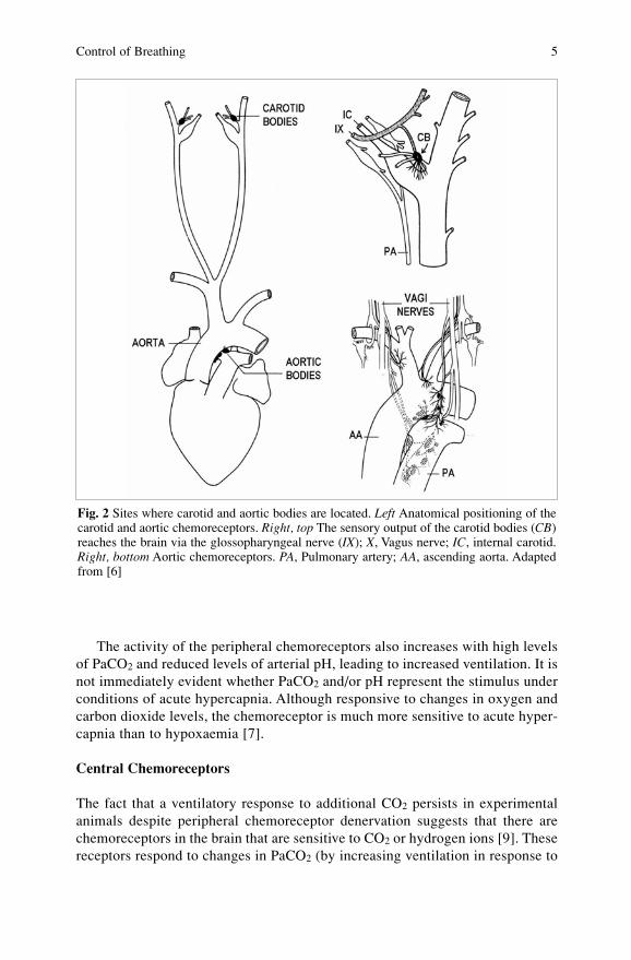
From their location in the carotid and aortic bodies, peripheral chemoreceptorsdirect the response to changes in PaO2, PaCO2 and pH. The carotid bodies arefound at the bifurcation of the common carotid artery into the internal and exter-nal carotid arteries (Fig. 2) and their sensory supply reaches the brain via theglossopharyngeal nerve. The aortic bodies are located around the ascendingaorta and send their afferent stimuli via the vagal nerves to the central nervoussystem. Since the arterial blood supply of these bodies amounts to approximate-ly 2 l/min/100 g tissue (they are located on the outside of the main arteries andreceive their own perfusion), they are one of the most highly perfused tissues inthe human body [4,5]. The carotid and aortic bodies consist of two different celltypes, glomus cells (type I) and sheath cells (type II). Afferent neurons terminateon glomus cells. There is also an unmyelinated supply to the sheath cells.
It is not clear how the carotid and aortic bodies sense hypoxaemia, but is clearthat the stimulus for increased ventilation is PaO2, not the oxygen content of theblood [1]. At normal levels of PaO2, some neural activity arises from thesechemosensors. At hyperoxic levels, this activity is only slightly reduced in normalpeople whereas in arterial hypoxaemia the intensity of the response varies in a non-linear manner according to the severity of the condition [7]. The greatest increasein activity in response to hypoxaemia occurs when PaO2 falls to ≤60 mmHg or anFIO2 ≤0.1 [1,7]. This increase in ventilation is manifested primarily by an increasein the depth of breathing (tidal volume or VT) but an increased respiratory rate isalso observed. These responses vary according to the degree of hypoxaemia.
In mammals, the carotid bodies account for about 90% of the ventilatoryresponse to hypoxaemia; the remaining 10% arises from the aortic bodies. Theformer are also responsible for 20–50% of the response to arterial hypercapniaand acidaemia, with the remaining 50–80% of the response mediated by centralbrainstem receptors [8].
F.B. Santos, L.K.S. Nagato, W.A. Zin4
Fig. 1 The control of breathing: basic elements. Sensors transmit information to the centralcontroller. Subsequently, stimuli are sent to the effectors–the respiratory muscles–to adjustventilatory responses. Adapted from [3]
The activity of the peripheral chemoreceptors also increases with high levelsof PaCO2 and reduced levels of arterial pH, leading to increased ventilation. It isnot immediately evident whether PaCO2 and/or pH represent the stimulus underconditions of acute hypercapnia. Although responsive to changes in oxygen andcarbon dioxide levels, the chemoreceptor is much more sensitive to acute hyper-capnia than to hypoxaemia [7].
Central Chemoreceptors
The fact that a ventilatory response to additional CO2 persists in experimentalanimals despite peripheral chemoreceptor denervation suggests that there arechemoreceptors in the brain that are sensitive to CO2 or hydrogen ions [9]. Thesereceptors respond to changes in PaCO2 (by increasing ventilation in response to
Control of Breathing 5
Fig. 2 Sites where carotid and aortic bodies are located. Left Anatomical positioning of thecarotid and aortic chemoreceptors. Right, top The sensory output of the carotid bodies (CB)reaches the brain via the glossopharyngeal nerve (IX); X, Vagus nerve; IC, internal carotid.Right, bottom Aortic chemoreceptors. PA, Pulmonary artery; AA, ascending aorta. Adaptedfrom [6]
increased PCO2 and vice versa) and pH (by increasing ventilation to a decreasedpH and vice versa). Although no definite chemoreceptors have been definedanatomically, results of experiments involving the local application of chemical,electrical and thermal stimuli suggest that central chemoreceptors are located ator near the ventral surface of the medulla [9]. This location may facilitate theability of the central chemoreceptors to monitor changes in PaCO2 and pH lev-els in the cerebrospinal fluid (CSF). Hydrogen ions enter and are found in theCSF and extracellular fluid in the vicinity of the central chemoreceptors. Thepresence of these ions is a result of CO2 dissociation and direct diffusion intoand out of the bloodstream. Elevated arterial CO2 easily crosses the blood–brainbarrier because this gas is highly membrane-permeable, is converted to carbon-ic acid (H2CO3) and rapidly dissociates into H+ and HCO3
- ions. As a result, H+
rises in the CSF and interstitium in parallel with PaCO2. This increased H+ stim-ulates respiration by a direct action on the central chemoreceptors [1,10].
There is an interaction between the responses of the peripheral and centralchemoreceptors. The blood–brain barrier exhibits different permeabilities toions, such as H+ (low permeability), and lipid-soluble molecules, such as carbondioxide (high permeability). In an acidic environment, the peripheral chemore-ceptors would trigger an increase in ventilation before the local environment inthe fluid bathing the medulla reflected the acid pH in the blood. As ventilationincreases owing to stimulation of peripheral chemoreceptors, PaCO2 decreases.The environment of central chemoreceptors would rapidly reflect the lowerPaCO2, but only later sense the elevated H+ concentration of the blood (becauseof the extra time needed for the H+ ions to cross the blood–brain barrier) [10].However, when PaCO2 level is chronically elevated, as might occur in a patientwith severe COPD, the activities of the peripheral and central chemoreceptorsdecrease within a few days, as pH normalises. At extremely high levels of car-bon dioxide (PaCO2>80–100 mmHg) an anaesthetic effect may be produced andventilation decreases rather than increases. This occurs because a chronicallyelevated PaCO2 results in renal compensation and consequent retention ofHCO3
-. This HCO3- gradually diffuses through the blood–brain barrier and into
the CSF, where it binds to the excess H+ produced by the elevated PaCO2, whichbalances the stimuli on ventilatory drive [10].
At moderate degrees of hypoxaemia—between 45 and 60 mmHg—ventila-tion rises moderately to about twice its normal level. Only when PaO2 fallsbelow 40 mmHg is there a sharp increase in ventilation. When hypercapniaoccurs simultaneously with acute hypoxaemia, a synergistic effect results andventilation rises substantially.
Pulmonary Receptors
Pulmonary receptors can be found in the airways and lung parenchyma and areinnervated by the vagus nerves.– Pulmonary stretch receptors are slowly adapting stretch receptors (SARs)
located among smooth muscle cells within the intra- and extra-thoracic air-
F.B. Santos, L.K.S. Nagato, W.A. Zin6
ways. These receptors are stimulated by pulmonary inflation and may play arole in the early termination of inspiration when tidal volume increases—Breuer-Hering inflation reflex [11]. In humans, this reflex is manifest only ata VT >3 l and seems to play a protective role in preventing excessive lunginflation. The SARs do not accommodate to a persistent stimulus, such asprolonged distension [12].
– Irritant receptors are also called rapidly adapting stretch receptors (RARs)and are located among the airway epithelial cells. RARs respond to noxiousstimuli, such as dust, cigarette smoke and histamine [13]. They are concen-trated in the carina and primary bronchi and are also believed to triggercough [12]. During normal quiet breathing, their discharge does not dependon the phases of the breathing cycle (inspiration and expiration); therefore,these receptors do not seem to influence to any great extent the baselinebreathing pattern at rest [14]. RARs also seem to trigger the augmented ven-tilation and sighs occurring sporadically during normal breathing, which helpto prevent atelectasis of the air spaces [15]. They have also been described astaking part in the dyspnoea, bronchoconstriction and rapid and shallowbreathing that occur in asthma [13,16,17].
– C fibres are unmyelinated fibres that carry information from a variety ofreceptors whose function is not totally understood [1]. Located within theairways, these receptors respond to either mechanical or chemical factors.
– Chest-wall and muscle mechanoreceptors respond to changes in length, ten-sion or movement. The primary mechanoreceptors in the chest are the mus-cle spindles, tendon organs of the respiratory muscles and the joint proprio-ceptors. Afferent information from these receptors reaches the respiratorycentres in the medulla [7]. Mechanoreceptors may also contribute to theincrease in ventilation that occurs during the early stages of exercise [18].Muscle spindles and tendon organs sense changes in the force of contractionof the respiratory muscles. While muscle spindles regulate muscle tonus, ten-don organs have an inhibiting effect on inspiration. Joint proprioceptorssense the degree of chest-wall movement and may also influence the leveland timing of respiratory activity [19].
Central Respiratory Controllers
The central respiratory controllers are divided into the brainstem group (invol-untary) and the cerebral cortex group (voluntary). The neural structures respon-sible for the automatic control of breathing are found in the medulla and pons.Two aggregates of neurons, termed the dorsal respiratory group (DRG) and theventrolateral respiratory group (VRG), contain both inspiratory and expiratoryneurons. The DRG seems to play an important role in processing informationfrom receptors in the lungs, chest wall and chemoreceptors that modulate breath-ing. Neural activity from the DRG is important to activate the diaphragm and theVRG. The DRG also exhibits a role in determining breathing rhythm and in reg-
Control of Breathing 7
ulating the changes in diameter of the upper airway that occur with breathing bystimulating the muscles to expand the upper airway during inspiration [1,2,7].The DRG is located in the nucleus of the tractus solitarius in the medulla andapparently represents the site of origin of the normal rhythmic respiratory drive,which consists of repetitive bursts of inspiratory action potentials [20]. Theexact mechanism by which this rhythm is generated remains unknown. The VRGis located within the nucleus ambiguus (rostrally) and nucleus retroambiguus(caudally). It innervates respiratory effector muscles by the phrenic, intercostaland abdominal respiratory motoneurons [20].
In the pons, the pontine respiratory group (PRG) contains neurons that maycontribute to the transitions or switching from inspiration to expiration. Damageto the respiratory neurons in the pons leads to an increase in inspiratory time, adecrease in respiratory frequency and an increase in tidal volume [1,2,7]. Nucleiso far located in the pons are the parabrachialis medialis and Kölliker-Fuse.
The breathing rhythm of the central pattern generator (CPG) has beenexplained as follows. Inspiration begins by the abrupt removal of inhibitoryimpulses to the DRG. An increased inspiratory motoneuron activity ensues inthe form of a slowly augmenting ramp of signals that is suddenly terminated byan off-switch mechanism. During expiration, another burst of inspiratory neu-ronal activity takes place [21]. In fact, so many different bursting patterns can bedetected in the respiratory neurons in the medulla and pons that, so far, anymodel or hypothesis of the triggering or interaction among the structuresremains speculative.
The cerebral cortex may temporarily influence or bypass the central respira-tory control mechanism in order to accomplish behaviour-related respiratoryactivity, such as cough, speech, singing and voluntary breath-holding [22,23].Discomfort and anxiety may also influence the respiratory rhythm. When expe-riencing pain or shortness of breath, most people increase their respiratory rate,and total ventilation increases. The pattern of breathing may also reflect attemptsto reduce the discomfort associated with ventilation. Patients with significantlyreduced respiratory system compliance tend to breath with a rapid, shallow pat-tern. For patients with increased airway resistance, on the other hand, the highflow required for rapid, shallow breathing requires considerable work. Thesepatients tend to adopt a slower breathing pattern with large tidal volumes [1].
Neural Control of Smooth Muscle in the Airways
The autonomous nervous system importantly participates in the regulation of thecalibre of the airways both in normal individuals and in those with pulmonaryillness.
Cholinergic fibres (parasympathetic) penetrate between the muscle fibres ofthe bronchi and their stimulation results in the contraction of airway smoothmuscle. Evidence for such an action stems from fact of that bronchodilatationensues after sectioning of the vagus nerves and after the administration of anti-cholinergic drugs. The cholinergic system participates in the maintenance of the
F.B. Santos, L.K.S. Nagato, W.A. Zin8

bronchial tonus at rest and in the majority of bronchoconstriction cases. In con-trast, the smooth muscle fibres in the airways present adrenergic innervation.While the amount of α-adrenoreceptors is reduced and their role seems insignif-icant, β-adrenoreceptors antagonise bronchoconstriction in asthmatic patients,by promoting the relaxation of airway smooth muscle [23].
Evidences show that the airways contain a system of innervation in which theneurotransmitters are neither adrenergic nor cholinergic. This system is knownas non-adrenergic non-cholinergic innervation (NANC). Its location cannot bedistinguished morphologically from those of the classic sympathetic andparasympathetic ways, but its stimulation can result in an excitatory responsebut its stimulation can result in a non-adrenergic non-cholinergic excitatory orinhibitory response. Among the neurotransmitters of this system, neuropeptides,such as substance P and neurokinin A, among others, can be found [24].
Effector System
The pathways and muscles involved in the actual performance of inspiration andexpiration make up the effector system. The spinal descending pathways connectthe DRG and VRG to the ventrolateral columns of the spinal cord; finally, thestimuli reach the α-motoneurons leading to the diaphragm, intercostal andabdominal muscles, and to other muscles promoting respiratory movements.
The respiratory muscles encompass the diaphragm and the intercostal,abdominal and accessory muscles of respiration. The diaphragm is responsiblefor the majority (75%) of gas movement during quiet inspiration, while the para-sternal internal intercostals and scalenes account for the remainder [25].
Control of Breathing in Disease
Chronic Obstructive Pulmonary disease
The patient with chronic obstructive pulmonary disease (COPD) presents alteredV’/Q’ distribution with hypoxaemia, with or without CO2 accumulation. Airflowobstruction could be the most important fact to explain the hypercapnia in COPDpatients. Inspiratory muscle dysfunction and the coexistence of nocturnalhypoventilation may worsen the hypercapnia. However, the true reason that somepatients present with CO2 retention while others do not, despite the same degreeof obstruction, remains unknown. The native ventilatory response to PaCO2 mightconstitute an inter-individual factor contributing to the variable hypercapnia inCOPD patients. This concept of inherent differences in the ventilatory responseto CO2 arose from observations of the considerable variability in the magnitudeof the ventilatory response to experimentally induced increases in arterial PCO2
in normal subjects. According to this paradigm, COPD patients have been classi-
Control of Breathing 9
fied into those with high ventilatory responses to abnormal blood gases (‘pinkpuffers’) and those with low responses (‘blue bloaters’) [26].
Another factor that may contribute to the variable arterial CO2 retention insevere COPD patients is a corresponding coincidence of sleep-related hypoven-tilation: patients with a larger amount of sleep-disordered breathing have day-time hypoventilation and those with normal ventilation during sleep only slighthypoventilation. Additionally, patients with obstructive sleep apnoea syndromeand concurrent COPD have higher daytime PaCO2 values than patients withoutCOPD [27].
The effects of a high inspiratory oxygen fraction are still controversial. Somepatients with CO2 retention worsen their respiratory acidosis when they inhalehigh O2 concentrations. This effect is usually explained by the loss of the hypox-ic stimulus to breathing. However, a reduction in the hypoxic ventilatory drivemay not be the only mechanism inducing hypercapnia in these patients. Theworst V’/Q’ mismatch results in a significantly increased dead space; this isanother explanation for the arterial hypercapnia associated with supplementaloxygen administration. Prior to the use of supplemental oxygen, areas of localalveolar hypoxia produce pulmonary hypoxic vasoconstriction, thereby divert-ing the flow of CO2-rich blood from poorly ventilated to better aerated lung seg-ments. When supplemental oxygen reverses local hypoxaemia, pulmonaryhypoxic vasoconstriction nullifies and allows the perfusion of very poorly ven-tilated lung segments, increasing the dead space and reducing the effective alve-olar ventilation. As a result, arterial CO2 rises. Finally, PaCO2 may increase inthe face of supplemental oxygen administration because of a concurrentdecrease in the CO2 carrying capacity of the haemoglobin molecule secondaryto the increasing oxygenation. This results in an altered steady-state relationshipbetween carbaminohaemoglobin and PaCO2, which raises the latter by severalmillimetres of mercury. This is known as the Haldane effect [28].
COPD patients exhibit an increased neural drive to their respiratory musclesthat seems to be larger in hypercapnic COPD patients than in normocapnic patients.This increased respiratory drive is probably needed to overcome both increased air-way resistance and mechanically disadvantaged respiratory muscles [26,29].
Neurological Diseases
Respiratory dysfunction may constitute an early and relatively major manifesta-tion of several neurological disorders, including structural or degenerative ail-ments of the central or peripheral nervous system or metabolic encephalopathies[30]. Neuromuscular diseases are often associated with abnormalities of ventila-tory control and their associated hypoventilation, particularly during sleep, andwith a reduced ventilatory response to CO2 and O2 [30,31]. Such patientsincrease their respiratory rate rather than VT in response to hypercapnia andhypoxaemia. This rapid and shallow breathing response is thought to be anattempted compensation aimed at increasing ventilation with minimal increase
F.B. Santos, L.K.S. Nagato, W.A. Zin10
in the work of breathing. Tachypnoea may then worsen respiratory musclefatigue, leading to a further reduction in tidal volume. Respiratory failure typi-cally complicates advanced neuromuscular disease by compromising effectiverespiratory muscle function. Death in these patients is usually due to progressiverespiratory failure and superimposed infections secondary to aspiration resultingfrom pharyngeal dysfunction [30,31].
Respiratory control may be affected acutely or subacutely, as in stroke ormultiple sclerosis. Lesions affecting the PRG, DRG, VRG or chemoreceptorsmay express an abnormal respiratory rhythm, central alveolar hypoventilation orboth. A unilateral lesion of the lateral medulla, including the VRG, leads toblunting of the ventilatory response to CO2 and sleep apnoea syndrome, partic-ularly when there is another predisposing factor such as nasal septum deviation.Cheyne-Stokes respiration typically accompanies bilateral infarcts of the cere-bral hemispheres, but also occurs in infratentorial ischaemic stroke [30,31].
Multiple sclerosis (MS) may yield respiratory dysfunction, in general asso-ciated with large lesions involving the upper cervical cord or medulla. Acutedemyelinating lesions involving the dorsolateral medulla may result in loss ofautomatic breathing, usually associated with impaired swallowing and coughreflex. Thus, there ensues a risk of aspiration pneumonia [30,31]. Paroxysmalhyperventilation may occur as a manifestation of an acute lesion in the upperbrainstem. Bulbar weakness, leading to aspiration followed by bronchopneumo-nia, is common in the terminal stages of MS. More rarely, loss of response toCO2 and hypercapnic respiratory insufficiency may occur early in the course ofthe disease [30,31].
Brainstem tumours may produce central neurogenic hyperventilation, centralsleep apnoea, irregular breathing, short breath-holding time and apneustic breath-ing. Occasionally, abnormalities of respiratory control are the only manifestationsof the tumour and resolve after its resection. Patients with severe traumatic brain-stem or high cervical-cord injury may lose both voluntary and autonomic controlof breathing. These patients require ventilatory support, which is given via a tra-cheostomy through which tracheal suction can also be performed [30,31].
Sudden Infant Death Syndrome
Sudden infant death syndrome (SIDS) is, according to the newly proposed defi-nition: ‘The sudden unexpected death of an infant <1 year of age, with onset ofthe fatal episode apparently occurring during sleep, that remains unexplainedafter a thorough investigation, including performance of a complete autopsy andreview of the circumstances of death and the clinical history’ [32]. Despite thefact that the diagnosis of SIDS originates from the exclusion of known causes ofdeath, there are common features in most cases. These observations have led tothe introduction of a triple-risk model for the understanding of SIDS. The modelproposed in 1993 implies that SIDS only occurs if three conditions occur simul-taneously: a vulnerable developmental stage of the CNS and the immune system;
Control of Breathing 11

predisposing factors, including a certain genetic pattern; and trigger events, suchas sleeping position, maternal smoking, or infection [32]. Despite many studiesin this area, the real aetiology of SIDS remains unknown.
Abnormal functioning of the central chemoreceptors represents one of thepossible mechanisms generating SIDS. The recently born with apparently lethalepisodes and the victims of SIDS studied before death presented a ventilatorypattern that was depressed with respect to the hypercapnic stimulus.Additionally, infants with episodes of apnoea in infancy present a slightly high-er PaCO2 as well as a lower sensibility to CO2 as a trigger alert during sleep. Thearcuate nuclei in the ventral medulla oblongata have been closely studied inSIDS victims. They are integrative sites for vital autonomic functions, includingbreathing and arousal, and are integrated with other regions that regulate arous-al and autonomic chemosensory function. Quantitative three-dimensionalanatomical studies indicated that some SIDS victims show hypoplasia of thearcuate nuclei, and as many as 56% of SIDS victims exhibit histopathologicalevidence of less extensive bilateral or unilateral hypoplasia. Studies on neuro-transmission in the arcuate nuclei have also identified receptor abnormalities insome SIDS victims that involve several receptor types relevant to state-depend-ent autonomic control overall and to ventilatory and arousal responsiveness inparticular. These deficits include significant decreases in binding to muscarinic,cholinergic and serotonergic receptors [33].
Cheyne-Stokes Respiration
Cheyne-Stokes respiration (CSR) with central sleep apnoea (CSA) is a breathingdisorder seen in patients with advanced congestive heart failure (CHF). It ischaracterised by central apnoeas and hypopnoeas that alternate with periods ofincreasing-decreasing tidal volume. CSR-CSA has been associated with increas-es in sympathetic nervous activity in CHF patients, which is an important pre-dictor of CHF progression, arrhythmias and mortality. Indeed, CSR-CSA, inde-pendent of other risk factors, elevates the risk of mortality in CHF by two- tothree-fold. Successful treatment of CSR by continuous positive airway pressure(CPAP) leads to a significant reduction in sympathetic nervous activity and mayreduce mortality by up to 40% in patients with CHF and CSR-CSA. Since CPAPhas salutary effects on cardiac function (independent of its effect on CSR), itremains uncertain whether CSR-CSA is a mere phenomenon of a failing heart ora major contributor to poor outcomes in patients with CHF. Supplemental oxy-gen may be used as treatment and tends to eliminate or decrease CSR in CHF byeliminating hypoxaemia, which contributes to respiratory cycling [34]. The clas-sic cases of CSR are caused by CNS dysfunction, such as a cerebrovascular acci-dent. In this setting, CSR is usually associated with bilateral supramedullarydamage in conjunction with a depressed level of consciousness, such as occursduring sleep, sedation or diffuse cortical injury [35].
F.B. Santos, L.K.S. Nagato, W.A. Zin12
References
1. Schwartzstein RM, Paker MJ (2006) Respiratory physiology. A clinical approach.Lippincott Williams & Wilkins, Philadelphia
2. Gallego J, Nsegbe E, Durand E (2001) Learning in respiratory control. Behav Modif25:495–512
3. West JB (2000) Respiration physiology. 6th edition. Lippincott Williams & Wilkins,Philadelphia
4. Ganong WF (1993) Regulation of respiration. In: Review of medical physiology, 16th edi-tion. Appleton & Lange, Norwalk, pp 611–619
5. Bee DH (1993) The carotid body: a review of its anatomy, physiology and clinical impor-tance. Monaldi Arch Chest Dis 48:48–53
6. Comroe JH (1974) Physiology of respiration: an introductory text. 2nd Edition. Chicago:Yearbook Publishers, Chicago
7. Montaldo-Caruana B, Gleeson K, Zwillich CW (2000) The control of breathing in clinicalpractice. Chest 117:205–225
8. Gonzales C, Almara L, Obeso A et al (1192) Oxygen and acid chemoreception in the carotidbody receptors. Trends Neurosci 15:146–153
9. Bruce EN, Cherniak NS (1987) Central chemoreceptors. J Appl Physiol 62:389–40210. Bledsoe SW, Hornbein TF (1981) Central chemoreceptors and the regulation of their chem-
ical environment. In: Hornbein TF (ed) Regulation of breathing (part I). Marcel Dekker,New York, pp 347–428
11. Manning HL, Schwartzstein RM (1995) Pathophysiology of dyspnea. N Engl J Med333:547–553
12. Sant’Ambrogio G (1987) Nervous receptors of the tracheobronchial tree. Ann Rev Physiol49:611–627
13. Widdicombe J (2006) Reflexes from the lungs and airways: historical perspective. J ApplPhysiol 101:628–634
14. Sampson SR, Vidruk EH (1975) Properties of ‘irritant’ receptors in canine lungs. RespirPhysiol 25:9–22
15. Sant’Ambrogio G (1982) Information arising from the tracheobronchial tree of mammals.Physiol Rev 62:531–569
16. Schwartzstein R, Lilly J, Israel E et al (1991) Breathlessness of asthma differs from that ofexternal resistive loading. Am Rev Respir Dis 143(suppl):A596
17. Widdicombe, J (2001) Airway receptors. Respir Physiol 125:3–1518. Mitchell RA, Berger AJ (1981) Neural regulation of respiration. In: Hornbein TF (ed)
Regulation of breathing (part I). Marcel Dekker, New York, pp 541–62019. Duron B (1981) Intercostal and diaphragmatic muscle endings and afferents. In: Hornbein
TF (ed) Regulation of breathing (part I). Marcel Dekker, New York, pp 473–54020. Berger AJ, Mitchell RA, Severinghaus JW (1977) Regulation of respiration, Part II. N Engl
J Med 297:138–14321. von Euler C (1983) On the central pattern generator for the basic breathing rhythmicity. J
Appl Physiol 55:1647–165922. Mithoeffer JC (1964) Breath holding. In: Handbook of physiology: respiration (section 3,
vol II). American Physiology Society, Washington, DC 38:1011–102523. Ramirez JM, Viemari JC (2005) Determinants of inspiratory activity. Respir Physiol
Neurobiol 147:145–15724. Adriaensen D, Brouns I, Pintelon I et al (2006) Evidence for a role of neuroepithelial bod-
ies as complex airway sensors: Comparison with smooth muscle-associated airway recep-tors. J Appl Physiol 101:960–970
25. Ganong WF (1993) Pulmonary function. In: Review of medical physiology. 16th ed.Appleton & Lange, Norwalk, pp 587–603
26. Mountain R, Zwillich CW, Weil JV (1978) Hypoventilation in obstructive lung disease. NEngl J Med 298:521–525
Control of Breathing 13

27. Chan CS, Bye PTP, Woolcock AJ et al (1990) Eucapnia and hypercapnia in patients withchronic airflow limitation. Am Rev Respir Dis 141:861–866
28. Kalhoff H, Werkmiester F, Kiwull-Schone L et al (1994) The Haldane effect under differentacid-base conditions in premature and adult humans. Adv Exp Med Biol 361:353–361
29. Gorini M, Spinelli A, Ginanni R et al (1990) Neural respiratory drive and neuromuscularcoupling in patients with chronic obstructive pulmonary disease. Chest 98:1179–1186
30. Nogués MA, Roncoroni AJ, Benarroch E (2002) Breathing control in neurological diseases.Clin Auton Res 12:440–449
31. Johnson DC, Homeyoun K (1994) Central control of ventilation in neuromuscular disease.Clin Chest Med 15:607–615
32. Opdal SH, Rognum TO (2004) New insight into sudden infant-death syndrome. Lancet364:825–826
33. Hunt CE (2005) Gene-environment interactions: implications for sudden unexpected deathsin infancy. Arch Dis Child 90:48–53
34. Sin DD, Man GCW (2003) Cheyne-Stokes respiration. A consequence of a broken heart?Chest 124:1627–1628
35. Hanly PJ, Zuberi-Khokhar S (1996) Increased mortality associated with Cheyne-Stokes res-piration in patients with congestive heart failure. Am J Respir Crit Care Med 153:272–276
F.B. Santos, L.K.S. Nagato, W.A. Zin14
Elastic and Resistive Properties of theRespiratory System
W.A. Zin
Introduction
This chapter will consider basic aspects of respiratory-system mechanics in orderto provide a background for the analysis of the most common disorders related tothe elastic and resistive components of the lung and chest wall. Excellent reviewsarticles can be consulted, if further details are desired [1–9b].
The movements of the lungs are entirely passive. Forces must be applied tothe respiratory system to move it from its resting position at the end of expiration.In spontaneous breathing, the respiratory muscles provide the external forces,whereas artificial ventilation moves the relaxed respiratory system. In either sit-uation, movement depends on the impedance of the lung and chest wall, the twocomponents of the respiratory system. This impedance stems mainly from theelastic and resistive mechanical properties that are found in the lung and in thechest wall. The inertial component of gas and tissue is usually negligible [10].
Elastic Properties
Both the lungs and the chest wall can be considered as elastic structures, withtransmural pressure gradients corresponding to stress and lung volume to strain.Over a certain range of volumes and pressures, lung and chest-wall structuresobey Hooke’s law, and the change in lung and chest-wall volumes divided by thetransmural pressures required to produce them defines the compliance (C).Elastance (E) is the reciprocal of compliance, i.e. ∆P/∆V, and is usually expressedin cmH2O per litre. Stiff structures present a high elastance. Respiratory-systemelastance equals the sum of lung plus chest wall elastances (Ers=EL+Ew, respec-tively), whereas respiratory-system compliance is more complex:1/Crs=1/CL+1/Cw.
U. Lucangelo, P. Pelosi, W.A. Zin, A. Aliverti (eds.) Respiratory System and ArtificialVentilation. © Springer 2008 15

Pleural Pressure
Since variations in lung and chest wall volumes are virtually identical, the com-pliances of the respiratory system, lung and chest wall vary according to thechange in the transmural pressure (i.e. inside minus outside pressures) acrossthese structures. Under static conditions, the distending pressure of the respirato-ry system (Prs), lung (PL) and chest wall (Pw) are (Fig. 1):
PL = Palv – Ppl (Eq. 1)
where Palv represents the alveolar pressure [which is equal to the airway pressure(Paw) or pressure at the airway opening (Pao) under static conditions and in theface of an open glottis] and Ppl stands for intrapleural pressure. PL is commonlyreferred to as the transpulmonary pressure:
Pw = Ppl – Pbs (Eq. 2)
where Pw represents the transthoracic or chest-wall pressure, and Pbs the pres-sure at the body surface (usually barometric pressure);
Prs = PL + Pw (Eq. 3)
or
Prs = Palv – Ppl + Ppl – Pbs = Palv – Pbs (Eq. 4)
As can be easily understood, precise determination of swings in intrapleuralpressure is of paramount importance when it is necessary to divide respiratory
W.A. Zin16
Fig. 1 Schematic representation of the structures and pressures involved in breathing. Pao,Pressure at the airway opening; Pbs, pressure at the body surface; Ppl, intrapleural pressure;Palv, alveolar pressure; PL, transpulmonary pressure; Pw, chest-wall pressure; Prs, pressuredifference across the respiratory system
system mechanics into their lung and chest-wall components. However, in clini-cal practice, pleural pressure is rarely measured because of all the risks involvedin the procedure. Instead, variations in oesophageal pressure (Poes) are deter-mined as these reflect quite accurately the changes in pleural pressure. Usually alatex balloon or a liquid-filled catheter is placed in the lower third of the oesoph-agus and its correct positioning must be accomplished to achieve a perfect read-ing of the changes in intrathoracic pressure [11]. Complete descriptions of thetechniques used to measure Poes can be found in the literature [12–14].
Elastic Recoil of the Lungs
The elastic recoil of the lungs tends to bring them down to their minimum vol-ume. Accordingly, the elastic component (Pel,rs) of the total pressure applied tothe respiratory system during inspiration is restored during expiration to promoteexpiration. In other words, the potential energy stored during inspiration returnsto the system as kinetic energy. The passive volume–pressure curve of the lung isalmost linear (constant compliance) up to volumes around 80% of the total lungvolume. Beyond this point the curve flattens (compliance decreases) mainlybecause the elastic limit of the lung is approached and the structures stiffen. Iftranspulmonary pressure rises above 30 cmH2O, the danger of tissue rupture mayensue.
Tissue Recoil
Two components account for the elastic recoil of the lungs [15]. One of them isrepresented by the elastic components of lung tissue (mainly collagenous andelastic fibres). It is believed that the elastic behaviour of the lung does not dependstrongly on the elongation of these fibres, but mainly on their geometric arrange-ment. The network of pulmonary connective tissue interconnects all pulmonarystructures (vessels, bronchioles, alveoli, and so forth) and, as a result, they dilateduring inspiration. This phenomenon is known as interdependence and con-tributes to keep the alveoli open, since if some of them collapsed, their neigh-bours would tether their walls, tending to reopen them. In addition to their tissueelastic properties, the lungs present another component that contributes impor-tantly to their elastic characteristics: the surface tension of the liquid lining thealveoli and distal air spaces.
Surface Tension
The air-liquid interface of the thin film of liquid that covers the surface of termi-nal respiratory units and probably also lines the luminal surface of terminal bron-chioles displays surface tension, i.e. the molecules in the film attract each otheralong its surface. This component must also be overcome during inspiration:
Elastic and Resistive Properties of the Respiratory System 17