restored with transfixed glass fiber post: 3d-finite
TRANSCRIPT
materials
Communication
Stress Concentration of Endodontically Treated MolarsRestored with Transfixed Glass Fiber Post: 3D-FiniteElement Analysis
Alexandre Luiz Souto Borges 1 , Manassés Tercio Vieira Grangeiro 1 , Guilherme Schmitt de Andrade 1 ,Renata Marques de Melo 1, Kusai Baroudi 2, Laís Regiane Silva-Concilio 2 and João Paulo Mendes Tribst 2,*
�����������������
Citation: Borges, A.L.S.;
Grangeiro, M.T.V.; de Andrade, G.S.;
de Melo, R.M.; Baroudi, K.;
Silva-Concilio, L.R.; Tribst, J.P.M.
Stress Concentration of
Endodontically Treated Molars
Restored with Transfixed Glass Fiber
Post: 3D-Finite Element Analysis.
Materials 2021, 14, 4249.
https://doi.org/10.3390/ma14154249
Academic Editor: Deog-Gyu Seo
Received: 10 June 2021
Accepted: 2 July 2021
Published: 29 July 2021
Publisher’s Note: MDPI stays neutral
with regard to jurisdictional claims in
published maps and institutional affil-
iations.
Copyright: © 2021 by the authors.
Licensee MDPI, Basel, Switzerland.
This article is an open access article
distributed under the terms and
conditions of the Creative Commons
Attribution (CC BY) license (https://
creativecommons.org/licenses/by/
4.0/).
1 Department of Dental Materials and Prosthodontics, Institute of Science and Technology, São Paulo StateUniversity (Unesp), São José dos Campos, São Paulo 12245-000, Brazil; [email protected] (A.L.S.B.);[email protected] (M.T.V.G.); [email protected] (G.S.d.A.);[email protected] (R.M.d.M.)
2 Graduate Program in Dentistry, Department of Dentistry, University of Taubaté (UNITAU),Taubaté 12020-270, Brazil; [email protected] (K.B.); [email protected] (L.R.S.-C.)
* Correspondence: [email protected]
Abstract: The loss of dental structure caused by endodontic treatment is responsible for a decrease intooth resistance, which increases susceptibility to fracture. Therefore, it is important that minimallyinvasive treatments be performed to preserve the dental structure and increase the resistance tofracture of endodontically treated posterior teeth. To evaluate under axial loads, using the finiteelement method, the stress distribution in endodontically treated molars restored with both transfixedor vertical glass fiber posts (GFP) and resin composite. An endodontically treated molar 3D-modelwas analyzed using finite element analyses under four different conditions, class II resin composite(G1, control model), vertical glass fiber post (G2), transfixed glass fiber posts (G3) and verticaland transfixed glass fiber posts (G4). Ideal contacts were considered between restoration/resincomposite and resin composite/tooth. An axial load (300 N) was applied to the occlusal surface.The resulting tensile stresses were calculated for the enamel and dentin tissue from five differentviewports (occlusal, buccal, palatal, mesial and distal views). According to the stress maps, similarstress trends were observed, regardless of the glass fiber post treatment. In addition, for the G1 model(without GFP), a high-stress magnitude can be noticed in the proximal faces of enamel (7.7 to 14 MPa)and dentin (2.1 to 3.3 MPa) tissue. The use of transfixed glass fiber post is not indicated to reduce thestresses, under axial loads, in both enamel and dentin tissue in endodontically treated molar with aclass II cavity.
Keywords: dental restoration failure; endodontically treated teeth; finite element analysis;dental materials
1. Introduction
The loss of dental structure caused by endodontic treatment is responsible for adecrease in tooth resistance, which increases susceptibility to fracture [1]. The longevityof endodontically treated teeth is influenced by several factors, such as, the preservationof remaining dental tissue, effectiveness of restorative procedures and occlusal force [2].Therefore, it is extremely important that minimally invasive dental treatments be performedto preserve the dental structure and obtain success. Additionally, the literature reportsthat different types of restoration parameters can increase the resistance to fracture ofendodontically treated posterior teeth [3].
One of the most common post-endodontic treatments is glass fiber post (GFP) asso-ciated with adhesively bonded resin composite restoration in order to increase fractureresistance and reduce the interfacial gap between dental tissues and restorative materi-
Materials 2021, 14, 4249. https://doi.org/10.3390/ma14154249 https://www.mdpi.com/journal/materials

Materials 2021, 14, 4249 2 of 11
als [4,5]. Some authors justify their use because GFP can distribute chewing stresses andocclusal loads on the restoration [6].
In addition, the elastic modulus of post and the direct restorative material must becompatible with the root dentin to reduce the possibility of fracture [7], as well as theroot stress magnitude during chewing loads. Aiming to improve the beneficial effectsof GFP usage in weakened teeth, several studies have investigated how different clinicalparameters can modify the mechanical response during loading, such as GFP geometries,relining, position, and length. Furthermore, previous in vitro studies reported that insertingtransfixed GFP could be a viable alternative procedure to reinforce the coronal dentalstructure, replacing metallic or ceramic posts [8,9]. According to a clinical study, thisprocedure is also economically viable and preserves the natural tooth structure comparedto full crown preparation [10].
In this sense, several studies aimed to evaluate the restorative techniques that could re-inforce the remaining dental structure, to reduce the stress concentration in the dental struc-ture [11,12] and the probability of fracture through alternative restorative procedures [13].One of these proposed techniques is the use of transfixed GFP in the tooth crown, and isreported as a contemporary conservative treatment [10]. According to the literature, thisrestorative treatment has satisfactory aesthetics and easy execution compared to full-crownpreparations [14]. However, the mechanical effect of transfixed post placement has not yetbeen extensively investigated in the literature. In vitro studies are controversial about themechanical improvements in the fracture load when transfixed glass fiber posts were usedto restore posterior teeth [8,9,15]. However, no study has evaluated how the transfixed GFPplacement can modify the tooth biomechanical behavior and how the stress can be reducedduring compressive loading. Therefore, the aim of the present study was to evaluate thestress distribution in endodontically treated molars restored with both transfixed or verticalglass fiber posts and resin composite under axial loads using 3D finite element analysis(3D-FEA). The use of 3D-FEA is the most widely used numerical method, allowing thereproduction of mechanical behavior under a mechanical load based on the properties ofthe materials [16–21].
2. Materials and Methods2.1. Modelling
Finite Element Analysis (FEA) was used to evaluate the mechanical behavior and stressdistribution in mesio-occluso-distal class II direct resin composite restoration of maxillaryfirst molar, restored endodontically, and treated with four post-endodontic restorativetreatments, including a no-post approach (G1, no-post approach), glass fiber cemented inthe palatal root canal (G2), two transfixed glass fiber posts (G3), two transfixed glass fiberposts, and one glass fiber post in the palatal root canal (G4) (Figure 1).
The three-dimensional models were designed in NURBS (non-uniform rational basisspline) CAD (computer-aided design) software (Rhinoceros 6.0SR8, McNell North America,Seattle, WA, USA). An intact first upper molar tooth model (previously reported [16],consisting of enamel layer, dentin layer, root, and pulp chamber) was used to generate theevaluated models. Endodontic treatment was designed using the crown-down techniquewith 4% conicity and 25% tapering. A large class II mesio-occluso-distal (MOD) cavitywas performed with 2 ± 0.5 mm thickness of remaining wall, 6 mm isthmus preparation,and gingival wall of 1.5 mm from the cementoenamel junction (Figure 2). An acrylicresin cylinder was designed to simulate the fixation support, in which the models werepositioned, exposing 2 mm below the restoration margin. The resulting model was used tosimulate the evaluated treatments.

Materials 2021, 14, 4249 3 of 11Materials 2021, 14, x FOR PEER REVIEW 3 of 11
Figure 1. Three-dimensional models created in the modeling software with different post-endodon-tic restorative treatments: (G1) no-post approach; (G2) glass fiber cemented in the palatal root canal; (G3) two transfixed glass fiber posts; and (G4) two transfixed glass fiber posts and one glass fiber post in the palatal root canal. In red the conventional glass fiber post and in blue the transfixed glass fiber posts.
Figure 2. CAD modeling. (A) Class II cavity design; (B) model components.
2.2. Pre-Processing The models were imported to a computer-aided engineering (CAE) software pro-
gram (ANSYS 19.2, ANSYS Inc., Houston, TX, USA) as a standard for the exchange of product model data (STP) file. Structural mechanical analysis was applied to each group and all materials were considered homogeneous, elastic, and isotropic, except for the glass
Figure 1. Three-dimensional models created in the modeling software with different post-endodonticrestorative treatments: (G1) no-post approach; (G2) glass fiber cemented in the palatal root canal;(G3) two transfixed glass fiber posts; and (G4) two transfixed glass fiber posts and one glass fiberpost in the palatal root canal. In red the conventional glass fiber post and in blue the transfixed glassfiber posts.
Materials 2021, 14, x FOR PEER REVIEW 3 of 11
Figure 1. Three-dimensional models created in the modeling software with different post-endodon-tic restorative treatments: (G1) no-post approach; (G2) glass fiber cemented in the palatal root canal; (G3) two transfixed glass fiber posts; and (G4) two transfixed glass fiber posts and one glass fiber post in the palatal root canal. In red the conventional glass fiber post and in blue the transfixed glass fiber posts.
Figure 2. CAD modeling. (A) Class II cavity design; (B) model components.
2.2. Pre-Processing The models were imported to a computer-aided engineering (CAE) software pro-
gram (ANSYS 19.2, ANSYS Inc., Houston, TX, USA) as a standard for the exchange of product model data (STP) file. Structural mechanical analysis was applied to each group and all materials were considered homogeneous, elastic, and isotropic, except for the glass
Figure 2. CAD modeling. (A) Class II cavity design; (B) model components.
2.2. Pre-Processing
The models were imported to a computer-aided engineering (CAE) software program(ANSYS 19.2, ANSYS Inc., Houston, TX, USA) as a standard for the exchange of productmodel data (STP) file. Structural mechanical analysis was applied to each group and all

Materials 2021, 14, 4249 4 of 11
materials were considered homogeneous, elastic, and isotropic, except for the glass fiberpost, which was considered orthotropic. The interfaces in all models were consideredperfectly bonded. Table 1 [20,22–29] summarizes the mechanical properties (elastic mod-ulus and Poisson ratio) used for the mechanical analysis. An average of 156,252 nodesand 128,242 tetrahedral, ten nodes, elements were used for the meshing process after aconvergence test with a 10% degree of freedom of the converged value and mesh size,based on the maximum principal stress (MPS) results. During the boundary conditions,the models were fixed (3-axis) on the bottom surface of a cylinder and loaded with 300 N(90◦) distributed in tripod contact area at the central fossa (Figure 3).
Materials 2021, 14, x FOR PEER REVIEW 4 of 11
fiber post, which was considered orthotropic. The interfaces in all models were considered perfectly bonded. Table 1 [20,22–29] summarizes the mechanical properties (elastic mod-ulus and Poisson ratio) used for the mechanical analysis. An average of 156,252 nodes and 128,242 tetrahedral, ten nodes, elements were used for the meshing process after a con-vergence test with a 10% degree of freedom of the converged value and mesh size, based on the maximum principal stress (MPS) results. During the boundary conditions, the models were fixed (3-axis) on the bottom surface of a cylinder and loaded with 300 N (90°) distributed in tripod contact area at the central fossa (Figure 3).
Figure 3. Boundary condition in the present simulation. (a) Mesh generation; (b) fixation of the system; (c) loading settings; (d) tripod contact area.
Figure 3. Boundary condition in the present simulation. (a) Mesh generation; (b) fixation of the system; (c) loading settings;(d) tripod contact area.
Materials 2021, 14, 4249 5 of 11
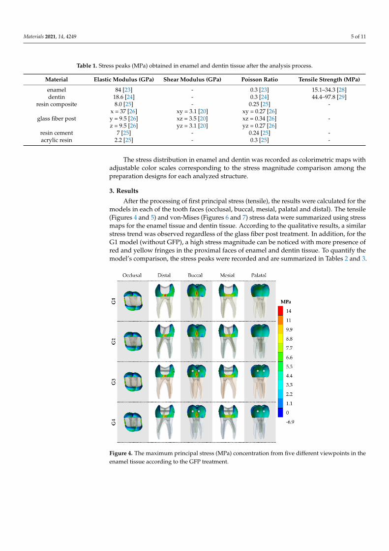
Table 1. Stress peaks (MPa) obtained in enamel and dentin tissue after the analysis process.
Material Elastic Modulus (GPa) Shear Modulus (GPa) Poisson Ratio Tensile Strength (MPa)
enamel 84 [23] - 0.3 [23] 15.1–34.3 [28]dentin 18.6 [24] - 0.3 [24] 44.4–97.8 [29]
resin composite 8.0 [25] - 0.25 [25] -
glass fiber postx = 37 [26]y = 9.5 [26]z = 9.5 [26]
xy = 3.1 [20]xz = 3.5 [20]yz = 3.1 [20]
xy = 0.27 [26]xz = 0.34 [26]yz = 0.27 [26]
-
resin cement 7 [25] - 0.24 [25] -acrylic resin 2.2 [25] - 0.3 [25] -
The stress distribution in enamel and dentin was recorded as colorimetric maps withadjustable color scales corresponding to the stress magnitude comparison among thepreparation designs for each analyzed structure.
3. Results
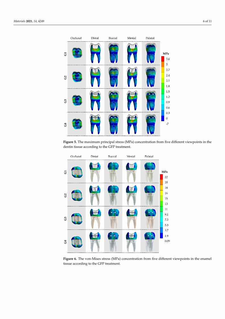
After the processing of first principal stress (tensile), the results were calculated for themodels in each of the tooth faces (occlusal, buccal, mesial, palatal and distal). The tensile(Figures 4 and 5) and von-Mises (Figures 6 and 7) stress data were summarized using stressmaps for the enamel tissue and dentin tissue. According to the qualitative results, a similarstress trend was observed regardless of the glass fiber post treatment. In addition, for theG1 model (without GFP), a high stress magnitude can be noticed with more presence ofred and yellow fringes in the proximal faces of enamel and dentin tissue. To quantify themodel’s comparison, the stress peaks were recorded and are summarized in Tables 2 and 3.
Materials 2021, 14, x FOR PEER REVIEW 6 of 11
Figure 4. The maximum principal stress (MPa) concentration from five different viewpoints in the enamel tissue according to the GFP treatment.
Figure 5. The maximum principal stress (MPa) concentration from five different viewpoints in the dentin tissue according to the GFP treatment.
Figure 4. The maximum principal stress (MPa) concentration from five different viewpoints in theenamel tissue according to the GFP treatment.
Materials 2021, 14, 4249 6 of 11
Materials 2021, 14, x FOR PEER REVIEW 6 of 11
Figure 4. The maximum principal stress (MPa) concentration from five different viewpoints in the enamel tissue according to the GFP treatment.
Figure 5. The maximum principal stress (MPa) concentration from five different viewpoints in the dentin tissue according to the GFP treatment.
Figure 5. The maximum principal stress (MPa) concentration from five different viewpoints in thedentin tissue according to the GFP treatment.
Materials 2021, 14, x FOR PEER REVIEW 7 of 11
Figure 6. The von-Mises stress (MPa) concentration from five different viewpoints in the enamel tissue according to the GFP treatment.
Figure 7. The von-Mises stress (MPa) concentration from five different viewpoints in the dentin tissue according to the GFP treatment.
4. Discussion First molars are teeth frequently involved in endodontic therapy [27]; therefore, the
use of resin composite restorations after endodontic treatments should improve the me-chanical resistance against the occlusal loads, as well as the restoration of missing dental tissue. However, this mechanical effect is not a consensus in the literature. The failures in endodontically treated teeth are still widely reported even after restorative procedures.
Figure 6. The von-Mises stress (MPa) concentration from five different viewpoints in the enameltissue according to the GFP treatment.

Materials 2021, 14, 4249 7 of 11
Materials 2021, 14, x FOR PEER REVIEW 7 of 11
Figure 6. The von-Mises stress (MPa) concentration from five different viewpoints in the enamel tissue according to the GFP treatment.
Figure 7. The von-Mises stress (MPa) concentration from five different viewpoints in the dentin tissue according to the GFP treatment.
4. Discussion First molars are teeth frequently involved in endodontic therapy [27]; therefore, the
use of resin composite restorations after endodontic treatments should improve the me-chanical resistance against the occlusal loads, as well as the restoration of missing dental tissue. However, this mechanical effect is not a consensus in the literature. The failures in endodontically treated teeth are still widely reported even after restorative procedures.
Figure 7. The von-Mises stress (MPa) concentration from five different viewpoints in the dentintissue according to the GFP treatment.
Table 2. Tensile stress peaks (MPa) obtained in enamel and dentin tissue after the analysis process.
Model Glass Fiber Post Approach Enamel Stress Dentin Stress
G1 No-post 14.5 3.7G2 Glass fiber in the palatal root canal 13.9 3.2G3 Two transfixed glass fiber posts 14.2 3.4
G4 Two transfixed glass fiber posts and one glassfiber post in the palatal root canal. 14.0 3.3
Table 3. Von-Mises stress peaks (MPa) obtained in enamel and dentin tissue after the analysis process.
Model Glass Fiber Post Approach Enamel Stress Dentin Stress
G1 No-post 18.17 11.68G2 Glass fiber in the palatal root canal 17.58 11.18G3 Two transfixed glass fiber posts 17.87 11.38
G4 Two transfixed glass fiber posts and one glassfiber post in the palatal root canal. 17.68 11.28
4. Discussion
First molars are teeth frequently involved in endodontic therapy [27]; therefore, theuse of resin composite restorations after endodontic treatments should improve the me-chanical resistance against the occlusal loads, as well as the restoration of missing dentaltissue. However, this mechanical effect is not a consensus in the literature. The failures inendodontically treated teeth are still widely reported even after restorative procedures.
Previous reports showed that the fracture resistance during in vitro compressive loadscould be enhanced with the GFP transfixed placement in posterior teeth [8,14,30]. Thereare reports affirming that the use of two transfixed GFPs in MOD-prepared cavities ledto recovery of approximately 23% more fracture strength than teeth without GFPs [30].According to the authors, a possible explanation would be the reduction of cusp deflectioncaused by anchoring of buccal and lingual walls of the cavity. The present study showed
Materials 2021, 14, 4249 8 of 11
a slight stress reduction with the use of GFP regardless of the post-endodontic treatmentthat could be associated with the reduction in cusp’s displacements; however, with valueslower than 2% in enamel tissue and 8% in dentin tissue comparing G1 and G3. Therefore,it is possible to suggest that the GFP effect would be more noticeable when the adhesion isnot ideal between resin composite restoration and cavity walls.
The mechanical properties of the restorative materials can determine the clinicalperformance of restored endodontically treated teeth, especially the elastic modulus ofthe post system [31]. Rigid posts, such as metallic and zirconia, generates less stress inthe cement layer, however, concentrate more on root dentin and, thus, catastrophic rootfractures can occur if the tooth is overloaded. On the other hand, less rigid posts, like thefiber-reinforced posts, can deflect under high loads, which can lead to loss of retention,or even post fracture, however, avoiding root fracture [32]. In this regard, the presentstudy is limited to the mechanical behavior with the use of GFP, however different postsystems or transfixed reinforcement systems may modify the calculated stress and shouldbe evaluated in further evaluations.
Another in vitro study [33] reported that the transfixed glass fiber post placementcould be an alternative treatment modality for the restoration of endodontically treatedteeth. According to the authors, this technique did not improve the fracture resistanceof endodontically treated teeth with MOD cavities; the present study corroborates this,since the difference in stress magnitude between models is less than 10%. According tothe authors, this mechanical behavior can be explained due to the minimal surface areabetween the GFP and the tooth structure; hence, it does not provide an adequate area forbonding. In addition, the GFP bond strength with resin cement is weaker than the bondstrength between the composite restoration and dental tissue. Finally, the presence of holesin the crown might have affected the fracture resistance of teeth. Therefore, the presentstudy suggests that the elastic modulus of GFP is lower than the enamel, and hence wouldpresent considerable flexible structure that cannot act as a stress reduction framework inthis case.
Cusp deflection mainly occurs along the bucco-lingual axis and usually occurs becausemost of the chewing forces on posterior teeth are directed laterally [15]. These directionaloblique components of the masticatory load can affect the adhesive layer in MOD cavities,being mandatory the use of effective adhesive systems to retain the restorative materials.In addition to that, with the interface property, the enamel tissue can be considered abrittle material, while dentine is more elastic damping the stress effect at the dentin-enameljunction [15].
On the other hand, when associated with the insertion of GFP in the palatal root canal(G2), there is slightly improvement in the absorption of occlusal loads, associated withstress dissipation along its axis [34], resulting in an improvement in the tooth resistance [35].This effect has been observed in clinical reports [36,37] corroborating with the results foundin the present study.
Another finite element study [38] showed that the use of adhesive GFP was neitherable to reduce the maximum stresses calculated on the occlusal surface nor to optimize thestress distribution regardless of different vertical post-approach. According to the authors,the placement of high amount of GFP can be deleterious to the remaining tooth structurewithout improving the mechanical response against chewing loading. The present studycorroborates with this indication, since there use of more GFPs (G4) was not beneficial forthe present model stress result.
Although in vitro laboratory tests of extracted human teeth are important to obtainuseful information about fracture load and the fracture mode, they are generally based ondestructive experiments and have limited capacity to investigate the stress-strain relation-ship in the tooth restoration complex [38,39]. Therefore, 3D-FEA is an engineering tool thatcan be applied to biology, medicine and dentistry and used to investigate the mechanicalbehavior of complex systems by a mathematical approach and simulation [37]. The nu-merical simulation consists of modeling a structure as close as possible to the real one, in
Materials 2021, 14, 4249 9 of 11
addition to the correct outputs that should be applicable and practical based in the clinicalresearch to define the boundary conditions. However, restorations have other problems,such as microleakage, polymerization shrinkage of resinous materials and postoperativesensitivity, that should be considered. As oral conditions cannot be completely reproducedby in vitro and in silico studies, further evaluations are still necessary to determine theeffectiveness and longevity of GFP treatment in class II MOD cavities [40].
In addition, based on what was said before, the FEA presents limitations relatedto numerical simulation. Initially, the endodontically treated teeth would be subject todifferent temperature cycles and pH variation in the oral cavity. In addition, the simulatedmaterials would present some defects that are not simulated in isotropic structures [41].There are, possible influences of oblique loading, sliding contacts and operator errors thatare simplified [42]. Therefore, further in vitro studies should be carried out to complementthe present findings, demonstrating transfixed GFP biological behavior, fatigue survival,and bond strength followed by clinical trials.
5. Conclusions
Based on this study’s limitations, the use of transfixed glass fiber post generatedstresses similar to the absence of a post and is not indicated to improve the endodonticallytreated molar mechanical response, in both enamel and dentin tissue. The conventionalglass fiber post placement is the most suitable technique to reduce the stress magnitudeduring axial loading.
Author Contributions: Conceptualization, A.L.S.B.; M.T.V.G.; G.S.d.A.; R.M.d.M.; and J.P.M.T.;formal analysis, A.L.S.B.; G.S.d.A.; and J.P.M.T.; investigation, A.L.S.B.; M.T.V.G.; G.S.d.A.; R.M.d.M.;K.B.; L.R.S.-C. and J.P.M.T.; data curation, A.L.S.B.; M.T.V.G.; G.S.d.A.; and J.P.M.T.; writing—originaldraft preparation, A.L.S.B.; M.T.V.G.; G.S.d.A.; and J.P.M.T.; writing—review and editing, A.L.S.B.;G.S.d.A.; R.M.d.M.; K.B.; L.R.S.-C. and J.P.M.T.; supervision, A.L.S.B.; R.M.d.M.; K.B.; L.R.S.-C. andJ.P.M.T. All authors have read and agreed to the published version of the manuscript.
Funding: This research received no external funding.
Institutional Review Board Statement: Not applicable.
Informed Consent Statement: Not applicable.
Data Availability Statement: The data presented in this study are available on request from thecorresponding author.
Conflicts of Interest: The authors declare no conflict of interest.
References1. da Silva, P.B.; Duarte, S.F.; Alcalde, M.P.; Duarte, M.A.H.; Vivan, R.R.; da Rosa, R.A.; Só, M.V.R.; do Nascimento, A.L. Influence
of cervical preflaring and root canal preparation on the fracture resistance of endodontically treated teeth. BMC Oral Health2020, 20, 111. [CrossRef]
2. Fernandes, A.S.; Dessai, G.S. Factors affecting the fracture resistance of post-core reconstructed teeth: A review. Int. J. Prosthodont.2001, 14, 355–363.
3. Göktürk, H.; Karaarslan, E.S.; Tekin, E.; Hologlu, B.; Sarıkaya, I. The effect of the different restorations on fracture resistance ofroot-filled premolars. BMC Oral Health 2018, 18, 196. [CrossRef]
4. Scotti, N.; Michelotto Tempesta, R.; Pasqualini, D.; Baldi, A.; Vergano, E.A.; Baldissara, P.; Alovisi, M.; Comba, A. 3D InterfacialGap and Fracture Resistance of Endodontically Treated Premolars Restored with Fiber-reinforced Composites. J. Adhes. Dent.2020, 22, 215–224. [CrossRef] [PubMed]
5. Thakur, A.; Ramarao, S. A comparative evaluation of fracture resistance of endodontically treated premolar teeth reinforcedwith different prefabricated and custom-made fiber-reinforced post system with two different post lengths: An in vitro study. J.Conserv. Dent. 2019, 22, 376. [CrossRef]
6. Pierrisnard, L.; Bohin, F.; Renault, P.; Barquins, M. Corono-radicular reconstruction of pulpless teeth: A mechanical study usingfinite element analysis. J. Prosthet. Dent. 2002, 88, 442–448. [CrossRef]
7. da Rocha, D.M.; Tribst, J.P.M.; Ausiello, P.; Dal Piva, A.M.D.O.; da Rocha, M.C.; Di Nicoló, R.; Borges, A.L.S. Effect of therestorative technique on load-bearing capacity, cusp deflection, and stress distribution of endodontically-treated premolars withMOD restoration. Restor. Dent. Endod. 2019, 44, e33. [CrossRef] [PubMed]

Materials 2021, 14, 4249 10 of 11
8. Scotti, N.; Forniglia, A.; Michelotto Tempesta, R.; Comba, A.; Saratti, C.M.; Pasqualini, D.; Alovisi, M.; Berutti, E. Effects offiber-glass-reinforced composite restorations on fracture resistance and failure mode of endodontically treated molars. J. Dent.2016, 53, 82–87. [CrossRef] [PubMed]
9. Karzoun, W.; Abdulkarim, A.; Samran, A.; Kern, M. Fracture Strength of Endodontically Treated Maxillary Premolars Supportedby a Horizontal Glass Fiber Post: An In Vitro Study. J. Endod. 2015, 41, 907–912. [CrossRef] [PubMed]
10. Kim, S.G.; Kim, S.S.; Levine, J.L.; Piracha, Y.S.; Solomon, C.S. A Novel Approach to Fracture Resistance Using Horizontal Postsafter Endodontic Therapy: A Case Report and Review of Literature. J. Endod. 2020, 46, 545–550. [CrossRef] [PubMed]
11. Reeh, E.S.; Ross, G.K. Tooth stiffness with composite veneers: A strain gauge and finite element evaluation. Dent. Mater. 1994, 10,247–252. [CrossRef]
12. Cobankara, F.K.; Unlu, N.; Cetin, A.R.; Ozkan, H.B. The effect of different restoration techniques on the fracture resistance ofendodontically-treated molars. Oper. Dent. 2008, 33, 526–533. [CrossRef] [PubMed]
13. De Carvalho, A.B.G.; de Andrade, G.S.; Tribst, J.P.M.; Grassi, E.D.A.; Ausiello, P.; Saavedra, G.D.S.F.A.; Bressane, A.; de Melo, R.M.;Borges, A.L. Mechanical behavior of different restorative materials and onlay preparation designs in endodontically treatedmolars. Materials 2021, 14, 1923. [CrossRef] [PubMed]
14. Favero, F.J.; De Melo, T.A.F.; Stona, D.; Mota, E.G.; Spohr, A.M.; Burnett, L.H. Strengthening effect of horizontally placed fiberglassposts in endodontically-treated teeth restored with direct resin composite. Am. J. Dent. 2015, 28, 143–149. [PubMed]
15. Daher, R.; Feilzer, A.J.; Krejci, I. Novel non-invasive reinforcement of MOD cavities on endodontically treated teeth. J. Dent.2016, 54, 77–85. [CrossRef]
16. Cantó-Navés, O.; Medina-Galvez, R.; Marimon, X.; Ferrer, M.; Figueras-Álvarez, Ó.; Cabratosa-Termes, J. A 3D Finite ElementAnalysis Model of Single Implant-Supported Prosthesis under Dynamic Impact Loading for Evaluation of Stress in the Crown,Abutment and Cortical Bone Using Different Rehabilitation Materials. Materials 2021, 14, 3519. [CrossRef]
17. Lee, J.-H.; Jang, H.Y.; Lee, S.Y. Finite Element Analysis of Dental Implants with Zirconia Crown Restorations: ConventionalCement-Retained vs. Cementless Screw-Retained. Materials 2021, 14, 2666. [CrossRef]
18. Cicciù, M. Bioengineering Methods of Analysis and Medical Devices: A Current Trends and State of the Art. Materials 2020, 13,797. [CrossRef]
19. Nabih, S.M.; Ibrahim, N.I.M.; Elmanakhly, A.R. Mechanical and thermal stress analysis of hybrid ceramic and lithium disilicatebased ceramic CAD-CAM inlays using 3-D finite element analysis. Braz. Dent. Sci. 2021, 24. [CrossRef]
20. Ausiello, P.; Ciaramella, S.; Garcia-Godoy, F.; Martorelli, M.; Sorrentino, R.; Gloria, A. Stress distribution of bulk-fill resincomposite in class II restorations. Am. J. Dent. 2017, 30, 227–232.
21. Martorelli, M.; Ausiello, P. A novel approach for a complete 3D tooth reconstruction using only 3D crown data. Int. J. Interact.Des. Manuf. 2013, 7, 125–135. [CrossRef]
22. Tribst, J.P.M.; Dal Piva, A.M.D.O.; Borges, A.L.S.; Araújo, R.M.; da Silva, J.M.F.; Bottino, M.A.; Kleverlaan, C.J.; de Jager, N. Effectof different materials and undercut on the removal force and stress distribution in circumferential clasps during direct retaineraction in removable partial dentures. Dent. Mater. 2020, 36, 179–186. [CrossRef]
23. Pałka, K.; Bienias, J.; Debski, H.; Niewczas, A. Finite element analysis of thermo-mechanical loaded teeth. Comput. Mater. Sci.2012, 64, 289–294. [CrossRef]
24. Holmes, D.C.; Diaz-Arnold, A.M.; Leary, J.M. Influence of post dimension on stress distribution in dentin. J. Prosthet. Dent.1996, 75, 140–147. [CrossRef]
25. Tribst, J.P.M.; Borges, A.L.S.; Silva-Concílio, L.R.; Bottino, M.A.; Özcan, M. Effect of Restorative Material on Mechanical Responseof Provisional Endocrowns: A 3D-FEA Study. Materials 2021, 14, 649. [CrossRef] [PubMed]
26. Lanza, A.; Aversa, R.; Rengo, S.; Apicella, D.; Apicella, A. 3D FEA of cemented steel, glass and carbon posts in a maxillary incisor.Dent. Mater. 2005, 21, 709–715. [CrossRef]
27. Skupien, J.A.; Opdam, N.; Winnen, R.; Bronkhorst, E.; Kreulen, C.; Pereira-Cenci, T.; Huysmans, M.-C. A Practice-based Study onthe Survival of Restored Endodontically Treated Teeth. J. Endod. 2013, 39, 1335–1340. [CrossRef] [PubMed]
28. Carvalho, R.M.; Santiago, S.L.; Fernandes, C.A.; Suh, B.I.; Pashley, D.H. Effects of prism orientation on tensile strength of enamel.J. Adhes. Dent. 2000, 2, 251–257. [PubMed]
29. Staninec, M.; Marshall, G.W.; Hilton, J.F.; Pashley, D.H.; Gansky, S.A.; Marshall, S.J.; Kinney, J.H. Ultimate tensile strength ofdentin: Evidence for a damage mechanics approach to dentin failure. J. Biomed. Mater. Res. 2002, 63, 342–345. [CrossRef]
30. Bromberg, C.R.; Alves, C.B.; Stona, D.; Spohr, A.M.; Rodrigues-Junior, S.A.; Melara, R.; Burnett, L.H. Fracture resistance ofendodontically treated molars restored with horizontal fiberglass posts or indirect techniques. J. Am. Dent. Assoc. 2016, 147,952–958. [CrossRef]
31. De Andrade, G.S.; Tribst, J.P.M.; Orozco, E.I.; Augusto, M.G.; Bottino, M.A.; Borges, A.L.; Anami, L.C.; Saavedra, G.D. Influenceof different post-endodontic restorations on the fatigue survival and biomechanical behavior of central incisors. Am. J. Dent.2020, 33, 227–234. [PubMed]
32. Stricker, E.J.; Göhring, T.N. Influence of different posts and cores on marginal adaptation, fracture resistance, and fracture modeof composite resin crowns on human mandibular premolars. An in vitro study. J. Dent. 2006, 34, 326–335. [CrossRef] [PubMed]
33. Abou-Elnaga, M.Y.; Alkhawas, M.-B.A.M.; Kim, H.-C.; Refai, A.S. Effect of Truss Access and Artificial Truss Restoration on theFracture Resistance of Endodontically Treated Mandibular First Molars. J. Endod. 2019, 45, 813–817. [CrossRef] [PubMed]

Materials 2021, 14, 4249 11 of 11
34. Pantvisai, P.; Messer, H.H. Cuspal deflection in molars in relation to endodontic and restorative procedures. J. Endod. 1995, 21,57–61. [CrossRef]
35. Mohammadi, N.; Kahnamoii, M.A.; Yeganeh, P.K.; Navimipour, E.J. Effect of Fiber Post and Cusp Coverage on Fracture Resistanceof Endodontically Treated Maxillary Premolars Directly Restored with Composite Resin. J. Endod. 2009, 35, 1428–1432. [CrossRef][PubMed]
36. Mannocci, F.; Qualtrough, A.J.E.; Worthington, H.V.; Watson, T.F.; Pitt Ford, T.R. Randomized clinical comparison of endodonti-cally treated teeth restored with amalgam or with fiber posts and resin composite: Five-year results. Oper. Dent. 2005, 30, 9–15.[PubMed]
37. Scotti, N.; Eruli, C.; Comba, A.; Paolino, D.S.; Alovisi, M.; Pasqualini, D.; Berutti, E. Longevity of class 2 direct restorations inroot-filled teeth: A retrospective clinical study. J. Dent. 2015, 43, 499–505. [CrossRef]
38. Zarow, M.; Vadini, M.; Chojnacka-Brozek, A.; Szczeklik, K.; Milewski, G.; Biferi, V.; D’Arcangelo, C.; De Angelis, F. Effect of FiberPosts on Stress Distribution of Endodontically Treated Upper Premolars: Finite Element Analysis. Nanomaterials 2020, 10, 1708.[CrossRef]
39. Alp, S.; Gulec Alagoz, L.; Ulusoy, N. Effect of Direct and Indirect Materials on Stress Distribution in Class II MOD Restorations: A3D-Finite Element Analysis Study. BioMed Res. Int. 2020, 2020, 7435054. [CrossRef]
40. Ausiello, P.; Dal Piva, A.M.D.O.; Borges, A.L.S.; Lanzotti, A.; Zamparini, F.; Epifania, E.; Mendes Tribst, J.P. Effect of Shrinkingand No Shrinking Dentine and Enamel Replacing Materials in Posterior Restoration: A 3D-FEA Study. Appl. Sci. 2021, 11, 2215.[CrossRef]
41. Matuda, A.G.N.; Silveira, M.P.M.; Andrade, G.S.D.; Piva, A.M.D.O.D.; Tribst, J.P.M.; Borges, A.L.S.; Testarelli, L.; Mosca, G.;Ausiello, P. Computer Aided Design Modelling and Finite Element Analysis of Premolar Proximal Cavities Restored with ResinComposites. Materials 2021, 14, 2366. [CrossRef] [PubMed]
42. Tribst, J.P.M.; Dal Piva, A.M.O.; Borges, A.L.S.; Anami, L.C.; Kleverlaan, C.J.; Bottino, M.A. Survival Probability, WeibullCharacteristics, Stress Distribution, and Fractographic Analysis of Polymer-Infiltrated Ceramic Network Restorations Cementedon a Chairside Titanium Base: An In Vitro and In Silico Study. Materials 2020, 13, 1879. [CrossRef] [PubMed]