restriction fragmentlength polymorphismevidence for ... · pseudomonas syringae pv. phaseolicola...
TRANSCRIPT
APPLIED AND ENVIRONMENTAL MICROBIOLOGY, Apr. 1994, p. 1093-1100 Vol. 60, No. 40099-2240/94/$04.00+0Copyright C 1994, American Society for Microbiology
Restriction Fragment Length Polymorphism Evidence for GeneticHomology within a Pathovar of Pseudomonas syringae
BRENDA K. SCHOLZ,t JUDY L. JAKOBEK, AND PETER B. LINDGREN*Department of Plant Pathology, North Carolina State University,
Raleigh, North Carolina 27695-7616
Received 29 July 1993/Accepted 31 January 1994
Pseudomonas syringae pv. phaseolicola NPS3121 hrp sequences were used as hybridization probes in arestriction fragment length polymorphism (RFLP) analysis of 24 P. syringae pv. tabaci strains as a means toevaluate the genetic and taxonomic relationship of pathovars of P. syringae. Southern blot analyses of genomicrestriction digests, with hrpA-S sequences as hybridization probes, and restriction analyses of PCR-amplifiedDNA of regions within hrpD were conducted. The resulting RFLP patterns were uniform for 23 of the 24 isolatestested, with strain BR2R having a unique pattern. BR2R is a pathogen of bean which was classified as pathovartabaci because of its ability to produce tabtoxin, but unlike the other 23 tabaci strains in this study, it does notincite disease symptoms on tobacco. When a DNA fragment containing hrpM sequences was used as ahybridization probe, the tabaci isolates could be divided into three groups on the basis of the RFLP patterns:BR2R, Pt11528R and PtMM3R, and the remaining strains. For all of the above analyses, BR2R shared identicalRFLP patterns with P. syringae pv. phaseolicola NPS3121, also a bean pathogen which does not cause diseaseon tobacco. However, BR2R and NPS3121 could be differentiated from each other on the basis of the RFLPpatterns from restriction analysis of PCR-amplified DNA of argF, while the remaining tabaci strains had athird pattern. These studies indicate that hrp genes and argF are conserved in strains ofP. syringae pathogenicto tobacco, suggesting that P. syringae strains pathogenic to specific hosts may have a high level of geneticsimilarity. We believe that these analyses have shown that distinct identifiable genetic differences may becorrelated with host range and suggest that such information may be useful for assigning pathovardesignations.
The genetic and taxonomic relationships that exist betweenstrains of Pseudomonas syringae are not well understood.Although these bacteria are primarily foliar pathogens, mem-bers of this species produce diverse types of disease symptomsincluding necrosis, chlorosis, galls, and cankers (4, 31). Signif-icantly, they induce hypersensitive reactions on nonhost plants(16). This response is characterized by a rapid localizednecrosis of plant tissue at the site of pathogen infection and isbelieved to be a mechanism of plant disease resistance whichlimits the multiplication and spread of incompatible patho-gens. At least 40 biotypes of P. syringae have been differenti-ated by the host plant on which they cause disease; thesebiotypes have been designated pathovars (4, 37). Althoughthese pathovars are specialized with respect to host range, theyare difficult to differentiate by standard bacterial taxonomictests, leading to their classification within the single species P.syringae (30, 31). However, P. syringae pathovars differ not onlyin their ability to cause disease on different plant hosts and thetypes of symptoms they incite but also in disease etiology,virulence, and ability to produce chemically distinct phytotox-ins (31). This phenotypic diversity suggests that there aregenetic differences among many of the P. syringae pathovarswhich cannot be detected by bacterial taxonomic tests.
Mutational analysis of P. syringae pathovars has led to theidentification of hypersensitive reaction and pathogenicity(hrp) genes (35). These genes control the ability of P. syringaestrains to elicit hypersensitive reactions on nonhost plants and
* Corresponding author. Phone: (919) 515-6698. Fax: (919) 515-7716.
t Present address: Department of Plant Pathology, WashingtonState University, Pullman, WA 99164-6430.
resistant cultivars of susceptible plants, as well as the ability toproduce disease symptoms on susceptible host plants. Initially,hrp genes were identified in P. syringae pv. phaseolicola (20);however, they have been found to be present in a large numberof P. syringae pathovars, including glycinea, tabaci, syringae,tomato, and pisi (35). hrp genes have also been found in otherbacterial plant pathogens including Erwinia amylovora, Xan-thomonas campestris pathovars, and Pseudomonas solanacea-rum (35). The precise functions of hrp genes have not beendetermined; however, recent studies suggest that they encodeproducts specifically required for the export of metabolitesinvolved in plant-bacterium interactions across the bacterialcell membrane (5, 9), while others have been shown to encodeproducts which function as elicitors of the hypersensitivereaction on nonhost plants (12, 34).
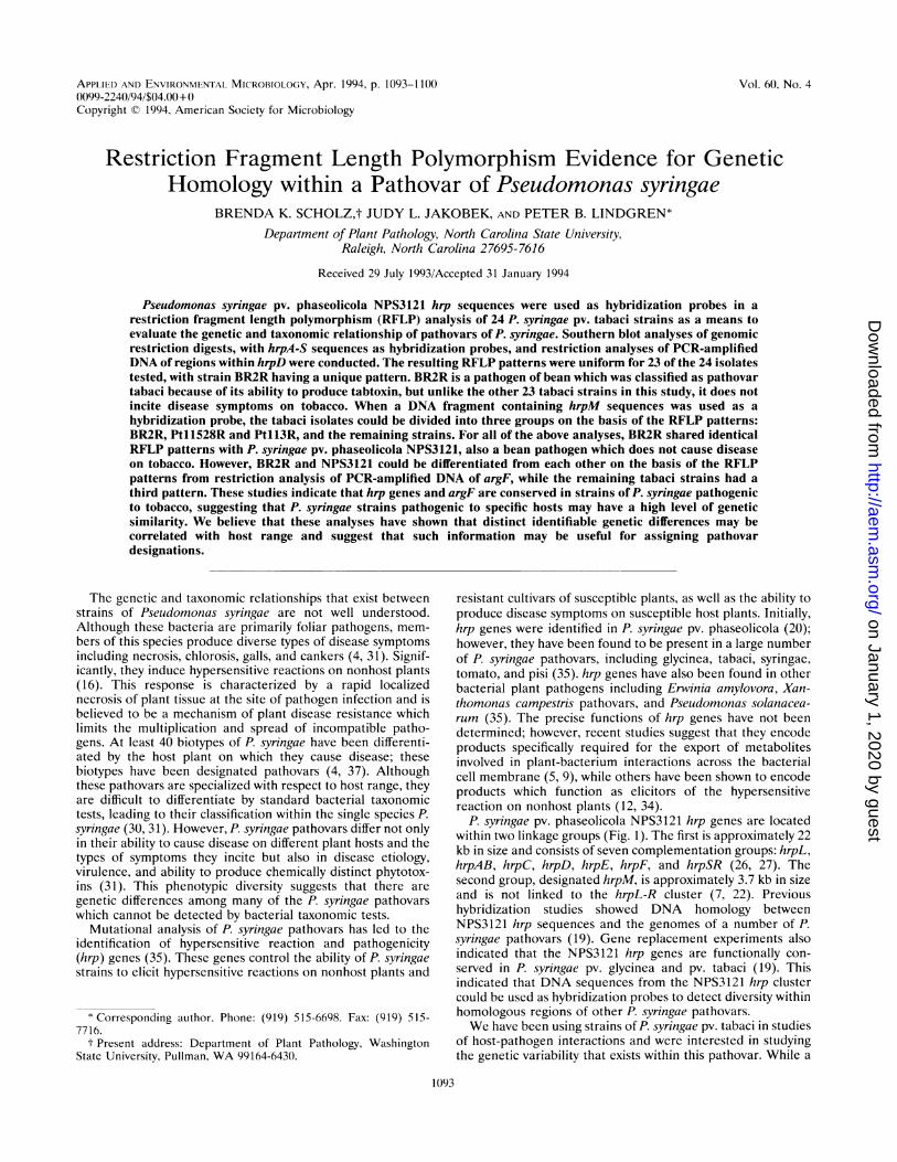
P. syringae pv. phaseolicola NPS3121 hrp genes are locatedwithin two linkage groups (Fig. 1). The first is approximately 22kb in size and consists of seven complementation groups: hrpL,hrpAB, hrpC, hrpD, hrpE, hrpF, and hrpSR (26, 27). Thesecond group, designated hrpM, is approximately 3.7 kb in sizeand is not linked to the hrpL-R cluster (7, 22). Previoushybridization studies showed DNA homology betweenNPS3121 hrp sequences and the genomes of a number of P.syringae pathovars (19). Gene replacement experiments alsoindicated that the NPS3121 hrp genes are functionally con-served in P. syringae pv. glycinea and pv. tabaci (19). Thisindicated that DNA sequences from the NPS3121 hrp clustercould be used as hybridization probes to detect diversity withinhomologous regions of other P. syringae pathovars.We have been using strains of P. syringae pv. tabaci in studies
of host-pathogen interactions and were interested in studyingthe genetic variability that exists within this pathovar. While a
1093
on January 1, 2020 by guesthttp://aem
.asm.org/
Dow
nloaded from
1094 SCHOLZ ET AL.
El B
R BBg
E Bg B b SIETh
iI 'I1_ __ __ __iE____
E Bg B E
IIEl
pC6l .01
4
2
2 kbpRF1 021
3
FIG. 1. P. syringae pv. phaseolicola NPS3121 hrp regions, plasmids, and DNA probes used in this study. The open rectangles at the top andletters within them depict complementation groups as defined previously (7, 26). B, Bg, H, E, S, Xb, and R designate cleavage sites for therestriction enzymes BamHI, BglII, HindIII, EcoRI, Sacl, XbaI, and EcoRV, respectively. Probes 1 and 2 are the 15.5-kb BamHI-EcoRI fragment(hrpA-E) and the 4.5-kb EcoRI-HindIII fragment (hrpF-S), respectively, from pPL11; probe 3 is the 3.2-kb BglII-XbaI fragment (hrpD) frompRF1021; and probe 4 is the 3.7-kb BamHI-EcoRI fragment (hrpM) from pC61.01. On plasmid diagrams, open regions correspond to cloned hrpsequences, and lines represent vector sequences.
number of laboratories have studied the genetic diversitywhich exists among the P. syringae pathovars, only limitedinformation is available about the genetic diversity within eachpathovar (17, 23, 24, 31). In this work, we describe a restrictionfragment length polymorphism (RFLP) analysis of P. syringaepv. tabaci with hrp sequences from the closely related pathovarP. syringae pv. phaseolicola as hybridization probes. Wepresent data which indicate that strains of P. syringae pv. tabacipathogenic on tobacco are genetically homologous withinhrpA-S and argF, indicating that host range is correlated witha high degree of genetic uniformity within this pathovar of P.syringae.
MATERIALS AND METHODS
Bacterial strains, culture conditions, and media. The bac-terial strains, plasmids, and phage used in this study are
presented in Table 1. The P. syringae strains were cultured at28°C in King's medium B (14), and the Escherichia coli strainswere grown at 37°C in Luria-Bertani medium (29). Bacto Agar(Difco, Detroit, Mich.) at 1.5% (wt/vol) was added to mediafor plate cultures. Antibiotics (Sigma, St. Louis, Mo.) wereused for selection at the following concentrations (microgramsper milliliter): rifampin, 100; tetracycline, 15; and ampicillin,50.DNA extraction. Total genomic DNA was extracted from
100-ml overnight broth cultures of P. syringae strains withhexadecyltrimethylammonium bromide (CTAB) as describedin Ausubel et al. (1). Plasmid DNA was extracted by analkaline lysis procedure (29). All DNA samples were furtherpurified by cesium chloride-ethidium bromide density gradientcentrifugation (29).
Southern hybridization. Total genomic DNA (4 p.g) was
digested with the restriction endonucleases EcoRI, BglII, XbaI,EcoRV, Hindlll, and Sacl, in single or double digests accord-ing to the manufacturer's specifications (Boehringer Mannheim,Indianapolis, Ind.). Restricted DNA was analyzed by electro-
phoresis through 0.7% SeaKem agarose (FMC Bioproducts,Rockland, Maine) in 1 x TBE buffer (1 x TBE = 89 mMTris-HCI [pH 8.0], 89 mM boric acid, and 2 mM EDTA) at 22V for 18 h. The DNA was denatured and transferred toGeneScreen Plus membranes according to the manufacturer'sprotocol (NEN Dupont, Boston, Mass.).The membranes were prehybridized and hybridized in a
solution consisting of 50% formamide, 5 x Denhardt's solution(1 x Denhardt's solution = 0.02% Ficoll, 0.02% polyvinylpyr-rolidone, 0.02% bovine serum albumin), 1% sodium dodecylsulfate (SDS), 1 M NaCl, 0.05 M Tris-HCl (pH 7.5), and 100,ug of denatured salmon sperm DNA per ml at 42°C. Hybrid-ization probes (Fig. 1) included the 15.5-kb BamHI-EcoRIfragment and the 4.5-kb EcoRI-HindIII fragment from pPL11containing P. syringae pv. phaseolicola hrpA-E and hrpF-Ssequences, respectively (18, 26); the 3.2-kb XbaI-BglII frag-ment from pRF1021 containing P. syringae pv. phaseolicolahrpD sequences (7); and the 3.7-kb EcoRI-BamHI fragmentfrom pC61.01 containing P. syringae pv. phaseolicola hrpMsequences (7). These fragments were labeled with [o-32P]dCTPby random hexamer labeling (6) and added to the hybridiza-tion buffer at a final concentration of 106 cpm/ml. The mem-branes were hybridized for 14 to 18 h and washed with constantagitation twice in 2 x SSC (1 x SSC = 0.15 M NaCl, 0.015 Msodium citrate, pH 7.0) at room temperature for 5 min, once in2 x SSC plus 1% SDS at 65°C for 30 min, once in 1.5 x SSCplus 1% SDS at 65°C for 30 min, and, finally, once in 0.1 x SSCat room temperature for 30 min. The membranes were ex-
posed to AIF/RX X-ray film (Fuji) for 10 h with one CronexLightning Plus intensifying screen (NEN Dupont) at - 80°C.PCR analysis. PCR primers were constructed from P.
syringae pv. phaseolicola NPS3121 hrpD (7) and argF sequencedata (10) with the computer program PC Gene (IntelliGenet-ics Inc., Mountain View, Calif.). Primers, 20 nucleotides inlength, were matched by Tn, that varied between 62 and 64°Cdepending upon the primer set. Two primer sets were con-structed to amplify two consecutive DNA segments within the
pPL 1 1
APPL. ENVIRON. MICROBIOL.
l
on January 1, 2020 by guesthttp://aem
.asm.org/
Dow
nloaded from
RFLP EVIDENCE FOR GENETIC HOMOLOGY WITHIN P. SYRINGAE
TABLE 1. Bacterial strains, plasmids, and phage
Strain, plasmid, or phage Relevant characteristics" Reference or source
StrainsPseudomonas syringae
pv. tabaciBR2RJS78-45RJS78-45-1RJS78-45-3RJS78-46RJS78-51 RJS78-53RJS78-64W#2RJS81-70RJS81-75RJS82-89RJS82-90RJS82-93RJS82-107RJS84-116RJS84-117RJS84-118RJS84-119RJS88-63RJSWF-1-1-IRPsa45RPsa52RPtl 13RPtl 1528R
Pseudomonas syringaepv. phaseolicola
NPS3121Escherichia coli
DH5ot
HB1OI
JM101MIKE
PlasmidspC61.01
pPL11
pRCP36
PhagepRF1021
Rifr; isolated in Brazil, 1979Rifr; isolated in Florida, 1978Rifr; isolated in Florida, 1978RifT; isolated in Florida, 1978RifT; isolated in Florida, 1978RifT; isolated in Pennsylvania, 1978RifT; isolated in Pennsylvania, 1978Rif'; unknown origin, 1978Rifr; isolated in Maryland, 1981Rifr; isolated in Maryland, 1981RifT; isolated in Maryland, 1982RifT; isolated in Maryland, 1982Rifr; isolated in Kentucky, 1982Rifr; isolated in Maryland, 1982RifT; isolated in Pennsylvania, 1984RifT; isolated in Pennsylvania, 1984Rif'; isolated in Maryland, 1984Rif'; isolated in Maryland, 1984RifT; origin unknown, 1988RifT; unknown origin and date of isolationRifr; isolated in Florida, unknown date of isolationRifr; isolated in Pennsylvania, unknown date of isolationRifr; isolated in Europe, 1965Rifr; origin unknown, 1923
Rifr; unknown origin and date of isolation
F- endAI hsdR1 7 (rk -Mk+) supE44 thi recA I gyrA 4801acZAMJ5 relAIA(lac-proAB-argF) X -
F- hsdS20 (hsdR hsdM) recA13 thi leu arg-14 proA2 lacYI galK2 rpsLxyl-5 mtl-l supE44 X-
supE thi A(lac-proAB) (F' traD36 proAB lacIq lacZAM15)Wild-type isolate from sewage
Tetr; carries the 3.4-kb BamHI-EcoRI fragment containing NPS3121hrpM in pLAFR3
Tetr; carries the 20-kb BamHI-HindIII fragment containing NPS3121hrpA-S sequences cloned in the synonymous sites of pWB5A
Tetr; carries the 2.1-kb BstEEII fragment containing the NPS3121 argF inpLAFR3
Ampr; M13mpl8 carrying the 3.2-kb BglII-XbaI fragment containingNPS3121 hrpD
28J. Skoog via H. SpurrJ. Skoog via H. SpurrJ. Skoog via H. SpurrJ. Skoog via H. SpurrJ. Skoog via H. SpurrJ. Skoog via H. SpurrJ. Skoog via H. SpurrJ. Skoog via H. SpurrJ. Skoog via H. SpurrJ. Skoog via H. SpurrJ. Skoog via H. SpurrJ. Skoog via H. SpurrJ. Skoog via H. SpurrJ. Skoog via H. SpurrJ. Skoog via H. SpurrJ. Skoog via H. SpurrJ. Skoog via H. SpurrJ. Skoog via H. SpurrJ. Skoog via H. SpurrR. DurbinR. Durbin28American Type Culture
Collection
25
Bethesda ResearchLaboratories
3
3615
7
18
25
7
" Rif, rifampin; Tet, tetracycline; Amp, ampicillin; r, resistant.
4.4-kb hrpD region. The hrpD set 1 (5'-GATCAGCAAGGlITl7CGGCGG-3' [upstream] and 5'-CAGTGCTGTICAGTCGTCGG-3' [downstream] amplified an estimated 1,100-bpfragment (nucleotide positions 253 to 1352 of hrpD codingregion), and the hrpD set 2 (5'-CTGAACAGCACTGGTCCGAA-3' [upstream] and 5'-CGACCAGAAACATGCCTTGC-3' [downstream]) amplified an estimated 1,023-bp fragment(nucleotide positions 1340 to 2362 of hrpD coding region). TheargF primer set (5'-CATCGAGGTCATTGAGGCGG-3' [up-stream] and 5'-CAGCAACTGACTCATGCGCG-3' [down-stream]) amplified an approximately 1,470-bp fragment con-taining the ornithine carbamoyltransferase (OCTase) codingregion. (Please note: in addition to the 918-nucleotide OCTasecoding region, this amplified fragment contains approximately540 nucleotides 5' to the argF initiation codon and 11 nucle-otides 3' of the termination codon). Amplification of DNA was
performed in 100-plI volumes containing 500 mM deoxynucleo-side triphosphates, 0.5 ,M each primer, 2.5 U of Taq DNApolymerase (Boehringer Mannheim or Promega [Madison,Wis.]), 50 mM KCl, 10 mM Tris-HCl (pH 9.0), 0.1% TritonX-100, 1.0 mM MgCl2, and 2 ng of DNA template. Amplifi-cations were performed in an MJR Programmable ThermalController model PTC-100 (MJ Research, Inc., Waterstown,Mass.) with an initial three-step cycle of 2 min at 95°C, 2 minat 55°C, and 2 min at 72°C, followed by 30 cycles of 1 min at94°C, I min at 55°C, and 2 min at 72°C. Positive controlsconsisting of pPL11 or pRCP36 (containing argF) and negativecontrols lacking DNA were included with each experiment.The amplified products were extracted in phenol-chloro-
form-isoamyl alcohol (25:24:1), precipitated with 0.3 M sodiumacetate and 95% ethyl alcohol at - 20°C for 24 h, washed with70% ethyl alcohol, dried, and resuspended in TE (10 mM
VOL. 60, 1994 1 095
on January 1, 2020 by guesthttp://aem
.asm.org/
Dow
nloaded from
1096 SCHOLZ ET AL.
TABLE 2. Tabtoxin production, disease symptoms, and summary of RFLP groupings observed for P. syringae pv. phaseolicolaNPS3121 and strains of P. syringae pv. tabaci
Disease symptoms on: RFLP groupingP. syringae strains Tabtoxin'
Tobacco Bean hrpA-S hrpM argF
P. syringae pv. phaseolicola NPS3121 - HRh Halo blight A A AP. syringae pv. tabaciBR2R + HR Wildfire A A BPtll3R, Pt1l528R + Wildfire HR B B CJS78-64W#2R, JS82-107R, JS84-116R, + Wildfire HR B C C
JS84-117R, JS84-118R, JS84-119R,JS88-63R, JSWF-1-1-IR
Psa45R, Psa52R, JS78-45R, JS78-45-1R, - Angular leaf spot HR B C CJS78-45-3R, JS78-46R, JS78-51 R,JS78-53R, JS81-70R, JS81-75R, JS82-89R, JS82-90R, JS82-93R
"Tabtoxin production determined by biological assay as described in Materials and Methods.HR, hypersensitive reaction.
Tris-Cl [pH 8.0], 1 mM EDTA) buffer. PCR products weredigested with various restriction endonucleases according tothe manufacturer's specifications (Boehringer Mannheim) andelectrophoresed through 4% agarose consisting of 0.8%SeaKem and 3.2% Nusieve agarose (FMC Bioproducts) in 1 xTBE buffer at 54 V for 12 h. Gels were stained with 10-4 mgof ethidium bromide per ml and photographed over a UVtransilluminator. All experiments were repeated at least twotimes.Growth and inoculation of plants. Nicotiana tabacum (to-
bacco) cv. Judy's Pride was grown from seed in the greenhouse.Seeds were germinated in Metro-Mix 220 planting mix andtransplanted 2 to 4 weeks after germination into clay potscontaining a 1:2 mixture of Metro-Mix 220 planting mix andclay. Inoculation of plants was 10 to 16 weeks after transplant-ing. P. syringae strains were grown overnight, pelleted bycentrifugation, and washed in sterile distilled water. Cellsuspensions were adjusted to approximately 105 CFU/ml (foranalysis of compatible interactions) and 108 CFU/ml (foranalysis of incompatible interactions) with sterile distilledwater and infiltrated into tobacco leaves with a disposableplastic Pasteur pipette as described previously (19). Standardplate count procedures were used to verify inoculum concen-trations. Plants were incubated in the greenhouse after inocu-lation, and observations of symptom development were madedaily. All inoculations were repeated three times.
Phaseolus vulgaris L. (bean) cv. Red Kidney plants weregrown and inoculated, by vacuum infiltration, as previouslydescribed (13).
Tabtoxin bioassay. P. syringae pv. tabaci strains were assayedfor tabtoxin production by the procedure of Gasson (8).Briefly, E. coli MIKE (15) was grown overnight in Luria-Bertani medium. A 100-pl aliquot was introduced into 5 ml ofmelted 0.9% M9 agar (29) at 50°C with 0.4% glycerol substi-tuted for glucose, overlaid onto Turner's minimal agar (33),and allowed to dry for 30 min. Overnight P. syringae pv. tabacicultures (5 ml) were centrifuged and washed with steriledistilled water. Cells were resuspended in 5 ml of steriledistilled water, and 5 .1I of each isolate was spotted onto platespreviously overlaid with strain MIKE and allowed to dry. Fourtest isolates were spotted per plate, with P. syringae pv.phaseolicola and E. coli as controls. The plates were incubatedat 28°C and observed 24 and 48 h after plating. Clear zonessurrounding P. syringae strains indicated tabtoxin production.Each strain was tested twice by this procedure.
RESULTS
Disease reactions on tobacco and bean. Disease phenotypesfor all P. syringae strains were determined by infiltration intotobacco and bean plants; the results of these analyses aresummarized in Table 2. Except for strain BR2R, all P. syringaepv. tabaci strains, at a concentration of 105 CFU/ml, causedeither wildfire or angular leaf spot on tobacco plants. BR2R,like P. syringae pv. phaseolicola NPS3121, did not cause diseaseon tobacco but elicited a hypersensitive reaction instead.Wildfire of tobacco has been associated with P. syringaetabtoxin production (21); therefore, we used an in vitro assayto determine tabtoxin production for each strain used in thisstudy (8, 15). All P. syringae pv. tabaci strains that causedwildfire also produced tabtoxin, while strains that causedangular leaf spot did not (Table 2). BR2R, which did not causewildfire of tobacco, but instead elicited a hypersensitive reac-tion, also produced tabtoxin in vitro (Table 2). P. syringae pv.phaseolicola NPS3121 did not produce tabtoxin in vitro. Againwith the exception of BR2R, all P. syringae pv. tabaci strains,when infiltrated into bean at a concentration of 108 CFU/ml,induced a hypersensitive reaction within 24 h after infiltration.Although strain BR2R produced tabtoxin, the plant reac-
tions on tobacco and bean were distinct from those induced bythe other P. syringae pv. tabaci isolates and more closelyresembled those of P. syringae pv. phaseolicola NPS3121 withsubtle differences. Like NPS3121, BR2R induced a hypersen-sitive reaction on tobacco within 24 h after infiltration with aconcentration of 108 CFU/ml. When infiltrated into bean, bothBR2R and NPS3121 incited disease but differed in the extentof tissue chlorosis and time required for lesion formation.When bean was infiltrated with BR2R at an inoculum concen-tration of 105 CFU/ml, sunken chlorotic lesions developed 48h after infiltration; these lesions spread rapidly, and infiltratedleaves became completely necrotic 5 to 7 days after infiltration.In contrast, bean infiltrated with an identical inoculum ofNPS3121 produced water-soaked lesions between 72 and 96 hafter infiltration. The degree of chlorosis on bean, observedwith NPS3121, was also much reduced compared with BR2Rand was observed primarily on young secondary leaves ratherthan on the primary leaves that had been infiltrated. Thus,BR2R produced reactions on tobacco which were very differ-ent from those of the other P. syringae pv. tabaci isolates andalso distinct from those of P. syringae pv. phaseolicolaNPS3121.
APPL. ENVIRON. MICROBIOL.
on January 1, 2020 by guesthttp://aem
.asm.org/
Dow
nloaded from
RFLP EVIDENCE FOR GENETIC HOMOLOGY WITHIN P. SYRINGAE 1097
C 1 2 3 4 5 6 7 8 9 10 11 12
A.
24-
9.4 -X0
4.3-t_ *l l- ;:
2._3*- |_l2><
2.0 - | . !Imm _ l
J78-45-1R; lane 2, JS_1-a5,lan 3, x,S_8-4f5R lan 4_BR2Rg; lAn 5
JS84-117R; .lan 6, JS8-4R lanei.0 7i KW,JS85R;ln ,9JSW 1> lRlan9,Pa2; lane 10 JS411R lan 11, JS29R lanL12v,w
JS82-89R.~~ ~Moeua siz makig ar gie in kilbs pars
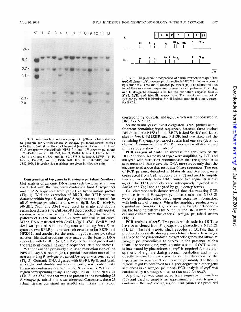
FIG. 2. Southern blot autoradiograph of BglII-EcoRI-digested to-tal genomic DNA from several P. syringae pv. tabaci strains probedwith the 15.5-kb BamHI-EcoRI fragment (hrpA-E) from pPL11. LaneC, P. syringae pv. phaseolicola NPS3121; lane 1, P. syringae pv. tabaciJS78-45-1R; lane 2, JS81-75R; lane 3, JS78-45R; lane 4, BR2R; lane 5,JS84-117R; lane 6, JS78-46R; lane 7, JS78-SlR; lane 8, JSWF-1-1-lR;lane 9, Psa52R; lane 10, JS84-116R; lane 11, JS82-9OR; lane 12,JS82-89R. Molecular size markings are given in kilobase pairs.
Conservation ofhap genes in P. syringae pv. tabaci. Southernblot analysis of genomic DNA from each bacterial strain wasconducted with the fragments containing hrpA-E sequencesand hrpF-S sequences from pPL11 as hybridization probes(Fig. 1). With the exception of BR2R, the RFLP patternsdetted within hrpA-E and hrpF-S regions were identical forall P. syringae pv. tabaci strains when BglII, EcoRI, EcoRV,HindIII, SacI, and XbaI were used in single and doublerestriction digests (the BglII-EcoRI digest probed with hrpA-Esequences is shown in Fig. 2). Interestingly, the bandingpatterns of BR2R and NPS3121 were identical in all cases.When DNA restricted with EcoRI, BglII, XbaI, EcoRV, andSacl was probed with the fragment containing hrpA-E se-quences, two RFLP patterns were observed, one for BR2R andNPS3121 and another for the remaining P. syringae pv. tabaciisolates. Identical groupings were made on the basis of DNArestricted with EcoRI, BglII, EcoRV, and Sacl and probed withthe fragment containing hrpF-S sequences (data not shown).With the aid of a previously published restriction map of the
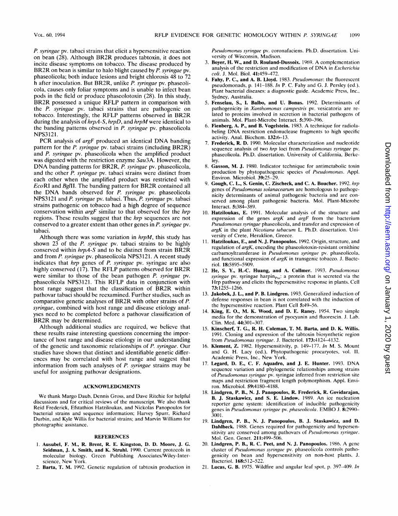
NPS3121 hrpL-R region (26), a partial restriction map of thecorresponding P. syringae pv. tabaci hrp region was constructed(Fig. 3). Genomic DNA digested with EcoRI, BglII, and XbaI,in single and double restriction digests, was probed withfragments containing hrpA-E and hrpD sequences. Within theregion corresponding to hrpD and hrpE in BR2R and NPS3121(Fig. 3), an XbaI site that was not present in the remaining 23P. syringae pv. tabaci strains was observed. Conversely, these 23tabaci strains contained an EcoRI site within the region
Bg EE gBg9 Xb E Ey H
I I I I I I
B. E1 E E Bg E Bg Bg E E9 HII I I I I I
2 kb
FIG. 3. Diagrammatic comparison of partial restriction maps of thehrpL-R cluster of P. syringae pv. phaseolicola NPS3121 (A) as reportedby Rahme et al. (26) and P. syringae pv. tabaci (B). The restriction sitesin boldface represent unique sites present in each pathovar. E, Xb, Bg,and H designate cleavage sites for the restriction enzymes EcoRI,XbaI, BglII, and HindIll, respectively. The restriction map of P.syringae pv. tabaci is identical for all isolates used in this study exceptfor BR2R.
corresponding to hrpAB and hrpC, which was not observed inBR2R or NPS3121.
Southern analysis of EcoRV-digested DNA, probed with afragment containing hrpM sequences, detected three distinctRFLP patterns: NPS3121 and BR2R lacked EcoRV restrictionsites in hrpM, Pt11528R and Pt113R had two sites, and theremaining P. syringae pv. tabaci strains had one site (data notshown). A summary of the RFLP groupings for all strains usedin this study is shown in Table 2.PCR analysis of hrpD. To increase the sensitivity of the
RFLP analysis, segments of hrpD were amplified by PCR andanalyzed with restriction endonucleases that recognize 4-basesequences and thus cleave the DNA more frequently than theenzymes used above that recognize 6-base sequences. Two setsof PCR primers, described in Materials and Methods, wereconstructed from hrpD sequence data (7) and used to amplifytwo approximately 1-kb-DNA, consecutive segments withinhrpD. The PCR products were subsequently digested withSau3A and TaqI and analyzed by gel electrophoresis.
Gel electrophoresis demonstrated that the resulting PCRproducts from all P. syringae pv. tabaci strains and NPS3121were the predicted size, based upon sequence information,with both sets of primers. When the amplified products weredigested with Sau3A or TaqI and analyzed by gel electrophore-sis, the banding patterns for NPS3121 and BR2R were identi-cal and distinct from the other P. syringae pv. tabaci strains(Fig. 4).PCR analysis of argF. Two genes which code for OCTase
have been cloned from P. syringae pv. phaseolicola NPS3121(11, 25). The first is argK, which encodes an OCTase that isproduced specifically during phaseolotoxin biosynthesis; argKis linked to the phaseolotoxin biosynthetic genes and allows P.syringae pv. phaseolicola to survive in the presence of thistoxin. The second gene, argF, encodes a form of OCTase thatis inactivated by phaseolotoxin; argF is required for the bio-synthesis of arginine during normal metabolism and is notdirectly involved in pathogenicity or the elicitation of thehypersensitive reaction. To address the possibility that the hrpregions might be conserved to a higher degree than other genesequences in P. syringae pv. tabaci, PCR analysis of argF wasconducted by a strategy similar to that used for hrpD.A primer set was constructed from sequence information
(10) and used to amplify an approximately 1.5-kb fragmentcontaining the argF coding region. This primer set produced
I L I I E I
VOL. 60, 1994
_ _ _ _
on January 1, 2020 by guesthttp://aem
.asm.org/
Dow
nloaded from

1098 SCHOLZ ET AL.
1 2 3 4 5 6 7 8 9 10 11
253-
165-
110-
FIG. 4. Sau3A-digested amplified PCR product from several P.syringae pv. tabaci strains with hrpD primer set 2. Lane 1, pPL11(containing NPS3121 hrp sequences); lane 2, JS78-45-1R; lane 3,JS81-75R; lane 4, JS78-45R; lane 5, BR2R; lane 6, JS84-117R; lane 7,JS78-46R; lane 8, JS78-51R; lane 9, JSWF-1-1-IR; lane 10, Psa52R;lane 11, JS84-116R. Molecular size markings (253, 165, and 110) aregiven in base pairs. Please note that the NPS3121 hrp region isrepresented by plasmid pPL11 in this figure and that banding patternsobserved with pPL11 were identical to those for NPS3121 (data notshown).
the expected amplification product with all P. syringae pv.tabaci strains and NPS3121. When the amplification productswere digested with the restriction enzyme Sau3A and analyzedby gel electrophoresis, all P. syringae pv. tabaci strains andNPS3121 produced the same DNA banding pattern (data notshown). However, when the amplification products were dou-ble-digested with BglII and EcoRI, three different groups wereidentified: NPS3121, BR2R, and the other P. syringae pv.tabaci strains (Fig. 5). The BR2R DNA banding pattern wassimilar to that of NPS3121, but in addition contained a DNAfragment which was similar in size to a fragment amplified inthe other P. syringae pv. tabaci strains.
DISCUSSION
There has been much interest and confusion regarding thegenetic relationships and taxonomy of P. syringae (30, 31).Many strains of P. syringae were originally classified as separatespecies on the basis of the host plant from which they wereisolated. In 1980, the International Committee of SystematicBacteriology reviewed the classification of pseudomonads andreduced the accepted number of species from over 100 to 23(32, 37). Many of the strains previously classified as uniquespecies were consequently grouped into P. syringae, on thebasis of determinative tests designed primarily for the identi-fication of saprophytic bacteria and members of the familyEnterobacteriaceae. Host range, however, is a very usefulcriterion to differentiate plant-pathogenic bacteria; therefore,strains of P. syringae have been further differentiated intosubspecies groupings, referred to as pathovars, on the basis ofhost-plant interactions (31, 37).For scientists studying plant-bacterium interactions, the
most significant feature of P. syringae strains is the fact thatthey are plant pathogens. It may be difficult to design biochem-ical tests to differentiate the unique physiological and/or
1 2 3 4 5 6 7 8 9 10 11 12 13
700 -
600 -
300 -
124 -
FIG. 5. EcoRI-BglII-digested amplified PCR product from severalP. syringae pv. tabaci strains with argF primers. Lane 1, P. syringae pv.phaseolicola NPS3121; lane 2, JS78-45-1R; lane 3, JS81-75R; lane 4,JS78-45R; lane 5, BR2R; lane 6, JS84-117R; lane 7, JS78-46R; lane 8,JS78-51R; lane 9, JSWF-1-1-IR; lane 10, Psa52R; lane 11, JS84-116R;lane 12, JS82-90R; lane 13, JS82-89R. Molecular size markings (700,600, 300, and 124) are given in base pairs.
biochemical attributes that are hypothesized to contribute tothe host range of each pathovar. However, those characteris-tics that contribute to host range may be reflected in differencesin genome organization within each pathovar. Consequently,genetic techniques may offer more data for characterization andidentification of these bacteria than do determinative tests.
In this study, 24 P. syringae pv. tabaci strains and 1 P. syringaepv. phaseolicola strain were compared by RFLP analysis. Thehybridization probes used included P. syringae pv. phaseolicolaNPS3121 DNA sequences containing hrpA-S (hrpA-E, hrpF-S,and hrpD) and hrpM. On the basis of the RFLP patterns withinthe hrpA-S cluster, the strains were divided into two groups, 23of the P. syringae pv. tabaci isolates forming one group andBR2R and NPS3121 forming the other (Table 2). Thus, exceptfor BR2R, these studies indicate that within the hrpA-S regionthe restriction sites examined are conserved within P. syringaepv. tabaci. For many of the restriction endonucleases analyzed,the DNA banding patterns of P. syringae pv. tabaci and P.syringae pv. phaseolicola NPS3121 were similar. However, andwith the exception of BR2R, P. syringae pv. tabaci containedunique Sacl, EcoRI, EcoRV, and XbaI restriction sites pro-ducing DNA banding patterns distinct from those forNPS3121. These same restriction enzymes resulted in DNAbanding patterns for BR2R that were distinct from those forthe P. syringae pv. tabaci strains but identical to those forNPS3121. Similar results were also seen in the PCR analysis ofhrpD.
Three RFLP groups were observed when genomic DNA wasdigested with EcoRV and probed with hrpM. These groupsconsisted of Pt11528R and Pt113R, BR2R and NPS3121, andthe remaining 21 P. syringae pv. tabaci strains. Although theRFLP pattern of Ptl 1528R and Ptl 13R was unique, nodifferences in disease symptoms were observed with thesestrains in comparison with other P. syringae pv. tabaci strainscausing wildfire of tobacco. A previous study of the DNAfingerprint patterns of P. syringae pv. tabaci also found thatPt11528R and Pt113R were unique in comparison with 29other P. syringae pv. tabaci strains (2). The significance of thisis currently not understood.
Strain BR2R, originally isolated from Phaseolus vulgaris, isthe causal agent of bean wildfire and is distinct from the other
APPL. ENVIRON. MICROBIOL.
on January 1, 2020 by guesthttp://aem
.asm.org/
Dow
nloaded from
RFLP EVIDENCE FOR GENETIC HOMOLOGY WITHIN P. SYRINGAE 1099
P. syringae pv. tabaci strains that elicit a hypersensitive reactionon bean (28). Although BR2R produces tabtoxin, it does notincite disease symptoms on tobacco. The disease produced byBR2R on bean is similar to halo blight caused by P. syringae pv.
phaseolicola; both induce lesions and bright chlorosis 48 to 72h after inoculation. But BR2R, unlike P. syringae pv. phaseoli-cola, causes only foliar symptoms and is unable to infect beanpods in the field or produce phaseolotoxin (28). In this study,BR2R possessed a unique RFLP pattern in comparison withthe P. syringae pv. tabaci strains that are pathogenic on
tobacco. Interestingly, the RFLP patterns observed in BR2Rduring the analysis of hrpA -S, hrpD, and hrpM were identical to
the banding patterns observed in P. syringae pv. phaseolicolaNPS3121.PCR analysis of argF produced an identical DNA banding
pattern for the P. syringae pv. tabaci strains (including BR2R)and P. syringae pv. phaseolicola when the amplified productwas digested with the restriction enzyme Sau3A. However, theDNA banding patterns for BR2R, P. syringae pv. phaseolicola,and the other P. syringae pv. tabaci strains were distinct fromeach other when the amplified product was restricted withEcoRI and BglII. The banding pattern for BR2R contained allthe DNA bands observed for P. syringae pv. phaseolicolaNPS3121 and P. syringae pv. tabaci. Thus, P. syringae pv. tabacistrains pathogenic on tobacco had a high degree of sequence
conservation within argF similar to that observed for the hrpregions. These results suggest that the hrp sequences are notconserved to a greater extent than other genes in P. syringae pv.
tabaci.Although there was some variation in hrpM, this study has
shown 23 of the P. syringae pv. tabaci strains to be highlyconserved within hrpA-S and to be distinct from strain BR2Rand from P. syringae pv. phaseolicola NPS3121. A recent studyindicates that hrp genes of P. syringae pv. syringae are alsohighly conserved (17). The RFLP patterns observed for BR2Rwere similar to those of the bean pathogen P. syringae pv.
phaseolicola NPS3121. This RFLP data in conjunction withhost range suggest that the classification of BR2R withinpathovar tabaci should be reexamined. Further studies, such as
comparative genetic analyses of BR2R with other strains of P.syringae, combined with host range and disease etiology anal-yses need to be completed before a pathovar classification ofBR2R may be determined.Although additional studies are required, we believe that
these results raise interesting questions concerning the impor-tance of host range and disease etiology in our understandingof the genetic and taxonomic relationships of P. syringae. Ourstudies have shown that distinct and identifiable genetic differ-ences may be correlated with host range and suggest thatinformation from such analyses of P. syringae strains may beuseful for assigning pathovar designations.
ACKNOWLEDGMENTS
We thank Margo Daub, Dennis Gross, and Dave Ritchie for helpfuldiscussions and for critical reviews of the manuscript. We also thankReid Frederick, Efstathios Hatziloukas, and Nickolas Panopoulos forbacterial strains and sequence information; Harvey Spurr, RichardDurbin, and Kyle Willis for bacterial strains; and Marvin Williams forphotographic assistance.
REFERENCES1. Ausubel, F. M., R. Brent, R. E. Kingston, D. D. Moore, J. G.
Seidman, J. A. Smith, and K. Struhl. 1990. Current protocols inmolecular biology. Green Publishing Associates/Wiley-Inter-science, New York.
2. Barta, T. M. 1992. Genetic regulation of tabtoxin production in
Pseudomonas syringae pv. coronafaciens. Ph.D. dissertation. Uni-versity of Wisconsin, Madison.
3. Boyer, H. W., and D. Rouland-Dussoix. 1969. A complementationanalysis of the restriction and modification of DNA in Escherichiacoli. J. Mol. Biol. 41:459-472.
4. Fahy, P. C., and A. B. Lloyd. 1983. Pseudomonas: the fluorescentpseudomonads, p. 141-188. In P. C. Fahy and G. J. Persley (ed.),Plant bacterial diseases: a diagnostic guide. Academic Press, Inc.,Sydney, Australia.
5. Fenselau, S., I. Balbo, and U. Bonas. 1992. Determinants ofpathogenicity in Xanthomonas campestris pv. vesicatoria are re-lated to proteins involved in secretion in bacterial pathogens ofanimals. Mol. Plant-Microbe Interact. 5:390-396.
6. Fienberg, A. P., and B. Vogelstein. 1983. A technique for radiola-beling DNA restriction endonuclease fragments to high specificactivity. Anal. Biochem. 132:6-13.
7. Frederick, R. D. 1990. Molecular characterization and nucleotidesequence analysis of two hrp loci from Pseudomonas syringae pv.phaseolicola. Ph.D. dissertation. University of California, Berke-ley.
8. Gasson, M. J. 1980. Indicator technique for antimetabolic toxinproduction by phytopathogenic species of Pseudomonas. Appl.Environ. Microbiol. 39:25-29.
9. Gough, C. L., S. Genin, C. Zischeck, and C. A. Boucher. 1992. hrpgenes of Pseudomonas solanacearum are homologous to pathoge-nicity determinants of animal pathogenic bacteria and are con-served among plant pathogenic bacteria. Mol. Plant-MicrobeInteract. 5:384-389.
10. Hatziloukas, E. 1991. Molecular analysis of the structure andexpression of the genes argK and argF from the bacteriumPseudomonas syringae phaseolicola, and transfer and expression ofargK in the plant Nicotiana tabacum L. Ph.D. dissertation. Uni-versity of Crete, Heraklion, Greece.
11. Hatziloukas, E., and N. J. Panopoulos. 1992. Origin, structure, andregulation of argK, encoding the phaseolotoxin-resistant ornithinecarbamoyltransferase in Pseudomonas syringae pv. phaseolicola,and functional expression of argK in transgenic tobacco. J. Bacte-riol. 18:5895-5909.
12. He, S. Y., H.-C. Huang, and A. Collmer. 1993. Pseudomonassyringae pv. syringae harpinps.: a protein that is secreted via theHrp pathway and elicits the hypersensitive response in plants. Cell73:1255-1266.
13. Jakobek, J. L., and P. B. Lindgren. 1993. Generalized induction ofdefense responses in bean is not correlated with the induction ofthe hypersensitive reaction. Plant Cell 5:49-56.
14. King, E. O., M. K. Wood, and D. E. Raney. 1954. Two simplemedia for the demonstration of pyocyanin and fluorescin. J. Lab.Clin. Med. 44:301-307.
15. Kinscherf, T. G., R. H. Coleman, T. M. Barta, and D. K. Willis.1991. Cloning and expression of the tabtoxin biosynthetic regionfrom Pseudomonas syringae. J. Bacteriol. 173:4124-4132.
16. Klement, Z. 1982. Hypersensitivity, p. 149-177. In M. S. Mountand G. H. Lacy (ed.), Phytopathogenic procaryotes, vol. II.Academic Press, Inc., New York.
17. Legard, D. E., C. F. Aquadro, and J. E. Hunter. 1993. DNAsequence variation and phylogenetic relationships among strainsof Pseudomonas syringae pv. syringae inferred from restriction sitemaps and restriction fragment length polymorphism. Appl. Envi-ron. Microbiol. 59:4180-4188.
18. Lindgren, P. B., N. J. Panopoulos, R. Frederick, R. Govidarajan,B. J. Staskawicz, and S. E. Lindow. 1989. An ice nucleationreporter gene system: identification of inducible pathogenicitygenes in Pseudomonas syringae pv. phaseolicola. EMBO J. 8:2990-3001.
19. Lindgren, P. B., N. J. Panopoulos, B. J. Staskawicz, and D.Dahlbeck. 1988. Genes required for pathogenicity and hypersen-sitivity are conserved among pathovars of Pseudomonas syringae.Mol. Gen. Genet. 211:499-506.
20. Lindgren, P. B., R. C. Peet, and N. J. Panopoulos. 1986. A genecluster of Pseudomonas syringae pv. phaseolicola controls patho-genicity on bean and hypersensitivity on non-host plants. J.Bacteriol. 168:512-522.
21. Lucas, G. B. 1975. Wildfire and angular leaf spot, p. 397-409. In
VOL. 60, 1994
on January 1, 2020 by guesthttp://aem
.asm.org/
Dow
nloaded from
1100 SCHOLZ ET AL.
G. B. Lucas (ed.), Diseases of tobacco. Biological ConsultingAssociates, Raleigh, N.C.
22. Mukhopadhyay, P., J. Williams, and D. Mills. 1988. Molecularanalysis of a pathogenicity locus in Pseudomonas syringae pv.syringae. J. Bacteriol. 170:5479-5488.
23. Palleroni, N. J., R. W. Ballard, E. Ralston, and M. Doudoroff.1972. Deoxyribonucleic acid homologies among some Pseudomo-nas species. J. Bacteriol. 110:1-11.
24. Pecknold, P. C., and R. C. Grogan. 1973. Deoxyribonucleic acidhomology groups among phytopathogenic Pseudomonas species.Int. J. Syst. Bacteriol. 23:111-121.
25. Peet, R. C., and N. J. Panopoulos. 1987. Ornithine carbamoyltrans-ferase genes and phaseolotoxin immunity in Pseudomonas syringaepv. phaseolicola. EMBO J. 6:3585-3591.
26. Rahme, L. G., M. M. Mindrinos, and N. J. Panopoulos. 1991.Genetic and transcriptional organization of the hrp cluster ofPseudomonas syringae pv. phaseolicola. J. Bacteriol. 173:575-586.
27. Rahme, L. G., M. M. Mindrinos, and N. J. Panopoulos. 1992. Plantand environmental sensory signals control the expression of hrpgenes in Pseudomonas syringae pv. phaseolicola. J. Bacteriol.174:3499-3507.
28. Ribiero, R. D. L. D., D. J. Hagedorn, R. D. Durbin, and T. F.Uchytil. 1979. Characterization of the bacterium inciting beanwildfire in Brazil. Phytopathology 69:208-212.
29. Sambrook, J., E. F. Fritsch, and T. Maniatis. 1989. Molecularcloning: a laboratory manual, 2nd ed. Cold Spring Harbor Labo-ratory, Cold Spring Harbor, N.Y.
30. Schaad, N. W. 1982. How phytopathogenic prokaryotes are clas-sified, p. 19-29. In M. S. Mount and G. H. Lacy (ed.), Phytopatho-genic procaryotes, vol. I. Academic Press, Inc., New York.
31. Schroth, M. N., D. C. Hildebrand, and M. P. Starr. 1981.Phytopathogenic members of the genus Pseudomonas, p. 701-718.In M. P. Starr, H. Stolp, H. G. Truper, A. Balows, and H. G.Schlegel (ed.), The procaryotes. Springer-Verlag, Berlin.
32. Skerman, V. B. D., V. McGowen, and P. H. A. Sneath. 1980.Approved lists of bacterial names. Int. J. Syst. Bacteriol. 30:225-420.
33. Turner, J. G., and R. R. Taha. 1984. Contribution of tabtoxin tothe pathogenicity of Pseudomonas syringae pv. tabaci. Physiol.Plant Pathol. 25:55-69.
34. Wei, Z.-M., R. J. Laby, C. H. Zumoff, D. W. Bauer, S. Y. He, A.Collmer, and S. V. Beer. 1992. Harpin, elicitor of the hypersensi-tive response produced by the plant pathogen Erwinia amylovora.Science 257:85-88.
35. Willis, D. K., J. J. Rich, and E. M. Hrabak. 1991. hrp genes ofphytopathogenic bacteria. Mol. Plant-Microbe Interact. 4:132-138.
36. Yanisch-Perron, C., J. Vieira, and J. Messing. 1985. ImprovedM13 phage cloning vectors and host strains: nucleotide sequencesof the M13mpl8 and pUC19 vectors. Gene 33:103-119.
37. Young, J. M., D. W. Dye, J. F. Bradbury, C. G. Panagopoulos, andC. F. Robbs. 1978. A proposed nomenclature and classification forplant pathogenic bacteria. N. Z. J. Agric. Res. 21:153-177.
APPL. ENVIRON. MICROBIOL.
on January 1, 2020 by guesthttp://aem
.asm.org/
Dow
nloaded from