results of radial shock wave treatment of sports...
TRANSCRIPT

161
17
16
Results of Radial Shock Wave Treatment of Sports-Induced Diseases (Achillodynia, Patella Tip Syndrome and
Tibialis Anterior Syndrome)J. Schöll, H. Lohrer, S. Arentz
Summary
This prospective pilot study was conducted to assess the effects of radialextracorporeal shock waves on chronic achillodynia, patella tip syndrome andtibialis anterior syndrome. Forty patients with chronic therapy-resistant achillo-dynia, 45 patients with chronic therapy-resistant patella tip syndrome and 17patients with chronic therapy-resistant tibialis anterior syndrome receivedrESWT for 3-5 sessions. The results show significant pain relief (at rest, pressureand exertion) as well as an increase of pain threshold. The long term outcomeshowed further improvement up to one year. Radial extracorporeal shock wavetherapy is a promising therapeutic option even for sports-related degenerativediseases. Further controlled and randomized studies are required to confirm thesefindings.
Introduction
Achillodynia, patella tip syndrome and tibialis anterior syndrome are wellknown as sports-induced degenerative diseases. The natural course primarilyseems to be chronic and progressive.
A large number of therapeutic options exist, but most of them do not have anevidence based approach. Regarding infiltration techniques in chronic achillo-
Chapter 16.fm Page 161 Tuesday, November 21, 2006 7:34 PM

162 EXTRACORPOREAL SHOCK WAVE THERAPY
dynia with corticoid substances, a potential risk of iatrogen-induced tendon rup-ture is well known.
From a pathological-anatomical point of view, we consider achillodynia to bea combination of paratendinosis and tendinosis of the Achilles tendon. Thesecomponents are more or less marked[32]. Repetitive microtrauma and insuffi-cient regeneration as well as local inflammatory changes in the paratendinumtrigger the initially latent process of Achilles tendon degeneration[16]. The ten-don 2-7 cm proximal of the calcanear insertion area is the most affected region.Achillodynia itself is characterized by stress-dependent local pain associated withpressure dolence and swelling of the Achilles tendon[19].
The issue of Achilles tendon injuries has recently once again come to beimportant in sports medicine. Within the German national track and field team atotal of 21 out of 70 athletes suffered relevant Achilles tendon injuries during the2000 Olympic Games in Sydney. Four of these athletes have undergone surgerysince then, 10 patients underwent significant long term sport restrictions. Achill-odynia is a typical sports injury induced by running. James et al. reported in 1978achillodynia in 11% of running-induced sports injuries[15]. Endogenous causes(e.g. hyperuricemia) are rare. Anomalies of the lower extremity and previoustendineous lesions bring about increased pronation. For that reason the Achillestendon is exposed to increased torsion[38].
As a typical insertion tendopathy, patella syndrome, so called jumper´s kneeinjury, features histologically mucoid denegeration, fibrinoid necroses, microrup-tures and regeneration at the tendon/bone junction[18]. The jumper´s knee isknown as one of the most common insertion tendopathies. Becker & Krahl in1978 reported patella pathologies in about 37.8% of all tendopathies in athletes[4].We observed 7.4% of all patients suffering from jumper´s knee in our own Institutefor Sports Medicine. Already among 14 to 18 year-old basketball players, the prev-alence of patella tip syndrome is 7%, but prevalence increases with age[8]. Mar-tens et al. in 1982 discussed volleyball and football as a predictive factor because2/3 of the patients participated in these sport activities[28]. Orava & Leppilahti in1999 found insertion endopathies of the lig. patellae most often (44%) in the dif-ferential diagnosis in patients with “anterior knee pain[30].” One third of allpatients report time-out periods of more than 6 months[7]. In terms of jumper’sknee, the pathology is mostly located close to the tip of the patella (79%). In 16%of the jumper´s knee patients, pain was found at the patella base, in 3% at thetuberositas tibiae and in 2% along the entire lig. patellae[30].
Chapter 16.fm Page 162 Tuesday, November 21, 2006 7:34 PM

RESULTS OF RADIAL SHOCK WAVE TREATMENT OF SPORTS-INDUCED DISEASES 163
In a prospective study conducted with sports students an incidence of 13.8%was found within a two year period[40].
Repetitive jumps, particularly vertical ones, seem to be a significant predic-tive factor in patella tip syndrome.
The medial tibialis anterior syndrome is characterized by a load dependentlocal pain at the tibialis anterior in the middle and lower third. An exudativehypertrophic tenosynovitis is correlated to the tibialis posterior tendon [16] andfrequently caused by intensive running and most often by sudden load peaks.Forefoot runners are more affected than heel runners[25].
Tibialis anterior syndrome occurs in 2.5% of top athletes[20]. Among activetrack and field athletes, the medial tibialis anterior syndrome is the third mostcommon sports injury after achillodynia and stress fractures[22]. Endurance ath-letes are more often afflicted by tibialis anterior syndrome.
The efficacy of extracorporeal shock wave therapy to treat different chronicinsertion endopathies has often been documented in controlled randomized andprospective studies [23, 34, 35] even though no consensus has been reached so faron the energy to be applied, on the number of shock waves or on treatment fre-quency. Considered today as standard indications in the musculoskeletal systemare fasciitis plantaris, epicondylitis humeri radialis, pseudarthrosis and calcifictendonitis of the shoulder[14]. The different treatment modalities (energy gener-ation, energy flux density, dose and treatment frequency) continue to be the sub-ject of scientific debate. Until now comparative studies of treatments with focusedand radial extracorporeal shock waves show no difference for fasciitis plantaris[36] or calcific tendonitis of the shoulder[12]. Apart from our own publication[26], clinical studies of the effect of ESWT for achillodynia, patella tip syndromeand tibialis anterior syndrome have not been published. In experiments con-ducted on rabbits Rompe et al. showed dose-dependent effects of extracorporealshock waves on the Achilles tendon[33]. High energies ( > 0.6 mJ/mm2) lead toinjuries, necroses and edema.
Objective of the study
This study is designed as a prospective trial to investigate the effect of rESWTon sports-induced enthesiopathies such as chronic achillodynia, chronic patellatip syndrome and chronic tibialis anterior syndrome.
Chapter 16.fm Page 163 Tuesday, November 21, 2006 7:34 PM

164 EXTRACORPOREAL SHOCK WAVE THERAPY
Patients, material and methods
Between September 1998 and October 2000, 102 subjects (40 with achillo-dynia, 45 with patella tip syndrome and 17 with tibialis anterior syndrome) havefulfilled all inclusion and exclusion criteria. They were enrolled in the outpatientorthopedic clinic of the Institute for Sports Medicine in Frankfurt am Main.
The patients of the study groups were on average 35.8 ± 10.5 years old forachillodynia, 24.2 ± 9.8 years old for patella tip syndrome and 21.2 ± 4.6 years oldfor tibialis anterior syndrome. For the athletes suffering from achillodynia, themean height was 178.5 ± 7.5 cm and the mean weight was 75.9 ± 12.6 kg. In theathlete group suffering from patella tip syndrome, their height was 179.1 ± 8.9 cmand their weight was 73.7 ± 13.7 kg. Athletes with tibialis anterior syndrome were172.7 ± 8.9 cm tall and weighed 57.7 ± 8.0 kg. All patients of the study groupswere active athletes. In the achillodynia and tibialis anterior syndrome groupsrunning sports were predominant. Patients with patella tip syndrome were pri-marily affected by jumps (Table 1).
Prior to enrollment, at least two of the conservative standard therapies suchas targeted sole inserts, physical and rehabilitation therapy, bandages, local infil-trations and radiation were performed without success (Table 2).
After one year, 33 patients (82.5%) with chronic achillodynia, 40 patients(88.9%) with patella tip syndrome and 15 patients (88.2%) with chronic tibialisanterior syndrome could be re examined (Table 3).
Sports Achillodynia Jumper´s knee Tibialis anterior syndrome
Jogging 8 3 0
Middle distance 5 1 3
Long distance 5 0 4
Soccer 4 6 2
Long jump 4 2 0
Handball 4 1 2
Tennis 3 6 3
Chapter 16.fm Page 164 Tuesday, November 21, 2006 7:34 PM
RESULTS OF RADIAL SHOCK WAVE TREATMENT OF SPORTS-INDUCED DISEASES 165
Table 1. Demographic data of patients enrolled in the rESWT trial.
Table 2. Treatments proir to the rESWT study.
Sports Achillodynia Jumper´s knee Tibialis anterior syndrome
Triathlon 2 1 0
Bike racing 2 0 0
Volleyball 1 8 0
Decathlon 1 1 1
Gym 1 0 0
Basketball 0 11 0
Ballett 0 3 0
High jump 0 1 2
Total 40 45 17
Prior treatment Achillodynia Jumper´s knee
Tibialis anterior syndrome
Physiotherapy 31 26 11
Massage 15 13 7
Electrotherapy 30 30 10
Medication (oral) 14 12 7
Eternal ointment 8 9 3
Tape 11 21 2
Infiltration (without cortsone) 22 13 3
Infiltration (with cortisone) 8 2 3
Inserts 7 7 3
Cast 6 7 1
Acupuncture 4 1 0
Röntrenreizbestralung 1 0 0
Operation 5 2 1
Chapter 16.fm Page 165 Tuesday, November 21, 2006 7:34 PM

166 EXTRACORPOREAL SHOCK WAVE THERAPY
Table 3. Patients examined from 1 to 52 weeks after rESWT.
Achillodynia and jumper´s knee were defined as an exertion-dependent andpressure-pain related injury of the Achilles tendon[21]. A thickening of the ten-don had to be shown.
The tibialis anterior syndrome was defined as an exertion-dependent andpressure pain injury to the medial tibialis anterior. This injury could not be local-ized presisely but had to extend over at least 5 cm in the middle and lower third ofthe tibialis anterior. X-rays had to exclude tibia stress fracture.
The sagital diameter of the Achilles tendon and the ligamentum patellae aswell as the central echogenity was documented by ultrasound prior to rESWT.The mean thickness of the Achilles tendon was 7.5 ± 2.0 mm and the mean thick-ness of the ligamentum patellae was 6.7 ± 2.5 mm. Following the treatment, nosignificant loss in tendon thickness was found.
Exclusion criteria were defined as systemic predispositions (hyperurikemia,positive HLA B 27, positive rheuma factors), distal Achilles tendopathies, sub-achillary bursitides, heel-adjacent pain syndrome imitating achillodynia, intraar-ticular knee joint injuries (in particular associated chondropathic injuries),instabilities of the patella and stress fractures of the tibia.
The treatment was performed with the Swiss DolorClast®. The applicator(handpiece) was pressed upon the treatment area up to the first ring with applica-tion pressure categorized as “medium.” As the patient adjusted to the shock wave-induced pain, the applied energy was increased during the treatment from 2 to 4bar. Analgesia of the treatment zone was not necessary.
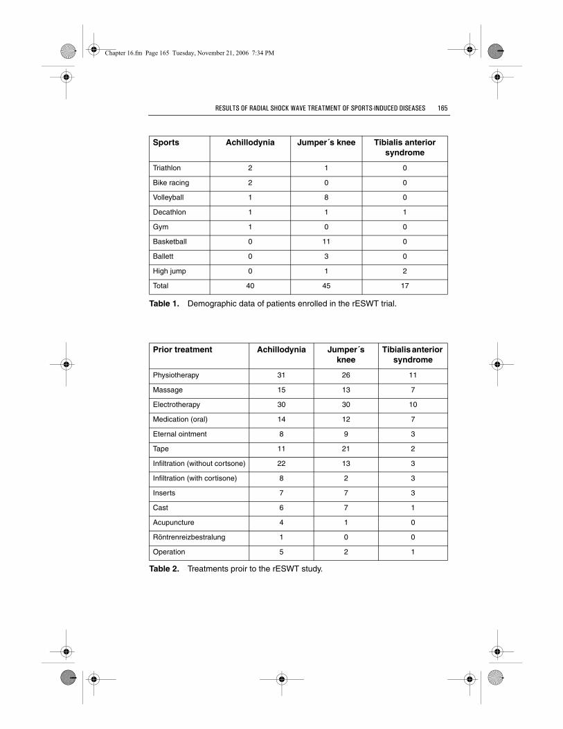
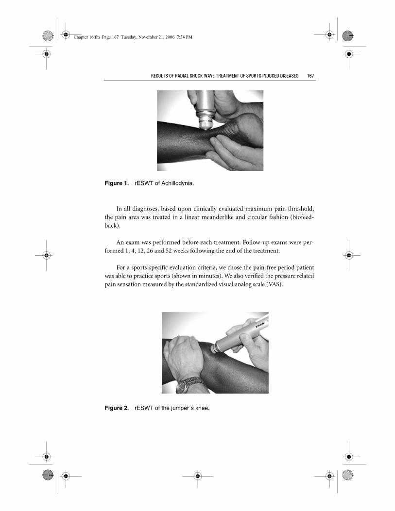
Therapy was delivered in five sessions at one-week intervals (Figures 1-3).Two thousand impulses per treatment session were applied. Treatment frequencywas 6 Hz.
Baseline Weeks after rESWT
1 4 12 26 52
Achillodynia 40 40 39 33 33 33
Jumper´s knee 45 45 45 42 40 40
Tibialis anterior syndrome 17 17 17 15 15 15
Chapter 16.fm Page 166 Tuesday, November 21, 2006 7:34 PM
RESULTS OF RADIAL SHOCK WAVE TREATMENT OF SPORTS-INDUCED DISEASES 167
Figure 1. rESWT of Achillodynia.
In all diagnoses, based upon clinically evaluated maximum pain threshold,the pain area was treated in a linear meanderlike and circular fashion (biofeed-back).
An exam was performed before each treatment. Follow-up exams were per-formed 1, 4, 12, 26 and 52 weeks following the end of the treatment.
For a sports-specific evaluation criteria, we chose the pain-free period patientwas able to practice sports (shown in minutes). We also verified the pressure relatedpain sensation measured by the standardized visual analog scale (VAS).
Figure 2. rESWT of the jumper´s knee.
Chapter 16.fm Page 167 Tuesday, November 21, 2006 7:34 PM

168 EXTRACORPOREAL SHOCK WAVE THERAPY
Figure 3. rESWT of the tibialis anterior syndrome.
We used the Dolormeter as a semi-objective measuring tool. It provides for astandardized application of defined pressure (Figure 4).
It allows evaluation of the threshold of local pain tolerance (in Newton) aswell as the subjective pain response (VAS) on a defined pressure of 30 Newton.
Results
After rESWT we observed an excellent clinical outcome (Table 4). The base-line value for pain at rest before rESWT was 1.7 ± 2.5 VAS for achillodynia, 1.6 ±2.4 VAS for patella tip syndromey and 1.7 ± 1.9 VAS for tibialis anterior syn-drome. One week after rESWT patients with achillodynia improved to 0.4 ±- 1.0VAS (p ≤ 0.01). The group with patella tip syndrome reached 0.8 ± 1.7 VAS (p ≤0.05) one week after treatment completion. Patients with tibialis anterior
Figure 4. The EMS Dolormeter for standardized local pain measurement.
Chapter 16.fm Page 168 Tuesday, November 21, 2006 7:34 PM

RESULTS OF RADIAL SHOCK WAVE TREATMENT OF SPORTS-INDUCED DISEASES 169
syndrome reached 0.6 ± 1.7 VAS (p≤ 0.05) one week after the last rESWT. The fol-low-up exam twelve weeks after the last rESWT showed 0.1 ± 0.3 VAS for achillo-dynia (p ≤ 0.01), 0.3 ± 0.7 VAS for patella tip syndrome (p ≤ 0.01) and 0.5 ± 1.6VAS for tibialis anterior syndrome (p ≤ 0.01). At the end of the observationperiod of one year, patients with achillodynia had improved to 0.1 ± 0.3 VAS (p ≤0.01), those with patella tip syndrome improved to 0.3 ± 0.7 VAS (p ≤ 0.01) andthose with tibialis anterior syndrome were down to 0.1 ± 0.2 VAS (p ≤ 0.01).
Prior to treatment, the pain threshold determined by the DolorMeter was14.1 ± 6.6 N in achillodynia patients, 15.1 ± 7.4 N in patella tip syndrome patientsand 16.1 ± 7.1 N in tibialis anterior syndrome patients. One week after the lastrESWT, patients with achillodynia improved to 27.5 ± 10.9 N (p ≤ 0.01), patientswith patella tip syndrome showed 28.7 ± 14.0 N (p ≤ 0.01) and those with tibialisanterior syndrome improved to 29.1 ± 12.3 N (p ≤ 0.01). The 12 week examthreshold was 38.4 ± 11.8 N in achillodynia (p ≤ 0.01), 32.9 ± 15.6 N in patella tipsyndrome (p ≤ 0.01) and 32.9 ± 11.2 N in tibialis anterior syndrome. After 1 year,patients with achillodynia had improved to 41.9 ± 11.6 N (p ≤ 0.01), those withpatella tip syndrome showed 35.3 ± 15.0 N (p ≤ 0.01) and patients with tibialisanterior syndrome also improved to 40.5 ± 10.4 N (p ≤ 0.01).
Prior to the treatment, the pressure pain determined by the DolorMeter at 30N and VAS was 6.7 ± 3.2 VAS for achillodynia, 5.5 ± 2.9 VAS for patella tip
Achillodynia
Baseline Weeks after rESWT
1 4 12 26 52
Pain at rest (VAS)
1.7±2.5 0.4±1.0** 0.3±1.3** 0.1±0.3** 0.1±0.2** 0.1±0.3**
Pain threshold (Newton)
14.1±6.6 27.5±10.9** 31.2±13.4** 38.4±11.8** 43.3±9.1** 41.9±11.6**
Local pressure (30 N, VAS)
6.7±3.2 2.6±3.6** 1.8±3.4** 0.7±1.8** 0.5±1.8** 0.9±2.6**
Pain during exercise (VAS)
7.8±1.7 2.2±2.5** 1.3±2.2** 0.5±1.1** 0.4±1.0** 0.7±1.6**
Pain-free running interval (min.)
14.4±18.5 63.0±37.0** 75.4±38.1** 87.5±35.2** 96.7±34.7** 90.0±43.0**
Chapter 16.fm Page 169 Tuesday, November 21, 2006 7:34 PM

170 EXTRACORPOREAL SHOCK WAVE THERAPY
Table 4. Outcome after rESWT (* = P ≤ 0.05; ** = P ≤ 0.01).
Jumper´s knee
Baseline Weeks after rESWT
1 4 12 26 52
Pain at rest (VAS)
1.6±2.4 0.8±1.7* 0.6±1.6** 0.3±0.7** 0.3±0.7** 0.3±0.7**
Pain threshold (Newton)
15.1±7.4 28.7±14.0** 30.6±14.5** 32.9±15.6** 34.6±15.5** 35.3±15.0**
Local pres-sure (30 N, VAS)
5.5±2.9 2.3±3.0** 2.2±3.0** 2.1±2.9** 1.9±2.9** 1.7±2.6**
Pain during exercise (VAS)
5.5±2.3 2.8±3.0** 2.5±2.9** 2.3±2.8** 1.9±2.4** 1.9±2.5**
Pain-free running interval (min.)
10.4±15.0 54.3±50.9** 57.0±48.9** 62.8±49.2** 71.2±49.1** 70.3±48.7**
Tibialis anterior syndrome
Baseline Weeks after rESWT
1 4 12 26 52
Pain at rest (VAS)
1.7±1.9 0.6±1.7* 0.6±1.7* 0.5±1.6** 0.5±1.7** 0.1±0.2**
Pain thresh-old (Newton)
16.1±7.1 29.1±12.3** 31.8±11.6** 32.9±11.2** 36.3±11.9** 40.5±10.4**
Local pres-sure (30 N, VAS)
6.2±2.8 2.4±3.3** 2.3±3.5** 1.7±2.8** 1.5±2.8** 0.9±1.9**
Pain during exercise (VAS)
7.8±1.7 1.8±3.1** 2.2±3.0** 1.9±2.9** 1.6±2.8** 0.9±1.9**
Pain-free running interval (min.)
11.2±14.4 62.9±48.9** 72.9±47.5** 91.3±48.8** 91.3±48.8** 105.0±34.8**
Chapter 16.fm Page 170 Tuesday, November 21, 2006 7:34 PM

RESULTS OF RADIAL SHOCK WAVE TREATMENT OF SPORTS-INDUCED DISEASES 171
syndrome and 6.2 ± 2.8 VAS for tibialis anterior syndrome. One week followingthe treatment series, patients with achillodynia improved to 2.6 ± 3.6 VAS (p ≤0.01), patients with patella tip syndrome to 2.3 ± 3.0 VAS (p ≤ 0.01) and patientswith tibialis anterior sybdrome to 2.4 ± 3.3 VAS (p ≤ 0.01). Twelve weeks afterrESWT, 0.7 ± 1.8 VAS was found in patients suffering from achillodynia (p ≤0.01), 2.1 ± 2.9 VAS in patients with patella tip syndrome (p ≤ 0.01) and 1.7 ± 2.8VAS in patients with tibialis anterior syndrome (p ≤ 0.01). At the end of theobservation period of 1 year, achillodynia improved to 0.9 ± 2.6 VAS (p ≤ 0.01),patella tip syndrome to 1.7 ± 2.6 VAS (p ≤ 0.01) and tibialis anterior syndrome to0.9 ± 1.9 VAS.
The pain during sports actvities prior to treatment was 7.8 ± 1.7 VAS forachillodynia, 5.5 ± 2.3 VAS for patella tip syndrome and 7.8 ± 1.7 VAS for tibialisanterior syndrome. One week after the last rESWT, patients with achillodyniaimproved to 2.2 ± 2.5 VAS (p ≤ 0.01), the group with patella tip syndromereached 2.8 ± 3.0 VAS (p ≤ 0.01) and the tibialis anterior syndrome group reached1.8 ± 3.1 VAS (p ≤ 0.01). Twelve weeks after the last rESWT, the scores were 0.5 ±1.1 VAS for achillodynia (p ≤ 0.01), 2.3 ± 2.8 VAS for patella tip syndrome (p ≤0.01) and 1.9 ± 2.9 VAS for tibialis anterior syndrome (p ≤ 0.01). At the end of thefollow up period of 1 year, patients with achillodynia had improved to 0.7 ± 1.6VAS (p ≤ 0.01), patients with patella tip syndrome showed 1.9 ± 2.5 VAS (p ≤0.01) and the tibialis anterior syndrome group was scored at 0.9 ± 1.9 VAS (p ≤0.01).
The pain-free running interval at baseline as identified by the patients them-selves was 14.4 ± 18.5 minutes for achillodynia, 10.4 ± 15.0 minutes for patella tipsyndrome and 11.2 ± 14.4 minutes for tibialis anterior syndrome. One week aftertha last rESWT, patients with achillodynia had improved to 63.0 ± 37.0 minutes(p ≤ 0.01). In the group with patella tip syndrome a pain-free interval of 54.3 ±50.0 minutes (p ≤ 0.01) was reached. One week after treatment completion, thetibialis anterior syndrome group showed a pain-free interval of at least 62.9 ±48.9 minutes, (p ≤ 0.01). The follow-up exam twelve weeks after the treatmentshowed a pain-free interval increase up to 87.5 ± 35.2 minutes for achillodynia (p≤ 0.01), up to 62.8 ± 49.2 minutes for patella tip syndrome (p ≤ 0.01) and up to91.3 ± 48.8 minutes for tibialis anterior syndrome (p ≤ 0.01). At the end of theobservation period of 1 year, patients with achillodynia had improved to 90.0 ±43.0 minutes (p ≤ 0.01), patients with patella tip syndrome to 70.3 ± 48.7 minutes(p ≤ 0.01) and patients with tibialis anterior syndrome to 105.0 ± 34.8 minutes (p≤ 0.01).
Chapter 16.fm Page 171 Tuesday, November 21, 2006 7:34 PM

172 EXTRACORPOREAL SHOCK WAVE THERAPY
Overall, 60% of patients with achillodynia, 40% of patients with patella tipsyndrome and 58.8% of patients with tibialis anterior syndrome were found to bepain-free one year after the last rESWT (Figure 5). Improvement was recorded by12.5% of patients with achillodynia, 24.4% of patients with patella tip syndromeand 17.7% of patients with tibialis anterior syndrome. In 27.5% of patients withachillodynia, 35.6% of patients with patella tip syndrome and 23.5% of patientswith tibialis anterior syndrome the pain was unchanged 12 month after the lastrESWT compared to baseline.
Discussion
Neither in terms of conservative treatment nor in terms of surgery forchronic degenerative injuries to the Achilles tendon (achillodynia), ligamentumpatellae (patella tip syndrome) and tibialis anterior syndrome, the treatmentoptions were evidence based[29, 9, 25]. In terms of nomenclature for chronic ten-don damage associated with running and jumping, concensus is still lacking[27].Even the precise pathology remains unclear[17]. Today, a degenerative-microtraumatic genesis is discussed but histologically relevant inflammatory cellshave never been described on a relevant scale[16].
Studies determining the natural course of these diseases are not avail-able[31].
Figure 5. Overall outcome 1 year after the last rESWT.
Chapter 16.fm Page 172 Tuesday, November 21, 2006 7:34 PM

RESULTS OF RADIAL SHOCK WAVE TREATMENT OF SPORTS-INDUCED DISEASES 173
From a diagnostic point of view, achillodynia is to be limited to the freecourse of the tendon some 2-7 cm above its insertion zone. It must be differenti-ated from other achillary (distal insertion endopathy, dorsal heel spur, bursitissubachillea) and periachillary pain syndromes[21].
Studies on the conservative therapy of achillodynia report from 41 to 67%excellent results[6, 1]. For surgery, Tallon et al. report an average success rate of77.4% in a literature review[39]. These authors point especially to a low method-ical quality of the published studies. Studies with greater methodological deficitsshow comparatively better results.
For the treatment of patella tip syndrome, Cook and Khan reported ten ran-domized studies for the conservative treatment but no randomized studies forsurgery[9]. These authors conclude that neither a specific conservative nor a spe-cial operative method for treating this injury can be recommended.
Achillodynia and patella tip syndrome feature histologically mucoid degener-ation fibrinoid necroses, microruptures and regeneration[16].
Tibialis anterior syndrome is an excsudative hypertrophic tenosynovitis ofthe tibialis posterior tendon[16]. Specific data on the efficacy of conservativeforms of treatment for medial tibialis anterior syndrome have not been pub-lished. Our own experience indicates that surgery is required. Good results areshown in retrospective, non-controlled design[25].
The efficacy of extracorporeal shock wave therapy in different chronic inser-tion tendopathies has been documented repeatedly in the literature through con-trolled, randomized and prospective studies[23, 11, 34, 35]. In other studies theefficacy of ESWT was not demonstrated because of some study limitations[5, 13].
Local anesthesia was not necessary in this study. In the meantime, it wasdemonstrated that local anesthesia has a negative impact on treatment suc-cess[3].
Today, standard indications of the musculoskeletal system are fasciitis planta-ris, eopicondylitus humeri radialis and calcific tendonitis of the shoulder[14].The various treatment modalities (energy generation, energy density, dosage andtreatment frequency) continue to be a subject of scientific debate. A comparisonof treatments with the focused and radial (unfocused) extracorporeal shock wave
Chapter 16.fm Page 173 Tuesday, November 21, 2006 7:34 PM

174 EXTRACORPOREAL SHOCK WAVE THERAPY
for fasciitis plantaris shows no difference[36]. Achillodynia, patella tip syndromeand tibialis anterior syndrome are sports-induced, chronic injuries that are differ-ent from insertion endopathies such as fasciitis plantaris. Compared to results ofconservative and surgical options, the results of this pilot study must be evaluatedas good, especially since at least two conservative pretreatment methods failed.The success rate after one year (pain-free or improvement over 50%) is 72.5% forAchillodynia, 67.4% for patella tip syndrome and 76.5% for tibialis anterior syn-drome and is hardly any different from the average success rate of 77.4% found byTallon et al. following surgery (77.4%)[39].
The study was limited by the uncontrolled study design. Therefore the resultscannot be considered as sufficiently certain on the basis of the criteria of evi-dence-based medicine. Controlled and randomized studies have to be performedto confirm these finings.
Literature
1. Angermann, P, Hovgaard, D (1999): Chronic achilles tendinopathy in athleticindividuals: results of nonsurgical treatment. Foot Ankle 20: 43-46
2. Arndt, K.-H. (1976): Achillessehnenruptur und Sport. Barth Verlag, Leipzig
3. Auersperg, V, Labek, G, Ziernhoeld, M, Poulios, N, Rompe, J-D, Boehler, N (2003):Lokalanaesthesie beeinflusst das Ergebnis der N-ESWT an der plantaren Fasciitis.In: Maier M, Gillesberger, F (Hrsg): Kongressband des 3. Drei-Länder-Treffens derÖsterreichischen, Schweizer und Deutschen Fachgesellschaften. München.
4. Becker, W, Krahl, H (1978): Die Tendopathien. Thieme Verlag, Stuttgart
5. Buchbinder, R, Green, S, White, M, Barnsley, L, Smidt, N, Assendelft, WJ (2002):Shock wave therapy for lateral elbow pain. The Cochrane database of systematicreviews (1), p: CD003524.
6. Clement, DB, Taunton, JE, Smart, GW (1984): Achilles tendinitis andperitendinitis: Etiology and treatment. Am J Sports Med 12: 179-184
7. Cook, JL, Khan, KM, Harcourt, PR, Grant, M, Young, DA, Bonar, SF (1997): Across sectional study of 100 athletes with jumper’s knee managed conservatively andsurgically. Br J Sports Med 31: 332-336
8. Cook, JL, Khan, KM, Kiss, ZS, Purdam, CR, Griffiths, L (2000): Prospectiveimaging study of asymptomatic patellar tendinopathy in elite junior basketballplayers. J Ultrasound Med 19: 473-479
9. Cook, JL, Khan, KM (2001): What is the most appropriate treatment for patellartendinopathy? Br J Sports Med 35: 291-294
10. Franke, K (1980): Traumatologie des Sports. Stuttgart, Thieme, 1980
11. Gerdesmeyer, L, Wagenpfeil, S, Haake, M, Maier, M, Loew, M, Wörtler, K, Lampe,R, Seil, R, Handle, G, Gassel, S, Rompe, J-D (2003): Extracorporeal shock wavetherapy for the treatment of chronic calcifyung tendonitis of the rotator cuff: arandomized controlled trial. JAMA: The Journal of the American MedicalAssociation, Vol. 290 (19), p:2573-80
Chapter 16.fm Page 174 Tuesday, November 21, 2006 7:34 PM

RESULTS OF RADIAL SHOCK WAVE TREATMENT OF SPORTS-INDUCED DISEASES 175
12. Gremion, G, Augros, R, Gobelet, Ch, Leyvraz, P-F (2000): Wirksamkeit derextrakorporalen Stosswellentherapie bei der Calcific tendinitis der Schulter.Schweizerische Zeitschrift für Sportmedizin und Sporttraumatologie 48: 8-11
13. Haake, M, Huenerkopf, M, Gerdesmeyer, L, Koenig, IR (2002): ExtrakorporaleStosswellentherapie (ESWT) bei epicondilytis humeri radialis – EineLiteraturübersicht. Der Orthopäde, Vol. 31 (7), p: 623-632
14. Heller, K-D, Niethard, FU (1998): Der Einsatz der extrakorporalenStosswellentherapie in der Orthopädie – eine Metaanalyse. Z Orthop. 136: 390-401
15. James, SL, Bates, BT, Osternig, LR (1978): Injuries to runners. Am J Sports Med 6:40-50
16. Józsa, L, Kannus, P (1997): Human tendons. Anatomy, physiology and pathology.Human Kinetics. 1-576
17. Khan, KM, Cook, JL (2000): Overuse Tendon Injuries: Where Does the Pain ComeFrom? Sports Medicine and Arthroscopy Review 8: 17-31
18. Krahl, H. (1980): Jumper’s Knee” – Ätiologie, Differentialdiagnose undtherapeutische Möglichkeiten. Orthopäde 9: 193-197
19. Lohrer, H. (1991): Seltene Ursachen und Differentialdiagnosen der Achillodynie.Sportverl Sportschad 5: 182-185
20. Lohrer, H. (1995): Sport orthopaedics in athletics – an analysis of the currentsituation. NSA (New Studies in Athletics) 10 (4): 11-21
21. Lohrer, H. (1996): Die Achillodynie – Eine Übersicht. Sportorthop Sporttraumatol12: 36-42
22. Lohrer, H., W. Alt, A. Gollhofer (1999): Sportschuhe – Design versus Biomechanik?Sonderheft Orthopädieschuhtechnik: 13-16
23. Lohrer, H, Schoell, J, Arentz, S, Froelich, T, Straub, T, Penninger, E, Diesch, R,Haupt, G (2001): Effectiveness of radial shockwave Therapy (rESWT) on tenniselbow and plantar fasciitis. Book of Abstracts. CASM/ACMS annual symposiumand sport medicine Conference, Calgary/CAN
24. Lohrer, H, Schöll, J (2001): Anterior knee pain in athletes. Book of abstracts. 6th
Annual Congress of the European College of Sport Science: 64
25. Lohrer, H. (2002): Überlastungsschäden. In C. J. WIRTH (Hg): Fuß: DasStandardwerk für Klinik und Praxis. Stuttgart – New York, Thieme, 2002
26. Lohrer, H, Schoell, J, Arentz S.(2002): Achillodynie und Patellaspitzensyndrom –Ergebnisse der Behandlung austherapierter, chronischer Fälle mit radialenStosswellen. Sportverletzung Sportschaden 2002;16; 108-114
27. Maffuli, N, Khan, KM, Puddu, G (1998): Overuse Tendon Conditions: Time toChange a Confusing Terminology. Arthroscopy 14: 840- 843
28. Martens, M., Wouters, P, Brussens, A, Mulier, JC (1982): Patellar tendinitis:Pathology and results of treatment. Acta Orthop Scand 53: 445-450
29. McLauchlan, GJ, Handoll, HH (2001): Interventions for treating acute and chronicAchilles tendinits. Cochrane Database Syst Rev (2) pCD000232
30. Orava, S, Leppilahti, J (1999): Overuse injuries of tendons in athletes. In: Jakob, RP,Fulford, P, Horan, F, Hrsg.; European Instructional Course Lectures 4: 128-131
31. Paavola; M, Kannus; P, Paakkala, T, Pasanen; M; Järvinen, M (2000): Long-TermPrognosis of Patients With Achilles Tendinopathy. Am J Sports Med 28: 634-642
32. Puddu, G., Ippolito, E., Postacchini, F.(1976): A classification of Achilles tendondisease. Am J Sports Med 4: 145-150
33. Rompe JD; Kirkpatrick CJ; Kullmer K; Schwitalle M; Krischek O (1998): Dose-related effects of shock waves on rabbit tendo Achillis. A sonographic andhistological study. J Bone Joint Surg (Br) 80:546-552
Chapter 16.fm Page 175 Tuesday, November 21, 2006 7:34 PM

176 EXTRACORPOREAL SHOCK WAVE THERAPY
34. Rompe, J.D., CH. Hopf, B. Nafe, R. Bürger (1996): Low energy extracorporal shockwave therapy for painful heel: a prospective controlled single-blind study. Arch.Orthop. Trauma Surg. 115:75-79
35. Rompe, J.D., CH. Hopf, K. Küllmer, J. Heine, R. Bürger, B. Nafe (1996): Low-energyextracorporal shock wave therapy for persistent tennis elbow. Int Orthop 20:23-27
36. Schöll, J, Lohrer, H (2000): Radiale Stosswellentherapie des Fersensporns.Orthopädie Mitteilungen 2/2000: A 14
37. Schöll, J, Lohrer, H (2001): Fasciitis Plantaris – eine Indikation zurStosswellentherapie. Orthopädie Schuhtechnik, 7/8, 2001, 66-70
38. Segesser, B., Nigg, B.M. (1980): Insertionstendinosen am Schienbein, Achillodynieund Überlastungsfolgen am Fuß – Ätiologie, Biomechanik, therapeutischeMöglichkeiten. Orthopäde 9: 207-214
39. Tallon, C, Coleman; BD, Khan, KM, Maffuli; N (2001): Outcome of Surgery forChronic Achilles Tendinopathy. Am J Sports Med 29: 315-320
40. Witvrouw, E, Bellemans, J, Lysens, R, Danneels, L, Cambier, D (2001): IntrinsicRisk Factors for the Development of Patellar Tendinitis in an Athletic Population.Am J Sports Med 29: 190-195
Chapter 16.fm Page 176 Tuesday, November 21, 2006 7:34 PM