rethinking patient care cme in polycythemia vera: the...
TRANSCRIPT
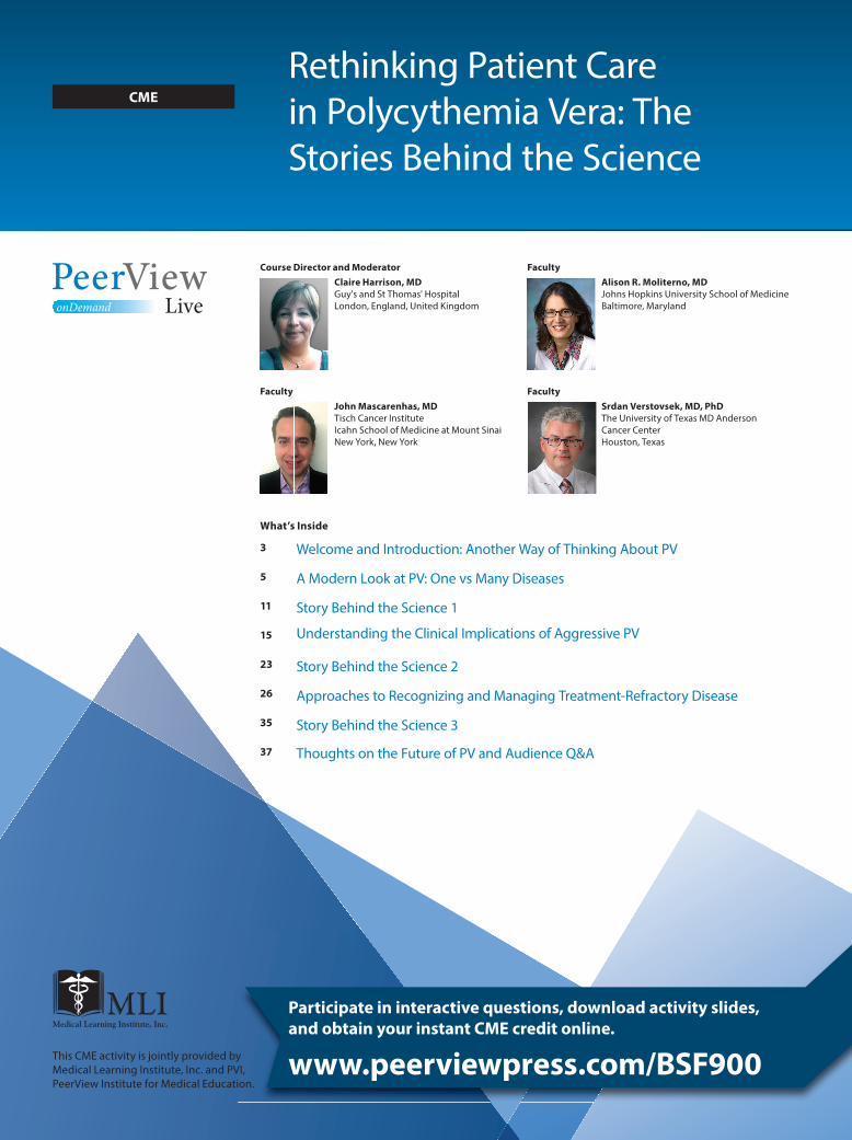
Claire Harrison, MDGuy's and St Thomas' HospitalLondon, England, United Kingdom
Alison R. Moliterno, MDJohns Hopkins University School of MedicineBaltimore, Maryland
John Mascarenhas, MDTisch Cancer InstituteIcahn School of Medicine at Mount SinaiNew York, New York
Srdan Verstovsek, MD, PhDThe University of Texas MD Anderson Cancer CenterHouston, Texas
Course Director and Moderator Faculty
Faculty Faculty
Participate in interactive questions, download activity slides, and obtain your instant CME credit online.
This CME activity is jointly provided by Medical Learning Institute, Inc. and PVI, PeerView Institute for Medical Education.
CME
Rethinking Patient Care in Polycythemia Vera: The Stories Behind the Science
What’s Inside
3
5
Welcome and Introduction: Another Way of Thinking About PV
A Modern Look at PV: One vs Many Diseases
Story Behind the Science 1
Understanding the Clinical Implications of Aggressive PV
Story Behind the Science 2
Approaches to Recognizing and Managing Treatment-Refractory Disease
Story Behind the Science 3
Thoughts on the Future of PV and Audience Q&A
11
15
23
26
35
37
www.peerviewpress.com/BSF900
2 Go online to complete the post-test and evaluation for CME credit
Activity Information
Activity Description and Educational ObjectivesNew science has driven innovative medical practice in the recognition and management of polycythemia vera (PV). At a recent live symposium, experts reviewed the latest evidence on how PV is categorized and assessed, its symptom burden, and best practices for clinical management. They also addressed a range of subjects related to these topics, from new thinking on PV diagnostic criteria and aggressive versus indolent disease phenotypes, to the therapeutic implications of higher risk, uncontrolled, or treatment-refractory disease. Personal stories and insights for patient care from these leading experts are also featured in this activity.
Upon completion of this activity, participants should be better able to:• Utilize clinical tools and evidence to accurately diagnose or characterize polycythemia vera (PV)
and distinguish it from other syndromes with similar presentations• Discuss the clinical implications of elevated hematocrit and uncontrolled blood counts in PV• Establish the presence of high-risk and treatment-refractory PV in patients with symptoms not
responding to current treatment such as phlebotomy or hydroxyurea• Select patient-appropriate therapy for individuals in higher risk PV treatment settings, including
refractory disease• Manage safety considerations in patients with PV on therapy across a range of treatment settings
Target AudienceThis activity has been designed to meet the educational needs of hematologists, hematologist-oncologists, medical oncologists, oncology physician assistants, advanced practice oncology nurses, and other clinicians involved in the care of patients with PV.
Requirements for Successful CompletionIn order to receive credit, participants must view the activity and complete the post-test and evaluation form. A score of 70% or higher is needed to obtain CME credit. There are no pre-requisites and there is no fee to participate in this activity or to receive CME credit. Statements of Credit are awarded upon successful completion of the post-test and evaluation form.
Media: Enduring MaterialRelease and Expiration Dates: December 22, 2016 - December 21, 2017Time to Complete: 120 minutes
Faculty & Disclosure / Conflict of Interest Policy Before the activity, all faculty and anyone who is in a position to have control over the content of this activity and their spouse/life partner will disclose the existence of any financial interest and/or relationship(s) they might have with any commercial interest producing healthcare goods/services to be discussed during their presentation(s): honoraria, expenses, grants, consulting roles, speakers bureau membership, stock ownership, or other special relationships. Presenters will inform participants of any off-label discussions. All identified conflicts of interest are thoroughly vetted by Medical Learning Institute, Inc. for fair balance, scientific objectivity of studies mentioned in the materials or used as the basis for content, and appropriateness of patient care recommendations.
The associates of Medical Learning Institute, Inc., the accredited provider for this activity, and PVI, PeerView Institute for Medical Education do not have any financial relationships or relationships to products or devices with any commercial interest related to the content of this CME activity for any amount during the past 12 months.
Course Director and ModeratorClaire Harrison, MDFRCP Consultant Haematologist &Clinical Director Cancer and HaematologyGuy's and St Thomas' HospitalLondon, England, United Kingdom
Claire Harrison, MD, has a financial interest/relationship or affiliation in the form of:Grant/Research Support from Novartis AG.Speakers Bureau participant with Baxalta; CTI BioPharma Corp.; Gilead; Incyte Corporation; Novartis AG; Sanofi; and Shire.Honoraria from Baxalta; CTI BioPharma Corp.; Gilead; Incyte Corporation; Novartis AG; Sanofi; and Shire.Advisory Board for Baxalta; CTI BioPharma Corp.; Gilead; Incyte Corporation; Novartis AG; Sanofi; and Shire.
Claire Harrison, MD, does intend to discuss either non–FDA-approved or investigational use for the following products/devices: a number of therapeutic options for polycythemia vera and other myeloproliferative neoplasms.
FacultyAlison R. Moliterno, MDAssociate Professor of MedicineHematology Division, Department of MedicineJohns Hopkins University School of MedicineBaltimore, Maryland
Alison R. Moliterno, MD, has a financial interest/relationship or affiliation in the form of:Consultant for Incyte Corporation.Advisory Board for Incyte Corporation.
Alison R. Moliterno, MD, does intend to discuss either non–FDA-approved or investigational use for the following products/devices: a number of therapeutic options for polycythemia vera and other myeloproliferative neoplasms.
John Mascarenhas, MDMyeloproliferative Disorders ProgramTisch Cancer Institute, Division of Hematology/OncologyAssociate Professor of MedicineIcahn School of Medicine at Mount SinaiNew York, New York
John Mascarenhas, MD, has a financial interest/relationship or affiliation in the form of:Consultant for Incyte Corporation and Novartis Corporation for Data Safety Monitoring Board and steering committee member.Grant/Research Support from CTI BioPharma Corp.; F. Hoffmann-La Roche Ltd.; Incyte Corporation; Janssen Global Services, LLC; Merck & Co., Inc.; Novartis Corporation; and Promedior, Inc. paid to institution.
John Mascarenhas, MD, does intend to discuss either non–FDA-approved or investigational use for the following products/devices: a number of therapeutic options for polycythemia vera and other myeloproliferative neoplasms.
Srdan Verstovsek, MD, PhDProfessor of MedicineChief, Section for Myeloproliferative Neoplasms (MPNs)Department of LeukemiaDirector, Clinical Research Center for MPNsThe University of Texas MD Anderson Cancer CenterHouston, Texas
Srdan Verstovsek, MD, PhD, has a financial interest/relationship or affiliation in the form of:Grant/Research Support from AstraZeneca; Bristol-Myers Squibb; Celgene Corporation; CTI BioPharma Corporation; Galena Biopharma, Inc.; Genentech, Inc.; Geron; Gilead; Hoffmann-La Roche Inc.; Incyte Corporation; Lilly Oncology; NS Pharma Inc.; Pfizer; Promedior, Inc.; and Seattle Genetics, Inc.
Srdan Verstovsek, MD, PhD, does intend to discuss either non–FDA-approved or investigational use for the following products/devices: a number of therapeutic options for polycythemia vera and other myeloproliferative neoplasms.
CME ReviewerVishwanath Sathyanarayanan, MD, DMApollo HospitalsBangalore, Karnataka, India
Vishwanath Sathyanarayanan, MD, DM, has no financial interests/relationships or affiliations in relation to this activity.
Medical DirectorsKirk Tacka, PhDPVI, PeerView Institute for Medical Education
Kirk Tacka, PhD, has no financial interests/relationships or affiliations in relation to this activity.
Carmine DeLucaPVI, PeerView Institute for Medical Education
Carmine DeLuca has no financial interests/relationships or affiliations in relation to this activity.
DisclaimerThe information provided at this CME activity is for continuing education purposes only and is not meant to substitute for the independent medical judgment of a healthcare provider relative to diagnostic and treatment options of a specific patient's medical condition. Recommendations for the use of particular therapeutic agents are based on the best available scientific evidence and current clinical guidelines. No bias towards or promotion for any agent discussed in this program should be inferred.
Providership, Credit & SupportThis activity has been planned and implemented in accordance with the accreditation requirements and policies of the Accreditation Council for Continuing Medical Education (ACCME) through the joint providership of Medical Learning Institute, Inc. and PVI, PeerView Institute for Medical Education. The Medical Learning Institute, Inc. is accredited by the ACCME to provide continuing medical education for physicians.
The Medical Learning Institute, Inc. designates this enduring material for a maximum of 2.0 AMA PRA Category 1 CreditsTM. Physicians should claim only the credit commensurate with the extent of their participation in the activity.
ProvidershipThis CME activity is jointly provided by Medical Learning Institute, Inc. and PVI, PeerView Institute for Medical Education.
SupportThis activity is supported by an educational grant from Incyte Corporation.
Disclosure of Unlabeled UseThe faculty of this educational activity may include discussions of products or devices that are not currently labeled for use by the FDA. Faculty members have been advised to disclose to the audience any reference to an unlabeled or investigational use.
No endorsement of unapproved products or uses is made or implied by coverage of these products or uses in our reports. No responsibility is taken for errors or omissions in reports.
Please refer to the official prescribing information for each product for discussion of approved indications, contraindications and warnings.
The materials presented here are used with the permission of the authors and/or other sources. These materials do not necessarily reflect the views of PeerView Press or any of its partners, providers, and/or supporters.
www.peerviewpress.com/BSF900

3
Rethinking Patient Care in Polycythemia Vera: The Stories Behind the Science
Dr. Harrison: Welcome to this educational session, “Rethinking Patient Care in Polycythemia Vera: The Stories Behind the Science.” Thank you for joining us this afternoon. I’m Claire Harrison. I’m a hematologist at Guy’s and St Thomas’ Hospital in London.
Joining me for this PeerView Live Symposium are Dr. John Mascarenhas from Icahn School of Medicine at Mount Sinai, Dr. Alison Moliterno from Johns Hopkins University School of Medicine, and Dr. Srdan Verstovsek from The University of Texas MD Anderson Cancer Center.
Narrator: After completing the activity, access the post-test and evaluation form by clicking the red “Get certificate” button.
I encourage you to download the slides, Practice Aids, and any other activity features that may interest you.
Welcome and Introduction: Another Way of Thinking About PV
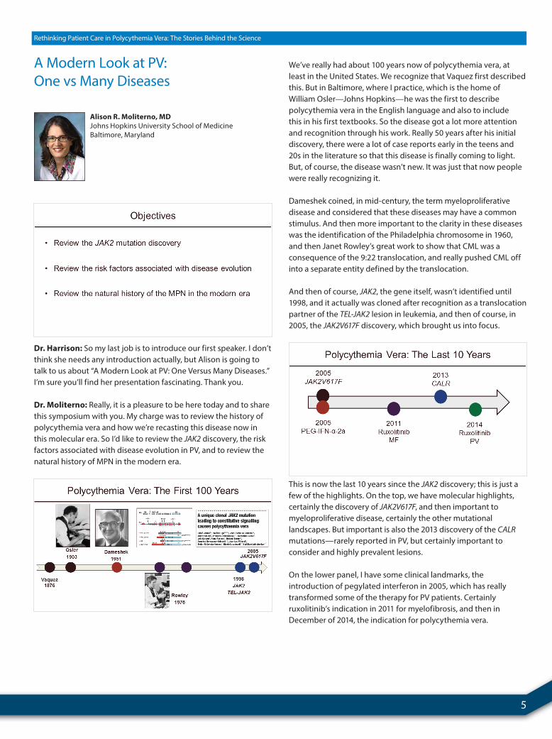
Dr. Harrison: So this afternoon we’re going to present another way of thinking about PV, and each of our esteemed speakers are presenting on a different aspect of this disease. And you’ll see this historical slide representing the beginning of PV with its first description by Vaquez, and also by William Osler right through to developments in the present day. Particularly, note the acceleration in developments regarding our clinical thinking about this disease and therapeutics, since the description of the JAK2V617F mutation just over 10 years ago.
So if we think about this disease at a glance, this is a chronic myeloproliferative neoplasm, as you will know, characterized by erythrocytosis and often, but not always, leukocytosis and thrombocytosis.
As we will hear more, the JAK2V617F mutation is positive in more than 90% of cases. And of the remaining JAK2V617F-negative patients, many of them will have exon 12 mutations. But there are some patients with PV who lack a JAK2 mutation.
Increasingly, we understand the clinical complexity of this disease and are trying to target our therapies to address them. And you’ll hear some interesting data with regard to this later in this symposium.
Claire Harrison, MDGuy's and St Thomas' HospitalLondon, England, United Kingdom

4 Go online to complete the post-test and evaluation for CME credit
Rethinking Patient Care in Polycythemia Vera: The Stories Behind the Science
So there are a number of unanswered questions in this field. The diversity of the disease, the use of “uncontrolled” PV as a term, when should we switch a patient from phlebotomy to cytoreductive therapy, which one should we choose, and when might we consider then, furthermore, changing the cytoreductive therapy? And now that we have further options for aggressive PV, when should we be thinking about applying these in our treatment algorithm? So these are the kind of questions that you will hear my colleagues addressing today.
So here is today’s agenda. First, we’ll take a look at the heterogeneity of the disease and tools which you can use to capture important aspects of this at the time of diagnosis. Then, we’ll turn to aggressive PV, discuss its characteristics and what we might consider for treatments. And there are some important advances and new data being presented at this ASH meeting. Finally, we’ll discuss treatment-refractory disease and how we can recognize and manage this challenging clinical setting. Each session will include a personal story illustrating an aspect of PV management.
www.peerviewpress.com/BSF900
5
Rethinking Patient Care in Polycythemia Vera: The Stories Behind the Science
A Modern Look at PV: One vs Many Diseases
Dr. Harrison: So my last job is to introduce our first speaker. I don’t think she needs any introduction actually, but Alison is going to talk to us about “A Modern Look at PV: One Versus Many Diseases.” I’m sure you’ll find her presentation fascinating. Thank you.
Dr. Moliterno: Really, it is a pleasure to be here today and to share this symposium with you. My charge was to review the history of polycythemia vera and how we’re recasting this disease now in this molecular era. So I’d like to review the JAK2 discovery, the risk factors associated with disease evolution in PV, and to review the natural history of MPN in the modern era.
We’ve really had about 100 years now of polycythemia vera, at least in the United States. We recognize that Vaquez first described this. But in Baltimore, where I practice, which is the home of William Osler—Johns Hopkins—he was the first to describe polycythemia vera in the English language and also to include this in his first textbooks. So the disease got a lot more attention and recognition through his work. Really 50 years after his initial discovery, there were a lot of case reports early in the teens and 20s in the literature so that this disease is finally coming to light. But, of course, the disease wasn’t new. It was just that now people were really recognizing it.
Dameshek coined, in mid-century, the term myeloproliferative disease and considered that these diseases may have a common stimulus. And then more important to the clarity in these diseases was the identification of the Philadelphia chromosome in 1960, and then Janet Rowley’s great work to show that CML was a consequence of the 9:22 translocation, and really pushed CML off into a separate entity defined by the translocation.
And then of course, JAK2, the gene itself, wasn’t identified until 1998, and it actually was cloned after recognition as a translocation partner of the TEL-JAK2 lesion in leukemia, and then of course, in 2005, the JAK2V617F discovery, which brought us into focus.
This is now the last 10 years since the JAK2 discovery; this is just a few of the highlights. On the top, we have molecular highlights, certainly the discovery of JAK2V617F, and then important to myeloproliferative disease, certainly the other mutational landscapes. But important is also the 2013 discovery of the CALRmutations—rarely reported in PV, but certainly important to consider and highly prevalent lesions.
On the lower panel, I have some clinical landmarks, the introduction of pegylated interferon in 2005, which has really transformed some of the therapy for PV patients. Certainly ruxolitinib’s indication in 2011 for myelofibrosis, and then in December of 2014, the indication for polycythemia vera.
Alison R. Moliterno, MDJohns Hopkins University School of MedicineBaltimore, Maryland

6 Go online to complete the post-test and evaluation for CME credit
Rethinking Patient Care in Polycythemia Vera: The Stories Behind the Science
So the first 100 years was really description, and now the last 10 years has been molecular discovery and targeted therapy.
So let’s review a little bit about JAK2 mutations. These are acquired somatic mutations with enhanced kinase activity. JAK2 participates by sending a signal for type 1 cytokine receptors that themselves do not have kinase activity.
JAK2 transmits a signal normally through the JAK-STAT pathway and other pathways, and in this sense, it is a signal transducer and tells the cell to divide and grow. JAK2 is essential for signal transduction. Without the JAK2 gene, we can’t make red cells, and it certainly cooperates to make white cells and platelets, so critical function normally.
Now activating mutations in JAK2 just transmit more of a normal signal. These activating mutations never spontaneously resolve, and clonal expansion of the JAK2 mutation–positive cells occur. These cells have an advantage, and I use these analogies, like a weed in the garden or a more aggressive flower that can take over other less empowered cells or flowers.
And unique to the JAK2 discovery early on was that the JAK2mutations occur in one allele and then frequently, due to a mitotic recombination event, you can get the JAK2 mutation on another allele in the same cell, so effectively making the cell homozygous or having two doses of the JAK2 mutation. And that introduces a lot of variability into the clones. You can have heterozygous clones, homozygous JAK2 clones, and wildtype clones in an individual at any time, and that these clones can continue to develop uniparental disomy, and develop homozygosity.
So in the 10 years or 11 years since the JAK2 discovery, we found that the JAK2 mutation really subtends a number of different entities. In this Venn diagram that I have made, this dark circle, if you can imagine, is all the individuals who have acquired the JAK2 mutation. And you can see the different phenotypes, clinical diseases, associated with that. In here, the largest circle is polycythemia vera, and if you look at 100 individuals who have the JAK2 mutation, polycythemia vera will be the most common phenotype. Here we have essential thrombocytosis, myelofibrosis, and then refractory anemia with ring sideroblasts and thrombocytosis. These four entities are the most common entities associated with JAK2V617F.
I have overlaid the circles here because we’ve all seen in our practice that patients may have ET for many years, or just isolated thrombocytosis and then evolve into polycythemia vera. And similarly, patients with polycythemia vera may evolve into myelofibrosis, so that there is some heterogeneity over time. Certainly, gene dosage also affects why some patients have just ET or PV and why others present with MF. So this variation can be explained by gene dosage effect of the JAK2 mutation. Certainly age, gender, and time seem to have an influence as to how you present and evolve with these entities, and certainly phenotypic evolution seems to be very common in these entities. So we’ll go over this in more detail later.
www.peerviewpress.com/BSF900

7
Rethinking Patient Care in Polycythemia Vera: The Stories Behind the Science
I did just now want to review the 2013 discovery of the CALRmutation. So CALR does not really participate directly in the signal transduction pathway that I have shown you here. We have the hormone binding to receptor—JAK—and then the JAK-STAT pathway being activated.
CALR is an endoplastic reticulum chaperone protein that seems to be important for calcium metabolism, but also many other factors, and it seems to influence MPL processing and perhaps folding of the MPL, the thrombopoietin receptor here, and in that sense it seems to drive JAK-STAT signaling through processes that aren’t completely elucidated. But similar to JAK2, when a cell develops the CALR mutation, this advantages the cell, allows the clone bearing the mutation to expand, and seems to generate too many platelets or hematopoietic cells.
In contrast to JAK2, there is not as much of a variation in terms of gene dosage as a modifier. JAK2, again, can—through this uniparental disomy—introduce two copies in a cell of the mutation, where this is rarely reported in CALR. So most clones that have the CALR mutation seem to have one copy, so less variation there.
Similarly, how I did with the JAK2 mutation, if we look at individuals with a CALR mutation, again, if we could include all of those individuals in this black circle, you’ll see about half of them have a phenotype of essential thrombocytosis, and the other half have myelofibrosis. And again, we have this overlap because we know that many patients with ET initially may progress to myelofibrosis. I have left PV pretty much out of the CALR spectrum, with just a little edge here. There have been few reports of CALR mutation
individuals who fulfill PV diagnostic criteria. And so it is possible to generate a PV phenotype with CALR, though they’re very rare.
So again, to summarize CALR, the gene dosage effect is much less with JAK2 mutation. Time and age certainly influence the phenotype here, and evolution is also a feature of CALR mutation.
So holding all this together, we can now look at polycythemia vera and the lesions and their combined frequency. And in these last 11 years, pretty much 99% of individuals who have polycythemia vera will be able to be defined by either a JAK2 mutation, a JAK2 exon 12 mutation, and potentially a rare patient with CALRmutation. So that, in this day and age, most individuals with a clinical phenotype of PV will have a molecular diagnosis that can be sought, and so I leave that at 99%.
I’m certain that there are individuals who generate a PV phenotype that may be due to a STAT mutation or something along that same pathway that we haven’t identified yet. But for the most part, most will be JAK mutations. You can see JAK2, CALR, and MPL mutations make up about 80% to 90% of individuals with ET and primary myelofibrosis. Again, 80% to 90% will have one of these three mutations. We’re focusing here on polycythemia vera today, again, predominantly JAK2 mutations.
So with this molecular discovery in hand, the WHO diagnostic criteria has been updated, and really has been updated to do two
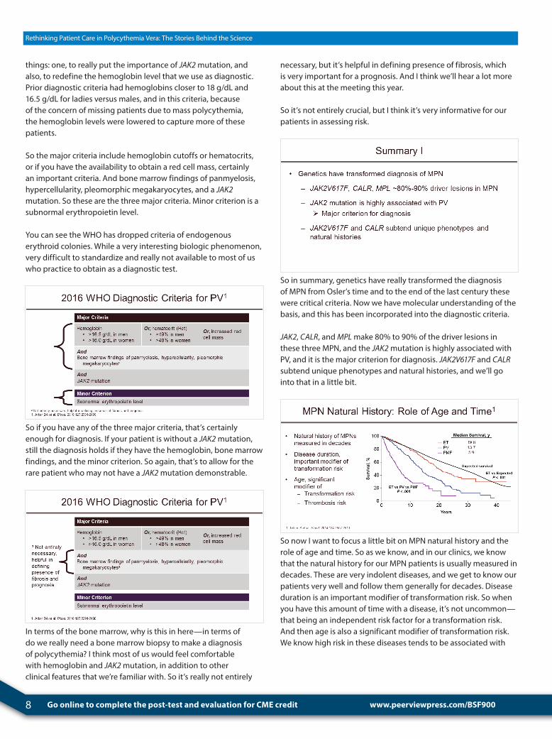
8 Go online to complete the post-test and evaluation for CME credit
Rethinking Patient Care in Polycythemia Vera: The Stories Behind the Science
things: one, to really put the importance of JAK2 mutation, and also, to redefine the hemoglobin level that we use as diagnostic. Prior diagnostic criteria had hemoglobins closer to 18 g/dL and 16.5 g/dL for ladies versus males, and in this criteria, because of the concern of missing patients due to mass polycythemia, the hemoglobin levels were lowered to capture more of these patients.
So the major criteria include hemoglobin cutoffs or hematocrits, or if you have the availability to obtain a red cell mass, certainly an important criteria. And bone marrow findings of panmyelosis, hypercellularity, pleomorphic megakaryocytes, and a JAK2mutation. So these are the three major criteria. Minor criterion is a subnormal erythropoietin level.
You can see the WHO has dropped criteria of endogenous erythroid colonies. While a very interesting biologic phenomenon, very difficult to standardize and really not available to most of us who practice to obtain as a diagnostic test.
So if you have any of the three major criteria, that’s certainly enough for diagnosis. If your patient is without a JAK2 mutation, still the diagnosis holds if they have the hemoglobin, bone marrow findings, and the minor criterion. So again, that’s to allow for the rare patient who may not have a JAK2 mutation demonstrable.
In terms of the bone marrow, why is this in here—in terms of do we really need a bone marrow biopsy to make a diagnosis of polycythemia? I think most of us would feel comfortable with hemoglobin and JAK2 mutation, in addition to other clinical features that we’re familiar with. So it’s really not entirely
necessary, but it’s helpful in defining presence of fibrosis, which is very important for a prognosis. And I think we’ll hear a lot more about this at the meeting this year.
So it’s not entirely crucial, but I think it’s very informative for our patients in assessing risk.
So in summary, genetics have really transformed the diagnosis of MPN from Osler’s time and to the end of the last century these were critical criteria. Now we have molecular understanding of the basis, and this has been incorporated into the diagnostic criteria.
JAK2, CALR, and MPL make 80% to 90% of the driver lesions in these three MPN, and the JAK2 mutation is highly associated with PV, and it is the major criterion for diagnosis. JAK2V617F and CALRsubtend unique phenotypes and natural histories, and we’ll go into that in a little bit.
So now I want to focus a little bit on MPN natural history and the role of age and time. So as we know, and in our clinics, we know that the natural history for our MPN patients is usually measured in decades. These are very indolent diseases, and we get to know our patients very well and follow them generally for decades. Disease duration is an important modifier of transformation risk. So when you have this amount of time with a disease, it’s not uncommon—that being an independent risk factor for a transformation risk. And then age is also a significant modifier of transformation risk. We know high risk in these diseases tends to be associated with
www.peerviewpress.com/BSF900
9
Rethinking Patient Care in Polycythemia Vera: The Stories Behind the Science
older age, both for thrombosis risk and for transformation risk.
And in here one of Dr. Tefferi’s studies was looking at median survival in several 100 patients. And you can see the expected survival for the group here in the black line, and then we have ET, polycythemia vera, and myelofibrosis in purple here.
We can see in ET there is a slight diminution in expected survival, and that’s more pronounced with both PV and myelofibrosis. So a lot of time with these diseases, and what are the risk factors associated with this.
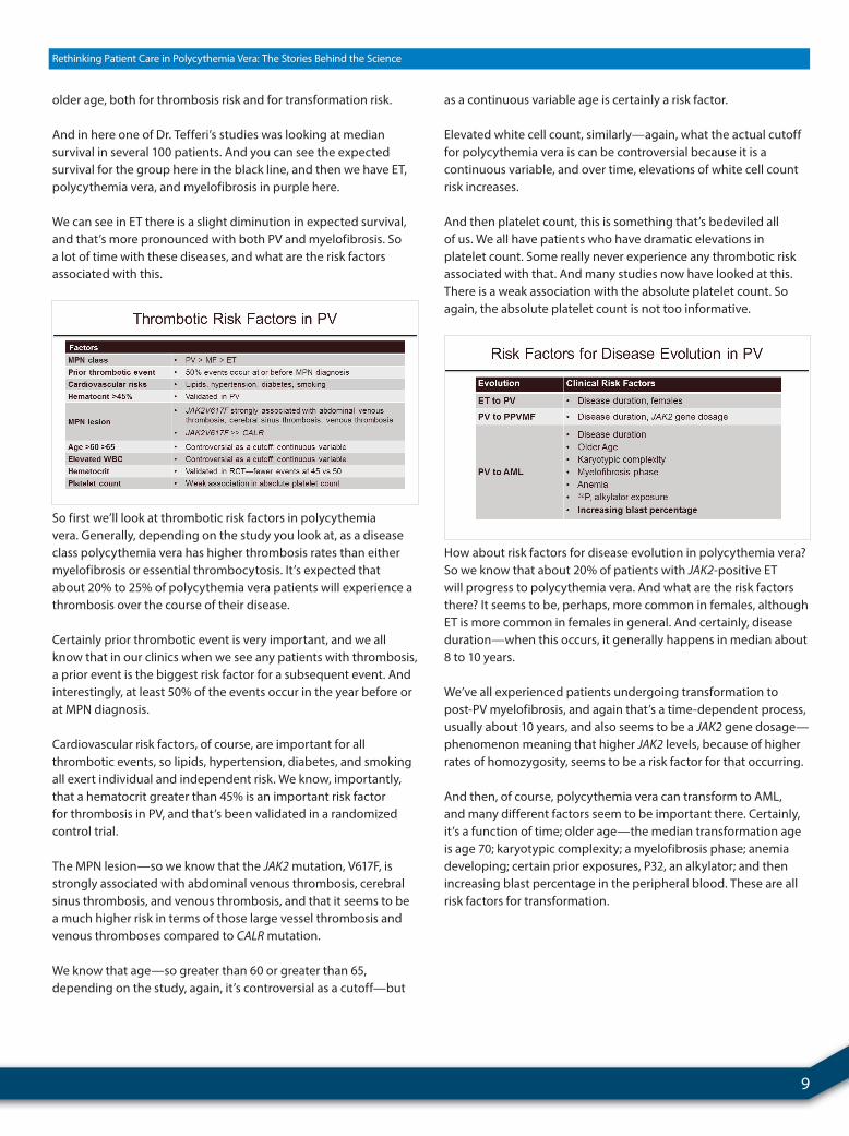
So first we’ll look at thrombotic risk factors in polycythemia vera. Generally, depending on the study you look at, as a disease class polycythemia vera has higher thrombosis rates than either myelofibrosis or essential thrombocytosis. It’s expected that about 20% to 25% of polycythemia vera patients will experience a thrombosis over the course of their disease.
Certainly prior thrombotic event is very important, and we all know that in our clinics when we see any patients with thrombosis, a prior event is the biggest risk factor for a subsequent event. And interestingly, at least 50% of the events occur in the year before or at MPN diagnosis.
Cardiovascular risk factors, of course, are important for all thrombotic events, so lipids, hypertension, diabetes, and smoking all exert individual and independent risk. We know, importantly, that a hematocrit greater than 45% is an important risk factor for thrombosis in PV, and that’s been validated in a randomized control trial.
The MPN lesion—so we know that the JAK2 mutation, V617F, is strongly associated with abdominal venous thrombosis, cerebral sinus thrombosis, and venous thrombosis, and that it seems to be a much higher risk in terms of those large vessel thrombosis and venous thromboses compared to CALR mutation.
We know that age—so greater than 60 or greater than 65, depending on the study, again, it’s controversial as a cutoff—but
as a continuous variable age is certainly a risk factor.
Elevated white cell count, similarly—again, what the actual cutoff for polycythemia vera is can be controversial because it is a continuous variable, and over time, elevations of white cell count risk increases.
And then platelet count, this is something that’s bedeviled all of us. We all have patients who have dramatic elevations in platelet count. Some really never experience any thrombotic risk associated with that. And many studies now have looked at this. There is a weak association with the absolute platelet count. So again, the absolute platelet count is not too informative.
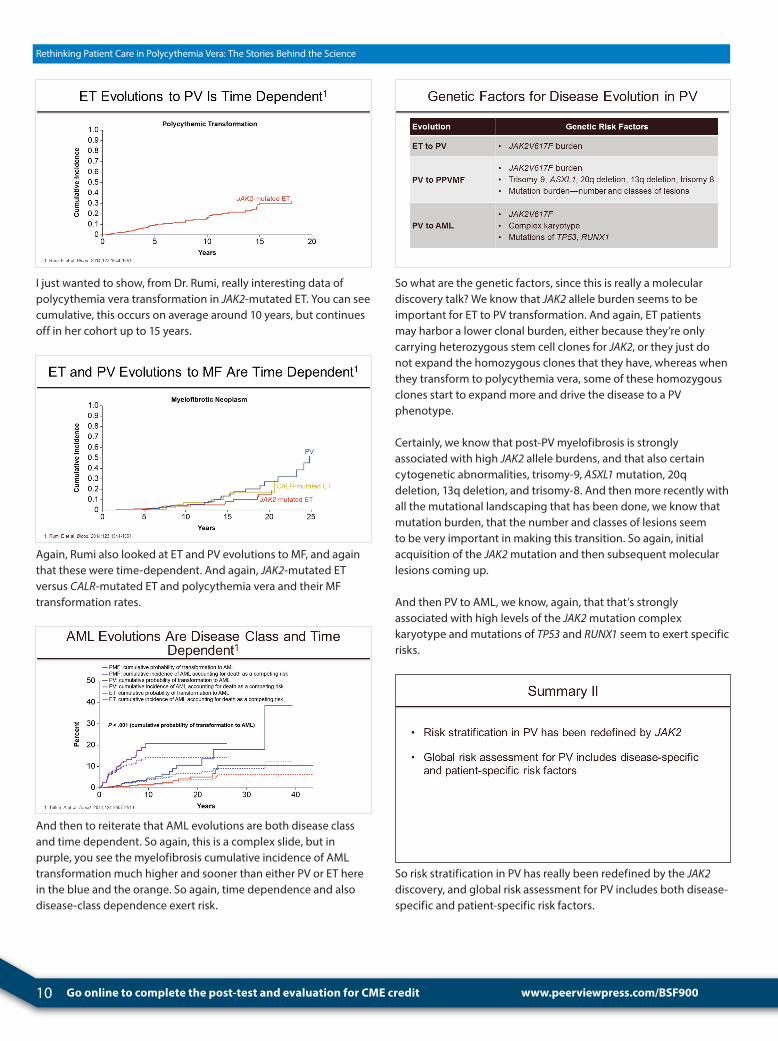
How about risk factors for disease evolution in polycythemia vera? So we know that about 20% of patients with JAK2-positive ET will progress to polycythemia vera. And what are the risk factors there? It seems to be, perhaps, more common in females, although ET is more common in females in general. And certainly, disease duration—when this occurs, it generally happens in median about 8 to 10 years.
We’ve all experienced patients undergoing transformation to post-PV myelofibrosis, and again that’s a time-dependent process, usually about 10 years, and also seems to be a JAK2 gene dosage—phenomenon meaning that higher JAK2 levels, because of higher rates of homozygosity, seems to be a risk factor for that occurring.
And then, of course, polycythemia vera can transform to AML, and many different factors seem to be important there. Certainly, it’s a function of time; older age—the median transformation age is age 70; karyotypic complexity; a myelofibrosis phase; anemia developing; certain prior exposures, P32, an alkylator; and then increasing blast percentage in the peripheral blood. These are all risk factors for transformation.
10 Go online to complete the post-test and evaluation for CME credit
Rethinking Patient Care in Polycythemia Vera: The Stories Behind the Science
I just wanted to show, from Dr. Rumi, really interesting data of polycythemia vera transformation in JAK2-mutated ET. You can see cumulative, this occurs on average around 10 years, but continues off in her cohort up to 15 years.
Again, Rumi also looked at ET and PV evolutions to MF, and again that these were time-dependent. And again, JAK2-mutated ET versus CALR-mutated ET and polycythemia vera and their MF transformation rates.
And then to reiterate that AML evolutions are both disease class and time dependent. So again, this is a complex slide, but in purple, you see the myelofibrosis cumulative incidence of AML transformation much higher and sooner than either PV or ET here in the blue and the orange. So again, time dependence and also disease-class dependence exert risk.
So what are the genetic factors, since this is really a molecular discovery talk? We know that JAK2 allele burden seems to be important for ET to PV transformation. And again, ET patients may harbor a lower clonal burden, either because they’re only carrying heterozygous stem cell clones for JAK2, or they just do not expand the homozygous clones that they have, whereas when they transform to polycythemia vera, some of these homozygous clones start to expand more and drive the disease to a PV phenotype.
Certainly, we know that post-PV myelofibrosis is strongly associated with high JAK2 allele burdens, and that also certain cytogenetic abnormalities, trisomy-9, ASXL1 mutation, 20q deletion, 13q deletion, and trisomy-8. And then more recently with all the mutational landscaping that has been done, we know that mutation burden, that the number and classes of lesions seem to be very important in making this transition. So again, initial acquisition of the JAK2 mutation and then subsequent molecular lesions coming up.
And then PV to AML, we know, again, that that’s strongly associated with high levels of the JAK2 mutation complex karyotype and mutations of TP53 and RUNX1 seem to exert specific risks.
So risk stratification in PV has really been redefined by the JAK2discovery, and global risk assessment for PV includes both disease-specific and patient-specific risk factors.
www.peerviewpress.com/BSF900

11
Rethinking Patient Care in Polycythemia Vera: The Stories Behind the Science
Story Behind the Science 1
Dr. Moliterno: So I would just like to go into sort of a personal and scientific story in polycythemia vera that we’ve been developing at Johns Hopkins.
When I came into my fellowship, we decided to develop a registry for MPN patients at our institution. And we’ve looked at more than 700 patients now in our registry, but between specifically 2005 and 2011, we had 273 of these individuals who had a PV phase.
You can see the number of patients and their age at diagnosis in this histogram. Again, PV seems to be a disease of middle age, so the median age at diagnosis was around 55 for the cohort. But you can see a broad left tail here. The youngest individual in our cohort with a JAK2 positive PV was a 5-year-old girl, and the oldest is a 91-year-old female at diagnosis.
Again, if we break this down into disease types, we can see ET, again, age at presentation—you can see ET is prevalent throughout the ages; polycythemia vera again seems to have this in middle-age bulge; myelofibrosis, older individuals; and certainly AML, the very oldest of this cohort. So again, this concept of age being a specific risk factor for how you present is evident in this histogram.
Now we know that aging and mutations go together. These were studies from The New England Journal of Medicine in the last few years. Many individuals have looked at cohorts, not hematopoietic cell disease cohorts, but just aging cohorts, and looked at mutational landscaping. And here among persons of greater than 60 years, males had increased likelihood of having detectable mutation as compared with women, the odds ratio of 1.3, which is very interesting. So again age is a risk factor, and male sex seems to be a risk factor for developing clonal lesions.
Alison R. Moliterno, MDJohns Hopkins University School of MedicineBaltimore, Maryland

12 Go online to complete the post-test and evaluation for CME credit
Rethinking Patient Care in Polycythemia Vera: The Stories Behind the Science
And if we look specifically at these, what are the lesions? Well, we’ve seen the DNMT3A, but also here is our friend, the JAK2mutation. It’s one of the most common aging lesions. And what’s striking to me to understand is if JAK2 is a common lesion of aging, why in our cohort with PV we seem to have this excess of females below the age of 50 compared to males.
So here’s the Hopkins cohort for age at PV diagnosis spread between females and males, and astonishingly, the majority of individuals below the age of 40 are females with the JAK2mutation, compared to males. And that seems to fly into the face of what I just showed you, that with normal aging in individuals, JAK2 mutations are common and are also more common in males as they age. And here we have something very unique going on in PV, where younger females are risk for developing this—so important risk factors that we need to understand.
When we look at our 620 MPN patient cohort, we can see that patients may start off with a pie chart of the original diagnoses—ET, PV, and myelofibrosis—and after a median of 8 years evolution occurs. And so you can see that after 8 years the cohort has changed to more aggressive MPN, including that fewer patients maintain their ET phenotype and move into PV or MF, and we even see the introduction of AML into this cohort. So again, time seems to be a very important risk in our cohort.
Importantly, I mentioned that JAK2 has this unique burden of mutation that is unique compared to other mutations. So, for instance, here if you look on the y axis here, this is the JAK2 allele burden or the number of alleles that you can measure in a person’s bloodstream. We know that there are differences between ET, PV, post-ET myelofibrosis, and post-PV myelofibrosis. So the higher JAK2 levels in the blood seems to correspond with more aggressive disease.
This is not really the case in CALR. There is not as huge a variation in CALR mutational burden within ET, and within MF, maybe it’s slightly higher than ET, but certainly we don’t see this broad increase up to 100%.
So again, JAK2 really introduces a lot of variability just by mutational burden, and that’s quite different compared to CALR.
And how do we understand this variation in a JAK2 allele burden? If you can imagine that these are a group of stem cells in different rows here—white means that they’re non-mutated. A stem cell in this lane gets a JAK2 mutation that’s heterozygous. You can see the allele burden would be fairly low.
www.peerviewpress.com/BSF900
13
Rethinking Patient Care in Polycythemia Vera: The Stories Behind the Science
But over time one of these stem cells develop two copies of the JAK2 mutation, so you have a homozygous clone, and now that clone may expand at the expense over time of even the heterozygous clone, and certainly over the wildtype clone. And so you can understand here how this pattern could give an allele burden of 100%, whereas this pattern here could give you an allele burden of very low. And so that’s both the feature of genomic instability and time in the stem cell pool to lead to this variation in burden.
Here we have about 200 PV patients where we looked at the JAK2allele burden, so each line across is an individual patient. The intensity of the color relates to the level of JAK2 allele burden, and we also have here karyotypic complexity, so all these patients had a high-density karyotype done. And the darker the color in the karyotype, the more complex their karyotypic lesions.
And you can see PV, very little karyotypic complexity, but a lot of variation in JAK2 allele burden, yet in the post-PV MF, the PV patients who went to MDS, and the PV patients who went to AML, you can see now there is a lot more activity in terms of karyotypic complexity, and that’s certainly associated with disease progression.
This is also being borne out in mutational landscaping, so again, instead of karyotypic complexity, if you just put number and types of mutations, again, you would see this increase in mutation burden with more advanced disease.
Joe Prchal did a lovely study a few years ago looking at 31 PV patients and looked for other lesions beyond JAK2, and you can see these common known lesions—ASXL1, NF1, TET2—as associated with post-PV MF transformation.
We at Johns Hopkins looked at gene expression, and this was an unbiased approach where we took 20 PV patients, and by just simple gene expression profiling, we could compare females and males to each other and found that they really had different gene expression and that this was different in PV compared to ET.
We found that we could demonstrate aggressive disease, just by gene expression profiling, compared to indolent disease, and this was not explained by typical clinical features, including JAK2 allele burden, but was really defined by the genes that were turned on.
14 Go online to complete the post-test and evaluation for CME credit
Rethinking Patient Care in Polycythemia Vera: The Stories Behind the Science
And in this histogram, this is just to show the group of indolent patients, compared to aggressive, compared to the normal controls. And you can see that there’s a vastly different gene expression profile.
So we know that disease duration is an important modifier of transformation risk. We know clonal expansion is time dependent. The allele burden is time and sex dependent. Instability is age and time dependant, and stem cell damage is age and time dependent. So these are all factors that lead to these different survival curves.
Polycythemia vera is one mutation basically, but we do have many different diseases, even within PV. We know that the age of onset, disease duration, gender, and stem cell expansion really all influence how an individual is going to process the mutation and how this is going to affect their clinical phenotype.
So on that, I’d like to finish and let Dr. Harrison introduce the next speaker.
www.peerviewpress.com/BSF900
15
Rethinking Patient Care in Polycythemia Vera: The Stories Behind the Science
Understanding the Clinical Implications of Aggressive PV
Dr. Harrison: Our next speaker is John Mascarenhas. He is going to talk to us about understanding clinical implications of aggressive PV.
Dr. Mascarenhas: Okay, thank you. Thanks, Dr. Harrison.
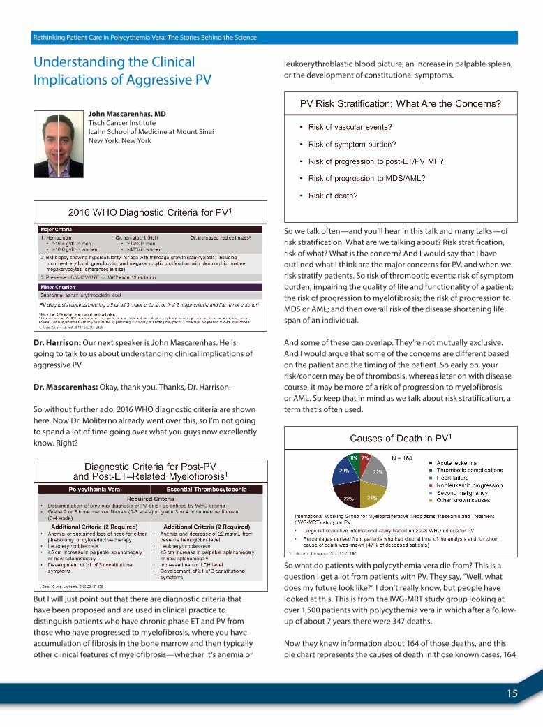
So without further ado, 2016 WHO diagnostic criteria are shown here. Now Dr. Moliterno already went over this, so I’m not going to spend a lot of time going over what you guys now excellently know. Right?
But I will just point out that there are diagnostic criteria that have been proposed and are used in clinical practice to distinguish patients who have chronic phase ET and PV from those who have progressed to myelofibrosis, where you have accumulation of fibrosis in the bone marrow and then typically other clinical features of myelofibrosis—whether it’s anemia or
leukoerythroblastic blood picture, an increase in palpable spleen, or the development of constitutional symptoms.
So we talk often—and you’ll hear in this talk and many talks—of risk stratification. What are we talking about? Risk stratification, risk of what? What is the concern? And I would say that I have outlined what I think are the major concerns for PV, and when we risk stratify patients. So risk of thrombotic events; risk of symptom burden, impairing the quality of life and functionality of a patient; the risk of progression to myelofibrosis; the risk of progression to MDS or AML; and then overall risk of the disease shortening life span of an individual.
And some of these can overlap. They’re not mutually exclusive. And I would argue that some of the concerns are different based on the patient and the timing of the patient. So early on, your risk/concern may be of thrombosis, whereas later on with disease course, it may be more of a risk of progression to myelofibrosis or AML. So keep that in mind as we talk about risk stratification, a term that’s often used.
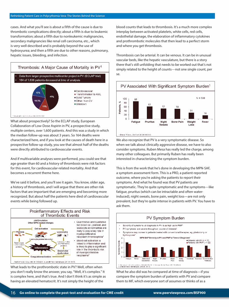
So what do patients with polycythemia vera die from? This is a question I get a lot from patients with PV. They say, “Well, what does my future look like?” I don’t really know, but people have looked at this. This is from the IWG-MRT study group looking at over 1,500 patients with polycythemia vera in which after a follow-up of about 7 years there were 347 deaths.
Now they knew information about 164 of those deaths, and this pie chart represents the causes of death in those known cases, 164
John Mascarenhas, MDTisch Cancer InstituteIcahn School of Medicine at Mount SinaiNew York, New York
16 Go online to complete the post-test and evaluation for CME credit
Rethinking Patient Care in Polycythemia Vera: The Stories Behind the Science
cases. And what you’ll see is about a fifth of the cause is due to thrombotic complications directly; about a fifth is due to leukemic transformation; about a fifth due to nonleukemic malignancies, secondary malignancies like renal cell carcinoma, etc., which is very well described and is probably beyond the use of hydroxyurea; and then a fifth are due to other reasons, pulmonary, hepatic issues, bleeding, and infection.
What about prospectively? So the ECLAP study, European Collaboration of Low-Dose Aspirin in PV, a prospective study, multiple centers, over 1,600 patients. And this was a study in which the median follow-up was about 3 years. So 164 deaths were characterized here, and if you look at the causes of death here in a prospective follow-up study, you see that almost half of the deaths were directly attributed to cardiovascular events.
And if multivariable analyses were performed, you could see that age greater than 60 and a history of thrombosis were risk factors for this event, for cardiovascular-related mortality. And that becomes a recurrent theme here.
We’ve said it before, and you’ll see it again. You know, older age, a history of thrombosis, and I will argue that there are other risk factors that are important that are emerging and becoming more recognized. But about half the patients here died of cardiovascular events while being followed up.
What leads to the prothrombotic state in PV? Well, often when you don’t really know the answer, you say, “Well, it’s complex.” It is complex here, and that’s true. And I don’t think it’s as simple as having an elevated hematocrit. It’s not simply the height of the
blood counts that leads to thrombosis. It’s a much more complex interplay between activated platelets, white cells, red cells, endothelial damage, the elaboration of inflammatory cytokines and prothrombotic molecules that then lead to a perfect storm and where you get thrombosis.
Thrombosis can be arterial. It can be venous. It can be in unusual vascular beds, like the hepatic vasculature, but there is a story there that’s still unfolding that needs to be worked out that’s not simply related to the height of counts—not one single count, per se.
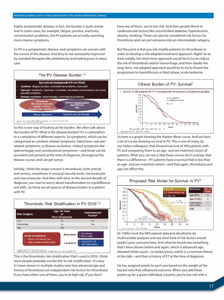
We also recognize that PV is a very symptomatic disease. So when we talk about clinically aggressive disease, we have to also consider symptoms. Ruben Mesa has really led the charge, among many other colleagues. But primarily Ruben has really been interested in characterizing the symptom burden.
This is from the work that he’s done in developing the MPN-SAF, a symptom assessment form. This is a PRO, a patient-reported outcome, where you’re asking the patients to report their symptoms. And what he found was that PV patients are symptomatic. They’re quite symptomatic and the symptoms—like fatigue, pruritus (which can be intractable and often water-induced), night sweats, bone pain, weight loss—are not only prevalent, but they’re quite intense in patients with PV. You have to ask them.
What he also did was he compared at time of diagnosis—if you compare the symptom burden of patients with PV and compare them to MF, which everyone sort of assumes or thinks of as a
www.peerviewpress.com/BSF900
17
Rethinking Patient Care in Polycythemia Vera: The Stories Behind the Science
highly symptomatic disease, in fact, the burden is quite similar. And in some cases, for example, fatigue, pruritus, inactivity, concentration problems, the PV patients are actually reporting more intense symptoms.
So PV is a symptomatic disease, and symptoms can worsen with the course of the disease. And they’re not necessarily improved by standard therapies like phlebotomy and hydroxyurea in every case.
So this is one way of looking at the burden. We often talk about the burden of PV. What is the disease burden? It’s a culmination or a cumulation of different aspects. So symptoms, which can be categorized as cytokine-related symptoms, listed here, vascular-related symptoms, or disease-evolution–related symptoms like splenomegaly and constitutional symptoms—and those can be prevalent and present at the time of diagnosis, throughout the disease course, and can get worse.
Initially, I think the major concern is thrombosis, both arterial and venous, sometimes in unusual vascular beds, microvascular and macrovascular. And then with time, in the second decade of diagnosis, you start to worry about transformation to myelofibrosis and AML. So these are all aspects of disease burden in a patient with PV.
This is the thrombotic risk stratification that’s used in 2016. I think most people probably ascribe this to risk stratification. It’s easy. It’s been shown in multiple studies now that advanced age and history of thrombosis are independent risk factors for thrombosis. If you have either one of them, you’re at high risk. If you don’t
have any of them, you’re low risk. And then people throw in cardiovascular factors like uncontrolled diabetes, hypertension, obesity, smoking. These can also be considered risk factors for thrombosis and can put someone into an intermediate category.
But the point is that you risk stratify patients for thrombosis in order to develop a risk-adapted treatment approach. Right? So at least initially, the short-term approach would be to try to reduce the risk of thrombosis and/or hemorrhage, and then ideally the long-term, risk-adapted approach would be to try to thwart the progression to myelofibrosis or blast phase, acute leukemia.
So here is a graph showing the Kaplan-Meier curve. And we have a lot of curves showing survival in PV. This is one of many, by our Italian colleagues, that showed survival of 396 patients with PV and comparing them to an age- and sex-matched cohort of patients. What you can see is that these curves don’t overlap, that there is a difference—PV patients have a survival that is less than an age- and sex-matched cohort—and that again, thrombosis and age can affect this.
Dr. Tefferi took the IWG patient data and decided to do multivariable analyses and see what kind of risk factors would predict poor outcome here. And what he found was something that’s been shown before and again, which is advanced age, elevated white count—so leukocytosis, which is a common theme in this talk—and then a history of ET at the time of diagnosis.
He has assigned points to each one based on the weight of the hazard ratio that influenced outcome. When you add those points up for a given individual, 0 points, you’re low risk with a

18 Go online to complete the post-test and evaluation for CME credit
Rethinking Patient Care in Polycythemia Vera: The Stories Behind the Science
median survival of about 30 years. If you have 1 or 2 points, you’re intermediate risk, median survival of about 20 years. And if you have 3 or more, it’s high risk, median survival of about 10 years.
So here is a prognostication tool that can be used based on this retrospective review of patients with PV to prognosticate for outcome, risk of mortality.
He also looked at risk for leukemic transformation. So in this cohort of about 1,500 patients, about 50 patients—3%—developed acute leukemia.
Risk factors associated with increased risk for leukemia included, again, age greater than 61; abnormal karyotype, which is not that frequent; leukocyte count, again, a common thing. Other things seemed to influence it like exposure to 32P, chlorambucil, pipobroman, an alkylating agent, but not necessarily hydroxyurea monotherapy.
What about prognosis and survival in PV as a relative survival? This was a study of 321 patients, an Italian study, that looked at, again, multivariable analysis for predictors of outcome. And here, age greater than 70, a white count greater than 13,000, a little bit less than the IWG criteria or history of ET, again factored into outcome.
If you add those up, 0 is low risk, 1 to 2 points intermediate risk, and 3 points high risk. And you see the survival curves are different here. Again, leukocytosis seems to factor into outcome.
What about risk-adapted management of PV? This was my take on risk-adapted approach to PV.
Everyone should address cardiovascular modifiable risk factors. I don’t think many people would disagree with that. I think everyone deserves aspirin 81 mg. Probably everyone in this audience should be taking aspirin 81 mg. I think the risk of aspirin is quite low, and the benefit is high. There are those that propose taking aspirin twice a day or more based on risk, and unfortunately, I don’t think that’s really evidence-based but interesting.
Therapeutic phlebotomy to keep the hematocrit less than 45%—now in the US at least that’s sort of been the standard practice for quite some time, based on the PVSG [Polycythemia Vera Study Group], but has now more recently, due to the efforts of our Italian colleagues, been shown prospectively to be meaningful.
And then first-line cytoreductive therapy for those high-risk individuals, hydroxyurea, I think whether you’re on this side of the ocean or the other, is typically the first line. Interferon-α is being evaluated more and more. And then I think busulfan is still a good drug, particularly low-dose intermittently in older patients can control counts.
Second-line cytoreductive therapy includes ruxolitinib, an FDA-approved drug for patients who have hydroxyurea-refractory, so resistant/intolerant disease—which we’ll talk about—and then interferon-α.
www.peerviewpress.com/BSF900

19
Rethinking Patient Care in Polycythemia Vera: The Stories Behind the Science
So this is from the ECLAP study, European Collaboration on Low-Dose Aspirin in PV. This was a prospective study, 518 patients, double-blinded, randomized, either you got placebo or you got aspirin. I think here it was 100 mg, what’s used in Europe.
And what you can see is that if you look at the combined endpoint, cardiovascular death and major thrombosis, there was a statistically significant reduction in that endpoint with aspirin. And there wasn’t really an increased risk of major bleeding. That sort of makes my point, I think, that aspirin is worthy of looking at.
Now if you look at this paper very closely, which I did, you do realize that there is something interesting. There is a table there that breaks down the outcomes with aspirin. And actually the real strongest benefit of aspirin, which didn’t really affect overall survival as an endpoint or cardiovascular mortality, really was in minor thrombotic events, particularly TIAs, which you can frequently see in PV and I think are a harbinger of CVAs to come. So that’s perhaps where you get the best benefit of aspirin, is reducing some of the minor thrombotic complications.
The CYTO-PV study, this was an important study, relatively recently done at this point, that took patients with PV, whether they were treatment naïve or previously treated. Patients were randomized, 365 total, randomized 1:1 to two cohorts, either stringent control of the hematocrit, so keeping it less than 45%, or less stringent, allowing the hematocrit to go between 45% and 50%.
The hypothesis here was that controlling the hematocrit wouldn’t necessarily make a difference in outcome or thrombotic outcome, something that had evolved through the PVSG studies. The median follow-up of this prospective study was 31 months.
But it does make a difference. So what they showed very nicely here, there was a four-fold reduction in thrombosis rate, cardiovascular event or major thrombosis in the stringently controlled hematocrit arm, arguing that hematocrit should be controlled stringently.
And again, this is patients who were on hydroxyurea, other drugs, and phlebotomy—it was a hodgepodge of patients. But it makes the point that controlling the hematocrit appears to be important.
Time-dependent multivariate analysis—so Dr. Barbui looked at these data and he noticed something, that there was an imbalance in the white count between the two arms, the hematocrit-controlled arm, the strictly controlled arm, and the less strictly controlled arm. So he hypothesized perhaps the white count—leukocytosis, again, a recurrent theme here—is really influencing the thrombotic outcomes here and should be looked at independently. And he did an analysis on the 28 patients who had events in this cohort of patients that were followed.
20 Go online to complete the post-test and evaluation for CME credit
Rethinking Patient Care in Polycythemia Vera: The Stories Behind the Science
They had white counts at the time of event, and he stratified those white counts by less than 7, 7 to 8.4, 8.5 to 11 or greater than 11 x 109/L. And what you can see if you follow the hazard ratio down the line is that white count greater than 11 x 109/L does appear to have a statistically significant hazard ratio, suggesting that leukocytosis is an important factor here.
And, of course, in the stringently controlled hematocrit arm, probably a higher use of cytoreductive agents in order to keep that white count in check, so again, leukocytosis playing a role.
Well, we control leukocytosis by cytoreductive agents, hydroxyurea first line for many years now. It stems from work that was done by Lou Wasserman at our institution at Mount Sinai. PVSG-08 was a phase 2 study, 51 patients prospectively treated with hydroxyurea. Median follow-up was 8.6 years. There was a 9.8% rate of thrombosis in this trial.
What they did was they went back and they looked at the PVSG-01. That was a randomized study. It was phlebotomy only, 32P, chlorambucil. Now, phlebotomy lost in terms of rate of thrombosis, but 32P and chlorambucil lost because of rate of leukemic transformation.
They took 134 patients from the phlebotomy-only arm and they compared them. Now today we wouldn’t probably do this, but they did it back then; they compared the two and there was a difference. And the rate appears to be better or improved in the hydroxyurea arm if you compare it to phlebotomy only.
You’ll also notice that at first glance the rate of leukemia looks to be higher too; not statistically significant, but this caused a lot of concern. I mean, hydroxyurea is a ribonucleotide reductase inhibitor. It’s mutagenic, potentially leukemogenic, and that was a concern from back then. It still remains a concern that we’ll talk about.
But this started to look at whether cytoreductive therapy can reduce that risk of thrombosis, whether it’s reducing the white count, the hematocrit, or all the counts.
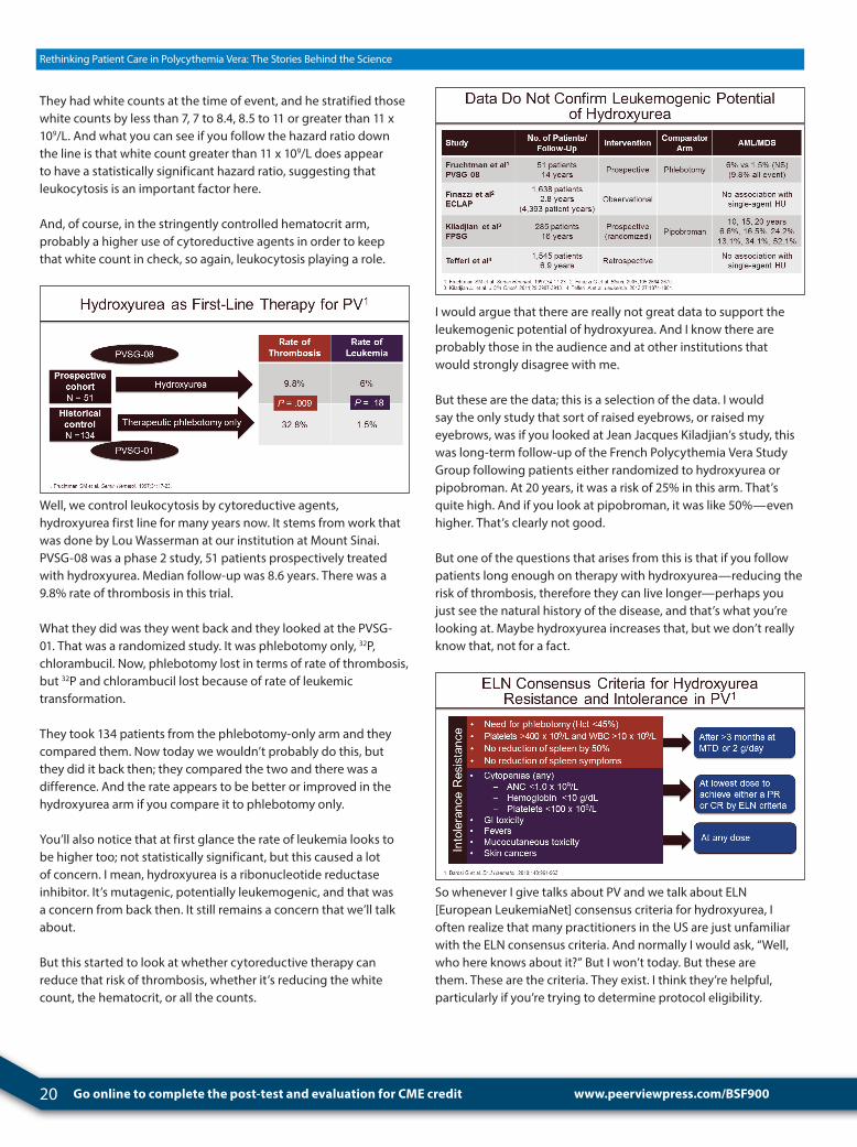
I would argue that there are really not great data to support the leukemogenic potential of hydroxyurea. And I know there are probably those in the audience and at other institutions that would strongly disagree with me.
But these are the data; this is a selection of the data. I would say the only study that sort of raised eyebrows, or raised my eyebrows, was if you looked at Jean Jacques Kiladjian’s study, this was long-term follow-up of the French Polycythemia Vera Study Group following patients either randomized to hydroxyurea or pipobroman. At 20 years, it was a risk of 25% in this arm. That’s quite high. And if you look at pipobroman, it was like 50%—even higher. That’s clearly not good.
But one of the questions that arises from this is that if you follow patients long enough on therapy with hydroxyurea—reducing the risk of thrombosis, therefore they can live longer—perhaps you just see the natural history of the disease, and that’s what you’re looking at. Maybe hydroxyurea increases that, but we don’t really know that, not for a fact.
So whenever I give talks about PV and we talk about ELN [European LeukemiaNet] consensus criteria for hydroxyurea, I often realize that many practitioners in the US are just unfamiliar with the ELN consensus criteria. And normally I would ask, “Well, who here knows about it?” But I won’t today. But these are them. These are the criteria. They exist. I think they’re helpful, particularly if you’re trying to determine protocol eligibility.
www.peerviewpress.com/BSF900

21
Rethinking Patient Care in Polycythemia Vera: The Stories Behind the Science
But essentially, they break down to resistance. So if you have myeloproliferation, despite using hydroxyurea at 2 g/dL or the maximally tolerated dose for at least 3 months, and you continue to need phlebotomies, you have a platelet count that’s elevated and leukocytosis, you don’t get a reduction in the spleen or the symptoms, then you’re resistant by definition.
And you have intolerance if you develop cytopenias at any dose, the lowest dose to achieve a CR or PR by ELN criteria, which is simply normalization of the hematologic profile, elimination of palpable splenomegaly, and major symptom burden. So if you’re developing cytopenias, that’s intolerance. If you develop extramedullary toxicity, GI toxicity, fevers, mouth ulcers, skin cancers, that’s also considered intolerance at any dose. So those are the criteria. They exist.
The Spanish group, they did something very interesting. They took a cohort of—I think it’s 261 patients that they follow, hydroxyurea-treated patients with PV. And they looked at how many of those patients meet the definition of hydroxyurea resistance or intolerance. And there was about 10% resistance, 10% intolerance.
They looked at what happens to the outcome of those patients if you meet that definition, and what they saw was if you met the definition of resistance, you had a worse outcome compared to those patients who don’t meet the definition of resistance.
Interestingly, if you achieve a complete hematologic remission as defined by the ELN, that doesn’t actually seem to translate to improved outcome and survival. But if you broke it down and you looked at the white count, leukocytosis, if you didn’t get a response in the white count itself, that alone, that did correlate and associate with worse outcome. So again, leukocytosis, in a different way—not responding to hydroxyurea therapy.
The same Spanish group just recently published an updated retrospective review where they had 890 patients, PV patients treated with hydroxyurea. What they did was they took the intolerance and resistance as a unified definition. And they took those patients who met that and they compared them to the patients who didn’t meet that definition. There really wasn’t a difference in survival if you looked at it as a unified entity.
But then what they did was they looked at all the different components of it, and the only component that seemed to affect outcome and survival was the development of cytopenias. So if you develop a cytopenia, whether it’s white count, red count, platelet count, at the lowest dose needed with hydroxyurea to achieve an ELN response, your outcome was actually much worse. So it’s about 63% versus 93% at 10 years—worse outcome.
What about progression to myelofibrosis? Well, if you meet the criteria for hydroxyurea intolerance and resistance, there does seem to be a difference in transformation rate to myelofibrosis, about 6.7% versus 17% in this analysis. It didn’t affect AML transformation rate. And again, the only thing that did was the development of cytopenias at the lowest dose to achieve an ELN response.

22 Go online to complete the post-test and evaluation for CME credit
Rethinking Patient Care in Polycythemia Vera: The Stories Behind the Science
I think Srdan will touch on this, but I’ll just say that same group—I think this was 533 patients looking at thrombosis, rate of thrombosis based on the need for supplemental therapeutic phlebotomy. So patients on hydroxyurea requiring three or more phlebotomies a year versus patients who needed two or less had a higher risk of developing thrombosis.
And this is interesting because if you go back to the PVSG-0 studies, if you look at 01, if you look at the phlebotomy, subset analyses were done there too that showed that if you had four phlebotomies a year or more that there was an increased risk of developing thrombosis. So sort of the concept is emerging of uncontrolled myeloproliferation or uncontrolled polycythemia vera.
So I’m going to try to summarize what I’ve said and then I’ll move on to the second part. So age greater than 60 and a prior thrombosis remain the mainstay of categorizing risk, although there are now risk stratifications that incorporate leukocytosis, which need to be considered.
PV patients have a significant symptom burden. Therapeutic phlebotomy and/or cytoreductive therapies may not really address these adequately. That’s where the role of other therapies that may come into place.
Then the concept of uncontrolled disease could be defined in several ways—or more than several ways; a hematocrit that’s not well controlled, despite a maximally tolerated dose of a cytoreductive therapy; persistent leukocytosis, and I think I showed enough data to support that; continued systemic or spleen-related symptoms; perhaps the frequency of phlebotomy, definitely when you’re on hydroxyurea may be an influencer of outcome; and then thrombocytosis. I didn’t really talk about thrombocytosis. And you know, often people focus on the platelet count, but what remains very unclear to me is whether the platelet count or the height of the platelet count truly predicts anything. Those data really aren’t very good, yet we all chase the platelet count. We want to normalize the platelet count.
And the Spanish have shown if you normalize the blood counts, it doesn’t necessarily improve the outcomes. So it’s unclear of attaining a CR by consensus criteria really affords a patient a benefit. That’s unfortunate because that’s what we do.
And then hydroxyurea resistance as defined by the ELN may be clinically meaningful. It may identify a subset of patients that are unlikely to do well, overall, and perhaps make you think differently about treating those patients.
Then ultimately, the management of PV really needs to be individualized—the age of the patient and the sex of the patient, and Dr. Moliterno reviewed that. Those become important clinically.
www.peerviewpress.com/BSF900

23
Rethinking Patient Care in Polycythemia Vera: The Stories Behind the Science
Story Behind the Science 2
John Mascarenhas, MDTisch Cancer InstituteIcahn School of Medicine at Mount SinaiNew York, New York
Dr. Mascarenhas: So when I was asked to do this, I was told, “Oh, there is going to be a story behind the science.” I kind of had a sense of what Alison and Srdan were going to use as their story behind the science. I didn’t want to be redundant, so I picked something that I thought was interesting and personal to me, and personal to many people in the audience today.
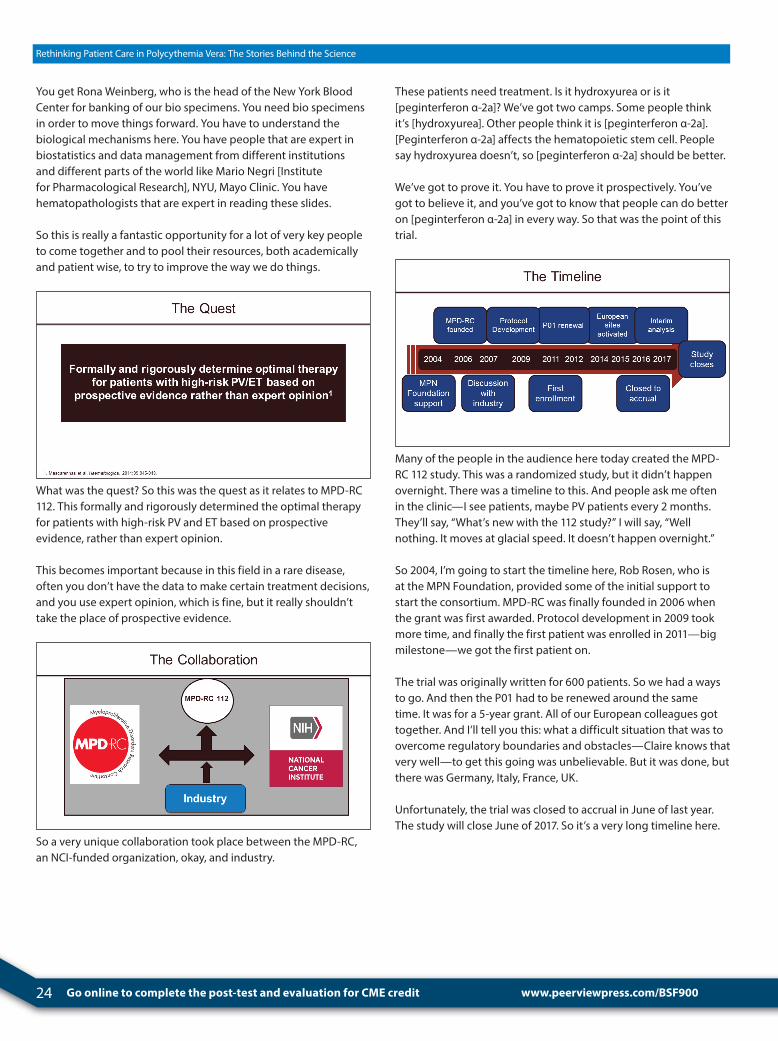
I think it’s an interesting story. It’s often not appreciated what goes into clinical trial development. So I thought today I would spend just a short amount of time talking about a clinical trial, specifically MPD-RC 112—that started way beyond me—that’s interesting.
And it starts with the MPD-RC. So the MPD-RC is an NCI-funded research consortium. It is a P01 grant. And basically, it’s a very unique story because it’s a group of very dedicated laboratory scientists and clinical investigators that have come together with a joint purpose. And this is a rare disease, so if you want to really have an impact, you’ve got to come together. You’ve got to really pool your resources, your ideas, your thoughts, your patients to really make a difference.
So this was a group of people that came together in a grant, to try to leverage different expertise, whether it’s laboratory or clinical, in order to improve the basic understanding of MPN pathophysiology and then translate that into effective therapies to benefit our patients. Sort of a novel way of treating PV/ET/MF in this era, or back then it was.
And what you see here is a star basically where every clinical site was. At one point, there were 45 sites in our consortium. Forty-five sites—that’s a lot of sites. And it’s spread across multiple countries in Europe, Israel, Canada, and the US.
And this is really a joint effort. This is a very special, unique thing, where all of these people cross continents, cross countries, languages, come together to truly translate and to capitalize on knowledge and to collaborate, so that there are different scientific projects that then work off each other and then ultimately contribute agents to the clinic in order to further the treatment of MPN patients.
This is my key contributor slide. So in the middle I put Ron Hoffman, who is my mentor, my colleague, my friend at Mount Sinai. He is the overall PI of the grant. But what you’ll notice is it’s not a one-man show by any means. If he were here, he would agree with this totally. It’s really a collaboration of people with knowledge in MPN, scientific understanding, and a desire to collaborate to move things along.
What you will see is there is the elder statesmen. I don’t mean that they’re old. They’ve just been doing this a long time, like Dr. Spivak, Dr. Silver, Dr. Barbui—and many of these people are here today—Dr. Barosi. And then there are people like myself, Vikas Gupta, Ruben Mesa, our clinical investigators. And the laboratory scientists, Ross Levine, Anna Rita Migliaccio, Joe Prchal. If you follow the field, these are people that really have been involved and really are dedicated. So this is very unique. You get all these people together.
24 Go online to complete the post-test and evaluation for CME credit
Rethinking Patient Care in Polycythemia Vera: The Stories Behind the Science
www.peerviewpress.com/BSF900
You get Rona Weinberg, who is the head of the New York Blood Center for banking of our bio specimens. You need bio specimens in order to move things forward. You have to understand the biological mechanisms here. You have people that are expert in biostatistics and data management from different institutions and different parts of the world like Mario Negri [Institute for Pharmacological Research], NYU, Mayo Clinic. You have hematopathologists that are expert in reading these slides.
So this is really a fantastic opportunity for a lot of very key people to come together and to pool their resources, both academically and patient wise, to try to improve the way we do things.
What was the quest? So this was the quest as it relates to MPD-RC 112. This formally and rigorously determined the optimal therapy for patients with high-risk PV and ET based on prospective evidence, rather than expert opinion.
This becomes important because in this field in a rare disease, often you don’t have the data to make certain treatment decisions, and you use expert opinion, which is fine, but it really shouldn’t take the place of prospective evidence.
So a very unique collaboration took place between the MPD-RC, an NCI-funded organization, okay, and industry.
These patients need treatment. Is it hydroxyurea or is it [peginterferon α-2a]? We’ve got two camps. Some people think it’s [hydroxyurea]. Other people think it is [peginterferon α-2a]. [Peginterferon α-2a] affects the hematopoietic stem cell. People say hydroxyurea doesn’t, so [peginterferon α-2a] should be better.
We’ve got to prove it. You have to prove it prospectively. You’ve got to believe it, and you’ve got to know that people can do better on [peginterferon α-2a] in every way. So that was the point of this trial.
Many of the people in the audience here today created the MPD-RC 112 study. This was a randomized study, but it didn’t happen overnight. There was a timeline to this. And people ask me often in the clinic—I see patients, maybe PV patients every 2 months. They’ll say, “What’s new with the 112 study?” I will say, “Well nothing. It moves at glacial speed. It doesn’t happen overnight.”
So 2004, I’m going to start the timeline here, Rob Rosen, who is at the MPN Foundation, provided some of the initial support to start the consortium. MPD-RC was finally founded in 2006 when the grant was first awarded. Protocol development in 2009 took more time, and finally the first patient was enrolled in 2011—big milestone—we got the first patient on.
The trial was originally written for 600 patients. So we had a ways to go. And then the P01 had to be renewed around the same time. It was for a 5-year grant. All of our European colleagues got together. And I’ll tell you this: what a difficult situation that was to overcome regulatory boundaries and obstacles—Claire knows that very well—to get this going was unbelievable. But it was done, but there was Germany, Italy, France, UK.
Unfortunately, the trial was closed to accrual in June of last year. The study will close June of 2017. So it’s a very long timeline here.
25
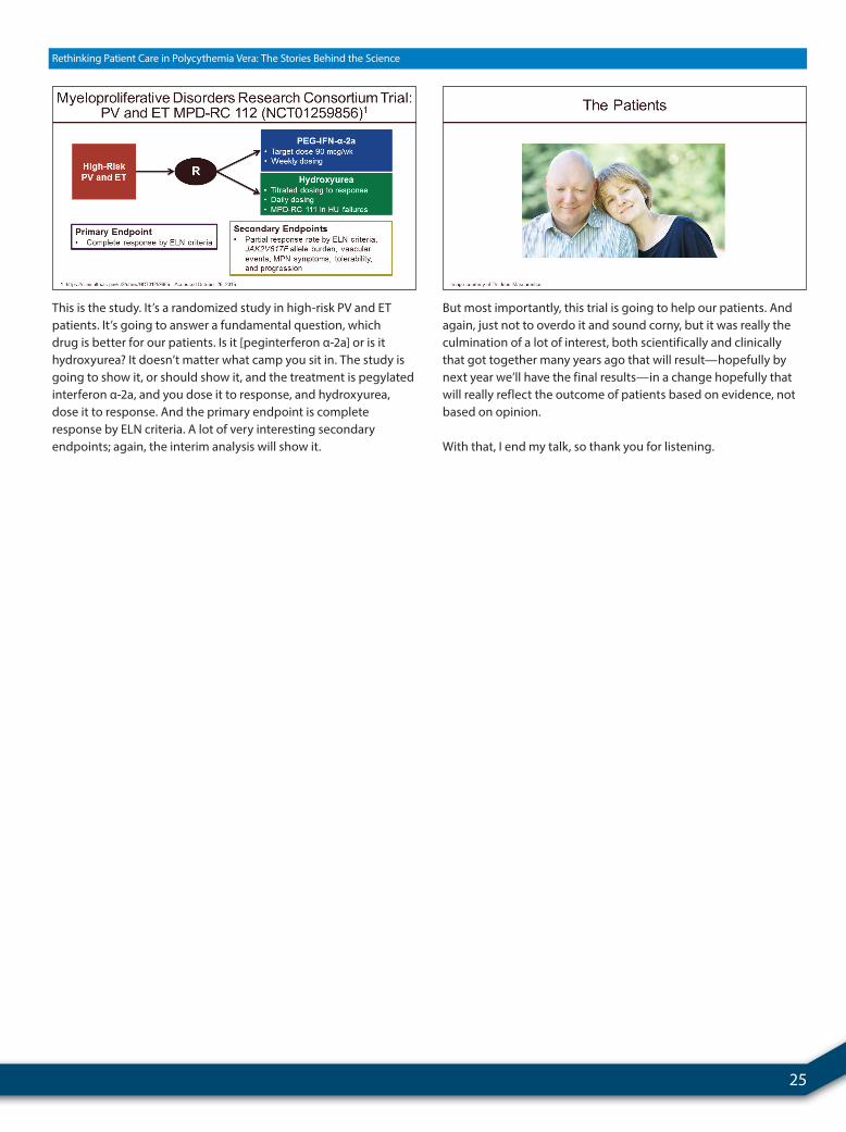
Rethinking Patient Care in Polycythemia Vera: The Stories Behind the Science
This is the study. It’s a randomized study in high-risk PV and ET patients. It’s going to answer a fundamental question, which drug is better for our patients. Is it [peginterferon α-2a] or is it hydroxyurea? It doesn’t matter what camp you sit in. The study is going to show it, or should show it, and the treatment is pegylated interferon α-2a, and you dose it to response, and hydroxyurea, dose it to response. And the primary endpoint is complete response by ELN criteria. A lot of very interesting secondary endpoints; again, the interim analysis will show it.
But most importantly, this trial is going to help our patients. And again, just not to overdo it and sound corny, but it was really the culmination of a lot of interest, both scientifically and clinically that got together many years ago that will result—hopefully by next year we’ll have the final results—in a change hopefully that will really reflect the outcome of patients based on evidence, not based on opinion.
With that, I end my talk, so thank you for listening.

26 Go online to complete the post-test and evaluation for CME credit
Rethinking Patient Care in Polycythemia Vera: The Stories Behind the Science
Approaches to Recognizing and Managing Treatment-Refractory Disease
Dr. Harrison: Our last speaker is Srdan Verstovsek. I don’t think he needs any introduction. He is going to speak on approaches to recognizing and managing treatment-refractory disease. Thank you, Srdan.
Dr. Verstovsek: Very good. Thank you very much for the opportunity to join you today. Thank you for invitation from the chair and to the colleagues that share the podium with me.
So we’re going to continue with the last talk, and we’ll just take off where we stopped basically, just to summarize what happened so far.
So by modern ways of looking at polycythemia vera, we would like to have a complete response defined as implementing five factors, and these are easy to see: control of the red blood cell count, white cells, platelets, control of the spleen, and the symptoms. So if everything is controlled well, we would say complete response. And then we have partial response and no response. So we are talking about five factors at this time and age.
Now, of course, then how do we find resistance or intolerance to hydroxyurea if you are looking at these five factors? The first three on this slide are related to control of the red blood cells. Myeloproliferation would be control of the platelets and white cells, and then splenomegaly—if the medication, and the hydroxyurea is the first-line medication, is given for a good number of months and at a good dose. The bottom two would be more or less toxicity. Sometimes cytopenia is even called resistance—this is in blue color. And the last one is nonhematologic toxicity at any hydroxyurea dose.
So these are official, from 2010, definitions of resistance or intolerance to hydroxyurea that we use all the time in the clinical studies for development of new medications in polycythemia vera.
Now the one factor that is not covered here is control of the symptoms.
Dr. Ruben Mesa—and his work has already been mentioned, a good friend and colleague from Mayo Clinic in Arizona—published in Journal of Clinical Oncology, not so long ago, this particular study assessing with the MPN-10—a symptomatic score that even in some clinics is now used to assess the quality of life of the patients—whether patients that are or are not on hydroxyurea have a good control of general symptoms that will be possibly related to polycythemia vera.
And as you can see here, without going into detail one by one, there does not appear to be very good control of general symptoms related to polycythemia vera with hydroxyurea, as assessed in this study.
Srdan Verstovsek, MD, PhDThe University of Texas MD Anderson Cancer CenterHouston, Texas
www.peerviewpress.com/BSF900

27
Rethinking Patient Care in Polycythemia Vera: The Stories Behind the Science
Now this is important. Quality of life is coming up in the myeloproliferative neoplasm area quite significantly in the front-line because we would like to control problems that people have, looking over their long longevity, particularly in polycythemia vera. But the quality of life as we control counts is important.
Now here is a little bit more on a very recent paper, 2 months ago, on perception of what’s wrong. So what we have in the red color on the left side is the patient responding to a question on whether they were symptomatic at the time of diagnosis. And we encircle 89% of the PV patients said, “Yes, I am symptomatic for the disease.” There is a huge imbalance between the perception from the patient perspective relative to the perception from the physician perspective. And I guide you to this particular paper. It’s quite interesting, published the last day of September this year in Cancer.
So it is not enough anymore to say, “Oh, I treat my polycythemia vera patients well. Hardly anybody requires anything else, or they all do well.” Let’s do a little tour around the globe and see what actually happens. This is a multicenter chart review of about 1,000 patients in Germany.
What we have on the left upper part is the dose of hydroxyurea used. Not too many patients are actually given more than 2 grams of hydroxyurea in everyday practice. As assessed by the doctors in the lower left panel, in the blue color, is what the perception is from the treating doctors on sensitivity or resistance to hydroxyurea, so in fact it’s not that everybody is sensitive to hydroxyurea.
And when the question was asked about particulars in terms of response, in the graph on the right side you see that the majority of the patients, 80% would have a good response to phlebotomy. That’s the longest blue line. But the other ones in terms of symptoms, splenomegaly, or control of white cells and platelets are not so satisfactory as it's defined through this chart review in Germany.
There is more to it. What we see here is, for example in the right upper part, hematological toxicity when it’s seen is the majority of the cases when we talk about toxicity related to hydroxyurea. In the right lower panel, which are nonhematological toxicities—perhaps we forget that hydroxyurea can actually cause ulcers, skin ulcers, and mouth ulcers in particular.
And then there are more particulars on the left upper and left lower panels in terms of what kind of intolerance we are talking about, particularly in terms of myelosuppression, which is the left lower panel. Particularly, anemia can happen with hydroxyurea.
So this is from the chart review. So it’s not that it’s super therapy that really works beautifully in every patient, as we just saw in the charts.
Now there is another study published recently, earlier this year. This is from the United States in about 1,300 patients with PV. And really without going into detail about why these people are not on [hydroxyurea] anymore, about 20% of the patients stopped their hydroxyurea out of these 1,300 patients. And the reasons are listed one by one with different colors provided on the right side of the slide, but the summary is in the box.
28 Go online to complete the post-test and evaluation for CME credit
Rethinking Patient Care in Polycythemia Vera: The Stories Behind the Science
So among the 20% of patients that actually stopped the hydroxyurea, about 30% are because of inadequate response, about 27% because of intolerance, and others because of progression. So it’s a combination of factors, and the front-line therapy is very effective, but not in everybody. And there are different reasons why people stop the therapy.
And then the focus of this particular paper in Experimental Hematology & Oncology was on the patients that are on hydroxyurea. So we are talking about 1,000 patients that are treated with hydroxyurea. The question here is how good is the control of CBC? And in the largest part of the circle, we see only about a third of the patients have normal blood cell count—white cells, red blood cells, and platelets. And the other ones are in different colors with certain abnormalities. So it’s only about a third of the patients actually that are treated with hydroxyurea that have normal blood cell count.
And the comparison in terms of overall response by using these five factors that I mentioned—normal white cells, red blood cells, platelets, symptoms, and spleen—does not distinguish a lot between the patients that are on hydroxyurea, which is on the left side, the left circle, and the right side, the right circle, these are patients that are not on hydroxyurea anymore.
So the patients may or may not be on hydroxyurea, but the type and degree of response or percent of the response does not appear to differ much when we talk about continuing therapy or discontinuing therapy for different reasons. And we’re talking about first-line therapy with hydroxyurea.
This is a little bit of a wordy slide, the one that recapitulates what was already shown, and I show this again because it may have some implications on how we treat the patients. This is from Spain. Now we moved from Germany to the United States and to Spain. Here are 533 patients, where the message here is that in patients who are on hydroxyurea, if they need three or more phlebotomies a year, they appear to have a worse outcome.
So in many instances, therefore, it appears that there are solid data to suggest if the cytoreductive therapy is used—and hydroxyurea in this case is the one—that we should be optimizing therapy to eliminate, if possible, the use of phlebotomy along with cytoreductive therapy because otherwise patients are not doing well.
The same Spanish group recently earlier in August of this year published an analysis of 890 patients, also shown and mentioned by John earlier on. In their hands, about 15% of the patients were intolerant or resistant to hydroxyurea for multiple reasons. That is the first bullet on the slide.
The bottom line is the second bullet, that there was increased risk of transformation to myelofibrosis or acute myeloid leukemia if the reason for intolerance was cytopenia. So the patients that developed low blood cell count were particularly identified as at increased risk for transformation.
www.peerviewpress.com/BSF900
29
Rethinking Patient Care in Polycythemia Vera: The Stories Behind the Science
Survival in this group of patients, as was shown before and here on the left side, did not differ, but a transformation to myelofibrosis overall did differ for patients that were intolerant or resistant to [hydroxyurea], as is shown on the right side.
One more time on intolerance of hydroxyurea, and this I put extra here with a photograph of one of my patients. Unfortunately, for some reason, it is not very well recognized in community practice that these skin ulcers—and this is a typical picture of one—are related to hydroxyurea. And quite a few patients come through the door with these complications without an involved doctor recognizing the connection to [hydroxyurea] and stopping the hydroxyurea. This is the most common. Nine out of 10 cases when we have intolerance to hydroxyurea, it is about skin ulcers or mouth ulcers.
So operationally, what does it mean clinically when we are talking about resistance or intolerance? This is a summary from Dr. Barosi
in a paper in 2010. What does it really mean when we are talking about these issues of intolerance or resistance?
So phlebotomy and thrombosis, certainly we are talking about thrombotic risk. Others, as you can see, may have influence on transformation or even overall survival in some papers, particularly when we’re talking about the resistance. Nonhematological toxicities are less relevant for the overall outcome of the patients, but the resistance appears to be significant.
What do we do in patients that are in need of a second-line therapy? This is the standard practice these days. Interferon, as you see, in cases of hydroxyurea intolerance or the other way around, the first two lines. Then the other ones, the standard less used medications that are still mentioned in the guidelines. And the new addition, as we all know now, is ruxolitinib, which is approved in the European Union and the United States for patients that have inadequate response or intolerance to hydroxyurea.
Now let’s talk about interferon. We do love interferon, and I use interferon, particularly in younger patients, even in the first line—almost all the time, if possible—because of these findings. We are very familiar with ability of interferon to affect the malignant clone, perhaps even normalize the bone marrow and reduce vascular events. And there are different ways of assessing the benefit. Whether we are really changing the natural course of the disease is open to evaluation in clinical studies, and John has described the ongoing comparison between [hydroxyurea] and interferon in the MPN Consortium study, which we will hear about later this week.

30 Go online to complete the post-test and evaluation for CME credit
Rethinking Patient Care in Polycythemia Vera: The Stories Behind the Science
Now there was a study in advanced polycythemia vera—43 patients with a median duration from diagnosis to therapy of 50 months. So it’s not in newly diagnosed patients. This is in advanced patients. Most of them, the great majority, were already previously treated where there was a very good hematological response, and what I’m showing here on the right side is the decrease in JAK2 allele burden. Operationally, we can say this is the number of cells in the bone marrow or blood sample with the JAK2 mutation. It’s going down. And in about 18% to 20% of the patients, it’s going away.
So there is a possibility of having complete molecular response in some patients with polycythemia vera on interferon. This is [peginterferon α-2a].
Now it’s not that simple. We know that the JAK2 mutation is not the cause for the disease on its own. It’s a driver of the disease. And there are other mutations that were elaborated on earlier on. And what is shown on this slide, perhaps a little bit difficult to see, here we have on the left side complete molecular responders, partial molecular responders, as it pertains to the JAK2 mutation. And what is here in different colors is presence of other mutations that we were able to test for. This is a few years ago, 2013; now we are much better in testing more samples. This is being done with better kits.
These five mutations were tested for and it seems that the greater complete molecular response change, or ability to achieve complete molecular response with JAK2 measurements is tied to presence or absence of other mutations. So it’s not that simple story about only JAK2 being involved in biology of the disease.
Presence or absence of other mutations that we now can test for, at least in academic settings, may influence the outcome of therapy with interferon. And Dr. Silver here at ASH will have an oral session on Sunday, his experience with testing for other mutations in interferon-treated patients with MPN.
And this particular study that I presented here was updated last ASH—not published yet. This is a 7-year median follow-up. This is the summary of the long-term follow-up. I would say 7 years is not trivial. The hematological response I already mentioned. The median duration, however, is 66 months; it does not last forever. Molecular response is durable only if there is complete molecular response. There are some toxicities over time. There are some discontinuations and there are some failures.
It is very effective, particularly in younger patients, I would say, but one needs to understand that there are also some limitations of the therapy, still with some toxicities and the failures, particularly if you use it in more advanced cases. Perhaps in earlier, newly diagnosed patients, the results would be different.
This is a study that is underway that has not been mentioned yet. This is another Myeloproliferative Disease Consortium study that is led by John here, which is just an open-label study to try to recapitulate what was seen in the earlier study that I showed results on. This is a study with 188 patients with high-risk ET and PV second line after hydroxyurea to try to objectivize these benefits perhaps a little bit better with the modern techniques.
www.peerviewpress.com/BSF900

31
Rethinking Patient Care in Polycythemia Vera: The Stories Behind the Science
Now we are aware of ruxolitinib as being an option in patients that are intolerant or refractory to hydroxyurea. This is just a summary slide that has been published before in The New England Journal of Medicine. Patients that were resistant or intolerant to [hydroxyurea] with the requirement for phlebotomy with splenomegaly were randomized between ruxolitinib and best available therapy, with the possibility of patients on best available therapy crossing over to ruxolitinib.
The benefits are summarized at the bottom. I’m going to analyze this a little bit more with updated results, some of which have not been published yet. So there was superior control, as you can see, in red blood cell counts, white cells, platelets, spleen, and symptoms, and the last one, trend for fewer thromboembolic events on a safety analysis. The study was designed to control the blood cell count, spleen, and symptoms, not to actually look at the thrombotic events, but that is part of the safety analysis.
So this is a longer term follow-up now, just published. The durability of hematocrit control on ruxolitinib, so if you basically achieve response, it does last long. This is perhaps not long enough for PV patients. We are talking about 80-week follow-up, but it’s very promising. People continue to respond.
This is a response in white cell count. Let’s look at the table for the sake of time. This is control of white cells and control of the platelets at week 32, which is the primary time of analysis. Now at week 80 it appears that over time there is more and more control of the white cells and the platelets in these patients that are treated with ruxolitinib.
This is perhaps more visible on analysis of a spleen response. This is a spleen decrease over time in patients with big spleen. This is at week 32, where the study was analyzed for approval. This is now continuation of the analysis of spleen response over time. It gets better.
The symptom control is very well described. Not to go into detail, but ruxolitinib, in the red color, is much better than the standard therapy in multiple aspects of quality of life, as you can see here, and more on the other side as well.

32 Go online to complete the post-test and evaluation for CME credit
Rethinking Patient Care in Polycythemia Vera: The Stories Behind the Science
Now let’s just show this particular table that was published 2 months ago in Haematologica. This is a safety analysis, and it’s a rather wordy table, but the circled part is the most important. These are the thromboembolic events in the ruxolitinib part and best-available-therapy part. It appears—and there is no statistical analysis of the safety analysis, these are just the data as is from the analysis of safety—there appears to be somewhat of a difference in the thromboembolic events between the two arms.
What about other events of interest? Infections are present in patients that are on ruxolitinib. They are also present in patients that are on the best-available-therapy arm. That appears to be equal. What we know already, and it is on the label for ruxolitinib, is that there are atypical infections that happen more often in patients on ruxolitinib, and one here that is listed is herpes. That is very well described. We treat through therapy with ruxolitinib those patients that do develop herpes. And these are some other issues here of perhaps non-melanoma skin cancer that were followed very closely in these groups of patients.
Finally, the disease progression, of course, as it is known very well from the myelofibrosis field, ruxolitinib does not prevent progression to myelofibrosis or acute myeloid leukemia. It does control symptoms and signs of the disease, but it does not prevent progression.
This is an important practical point. These are the doses of patients that are treated for polycythemia vera with ruxolitinib. Starting dose, unlike myelofibrosis, is 10 mg twice a day. So everybody who has PV is started equally at 10 mg twice a day. And then during the first 3 months, usually you require in a majority of the patients, as you can see, to increase in dose. It has to be given twice a day—not once a day, but twice a day—because of the short half-life in the body. And about a third of the patients will go to 15 and some to 20 and some to 25 mg. Only 10% of patients will require less than 10 mg twice a day.
Now let’s put into everyday practice perspective the phlebotomy need in patients that are on ruxolitinib. This is the elimination of the phlebotomy need completely in the majority of the patients. These are the patients that require one or two during the first 8 weeks when the assessment of response was not even in place yet. This is assessment of response as per study design, 80% response rate for the phlebotomy—no need for it. And this is a long-term follow-up. As I said before, if you have response, you tend to continue to respond, 90% probability of continuing without phlebotomy at all.
www.peerviewpress.com/BSF900

33
Rethinking Patient Care in Polycythemia Vera: The Stories Behind the Science
So my question usually is, “What about those that did not respond? What happened to those,” because protocol definitions usually are very strict, to make sure that we can objectivize in the eyes of administrative bodies. And this is very proper for drug approval what the drug does. But let’s answer the question, “What happens with patients that are nonresponders by protocol definitions?”
These are best-available-therapy arm patients and, in red color, ruxolitinib in nonresponders—time to phlebotomy in one versus the other. So time to next phlebotomy was 21 weeks in patients on best-available-therapy arm, and it was a full year in patients that did not respond to ruxolitinib.
So there is a benefit that is not properly objectivized in the trial design, but you can see here this is one example where there is a difference even in patients that are nonresponders with ruxolitinib.
This is perhaps even better seen when we look at the control of symptoms. This is control of symptoms on the best-available-therapy arm, 4% response rate. In red color are official responders. In black color are nonresponders. So these people did not respond, and yet you see that control of the symptoms was actually excellent, even though they needed one phlebotomy here and there, as you can see. So control of the symptoms was excellent across the board.
This is also seen here. This is the so-called easy questionnaire, PGIC score—no change, improved little, more, or very much improved. Best-available-therapy arm showed no change over time, and responders and nonresponders feeling much better on the ruxolitinib. So it’s excellent for control of the symptoms.
Now there was another study, which is called the RESPONSE-2 study, not published yet, where the same trial design was in place, but in patients without splenomegaly. So ruxolitinib in hydroxyurea-resistant or intolerant patients with PV in second line, similar results in terms of controlling hematocrit, overall control of the CBC, and symptoms.
So one supports the other. In other words, the presence of splenomegaly for success in therapy of patients in the second line with ruxolitinib is not a prerequisite. The spleen was there in one study, was not there in the other study, and results in terms of control of signs and symptoms is the same.
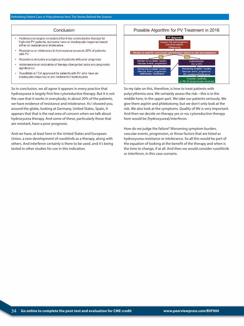
34 Go online to complete the post-test and evaluation for CME credit
Rethinking Patient Care in Polycythemia Vera: The Stories Behind the Science
So in conclusion, we all agree it appears in every practice that hydroxyurea is largely first-line cytoreductive therapy. But it is not the case that it works in everybody; in about 20% of the patients, we have evidence of resistance and intolerance. As I showed you, around the globe, looking at Germany, United States, Spain, it appears that that is the real area of concern when we talk about hydroxyurea therapy. And some of these, particularly those that are resistant, have a poor prognosis.
And we have, at least here in the United States and European Union, a new development of ruxolitinib as a therapy, along with others. And interferon certainly is there to be used, and it’s being tested in other studies for use in this indication.
So my take on this, therefore, is how to treat patients with polycythemia vera. We certainly assess the risk—this is in the middle here, in the upper part. We take our patients seriously. We give them aspirin and phlebotomy, but we don’t only look at the risk. We also look at the symptoms. Quality of life is very important. And then we decide on therapy yes or no; cytoreductive therapy here would be [hydroxyurea]/interferon.
How do we judge the failure? Worsening symptom burden, vascular events, progression, or those factors that are listed as hydroxyurea resistance or intolerance. So all this would be part of the equation of looking at the benefit of the therapy and when is the time to change, if at all. And then we would consider ruxolitinib or interferon, in this case scenario.
www.peerviewpress.com/BSF900
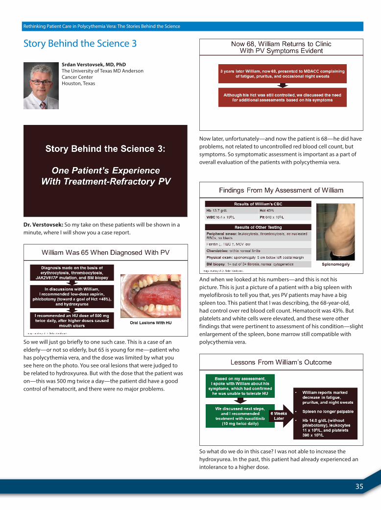
35
Rethinking Patient Care in Polycythemia Vera: The Stories Behind the Science
Story Behind the Science 3
Dr. Verstovsek: So my take on these patients will be shown in a minute, where I will show you a case report.
So we will just go briefly to one such case. This is a case of an elderly—or not so elderly, but 65 is young for me—patient who has polycythemia vera, and the dose was limited by what you see here on the photo. You see oral lesions that were judged to be related to hydroxyurea. But with the dose that the patient was on—this was 500 mg twice a day—the patient did have a good control of hematocrit, and there were no major problems.
Srdan Verstovsek, MD, PhDThe University of Texas MD Anderson Cancer CenterHouston, Texas
Now later, unfortunately—and now the patient is 68—he did have problems, not related to uncontrolled red blood cell count, but symptoms. So symptomatic assessment is important as a part of overall evaluation of the patients with polycythemia vera.
And when we looked at his numbers—and this is not his picture. This is just a picture of a patient with a big spleen with myelofibrosis to tell you that, yes PV patients may have a big spleen too. This patient that I was describing, the 68-year-old, had control over red blood cell count. Hematocrit was 43%. But platelets and white cells were elevated, and these were other findings that were pertinent to assessment of his condition—slight enlargement of the spleen, bone marrow still compatible with polycythemia vera.
So what do we do in this case? I was not able to increase the hydroxyurea. In the past, this patient had already experienced an intolerance to a higher dose.

36 Go online to complete the post-test and evaluation for CME credit
Rethinking Patient Care in Polycythemia Vera: The Stories Behind the Science
So we began treating this patient with ruxolitinib. And again, the starting dose is 10 mg twice a day. And you see results like you see in myelofibrosis, within month two, and maximum three, you see all the benefits. Particularly, within a month, control of all the symptoms. There would be control in the spleen, and the blood cell count may be improved as well—so the combination of factors that control the JAK-STAT pathway, that is the target for the JAK inhibitors. The JAK-STAT pathway would lead to improved signs and symptoms of the disease.
Now I should take this further just to highlight one more time, which is in the first bullet, that it’s not only about the control of red blood cells, meaning hematocrit or hemoglobin. It is about the whole person, including the control of the symptoms. So you look at the CBC, spleen, and symptoms as well, and in this setting there are up to one-quarter of the patients that actually are not really doing well on hydroxyurea. And as you have seen here, ruxolitinib is a good option in that setting.
There are other patients, of course, that are enjoying life with PV now that we are able to control their signs and symptoms. Those that were here last year may recognize the gentleman on the left side. Jack was here talking about his life experience. He is fully engaged with a community of patients with PV and is enjoying life with 10 mg twice a day of ruxolitinib. And there are some other patients here as well.
So I thank you very much for your attention, and I give the podium to our Chair. Thank you.
www.peerviewpress.com/BSF900
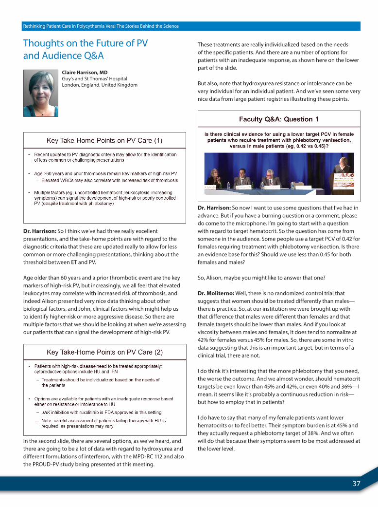
37
Rethinking Patient Care in Polycythemia Vera: The Stories Behind the Science
Thoughts on the Future of PV and Audience Q&A
Claire Harrison, MDGuy's and St Thomas' HospitalLondon, England, United Kingdom
Dr. Harrison: So I think we’ve had three really excellent presentations, and the take-home points are with regard to the diagnostic criteria that these are updated really to allow for less common or more challenging presentations, thinking about the threshold between ET and PV.
Age older than 60 years and a prior thrombotic event are the key markers of high-risk PV, but increasingly, we all feel that elevated leukocytes may correlate with increased risk of thrombosis, and indeed Alison presented very nice data thinking about other biological factors, and John, clinical factors which might help us to identify higher-risk or more aggressive disease. So there are multiple factors that we should be looking at when we’re assessing our patients that can signal the development of high-risk PV.
In the second slide, there are several options, as we’ve heard, and there are going to be a lot of data with regard to hydroxyurea and different formulations of interferon, with the MPD-RC 112 and also the PROUD-PV study being presented at this meeting.
These treatments are really individualized based on the needs of the specific patients. And there are a number of options for patients with an inadequate response, as shown here on the lower part of the slide.
But also, note that hydroxyurea resistance or intolerance can be very individual for an individual patient. And we’ve seen some very nice data from large patient registries illustrating these points.
Dr. Harrison: So now I want to use some questions that I’ve had inadvance. But if you have a burning question or a comment, pleasedo come to the microphone. I’m going to start with a questionwith regard to target hematocrit. So the question has come fromsomeone in the audience. Some people use a target PCV of 0.42 forfemales requiring treatment with phlebotomy venisection. Is therean evidence base for this? Should we use less than 0.45 for bothfemales and males?
So, Alison, maybe you might like to answer that one?
Dr. Moliterno: Well, there is no randomized control trial thatsuggests that women should be treated differently than males—there is practice. So, at our institution we were brought up withthat difference that males were different than females and thatfemale targets should be lower than males. And if you look atviscosity between males and females, it does tend to normalize at42% for females versus 45% for males. So, there are some in vitrodata suggesting that this is an important target, but in terms of aclinical trial, there are not.
I do think it’s interesting that the more phlebotomy that you need, the worse the outcome. And we almost wonder, should hematocrit targets be even lower than 45% and 42%, or even 40% and 36%—I mean, it seems like it’s probably a continuous reduction in risk—but how to employ that in patients?
I do have to say that many of my female patients want lower hematocrits or to feel better. Their symptom burden is at 45% and they actually request a phlebotomy target of 38%. And we often will do that because their symptoms seem to be most addressed at the lower level.
38 Go online to complete the post-test and evaluation for CME credit
Rethinking Patient Care in Polycythemia Vera: The Stories Behind the Science
www.peerviewpress.com/BSF900
Dr. Harrison: Yeah. I mean, I suppose in my practice I would lower a hematocrit target for a female if they’re still symptomatic. And I certainly use a lower target for patients who are pregnant and patients who have got a splenic vein thrombosis.
Dr. Harrison: There is a follow-up question that maybe we could think about because the consequence of driving the hematocrit lower—especially by using phlebotomy—is iron deficiency. And so there is a question that actually came in by email: Is it useful to give iron replacement therapy to a patient with PV who develops an iron-deficient state? John, do you want to comment on that?
Dr. Mascarenhas: I think it would be unwise to give iron to a patient with PV, as it would simply act to induce erythrocytosis. So I typically don’t give iron in that setting.
There has been a lot of discussion about whether the iron-deficient state also contributes to the fatigue and a lot of the symptomatology. I think a lot of the earlier studies looking at iron and whether there is a role there are actually from Johns Hopkins University. We know that iron deficiency definitely gives you pica syndrome. It gives you nail-bed changes. It likely induces some degree of fatigue. But it would seem unwise to give iron in this setting—so I typically don’t do that.
I guess one possibility, or one thought, is that if you are developing a situation where excessive phlebotomy is required and you’re developing a very iron-deficient state, maybe that is a patient that then deserves cytoreductive therapy, where you would move away from developing an iron-deficient state, and therefore, control the absolute number of cells rather than the size of the cells.
Dr. Harrison: I suppose for me sometimes with iron deficiency, I think it might worsen pruritus and fatigue, as well, which are really dominant features.
Dr. Harrison: So, there is another question about treatments for pruritus. I know that’s a really difficult issue. I don’t know if somebody wants to handle that—maybe Srdan—do you want to comment on therapy for pruritus?
Dr. Verstovsek: In terms of the benefit of ruxolitinib in patients who are refractory to hydroxyurea, because hydroxyurea is usually given to control the counts. But as I showed you, we would like to control the symptoms as well. And if that is the problem, and it’s making patients’ quality of life so bad—and I have patients like that that are really not able to perform regular duties because of intractable pruritus—then that would be a reason in my mind to consider alternative therapies, either the interferon—interferon has also been reported to control symptoms very well in patients, even with low dose—or ruxolitinib, in that case.
Certainly, there are other interventions that one can try; those are antihistamines, or some [paroxetine]-like antidepressant medications, or sometimes I had a couple of patients that were seen by the dermatologists and UV light was applied. But these are extremes. These are anecdotal cases. Usually a change in cytoreductive therapy, if the quality of life is so bad, is the one approach that I take most often.
Dr. Harrison: Another good question here about the management of JAK2-negative PV. Alison, should I pass that to you?
Dr. Moliterno: I get asked that question a lot, and I see a lot of patients, as we all do, who have erythrocytosis or high hemoglobin, who have been referred to us for this JAK2-negative PV.
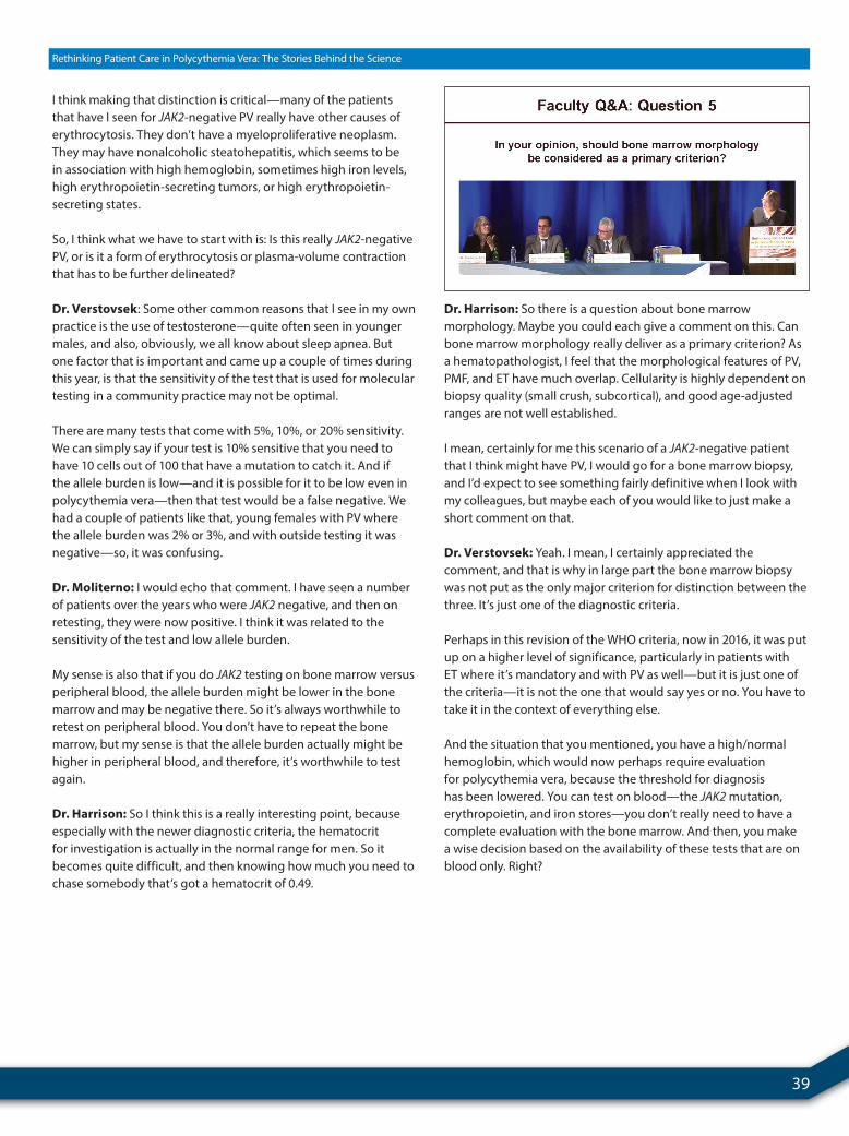
39
Rethinking Patient Care in Polycythemia Vera: The Stories Behind the Science
I think making that distinction is critical—many of the patients that have I seen for JAK2-negative PV really have other causes of erythrocytosis. They don’t have a myeloproliferative neoplasm. They may have nonalcoholic steatohepatitis, which seems to be in association with high hemoglobin, sometimes high iron levels, high erythropoietin-secreting tumors, or high erythropoietin-secreting states.
So, I think what we have to start with is: Is this really JAK2-negative PV, or is it a form of erythrocytosis or plasma-volume contraction that has to be further delineated?
Dr. Verstovsek: Some other common reasons that I see in my own practice is the use of testosterone—quite often seen in younger males, and also, obviously, we all know about sleep apnea. But one factor that is important and came up a couple of times during this year, is that the sensitivity of the test that is used for molecular testing in a community practice may not be optimal.
There are many tests that come with 5%, 10%, or 20% sensitivity. We can simply say if your test is 10% sensitive that you need to have 10 cells out of 100 that have a mutation to catch it. And if the allele burden is low—and it is possible for it to be low even in polycythemia vera—then that test would be a false negative. We had a couple of patients like that, young females with PV where the allele burden was 2% or 3%, and with outside testing it was negative—so, it was confusing.
Dr. Moliterno: I would echo that comment. I have seen a number of patients over the years who were JAK2 negative, and then on retesting, they were now positive. I think it was related to the sensitivity of the test and low allele burden.
My sense is also that if you do JAK2 testing on bone marrow versus peripheral blood, the allele burden might be lower in the bone marrow and may be negative there. So it’s always worthwhile to retest on peripheral blood. You don’t have to repeat the bone marrow, but my sense is that the allele burden actually might be higher in peripheral blood, and therefore, it’s worthwhile to test again.
Dr. Harrison: So I think this is a really interesting point, because especially with the newer diagnostic criteria, the hematocrit for investigation is actually in the normal range for men. So it becomes quite difficult, and then knowing how much you need to chase somebody that’s got a hematocrit of 0.49.
Dr. Harrison: So there is a question about bone marrow morphology. Maybe you could each give a comment on this. Can bone marrow morphology really deliver as a primary criterion? As a hematopathologist, I feel that the morphological features of PV, PMF, and ET have much overlap. Cellularity is highly dependent on biopsy quality (small crush, subcortical), and good age-adjusted ranges are not well established.
I mean, certainly for me this scenario of a JAK2-negative patient that I think might have PV, I would go for a bone marrow biopsy, and I’d expect to see something fairly definitive when I look with my colleagues, but maybe each of you would like to just make a short comment on that.
Dr. Verstovsek: Yeah. I mean, I certainly appreciated the comment, and that is why in large part the bone marrow biopsy was not put as the only major criterion for distinction between the three. It’s just one of the diagnostic criteria.
Perhaps in this revision of the WHO criteria, now in 2016, it was put up on a higher level of significance, particularly in patients with ET where it’s mandatory and with PV as well—but it is just one of the criteria—it is not the one that would say yes or no. You have to take it in the context of everything else.
And the situation that you mentioned, you have a high/normal hemoglobin, which would now perhaps require evaluation for polycythemia vera, because the threshold for diagnosis has been lowered. You can test on blood—the JAK2 mutation, erythropoietin, and iron stores—you don’t really need to have a complete evaluation with the bone marrow. And then, you make a wise decision based on the availability of these tests that are on blood only. Right?
40 Go online to complete the post-test and evaluation for CME credit
Rethinking Patient Care in Polycythemia Vera: The Stories Behind the Science
www.peerviewpress.com/BSF900
Dr. Harrison: John?
Dr. Mascarenhas: Yeah. I would just add one other thing too, which is sometimes overlooked, and I think it can be important. Sometimes when you look at a bone marrow biopsy, and you believe the patient has PV, they may have PV, but they also may have other things that you’re not anticipating. And I see this not infrequently, where you find mast cells and they may have an SM-AHNMD [systemic mastocytosis with associated clonal hematological non–mast cell lineage disease], or you may find eosinophilia, or even an increase in monotypic plasma cells and an MGUS [monoclonal gammopathy of undetermined significance] that can evolve.
So, there are other things that can often coincide. And this is well described in the literature, so it’s not always just PV. You sort of characterize them at baseline, so that if something changes clinically in the future, you have something to go back to and be able to tell. The other day, I saw a patient and attributed the symptoms to PV, did the bone marrow biopsy, and he had mastocytosis as well. So sometimes it’s not always just PV.
Dr. Harrison: Alison? Sorry to come to you last.
Dr. Moliterno: Yeah. I’ve had similar experiences as well. I think that also where you practice is helpful in answering this question. If you have the ability to sit down with hematopathologists to really review the situation, that’s a much better context than just getting a report and trying to integrate that into your practice.
Now a lot of us, including myself, we don’t often have time to review every bone marrow biopsy or possibility with the hematopathologist, so I share the hematopathologist’s comments that the morphology itself really can’t distinguish very well between the three entities, but it can distinguish—is this a myeloproliferative neoplasm? And so in that sense, if you’re really trying to make that call, hopefully there will be information on the biopsy to help move an MPN diagnosis forward. Again, whether it’s PV or ET or MF—sometimes, I think that’s a hard call.
Dr. Harrison: I think, as well, not overcalling a biopsy that’s suboptimal—if a biopsy is suboptimal, it should be repeated, and I agree with your comment about the dialogue, and I think the WHO criteria are encouraging that dialogue and the summation of all of that information. I do think a biopsy is important.
So it’s time, perhaps, for us to end the symposium, to thank all of our speakers—they did a really great job—and to thank you in the audience for participating today, for your great questions. I hope you have a good ASH meeting, and look forward to seeing you in the future. Thank you very much.


CME
Rethinking Patient Care in Polycythemia Vera: The Stories Behind the Science
www.peerviewpress.com/BSF900
Sign up for e-mail alerts on new clinical advances and educational activities in your specialty: www.peerviewpress.com/signup
Copyright © 2000-2016, PeerView Press
This CME activity is jointly provided by Medical Learning Institute, Inc. and PVI, PeerView Institute for Medical Education.
This activity is supported by an educational grant from Incyte Corporation.
PVI, PeerView Institute for Medical Education, and Medical Learning Institute, Inc. are responsible for the selection of this activity's topics, the preparation of editorial content, and the distribution of this activity. The preparation of PeerView activities is supported by educational grants subject to written agreements that clearly stipulate and enforce the editorial independence of PVI and Medical Learning Institute, Inc. Our activities may contain references to unapproved products or uses of these products in certain jurisdictions. For approved prescribing information, please consult the manufacturer's product labeling. No endorsement of unapproved products or uses is made or implied by coverage of these products or uses in our activities. No responsibility is taken for errors or omissions in activities.
New and improved sourcefor free CME/CE
Based on a panel discussion and on data from recent medical literature. The materials presented here are used with the permission of the authors and/or other sources. These materials do not necessarily reflect the views of PeerView Press or any of its partners, providers, and/or supporters.