retinoic acid and pattern formation in the developing...
TRANSCRIPT

/. Embryol. exp. Morph. 90,139-169 (1985) 139Printed in Great Britain © The Company of Biologists Limited 1985
Retinoic acid and pattern formation in the developingchick wing: SEM and quantitative studies of earlyeffects on the apical ectodermal ridge and budoutgrowth
J. LEE AND C. TICKLEDept. of Anatomy and Biology as Applied to Medicine, The Middlesex HospitalMedical School, Cleveland St., London W1P6DB, U.K.
SUMMARYWhen retinoic acid is locally applied to the anterior margin of developing chick wing buds on
ion-exchange beads, dose-dependent changes in the skeletal pattern result. At low doses,additional digits develop. At high doses, there is thinning of the symmetrical wing. Localapplication of retinoic acid to the apex of the bud also leads to pattern changes, but in contrastnormal wing patterns are almost always obtained following application posteriorly. These effectsare manifest at 6-7 days after the operation although only a brief exposure (14-20 h) to retinoicacid is required. Therefore the morphology of wing buds was studied at shorter times after thestart of treatment.
The local application of retinoic acid to the wing bud margin leads to changes in extent of theapical ridge that can be detected at 24 h after application. The behaviour of the apical ridge withvarying doses and positions of retinoic acid application has been analysed quantitatively anddose response curves obtained. At low doses of retinoic acid, the length of the apical ridgeincreases or remains constant, but then progressively decreases with higher doses. The progress-ive obliteration of the ridge starts first near the bead and then involves more distant parts of thebud. Thus the region of the ridge affected depends on the position at which the retinoic acid isapplied.
We propose that these effects on the apical ridge reflect dose-dependent responses to the localconcentration of retinoic acid that varies with distance from the source. At high doses, the apicalridge disappears but at low doses it is maintained. Since grafts of polarizing region tissue alsohave a graded effect on ridge morphology, a possible interpretation of the retinoic acid effects isthat tissue adjacent to the source is converted into polarizing region tissue. Alternatively,retinoic acid may act directly on the ridge cells.
The changes in the extent of the apical ridge produced by retinoic acid lead to different formsof bud outgrowth. The form of the outgrowth depends on the dose of retinoic acid, the positionof application and the interaction between the effects of the local source of retinoic acid andthose of the polarizing region of the host bud. These considerations give some insights into whyanterior application of retinoic acid leads to the development of additional digits whereasposterior application generally gives normal wings.
INTRODUCTION
Local application of retinoic acid to the developing wing bud brings aboutpattern changes (Tickle, Alberts, Wolpert & Lee, 1982; Summerbell, 1983;Summerbell & Harvey, 1983). When ion-exchange beads (AG1-X2) are used as
Key words: retinoic acid, apical ectodermal ridge, limb development, chick wing, SEM, budoutgrowth.
140 J. LEE AND C. TICKLE
carriers for retinoic acid (Eichele, Tickle & Alberts, 1984) and implanted beneaththe apical ectodermal ridge at the anterior margin of the wing bud, there is a cleardose response. A highly reproducible series of wing patterns results with effects onpattern formation across the anteroposterior axis (Tickle, Lee & Eichele, 1985).Beads soaked in low concentrations of retinoic acid (0-01-04 mg ml"1) whichresult in 3-20 picograms of retinoic acid in the wing tissue (0-9-25 nivr) at 14 h leadto the sequential formation of additional digits (digit patterns 2234, 32234 or 3234and 432234 or 43234). When beads are soaked in higher concentrations of retinoicacid (1-10 mg ml"1), giving over 100 picograms of retinoic acid in the bud tissue at14 h, there is a progressive thinning of the symmetrical wing (digit patterns 43234,4334, 434 and a single symmetrical digit 4). Beads soaked in very high concen-trations of retinoic acid (lOmgrnP1) frequently result in truncated wings with nodigits at all.
When beads soaked in a low concentration of retinoic acid (0-01 mg ml"1) areimplanted to the apex of the wing bud beneath the apical ridge (Tickle et al. 1985),about 50% of the wings develop digits 234 anterior to the implant and asymmetrical digit 4 posteriorly (digit pattern 234, 4), while the remainder aremostly normal or lack one or more digits. With higher concentrations of retinoicacid (1-10 mg ml"1), truncations result. In contrast to the above changes, normaldigit patterns (although reduced in size) result from implantation of retinoic-acid-impregnated beads to the posterior margin of the bud except at the highestconcentration when truncations result (Tickle et al. 1985). A striking feature ofthese position-dependent effects of retinoic acid treatment is that additionalstructures appear to be generated only from anterior tissue.
The effects of local application of low doses of retinoic acid mimic thoseproduced by grafts from the polarizing region, which is a signalling region at theposterior of the bud (Saunders & Gasseling, 1968; Tickle, Summerbell & Wolpert,1975). Thus the sequential formation of additional digits is obtained with anteriorgrafts of increasing numbers of cells from the polarizing region (Tickle, 1981), digitpatterns such as 234,4 follow grafts of polarizing tissue to the apex of the bud andnormal digit patterns result from posterior grafts of polarizing tissue (Tickle et al.1975; Wolpert & Hornbruch, 1981).
The patterns described above following retinoic acicl treatment are assayed at6-7 days after application when the skeletal elements are clearly recognizable.However, we have found that exposures of 14-20 h are sufficient to bring aboutpattern changes (Eichele, Tickle & Alberts, 1985). Thus to understand how reti-noic acid exerts its effects, events occurring at much shorter times after treatmentmust be followed. As a start to this analysis, bud outgrowth following implantationof beads soaked in a range of retinoic acid concentrations was examined. Since theapical ectodermal ridge, a specialized epithelium that runs anteroposteriorly alongthe tip of the bud has been found essential for bud outgrowth (Saunders, 1948;Summerbell, 1974), particular attention was paid to its extent and disposition.
Polarizing tissue grafts have previously been shown to lead to alterations in budform and extent of the apical ridge before pattern changes become apparent.

Effects of retinoic acid on wing bud outgrowth 141
For example, the formation of additional digits following anteriorly positionedpolarizing region grafts is accompanied by an increase in width of the outgrowth ofthe developing bud across the anteroposterior axis (Tickle et al. 1975; Smith &Wolpert, 1981). This may reflect changes in the extent of the apical ridge.Although the apical ridge immediately adjacent to a polarizing region graftdisappears, the ridge persists as a thickened epithelium over the anterior margin ofthe bud (Saunders & Gasseling, 1968). Irradiation of buds following anteriorlygrafted polarizing tissue leads to a reduction in the width of outgrowth and wingswith digit patterns similar to those produced by high concentrations of retinoicacid are obtained (Smith & Wolpert, 1981). Digit patterns such as 434 also resultwhen polarizing region cells are grafted beneath the apical ridge over extensivedistances along the anterior of the bud (Honig, 1983) and here too outgrowth ispresumably affected. In principle, severe width reductions could ultimately lead totruncations.
The observations reported here reveal that shortly after implantation ofretinoic-acid-impregnated beads, the shape of the bud outgrowth was affected andreflected consistent changes in the extent and disposition of the apical ridge. Incontrast to the pattern changes that are position-dependent, the graded effect ofretinoic acid on the ridge occurred irrespective of the position of application. Thisparadox can be understood in terms of the interaction between the locally appliedretinoic acid and the polarizing region of the host bud that acts as a boundary.
MATERIALS AND METHODS
Implanting carrier beadsThis procedure has been described previously by Tickle et al. (1985). Briefly, AG1-X2 beads
(200 jum diameter) were soaked in solutions of all-frww-retinoic acid (Sigma lot Nos. 41F. 0440and 63F-0476) dissolved in dimethyl sulphoxide (DMSO), then rinsed in tissue culture mediumand implanted beneath the apical ectodermal ridge of the right wing bud of stage-20 embryos(Hamilton-Hamburger stages). The left wing bud served as an unoperated control. Controlbeads were soaked in DMSO, rinsed and implanted in the same way. The main series of beadimplants was made beneath the ridge at the anterior margin of buds but some beads wereimplanted under the ridge at the apex or at the posterior margin.
Grafting tissueThe limb buds of embryos at stage 21-22 were dissected off and placed in 2 % trypsin in
calcium- and magnesium-free saline, for 1 h at 4°C to loosen the ectoderm from the underlyingmesoderm. The buds were then transferred to tissue culture medium (MEM+10% foetal calfserum; Gibco: Biocult) at 4°C and the ectoderm removed. Next cubes of polarizing regionmesenchyme, of size 200 jon3, were dissected out of the posterior region of the bud. For controltissue, cubes of mesenchyme were similarly dissected from the anterior of the buds. The tissuewas grafted beneath the apical ridge in the same way that the bead implants were made.
Preparation of specimens for SEMAt various times (4-48h) after performing the bead implants or tissue grafts, the embryos
were dissected out of the egg and fixed in half-strength Karnovsky fixative (Karnovsky, 1965) forat least 24 h at 4 °C. Next, the right and left wing bud of each embryo was carefully removed andplaced into a small bag made out of lens tissue (Whatman 105). Bags containing a pair of wing

142 J. L E E AND C. T ICKLE
buds from each embryo could then be processed further without damage to the buds. Theprocessing involved a rinse in 0-OlM-sodium cacodylate and postfixation in 1% osmiumtetroxide for 1 h. Following dehydration through a series of alcohols, the buds were transferredin trichloroethane (Arclone) to liquid CO2 for critical-point drying in a Polaron critical-pointdryer. The dried specimens were mounted onto aluminium stubs with double-sided tape.A conducting layer of silver paint was applied closely around the specimens and the stub surface.These specimens were then coated with a 50 nm thick layer of gold and palladium using an SEM-coating unit E5000. Prepared in this way, all specimens were viewed with a Jeol JSM-35 scanningelectron microscope.
Measurements of bud perimeter and extent of apical ridgeSome data were obtained by making measurements on scanning electron micrographs of buds
in profile using an IB AS computer. Since the buds had shrunk considerably (up to 42 %) duringpreparation and in addition could not always be orientated to give suitable profile views, anotherseries of treated buds was used to provide the bulk of the data. In this series, beads soaked in arange of retinoic acid concentrations were implanted to the right wing bud as before. At 24 h or48 h, the embryos were removed from the egg, placed in Tyrodes solution, and stained with Nileblue sulphate (0005% in Tyrodes). Both right and left (untreated) buds were dissected out.Each wing bud was positioned in a Petri dish so that a profile view was obtained. Drawings of thebud and the extent of the apical ridge were made using a camera lucida. Measurements weremade from the camera-lucida drawings using an IB AS computer as before. A minimum of fourbuds was usually measured for each data point.
RESULTS
SEM observations
Beads were soaked in 10,1,0-1 or 0-01 mgml"1 retinoic acid. The buds to whichthese treated beads were implanted were compared with limbs to which controlbeads soaked in DMSO only had been implanted and which were fixed after thesame time periods. Treated buds were also compared with left unoperated buds,which served as controls. The complete series of experiments is listed in Table 1.
Effects of beads implanted anteriorly
4-6 h
At this time, no differences could be detected between limb buds irrespective oftheir treatment. The shape of retinoic-acid-treated buds closely resembled that oftheir left counterparts (Fig. 1A,B), except that the bead was clearly visible bulgingbeneath the apical ridge at the anterior of the bud. These buds were alsoindistinguishable from buds which received control beads soaked in DMSO only.In buds to which beads had been implanted, the apical ridge extended over thebead and along the bud profile to its posterior margin (Fig. 1A). In the region ofthe bead, the ridge was stretched to accommodate the carrier and so its thickenedmorphology was not apparent. However, extending posteriorly from the bead, thethickened ridge was well defined and could be estimated as being about 10 cellswide. Cells at the edge of the cut tissue had begun to migrate over the surface ofthe implanted bead (Fig. 1C,D). This marked the first stages of the healing processduring which the beads become completely embedded in the buds. The extent ofcell migration onto the beads was variable and did not appear to be correlated with

Tab
le 1
. Su
mm
ary
tabl
e sh
owin
g th
e to
tal n
umbe
r of
bud
s ex
amin
ed in
eac
h ca
se a
nd th
e di
git p
atte
rns
that
res
ulte
d af
ter
6 da
ysC
one.
RA
Po
sitio
n in
win
g bu
d to
whi
ch r
etin
oic
acid
is a
pplie
d or
tiss
ue g
raft
edin
soa
king
soln
. (m
gml"
1 )or
tis
sue
graf
ted
Ant
erio
rA
pex
Post
erio
r
4-6
h 18
-24
h 48
h
Dig
it pa
tter
ns!
24 h
48
h
Dig
it pa
tter
ns!
24 h
48
h
Dig
it pa
tter
ns!
DM
SO
10 01
00
1
Pola
rizi
ngre
gion
Ant
erio
rm
esen
chym
e
—
234
2 tr
unca
tions
34 434
4334
4334
3 tr
unca
tions
4 34 434
4334
4323
4—
4 43
443
34
4334
4323
443
2234
2 43
443
34
4334
4323
443
2234
3223
423
4
3 43
3443
234
—
234
(11)
*(1
6) (4)
(2)
(3)
(7)
(6)
(3)
(1)
(2)
(2)
(2)
(3)
(1)
(4)
(4)
(6)
(1)
(1)
(4)
(1)
(8)
(3)
(1)
(1)
(2)
(3)
(2)
—
234
5 tr
unca
tions
^(3
) 3
—
234
(4)
- -
-
11
10
23 234
5 6
234
(2)
(1)
(8)
(6)
& o a
2 34 334
2,4
234,
423
4 +
pos
tel
emen
ts23
423
4,4
234,
4423
4,43
423
423
4
(1)
(1)
(1)
(1)
(3)
(1)
(7)
(3)
(1)
(1)
(1)
(2)
3 I23
4(2
)
—
234+
extr
a (5
)el
emen
t at
elbo
w p
roje
ctin
gve
ntra
lly
* N
umbe
rs in
bra
cket
s in
dica
te n
umbe
r of
cas
es.
~ In
dica
tes
fuse
d di
gits
, t
Cum
ulat
ive
data
from
all
expe
rim
ents
usi
ng a
ll-fr
a/is
-ret
inoi
c ac
id fr
om S
igm
aL
ot.
41F-
0440
and
63F
-047
6.
X O
btai
ned
with
ret
inoi
c ac
id L
ot. N
o. 1
2F-0
598.
144 J. LEE AND C. TICKLE
V-' * /
D '
Effects of retinoic acid on wing bud outgrowth 145
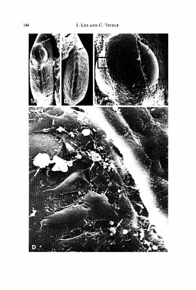
the treatment of the bead. In addition to the migrating cells, which resembledfibroblasts, a few macrophages could be seen, particularly at the corners of thehealing wound.
18-24h
Differences were now observed between buds, depending on the treatment ofthe implanted beads. At 24 h, the difference in shape was most marked betweencontrol buds (or those with control beads) and buds treated with the highestconcentration of retinoic acid. Buds to which beads soaked in progressively lowerconcentrations of retinoic acid had been implanted showed a dose-dependenteffect: the difference between the control and treated buds becoming progress-ively reduced as lower concentrations of retinoic acid were applied on the beads.
In buds to which control beads had been implanted (Fig. 2A,B), there was aslight bulge anteriorly marking the position of the bead that had become com-pletely embedded in the wing tissue. The wing bud was now elongated. The apicalridge was well-defined and extended from beneath the embedded bead at theanterior, around the rim of the bud to the posterior. The contours of these budscontaining control beads were identical to those of normal untreated left-handbuds (approximately stage 24).
In striking contrast, wing buds which had received an implant soaked inlOmgml"1 retinoic acid were considerably shortened and steeply tapered to apointed tip at the posterior margin. The extent of the apical ridge was considerablyreduced, being absent over most of the bud margin and present only over thepointed tip at the very posterior of the bud. Buds to which beads soaked in1 mg ml"1 retinoic acid had been implanted also resulted in shortened buds and theridge was similarly present only over the posterior tip (Fig. 2C,D).
With beads soaked in lower concentrations of retinoic acid, the buds wereprogressively less tapered. For example, with beads soaked in 0-1 mgrnP1 retinoicacid, the bud profile was narrow, spadelike and the anterior edge was not ascurved as that of the normal bud at this stage. The extent of the apical ridge wasreduced but not as severely as those buds with beads soaked in higherconcentrations of retinoic acid. Thus the apical ridge was absent anteriorly nearthe site of the bead but its anterior limit was closer to the bead than in the treatedbuds just described.
Fig. 1. (A) Edge view of a right wing bud fixed 4-6 h after implanting a bead soaked inlOmgml"1 retinoic acid at the anterior margin. The apical ridge (arrowed) runs overthe bead and extends along the bud profile to the posterior margin. Note that in theregion of the bead, the ridge (double arrowed) is stretched to accommodate thecarrier. Picture width (PW) = 0-37 mm. (B) Edge view of the contralateral left wingbud to that shown in A. The apical ridge can be distinctly seen extending from theanterior to the posterior margin (extent indicated between arrows). PW = 0-36mm.(C) Enlarged view of A showing the bead positioned directly beneath the apical ridge.Flattened cells can be seen moving out over the surface of the bead from under theridge. PW = 0-22 mm. (D) A highly magnified view of C in region indicated, showingpavement of apical ridge cells and fibroblast-like cells migrating over bead surface. Afew macrophage-like cells (arrowed) are also present. PW = 0-051 mm.

146 J. LEE AND C. TICKLE

Effects ofretinoic acid on wing bud outgrowth 147
Finally, with implanted beads soaked in the lowest concentration of retinoic acid(0-01 mgml"1), the buds appeared to be about the same width as normal buds,(Fig. 2E) but were distally more symmetrical (compare Fig. 2A,E). The apicalridge was present over the entire wing bud margin except in the region of theembedded bead (Fig. 2F).
48 h
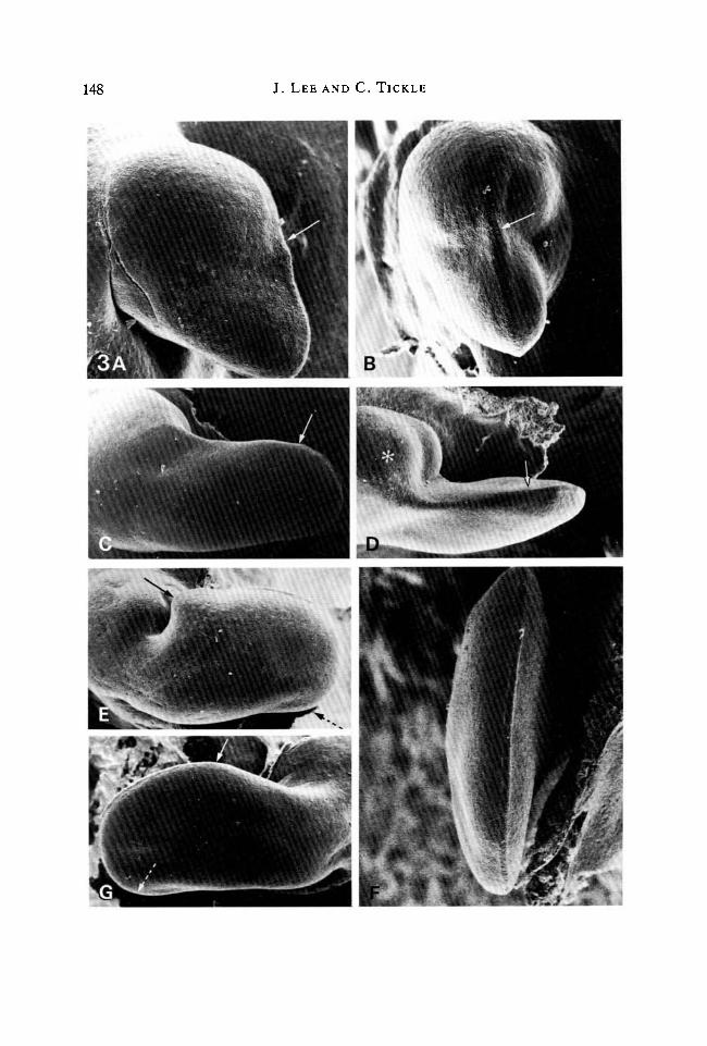
Further development had produced even greater differences in the shapes ofbud outgrowth. Application of the highest concentration of retinoic acid(lOmgmn1), which had greatly reduced the extent of the apical ridge at 24 h, hadnow resulted in stunted buds with a very narrow region of outgrowth posteriorly(Fig. 3A). The apical ridge was present only over the posterior outgrowth (Fig.3B). It should be noted that where the apical ridge had disappeared anteriorly andoutgrowth ceased, the bud bulged and was not flattened dorsoventrally as normal.Beads soaked in lmgml"1 retinoic acid produced narrow, finger-like outgrowthswith apical ridges running along their tips (Fig. 3C,D). These narrow contourswere quite unlike the shape of the left-hand bud which was paddle-shaped. Withbeads soaked in the lowest concentrations of retinoic acid, the buds were broadwith additional tissue at the anterior margin (compare Fig. 3E with contralateralnormal bud in Fig. 3G). The apical ridge was present over the entire bud margin,and thus was longer than usual because it extended along the perimeter of thewidened outgrowth (Fig. 3F, also see quantitative analysis).
Effects of beads implanted at the wing bud apex
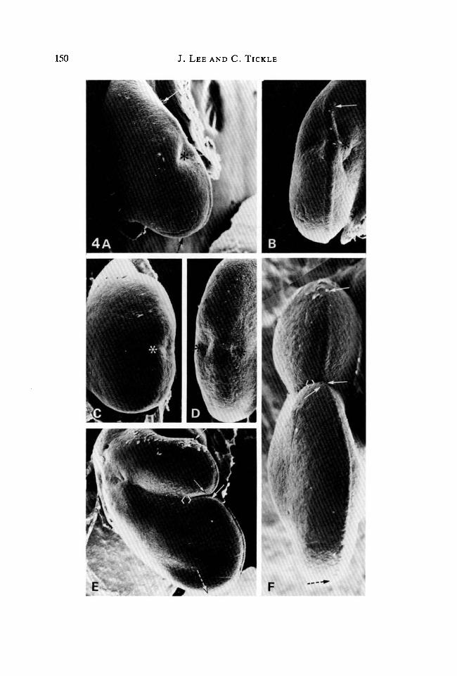
At 24 h, control beads placed at the apex of the bud had led to the developmentof an indentation in the margin of the bud which corresponds to the site ofimplantation (Fig. 4A,B). The indentation had been displaced anteriorly bygreater outgrowth posteriorly. Although the ridge was perturbed in the region ofthe indentation, it appeared to be virtually continuous around the bud margin.
In contrast, implants of beads soaked in high concentrations of retinoic acid(lOmgrnl"1) after 24 h had produced extremely stunted wing buds in which the
Fig. 2. Buds 24 h after implanting beads anteriorly. (A) Dorsal view. Control beadsoaked in DMSO only. A bulge at the anterior margin marks the site of the embeddedbead, shown by *. Anterior limit of apical ridge indicated by arrow. Posterior limitdetermined by rotation of specimen, indicated by doi:ted arrow. PW= 1-2 mm. (B)Edge view of A. Note extent of apical ridge along the wing bud margin (anterior limitof ridge indicated by arrow). PW = 0-47mm. (C) Dorsal view. Bead soaked in1 mgml"1 retinoic acid. Note the bud is much shorter (compare shape of bud with thatof bud in A). Extent of the apical ridge is indicated between the arrows. Note that theridge is confined to the posterior part of the bud. PW = 1-17 mm. (D) Edge view of C.The site of the embedded bead is indicated by *. Anterior limit of apical ridge isindicated by arrow. PW = 0-46mm. (E) Dorsal view. Bead soaked in 0-01 mg ml"1
retinoic acid. The shape of the bud is similar to that of the control bud except that thetip is more symmetrical (compare with A). Anterior limit of apical ridge indicated byarrow. PW = 1-46 mm. (F) Edge view of E. Anterior limit of apical ridge indicated byarrow (compare with A). PW = 0-68 mm.
148 J. LEE AND C. TICKLE
0***- - * •

Effects of retinoic acid on wing bud outgrowth 149
apical ridge had completely disappeared (Fig. 4C,D). By 48h, still no furtheroutgrowth had occurred.
With beads soaked in low concentrations of retinoic acid (0-01 mgml"1),asymmetrically bilobed wing buds developed after 24 h. The apical ridge wasclearly seen extending along each lobe. However, between the two lobes the ridgewas more difficult to distinguish and was raised very little above the surroundingectoderm. By 48 h the buds had developed into two lobes that had elongated inparallel and were separated by a narrow groove (Fig. 4E). The apical ridge couldbe distinctly seen extending around the apex of each elongated lobe (Fig. 4F).
Effects of beads implanted at the posterior margin
Implants of beads soaked in DMSO only, resulted in normal wing budoutgrowth with the normal extent of apical ridge at 24 h. The outline of suchtreated buds was slightly perturbed posteriorly by a bulge marking the position ofthe embedded bead.
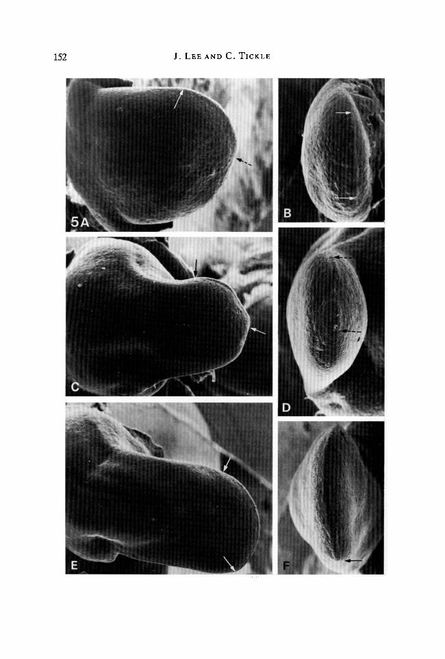
By 24 h, beads soaked in 1 mgml"1 retinoic acid had resulted in short, roundedwing buds (Fig. 5A). Posterior outgrowth was reduced. The shape of the budapproximated to an inverted version of that which resulted from anteriorlypositioned beads soaked in the same concentration of retinoic acid (compare Fig.5A with Fig. 2C). This was because, in contrast to normal buds, the apical ridgewas absent posteriorly in the region of the implanted bead but persisted over theanterior margin (Fig. 5A,B).
After 48 h, outgrowth of wing buds had resumed. However, instead of theoutgrowth involving posterior tissue, outgrowth continued from the anterior partof the bud (Fig. 5C). Thus the bud was abnormally positioned. It was noticeablyshort and the apex narrower than control buds. The posterior limit of the apicalridge had been shifted more anteriorly (Fig. 5D). The original posterior margin ofthe bud over which the apical ridge had disappeared remained as a bulbous mass oftissue posteriorly.
Fig. 3. Buds 48h after implanting beads anteriorly. (A) Dorsal view. Bead soaked in10 mg ml"1 retinoic acid. Note the stunted shape of the bud. The apical ridge (anteriorlimit indicated by arrow) is present over the pointed posterior part of the bud.PW = 1-26 mm. (B) Edge view of A. Anterior limit of apical ridge indicated by arrow.Note that where the apical ridge has disappeared anteriorly, the bud bulgesdorsoventrally. Compare with dorsoventral flattening of posterior part of the budwhere the ridge is still present. PW = l-28mm. (C) Dorsal view. Bead soaked in1 mgml"1 retinoic acid. Note narrow symmetrical outgrowth. Anterior limit of apicalridge indicated by arrow. PW = l-6mm. (D) Anterior view of C. Bulbous mass oftissue (*), covers bead. Anterior limit of apical ridge indicated by arrow.PW = l-7mm. (E) Dorsal view. Bead soaked in 0-01 mg ml"1 retinoic acid. Notebroadened bud with extensive apical ridge. Anterior limit indicated by arrow andposterior limit determined by rotation of specimen, indicated by dotted arrow.PW = 1-7 mm. (F) Edge view of anterior margin of bud shown in E with extensiveapical ridge. PW = 1-0 mm. (G) Dorsal view. Contralateral unoperated bud to thatshown in E for comparison with experimental buds. The shape of this bud and extent ofits apical ridge (indicated between arrows as previously) are normal. PW = 1-64 mm.
150 J. LEE AND C. TICKLE

Effects of retinoic acid on wing bud outgrowth 151
Beads soaked in O-lmgml"1 retinoic acid produced a similar but less-pronounced anterior shift in the position of outgrowth. This could be detectedafter 24 h but was more noticeable after 48 h. In addition, outgrowth was lessretarded than when beads soaked in lmgml"1 retinoic acid were implanted.However, the apex of the buds was still narrower than that of control buds (Fig.5E). The apical ridge extended from the anterior to the new posterior margin,which had been displaced anteriorly, only a short distance from its originalposition (Fig. 5F).
Effects of grafting polarizing region or anterior tissue to (a) the anterior margin; (b) theapex of the wing bud
Grafts of polarizing tissue were performed so thai: the effects of these could becompared with those obtained from beads soaked in retinoic acid. Grafts ofanterior wing bud tissue served as controls for polarizing region grafts.
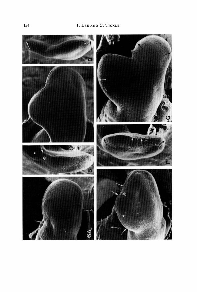
(a) Grafts to the anterior margin
24 h. At this time, no change in the gross shape of the wing bud could bedetected following a polarizing region graft. The bud was a paddle-shapedoutgrowth not noticeably wider than a normal bud of the same stage (Fig. 6A:compare with normal bud in Fig. 2A). However, the bud was distally moresymmetrical and strikingly similar in shape to the buds to which beads soaked in0-01 mgml"1 retinoic acid had been implanted anteriorly (compare with Fig. 2E).The apical ridge was absent at the graft site but extended around the remainder ofthe bud rim to the posterior margin (Fig. 6A,B). Wing buds which received graftsof anterior tissue developed normal outgrowths. The extent of the apical ridge wasunaffected by these grafts.
48 h. Buds which had received grafts of polarizing region were by this timemuch broader than controls (Fig. 6C). This widening resulted from the outgrowth
Fig. 4. Buds following implants of beads to the apex. (A) Dorsal view. 24 h afterimplanting a control bead soaked in DMSO only. A slight indentation (*) anteriorlymarks the site of the implant. Anterior limit of apical ridge indicated by arrow.Posterior limit determined by rotation of specimen, indicated by dotted arrow.PW = 1-12 mm. (B) Edge view of A. Note that the apical ridge is continuous over thesite of the embedded bead (*) and along the wing bud margin (anterior limit of ridgeindicated by arrow). PW = 0-60mm. (C) Dorsal view. 24h after implanting a beadsoaked in lOmgml"1 retinoic acid. A slight indentation (*) at the bud apex marks thesite of the embedded bead. Note the absence of apical lidge. PW = 0-75 mm. (D) Edgeview of C. Shallow indentations (*) either side of the apex mark the site of theembedded bead. The apical ridge is completely absent. PW = 0-46mm. (E) Dorsalview. 48 h after implanting a bead soaked in 0-01 mgml"1 retinoic acid. A narrowgroove (< >), separates the anterior outgrowth from the posterior one. Apical ridgeextends along the margin of both outgrowths (as indicated between solid arrows onanterior outgrowth; on posterior outgrowth between solid and dotted arrows as usedpreviously). PW = 0-47mm. (F) Enlarged view of E. The narrow groove separatinganterior from posterior outgrowth is indicated by (<>) . Note the apical ridgeextending along the margin of each separate outgrowth (as indicated between arrows,posterior limit by dotted arrow). PW =1-51 mm.
152 J. LEE AND C. TICKLE
5AB
m
1
D
% .
Effects of retinoic acid on wing bud outgrowth 153
of an extra portion of tissue from the anterior margin. A well-defined apical ridgeextended along the broadened rim of the bud (Fig. 6D). These buds were similarin shape to those which developed following anteriorly implanted beads soaked in0-01 mgml"1 retinoic acid (compare Figs 6C & 3E).
(b) Grafts to the apex
24h. Buds which had received grafts of polarizing tissue at their apex wereslightly bilobed (Fig. 6E). The bud margin was slightly indented in the regionof graft. The apical ridge was absent at the graft site but extended on eitherside both posteriorly and anteriorly from the graft around the rim of the bud(Fig. 6F). Following control anterior tissue grafts, the bud had a distorted shape.The outline of the bud was indented in the region of the graft where the apicalridge had disappeared directly over it. This indentation had then been displacedanteriorly.
48 h. The indentation in the bud margin produced by a polarizing region graftwas much more marked at this time. The bud had become distinctly bilobed(Fig. 6G). If these bud shapes are compared with those that developed followingimplants of beads soaked in 0-01 mgml"1 retinoic acid, after 24 and 48 h, it can beseen that the sequence of shape changes is superficially similar. However, buds towhich retinoic acid had been applied became distinctly bilobed at 24 h, whereassuch a bilobed shape was only beginning to become apparent with buds whichreceived grafts of polarizing tissue. Furthermore, later outgrowth of the buds ledto different relationships between the two lobes. With bead implants, the twolobes lay close together whereas with polarizing tissue, the two lobes were splayedout and separate.
Examination of buds with anterior tissue grafts showed that after 48 h furtheranterior displacement of the grafted tissue had occurred and normal budoutgrowth had been restored.
Fig. 5. Buds following implants of beads to the posterior margin. (A) Dorsal view. 24 hafter implanting a bead soaked in lmgml"1 retinoic acid. The shape of this bud isinverted compared to that shown in Fig. 2C. The apical ridge extends over the anteriormargin only (anterior limit of ridge indicated by arrow, posterior limit determined byrotation of specimen, indicated by dotted arrow). PW == 1-57 mm. (B) Edge view of A.Note that the extent of the apical ridge has been shifted anteriorly (as indicatedbetween arrows). PW = 0-75 mm. (C) Dorsal view. 48 h after implanting a bead soakedin 1 mgrnl"1 retinoic acid. The bud is shorter and the apex narrower than normal. Theapical ridge runs along the anterior margin only (as indicated between arrows).PW=l-82mm. (D) Edge view of C. Note the complete absence of apical ridgeposteriorly. Extent of apical ridge is indicated between arrows. PW = 0-61mm.(E) Dorsal view. 48h after implanting a bead soaked in 0-01 mgml"1 retinoic acid.Note that the shape changes of this bud are not as marked as those shown in C. Theextent of the apical ridge is indicated between arrows. PW = 1-86 mm. (F) Edge view ofE. The extent of the apical ridge (indicated between arrows) has been shifted onlyslightly anteriorly compared with D. PW = 0-70 mm.
154 J. LEE AND C. TICKLE
\

Effects of retinoic acid on wing bud outgrowth 155
Quantitative analyses of the effects of retinoic acid application on the extent anddisposition of the apical ridge
For analysis, the perimeter of the wing bud has been divided into three zones;anteriorly, a zone extending from the body wall to the anterior limit of the ridge -the anterior margin; a zone occupied by the apical ridge; and posteriorly, a zoneextending from the posterior limit of the ridge to the body wall - the posteriormargin (Fig. 7).
Measurements of the length of these stretches have been plotted as a function ofretinoic acid concentration in the solution in which the beads were soaked. Thecurves obtained illustrate quantitatively the effects seen morphologically in thescanning electron micrographs.
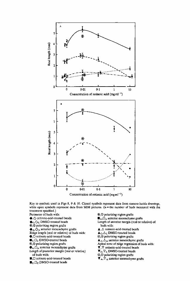
Fig. 8A,B shows how the perimeter of the bud and the lengths of the three zonesare affected by local application of retinoic acid anteriorly at 24 h and 48 h. Forcomparison, measurements following anteriorly grafted polarizing region tissueand anterior mesenchyme are plotted with the data for beads soaked in*0-01 mgml"1 retinoic acid and for normal buds respectively (see key).
The curves at 24 h show that with low doses of retinoic acid the perimeter of thebud had increased (by 1-5 mm). With increasing doses the perimeter progressivelydecreased so that with beads soaked in lmgml"1 retinoic acid or more theperimeter was shorter than that of normal buds. At lOmgml"1 the perimeter was0-05 mm shorter than normal. These changes in the length of the bud perimeterreflect the different bud shapes just described. At low doses, the buds are morerounded distally, and at high doses bud outgrowth is stunted.
The increase in bud perimeter at low doses appears to be accompanied by aslight increase in the length of the apical ridge (Fig. 8A). At higher doses, thedecrease in bud perimeter was accompanied by a marked decrease in the length ofthe ridge. This was due to an increase in the length of the anterior margin.
The inverse relationship between the lengths of apical ridge and anterior marginis clearly shown in curves drawn for data obtained over the whole concentrationrange at 48 h (Fig. 8B). As the length of the ridge first increased at low doses and
Fig. 6. Buds following grafts of polarizing region tissue. (A) Dorsal view. 24 h aftergrafting tissue anteriorly. Bud is not significantly broader (compare with normal budprofile in Fig. 2A). Anterior limit of apical ridge indicated by arrow. PW = 0-95 mm.(B) Edge view of A. Site of graft (*) is indicated by indentations on either side of bud.Anterior limit of apical ridge indicated by arrow. PW = 0-54 mm. (C) Dorsal view. 48 hafter grafting tissue anteriorly. Note broadening of bud. PW= 1-37 mm. (D) Edgeview of C. Continuous apical ridge extends around the broadened bud margin asindicated by arrows. PW = 0-56 mm. (E) Dorsal view. 24 h after grafting tissue at apexof bud. The bud is broad and slightly indented at the graft site (*). The ridge hasdisappeared at the site of the graft but is present anteriorly and posteriorly (ridgeindicated by arrows). PW = l-34mm. (F) Edge view of E. Indentations either sideindicate graft site. Note the ridge is absent in this region of the bud margin but ispresent anteriorly (between arrows) and also posteriorly (similarly indicated).PW = 0-77mm. (G) Dorsal view. 48 h after grafting tissue at the apex. Note twodistinct outgrowths, the anterior outgrowth is asymmetrical whereas the posterior oneis symmetrical. Apical ridge (arrowed) is present at the apex of both outgrowths.PW=l-60mm.

156 J. L E E AND C. T ICKLE
Fig. 7. Diagram to show how measurements were made for the quantitative analysis ofbud outgrowth (A) normal bud at 24 h, (B) normal bud after 48 h. p, Perimeter;a, Anterior margin; b, Apical ridge; c, Posterior margin. Note that for the 48 h budprofile, measurements were made around the distal paddle-shaped part of the bud onlyand did not extend to the body wall. This was because the proximal part of the bud liesin a different plane and accurate measurements of its profile could not be made.
then decreased at high doses so the length of the anterior margin initiallydecreased then increased. In contrast to these changes in the anterior part of thebud, the length of the posterior margin remained constant.
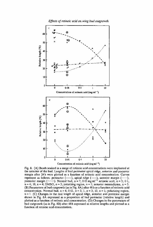
To see how the lengths of apical ridge, anterior and posterior margin change inrelation to each other, their lengths are expressed in terms of the proportion ofbud perimeter they occupy (relative lengths) and plotted against the concentrationof retinoic acid (Fig. 8C,D). Since proportions are now being considered, datafrom scanning electron micrographs can be included (see key). These data pointsare in close agreement with those obtained from camera-lucida drawings ofunfixed buds. The curves now demonstrate clearly the inverse relationshipbetween the relative length of the apical ridge and anterior margin at both 24 and48 h. This verifies quantitatively the impressions gained from the morphologicalobservations. Modulation of the anterior bud margin only is involved and theposterior margin remains constant.
A further interesting point is that the increase in the length of the ridge at 24 hwith low doses of retinoic acid can be accounted for by the increase in perimeter ofthe bud. In fact the relative ridge length rather than increasing appears to decreaseslightly. It is only at 48 h that a significant increase in the length of the ridge leads tothis occupying a greater proportion of the limb bud margin.
When retinoic acid is applied to the apex of the bud, as just describedmorphologically, the ridge disappears or becomes flatter in the region of the bead.Thus in addition to the three regions of the bud perimeter that we have defined,there is a stretch at the apex to be considered where the ridge has disappeared. Wewill call this the apical zone of ridge regression. It should also be noted that therelative length of the ridge has been calculated from the sum of the lengths of ridgepresent on both sides of the implant.
Fig. 9A,B shows the relative lengths of the apical ridge, the anterior andposterior margins and the apical zone of ridge regression at 24 and 48 h plotted as afunction of the concentration of retinoic acid.
Effects of retinoic acid on wing bud outgrowth 157
As for anterior implants, the relative length of the apical ridge remains virtuallyconstant at low doses but then progressively decreases. The decrease in therelative length of the ridge in this case, however, is brought about by the increasein the length of the apical zone of ridge regression with increasing doses of retinoicacid. At doses above O-lmgml"1, the ridge is completely obliterated by 48h. Tocalculate at these high doses the proportion of the perimeter occupied by the apicalzone of ridge regression, the proportions of the anterior and posterior marginswere subtracted from 100 %. This treatment of the data is used to illustrate theinverse relationship between the length of the ridge and that of the apical zone ofridge regression over the whole concentration range of retinoic acid. In thistreatment, the lengths of anterior and posterior margins remain constant.
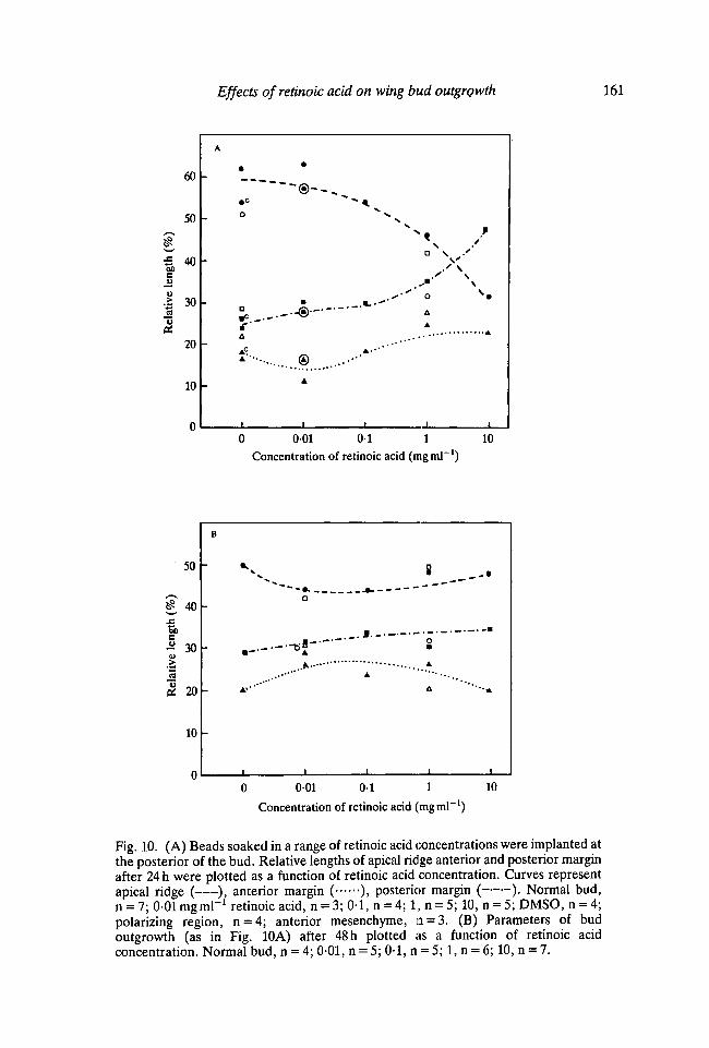
Fig. 10A,B shows the relative lengths of apical ridge, anterior and posteriormargins plotted as a function of retinoic acid concentration applied posteriorlyafter 24 and 48 h respectively. At 24 h with low doses the relative length of theapical ridge is unchanged but then progressively decreases as higherconcentrations of retinoic acid are applied. This is a similar dose response to thatobserved when retinoic acid is applied anteriorly or to the apex. The decrease inthe relative length of the ridge is due to an increase of the posterior margin. Thisincrease in the length of the posterior margin is particularly marked at highconcentrations. This is consistent with the impressions gained from scanningelectron microscope observations, that the ridge was shifted to more anteriorpositions with increasing doses of retinoic acid.
When implants are made at the anterior or apex of the bud, the samerelationship between the relative ridge length and the region of ridge regression inthe vicinity of the bead is found after 48 h as at 24 h. However, the relative lengthcurves for the parameters of bud outgrowth at 48 h after posterior application arequite different from those at 24 h. The decrease in the relative length of the ridgeseen at high concentrations of retinoic acid at 24 h is not continued. Even thoughthe perimeter of the bud steadily decreases with higher doses, the length of theridge only decreases slightly. Thus the relative length of the ridge actually appearsto increase slightly. This maintenance of a more normally proportioned ridgeinvolves changes in both the anterior and posterior margins. The anteriordecreases while the posterior increases, reflecting that bud outgrowth is now frommore anterior parts of the bud. At 48h, the differences between the curvesshowing the effects of applying retinoic acid posteriorly and to other positionsreflect the re-establishment of a correctly proportioned ridge rather than anamplification of the changes that had taken place at 24 h.
DISCUSSION
When all-frms-retinoic acid is locally applied to chick wing buds on AG1-X2beads, there are dose-dependent effects on the pattern of structures that developacross the anteroposterior axis (Tickle etal. 1985). These studies have shown thatthere are reproducible, dose-dependent changes in the form of bud outgrowth
5 -
? 4
g> 3
•aPi 2
1 -
A j
i i 1 1 i
0 001 0 1 1 10Concentration of retinoic acid (ing ml"1)
0 001 0 1 1 10Concentration of retinoic acid (mgml"1)
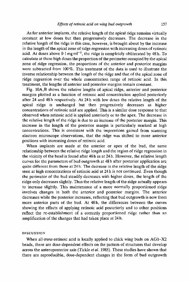
Key to symbols used in Figs 8, 9 & 10. Closedwhile open symbols represent data from SEMtreatment specified.)Perimeter of buds with:• , O retinoic-acid-treated beads• c . O c DMSO-treated beads®,© polarizing region grafts• A . O A anterior mesenchyme graftsRidge length (real or relative) of buds with:• , 0 retinoic-acid-treated beads•c ,Oc DMSO-treated beads®,@ polarizing region grafts• A I O A anterior mesenchyme graftsLength of posterior margin (real or relative)
of buds with:• , • retinoic-acid-treated beads•c ,Dc DMSO-treated beads
symbols represent data from camera-lucida drawings,pictures, (n = the number of buds measured with the
®,® polarizing region grafts• A , D A anterior mesenchyme graftsLength of anterior margin (real or relative) of
buds with:A, A retinoic-acid-treated beadsA c , A c DMSO-treated beads®,® polarizing region graftsA A, A A anterior mesenchyme graftsApical zone of ridge regression of buds with:T,V retinoic-acid-treated beads• c , V c DMSO-treated beads®,® polarizing region graftsT A , VA anterior mesenchyme grafts
Effects of retinoic acid on wing bud outgrowth
au
tiv
.213CC
60
50
40
30
20
10
n
c
•
-
-
-
-
• A
O
A'
•© ^O
a.dp
—8—®®
N
S
NS
.-' \o-' S
\J • • * . —
_ . A g ' '
,A
1
0 001 01 1 10
Concentration of retinoic acid (ing ml"1)
60 -
50 -
to 40c
1 30
20 -
10 -
D
*S
- f
o
•
AA
-
O
©
9m
A
®
\
. . - • " •
I
A
S\
\\ •
\\
.' \N
\
A
o•
1
A
A
8Q\
N\
\
t
001 0 1 1 10
Concentration of retinoic acid (nig ml"1)
Fig. 8. (A) Beads soaked in a range of retinoic acid concentrations were implanted atthe anterior of the bud. Lengths of bud perimeter apical ridge, anterior and posteriormargin after 24 h were plotted as a function of retinoic acid concentration. Curvesrepresent as follows: perimeter ( ), apical ridge ( ), anterior margin ( ),posterior margin ( ). Normal bud, n = 7; 0-01 mgml"1 retinoic acid, n = 3; 0-1,n = 4; 10, n = 4; DMSO, n = 5; polarizing region, n = 5; anterior mesenchyme, n = 5.(B) Parameters of bud outgrowth (as in Fig. 8A) after 48 h as a function of retinoic acidconcentration. Normal bud, n = 4;001,n = 3; l , n = 3; 10, n = 1; polarizing region,n = 1. (C) Changes in the real length of apical ridge, anterior and posterior marginshown in Fig. 8A expressed as a proportion of bud perimeter (relative length) andplotted as a function of retinoic acid concentration. (D) Changes in the parameters ofbud outgrowth (as in Fig. 8B) after 48 h expressed as relative lengths and plotted as afunction of retinoic acid concentration.
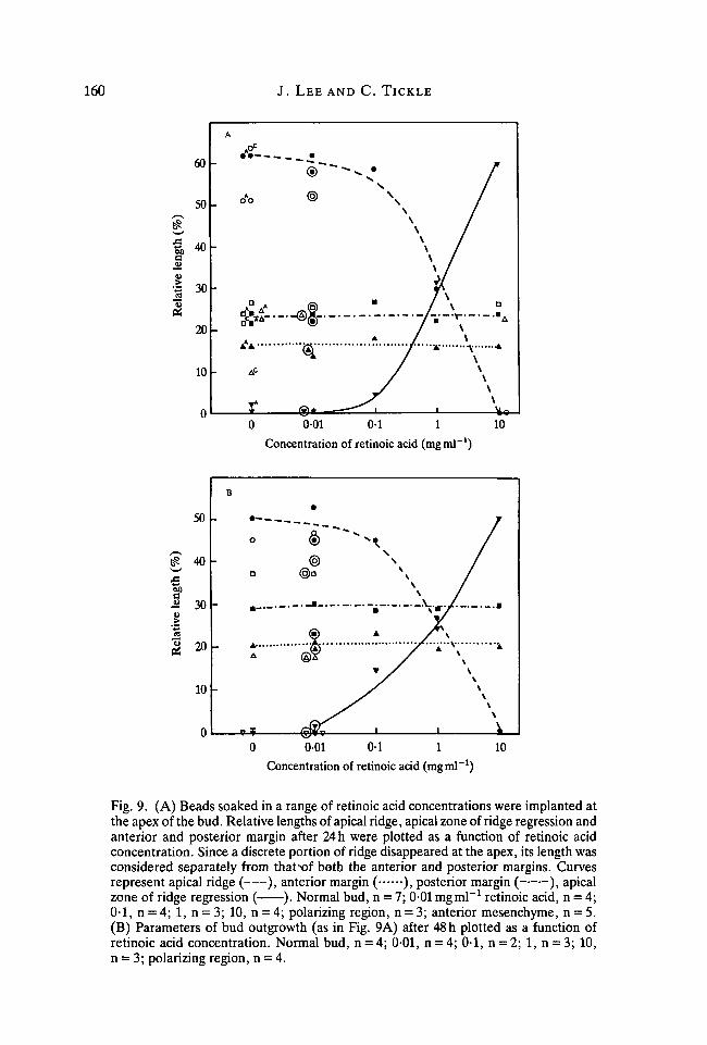
160 J. LEE AND C. TICKLE
0 001 01 1 10
Concentration of retinoic acid (mgml"1)
0 001 01 1
Concentration of retinoic acid (mgmT1)
10
Fig. 9. (A) Beads soaked in a range of retinoic acid concentrations were implanted atthe apex of the bud. Relative lengths of apical ridge, apical zone of ridge regression andanterior and posterior margin after 24 h were plotted as a function of retinoic acidconcentration. Since a discrete portion of ridge disappeared at the apex, its length wasconsidered separately from thatxrf both the anterior and posterior margins. Curvesrepresent apical ridge ( ), anterior margin ( ), posterior margin ( ), apicalzone of ridge regression ( ). Normal bud, n = 7; 0-01 mgml"1 retinoic acid, n = 4;0-1, n = 4; 1, n = 3; 10, n = 4; polarizing region, n = 3; anterior mesenchyme, n = 5.(B) Parameters of bud outgrowth (as in Fig. 9A) after 48 h plotted as a function ofretinoic acid concentration. Normal bud, n = 4; 0-01, n = 4; 0-1, n = 2; 1, n = 3; 10,n = 3; polarizing region, n = 4.
Effects of retinoic acid on wing bud outgrowth 161
leng
th (
%R
elat
ive
60
50
40
30
20
10
n
A
-
-
-
-
-
•_
• c
o
a
A
A '•••..,
1
•
A
A-*"'
1
DNN y
.*'' o s»AA
0 001 01 1 10Concentration of retinoic acid (mgml"1)
0 001 01 1Concentration of retinoic acid (rngml"1)
Fig. 10. (A) Beads soaked in a range of retinoic acid concentrations were implanted atthe posterior of the bud. Relative lengths of apical ridge anterior and posterior marginafter 24 h were plotted as a function of retinoic acid concentration. Curves representapical ridge ( ), anterior margin ( ), posterior margin ( ). Normal bud,n = 7; 0-01 mgml"1 retinoic acid, n = 3; 0-1, n = 4; 1, n = 5; 10, n = 5; DMSO, n = 4;polarizing region, n = 4; anterior mesenchyme, n = 3. (B) Parameters of budoutgrowth (as in Fig. 10A) after 48 h plotted as a function of retinoic acidconcentration. Normal bud, n = 4; 0-01, n = 5; 0-1, n = 5; 1, n = 6; 10, n = 7.

162 J. LEE AND C. TICKLE
following retinoic acid application. The changes in bud form are brought about byalterations in the extent of the apical ridge and its position around the budperimeter. These alterations are detected at 24 h of treatment. They aresubsequently modified and growth takes place so that by 48 h clearly different budforms result. The changes in the extent of the apical ridge have been documentedquantitatively and dose-response curves obtained.
To summarize, we will consider first the effects of application of retinoic acid tothe anterior of the bud. With low doses, the length of the ridge increases.However, since at 24 h the perimeter of the bud has also increased, the proportionof the perimeter occupied by the ridge is not significantly changed. By 48 h, thelength of the ridge has increased still further and now occupies a larger proportionof the bud perimeter. The increase in ridge length is due to its extension along theanterior margin of the bud. These changes lead to additional outgrowth anteriorly.With application of higher doses of retinoic acid, the length of the ridge, instead ofincreasing, becomes restricted to more posterior parts of the bud. Outgrowth isthen confined to these narrow posterior portions.
When retinoic acid is applied at the apex or to the posterior margin, similardose-dependent changes in the extent of the apical ridge are seen at 24 h but theregion affected depends on the position of application. With application of lowdoses of retinoic acid at the apex, the proportion of the bud perimeter occupied bythe ridge is unchanged. The ridge, although not significantly longer as withanterior application, similarly tends to persist over anterior parts of the bud.This leads by 48 h to additional outgrowth anteriorly and a bilobed bud. Withapplication of high doses, the length of the ridge decreases as it does followinganterior application. However, since the ridge is obliterated, first near the implantand then at progressively greater distances away, the ridge first disappears at theapex and then the ridge both anterior and posterior to the implant is progressivelyaffected. The total obliteration of the ridge that soon results halts outgrowth.
When low doses of retinoic acid are applied posteriorly, the ridge becomesshorter and confined to more anterior parts of the bud. This progressive restrictionof the ridge is, at 24 h, a mirror image of that following anterior application. At48 h, an almost normally proportioned ridge has been re-established. The anteriormargin is progressively reduced as the ridge is shifted more anteriorly.
The apical ridge and bud shape
One interesting observation is relevant to the possible role of the apical ridge inshaping the contours of the bud. It was seen that where the apical ridge haddisappeared, the bud bulged instead of being dorsoventrally flattened. Althoughthe overall shape of the bud will be the net result of forces generated by both themesenchyme and the ectoderm, this observation suggests that the ridge acts as aseam to maintain the shape of the ectodermal casing of the bud (Hornbruch &Wolpert, 1970). This is in contrast to the view that the mesoderm is the mostimportant determinant of bud shape (Ede, 1971).
Effects of retinoic acid on wing bud outgrowth 163
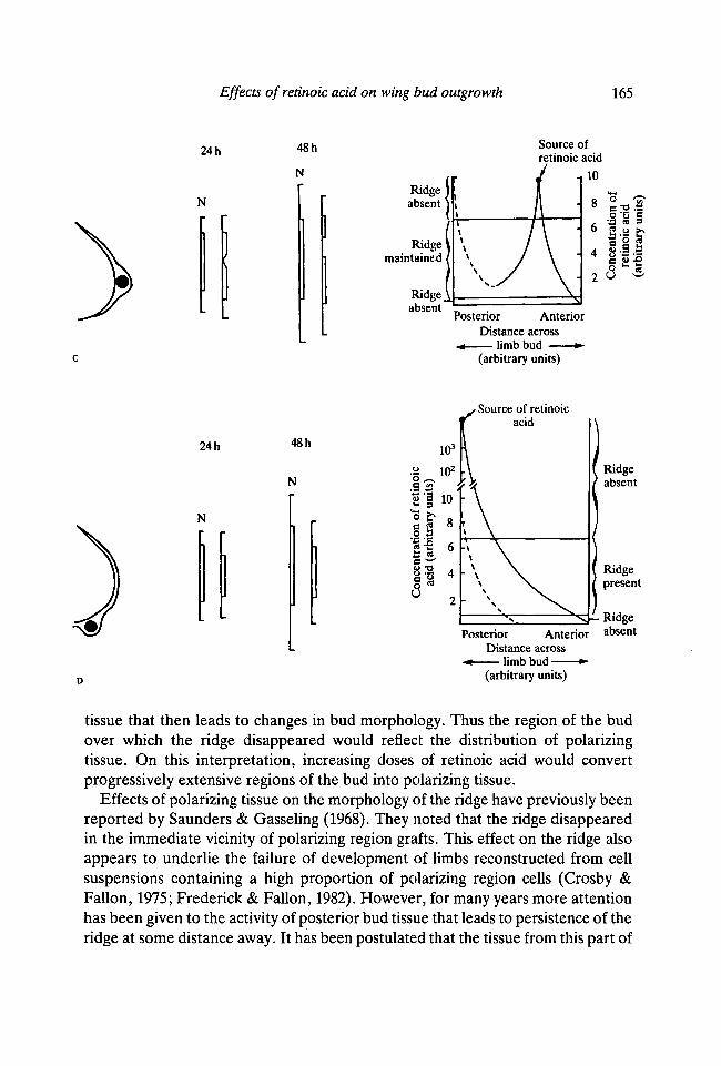
A model to account for the effects of retinoic acid on the extent of the ridge
Changes in the extent and disposition of the ridge can be interpreted in terms ofa graded effect of retinoic acid. We have already shown that when a bead soaked inretinoic acid is implanted at the anterior margin of the bud, a concentrationgradient is soon established across the bud tissue (Tickle et al. 1985). A similargradient is also established when the bead is implanted posteriorly. Thus, wepropose that the effects on the apical ridge reflect dose-dependent responses to thelocal concentration of retinoic acid that varies with distance from the source. Athigh concentrations, above a certain threshold concentration, the ridge woulddisappear but at lower concentrations of retinoic acid the ridge would persist. Atextremely low concentrations, the effects on the ridge are negligible and it is notmaintained. The model is shown in Fig. 11 and can account for all the main effectsof retinoic acid found here. These include the apparently paradoxical effect of firstthe increase and then the gradual decrease in ridge length found when beadssoaked in progressively higher concentrations of retinoic acid are implantedanteriorly. Furthermore, the model illustrates why the ridge is confined todifferent regions of the bud perimeter when the position of application is varied.
The changes in the extent of apical ridge give rise to the different forms of budoutgrowth. An increase in extent of the apical ridge leads to a broadenedoutgrowth and a decrease to a narrower one. Complete absence of the apical ridgewould lead to truncations. In this respect, it is interesting that the apical ridge oflimb buds of mouse embryos was reported to disappear when the embryos weresystematically treated with vitamin A at doses which lead to truncations (Yasuda&Nakamura, 1983).
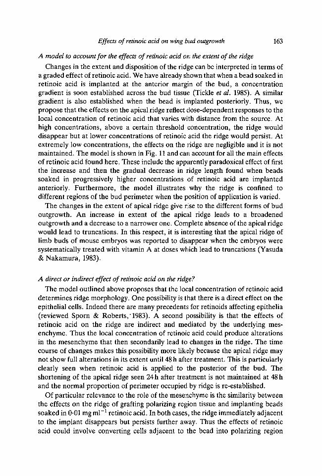
A direct or indirect effect of retinoic acid on the ridge?
The model outlined above proposes that the local concentration of retinoic aciddetermines ridge morphology. One possibility is that there is a direct effect on theepithelial cells. Indeed there are many precedents for retinoids affecting epithelia(reviewed Sporn & Roberts,'1983). A second possibility is that the effects ofretinoic acid on the ridge are indirect and mediated by the underlying mes-enchyme. Thus the local concentration of retinoic acid could produce alterationsin the mesenchyme that then secondarily lead to changes in the ridge. The timecourse of changes makes this possibility more likely because the apical ridge maynot show full alterations in its extent until 48 h after treatment. This is particularlyclearly seen when retinoic acid is applied to the posterior of the bud. Theshortening of the apical ridge seen 24 h after treatment is not maintained at 48 hand the normal proportion of perimeter occupied by ridge is re-established.
Of particular relevance to the role of the mesenchyme is the similarity betweenthe effects on the ridge of grafting polarizing region tissue and implanting beadssoaked in 0-01 mg ml"1 retinoic acid. In both cases, the ridge immediately adjacentto the implant disappears but persists further away. Thus the effects of retinoicacid could involve converting cells adjacent to the bead into polarizing region

164 J. LEE AND C. TICKLE
24 h
N
48hN
Ridgeabsent
Ridgemaintained
Source ofretinoic acid
Posterior AnteriorDistance across
•+ limb bud •(arbitrary units)
24 h
N
48 h
N
Ridgeabsent
Ridgemaintained
Ridge^absent
iource ofretinoic acid104
Posterior AnteriorDistance across
« limb bud •(arbitrary units)
Fig. 11. Diagrams to illustrate the effects of retinoic acid on the extent and position ofthe apical ridge and a model that accounts for these changes. On the left is depicted theposition of the bead immediately after, implantation. Next to this are linearrepresentations of the bud perimeter drawn to scale (1:10) showing the extent andposition of the apical ridge (indicated by boxed section) at the times shown. Forcomparison similar representations of normal buds (N) are shown alongside. On theright is shown diagrammatically the gradient of retinoic acid (solid line) that isestablished across the bud. The horizontal lines represent the thresholds for the limitsof the ridge. The dotted curve represents the gradient of the hypothetical morphogenfrom the polarizing region. (A) Anterior application of low concentrations of retinoicacid. (B) Anterior application of high concentrations of retinoic acid. (C) Apicalapplication of low concentrations of retinoic acid. (D) Posterior application of highconcentrations of retinoic acid. Note that the linear diagrams are aligned byv theirmidpoints and this makes the anterior shift of the ridge less obvious.
Effects of retinoic acid on wing bud outgrowth 165
24 h
N
48 h
NRidgeabsent \
Ridgemaintained /
Source ofretinoic acid
10
Posterior AnteriorDistance across
~4 limb bud •(arbitrary units)
24 h
N
48 h
N
• Source of retinoicacid
Ridgeabsent
PosteriorDistance across
•+ limb bud —(arbitrary units)
Ridgepresent
i-RidgeAnterior absent
tissue that then leads to changes in bud morphology. Thus the region of the budover which the ridge disappeared would reflect the distribution of polarizingtissue. On this interpretation, increasing doses of retinoic acid would convertprogressively extensive regions of the bud into polarizing tissue.
Effects of polarizing tissue on the morphology of the ridge have previously beenreported by Saunders & Gasseling (1968). They noted that the ridge disappearedin the immediate vicinity of polarizing region grafts. This effect on the ridge alsoappears to underlie the failure of development of limbs reconstructed from cellsuspensions containing a high proportion of polarizing region cells (Crosby &Fallon, 1975; Frederick & Fallon, 1982). However, for many years more attentionhas been given to the activity of posterior bud tissue that leads to persistence of theridge at some distance away. It has been postulated that the tissue from this part of
166 J. LEE AND C. TICKLE
the bud produces a diffusible substance that has been called the apical ridgemaintenance factor (Zwilling, 1956; Saunders & Gasseling, 1968). More recently,studies in culture have demonstrated a long-range effect of polarizing tissue onridge morphology. When anterior tips of wing buds are cultured in contact withpolarizing tissue, the apical ridge over the anterior part of the bud maintains itsthickened morphology (MacCabe & Parker, 1975). Furthermore, this effect on theridge was produced even when the polarizing tissue was not in direct contact withthe responding tissue. Some progress has been made in purifying the factornecessary to maintain the ridge (MacCabe, Leal & Leal, 1983).
The experimental effects of polarizing tissue on the apical ectodermal ridgedraw attention to the relationship between the two in the normal bud. The apicalridge is absent in the immediate vicinity of the polarizing region. Thus thepolarizing region in the normal bud appears to define the posterior margin ofoutgrowth. The apical ridge is thickened over the posterior part of the budadjacent to the polarizing region. Furthermore, there is some evidence for agraded effect on ridge morphology over the posterior part of the bud; the thicknessof the ridge varies with distance from the polarizing region (Todt & Fallon, 1984).However, the ridge is not maintained over the anterior part of the bud. We suggestthat this is due to the tissue being out of range of influence of the polarizing regionrather than the presence of an anterior boundary region.
Interaction between signal generated by retinoic acid application and signal from hostpolarizing region
To appreciate the effects of retinoic acid application on bud outgrowth,interaction with the host polarizing region must be considered (see Fig. 11). Thus,the effects of retinoic acid that lead to ridge persistence are always seen at theanterior of the bud since the ridge here is not maintained by the host polarizingregion. For example, with anterior application of low doses of retinoic acid, thetotal length of the ridge can increase because of summation of the lengthmaintained as a result of retinoic acid release from the bead and that maintainedby host polarizing tissue. A second example concerns the effect of retinoic acidwhen applied posteriorly over a fairly wide range of concentrations; again theridge persists over the anterior parts of the bud. In this case, however, the totallength of the ridge does not increase because the high concentrations posteriorlyapparently override the maintenance effect of the host polarizing region and leadto disappearance of the ridge posteriorly. The displacement of the ridge anteriorlyis allowed because the anterior limit of the ridge is not fixed by a boundary region.The ridge is therefore free to extend along the anterior margin as the posteriorboundary is shifted anteriorly. These considerations provide some insights intowhy anterior application of retinoic acid can lead to the development of additionaldigits whereas posterior application almost always gives normal wings.
The phenomenon of high concentrations of retinoic acid overriding themaintenance activity of the host polarizing region is also seen when high
Effects ofretinoic acid on wing bud outgrowth 167
concentrations are applied anteriorly or at the apex. In these cases displacement ofthe ridge posteriorly cannot occur because of the boundary specified by thepolarizing region. The apparent synergism between the effects of the local sourceof retinoic acid and the polarizing region suggest that the same signals areinvolved: either the polarizing region produces retinoic acid (or a related retinoid)or retinoic acid converts cells adjacent to the source into polarizing tissue that thenproduces the appropriate signal.
Establishment of a posterior boundary for limb outgrowth
The changes in the extent of the apical ridge induced by local application ofretinoic acid appear to do more than just mark the limits of bud outgrowth. Insome way, new posterior boundaries are defined near the retinoic acid implant,just as in the normal bud the polarizing region is sited at its posterior margin. Anattractive hypothesis is that the same signalling mechanism controls both theextent of the ridge and pattern formation across the anteroposterior axis. Thusthe apical ridge maintenance factor might be equivalent to the hypotheticalmorphogen produced by the polarizing region (reviewed Tickle, 1980). In thiscontext, the ideas of Meinhardt (1984) are relevant. He has proposed thatboundaries act as organizing regions for pattern formation. At the cellular level, itmay be significant that the cells of the apical ridge are extensively linked by gapjunctions (Kelley & Fallon, 1976; Fallon & Kelley, 1978). Thus cell-cellcommunication along the anteroposterior axis might be mediated via the ridgecells. When the ridge disappears or becomes thinner, as, for example, at the apexof the bud as a result of grafting polarizing tissue here or implanting beads soakedin retinoic acid, this pathway of communication could be interrupted. One canspeculate that this might be an important factor in establishing a boundary region.
This work was supported by the MRC. We thank Professor N. Woolf, The Bland SuttonInstitute of Pathology, The Middlesex Hospital Medical School for the use of the scanningelectron microscope, P. Rowles for technical advice, and Professor L. Wolpert, S. E. Wedden,Professor B. Alberts, and Dr G. Eichele for reading the manuscript.
REFERENCESCROSBY, G. M. & FALLON, J. F. (1975). Inhibitory effect on limb morphogenesis by cells of the
polarising zone coaggregated with pre- or post-axial wing bud mesoderm. Devi Biol. 46,28-39.
EDE, D. A. (1971). Control of form and pattern in the vertebrate limb. In Control Mechanisms ofGrowth and Differentiation (ed. D. D. Davies & M. Balls) pp. 235-254. Cambridge:Cambridge University Press.
EICHELE, G., TICKLE, C , & ALBERTS, B. M. (1984). Micro-controlled release of biologically activecompounds in chick embryos: Beads of 200 jum diameter for the local release of retinoids.Analytical Biochemistry 142, 542-555.
EICHELE, G., TICKLE, C. & ALBERTS, B. M. (1985). Studies on the mechanism of retinoid-inducedpattern duplications in the early chick limb bud: temporal and spatial aspects. /. Cell Biol. (inpress).
168 J. L E E AND C. T ICKLE
FALLON, J. F. & KELLEY, R. (1977). Ultrastructural analysis of the apical ectodermal ridge duringvertebrate limb morphogenesis ii. Gap junctions as distinctive ridge structures common tobirds and mammals. /. Embryol. exp. Morph. 41, 223-232.
FREDERICK, J. M. & FALLON, J. F. (1982). The proportion and distribution of polarising zone cellscausing morphogenetic inhibition when coaggregated with anterior half wing mesoderm inrecombinant limbs. /. Embryol. exp. Morph. 67, 13-25.
HONIG, L. S. (1983). Polarising activity of the avian limb examined on a cellular basis. In LimbDevelopment and Regeneration (ed. J. F. Fallon & A. I. Caplan), Part A, pp. 99-108. NewYork: Alan R. Liss, inc.
HORNBRUCH, A. & WOLPERT, L. (1970). Cell division in the early growth and morphogenesis of thechick limb. Nature, Lond. 226, 764-766.
KARNOVSKY, M. J. (1965). A formaldehyde glutaraldehyde fixative of high osmolarity for use inelectron microscopy. /. Cell Biol. 27, 137a (Abstr.).
KELLEY, R. O. & FALLON, J. F. (1976). Ultrastructural analysis of the apical ectodermal ridgeduring vertebrate limb morphogenesis. I. The human forelimb with special reference to gapjunctions. Devi Biol. 51, 241-256.
MACCABE, J. A., LEAL, K. W. & LEAL, C. W. (1983). The control of axial polarity: A. A lowmolecular weight morphogen affecting the ectodermal ridge. In Limb Development andRegeneration (ed. J. F. Fallon & A. I. Caplan), Part A, pp. 237-244. New York: Alan R. Liss,inc.
MACCABE, J. A. & PARKER, B. W. (1975). The In vitro maintenance of the apical ectodermalridge of the chick embryo wing bud: An assay for polarising activity. Devi Biol. 45, 349-357.
MACCABE, J. A. & RICHARDSON, K. E. (1982). Partial characterisation of a morphogenetic factorin the developing chick limb. /. Embryol. exp. Morph. 67,1-12.
MEINHARDT, H. (1984). A boundary model for pattern formation in vertebrate limbs. /. Embryol.exp. Morph. 76, 115-137.
SAUNDERS, J. W. (1948). The proximo-distal sequence of origin of parts of the chick wing and therole of the ectoderm. /. exp. Zool. 108, 363-403.
SAUNDERS, J. W. & GASSELING, M. T. (1968). Ectodermal-mesenchymal interactions in the originof limb symmetry. In Epithelial-Mesenchymal Interactions (ed. R. Fleischmajer & R.. E.Billingham), pp. 78-97. Baltimore: Williams & Wilkins.
SMITH, J. C. & WOLPERT, L. (1981). Pattern formation along the antero-posterior axis of the chickwing: the increase in width following a polarising region graft and the effect of X-irradiation./. Embryol. exp. Morph. 63,127-144.
SPORN, M. B. & ROBERTS, A. B. (1983). Role of retinoids in differentiation and carcinogenesis.Cancer Research 43, 3034-3040.
SUMMERBELL, D. (1974). A quantitative analysis of the effect of excision of the apical ectodermalridge from the chick limb bud. /. Embryol. exp. Morph. 32, 651-660.
SUMMERBELL, D. (1983). The effect of local application of retinoic acid to the anterior margin ofthe developing chick limb. /. Embryol. exp. Morph. 78, 269-289.
SUMMERBELL, D. & HARVEY, F. (1983). Vitamin A and the control of pattern in developing limbs.In Limb Development and Regeneration (ed. J. F. Fallon & A. I. Caplan), part A, pp. 109-118.New York: Alan R. Liss, inc.
TICKLE, C. (1980). The polarising region and limb development. In Development in Mammals,vol. 4 (ed. M. H. Johnson), pp. 101-136. Amsterdam: Elsevier/North-Holland BiomedicalPress.
TICKLE, C. (1981). The number of polarising region cells required to specify additional digits inthe developing chick wing. Nature, Lond. 289, 295.
TICKLE, C. SUMMERBELL, D. & WOLPERT, L. (1975). Positional signalling and specification of digitsin chick limb morphogenesis. Nature, Lond. 254,199-202.
TICKLE, C , ALBERTS, B., WOLPERT, L. & LEE, J. (1982). Local application of retinoic acid to thelimb bud mimics the action of the polarising region. Nature, Lond. 296, 564-565.
TICKLE, C , LEE, J. &EICHELE, G. (1985). A quantitative analysis of the effect of all-fnz/w-retinoicacid on the pattern of chick wing development. Devi Biol. 109, 82-95.

Effects of retinoic acid on wing bud outgrowth 169
TODT, W. & FALLON, J. F. (1984). Development of the apical ridge in the chick wing bud.J. Embryol. exp. Morph. 80, 21-41.
WOLPERT, L. & HORNBRUCH, A. (1981). Positional signalling along the antero-posterior axis of thechick wing. The effect of multiple polarising region grafts. /. Embryol. exp. Morph. 63,145-159.
YASUDA, M. & NAKAMURA, H. (1983). Pathogenesis of limb malformations in mice: An electronmicroscopic study. In Limb Development and Regeneration (ed. J. F. Fallon & A. I. Caplan),part A, pp. 301-310. New York: Alan R. Liss, inc.
ZWILLING, E. (1956). Interaction between limb bud ectoderm and mesoderm in the chick embryo.iv. Experiments with a wingless mutant. /. exp. Zool. 132, 241-253.
{Accepted 10 June 1985)
