rev iss web jpy 12174-13-086 50-2 388. · dinoflagellate scrippsiella nutricula, a species...
TRANSCRIPT

BRANDTODINIUM GEN. NOV. AND B. NUTRICULA COMB. NOV. (DINOPHYCEAE), ADINOFLAGELLATE COMMONLY FOUND IN SYMBIOSIS WITH POLYCYSTINE
RADIOLARIANS1
Ian Probert2
UPMC-CNRS, FR2424, Roscoff Culture Collection, Station Biologique de Roscoff, Place Georges Teissier, Roscoff 29682, France
Raffaele Siano
IFREMER, Centre de Brest, DYNECO/Pelagos, ZI de la Pointe du Diable CS 170, Plouzan�e 29280, France
Camille Poirier, Johan Decelle, Tristan Biard
UPMC-CNRS, UMR 7144, Station Biologique de Roscoff, Place Georges Teissier, Roscoff 29682, France
Akihiro Tuji
Department of Botany, National Museum of Nature and Science, 4-1-1 Amakubo, Tsukuba 305-0005, Japan
Noritoshi Suzuki
Institute of Geology and Paleontology, Graduate School of Science, Tohoku University, Sendai 980-8578, Japan
and Fabrice Not
UPMC-CNRS, UMR 7144, Station Biologique de Roscoff, Place Georges Teissier, Roscoff 29682, France
Symbiotic interactions between pelagic hosts andmicroalgae have received little attention, although theyare widespread in the photic layer of the world ocean,where they play a fundamental role in the ecologyof the planktonic ecosystem. Polycystine radiolarians(including the orders Spumellaria, Collodaria andNassellaria) are planktonic heterotrophic protists thatare widely distributed and often abundant in theocean. Many polycystines host symbiotic microalgaewithin their cytoplasm, mostly thought to be thedinoflagellate Scrippsiella nutricula, a species originallydescribed by Karl Brandt in the late nineteenthcentury as Zooxanthella nutricula. The free-living stageof this dinoflagellate has never been characterized interms of morphology and thecal plate tabulation. Weexamined morphological characters and sequencedconservative ribosomal markers of clonal culturesof the free-living stage of symbiotic dinoflagellatesisolated from radiolarian hosts from the threepolycystine orders. In addition, we sequencedsymbiont genes directly from several polycystine-symbiont holobiont specimens from different oceanicregions. Thecal plate arrangement of the free-livingstage does not match that of Scrippsiella or relatedgenera, and LSU and SSU rDNA-based molecularphylogenies place these symbionts in a distinct cladewithin the Peridiniales. Both phylogenetic analyses andthe comparison of morphological features of culture
strains with those reported for other closely relatedspecies support the erection of a new genus that wename Brandtodinium gen. nov. and the recombinationof S. nutricula as B. nutricula comb. nov.
Key index words: dinoflagellate; Peridiniales; polycys-tines; Radiolaria; Scrippsiella; symbiosis; taxonomy;Zooxanthella
Abbreviations: ICBN, International Code for Botani-cal Nomenclature; ITS, internal transcribed spacer;LM, light microscopy; LSU, large subunit (ribosomalDNA); ML, maximum likelihood; PCR, polymerasechain reaction; RCC, roscoff culture collection;SEM, scanning electron microscopy; SSU, small sub-unit (ribosomal DNA)
Mutualistic associations involving photosyntheticmicroalgae are common in both benthic and pela-gic ecosystems and are essential for establishing andmaintaining the structure of marine communities(Caron 2000). Symbiosis between corals and thedinoflagellate genus Symbiodinium Freudenthal isfundamental for the survival and ecological successof coral reef ecosystems. Members of the genusSymbiodinium have been intensively studied withrespect to their morphology and life cycle (Freuden-thal 1962, Fitt and Trench 1983, Trench and Blank1987), and genetic diversity (Coffroth and Santos2005, Sampayo et al. 2009, LaJeunesse and Thornhill2011, Stat et al. 2011). Studies on this coastal ben-thic symbiotic relationship significantly increased
1Received 21 May 2013. Accepted 27 August 2013.2Author for correspondence: e-mail [email protected] Responsibility: C. Lane (Associate Editor)
J. Phycol. 50, 388–399 (2014)© 2014 Phycological Society of AmericaDOI: 10.1111/jpy.12174
388

when the coral-bleaching phenomenon was broughtto global attention and associated to increases insea surface temperature, enhanced light intensity,and ocean acidification (Hoegh-Guldberg et al.2007).
Symbiotic interactions between pelagic hosts andmicroalgae have received less attention, althoughthey are widespread in the photic layer of the worldocean where they play a fundamental role in theecology of the planktonic ecosystem (Stoecker et al.2009, Decelle et al. 2012a,b). Recent studies havedemonstrated that dinoflagellate symbionts of Fora-minifera belong to Pelagodinium Siano, Montresor,Probert et de Vargas, a genus that is related toSymbiodinium within the order Suessiales (Sianoet al. 2010), and that Acantharia typically associatewith members of the prymnesiophyte genus Phaeo-cystis Lagerheim (Decelle et al. 2012a,b), althoughone taxon, Acanthochiasma sp., can contain multiplesymbiotic partners, including distantly related dino-flagellates (from the genera Pelagodinium, Hetero-capsa Stein, Azadinium Elbr€achter et Tillmann andScrippsiella Balech ex Loeblich III) as well as a ha-ptophyte (Decelle et al. 2012b).
Polycystine radiolarians (including the ordersSpumellaria, Collodaria, and Nassellaria) are single-celled, heterotrophic, biomineralizing planktonicprotists from the Rhizaria lineage that are widelydistributed in the ocean and are found throughoutthe entire water column (Boltovskoy et al. 2010).Many polycystines host microalgae within their cyto-plasm (Anderson 1983). Cells containing photosyn-thetic microalgae have been shown to survive forlonger periods in nutrient-poor water than thosethat do not have microalgal partners and the micro-algae are therefore assumed to be symbionts thatplay a nutritive role for the hosts (Anderson 1983).
Polycystines form associations with various dinofla-gellate, prymnesiophyte and prasinophyte partners(usually not at the same time), with dinoflagellatesbeing the most common symbiotic partners (Ander-son 1976, 1983, Anderson et al. 1983). In the latenineteenth century, Karl Brandt was the first to rec-ognize that the “yellow cells” within polycystines,actinian corals and hydrozoans were microalgae,which he collectively described in the new genusZooxanthella Brandt (Brandt 1881), although theywere not immediately recognized as dinoflagellates.Soon afterward, the species Z. nutricula Brandt wasproposed for the symbiont of the collodarian poly-cystine Collozoum inerme collected from the westernMediterranean Sea and it was stated in the descrip-tion that this species was presumably identical tothe yellow cells of other polycystines (Brandt 1882).The subsequent taxonomic history of this genus andspecies have been very confused (see review byBlank and Trench 1986), and the plural noun “zoo-xanthellae” has persisted as a colloquialism usedto describe marine microalgal endosymbionts ingeneral.
The symbionts of the “by-the-wind sailor”hydrozoan jellyfish Velella velella were reported to besimilar to those of polycystines by Hovasse (1922),who initially described the in hospite symbionts ofMediterranean V. velella as Endodinium chattoniHovasse (E. chattonii under International Code forBotanical Nomenclature (ICBN) Art. 73). Taylor(1971) and Hollande and Carr�e (1974) furthercharacterized the in hospite stage of E. chattonii andthe latter authors proposed the reclassification ofthe polycystine symbionts (Z. nutricula) as E. nutrico-la (Brandt) Hollande et Carr�e (E. nutricula underICBN Art. 73), although Hovasse (1924) had in factpreviously recombined E. chattonii as Z. chattonii(Hovasse) Hovasse. Banaszak et al. (1993) isolated aculture of the symbiont of V. velella from the Pacific,which they considered slightly different fromE. chattonii (larger cell size and presence of trich-ocysts in hospite and in culture). Based on scanningelectron microscopy (SEM) observations of the mor-phology and arrangement of thecal plates in themotile stage, Banaszak et al. (1993) classified theirorganism in the genus Scrippsiella as a new species,S. velellae Banaszak, Iglesias-Prieto et Trench (aname later validated by Trench 2000). Theseauthors also transferred E. chattonii and E. nutriculato Scrippsiella as S. chattonii (Hovasse) Banaszak, Igle-sias-Prieto et Trench, and S. nutricula (Brandt) Ban-aszak, Iglesias-Prieto et Trench, respectively(Banaszak et al. 1993), but these names remaintechnically invalid because reference was not madeto the exact page of the basionym.Using molecular methods, Gast and Caron (1996)
found that the dinoflagellate symbionts in six differ-ent polycystine species from the Sargasso Sea (thecollodarians Collozoum caudatum and Thalassicollanucleata, three unidentified collodarian species andthe spumellarian Spongostaurus sp.) had identicalSSU rDNA sequences that they assigned to S. nutri-cula. These molecular analyses indicate that taxo-nomically divergent radiolarians can contain thesame symbiotic dinoflagellate. Since these analyseswere conducted directly on symbionts extractedfrom the hosts (i.e., not cultured), the morphologyof the motile stage of the symbiotic algae assignedto S. nutricula was not investigated, and has stillnever been reported. Gast and Caron (1996) alsosequenced the SSU rDNA of the symbiont of V. vel-ella from the Sargasso Sea and found that thesequence was very similar to those of the radiolariansymbionts (four differences out of 1,802 base pairs).They therefore also assigned this V. velella symbiontto S. nutricula.Here, we examined the morphology and molecular
phylogenetic position of clonal cultures of the free-living stage of dinoflagellates isolated from severaldifferent polycystine radiolarian hosts, includingCollozoum, the taxon from which Z. nutricula was orig-inally described. In addition, we sequenced symbiontgenes directly from several polycystine-symbiont
BRANDTODINIUM NUTRICULA GEN. NOV. , COMB. NOV. 389

holobiont specimens (including collodarian, spumel-larian and nassellarian hosts) from different oceanicregions. Accurate morpho-molecular characterizationand taxonomic designation of symbionts from thegenus Symbiodinium has been key for studies of theecology and functioning of coral reef systems and it islikewise likely to prove important for future studieson the widespread pelagic symbiosis involving poly-cystine radiolarian hosts.
MATERIAL AND METHODS
Samples and culture isolation. The radiolarian specimensfrom which the holobiont sequences or cultures originatedwere isolated from samples collected in 2010–2012 by nettows (20–150 lm mesh size) in the bay of Villefranche-sur-Mer (France), off Sesoko Island, Okinawa (Japan) and in theSouth Pacific Ocean during the Tara Oceans expedition(Table 1; Figs. S1 and S2 in the Supporting Information).The polycystines were first sorted from fresh net samplesunder a binocular microscope, cleaned by successive transfersin sterile seawater in petri dishes, and then left in an illumi-nated and temperature-regulated incubator for several hoursto self-clean. Individual clean specimens were then identifiedbased on their morphology and imaged under an invertedmicroscope. Some specimens were then transferred to guan-idinium isothiocyanate buffer for direct DNA extraction fromholobionts. The dinoflagellate cultures were obtained bymicropipette isolation of single symbiont cells released fromlive radiolarian specimens that were microdissected under aninverted microscope. The resulting monoclonal cultures weremaintained in filter-sterilized seawater with K/2(-Tris, -Si)medium supplements (Keller et al. 1987) at 22°C with anirradiance of 70–80 lmol photons � m�2 � s�1 in a 12:12light:dark regime. The cultures have been deposited in theRoscoff Culture Collection (RCC; http://www.roscoff-culture--collection.org). Light microscopy (LM) images of radiolarianholobionts from which sequences/cultures were obtained areshown in Figures S1 and S2. Detailed information related toeach of the samples used in this study can be found in theRENKAN database at http://abims.sb-roscoff.fr/renkan/.
Microscopy preparations and observations. Light micrographsof living cells were taken using a Zeiss Axiophot light micro-scope equipped with a Zeiss AxioCam digital camera system(Carl Zeiss, Oberkochen, Germany). For SEM, dinoflagellatecells were fixed in 1% (v:v) formol for 2 h at room tempera-ture. Samples were then gently filtered onto 3 lm pore-sizeNucleopore polycarbonate filters (Pleasanton, CA, USA),washed with distilled water, dehydrated in an ethanol series(25%, 50%, 75%, 95%, 100%), and critical-point-dried. Thefilters were mounted on stubs, sputter coated with gold, andexamined with a FEI QuantaTM 200 SEM (FEI, Hillsboro, OR,USA).
DNA extraction, sequencing, and phylogenetic analyses. Geno-mic DNA was extracted from exponentially growing culturesof the strains using a NucleoSpin Plant II DNA extraction kit(Macherey-Nagel), or from holobionts using the methoddescribed in De Vargas et al. (2002).
Partial nuclear large subunit (LSU) and small subunit(SSU) rDNA genes were polymerase chain reaction (PCR)amplified using Phusion high-fidelity DNA polymerase(Finnzymes, Vantaa, Finland) in a 25 lL reaction volume andthe following thermocycler steps: an initial denaturation stepat 98°C for 30 s, followed by 35 cycles at 98°C for 10 s, 30 sat the temperature of semi-hybridization chosen for each setof primers, and 30 s at 72°C, with a final elongation step of10 min at 72°C. The eukaryote primer set 63F (ACGCTT
GTCTCAAAGATT)/1818R (ACGGAAACCTTGTTACGA; Tm50°C; Lepere et al. 2011) was used to amplify the SSU rDNAof the dinoflagellate cultures, whereas the dinoflagellate spe-cific primer set DIN464F (TAACAATACAGGGCATCCAT)/S69 (CCGTCADTTCCTTTRAGDTT; Tm 53°C) was used totarget the dinoflagellates in the holobiont samples. The D1-D2 fragment of the LSU rDNA was amplified using the dino-flagellate specific primers Ldino6 (MCC CGCTGAATTTAAG-CATA)/Ldino1 (AACGATTTGCAGGTCAGTACCGC; Tm55°C) from both cultures and holobionts. PCR products werethen sequenced at the GENOSCOPE (CEA, Evry, France).
The sequences generated from the studied strains andholobionts (GenBank accession numbers KF557491 toKF557545) were aligned with other LSU and SSU rDNAsequences from GenBank (release 194.0, February 2013)attributed to Scrippsiella and related Peridiniales genera, aswell as representatives of the Suessiales as an outgroup. Align-ments were generated using MUSCLE implemented in Sea-view v.4.0 (Gouy et al. 2010) with subsequent manualverification. The LSU rDNA data set contained 48 sequences(675 unambiguously aligned positions) and the SSU rDNAdata set contained 57 sequences (652 unambiguously alignedpositions).
Phylogenetic analyses were conducted with maximum like-lihood (ML) and Bayesian methods. The ML analysis was car-ried out using MEGA v. 5.1 (Tamura et al. 2011) with thegeneral time reversible as the best model of nucleotide substi-tution and considering a gamma distribution with a propor-tion of invariable sites (I) set at 5 by default. Bootstrapsupports for the tree were obtained after 1,000 replicates.Bayesian analyses were conducted using Mr Bayes v.3.2.1(Huelsenbeck and Ronquist 2001) using the same model ofevolution. For each gene marker, two Markov chain MonteCarlo chains were run for 1 million generations, samplingevery 500 generations (diagnostic frequency = 5,000). Thestandard deviation of split frequencies between the 2 runswas <0.01 in both LSU and SSU rDNA analyses. For both MLand Bayesian analyses, the trees were visualized and edited inFig Tree v. 1.3.1 (Rambaut 2010). In the trees presentedherein the posterior probabilities associated to each node inthe Bayesian topologies are reported on the ML topologies.
RESULTS
Microscopy observations. In our culture conditions,the clonal strains of polycystine symbionts tended tocontain a mixture of motile thecate cells and larger,irregularly shaped nonmotile cells devoid of the typ-ical features of motile cells (theca, cingulum, sul-cus), the latter more closely resembling the inhospite symbiotic state. The proportion of motileand nonmotile cells varied between strains andthrough growth cycles for each strain. The overallmorphology and thecal plate pattern of motile cellswas identical for several different strains observed.The following descriptions and illustrations arebased on observations of strain RCC3387.Cells are 10.5–15 lm in length (average 13.1 lm,
n = 30) and 9.1–11.2 lm in width (average 10.4 lm,n = 30). The epitheca is larger than the hypotheca.Observed under LM, cells have a slightly convexconical epitheca with a well-pronounced apical horn(Fig. 1, A, B, D). The hypotheca is rounded (Fig. 1,A and D). The nucleus is large and occupies thecenter of the cells (Fig. 1, B and D). One or two
390 IAN PROBERT ET AL.

TABLE 1. List of specimens used to obtain symbiont sequences (images of host cells are shown in Figs. S1 and S2).
Host ID Host taxonomy Sampling site Strain code
GenBank accession number
SSU rDNA LSU rDNA
HolobiontsPAC1 Collodaria (solitary) South Pacific
21°17.462 S, 105°9.476 Wn.a. KF557503 KF557534
PAC2 Collodaria (colony) South Pacific21°17.462 S, 105°9.476 W
n.a. KF557504 n.a.
PAC3 Collodaria (solitary) South Pacific23°42.949 S, 107°20.141 W
n.a. KF557505 KF557535
PAC4 Collodaria (solitary) South Pacific23°42.949 S, 107°20.141 W
n.a. KF557506 KF557536
PAC6 Collodaria (solitary) South Pacific24°48.085 S, 110°33.307 W
n.a. KF557507 n.a.
PAC7 Collodaria (colony) South Pacific24°48.085 S, 110°33.307 W
n.a. KF557508 n.a.
PAC8 Collodaria (colony) South Pacific24°48.085 S, 110°33.307 W
n.a. n.a. KF557537
PAC9 Collodaria (colony) South Pacific24°48.085 S, 110°33.307 W
n.a. KF557509 KF557538
PAC10 Collodaria (solitary) South Pacific24°48.085 S, 110°33.307 W
n.a. KF557510 n.a.
PAC11 Collodaria (solitary) South Pacific24°23.025 S, 113°58.068 W
n.a. KF557511 KF557539
PAC14 Collodaria (solitary) South Pacific24°23.025 S, 113°58.068 W
n.a. KF557512 KF557540
PAC15 Collodaria (solitary) South Pacific24°23.025 S, 113°58.068 W
n.a. KF557513 n.a.
PAC16 Collodaria (colony) South Pacific24°23.025 S, 113°58.068 W
n.a. KF557514 n.a.
PAC17 Collodaria (colony) South Pacific23°42.289 S, 131°12.744 W
n.a. KF557515 KF557541
PAC19 Collodaria (colony) South Pacific23°42.289 S, 131°12.744 W
n.a. KF557516 KF557542
PAC21 Collodaria (colony) South Pacific23°42.289 S, 131°12.744 W
n.a. KF557517 KF557543
PAC22 Collodaria (colony) South Pacific23°42.289 S, 131°12.744 W
n.a. KF557518 KF557544
PAC24 Collodaria (colony) South Pacific23°42.289 S, 131°12.744 W
n.a. KF557519 n.a.
PAC26 Collodaria (colony) South Pacific23°42.289 S, 131°12.744 W
n.a. KF557520 n.a.
PAC27 Collodaria (colony) South Pacific23°42.289 S, 131°12.744 W
n.a. KF557521 n.a.
SES47 Collodaria (colony) Sesoko, Japan26°370 20 N, 127°520 15 E
n.a. KF557502 KF557546
SES19 Spumellaria Sesoko, Japan26°370 20 N, 127°520 15 E
n.a. KF557501 n.a.
SES28 Nassellaria Sesoko, Japan26°370 20 N, 127°520 15 E
n.a. n.a. KF557545
Vil 210 Spumellaria? Villefranche-sur-Mer, France43°410 10 N, 7°180 50 E
n.a. KF557522 n.a.
Vil 217 Spumellaria Villefranche-sur-Mer, France43°410 10 N, 7°180 50 E
n.a. KF557523 n.a.
Vil 219 Spumellaria Villefranche-sur-Mer, France43°410 10 N, 7°180 50 E
n.a. KF557524 n.a.
Vil 231 Spumellaria Villefranche-sur-Mer, France43°410 10 N, 7°180 50 E
n.a. KF557525 n.a.
Culture strainsSES46 Collodaria (Collozoum colony) Sesoko, Japan
26°370 20 N, 127°520 15 ERCC3378 KF557500 KF557526RCC3379 KF557499 n.a.
VFPO14 Collodaria (Collozoum colony) Villefranche-sur-Mer, France43°410 10 N, 7°180 50 E
RCC3380 KF557494 KF557530RCC3381 KF557495 KF557531RCC3382 KF557496 KF557532
VFPO2 Spumellaria Villefranche-sur-Mer, France43°410 10N, 7°180 50E
RCC3383 KF557491 KF557527RCC3384 n.a. KF557528
BRANDTODINIUM NUTRICULA GEN. NOV. , COMB. NOV. 391
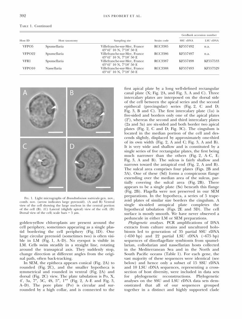
golden-yellow chloroplasts are present around thecell periphery, sometimes appearing as a single plas-tid bordering the cell periphery (Fig. 1D). Onelarge circular pyrenoid (sometimes two) is often visi-ble in LM (Fig. 1, A–D). No eyespot is visible inLM. Cells swim steadily in a straight line, rotatingaround the transapical axis. They suddenly stop,change direction at different angles from the origi-nal path, often back-tracking.
In SEM, the epitheca appears conical (Fig. 2A) torounded (Fig. 2C), and the smaller hypotheca issymmetrical and rounded in ventral (Fig. 2A) anddorsal (Fig. 2C) view. The plate tabulation is Po, X,40, 3a, 7″, 5C, 4S, 5‴, 1″″ (Fig. 2, A–E and Fig. 3,A–D). The pore plate (Po) is circular and sur-rounded by a high collar, and is connected to the
first apical plate by a long well-defined rectangularcanal plate (X; Fig. 2A, and Fig. 3, A and C). Threeintercalary plates are interposed on the dorsal sideof the cell between the apical series and the secondepithecal (precingular) series (Fig. 2, C and D;Fig. 3, B and C). The first intercalary plate (1a) isfive-sided and borders only one of the apical plates(20), whereas the second and third intercalary plates(2a and 3a) are six-sided and both border two apicalplates (Fig. 2, C and D; Fig. 3C). The cingulum islocated in the median portion of the cell and des-cends slightly, displaced by approximately one-thirdof its own width (Fig. 2, A and C; Fig. 3, A and B).It is very wide and shallow and is constituted by asingle series of five rectangular plates, the first beingmuch narrower than the others (Fig. 2, A–C, E;Fig. 3, A and B). The sulcus is fairly shallow andnarrows toward the antapical end (Fig. 2, A and B).The sulcal area comprises four plates (Figs. 2B and3A). One of these (Sd) forms a conspicuous flangeextending over the median area of the sulcus, par-tially covering the sulcal area (Fig. 2B). Thereappears to be a single plate (Ss) beneath this flange(Fig. 2B). Flagella were not preserved in our SEMpreparations. In the hypotheca, a series of 5 trape-zoid plates of similar size borders the cingulum. Asingle six-sided antapical plate completes thehypothecal tabulation (Figs. 2E and 3D). The cellsurface is mostly smooth. We have never observed apeduncule in either LM or SEM preparations.Phylogenetic analyses. PCR amplifications of DNA
extracts from culture strains and uncultured holo-bionts led to generation of 35 partial SSU rDNA(~650 bp) and 22 partial LSU rDNA (~675 bp)sequences of dinoflagellate symbionts from spumel-larian, collodarian and nassellarian hosts collectedin the Mediterranean Sea and in the North andSouth Pacific oceans (Table 1). For each gene, thevast majority of these sequences were identical (seebelow) and hence only a subset of 15 SSU rDNAand 10 LSU rDNA sequences, representing a cross-section of host diversity, were included in data setsfor phylogenetic reconstructions. Phylogeneticanalyses on the SSU and LSU rDNA data sets dem-onstrated that all of our sequences groupedtogether in a distinct and highly supported clade
TABLE 1. Continued
Host ID Host taxonomy Sampling site Strain code
GenBank accession number
SSU rDNA LSU rDNA
VFPO5 Spumellaria Villefranche-sur-Mer, France43°410 10 N, 7°180 50 E
RCC3385 KF557492 n.a.
VFPO22 Spumellaria Villefranche-sur-Mer, France43°410 10 N, 7°180 50 E
RCC3386 KF557497 n.a.
VFR1 Spumellaria Villefranche-sur-Mer, France43°410 10 N, 7°180 50 E
RCC3387 KF557498 KF557533
VFPO10 Nassellaria Villefranche-sur-Mer, France43°410 10 N, 7°180 50 E
RCC3388 KF557493 KF557529
FIG. 1. Light micrographs of Brandtodinium nutricula gen. nov.,comb. nov. (arrow indicates large pyrenoid). (A and B) Ventralview of the cell showing the large nucleus in the central portionof the cell (B). (C) Lateral (slightly apical) view of the cell. (D)Dorsal view of the cell; scale bars = 5 lm.
392 IAN PROBERT ET AL.

(hereafter called clade B) within the dinoflagellateorder Peridiniales (full ML and Bayesian statisticalsupport; Figs. 4 and 5). In both SSU and LSU rDNA
phylogenies, this clade included two distinct subc-lades, B1 and B2, each containing sequences thatare 100% identical irrespective of host taxon andoceanic region. In our SSU rDNA phylogenetic tree(Fig. 4), subclade B1 included the majority of symbi-ont sequences recovered in this study (includingthose from five culture strains isolated from Collo-zoum colonies from the Mediterranean Sea and Paci-fic Ocean), as well as published sequences thatcorrespond to the symbionts of five collodariansand one spumellarian collected in the AtlanticOcean (Gast and Caron 1996). Subclade B2 con-tained the sequences generated in the present studyof the symbionts of two collodarian holobionts aswell as one published sequence (U52357) of thesymbiont of the jellyfish V. velella (Gast and Caron1996). In both phylogenetic reconstructions, themonophyletic clade B containing the sequences ofpolycystine symbionts was phylogenetically distinctfrom the well-supported clade containing membersof the genus Scrippsiella (including the holotype spe-cies S. sweeneyae Loeblich III), but overall the phylo-genetic relationships between clades within thePeridiniales were not clearly resolved in our analy-ses. When sequences of members of the genusBysmatrum, which have a plate tabulation patternsimilar to Scrippsiella-like peridinaleans (Table 2),were included in phylogenetic analyses, they formeda distinct mono-generic clade which fell on a longbranch that altered overall tree topology (Fig. S3 inthe Supporting Information). In the SSU rDNA phy-logeny (Fig. 4), note that the sequence labeled
FIG. 2. SEM micrographs ofBrandtodinium nutricula gen. nov.,comb. nov. (enumeration of platesfollows the Kofoidian tabulationsystem). (A) Ventral view of a cell(flagella lost during fixation). (B)Detail of the sulcal region. (C)Dorsal view. (D) Apical view. (E)Antapical view; scale bars = 2 lm.
FIG. 3. Schematic representation of plate patterns of Brandtodi-nium nutricula gen. nov., comb. nov. (enumeration of plates fol-lows the Kofoidian tabulation system). (A) Ventral view(generalized). (B) Dorsal view (generalized). (C) Apical view(generalized). (D) Antapical view (generalized).
BRANDTODINIUM NUTRICULA GEN. NOV. , COMB. NOV. 393

“uncultured alveolate from Nasselaria” (DQ916409)and the two sequences labeled “Dinophyceae fromCollodaria” (DQ116021 and DQ116022) correspondto nonphotosynthetic dinoflagellate parasites ofRadiolaria (Gast 2006).
DISCUSSION
Dinoflagellates that form symbiotic relationshipswith metazoan or protistan hosts are characterized bycomplex life cycles, with an alternation of symbioticand free-living stages with considerable morphologicaland physiological differentiation between them.Within the host cells, the symbionts are typically coc-coid without flagella, and the cingulum and sulcus areno longer apparent (Trench and Blank 1987). In thefree-living stage, cells tend to regain their original mor-phology (Freudenthal 1962, Spero 1987, Siano et al.2010). Since the taxonomy of dinoflagellates is largelybased on comparison of the number, shape, andarrangement of the thecal plates (or amphiesmal vesi-cles in athecate species) that form the periplast offree-living motile cells, the establishment of clonal cul-tures from symbionts extracted from their hosts is criti-cal for accurate taxonomic assignation.
The genus Zooxanthella was originally created tocollectively describe the symbionts of diverse hosts
from the Mediterranean Sea, including polycystines,corals, and hydrozoans (Brandt 1881) and Z. nutri-cula was created to describe the symbionts of thecollodarian polycystine Collozoum inerme (Brandt1882). The taxonomic history of Zooxanthella hassubsequently been confusing, with Z. nutricula beingalternately combined within Endodinium, Amphidini-um Claper�ede et Lachmann (see review of thenomenclatural history of endosymbiotic dinoflagel-lates by Blank and Trench 1986) and most recently(albeit technically invalidly) within Scrippsiella (Ban-aszak et al. 1993).Our observations of the plate tabulation pattern
of cultured motile cells of the free-living stage ofthe dinoflagellate isolated from diverse polycystinehosts clearly show that it is a member of the orderPeridiniales (bilateral symmetry, cingulum onlyslightly displaced, presence of Po and X plates, pres-ence of 3 intercalary plates in the epitheca) andthat it should not be classified in the genus Scrippsi-ella, nor in the related genera Calciodinellum, Bysma-trum, Pentapharsodinium, or Ensiculifera. All of theselatter genera are described as possessing 2 antapicalplates, whereas the polycystine symbiont reportedhere possesses a single antapical plate (Table 2,Figs. 2E and 3D). The presence of a single antapicalplate is rare in the order Peridiniales, occurring
FIG. 4. Small subunit rDNAphylogenetic tree inferred bymaximum likelihood (ML) analy-sis. 652 unambiguously alignedpositions were considered from analignment of 57 sequences,including Brandtodinium gen. nov.Sequences obtained in this studyare indicated in bold (followed bythe type of host from which thesequence was obtained and thenumber of holobiont specimensor culture strains in parentheses).The tree was rooted withSuessiales (Symbiodinium spp. andPelagodinium b�eii) as the outgroup.Branch lengths are drawn to scale,with the scale bar indicating thenumber of nucleotidesubstitutions per site. Numbers onbranches are statistical supportvalues for the clusters to the rightof them (first: ML bootstrapsupport values, values under 0.5are not shown; second: Bayesianpost-erior probabilities, valuesunder 0.5 are not shown; blackdots at nodes represent a statisticalsupport of 1 for both methods).
394 IAN PROBERT ET AL.

notably in a group of heterotrophic genera (Podo-lampas Stein, Blepharocysta Ehrenberg, and Lissodini-um Matzenauer) characterized by the absence ofboth a cingulum and a depressed sulcus (G�omezet al. 2010) and a group of heterotrophic taxa(Diplopsalis Bergh, Preperidinium Mangin, BoreadiniumDodge et Hermes) characterized by having largelenticular-shaped cells. The radiolarian symbiontsare clearly morphologically and ecologically distinctfrom these other peridinialeans that have a singleantapical plate.
The polycystine symbionts also differ from Scrip-psiella and Bysmatrum (but not from Pentapharsodini-um and Ensiculifera) in possessing 5 (rather than 6)cingular plates. The wing-like flange that covers thesulcal area has not been described in any of theserelated genera. This structure resembles the pedun-cule cover plate (PC) of heterotrophic dinoflagel-lates in the peridinialean family PfiestereaceaeSteidinger et Burkholder emend. Litaker. Motileforms of members of the Pfiestereaceae feed myzo-cytotically by means of a peduncule that emergesclose to the flagella and that can attach to microal-gal prey or epidermal cells of live fish (e.g., Stei-dinger et al. 2006). We have not observed apeduncle in the taxon described here, but should itbe present, the Sd plate should rather be termedPC and the plate formula would become: Po, X, 40,3a, 7″, 5c, 3s, PC, 5‴, 1″″.Comparison of morphological characters
strongly supports a generic level separation of the
FIG. 5. Large subunit rDNAphylogenetic tree inferred bymaximum likelihood (ML) analysis.675 unambiguously alignedpositions were considered from analignment of 48 sequences,including Brandtodinium gen. nov.Sequences obtained in this studyare indicated in bold (followed bythe type of host from which thesequence was obtained and thenumber of holobiont specimens orculture strains in parentheses). Thetree was rooted with Suessiales(Symbiodinium spp. and Pelagodiniumb�eii) as the outgroup. Branchlengths are drawn to scale, with thescale bar indicating the number ofnucleotide substitutions per site.Numbers on branches are statisticalsupport values for the clusters tothe right of them (first: MLbootstrap support values, valuesunder 0.5 are not shown; second:Bayesian posterior probabilities,values under 0.5 are not shown;black dots at nodes represent astatistical support of 1 for bothmethods).
TABLE 2. Kofoidian plate tabulation of Brandtodinium andrelated genera.
Scrippsiella Po, X, 40, 3a, 6-7″, 6c, 4-7s, 5‴, 2″″Calciodinellum Po, X, 40, 3a, 7″, 6c, 5s, 5‴, 2″″Bysmatrum Po, X, 40, 3a, 7″, 6c, 4-5s, 5‴, 2″″Pentapharsodinium Po, X, 40, 3a, 7″, 5c, 4s, 5‴, 2″″Ensiculifera Po, X, 40, 3a, 7″, 5c, 5s, 5‴, 2″″Brandtodinium Po, X, 40, 3a, 7″, 5c, 4s, 5‴, 1″″
BRANDTODINIUM NUTRICULA GEN. NOV. , COMB. NOV. 395

polycystine symbiont reported here from otherdescribed Peridiniales taxa, a conclusion that is cor-roborated by phylogenetic analyses. In both SSUand LSU phylogenies (Figs. 4 and 5), the analyzedpolycystine symbionts (including several cultures iso-lated from Collozoum colonies) formed a well-sup-ported clade within the Peridiniales, clearly distinctfrom Scrippsiella and related genera and distant fromother dinoflagellate taxa known to form symbioticrelationships such as the suessialeans Symbiodiniumand Pelagodinium.
In light of both morphological and genetic differ-ences from existing genera, this taxon should clearlybe classified in a distinct genus. Although S. nutricu-la was previously classified within the genus Endodi-nium, this genus was created to describe thesymbiont of V. velella from the Mediterranean andthere is sufficient doubt as to whether these organ-isms are actually closely related (see below) to pre-clude reinstatement of this combination, which inany case should be considered synonymous withZ. nutricula. Strict adherence to nomenclatural ruleswould hence dictate the use of the genus Zooxan-thella for this species, but we agree with numerousprevious authors (e.g., Blank and Trench 1986,Trench and Blank 1987, Banaszak et al. 1993) whohave convincingly argued that Zooxanthella shouldbe rejected as a confusing name that has beenwidely applied to divergent taxa. We therefore pro-pose the erection of a new genus, which we nameBrandtodinium Probert et Siano in reference to KarlBrandt who first described this species (Brandt1882), and the transfer of Z. nutricula to this newgenus as Brandtodinium nutricula comb. nov. In theabsence of a holotype, not provided in the originaldescription of the species, we designate Figure 2,SEM illustrations of plate tabulation of the motilestage of the culture strain RCC3387 of this species,as the neotype for the species.
Although the generic level distinction of Brandto-dinium from other peridinialeans is obvious, therelationship of this genus to other genera withinthe Peridiniales is not clear. In terms of overallmorphology of the motile stage (e.g., cell size andshape, plate tabulation), Brandtodinia has several fea-tures in common with members of the Calciodinell-aceae Taylor, a family that includes Scrippsiella. TheCalciodinellaceae, however, are characterized by theproduction of calcified resting cysts, a feature thatwe have not observed in Brandtodinium. As discussedabove, Brandtodinium also has certain morphologicalsimilarities with members of other groups such asthe Pfiestereaceae. An unexpectedly close geneticrelationship between B. nutricula (as Z. nutricula)and a small group of taxa in which photosynthesistakes place by a tertiary endosymbiont derived froma diatom (Horiguchi and Pienaar 1994), the “dino-toms” (Imanian et al. 2011), was recently reported(Gottschling and McLean 2013). These investigatorsemployed a “maximal taxon sample” approach by
inferring relationships based on a concatenatedSSU, LSU and internal transcribed spacer (ITS)rDNA sequence alignment irrespective of whetherall of these sequences were available for the taxaincluded (i.e., an alignment with significant gaps).Our individual SSU and LSU phylogenies do notrecover this relationship. This study provides strongevidence from two highly conserved phylogeneticmarkers (SSU and LSU rDNA) to support the con-clusion from our observations of the morphology offree-living cells that Brandtodinium is a taxonomicallydistinct genus within the Peridiniales. We chose notto employ an approach comparable to that of Got-tschling and McLean (2013) because in-depth assess-ment of evolutionary and phylogenetic relationshipsbetween Brandtodinium and other members of theorder Peridiniales goes beyond the scope of ourresearch. We nevertheless provide evidence thatBrandtodinium is distinct from the dinotom genera(Durinskia Carty et Cox, Galeidinium Tamura etHoriguchi, Kryptoperidinium Lindemann, and somespecies currently assigned to Peridiniopsis Lemmermannor Peridinium Ehrenberg) on the basis of morphologi-cal criteria, notably because dinotom genera all havetwo antapical plates, whereas B. nutricula possesses asingle antapical plate, but also because the charac-teristic highly visible eyespot of dinotoms is absentin B. nutricula.Banaszak et al. (1993) described the dinoflagel-
late symbiont of the jellyfish V. velella from the Paci-fic as S. velellae and also (albeit invalidly) transferredEndodinium (=Zooxanthella) chattonii, the symbiont ofMediterranean V. velella, to Scrippsiella, as S. chattonii.These authors gave the thecal plate formula forS. velellae as pp (=Po, X), 40, 3a, 7″, 5c, 3s, 5‴, 2″″,which corresponds neither to that of Scrippsiella norto that of Brandtodinium (Table 2). The spine-likeprotuberance on the first cingular plate illustratedin figure 11 (p. 520) of Banaszak et al. (1993) is acharacteristic feature of the genus Ensiculifera, towhich we believe this species should have beenassigned. However, the SEM images illustrated inBanaszak et al. (1993) do not permit verification ofwhether this organism really has 3 sulcal plates (asstated in the description), rather than 5, as diagnos-tic for members of the genus Ensiculifera. It couldalso be inferred that S. chattonii, the symbiont ofMediterranean V. velella, might also be transferredto Ensiculifera, but unfortunately no morphologicaldata has ever been provided for the free-living stageof this taxon. It is noteworthy that the only existingsequence (SSU rDNA) of a symbiont of V. velella(from the Sargasso Sea, Atlantic Ocean) producedby Gast and Caron (1996) falls within our Brandtodi-nium clade, in the subclade B2 composed of threeidentical sequences, two of which we generatedfrom Pacific polycystine holobionts. This subclade isdistinct from the subclade B1 formed by the groupof identical sequences from all of our Pacific (Southand North) and Mediterranean culture strains of
396 IAN PROBERT ET AL.

B. nutricula isolated from polycystines, from severalPacific polycystine holobionts that we sequenced,and from the Sargasso Sea polycystine symbiontssequenced by Gast and Caron (1996). Gast andCaron (1996) did not observe the morphology ofthe dinoflagellate symbionts of Sargasso Sea V. velel-la that they sequenced, but we predict that theywould have plate tabulation consistent with ourdescription of Brandtodinium. If this was the case, itwould mean that V. velella is capable of formingsymbiotic associations with different dinoflagellategenera (Brandtodinium and Scrippsiella (or Ensiculifera)),possibly with a biogeographical pattern (Brandtodini-um in the Atlantic and possibly Mediterranean, Scrip-psiella (or Ensiculifera) in the Pacific). The capacityof hosts to form associations with different symbio-nts has already been observed for other pelagicorganisms (Siano et al. 2010, Decelle et al. 2012b).A comparison of genetic sequences from morpho-logically characterized cultured V. velella symbiontsfrom the Pacific Ocean, Sargasso Sea, and Mediter-ranean Sea could be helpful in establishing thevalidity of historical descriptions of these symbiontsand their relationship with B. nutricula.
Brandtodinium has been found (in this and previ-ous studies) in association with diverse polycystineradiolarian hosts from the North and South PacificOcean, Sargasso Sea, and Mediterranean Sea. Inlight of the abundance of symbiotic polycystines inthe world ocean, Brandtodinium likely plays a keyecological role in primary and secondary productionat a global scale. Putting aside associations with par-asitic alveolates (Gast 2006, Br�ate et al. 2012) thatcan be considered as a form of symbiosis, all Col-lodaria investigated so far harbor only Brandtodiniumspecies as symbionts. At present, Brandtodinium isthe only symbiont identified for Nassellaria, butinformation for this radiolarian group remainsextremely scarce. Brandtodinium has now been foundin association with numerous spumellarian hosts,but unlike the other polycystine lineages, othertypes of (non dinoflagellate) microalgal and cyano-bacterial symbionts have also been reported for thisgroup (Anderson 1983, Gast and Caron 2001, Yuasaet al. 2005). With Brandtodinium also probably foundin symbiosis with jellyfish, it is clear that Brandtodini-um, like the suessialean dinoflagellates Pelagodiniumand Symbiodinium, is a generalist symbiont. In thiscontext it is interesting to note that the knowngenetic diversity (in terms of SSU and LSU rDNAsequences) of Brandtodinium and Pelagodinium, bothof which form symbiotic relationships with plank-tonic hosts, is relatively low (2 clades describedwithin each of these genera) compared to that ofSymbiodinium (9 divergent clades and multiple subc-lades, Stat et al. 2008, Pochon and Gates 2010) thatis predominately found in association with benthichost organisms. This apparent trend might beexplained by the relatively low number of studies onsymbiosis in the pelagic realm, but might also be
real and reflect inherent differences between lifeand symbiotic processes in planktonic and benthicecosystems (Decelle 2013).Taxonomic appendix. Brandtodinium Probert et
Siano gen. nov.Diagnosis: Photosynthetic dinoflagellate. Motile
cells covered by 6 series of thecal plates: 3 in theepitheca, 2 in the hypotheca (including single ant-apical plate), and 1 in the cingulum. One transverseand one longitudinal flagellum. Large nucleuslocated in central part of cell. One or two periph-eral chloroplasts, golden-yellow in color. One or twolarge circular pyrenoids.Type species: Brandtodinium nutricula (Brandt)
Probert et Siano comb. nov.Etymology: the genus name for this dinoflagellate
(= dinos) derives from Karl Brandt who firstdescribed Zooxanthella in 1882.Brandtodinium nutricula (Brandt) Probert et Siano
comb. nov.Basionym: Zooxanthella nutricula Brandt in Brandt
(1882) Archiv f€ur Anatomie und Physiologie Leipzig1882: 140.Synonyms: Endodinium nutricula (Brandt) Hollande
et Carr�e in Hollande and Carr�e (1974); S. nutricula(Brandt) Banaszak, Iglesias-Prieto et Trench inBanaszak et al. (1993).Neotype: Fig. 2 in this publication.Diagnosis: Plate tabulation: Po, X, 40, 3a, 7″, 5c, 4s,
5‴, 1″″. Epitheca larger than hypotheca. Epithecaconvex conical with well-pronounced apical horn.Hypotheca rounded. Wide and shallow cingulumlocated in the median portion of the cell, displacedby a small fraction of its own width. Sulcal area with4 plates, one of which forms a wing-like flange overthe median part of the sulcus. Single antapical plate.Cells on average 13.1 lm in length by 10.4 lm inwidth. Symbiont of polycystine radiolarians.Type locality: Bay of Villefranche-sur Mer
(France), Western Mediterranean SeaAuthentic culture strain: RCC3387 in the RCC:
Following the production process of this manu-script, the authors will submit a formal proposal tothe ICN to reject the genus Zooxanthella.
We thank staff members(in particular John Dolan and SophieMarro) of the Laboratoire d’Oc�eanographie de Villefranche-sur-Mer (UPMC-CNRS) and of the Sesoko Marine Station(University of Ryukyus) as well as the Tara Oceans Expedition(doi: 10.1371/journal.pbio.1001177) for providing samplingfacilities. We thank Nicolas Gayet of the Laboratoire Envi-ronnement Profond (PDG-REM-EEP-LEP) of Ifremer Centrede Brest for his technical support for electron microscopyanalyses and Julien Qu�er�e of the Dyneco/Pelagos laboratory(PDG-ODE-DYNECO-PELAGOS) for cultivating stains at Ifr-emer. This research was supported by a JST-CNRS exchangeprogram to F.N. and N.S., the “Biblioth�eque du Vivant” net-work funded by the CNRS, the Mus�eum National d’HistoireNaturelle, the INRA and the CEA (Centre National deS�equenc�age), the EU FP7 projects ASSEMBLE (grant agree-ment 227799) and MACUMBA, and the French Investisse-ments d’Avenir project EMBRC-France.
BRANDTODINIUM NUTRICULA GEN. NOV. , COMB. NOV. 397

Anderson, O. R. 1976. Ultrastructure of a colonial radiolarian Col-lozoum inerme and a cytochemical determination of the roleof its zooxanthellae. Tissue Cell 8:195–208.
Anderson, O. R. 1983. Radiolaria. Springer-Verlag, New York, 363 pp.Anderson, O. R., Swanberg, N. R. & Bennet, P. 1983. Assimilation
of symbiont-derived photosynthates in some solitary andcolonial radiolaria. Mar. Biol. 77:265–9.
Banaszak, A. T., Iglesias-Prieto, R. & Trench, R. K. 1993. Scrippsiel-la velellae sp. nov. (Peridiniales) and Gloeodinium viscum sp.nov (Phytodiniales), dinoflagellate symbionts of hydrozoans(Cnidaria). J. Phycol. 29:517–28.
Blank, R. J. & Trench, R. K. 1986. Nomenclature of endosymbi-otic dinoflagellates. Taxon 35:286–94.
Boltovskoy, D., King, S. A., Takahashi, K. & Bjorklund, K. 2010.World atlas of distribution of recent polycystina (Radiolaria).Paleontologia Electronica 13 18A:230.
Brandt, K. 1881. €Uber das Zusammenleben von Thieren undAlgen. Verh. Physiol. Ges. Berlin 1881–1882:22–6.
Brandt, K. 1882. €Uber die morphologische und physiologischeBedeutung des Chlorophylls bei Thieren. Archiv f€ur Anatomieund Physiologie Leipzig 1882:125–51.
Br�ate, J., Krabberød, A. K., Dolven, J. K., Ose, R. F., Kristensen, T.,Bjørklund, K. R. & Schalchian-Tabrizi, K. 2012. Radiolaria associ-ated with large diversity of marine alveolates. Protist 163:767–77.
Caron, D. 2000. Symbiosis and mixotrophy among pelagic micro-organisms. In Kirchman, D. L. (ed.) Microbial Ecology of theOceans. Wiley-Liss Inc., New York, pp. 495–523.
Coffroth, M. A. & Santos, S. R. 2005. Invited Review: geneticdiversity of symbiotic dinoflagellates in the genus Symbiodini-um. Protist 156:19–34.
De Vargas, C., Bonzon, M., Rees, N. W., Pawlowski, J. & Zaninetti,L. 2002. A molecular approach to biodiversity and biogeogra-phy in the planktonic foraminifer Globigerinella siphonifera(d’Orbigny). Mar. Micropal. 45:101–16.
Decelle, J. 2013. New perspectives on the functioning and evolu-tion of photosymbiosis in plankton: mutualism or parasitism?Commun. Integr. Biol. 6:e24560.
Decelle, J., Probert, I., Bittner, L., Desdevises, Y., Colin, S., deVargas, C., Gali, M., Simo, R. & Not, F. 2012a. An originalmode of symbiosis in open ocean plankton. Proc. Natl. Acad.Sci. USA 109:18000–5.
Decelle, J., Siano, R., Probert, I., Poirier, C. & Not, F. 2012b. Mul-tiple microalgal partners in symbiosis with the AcanthariaAcanthochiasma sp. (Radiolaria). Symbiosis 58:233–44.
Fitt, W. K. & Trench, R. K. 1983. The relation of diel patterns ofcell division to diel patterns of motility in the symbiotic dino-flagellate Symbiodinium microadriaticum Freudenthal in cul-ture. New Phytol. 94:421–32.
Freudenthal, H. D. 1962. Symbiodinium gen. nov. and Symbiodiniummicroadriaticum sp. nov., a zooxanthella: taxonomy, life cycleand morphology. J. Protozool. 9:45–52.
Gast, R. J. 2006. Molecular phylogeny of a potentially parasiticdinoflagellate isolated from the solitary radiolarian, Thalassi-cola nucleate. J. Eukaryot. Microbiol. 53:43–5.
Gast, R. J. & Caron, D. A. 1996. Molecular phylogeny of symbioticdinoflagellates from planktonic foraminifera and radiolaria.Mol. Biol. Evol. 13:1192–7.
Gast, R. J. & Caron, D. A. 2001. Photosymbiotic associations inplanktonic foraminifera and radiolaria. Hydrobiologia 461:1–7.
G�omez, F., Moreira, D. & L�opez-Garc�ıa, P. 2010. Molecular phy-logeny of the dinoflagellates Podolampas and Blepharocysta(Peridiniales, Dinophyceae). Phycologia 49:212–20.
Gottschling, M. & McLean, T. I. 2013. New home for tiny symbio-nts: dinophytes determined as Zooxanthella are Peridinialesand distantly related to Symbiodinium. Mol. Phylogenet. Evol.67:217–22.
Gouy, M., Guindon, S. & Gascuel, O. 2010. SeaView version 4: a mul-tiplatform graphical user interface for sequence alignment andphylogenetic tree building. Mol. Biol. Evol. 27:221–4.
Hoegh-Guldberg, O., Mumby, P. J., Hooten, A. J., Steneck, R. S.,Greenfield, P., Gomez, E., Harvell, C. D. et al. 2007. Coralreefs under rapid climate change and ocean acidification.Science 318:1737–42.
Hollande, A. & Carr�e, D. 1974. Les Xanthelles des RadiolairesSphaerocollides, des Acanthaires et de Velella velella: infra-structure - cytochimie - taxonomie. Protistologica 10:573–601.
Horiguchi, T. & Pienaar, R. N. 1994. Ultrastructure of a new mar-ine sand-dwelling dinoflagellate, Gymnodinium quadrilobatumsp. nov. (Dinophyceae) with special reference to its endosym-biotic alga. European J. Phycol. 29:237–45.
Hovasse, R. 1922. Endodinium chattoni (nov. gen. et sp.). Son cyclede multiplication endog�ene. Variation du nombre de seschromosomes. Compte Rendu Hebdomadaire des S�eances del’Acad�emie des Sciences, Paris 87:845–6.
Hovasse, R. 1924. “Zooxanthella chattonii” (Endodinium chattonii).Bull. Biol. Fr. Belg. 58:34–8.
Huelsenbeck, J. P. & Ronquist, F. 2001. MRBAYES: Bayesian infer-ence of phylogenetic trees. Bioinformatics 17:754–5.
Imanian, B., Pombert, J. F. & Keeling, P. J. 2011. The completeplastid genomes of the two ‘dinotoms’ Durinskia baltica andKryptoperidinium foliaceum. PLoS ONE 5:e10711.
Keller, M. D., Selvin, R. C., Claus, W. & Guillard, R. R. L. 1987.Media for the culture of oceanic ultraphytoplankton. J. Phycol.23:633–8.
LaJeunesse, T. C. & Thornhill, D. J. 2011. Improved resolution ofreef-coral endosymbiont (Symbiodinium) species diversity,ecology, and evolution through psbA non-coding regiongenotyping. PLoS ONE 6:e29013.
Lepere, C., Demura, M., Kawachi, M., Romac, S., Probert, I. &Vaulot, D. 2011. Whole Genome Amplification (WGA) ofmarine photosynthetic eukaryote populations. FEMS Micro-biol. Ecol. 76:513–23.
Pochon, X. & Gates, R. D. 2010. A new Symbiodinium clade (Dino-phyceae) from soritid foraminifera in Hawai’i. Mol. Phyloge-net. Evol. 56:492–7.
Rambaut, A. 2010. FigTree 1.3.1. Available at http://tree.bio.ed.ac.uk/software/figtree/ (accessed April 2013).
Sampayo, E. M., Dove, S. & Lajeunesse, T. C. 2009. Cohesivemolecular genetic data delineate species diversity in thedinoflagellate genus Symbiodinium. Mol. Ecol. 18:500–19.
Siano, R., Montresor, M., Probert, I., Not, F. & de Vargas, C. 2010.Pelagodinium gen. nov. and P. beii comb. nov., a dinoflagellatesymbiont of planktonic foraminifera. Protist 161:385–99.
Spero, H. J. 1987. Symbiosis in the planktonic foraminifer, Orbuli-na universa, and the isolation of its symbiotic dinoflagellate.Gymnodinium b�eii sp. nov. J. Phycol. 23:307–17.
Stat, M., Bird, C. E., Pochon, X., Chasqui, L., Chauka, L. J.,Concepcion, G. T., Logan, D., Takabayashi, M., Toonen, R. J.& Gates, R. D. 2011. Variation in Symbiodinium ITS2 sequenceassemblages among coral colonies. PLoS ONE 6:e15854.
Stat, M., Loh, W. K. W., Hoegh-Guldberg, O. & Carter, D. A. 2008.Symbiont acquisition strategy drives host–symbiont associationsin the southern Great Barrier Reef. Coral Reefs 27:763–72.
Steidinger, K. A., Landsberg, J. H., Mason, P. L., Vogelbein, W.K., Tester, P. A. & Litaker, R. W. 2006. Cryptoperidiniopsis bro-dyi gen. et sp. nov. (Dinophyceae), a small lightly armoreddinoflagellate in the Pfiesteriaceae. J. Phycol. 42:951–61.
Stoecker, D. K., Johnson, M. D., de Vargas, C. & Not, F. 2009.Acquired phototrophy in aquatic protists. Aquat. Microb. Ecol.57:279–310.
Tamura, K., Peterson, D., Peterson, N., Stecher, G., Nei, M. &Kumar, S. 2011. MEGA5: molecular evolutionary geneticsanalysis using maximum likelihood, evolutionary distance,and maximum parsimony methods. Mol. Biol. Evol. 28:2731–9.
Taylor, D. L. 1971. Ultrastructure of the “zooxanthellae” Endodini-um chattonii in situ. J. Mar. Biol. Ass. UK 51:227–34.
Trench, R. 2000. Validation of some currently used invalid namesof dinoflagellates. J. Phycol. 36:972.
Trench, R. K. & Blank, R. J. 1987. Symbiodinium microadriaticumFreudenthal; S. goreauii sp. nov; S. kawagutii sp. nov. andS. pilosum sp. nov.: gymnodinioid dinoflagellate symbionts ofmarine invertebrates. J. Phycol. 23:469–81.
Yuasa, T., Takahashi, O., Honda, D. & Mayama, S. 2005. Phyloge-netic analyses of the polycystine Radiolaria based on the 18srDNA sequences of the Spumellarida and the Nassellarida.Eur. J. Protistol. 41:287–98.
398 IAN PROBERT ET AL.

Supporting Information
Additional Supporting Information may befound in the online version of this article at thepublisher’s web site:
Figure S1. LM images of host cells from whichuncultured symbiont (holobiont) sequences wereretrieved.
Figure S2. LM images of host cells from whichcultures were isolated.
Figure S3. SSU rDNA phylogenetic treeinferred by ML analysis. 652 unambiguouslyaligned positions were considered from an align-
ment of 59 sequences, including Bysmatrum. Thetree was rooted with Suessiales (Symbiodinium spp.and Pelagodinium b�eii) as the outgroup. Branchlengths are drawn to scale, with the scale bar indi-cating the number of nucleotide substitutions persite. Numbers on branches are statistical supportvalues for the clusters to the right of them (first:ML bootstrap support values, values under 0.5 arenot shown; second: Bayesian posterior probabili-ties, values under 0.5 are not shown; black dots atnodes represent a statistical support of 1 for bothmethods).
BRANDTODINIUM NUTRICULA GEN. NOV. , COMB. NOV. 399