review article antioxidant and anti-inflammatory medicinal ... · risk factors cause alteration in...
TRANSCRIPT

Am J Cardiovasc Dis 2017;7(2):19-32www.AJCD.us /ISSN:2160-200X/AJCD0052289
Review Article Antioxidant and anti-inflammatory medicinal plants have potential role in the treatment of cardiovascular disease: a review
Peter Adegbola1, Ifewumi Aderibigbe1, Wasiu Hammed2, Tolulope Omotayo1
1Department of Biochemistry, Faculty of Basic Medical Sciences, Ladoke Akintola University of Technology, Ogbo-moso, Oyo State Nigeria; 2Department of Biochemistry, Faculty of Basic Medical Sciences, University of Ibadan, Oyo State Nigeria
Received January 5, 2017; Accepted March 20, 2017; Epub April 15, 2017; Published April 30, 2017
Abstract: Cardiovascular disease is a compound name for clusters of disorders afflicting the heart and blood ves-sels; it is assuming an increasing role as a major cause of morbidity and mortality. Unhealthy practices such as smoking, high intake of saturated fat and cholesterol, diabetes and physical inactivity are predisposing factors. The risk factors cause alteration in vascular integrity, compromised membrane integrity, increase free radical genera-tion and reduced endogenous antioxidant system resulting in oxidative stress. Substance with ability to maintain vascular integrity, prevent, or reduce radical formation are able to treat cardiovascular disease. Conventional drugs in use to this effect are with side effect and as alternative, medicinal plants are increasingly gaining acceptance from the public and medical professionals. Reports have shown that bioactive compounds in plants with antioxi-dant, anti-inflammatory, ability to protect vascular endothelium, prevent lipid oxidation, and augment endogenous antioxidant system are cardioprotective. Phenolics and flavonoids in medicinal plants have been widely reported to play these major roles. This study reviewed the role of bioactive compounds in medicinal plants using a wide range database search.
Keywords: Cardiovascular disease, bioactive compounds, medicinal plants, predisposing factors
Introduction
The burden of chronic diseases among which cardiovascular disease (CVD) is named is increasing rapidly worldwide [1]. Cardiovascular disease is a compound name for clusters of disorders afflicting the heart and blood ves-sels. They include high blood pressure (hyper-tension), coronary heart failure (heart attack) cerebrovascular disease (stroke) and heart fail-ure [1, 2].
CVD is assuming an increasing role as a major cause of morbidity and mortality [3]. About 17.3 million death was estimated because of CVD by WHO in 2013 [4] and it has been fore-cast to increase up to 23.3 million in 2030. Different habits and unhealthy practices have been termed predisposing factors to CVD, this include high intake of saturated fat and choles-terol [5], stress, cigarette smoking, physical
inactivity and diabetes [2], atherosclerosis and hypertension [6]. The pathogenesis of CVD through these factors is by causing oxidative stress, which is characterized by upsurge in reactive oxygen species beyond the threshold of the endogenous antioxidant system [7], endothelial dysfunction [8] or alteration in the vasculature/vascular injury resulting in the mobilization of inflammatory markers [9].
Endothelial dysfunction refers to impairment of endothelium dependent vaso-relaxation caused by a loss of Nitric oxide bioavailability in the vessel wall observed in the presence of car-diovascular risk factors [10, 11]. The decreased bioavailability of NO allows diminished anti-in- flammatory properties of the endothelial cell, permitting the activity of growth factors on the cell surface and platelet activation to act as chemo attractants to a parade of inflammatory events [12]. However, any substance with anti-

Cardiovascular disease treatment
20 Am J Cardiovasc Dis 2017;7(2):19-32
inflammatory and antioxidant activity as well as substance with protective activity on the vascu-lar endothelium will be important in treating CVD. Bioactive compounds present in medici-nal plants play these active roles [13]. The interest of this review is to know the major roles bioactive compounds in plants could play in the CVD treatment.
Commonly used conventional drugs in the treatment of CVD are with side effects [14] and are very expensive [15], hence the need for a safer, cheaper, and more potent alternative. Herbal medicine is increasingly gaining accep-tance from the public and medical profession-als due to advances in the understanding of the mechanisms by which herbs positively influ-ence health and quality of life [16]. There is increasing trend in the use of medicinal plants to treat CVD [17, 18]. The therapeutic proper-ties of medicinal plants arise from the charac-teristics bioactive secondary metabolites pres-ent in them [19]. These secondary metabolites with biological activity in plants and other tradi-tional nutrients that have beneficial effect on human health are termed phytochemicals [20]. Several drug lead from plants have developed and being derived for the development of com-mercial drug preparation [21]. Over 2000 plants have been listed in the traditional i.e. herbal/alternative systems of medicine and some of these are providing comprehensive relief to the people suffering from CVD, espe-cially hyperlipidemia and ischemic heart dis-ease [22]. In addition, herbal treatments have been used in patients with congestive heart failure and atherosclerosis [23].
Risk factors in the pathogenesis of CVD
Several processes underline the pathogenesis of CVD. Evidence has suggest that most risk factors of CVD increase the risk of ROS produc-tion [7]. In addition, other processes such as the expression of adhesion molecules, the pro-liferation, and migration of smooth muscle cells, the apoptosis of endothelial cells, the oxi-dation of lipids, alteration of vasomotor activity, the activation of metalloproteinases [24, 25] are triggered by CVD risk factors. The factors include hypercholesterolemia, diabetes melli-tus, arterial hypertension, smoking, age and nitrate intolerance [7] and cancer [26].
Atherosclerosis
Simply speaking, inflammation occupies a very important central position in all phases of ath-erosclerosis and this is the underlying cause of heart attack [27]. There is a general agree- ment regarding atherosclerosis as an inflam-matory disease that is associated with lipid and protein oxidation in the vascular wall [28, 29]. Furthermore, high plasma concentration of cholesterol, in particular those of low-density lipoprotein (LDL) cholesterol is one of the princi-pal risk factors [105] and the process of ath- erogenesis has been considered by many to consist largely of the accumulation of lipids within the artery wall and even much more [106]. At inflammatory sites, the local cellular environment is enriched with cytokines, che-mo-attractant, chemokines and reactive oxy-gen species such as superoxide anion that are mainly produced by the activated leukocytes adhering to the endothelium [30] which can result in atheroma buildup and consequently block the free flow of blood in the artery [31]. Oxidative stress has been implicated in athero-genesis through the oxidative modification of low-density lipoprotein in the arterial wall by ROS [7], which can result in atherosclerotic lesions [32]. Atherosclerosis, which simply means the buildup of plague in the inner lining of coronary artery [33]. The plague is an unsta-ble collection of lipids and white blood cell (WBC) in the wall of artery [33]. When this plague ruptures, catastrophic thrombus forma-tion occurs, occluding the artery and prevent-ing blood flow downstream, a process that results in myocardial infarction (heart attack) [34]. The myocardial tissue is typically aerobic, meanwhile metabolic processes are almost dependent upon oxygen availability, and this is confirm by the abundance of mitochondria and myoglobin in the cardiomyocytes [35]. Because of this, the myocardial cells are highly sensitive to oxygen deficiency and so when blockage of blood flow to the myocardial tissue occur and oxygen shortage environment (ischemia) char-acterized by decreased energy supply and aci-dosis caused by anaerobic glycolysis induced by oxygen deficiency, the result in serious car-diac damage in the ischemic region [36, 37] (Figure 1).
Endothelial dysfunction is characterize by the loss of nitric oxide bioavailability in the vessel

Cardiovascular disease treatment
21 Am J Cardiovasc Dis 2017;7(2):19-32
wall. It occurs in the presence of elevated and modified LDL, increased free radical genera- tion caused by cigarette smoking, hyperten-sion, diabetes mellitus and genetic alterations; elevated plasma homocysteine concentrations, infectious microorganisms such as herpes vir- uses or Chlamydia pneumonia; and combina-tion of these or other factors [106] have been described in the pathogenesis of CVD including atherosclerosis [6]. The consequent endotheli-al dysfunction is the alteration in the compen-satory responses of the normal homeostatic properties of the endothelium such as increase in the adhesiveness of the endothelium with
ol have been identified as primary cause of CVD [38]. Total cholesterol can be broken into diagnostic lipoprotein profile, including high-density lipoprotein (HDL), low-density lipopro-tein (LDL), intermediate density lipoprotein (IDL), very low-density lipoprotein (VLDL), chylo-micron remnants, and triglycerides [9]. Hyper- cholesterolemia stimulates the production of superoxide anion radicals from the smooth muscle cells of vessels, an event that leads to increased oxidation of LDL (Figure 2), conse-quently, mobilization of macrophages for oxi-dized-LDL uptake. LDL as an atherogenic lipo-protein with access to the sub-endothelial
Figure 1. Atherosclerosis in the pathogenesis of myocardial infarction.
Figure 2. Hypercholesterolemia induced oxidative stress in the pathogenesis of CVD. Source; [7].
respect to leukocytes or pla- telets, as well as its permea-bility, presence of procoagu-lant instead of anticoagul- ant properties and formation of vasoactive molecules, cy- tokines, and growth factors [106]. Cardiovascular diseas-es (CVDs) like myocardial in- farction (heart attack), acute coronary syndrome, or stroke arise on a background of plaques and lesions inside the arteries [107, 108]. Hen- ce, hypercholesterolemia, hy- pertension, and obesity are high risk factors in the pro-gression of CVD [109]. The lesions of atherosclerosis rep-resent a series of highly spe-cific cellular and molecular responses that can best be described, in aggregate, as an inflammatory disease [110-112] which occur principally in large and medium-sized elastic and muscular arteries and can lead to ischemia of the heart, brain, or extremi-ties, resulting in infarction [106], a lesions that may be present throughout lifetime.
Hyperlipidemia/hypercholes-terolemia
Risk factors including elev- ated low-density lipoprotein cholesterol, low level of high-density lipoprotein cholester-

Cardiovascular disease treatment
22 Am J Cardiovasc Dis 2017;7(2):19-32
space undergoes oxidative modification when trapped in the intracellular matrix [39]. The uptake of oxidized LDL by macrophages is easy compared to non-oxidized lipoprotein [7]. The LDL uptake by the macrophage begins to devel-op into foam cells between the basal lamina of the endothelium and the smooth muscle layer [40, 42]. These foam cells lead to the produc-tion of numerous inflammatory and oxidative stress markers, cytokines, chemokines, and growth factors, which aggravate the balance of endothelial equilibrium leading to vascular dys-function [42]. Harrison [43] report that leuko-cytes (macrophages) that are recruited due to endothelial injury signal and inefficiencies with-in smooth muscle cell mitochondrial metabo-lism are one of the major source of oxidant within the vasculature.
Studies showed hypercholesterolemia leads to an inflammatory response within the vascula-ture, reflected by endothelial cell activation, leukocyte recruitment, rolling and adherence as well as platelet activation and adhesion [44, 45]. Leukocyte activation can subsequently obstruct capillary networks and thus reduce pumping [46] i.e. atherosclerosis (Figure 2). In hyperlipidemia/atherosclerotic patients, adap-tation to oxidative stress is poor; this is due to the impairment in the endogenous mecha-nisms against myocardial stress [47]. The role of cholesterol in the interruption and alteration of vascular structure and function as it builds within the lining of the vascular wall leading to lesions, plagues, occlusion, reduction in healing, recovery and appropriate management of ischemia/reperfusion injury has been de- scribed [9, 48].
The overall activity of high cholesterol level has been described as induction of hyperlipidemia consequently resulting in the plaguing of arte-rial wall, alteration of vascular function result-ing in the decreased bioavailability of nitric oxide, diminishing of the anti-inflammatory properties of the endothelial cell.
Drugs
Chemotherapy as a method for treating can- cer has yielded significant clinical benefits although its full therapeutic effectiveness is mask with severe side effects [49] one of which includes cytotoxicity. Conventional drugs in cancer treatment have been implicated in car-
dio toxicity induction [50, 51] through the gen-eration of highly cytotoxic free radicals [51, 52].
In many models, Doxorubicin and isoprotenol are commonly in use for the induction of cardio-vascular disease. Several research described the method by which the two anticancer drugs induce cardio toxicity [34, 50]. Isoprotenol gen-erates highly cytotoxic free radicals that stimu-late peroxidation of membrane phospholipids leading to myocardial membrane damage [16].
Reactive oxygen species (ROS) in the progres-sion of CVD
Since CVD is typically progressive and often associated with inter-related disease states (i.e. atherosclerosis, hypertension) several re- cent studies have demonstrated that altered oxygen utilization and/or increased formation of reactive oxygen species (ROS) contribute to CVD progression [53]. ROS as well as RNS are normal cellular metabolic products [30]. They participate in normal cell signaling as media-tors that regulate vascular function [54-56]. The endothelium smooth muscles and adventi-tia layers of the vascular wall produce ROS [57]. Under physiological condition, production of free radicals is in low concentration and play regulatory roles in the vascular system [59, 60]. For example, studies in animal models have described the pathway in the etiology of MI and implicated it to increase free radical activity with consequent lipid peroxidation [113-115].
Vascular risk factors such as hypertension, dia-betes, smoking, dyslipidemia, and atheroscle-rosis are associated with a marked increase in vascular ROS production [7, 60]. In oxidative stress (i.e. increase production of free radicals or deficiency of enzymatic or non-enzymatic antioxidants), oxidant metabolites exert their toxic effect because of altered cellular mecha-nism of protection [61] consequently, modifica-tion of LDL to oxidized LDL or cellular lipids, proteins and DNA [30]. Reduced production and increased consumption of nitric oxide with possible vascular impairment is implicative of oxidative stress [62]. The consequent impair-ment of NO production results in vasoconstric-tion, platelet aggregation, and leukocyte-endo-thelium adhesion [63], which is the hallmark of cardiovascular risk and even progression of atherosclerotic disease [64].

Cardiovascular disease treatment
23 Am J Cardiovasc Dis 2017;7(2):19-32
Alteration in the vascular tone (vascular homeo-stasis) from the toxic effect of free radicals as a consequent of increased production of ROS, leads to the accumulation of damage in various cellular locations and to the deregulation of sensitive metabolic and signaling pathway [30].
Nitric oxide is a key signaling messenger in the cardiovascular system [65], in addition to its function as endothelial derived relaxing factor, other functions in cardiovascular physiology include maintenance of vascular integrity by inhibiting platelet aggregation [66, 67], leuko-cyte-endothelium adhesion [69] and vascular smooth muscle proliferation [69]. Endothelial nitric oxide synthase activity on L-arginine results in NO production [24].
Evidence has shown that the protective role of nitric oxide (NO) can turn around to be pro-ath-erosclerotic elements, producing several vas-cular contracting pro-aggregating and pro-inflammatory factors including cyclooxygenase derived products and ROS which in turn causes NO breakdown in the presence of CVD risk fac-tors [70]. In other words, endothelial dysfunc-tion in impaired NO availability is considered an early and major promoter for thrombosis and atherosclerosis [64]. When endothelial nitric oxide synthase (eNOS) is uncoupled, NO and superoxide radical can react together to pro-duce peroxynitrite anion. The anion is a potent oxidizing agent with ability to causing oxidative damage to biomolecules with subsequent inhi-bition of their biological function [71].
Conclusively, increased ROS generation impairs NO bioavailability and consequently endotheli-um dysfunction that has been describe in the pathogenesis of cardiovascular disease.
Role of bioactive compounds in medicinal plants investigated as cardio-protective
Research has reported the cardio-protective properties of different medicinal plants and the roles are link to bioactive compounds that are present in these plants.
The use of medicinal plant as remedies for human disease has been known over centuries [14]. Medicinal plants throughout their exis-tence contributed immensely to the health needs of human categorically due to the pres-ence of component with therapeutic value in
them [14]. The use of conventional drugs in the treatment of cardiovascular disorders has proved effective but not without side effects, making their use limited [14] and necessitating alternative search.
Medicinal plants considered cheap and safe are gaining attention and their cardio-protec-tive action is by providing nutritional substanc-es mainly phytochemicals with potentials of restoring and maintaining balanced body sys-tem [72]. Different research has reported the cardio-protective activity of medicinal plants pointing to the antioxidant potentials as possi-ble mechanism. Epidemiological, clinical and experimental studies have provided evidence that myocardial infarction is largely preventable by suppression of free radical generation and augmentation of endogenous antioxidant [50]. However, several investigation has provided evidence justifying medicinal plants to play important roles.
Aside the antioxidant role played by medicinal plants, anti-inflammatory potential plants has been reported with cardioprotective effect.
Antioxidant role
Normal aerobic metabolic processes as well as exposure to radiation, redox cycling materials and other environmental substances result in free radical generation in the body [73]. ROS such as superoxide anion, hydrogen peroxide, and hydroxyl radical are produced in-vivo due to successive reduction of oxygen [74]. ROS have been reported to play important role as media-tor of the progression of cardiovascular disor-der and other diseases such as diabetes and neurodegenerative disease [75]. When the pro-duction of free radicals exceeds the threshold of the antioxidant defense system, oxidative stress occurs with successive oxidation of mac-romolecules such as DNA, protein, and lipids [76]. The antioxidant defense system in human includes enzymes such as glutathione peroxi-dase, superoxide dismutase, catalase, and reduced glutathione [77]. However, exogenous antioxidants such as vitamin E, ascorbic acid can augment the effects of the endogenous antioxidant system.
Research has shown a reduction in the endog-enous antioxidant enzyme in cardiovascular disease [18, 34, 50]. Increased lipid peroxida-

Cardiovascular disease treatment
24 Am J Cardiovasc Dis 2017;7(2):19-32
tion often accompany the increased ROS gen-eration in the pathogenesis of CVD with resul-tant vascular membrane damage [34, 79]. Since the formation of ROS play a key role in cardiac pathophysiology, targeting oxidative stress will substantially improve the treatment of CVD (Figure 3). Furthermore, with oxidative stress as an underlying factor, antioxidants may decrease cellular injury and apoptosis through a radical-scavenging mechanism [116]. Therapeutic intervention via suppression of free radical generation and/or augment action of endogenous antioxidant enzymes may for example attenuate myocardial dysfunc-tion [51]. The use of antioxidants in pharmacol-ogy is intensively studied. Antioxidants have gained popularity recently for their many health benefits. They have been shown to lower the risk of heart disease [117].
These antioxidant compounds are present in plants. Nowadays, interest has grown towards the use of natural antioxidants as protective strategy against cardiovascular related prob-lems such as ischemia reperfusion [118].
In a study by Jahan et al. [15], Terminela arjuna was reported to regulate the activities of anti-oxidant enzymes in Isoprotenol induced myo-cardial infarction. The authors demonstrated the cardio protective activity of the polypheno-lic rich extract of T. arjuna in both curative and
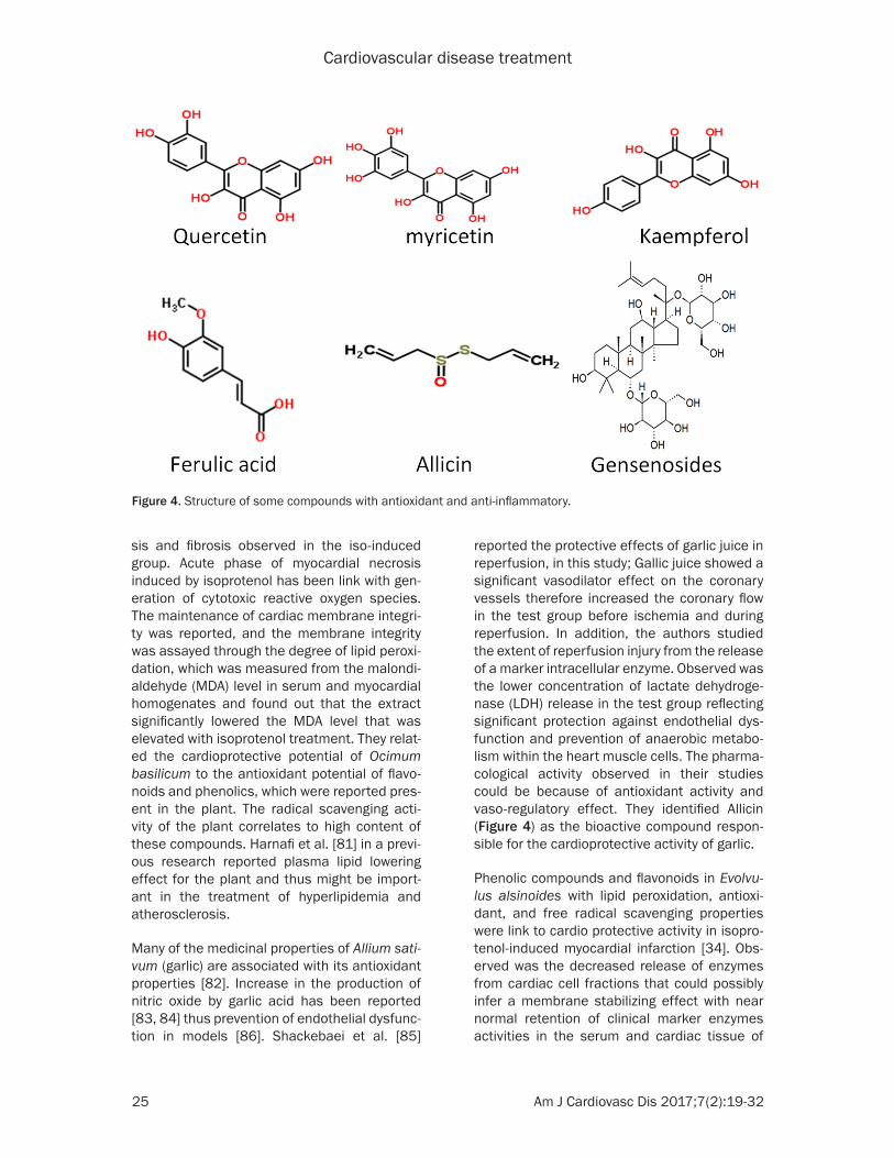
induction were restored to near normal when treated with T. arjuna. The activity of the plant extracts were compared with the combined activity of propranolol and gemfibrazole stan-dard drug and was found to be comparable. They related the potentials of the plant to the polyphenols fraction of the extract and its anti-oxidant activity. The presence of flavonol (quer-cetin, myricetin), kaempferol, phenolic acids (garlic and ferulic acid) (Figure 4) was higher in T. arjuna as observed in the HPLC analysis.
The potentials of flavonoids in the treatment of CVD have been reported. The possible mecha-nism includes its ability to ameliorate/stop endothelial dysfunction [80], reduce LDL oxida-tion and inhibition of platelet aggregation [80]. They concluded by correlating the superlative cardioprotective activity of T. arjuna mainly to the antioxidant constituents.
The cardio protective potentials of Ocimum basilicum have been studied. O. basilicum belong to the lamiaceae family. Studies have revealed the essential oil of the plant is com-posed of interesting terpenoids. Phytochemical reports reveal the presence of triterpenoids, polyphenols, and steroids. Studies by Fathiazad et al. [17], reported an ameliorative effects of the plant extract on the changes in blood pres-sure observed in the isoprotenol induced car-dio toxicity. The plant extract prevent the necro-
Figure 3. Interruption of oxidative stress by plant compounds, prevent cardio-vascular development.
preventive studies. The result shows that isoprotenol signi- ficantly increased the level of serum cardiac marker enzy- mes like LDH, AST, and ALT reflecting the severity of iso-induced myocardial cell ne- crosis. The myocardial cell necrosis can be due to increase in lipid peroxidation, but treatment with the poly-phenolic rich extract lowered the serum cardiac marker enzymes demonstrating the plant’s ability to maintain membrane integrity conse-quently constrain the leakag-es of enzymes. In addition, the activity of the myocardial antioxidant enzymes such as SOD, CAT and Peroxidase that were lowered by isoprotenol
Cardiovascular disease treatment
25 Am J Cardiovasc Dis 2017;7(2):19-32
sis and fibrosis observed in the iso-induced group. Acute phase of myocardial necrosis induced by isoprotenol has been link with gen-eration of cytotoxic reactive oxygen species. The maintenance of cardiac membrane integri-ty was reported, and the membrane integrity was assayed through the degree of lipid peroxi-dation, which was measured from the malondi-aldehyde (MDA) level in serum and myocardial homogenates and found out that the extract significantly lowered the MDA level that was elevated with isoprotenol treatment. They relat-ed the cardioprotective potential of Ocimum basilicum to the antioxidant potential of flavo-noids and phenolics, which were reported pres-ent in the plant. The radical scavenging acti- vity of the plant correlates to high content of these compounds. Harnafi et al. [81] in a previ-ous research reported plasma lipid lowering effect for the plant and thus might be import- ant in the treatment of hyperlipidemia and atherosclerosis.
Many of the medicinal properties of Allium sati-vum (garlic) are associated with its antioxidant properties [82]. Increase in the production of nitric oxide by garlic acid has been reported [83, 84] thus prevention of endothelial dysfunc-tion in models [86]. Shackebaei et al. [85]
reported the protective effects of garlic juice in reperfusion, in this study; Gallic juice showed a significant vasodilator effect on the coronary vessels therefore increased the coronary flow in the test group before ischemia and during reperfusion. In addition, the authors studied the extent of reperfusion injury from the release of a marker intracellular enzyme. Observed was the lower concentration of lactate dehydroge-nase (LDH) release in the test group reflecting significant protection against endothelial dys-function and prevention of anaerobic metabo-lism within the heart muscle cells. The pharma-cological activity observed in their studies could be because of antioxidant activity and vaso-regulatory effect. They identified Allicin (Figure 4) as the bioactive compound respon-sible for the cardioprotective activity of garlic.
Phenolic compounds and flavonoids in Evolvu- lus alsinoides with lipid peroxidation, antioxi-dant, and free radical scavenging properties were link to cardio protective activity in isopro-tenol-induced myocardial infarction [34]. Obs- erved was the decreased release of enzymes from cardiac cell fractions that could possibly infer a membrane stabilizing effect with near normal retention of clinical marker enzymes activities in the serum and cardiac tissue of
Figure 4. Structure of some compounds with antioxidant and anti-inflammatory.
Cardiovascular disease treatment
26 Am J Cardiovasc Dis 2017;7(2):19-32
experimental animals [34]. Membrane lipid per-oxidation by free radicals is known to compro-mise membrane integrity.
Conclusively, the antioxidant role played by phenolics and flavonoids reported in various research resulted in the inhibition of platelet aggregation, restoration of vascular function, inhibition of lipid peroxidation, membrane sta-bilization, restoration of antioxidant enzyme and inhibition of reactive oxygen species. A similar mechanism reported by Mohanty et al. [86] in a combination treatment research he conducted for Withania somnifera, Curcuma longa and Ocimum sanctum. Previous investi-gation has also reported the cardioprotective activity of Ginkgo biloba and Ocinum sanctum. In both plant, the cardioprotective activity was attributed to the antioxidant activities ass- ociated with the presence of flavonoids and phenolics. The report also associated reduced glutathione augmenting effect, elevated anti-oxidant enzyme level and the inhibition of lipid peroxidation [87, 88] to these compounds in both plant. Panda and Naik [16] validated the cardio protective claim for these bioactive com-pounds in the two medicinal plants when he combined Ginkgo biloba and Ocinum sanctum treatment in isoprotenol induced myocardial necrosis in rats, suggesting the possibility of similar mechanism previously reported, alth- ough no significant synergistic effect was observed in the combination treatment.
The high radical scavenging activity in pheno-lics and flavonoids is associated to their struc-ture activity relationship. The high reactivity of the hydroxyl group of flavonoids is responsible for their free radical scavenging activity [16].
Anti-inflammatory role
Inflammation is a complex biological response of vascular tissues to harmful stimuli [89]. It is also protective in nature in the attempt by organism to remove injurious stimuli and initi-ate healing process [90]. Inflammation respon- se involves cell activation and the release of inflammatory mediators [89]. Inflammation activation has been implicated as an important pathway in the pathogenesis and progression of CVD including chronic heart failure [27, 91] and their mediators can serve as relevant markers of disease severity [92]. Markers such as interleukins, cytokines are reported to cre-
ate inflammatory condition in the arterial wall [93]. Other inflammation factors are leukocyte adhesion molecules and chemokines [94]. Risk factors including hypercholesterolemia can lead to inflammatory response within the micro-vasculature, which is reflected, by endothelial cell activation, leukocyte recruitment, rolling and adherence as well as platelet activation and adhesion [44, 45]. This is preventable by NO availability in the vasculature. However, diminished availability of NO result in vascular dysfunction and diminished anti-inflammatory properties of the endothelial cell [9].
Wallace [95] in his review reported the health benefit of diets rich in natural bioactive com-pounds especially the contributory role in main-taining and improving cardiovascular health. He reported flavonoids, a member of the polyphe-nols as potential candidate to protect against CVD.
Several medicinal plants have been reported with anti-inflammatory activity. Kadian and Parle [96] in a review reported the anti-inflam-matory activity of Ocimum sanctum. The plant was reported to possess cardio protective effect as well as anti-hypertensive effect and the mechanism for this pharmacological effi-cacy is by inhibiting specific inflammatory mol-ecules [97]. Singh and Majumdar [98] sugg- ested the possible mechanism for the anti-inflammatory activity of linolenic acid present in Ocimum sanctum to be by blocking both the cyclooxygenase and lipooxygenase pathways of arachidonate metabolism. Arachidonic acid-derived lipid mediators are intimately involved in inflammation and are biosynthesized by pathways dependent on cyclooxygenase (COX) and lipoxygenase (LOX) enzymes.
The anti-inflammatory effect of Gensenosides (Figure 4), a triterpene dammarane-type sapo-nins was reported by Xing et al. [99]. According to the research, a combination of the Chinese herbal medicines Ginseng Radix Et Rhizoma Rubra (the root of Panax ginseng C.A.Mey.), Ophio- pogonis Radix (the root of Ophiopogon japoni-cas (L.f) Ker-Gawl), and Schisandrae Chinensis Fructus (the fructus of Schisandra chinensis (Turcz.) Baill) termed YQFM could suppress the expressions of inflammatory mediators. A con-stituent of one of the main bioactive compo-nents, ginsenosides according to the research contributed to the measured anti-inflammatory

Cardiovascular disease treatment
27 Am J Cardiovasc Dis 2017;7(2):19-32
effects. The three herbs of YQFM were report- ed to be significant in the enhancement of myo-cardial contractility, blood-vessel dilation, anti-lipid peroxidation, and anti-inflammatory effect [100-104].
Conclusion
With cardiovascular disease assuming an increasing role as a major cause of mobility and mortality and the limitations in the use of con-ventional drugs in its treatment, more intensive approach is required in the current Epidemiolo- gical, clinical and experimental studies of com-pounds that are able to suppress free radical generation, augment endogenous antioxidant and maintain the integrity of the vasculature in medicinal plants. Critical analysis of the roles of bioactive compounds as observed in this review will be important as a guide to identify potential compounds in plant and consequent-ly their characterization. Aside the commonly reported phenolics and flavonoids, other com-pounds with great potentials in treating cardio-vascular disease and their roles should be elucidated.
Acknowledgements
The authors appreciate Dr. A. Adetutu for his mentorship and tutorship and the Department of Biochemistry, Ladoke Akintola University of Technology, Ogbomoso, Oyo State. Nigeria for giving us the platform to carry out this study.
Disclosure of conflict of interest
None.
Authors’ contribution
PI conceived the idea, PI, IA, WH and TO outline the content, WH and IA search the internet, PI wrote the manuscript. All author read and approved the final manuscript for submission.
Address correspondence to: Peter Adegbola, De- partment of Biochemistry, Faculty of Basic Medical Sciences, Ladoke Akintola University of Technology, Ogbomoso, Oyo State Nigeria. Tel: 07069023126; E-mail: [email protected]
References
[1] Mayakrishnan V, Kannappan P, Abdullah N and Ali A. Cardioprotective activity of polysaccha-
rides derived from marine algae: an overview. Trends Food Sci Technol 2013; 1-7.
[2] Olorunnisola OS, Bradley G and Afolayan AJ. Ethnobotanical information on plants used for the management of cardiovascular diseases in Nkonkobe municipality, South Africa. J Med Plant Res 2011; 5: 4256-4260.
[3] Krisela S. The heart and stroke foundation South Africa heart disease in South Africa me-dia data document. http://www.heartfounda-tion.co.za/docs/heartmonth/HeartDiseasein-SA.pdf 2007.
[4] WHO: Cardiovascular disease http://www.who.int/cardiovascular_diseases/en/ (2013). Accessed 27 March 2015.
[5] Thompkinson D, Bhavana V and Kanika P. Di-etary approaches for management of cardio vascular health-a review. J Food Sci Technol 2012; 51: 2318-2330.
[6] Brunner H, Cockcroft JR, Deanfield J, Donald A, Ferrannini E, Halcox J, Kiowski W, Lüscher TF, Mancia G, Natali A, Oliver JJ, Pessina AC, Riz-zoni D, Rossi GP, Salvetti A, Spieker LE, Taddei S and Webb DJ; Working Group on Endothelins and Endothelial Factors of the European Soci-ety of Hypertension. Endothelial function and dysfunction. Part II: association with cardiovas-cular risk factors and diseases. A statement by the working group on endothelins and endo-thelial factors of the European society of hy-pertension. J Hypertens 2005; 23: 233-246.
[7] Vogiatzi G, Tousoulis D and Stefanadis C. The role of oxidative stress in atherosclerosis hel-lenic. J Cardiol 2009; 50: 402-409.
[8] Liu VW and Huang PL. Cardiovascular roles of nitric oxide: a review of insights from nitric ox-ide synthase gene disrupted mice. Cardiovasc Res 2007; 77: 19-29.
[9] Stapleton PA, Goodwill AG, James ME, Brock RW and Frisbee JC. Hypercholesterolemia and microvascular dysfunction: interventional strategies. J Inflamm 2010; 7: 1-10.
[10] Christensen HM, Schou M, Goetze JP, Faber J, Frystyk J, Flyvbjerg A and Kistorp C. Body mass index in chronic heart failure: association with biomarkers of neurohormonal activation, in-flammation and endothelial dysfunction. BMC Cardiovasc Disord 2013; 13: 80.
[11] Schachinger V, Britten MB and Zeiher AM. Prognostic impact of coronary vasodilator dys-function on adverse long-term outcome of cor-onary heart disease. Circulation 2010; 101: 1899-1906.
[12] Stapleton PA, Goodwill AG, James ME, D’Audi- ffret AC and Frisbee JC. Differential impact of familial hypercholesterolemia and combined hyperlipidemia on vascular wall and network remodeling in mice. Microcirculation 2010; 17: 47-58.
Cardiovascular disease treatment
28 Am J Cardiovasc Dis 2017;7(2):19-32
[13] Viswanatha GL, Vaidya S, Ramesh C, Krishna-das N and Rangappa S. Antioxidant and anti-mutagenic activities of bark extract of Termina-lia arjuna. Asian Pac J Trop Med 2010; 3: 965-970.
[14] Saman H, Khalil-ur-R, Zahoor-ul-hassan D, Na-zish J, Mansoor H, Zafar IK, Kafeel A, Khalid M and Ehsan EV. Cardioprotective effect of gem-motherapeutically treated withania somnifera against chemically induced myocardial injury Pak. J Bot 2010; 42: 1487-1499.
[15] Jahan N, Rahman K and Ali S. Cardioprotective and antilipidemic potential of cyperus rotun-dus in chemically induced cardiotoxicity. Int J Agric Biol 2012; 14: 989-992.
[16] Panda VS and Naik SR. Evaluation of cardio-protective activity of ginkgo biloba and Oci-mum sanctumin rodents. Altern Med Rev 2009; 14: 161-171.
[17] Fathiazad F, Matlobi A, Khorrami A, Hamedey-azdan S, Soraya H, Hammami M, Maleki-Dizaji N and Garjan A. Phytochemical screening and evaluation of cardioprotective activity of etha-nolic extract of Ocimum basilicum L. (basil) against isoproterenol induced myocardial in-farction in rats. Daru 2012; 20: 87.
[18] Ojha S, Bharti S, Sharma AK, Rani N, Bhatia J, Kumari S and Arya DS. Effect of Inula racemo-sa root extract on cardiac function and oxida-tive stress against isoproterenol induced myo-cardial infarction. Indian J Biochem Biophys 2011; 48: 22-28.
[19] Sangwan RS, Chaurasiya ND, Misra LN, Lal P, Uniyal GC, Sharma R, Sangwan NS, Suri KA, Oazi GN and Tuli R. Phytochemical variability in commercial herbal products and preparations of withania somnifera (Ashwagandha). Current Science 2004; 86: 461-465.
[20] Hasler CM. Functional foods: their role in dis-ease prevention and health promotion. Food Tech 1998; 52: 63-70.
[21] Suroowan S and Mahomoodally F. Common phyto-remedies used against cardiovascular diseases and their potential to induce adverse events in cardiovascular patients. Clinical Phy-toscience 2015; 1: 1-13.
[22] Rajalakshmy I, Ramya P and Kavimani S. Car-dioprotective medicinal plants-A review. Inter-national Journal of Pharmaceutical Invention 2011; 1: 24-41.
[23] Xin YF, Wan LL, Peng JL and Guo C. Alleviation of acute doxorubicin induced cardiotoxicity by lycium barbarum polysaccharides through the suppression of oxidative stress. Food Chem Toxicol 2011; 49: 259-264.
[24] Luscher TF and Vanhoutte PM. The endotheli-um: modulator of cardiovascular function, CRC press, Boca Raton, FL, USA. 1990; pp. 1-215.
[25] Madamanchi NR, Vendrov A and Runge MS. Oxidative stress and vascular disease. Arterio-scler Thromb Vasc Biol 2005; 25: 29-38.
[26] Young IS and Woodside JV. Antioxidants in health and disease. J Clin Pathol 2001; 54: 176-186.
[27] Libby P. Vascular biology of atherosclerosis: overview and state of art. Am J Cardiol 2003; 91: 3-6.
[28] Navab M, Ananthramaiah GM, Reddy ST, Van-Lenten BJ, Ansell BJ, Fonarow GC, Vahabzadeh K, Hama S, Hough G, Kamranpour N, Berliner JA, Lusis AJ and Fogelman AM. The oxidation hypothesis of atherogenesis: the role of oxi-dized phospholipids and HDL. J Lipid Res 2004; 45: 993-1007.
[29] Puddu GM, Cravero E, Arnone G, Muscari A and Puddu P. Molecular aspects of atherogen-esis: new insights and unsolved questions. J Biomed Sci 2005; 12: 839-853.
[30] Puddu P, Puddu GM, Cravero E, Pascalis S and Muscari A. The emerging role of cardiovascular risk factor-induced mitochondrial dysfunction in atherogenesis. J Biomed Sci 2009; 16: 1-9.
[31] Thygesen K, Alpert JS and White HD. “Univer-sal definition of myocardial infarction”. Eur Heart J 2007; 28: 2525-38.
[32] Radhika S, Smila KH and Muthezhilan R. Car-dioprotective activity of hybanthus enneasper-mus (Linn.) on isoproterenol induced rats. In-dian Journal of Fundamental and Applied Life Sciences 2011; 1: 90-97.
[33] Mohale DS, Dewani AP, Saoji AN and Khadse CD. Antihyperlipidemic activity of isolated con-stituents from lagenaria siceraria in albino rats. Int J Green Pharm 2008; 2: 104-107.
[34] Sudhakumari Anil KH, Aamir J, Manish J and Muralidhar ST. Cardioprotective effects in methanolic extract of Evolvulus alsinoides linn on isoproterenol-induced myocardial infarction in albino rats. International Journal of Basic Medical Sciences and Pharmacy (IJBMSP) 2012; 2: 53-57.
[35] Levick JR. An introduction to cardiovascular physiology. 4th edition. London, UK: Oxford University Press Inc; 2003.
[36] Chien KR, Han A, Sen A, Buja LM and Willerson JT. Accumulation of unesterified arachidonic acid in ischemic canine myocardium. Relation-ship to a phosphatidylcholine deacylation-reacylation cycle and the depletion of mem-brane phospholipids. Circ Res 1984; 54: 313-322.
[37] Shug AL, Thomsen JH, Folts JD, Bittar N, Klein MI, Koke JR and Huth PJ. Changes in tissue lev-els of carnitine and other metabolites during myocardial ischemia and anoxia. Arch Bio-chem Biophys 1978; 187: 25-33.

Cardiovascular disease treatment
29 Am J Cardiovasc Dis 2017;7(2):19-32
[38] Toth PP. Making a case for quantitative assess-ment of cardiovascular risk. J Clin Lipidol 2007; 1: 34-41.
[39] Tabas I, Williams KJ and Borén J. Subendo- thelial lipoprotein retention as the initiating process in atherosclerosis: update and thera-peutic implications. Circulation 2007; 116: 1832-44.
[40] Dilaveris P, Giannopoulos G, Riga M, Synetos A and Stefanadis C. Beneficial effects of statins on endothelial dysfunction and vascular stiff-ness. Curr Vasc Pharmacol 2007; 5: 227-237.
[41] Lakshmi SV, Padmaja G, Kuppusamy P and Kutala VK. Oxidative stress in cardiovascular disease. Indian J Biochem Biophys 2009; 46: 421-440.
[42] Simionescu M. Implications of early structural-functional changes in the endothelium for vas-cular disease. Arterioscler Thromb Vasc Biol 2007; 27: 266-274.
[43] Harrison DG. Endothelial function and oxidant stress. Clin Cardiol 1997; 20: 2-7.
[44] Scalia R, Appel JZ and Lefer AM. Leukocyte-endothelium interaction during the early stag-es of hypercholesterolemia in the rabbit: role of P-selectin, ICAM-1, and VCAM-1. Arterioscler Thromb Vasc Biol 1998; 18: 1093-1100.
[45] Stokes KY, Calahan L, Russell JM, Gurwara S and Granger DN. Role of platelets in hypercho-lesterolemia-induced leukocyte recruitment and arteriolar dysfunction. Microcirculation 2006; 13: 377-388.
[46] Stokes KY, Cooper D, Tailor A and Granger DN. Hypercholesterolemia promotes inflammation and microvascular dysfunction: role of nitric oxide and superoxide. Free Radic Biol Med 2002; 33: 1026-1036.
[47] Roberts WC. Preventing and arresting coronary atherosclerosis. Am Heart J 1995; 130: 580-600.
[48] Choudhury RP, Fuster V and Fayad ZA. Molecu-lar, cellular and functional imaging of athero-thrombosis. Nat Rev Drug Discov 2004; 3: 913-925.
[49] Mahsa Z, Komal KJ, Mehrdad Z and Syed B. Cardioprotective effect of the root extract of Hemidesmus indicus against doxorubicin-in-duced oxidative stress in mice. Der Pharmacia Lettre 2013; 5: 334-339.
[50] Sakthivel K, Palani S, Santhosh K, Devi K and Kumar BS. Phytoconstituents analysis by GC-MS, cardioprotective and antioxidant activity of Buchanania axillaris against doxorubicin-in-duced cardio toxicity in albino rats. Internation-al Journal of Pharmaceutical Studies and Re-search 2010; 1: 34-48.
[51] Yousefi K, Soraya H, Fathiazad F, Khorrami A, Hamedeyazdan S, Maleki-Dizaji N and Garjani A. Cardioprotective effect of methanolic extr-
act of Marribum vulgare L. On isoprotenol-in-duced acute myocardial infarction in rats. Indi-an J Exp Biol 2013; 51: 653-660.
[52] El-Sayed EM, Abd El-azeem AS, Afify AA, Sha-bana MH and Ahmed HH. Cardioprotective ef-fects of Curcuma longa L. Extracts against doxorubicin-induced cardiotoxicity in rats. J Med Plant Res 2011; 5: 4049-4058.
[53] Wattanapitayakul SK and Bauer JA. Oxidative pathways in cardiovascular disease roles, mechanisms, and therapeutic implications. Pharmacol Ther 2001; 89: 187-206.
[54] Finkel T. Signal transduction by ROS in non-phagocytic cells. J leukoc Biol 1999; 65: 337-340.
[55] Sizuki YJ, Forman HJ and Sevanian A. Oxidants as stimulators of signal transduction. Free Radic Biol Med 1997; 22: 269-285.
[56] Wolin MS. Interactions of oxidants with vascu-lar signaling systems. Artrioscler throm vasc. Biol 2000; 20: 1430-1442.
[57] Lassegue B and Clempus RE. Vascular NAD(P)H oxidases: specific features, expression and regulation. Am J Physiol Regul Integr Comp Physiol 2003; 285: 277-297.
[58] Touyz RM and Schiffrin EL. Ang 11-stimulated superoxide production is mediated via phos-pholipase D in human vascular smooth muscle cells. Hypertension 1999; 34: 976-982.
[59] Zafari AM, Ushio-fukai M, Akers M, Yin Q, Shah A, Harrison DG, Taylor WR and Griendling KK. Role of NADH/NADPH oxidase derived H2O2 in angiotensin 11-induced vascular hypertrophy. Hypertension 1998; 32: 488-495.
[60] Gozin A, Franzini E, Andrieu V, Da Costa L, Rollet-Labelle E and Pasquier C. Reactive oxy-gen species activate focal adhesion kinase, paxillin and p130cas tyrosine phosphorylation in endothelial cells. Free Radic Biol Med 1998; 25: 1021-1032.
[61] Block G, Dietrich M, Norkus EP, Morrow JD, Hudes M and Cann B. Factors associated with oxidative stress in human populations. Am J Epidemiol 2002; 156: 274-85.
[62] Victor VM, Rocha M, Solá E, Bañuls C, Gar- cia-Malpartida K and Hernández-Mijares A. Oxidative stress, endothelial dysfunction and atherosclerosis. Curr Pharm Des 2009; 15: 2988-3002.
[63] Vepa S, Scribner WM, Parinandi NL, English D, Garcia JG and Natarajan V. Hydrogen peroxide stimulates tyrosine phosphorylation of focal adhesion kinase in vascular endothelial cells. Am J Physiol 1999; 277: 150-158.
[64] Versari D, Daghini E, Virdis A, Ghiadoni L and Taddei S, The ageing endothelium, cardiovas-cular risk and disease in man. Exp Physiol 2008; 94: 317-321.

Cardiovascular disease treatment
30 Am J Cardiovasc Dis 2017;7(2):19-32
[65] Bredt DS and Snyder SH. Nitric oxide: a physi-ologic messenger molecule. Ann Rev Biochem 1994; 63: 175-195.
[66] Freedman JE, Sauter R, Battinelli EM, Ault K, Knowles C, Huang PL and Loscalzo J. Deficient platelet-derived nitric oxide and enhanced he-mostasis in mice lacking the NOSIII gene. Circ Res 1999; 84: 1416-1421.
[67] Radomski MW, Palmer RM and Moncada S. Modulation of platelet aggregation by an L-argi-nine-nitric oxide pathway. Trends Pharmacol Sci 1991; 12: 87-88.
[68] Lefer DJ, Jones SP, Girod WG, Baines A, Grish-am MB, Cockrell AS and Huang PL, Scalia R. Leukocyte-endothelial cell interactions in nitric oxide synthase deficient mice. Am J Physiol 1999; 276: 1943-1950.
[69] Garg UC and Hassid A. Nitric oxide-generating vasodilators and 8-bromo-cyclic guanosine monophosphate inhibit mitogenesis and prolif-eration of cultured rat vascular smooth muscle cells. J Clin Invest 1989; 83: 1774-1777.
[70] Rosz R. The pathogenesis of atherosclerosis: a perspective for the 1990s. Nature 1993; 362: 801-809.
[71] Valko M, Leibfritz D, Moncol J, Cronin MT, Ma-zur M and Telser J. Free radicals and antioxi-dants in normal physiological functions and human disease. Int J Biochem Cell Biol 2007; 39: 44-84.
[72] Hertog MG, Feskens EJ, Hollam PC, Katan MB and Kromhout D. Dietary antioxidant flavo-noids and risk of coronary heart diseases: the Zutphen elderly study. Lancet 1993; 342: 1007-1020.
[73] Yamagishi SI, Edelstein D, Du XL and Brownlee M. Hyperglycaemia potenciates collagen-in-duced platelet activation through mitochondri-al superoxide overproduction. Diabetes 2001; 50: 1491-1494.
[74] Nabavi SF, Nabavi SM, Ebrahimzadeh MA, Esl-ami SH, Jafari N and Hajizadeh M. The protec-tive effect of curcumin against sodium fluo-ride-induced oxidative stress in rat heart. Arch Biol Sci Belgrade 2011; 63: 563-569.
[75] Bartsch H and Nair J. Ultrasensitive and spe-cific detection method for exocyclic DNA ad-ducts markers for lipid peroxidation and oxida-tive stress. Toxicology 2000; 153: 105-114.
[76] Halliwell B and Guttteridge JM. Free radicals in biology and medicine. 2nd edition. Oxford, Lon-don: Oxford university press (Clarendon); 2007.
[77] Sheweita SA, Abd El-Gabar M and Bastawy M. Carbon tetrachloride induced changes in the activity of phase II drug metabolizing enzyme in the liver of male rats: role of antioxidants. Toxicology 2001; 165: 217-224.
[78] Nandave M, Mohanty I, Nag TC, Ojha SK, Mittal R, Kumari S and Arya DS. “Cardioprotective re-sponse to chronic administration of vitamin E in isoproterenol induced myocardial necrosis: hemodynamic, biochemical and ultrastructur-al studies”. Indian J Clin Biochem 2007; 22: 22-28.
[79] Kurosawa T, Itoh F, Nozaki A, Nakano Y, Katsu-da S, Osakabe N, Tsubone H, Kondo K and Ita-kura H. Suppressive effects of cacao liquor polyphenols (CLP) on LDL oxidation and the development of atherosclerosis in Kurosawa and Kusanagi hypercholesterolemic rabbits. Atherosclerosis 2005; 179: 237-246.
[80] Keevil JG, Osman HE, Reed JD and Folts JD. Grape juice, but not orange juice or grape fruit juice, inhibits human platelet aggregation. J Nutr 2000; 130: 53-56.
[81] Harnafi H, Serghini CH, Bouanani NH, Aziz M and Amrani S. Hypolipemic activity of polyphe-nol-rich extracts from Ocimum basilicum in Tri-ton WR-1339-induced hyperlipidemic mice. Food Chem 2008; 108: 205-212.
[82] Banerjee SK, Mukherjee PK and Maulik SK. Garlic as an antioxidant: the good, the bad and the ugly. Phytother Res 2003; 17: 97-106.
[83] Maslin DJ, Brown CA, Das I and Zhang XH. Ni-tric oxide-a mediator of the effects of garlic? Biochem Soc Trans 1997; 25: 408.
[84] Sooranna SR, Hirani J and Das I. Garlic can in-duce both GTP cyclohydrolase and nitric oxide synthase activity in choriocarcinoma cells. Bio-chem Soc Trans 1995; 23: 543.
[85] Shackebaei D, Ghazvineh S, Godini A, Pilehvar-ian A and Reshadat S. Cardioprotective effect of garlic juice on the isolated rat heart in isch-emia-reperfusion. Journal of Medicinal Plants 2010; 9: 71-79.
[86] Mohanty IR, Gupta SK, Arya DS, Mohanty N and Deshmukh Y. Medicinal herbs can play sig-nificant role in attenuation of ischemia and re-perfusion. Injury J Homeop Ayurv Med 2013; 2: 1-11.
[87] Naik SR, Pilgaonkar VW and Panda VS. Evalua-tion of antioxidant activity of ginkgo biloba phy-tosomes in rat brain. Phytother Res 2006; 20: 1013-1016.
[88] Uma DP, Ganasoundari A, Vrinda B, Srinivasan KK and Unnikrishnan MK. Radiation 18. Pro-tection by the Ocimum flavonoids orientin and vicenin: mechanisms of action. Radiat Res 2000; 154: 455-460.
[89] Anosike CA, Obidoa O and Ezeanyika LU. Mem-brane stabilization as a mechanism of the anti-inflammatory activity of methanol extract of garden egg (solanum aethiopicum). DARU 2012; 20: 2-7.
Cardiovascular disease treatment
31 Am J Cardiovasc Dis 2017;7(2):19-32
[90] Ferrero-Miliani L, Nielson OH, Andersen PS and Girardin SE. Chronic inflammation: importance of NOD2 and NALP3 in interleukin-1β genera-tion. Clin Exp Immunol 2007; 147: 227-235.
[91] Heymans S, Hirsch E, Anker SD, Aukrust P, Bal-ligand JL, Cohen-Tervaert JW, Drexler H, Filip-patos G, Felix SB, Gullestad L, Hilfiker-Kleiner D, Janssens S, Latini R, Neubauer G, Paulus WJ, Pieske B, Ponikowski P, Schroen B, Schul-theiss HP, Tschöpe C, Van Bilsen M, Zannad F, McMurray J and Shah AM. Inflammation as a therapeutic target in heart failure? A scientific statement from the tanslational research com-mittee of the heart failure association of the European society of cardiology. Eur J Heart Fail 2009; 11: 119-129.
[92] Bozkurt B, Mann DL and Deswal A. Biomarkers of inflammation in heart failure. Heart Fail Rev 2010; 15: 331-341.
[93] Chang HL and Jong-Hoon K. A review on the medicinal potentials of ginseng and ginsen-osides on cardiovascular diseases. J Ginseng Res 2014; 38: 161-166.
[94] Candia AM, Villacorta J and Mesquita ET. Im-mune-inflammatory activation in heart failure. Arquivos Brasileiros de Cardiologia 2007; 89: 183-190.
[95] Wallace TC. Anthocyanins in cardiovascular disease. American society for nutrition. Adv Nutr 2011; 2: 1-7.
[96] Kadian R and Parle M. Therapeutic potential and phytopharmacology of tulsi. Int J of Pharm Life Sci (IJPLS) 2012; 3: 1858-1867.
[97] Pandey G and Madhuri S. Pharmacological ac-tivities of Ocimum sanctum (Tulsi): a review. Int J Pharmaceutical Sci Rev Res 2010; 5: 61-66.
[98] Singh S and Majumdar DK. Evaluation of anti-inflammatory activity of fatty acids of Ocimum sanctum fixed oil. Indian J Exp Biol 1997; 35: 380-383.
[99] Xing L, Jiang M, Dong L, Gao J, Hou Y, Bai G and Luo G. Cardioprotective effects of the YiQi-FuMai injection and isolated compounds on attenuating chronic heart failure via NF-κB in-activation and cytokine suppression. J Ethno-pharmacol 2013; 148: 239-245.
[100] Kou J, Sun Y, Lin Y, Cheng Z, Zheng W, Yu B and Zu Q. Anti-inflammatory activities of aqueous extract from radix ophiopogon japonicus and its two constituents. Biol Pharm Bull 2005; 28: 1234-1238.
[101] Lee DC and Lau AS. Effects of Panax ginseng on tumor necrosis factor-alpha mediated in-flammation: a mini-review. Molecules 2011; 16: 2802-2816.
[102] Lu JM, Yao Q and Chen C. Ginseng compounds: an update on their molecular mechanisms and medical applications. Curr Vasc Pharmacol 2009; 7: 293-302.
[103] Oh SY, Kim YH, Bae DS, Um BH, Pan CH, Kim CY, Lee HJ and Lee JK. Anti-inflammatory ef-fects of gomisin N, gomisin J, and schisandrin C isolated from the fruit of schisandra chinen-sis. Biosci Biotechnol Biochem 2010; 74: 285-291.
[104] Tian YQ, Yu BY and Kou JP. Progress in pharma-cological actions of Ophiopogon japonicus. The Chinese Academic Medical Magazine of Organisms 2004; 1671: 1-5.
[105] National Cholesterol Education Program (NCEP). Second report of the expert panel on detection, evaluation, and treatment of high blood cholesterol in adults (adult treatment panel II). Bethesda, Md.: National Heart, Lung, and Blood Institute, 1993. (NIH publication no. 93-3095).
[106] Ross R. Atherosclerosis-an inflammatory dis-ease. N Engl J Med 1999; 340: 115-126.
[107] Steinberg D and Witztum JL. Lipoproteins, lipo-protein, oxidation, and atherogenesis, WB Saunders, Philadelphia, Pa, USA. 1999.
[108] Keaney JF. Atherosclerosis: from lesion forma-tion to plaque activation and endothelial dys-function. Mol Aspects Med 2000; 21: 99-166.
[109] Shamala S, Baskaran G, Noor AS, Siti AA and Mohd YS. Anti-artherosclerotic effects of plant flavonoids. Bio Med Research International 2014; 2014: 1-11.
[110] Ross R and Glomset JA. Atherosclerosis and the arterial smooth muscle cell: proliferation of smooth muscle is a key event in the genesis of the lesions of atherosclerosis. Science 1973; 180: 1332-9.
[111] Ross R. Rous-Whipple Award Lecture. Athero-sclerosis: a defence mechanism gone awry. Am J Pathol 1993; 143: 987-1002.
[112] Ross R. The pathogenesis of atherosclerosis: a perspective for the 1990s. Nature 1993; 362: 801-809.
[113] Lee V, Randhawa AK and Singal PK. Adriamy-cin-induced myocardial dysfunction in vitro is mediated by free radicals. Am J Physiol 1991; 261: 989-995.
[114] Gurvinder S, Anu ST, Aji A, Beena B, Ashok M, Riru V, Shiv KA, Shivesh J, Rama M and Anand CB. Protective effects of Terminalia arjuna against doxorubicin-induced cardiotoxicity. J Ethnopharmacol 2008; 117: 123-129.
[115] Vandana SP and Suresh RN. Evaluation of car-dioprotective activity of ginkgo biloba and Oci-mum sanctum in rodents. Altern Med Rev 2009; 14: 161-171.
[116] Angeloni C, Spencer JP, Leoncini E, Biagi PL and Hrelia S. Role of Quercetin and its in vivo metabolites in protecting H9c2 cells against oxidative stress. Biochimie 2007; 89: 73-82.
[117] Devasagayam TP, Tilak JC, Boloor KK, Sane KS, Ghaskadbi S and Lele RD. Free radicals

Cardiovascular disease treatment
32 Am J Cardiovasc Dis 2017;7(2):19-32
and antioxidants in human health: current sta-tus and future prospects. J Assoc Physicians India 2004; 52: 794-804.
[118] You JS, Pan TL and Lee YS. Protective effects of danshen (Salvia miltiorrhiza) on adriamycin-
induced cardiac and hepatic toxicity in rats. Phytother Res 2007; 21: 1146-52.