review article arenavirusbuddingdownloads.hindawi.com/journals/av/2011/180326.pdf · mediate...
TRANSCRIPT
Hindawi Publishing CorporationAdvances in VirologyVolume 2011, Article ID 180326, 9 pagesdoi:10.1155/2011/180326
Review Article
Arenavirus Budding
Shuzo Urata1, 2 and Juan Carlos de la Torre2
1 Department of Emerging Infectious Disease, Institute of Tropical Medicine, Nagasaki University, 1-12-4 Sakamoto,Nagasaki 852-8523, Japan
2 Department of Immunology and Microbial Science, The Scripps Research Institute, 10550 North Torrey Pines Road,La Jolla, CA 92037, USA
Correspondence should be addressed to Juan Carlos de la Torre, [email protected]
Received 19 February 2011; Accepted 23 May 2011
Academic Editor: Fadila Bouamr
Copyright © 2011 S. Urata and J. C. de la Torre. This is an open access article distributed under the Creative Commons AttributionLicense, which permits unrestricted use, distribution, and reproduction in any medium, provided the original work is properlycited.
Several arenaviruses cause hemorrhagic fever disease in humans and pose a significant public health concern in their endemicregions. On the other hand, the prototypic arenavirus LCMV is a superb workhorse for the investigation of virus-host interactionsand associated disease. The arenavirus small RING finger protein called Z has been shown to be the main driving force of virusbudding. The budding activity of Z is mediated by late (L) domain motifs, PT/SAP, and PPXY, located at the C-terminus of Z.This paper will present the current knowledge on arenavirus budding including the diversity of L domain motifs used by differentarenaviruses. We will also discuss how improved knowledge of arenavirus budding may facilitate the development of novel antiviralstrategies to combat human pathogenic arenaviruses.
1. Introduction
Arenaviruses are enveloped viruses with a bisegmented neg-ative strand (NS) RNA genome with coding capabilityfor four known genes: nucleoprotein (NP), surface gly-coprotein precursor (GPC), polymerase (L), and matrix-like (Z) proteins. Despite their limited genome and pro-teomic complexity, arenaviruses are able to exhibit verydifferent phenotypic infection outcomes ranging from long-term subclinical chronic infections on their natural rodenthosts [1] to hemorrhagic fever (HF) disease in humans,infected through mucosal exposure to aerosols or by directcontact of abrade skin with infectious material. Thus, Lassavirus (LASV), the causative agent of Lassa fever (LF) isestimated to infect several hundred thousand individualsyearly in its endemic regions of West Africa, resulting ina high number of LF cases associated with high morbidityand significant mortality. Likewise, Junin virus (JUNV)causes Argentine HF, a severe illness with hemorrhagic andneurological manifestations and a case fatality of 15–30%,whereas the Machupo (MACV) and Guanarito (GTOV)arenaviruses emerged as causative agents of HF in Bolivia andVenezuela, respectively. On the other hand, the prototypic
arenavirus, lymphocytic choriomeningitis virus (LCMV),is a superb workhorse for the investigation of virus-hostinteractions including mechanisms of virus control andclearance by the host immune defenses, as well as viralcounteracting measures leading to chronic infection andassociated disease [2, 3]. Moreover, evidence indicates thatthe globally distributed prototypic arenavirus LCMV is aneglected human pathogen of clinical significance, especiallyin cases of congenital infection. In addition, LCMV posesa special threat to immunocompromised individuals, asillustrated by cases of transplant-associated infections byLCMV with a fatal outcome in the USA and Australia. Publichealth concerns about arenavirus infections are aggravatedby the lack of licensed vaccines and current therapy beinglimited to the use of the nucleoside analog ribavirin, whichis only partially effective, requires early and intravenousadministration for optimal activity, and can cause significantside effects. Therefore, it is important to develop noveland effective antiarenaviral strategies, a task that shouldbe facilitated by a better understanding of the arenavirusmolecular and cell biology.

2 Advances in Virology
Arenavirus
NPNP
ZGPC
GPC
Virion structure
Ribosome
Ribosome
5
5
3
3
Z
L
L
Genome organization
IGR
IGR
Figure 1: Arenavirus virion structure and genome organization.Arenaviruses are enveloped viruses with a bisegmented negativestrand RNA genome. Each genome segment uses an ambisense-coding strategy to direct the synthesis of two viral polypeptides. TheS (ca 3.5 kb) segment encodes for the viral nucleoprotein (NP) andglycoprotein precursor (GPC). GPC is posttranslational processedby the cellular protease S1P into the mature virion surface GP1and GP2. The L (ca 7.3 kb) segment encodes for the virus RNA-dependent RNA polymerase (L) and a small RING finger protein(Z) that is functionally the arenavirus counterpart of the matrix (M)protein found in many enveloped negative strand RNA viruses. IGR,noncoding intergenic region.
The arenavirus small RING finger Z protein has beenshown to be the main driving force of budding. This paperwill examine our current understanding of arenavirus bud-ding and discuss potential implications for the developmentof novel targeting strategies to combat human pathogenicarenaviruses.
2. Arenavirus Genome Organization andLife Cycle
Arenaviruses are enveloped viruses with a bisegmented nega-tive strand (NS) RNA genome and a life cycle restricted to thecell cytoplasm. Virions are pleomorphic but often sphericaland covered with surface glycoprotein spikes. Both the large,L (ca 7.3 kb) and small, S (ca 3.5 kb) genome RNA speciesuse an ambisense-coding strategy to direct the synthesis oftwo polypeptides in opposite orientation, separated by anoncoding intergenic region (IGR) with a predicted foldingof a stable hairpin structure [1] (Figure 1). The S RNAencodes the viral glycoprotein precursor, GPC, (ca 75 kDa)and the nucleoprotein, NP, (ca 63 kDa), whereas the L RNAencodes the viral RNA-dependent RNA polymerase (RdRp,or L polymerase) (ca 200 kDa) and a small (ca 11 kDa)RING finger protein Z that is functionally the counterpartof the matrix (M) protein found in many enveloped NS RNAviruses.
Consistent with a broad host range and cell-type tropism,a highly conserved and widely expressed cell surface proteinα-Dystroglycan (α-DG) has been identified as a main recep-tor for LCMV, LASV, and several other arenaviruses [4,5], whereas the human transferrin receptor (TfR) wasidentified as the primary receptor used by several New World(NW) arenavirus [6]. Upon receptor binding, virions are
internalized using an endocytotic pathway that is eitherclathrin-independent or clathrin-dependent for Old World(OW) and NW arenavirus, respectively [5]. Interestingly,cell entry of OW LCMV and LASV are independent ofcaveolin, dynamin, actin, or small GTPases Rab5 and Rab7but cholesterol-dependent [7–9]. Following the release of theviral ribonucleoprotein into the cytoplasm of the infectedcells, the associated polymerase directs the biosynthetic pro-cesses involved in RNA replication and gene transcription.Assembly and cell release of infectious progeny involve theassociation of the viral ribonucleoprotein core with thesurface GP complex, a process that is required for theproduction of infectious virions, which bud from the plasmamembrane (PM).
3. Arenavirus Z Structure and Function
Results derived from minigenome- (MG-) based assaysidentified NP and L as the minimal viral transacting factorsrequired for efficient RNA synthesis mediated by the viruspolymerase [10–12]. Z was not required for RNA replicationor transcription, but rather Z has been shown to exhibita dose-dependent inhibitory effect on both transcriptionand replication of LCMV, Tacaribe virus (TACV), and LASVMGs [10, 12–14]. The inhibitory activity of Z on RNAsynthesis by the LCMV polymerase did not require the N-terminus or C-terminus of Z, whereas the RING domainwas strictly required but not sufficient [13, 14]. RINGdomains are known to mediate protein-protein interactions,and Z protein has been documented to interact with avariety of host cellular proteins including PML [15, 16]and translation initiation factor eIF4E [15–18]. The Z-PML interaction was reported to result in disruption ofPML nuclear bodies and redistribution of PML to thecytoplasm, but the biological implications of this remain tobe determined. On the other hand the Z-eIF4E interactionwas found to impair eIF4E-dependent translation throughits RING domain [16]. Interestingly, expression of Interferonregulatory factor 7 (IRF7), a key factor in the regulationof type I interferon (IFN) production by pDCs, is highlydependent on 4E [19]. Therefore, it is plausible that Z mightmediate inhibition of IRF7 expression in arenavirus-infectedpDCs and, thus, contributing to the mechanisms by whicharenaviruses overcome the innate immune response by thehost.
The possible contribution of RING-mediated Z-host cel-lular protein interactions to arenavirus budding is currentlyunknown. Notably, arenavirus Z proteins have a strictlyconserved W residue in proximity to the second conservedC residue within the RING, a feature characteristic ofRING proteins with E3 ligase activity involved in ubiquitin-dependent protein degradation. However, preliminary evi-dence indicated that LASV Z protein lacked ubiquitin-ligating activity in the presence of a variety of E2 enzymesincluding Ubc4 and Cdc34/Ubc3 [16]. Whether arenavirusZ proteins may exhibit E3 ligase activity in the presence ofother E2 ubiquitin-conjugating enzymes and their biologicalimplications remain to be determined. Z has also beenimplicated in antagonizing the host innate immune response.

Advances in Virology 3
Table 1: Summary of different matrix (M) protein L domain motifs and cellular-interacting partners. Characterized viral M proteins,accessory proteins, and their L domains are shown. ∗1: Alix/AIP1 has been shown to connect Z and NP [45, 46]. ∗2: there is a discrepancybetween the groups for Alix/AIP1 and Vps4 necessity for the budding [34, 35, 47].
Virus M protein Accessary protein L domain Nedd4-like Ubiquitin ligase Tsg101 Alix/AIP1 Vps4
HIV-1 GagPTAP © © ©
YPXnL ©RSV Gag
PPPY © ©YPXnL
VSV MPTAP x
xPPPY ©
Ebola virus VP40PTAP © ©PPPY ©
Marburg virusVP40 PPPY © © ©
NP PSAP ©Lassa virus Z
PTAP © ©PPPY
LCMV Z PPPY © ©Tacaribe virus Z ASAP x ©
Mopeia virusZ
PTAP©∗1PPPY
NP
Sendai virusM YLDL ©∗2 ©∗2
C ©∗2
Influenza virus x
NW, but not OW, arenavirus Z was shown to bind RIG-I andinhibits IFN-β activation [20]. The recently reported NMRstructure of LASV Z [17] should facilitate future structure-function studies aimed at the elucidation of the likely severalroles played by Z in the arenavirus life cycle.
4. The Z Protein Is the Driving Force ofArenavirus Budding
The arenavirus Z protein has been shown to have bona fidebudding activity [21–23]. Many enveloped viruses possessa matrix (M) protein that is often the main driving forceof viral budding. Accordingly, the sole expression of this Mprotein can produce virus-like particles (VLPs). FrequentlyM proteins contain short amino acid motifs, called L (late)domains that play a critical role in virus budding. To date,the sequences PT/SAP, PPXY, and YPXnL (YPXL) have beenwell established as L domain motifs [24–26]. In addition tothese L domains, the FPDL motif and several other shortamino acid motifs have also been reported to function asL domains [24–26]. These L domain motifs exert theiractivity in virus budding by mediating the interaction withspecific host cellular factors. Thus, the PT/SAP motif bindsto Tsg101, a component of ESCRT-I (endosomal sortingcomplex required for transport-I) and initiates the buddingprocess [27, 28]. Vps4A/B is AAA-type ATPases involved incatalyzing the disassembly and recycling of the membrane-bound ESCRT complexes [29–31]. Evidence indicates thatthe M protein of many, but not all, enveloped viruses have
the ability to recruit ESCRT complex to their budding sites[24–26]. In addition to M, several other viral proteins,including Sendai virus (SeV) C protein and Marburg virus(MARV) NP, have been found to bind directly to ESCRTproteins, Alix/AIP1, or both and contribute to the buddingprocess [24, 32–35]. Interestingly, some enveloped viruses,including influenza, are able to execute very efficiently thebudding process without the ESCRT machinery [24, 36]. It isworth noting that although the M protein of VSV containsboth PTAP and PPPY L domain motifs that interact withTsg101 and Nedd4; respectively, Tsg101 and Vps4A were notrequired for efficient budding of VSV [37]. Rous sarcomavirus (RSV) Gag has a PPPY motif whose activity has beenshown to be regulated by late-domain-interacting protein(LDI-1, Nedd4 chicken homolog) [38, 39]. Recently LYPSLmotif in RSV Gag was shown to serve as a second L domainmotif [40]. Table 1 summaries the variety of interactionsobserved between L domains present in M proteins ofenveloped viruses and their host cellular interacting partners.
The family Arenaviridae currently includes 23 antigeni-cally related viruses classified into two groups: OW andNW. This classification was originally established based onserological cross-reactivity but is well supported by recentsequence-based phylogenetic studies. OW arenaviruses con-stitute a single lineage, while NW arenaviruses segregate intoclades A, B, and C. Recently, Lujo (LUJV) and Merino Walk(MWAV) viruses were identified as newly identified membersof the OW group [41, 42]. Interestingly, among differentarenaviruses, there are significant differences in the type of

4 Advances in Virology
LASV
LCMV
PTAP
PTAP
PTAP
PTAP
PTAP
PPPY
PPPY
PPPYN-
N-
N-
N-
N-
N-
N-
N-
N-
N-
N-
N-
-C
-C
-C
-C
-C
-C
-C
-C
-C
-C
-C
-C
STAP
PICV
PSAPPY
PSAPPY
JUNV
MACV
TACV
LATV
WWAV
Clade A
Clade B
Clade C
PSAP
PSAP
ASAP
Old
Wor
ldN
ewW
orld
Z protein
N-terminus C-terminusRING domain
MOPV
LUJV
G YxxL
MWAV
SABV
L-domain (s)
2
1
1
1
1
1
1
1
1
1
1
1
1
90
103
95
94
91
81 84 94 9997
79 82 85
85
88
86 89 98 101
95
95
95
88
88
88
93
89 92
9489 92
90
93
93
9385
90 93
YREL
77 80
10096
PTCP
8984 87
YLCRHCL
YLCHKCL
YLCL
YLCL
YLCL
YLCL
YLCL
YLCL
YLCL
IY CL
YLCL
YLCL
48 51
53 56
57 60
55
56 59
56 59
60 63
57 60
52 55
55 58
51 57
49
55
Figure 2: Organization of arenavirus Z protein. All arenavirus Z proteins have G at position 2 (G2). Within the centrally located RINGdomain, all known arenavirus Z proteins possess an YxxL (or YxxL-like) motif. At their C-terminus, arenavirus Z proteins have differenttypes of L domains. Lassa virus (LASV), lymphocytic choriomeningitis virus (LCMV), Mopeia virus (MOPV), Merino Walk virus (MWAV),Lujo virus (LUJV), Pichinde virus (PICV), Junin virus (JUNV), Machupo virus (MACV), Sabia virus (SABV), Tacaribe virus (TACV), Latinovirus (LATV), Whitewater Arroyo virus (WWAV).
L domain motifs present within their Z proteins (Figure 2).LCMV Z contains a canonical PPPY L domain and thePT/SAP-like domain STAP, whereas the Z of LASV andMopeia virus (MOPV) which as LCMV are also membersof the OW arenavirus group, contain, however, both PTAPand PPPY canonical L domains, but the Z of the also OWmember Lujo virus contains only the PTAP L domain. On theother hand, the Z of many of the NW arenaviruses includingJUNV, MACV, GATV, and Sabia (SABV) viruses contain thePT/SAP L domain. In addition Z proteins of Pichinde (PICV)
and Whitewater Arroyo- (WWAV) viruses contain PTAPand APPY- (PPPY-like-) overlapping L domains, similar tothose of Ebola virus (EBOV) VP40 L domain (PTAPPEY).Some arenavirus Z proteins do not contain canonical Ldomains but rather closely related motifs as in the case ofthe NW TACV and OW MWAV whose Z proteins containASAP and PTCP, respectively, L-like domains. In addition, allknown arenavirus Z proteins contain an YxxL motif withinthe RING domain, but at least for TACV Z protein, it didnot influence the Z budding activity [43, 44] (Figure 2).
Advances in Virology 5
The relative contribution of the different types of L domainsto Z-mediated budding appears to be influenced by differentfactors including the virus species. Thus, for LASV the PPPYL domain appears to have a stronger contribution to buddingthan the PTAP motif [22, 23]. PPPY motif seems to havecritical function compared to PTAP motif. In the case ofTACV, the L-like domain ASAP was found to lack buddingactivity, and TACV Z-mediated budding was also Tsg101-independent but Vps4A/B-dependent [43, 44].
In addition to the critical role played by the L domainmotifs in Z-mediated budding, glycine at position two(G2) was strictly required for Z-mediated budding. G2 isconserved among all known arenavirus Z proteins (Figure 2)and is required for Z myristoylation and its subsequenttargeting of membranes [43, 48, 49]. Accordingly, mutationG2A abrogated Z-mediated budding. Consistent with thesefindings, treatment with 2-OHM (DL-2-hydroxymyristicacid), an inhibitor of protein myristoylation, caused adramatic reduction on Z-mediated budding and productionof infectious virus progeny [43, 48].
5. Novel Strategies to Identify CellularFactors Contributing to Z-MediatedBudding and Small Molecule Inhibitors ofZ-Mediated Budding
As with many other bona fide viral budding proteins,Z-mediated budding requires its interaction with specificcellular factors within the endosomal/multivesicular bodypathway as we discuss below. The identification and char-acterization of LASV-Z-host protein interactions involved invirus budding may uncover novel anti-arenavirus targets,and facilitate the development of screening strategies toidentify drugs capable of disrupting viral budding andthereby preventing virus propagation. The ability of Z todirect self-budding in the absence of other viral proteinsshould facilitate the development of assays amenable toboth genetics and chemical High Throughput Screening(HTS) to identify host cellular proteins required for Z-mediated budding, as well as small molecule inhibitors of thisprocess. To this end, the emergence of RNA interference as apathway that allows the modulation of gene expression hasenabled functional genetic screens in mammalian cell types.Likewise, combinatorial chemical libraries have emerged asa leading source of compounds for biological screens, and;therefore, it should be feasible to identify small moleculeinhibitors of Z-mediated budding by screening chemicallibraries using appropriately designed cell-based assays ofZ-mediated budding. In this regard, recent findings haveshown that the fusion of the smaller (185 amino acids)luciferase from Gaussia princeps (GLuc) to Z resulted in achimeric protein (Z-GLuc) that retain wild-type Z buddingactivity that could be monitored by direct measuring of GLucactivity in tissue culture supernatant of Z-GLuc transfectedcells. Initial studies have shown that this Z-GLuc-basedbudding assay consistently exhibits high signal-to-noise ratio(S/N) values (average 10-fold) [50], suggesting that it shouldbe amenable for the development of both genetic and
chemical HTS to identify host cellular genes contributingto Z-mediated budding and small molecule inhibitors of Z-mediated budding, respectively.
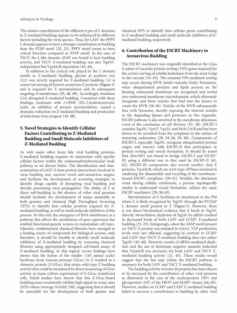
6. Contribution of the ESCRT Machinery toArenavirus Budding
The ESCRT machinery was originally identified as the ClassE subset of vacuolar protein sorting (VPS) genes required forthe correct sorting of soluble hydrolases from the yeast Golgito the vacuole [51–53]. The essential VPS-mediated sortingstep occurs during MVB (multi vesicular body) formation,when ubiquitinated proteins and lipids present on thelimiting endosomal membrane are recognized and sortedinto endosomal membrane microdomains, which ultimatelyinvaginate and form vesicles that bud into the lumen tocreate the MVB [54–56]. Vesicles of the MVB subsequentlyfuse with lysosome, thereby exposing the internal vesiclesto the degrading lipases and proteases in this organelle.ESCRT pathway is also involved in the membrane abscissionevent at the conclusion of cell division [57, 58]. ESCRT-Icontains Tsg101, Vps37, Vps23, and Mvb12A/B and has beenshown to be recruited from the cytoplasm to the surface ofmaturing endosomes [29, 54–56, 59, 60]. Components ofESCRT-I, especially Tsg101, recognize ubiquitinated proteincargos and interact with ESCRT-II that participates inprotein sorting and vesicle formation. It should be notedthat Alix/AIP1 was found to bridge ESCRT-I and ESCRT-III using a different way to that used by ESCRT-II [61,62]. ESCRT-III components also recruit another class Eproteins, Vps4A/B, which are AAA-type ATPases involved incatalyzing the disassembly and recycling of the membrane-bound ESCRT complexes [29–31]. Notably, the abscissionevent during cellular cytokinesis, a process topologicallysimilar to endosomal vesicle formation, utilizes the sameESCRT machinery [29, 58, 63].
Myristoylation of Z facilitates its attachment to the PMwhere Z is likely recognized by Tsg101 through the PT/SAPL domain motif present in Z (Figure 3). However, thereis not direct biochemical evidence that Z binds to Tsg101directly. Nevertheless, depletion of Tsg101 by siRNA resultedin decreased levels of both LASV and LCMV Z-mediatedbudding [21, 23]. Intriguingly, when the ASAP motif presenton TACV-Z protein was mutated to AAAA, VLP productionlevels were not affected, suggesting in contrast to LCMVand LASV that TACV Z-mediated budding does not utilizeTsg101 [43, 44]. However, results of siRNA-mediated deple-tion and the use of dominant negative mutants indicatedthat Vps4A/B was necessary for both LASV and TACV Z-mediated budding activity [21, 43]. These results wouldsuggest that the last step within the ESCRT pathway isnecessary for both LASV and TACV Z-mediated budding.
The budding activity of some M proteins has been shownto be increased by the contribution of other viral proteinsas illustrated in the case of the nucleoprotein (NP) andglycoprotein (GP) of the EBOV and MARV viruses [64, 65].However, studies on LCMV and LASV Z-mediated buddingfailed to uncover a contribution to budding by other viral

6 Advances in Virology
N-t
erm
RIN
GC
-ter
m Ldo
mai
n(s
)
Z protein
Plasma emm brane
Tsg101
Ubiquitinligase?
ESCRT-Icomplex
ESCRT pathway
Myr
?
NP
NP
IGR IGR
LL
L segment S segment
NP
SSP
GP1
GP2
Virus particle
Cytoplasm
Figure 3: Model of arenavirus budding. Myristoylation of Z at G2facilitates its interaction with the plasma membrane (PM), where Zlikely forms higher-order complexes. L domains located within theC-terminus of Z facilitate its interactions with host cellular factorsto allow Z to utilize the ESCRT machinery of the cell for cell egress(budding). SSP, stable signal peptide.
protein. In contrast, TACV NP was shown to enhance Z-mediated VLP production [44].
Nevertheless, recently published work has shown thatMopeia virus (MOPV) NP incorporation into VLP ismediated by AIP1/Alix via interaction with the YLCL motifpresent on Z [45, 46].
7. Tetherin/BST-2/CD317 as an Antagonist ofArenavirus Budding
Tetherin was identified as an IFN-inducible antiviral cellularfactor that tether HIV virions at the PM [66, 67]. Subse-quently studies have extended these findings to other viruses[68]. As with other host innate immune defense factors,several viruses have evolved mechanisms to counteracttetherin-mediated antiviral activity [69]. Tetherin has beenshown to inhibit LASV Z-mediated budding [70]. Accord-ingly, 293T cells constitutively expressing tetherin resultedin decreased production levels of LASV and MACV virionparticle production, whereas siRNA-mediated knockdownof endogenous tetherin in HeLa cells resulted in increasedproduction levels of LASV and MACV virion particleproduction. These results would suggest that LASV does notpossess any tetherin-antagonizing function as described for
other viral proteins including HIV-1 Vpu, EBOV GP, andKSHV K5 [66, 71–73].
An issue that remains to be investigated relates to the con-tribution of tetherin to host protection and viral pathogene-sis. An attractive hypothesis, but still without experimentalsupport, would be that tetherin could do to some degreeslow the process of virus propagation in vivo and therebyfacilitating both the action of the host innate immunedefense mechanisms and antigen presentation leading toa more robust host adaptive immune response that couldcontrol and eliminate the virus.
8. Perspectives on ArenavirusZ-Mediated Budding
Current evidence indicates that many enveloped virusesuse the ESCRT machinery to exit from the cell. In thecase of arenavirus budding, Z-Tsg101 interaction appearsto facilitate access of Z to the ESCRT machinery. Despitesignificant recent progress in defining the basic aspects ofarenavirus budding, there are still a large number of issuesthat have not been investigated including: (1) Identificationand functional characterization of host cellular factors thatinteract with the PPXY L domain motif present in LASV,LCMV, and some other arenavirus Z to facilitate virus bud-ding. PPXY L domain motifs present in EBOV and MARVVP40 have shown to mediate interaction with Nedd4.1 [74–76]. Likewise, for several retroviruses, the interaction of GagPPXY L domains with ubiquitin ligases has been shown tocontribute to the regulation of viral budding [24, 25, 38,39]. How ubiquitin ligases may regulate budding remainsunknown, but recent published data have shown arrestin-related proteins to connect ubiquitin ligases and the ESCRTmachinery [77]. It is important to know whether specificubiquitin ligase may regulate Z-mediated budding, and ifso, what are the mechanism underlying this regulation. (2)Ubiquitin or ubiquitin-like molecules (UBLs) have beenshown to modify the properties and budding activity ofHIV-1 Gag and EBOV VP40 proteins [78]. It is currentlyunknown whether these protein modifiers may also havea role in the regulation of arenavirus budding. (3) Themechanisms by which arenavirus RNP interacts with Z andGP to form budding mature infectious progeny are largelyunknown. (4) Whether the species-specific and type of cellinfluence arenavirus budding and the biological implicationsregarding the outcome of infection are issues that have notbeen investigated.
Detailed understanding of the virus-host cell proteininteractions that direct arenavirus budding may uncovernovel targets for the development of antiviral drugs tocombat human pathogenic arenaviruses.
Acknowledgments
This is Publication no. 21130 from the Department ofImmunology and Microbial Science, The Scripps ResearchInstitute (TSRI), La Jolla, Calif, USA. Because space limita-tions, we have relied extensively in recent reviews where more

Advances in Virology 7
extensive detailed information, including all the original cita-tions, can be found regarding the different topics discussed inthis paper. Recognition and apologies are given in advance tothe many colleagues whose original contributions have notbeen possible to cite. The research contributed by the authorsof this paper was supported by NIH grants RO1 AI047140,RO1 AI077719 and RO1 AI079665 to J. C. de la Torre andT32-AI0735419 to S. Urata.
References
[1] M. J. Buchmeier, C. J. Peters, and J. C. de la Torre, “Arenaviri-dae: the virus and their replication,” Fields Virology, vol. 2, pp.1792–1827, 2007.
[2] M. B. Oldstone, “Biology and pathogenesis of lymphocyticchoriomeningitis virus infection,” Current Topics in Microbi-ology and Immunology, vol. 263, pp. 83–117, 2002.
[3] R. M. Zinkernagel, “Lymphocytic choriomeningitis virus andimmunology,” Current Topics in Microbiology and Immunol-ogy, vol. 263, pp. 1–5, 2002.
[4] W. Cao, M. D. Henry, P. Borrow et al., “Identification of α-dystroglycan as a receptor for lymphocytic choriomeningitisvirus and Lassa fever virus,” Science, vol. 282, no. 5396, pp.2079–2081, 1998.
[5] S. Kunz, P. Borrow, and M. B. Oldstone, “Receptor structure,binding, and cell entry of arenaviruses,” Current Topics inMicrobiology and Immunology, vol. 262, pp. 111–137, 2002.
[6] S. R. Radoshitzky, J. Abraham, C. F. Spiropoulou et al., “Trans-ferrin receptor 1 is a cellular receptor for New Worldhaemorrhagic fever arenaviruses,” Nature, vol. 446, no. 7131,pp. 92–96, 2007.
[7] S. Kunz, “Receptor binding and cell entry of Old Worldarenaviruses reveal novel aspects of virus-host interaction,”Virology, vol. 387, no. 2, pp. 245–249, 2009.
[8] J. M. Rojek, M. Perez, and S. Kunz, “Cellular entry oflymphocytic choriomeningitis virus,” Journal of Virology, vol.82, no. 3, pp. 1505–1517, 2008.
[9] J. M. Rojek, A. B. Sanchez, N. T. Nguyen, J. C. de La Torre,and S. Kunz, “Different mechanisms of cell entry by human-pathogenic Old World and New World arenaviruses,” Journalof Virology, vol. 82, no. 15, pp. 7677–7687, 2008.
[10] M. Hass, U. Golnitz, S. Muller, B. Becker-Ziaja, and S.Gunther, “Replicon system for Lassa virus,” Journal of Virology,vol. 78, no. 24, pp. 13793–13803, 2004.
[11] K. J. Lee, I. S. Novella, M. N. Teng, M. B. Oldstone, and J. C. deLa Torre, “NP and L proteins of lymphocytic choriomeningitisvirus (LCMV) are sufficient for efficient transcription andreplication of LCMV genomic RNA analogs,” Journal ofVirology, vol. 74, no. 8, pp. 3470–3477, 2000.
[12] N. Lopez, R. Jacamo, and M. T. Franze-fernandez, “Tran-scription and RNA replication of tacaribe virus genome andantigenome analogs require n and l proteins: Z protein is aninhibitor of these processes,” Journal of Virology, vol. 75, no.24, pp. 12241–12251, 2001.
[13] T. I. Cornu and J. C. de La Torre, “Ring finger z protein oflymphocytic choriomeningitis virus (lcmv) inhibits transcrip-tion and rna replication of an lcmv s-segment minigenome,”Journal of Virology, vol. 75, no. 19, pp. 9415–9426, 2001.
[14] T. I. Cornu and J. C. de la Torre, “Characterization ofthe arenavirus RING finger Z protein regions required forZ-mediated inhibition of viral RNA synthesist,” Journal ofVirology, vol. 76, no. 13, pp. 6678–6688, 2002.
[15] K. L. Borden, E. J. Campbell Dwyer, and M. S. Salvato, “Anarenavirus RING (zinc-binding) protein binds the oncopro-tein promyelocyte leukemia protein (PML) and relocates PMLnuclear bodies to the cytoplasm,” Journal of Virology, vol. 72,no. 1, pp. 758–766, 1998.
[16] A. Kentsis, E. C. Dwyer, J. M. Perez et al., “The RING domainsof the promyelocytic leukemia protein PML and the arenaviralprotein Z repress translation by directly inhibiting translationinitiation factor eIF4E,” Journal of Molecular Biology, vol. 312,no. 4, pp. 609–623, 2001.
[17] L. Volpon, M. J. Osborne, A. A. Capul, J. C. de La Torre, andK. L. Borden, “Structural characterization of the Z RING-eIF4E complex reveals a distinct mode of control for eIF4E,”Proceedings of the National Academy of Sciences of the UnitedStates of America, vol. 107, no. 12, pp. 5441–5446, 2010.
[18] E. J. Campbell Dwyer, H. Lai, R. C. MacDonald, M. S. Salvato,and K. L. Borden, “The lymphocytic choriomeningitis virusRING protein Z associates with eukaryotic initiation factor4E and selectively represses translation in a RING-dependentmanner,” Journal of Virology, vol. 74, no. 7, pp. 3293–3300,2000.
[19] R. Colina, M. Costa-Mattioli, R. J. Dowling et al., “Transla-tional control of the innate immune response through IRF-7,”Nature, vol. 452, no. 7185, pp. 323–328, 2008.
[20] L. Fan, T. Briese, and W. I. Lipkin, “Z proteins of new worldarenaviruses bind RIG-I and interfere with type i interferoninduction,” Journal of Virology, vol. 84, no. 4, pp. 1785–1791,2010.
[21] S. Urata, T. Noda, Y. Kawaoka, H. Yokosawa, and J. Yasuda,“Cellular factors required for Lassa virus budding,” Journal ofVirology, vol. 80, no. 8, pp. 4191–4195, 2006.
[22] T. Strecker, R. Eichler, J. Meulen et al., “Lassa virus Z proteinis a matrix protein sufficient for the release of virus-likeparticles,” Journal of Virology, vol. 77, no. 19, pp. 10700–10705,2003.
[23] M. Perez, R. C. Craven, and J. C. de la Torre, “The small RINGfinger protein Z drives arenavirus budding: implications forantiviral strategies,” Proceedings of the National Academy ofSciences of the United States of America, vol. 100, no. 22, pp.12978–12983, 2003.
[24] B. J. Chen and R. A. Lamb, “Mechanisms for enveloped virusbudding: can some viruses do without an ESCRT?” Virology,vol. 372, no. 2, pp. 221–232, 2008.
[25] P. D. Bieniasz, “Late budding domains and host proteins inenveloped virus release,” Virology, vol. 344, no. 1, pp. 55–63,2006.
[26] E. Morita and W. I. Sundquist, “Retrovirus budding,” AnnualReview of Cell and Developmental Biology, vol. 20, pp. 395–425,2004.
[27] J. E. Garrus, U. K. von Schwedler, O. W. Pornillos et al.,“Tsg101 and the vacuolar protein sorting pathway are essentialfor HIV-1 budding,” Cell, vol. 107, no. 1, pp. 55–65, 2001.
[28] L. VerPlank, F. Bouamr, T. J. LaGrassa et al., “Tsg101, ahomologue of ubiquitin-conjugating (E2) enzymes, binds theL domain in HIV type 1 Pr55Gag,” Proceedings of the NationalAcademy of Sciences of the United States of America, vol. 98, no.14, pp. 7724–7729, 2001.
[29] J. H. Hurley and P. I. Hanson, “Membrane budding andscission by the ESCRT machinery: it’s all in the neck,” NatureReviews Molecular Cell Biology, vol. 11, no. 8, pp. 556–566,2010.

8 Advances in Virology
[30] M. Babst, T. K. Sato, L. M. Banta, and S. D. Emr, “Endosomaltransport function in yeast requires a novel AAA-type ATPase,Vps4p,” EMBO Journal, vol. 16, no. 8, pp. 1820–1831, 1997.
[31] M. Babst, B. Wendland, E. J. Estepa, and S. D. Emr, “The Vps4pAAA ATPase regulates membrane association of a Vps proteincomplex required for normal endosome function,” EMBOJournal, vol. 17, no. 11, pp. 2982–2993, 1998.
[32] O. Dolnik, L. Kolesnikova, L. Stevermann, and S. Becker,“Tsg101 is recruited by a late domain of the nucleocapsidprotein to support budding of Marburg virus-like particles,”Journal of Virology, vol. 84, no. 15, pp. 7847–7856, 2010.
[33] T. Irie, N. Nagata, T. Yoshida, and T. Sakaguchi, “Paramyx-ovirus Sendai virus C proteins are essential for maintenance ofnegative-sense RNA genome in virus particles,” Virology, vol.374, no. 2, pp. 495–505, 2008.
[34] T. Sakaguchi, A. Kato, F. Sugahara et al., “AIP1/Alix is abinding partner of Sendai virus C protein and facilitates virusbudding,” Journal of Virology, vol. 79, no. 14, pp. 8933–8941,2005.
[35] T. Irie, Y. Shimazu, T. Yoshida, and T. Sakaguchi, “The YLDLsequence within sendai virus M protein is critical for buddingof virus-like particles and interacts with Alix/AIP1 indepen-dently of C protein,” Journal of Virology, vol. 81, no. 5, pp.2263–2273, 2007.
[36] J. S. Rossman and R. A. Lamb, “Influenza virus assembly andbudding,” Virology, vol. 411, no. 2, pp. 229–236, 2011.
[37] T. Irie, J. M. Licata, J. P. McGettigan, M. J. Schnell, and R.N. Harty, “Budding of PPxY-containing rhabdoviruses is notdependent on host proteins TGS101 and VPS4A,” Journal ofVirology, vol. 78, no. 6, pp. 2657–2665, 2004.
[38] A. Kikonyogo, F. Bouamr, M. L. Vana et al., “Proteins relatedto the Nedd4 family of ubiquitin protein ligases interact withthe L domain of Rous sarcoma virus and are required for gagbudding from cells,” Proceedings of the National Academy ofSciences of the United States of America, vol. 98, no. 20, pp.11199–11204, 2001.
[39] M. L. Vana, Y. Tang, A. Chen, G. Medina, C. Carter, and J.Leis, “Role of Nedd4 and ubiquitination of rous sarcoma virusGag in budding of virus-like particles from cells,” Journal ofVirology, vol. 78, no. 24, pp. 13943–13953, 2004.
[40] K. A. Dilley, D. Gregory, M. C. Johnson, and V. M. Vogt,“An LYPSL late domain in the Gag protein contributes tothe efficient release and replication of Rous sarcoma virus,”Journal of Virology, vol. 84, no. 13, pp. 6276–6287, 2010.
[41] T. Briese, J. T. Paweska, L. K. McMullan et al., “Genetic detec-tion and characterization of Lujo virus, a new hemor-rhagic fever-associated arenavirus from southern Africa,” PLoSPathogens, vol. 5, no. 5, Article ID e1000455, 2009.
[42] G. Palacios, N. Savji, J. Hui et al., “Genomic and phylogeneticcharacterization of Merino Walk virus, a novel arenavirusisolated in South Africa,” Journal of General Virology, vol. 91,no. 5, pp. 1315–1324, 2010.
[43] S. Urata, J. Yasuda, and J. C. de La Torre, “The Z protein ofthe new world arenavirus tacaribe virus has bona fide buddingactivity that does not depend on known late domain motifs,”Journal of Virology, vol. 83, no. 23, pp. 12651–12655, 2009.
[44] A. Groseth, S. Wolff, T. Strecker, T. Hoenen, and S. Becker,“Efficient budding of the tacaribe virus matrix protein Zrequires the nucleoprotein,” Journal of Virology, vol. 84, no. 7,pp. 3603–3611, 2010.
[45] O. Shtanko, M. Imai, H. Goto et al., “A role for the C terminusof mopeia virus nucleoprotein in its incorporation into Zprotein-induced virus-like particles,” Journal of Virology, vol.84, no. 10, pp. 5415–5422, 2010.
[46] O. Shtanko, S. Watanabe, L. D. Jasenosky, T. Watanabe, andY. Kawaoka, “ALIX/AIP1 is required for NP incorporationinto Mopeia virus Z-induced virus-like particles,” Journal ofVirology, vol. 85, no. 7, pp. 3631–3641, 2011.
[47] A. S. Gosselin-Grenet, J. B. Marq, L. Abrami, D. Garcin, andL. Roux, “Sendai virus budding in the course of an infectiondoes not require Alix and VPS4A host factors,” Virology, vol.365, no. 1, pp. 101–112, 2007.
[48] M. Perez, D. L. Greenwald, and J. C. de La Torre, “Myristoy-lation of the RING finger Z protein is essential for arenavirusbudding,” Journal of Virology, vol. 78, no. 20, pp. 11443–11448,2004.
[49] T. Strecker, A. Maisa, S. Daffis, R. Eichler, O. Lenz, andW. Garten, “The role of myristoylation in the membraneassociation of the Lassa virus matrix protein Z,” VirologyJournal, vol. 3, article 93, 2006.
[50] A. A. Capul and J. C. de la Torre, “A cell-based luciferaseassay amenable to high-throughput screening of inhibitors ofarenavirus budding,” Virology, vol. 382, no. 1, pp. 107–114,2008.
[51] V. A. Bankaitis, L. M. Johnson, and S. D. Emr, “Isolation ofyeast mutants defective in protein targeting to the vacuole,”Proceedings of the National Academy of Sciences of the UnitedStates of America, vol. 83, no. 23, pp. 9075–9079, 1986.
[52] J. S. Robinson, D. J. Klionsky, L. M. Banta, and S. D. Emr,“Protein sorting in Saccharomyces cerevisiae: isolation ofmutants defective in the delivery and processing of multiplevacuolar hydrolases,” Molecular and Cellular Biology, vol. 8, no.11, pp. 4936–4948, 1988.
[53] C. K. Raymond, I. Howald-Stevenson, C. A. Vater, andT. H. Stevens, “Morphological classification of the yeastvacuolar protein sorting mutants: evidence for a prevacuolarcompartment in class E vps mutants,” Molecular Biology of theCell, vol. 3, no. 12, pp. 1389–1402, 1992.
[54] D. J. Katzmann, M. Babst, and S. D. Emr, “Ubiquitin-dependent sorting into the multivesicular body pathwayrequires the function of a conserved endosomal proteinsorting complex, ESCRT-I,” Cell, vol. 106, no. 2, pp. 145–155,2001.
[55] M. Babst, D. J. Katzmann, E. J. Estepa-Sabal, T. Meerloo,and S. D. Emr, “ESCRT-III: an endosome-associated het-erooligomeric protein complex required for MVB sorting,”Developmental Cell, vol. 3, no. 2, pp. 271–282, 2002.
[56] M. Babst, D. J. Katzmann, W. B. Snyder, B. Wendland, andS. D. Emr, “Endosome-associated complex, ESCRT-II, recruitstransport machinery for protein sorting at the multivesicularbody,” Developmental Cell, vol. 3, no. 2, pp. 283–289, 2002.
[57] E. Morita, V. Sandrin, H. Y. Chung et al., “Human ESCRTand ALIX proteins interact with proteins of the midbody andfunction in cytokinesis,” EMBO Journal, vol. 26, no. 19, pp.4215–4227, 2007.
[58] J. G. Carlton and J. Martin-Serrano, “Parallels betweencytokinesis and retroviral budding: a role for the ESCRTmachinery,” Science, vol. 316, no. 5833, pp. 1908–1912, 2007.
[59] T. Chu, J. Sun, S. Saksena, and S. D. Emr, “New componentof ESCRT-I regulates endosomal sorting complex assembly,”Journal of Cell Biology, vol. 175, no. 5, pp. 815–823, 2006.
[60] E. Morita, V. Sandrin, S. L. Alam, D. M. Eckert, S. P. Gygi, andW. I. Sundquist, “Identification of human MVB12 proteins asESCRT-I subunits that function in HIV budding,” Cell Hostand Microbe, vol. 2, no. 1, pp. 41–53, 2007.
[61] B. Strack, A. Calistri, S. Craig, E. Popova, and H. G. Gottlinger,“AIP1/ALIX is a binding partner for HIV-1 p6 and EIAV p9

Advances in Virology 9
functioning in virus budding,” Cell, vol. 114, no. 6, pp. 689–699, 2003.
[62] J. Martin-Serrano, A. Yarovoy, D. Perez-Caballero, and P. D.Bieniasz, “Divergent retroviral late-budding domains recruitvacuolar protein sorting factors by using alternative adaptorproteins,” Proceedings of the National Academy of Sciences ofthe United States of America, vol. 100, no. 21, pp. 12414–12419,2003.
[63] E. Morita, L. A. Colf, M. A. Karren, V. Sandrin, C. K. Rodesch,and W. I. Sundquist, “Human ESCRT-III and VPS4 proteinsare required for centrosome and spindle maintenance,” Pro-ceedings of the National Academy of Sciences of the United Statesof America, vol. 107, no. 29, pp. 12889–12894, 2010.
[64] S. Urata, T. Noda, Y. Kawaoka, S. Morikawa, H. Yokosawa, andJ. Yasuda, “Interaction of Tsg101 with Marburg virus VP40depends on the PPPY motif, but not the PT/SAP motif as inthe case of Ebola virus, and Tsg101 plays a critical role in thebudding of Marburg virus-like particles induced by VP40, NP,and GP,” Journal of Virology, vol. 81, no. 9, pp. 4895–4899,2007.
[65] J. M. Licata, R. F. Johnson, Z. Han, and R. N. Harty,“Contribution of Ebola virus glycoprotein, nucleoprotein, andVP24 to budding of VP40 virus-like particles,” Journal ofVirology, vol. 78, no. 14, pp. 7344–7351, 2004.
[66] S. J. Neil, T. Zang, and P. D. Bieniasz, “Tetherin inhibitsretrovirus release and is antagonized by HIV-1 Vpu,” Nature,vol. 451, no. 7177, pp. 425–430, 2008.
[67] N. Van Damme, D. Goff, C. Katsura et al., “The interferon-induced protein BST-2 restricts HIV-1 release and is downreg-ulated from the cell surface by the viral Vpu protein,” Cell Hostand Microbe, vol. 3, no. 4, pp. 245–252, 2008.
[68] D. T. Evans, R. Serra-Moreno, R. K. Singh, and J. C. Guatelli,“BST-2/tetherin: a new component of the innate immuneresponse to enveloped viruses,” Trends in Microbiology, vol. 18,no. 9, pp. 388–396, 2010.
[69] J. L. Douglas, J. K. Gustin, K. Viswanathan, M. Mansouri, A.V. Moses, and K. Fruh, “The great escape: viral strategies tocounter BST-2/tetherin,” PLoS Pathogens, vol. 6, no. 5, ArticleID e1000913, 2010.
[70] T. Sakuma, T. Noda, S. Urata, Y. Kawaoka, and J. Yasuda, “Inhi-bition of lassa and marburg virus production by tetherin,”Journal of Virology, vol. 83, no. 5, pp. 2382–2385, 2009.
[71] R. L. Kaletsky, J. R. Francica, C. Agrawal-Gamse, and P.Bates, “Tetherin-mediated restriction of filovirus budding isantagonized by the Ebola glycoprotein,” Proceedings of theNational Academy of Sciences of the United States of America,vol. 106, no. 8, pp. 2886–2891, 2009.
[72] S. R. Radoshitzky, L. Dong, X. Chi et al., “Infectious Lassavirus, but not filoviruses, is restricted by BST-2/tetherin,”Journal of Virology, vol. 84, no. 20, pp. 10569–10580, 2010.
[73] E. Bartee, A. McCormack, and K. Fruh, “Quantitative mem-brane proteomics reveals new cellular targets of viral immunemodulators,” PLoS Pathogens, vol. 2, no. 10, article e107, 2006.
[74] S. Urata and J. Yasuda, “Regulation of Marburg virus (MARV)budding by Nedd4.1: a different WW domain of Nedd4.1 iscritical for binding to MARV and Ebola virus VP40,” Journalof General Virology, vol. 91, no. 1, pp. 228–234, 2010.
[75] J. Yasuda, M. Nakao, Y. Kawaoka, and H. Shida, “Nedd4regulates egress of Ebola virus-like particles from host cells,”Journal of Virology, vol. 77, no. 18, pp. 9987–9992, 2003.
[76] J. Timmins, G. Schoehn, S. Ricard-Blum et al., “Ebola virusmatrix protein VP40 interaction with human cellular factorsTsg101 and Nedd4,” Journal of Molecular Biology, vol. 326, no.2, pp. 493–502, 2003.
[77] S. Rauch and J. Martin-Serrano, “Multiple interactionsbetween the ESCRT machinery and arrestin-related proteins:implications for PPXY-dependent budding,” Journal of Virol-ogy, vol. 85, no. 7, pp. 3546–3556, 2011.
[78] R. N. Harty, P. M. Pitha, and A. Okumura, “Antiviral activityof innate immune protein ISG15,” Journal of Innate Immunity,vol. 1, no. 5, pp. 397–404, 2009.

Submit your manuscripts athttp://www.hindawi.com
Hindawi Publishing Corporationhttp://www.hindawi.com Volume 2014
Anatomy Research International
PeptidesInternational Journal of
Hindawi Publishing Corporationhttp://www.hindawi.com Volume 2014
Hindawi Publishing Corporation http://www.hindawi.com
International Journal of
Volume 2014
Zoology
Hindawi Publishing Corporationhttp://www.hindawi.com Volume 2014
Molecular Biology International
GenomicsInternational Journal of
Hindawi Publishing Corporationhttp://www.hindawi.com Volume 2014
The Scientific World JournalHindawi Publishing Corporation http://www.hindawi.com Volume 2014
Hindawi Publishing Corporationhttp://www.hindawi.com Volume 2014
BioinformaticsAdvances in
Marine BiologyJournal of
Hindawi Publishing Corporationhttp://www.hindawi.com Volume 2014
Hindawi Publishing Corporationhttp://www.hindawi.com Volume 2014
Signal TransductionJournal of
Hindawi Publishing Corporationhttp://www.hindawi.com Volume 2014
BioMed Research International
Evolutionary BiologyInternational Journal of
Hindawi Publishing Corporationhttp://www.hindawi.com Volume 2014
Hindawi Publishing Corporationhttp://www.hindawi.com Volume 2014
Biochemistry Research International
ArchaeaHindawi Publishing Corporationhttp://www.hindawi.com Volume 2014
Hindawi Publishing Corporationhttp://www.hindawi.com Volume 2014
Genetics Research International
Hindawi Publishing Corporationhttp://www.hindawi.com Volume 2014
Advances in
Virolog y
Hindawi Publishing Corporationhttp://www.hindawi.com
Nucleic AcidsJournal of
Volume 2014
Stem CellsInternational
Hindawi Publishing Corporationhttp://www.hindawi.com Volume 2014
Hindawi Publishing Corporationhttp://www.hindawi.com Volume 2014
Enzyme Research
Hindawi Publishing Corporationhttp://www.hindawi.com Volume 2014
International Journal of
Microbiology