review article gingival mesenchymal stem/progenitor cells...
TRANSCRIPT

Review ArticleGingival Mesenchymal Stem/Progenitor Cells:A Unique Tissue Engineering Gem
Karim M. Fawzy El-Sayed1,2 and Christof E. Dörfer1
1Clinic of Conservative Dentistry and Periodontology, Christian Albrechts University of Kiel, Arnold Heller Straße 3,Haus 26, 24105 Kiel, Germany2Oral Medicine and Periodontology Department, Faculty of Oral and Dental Medicine, Cairo University, Egypt
Correspondence should be addressed to Karim M. Fawzy El-Sayed; [email protected]
Received 19 January 2016; Revised 28 March 2016; Accepted 28 April 2016
Academic Editor: Mahmoud Rouabhia
Copyright © 2016 K. M. Fawzy El-Sayed and C. E. Dorfer. This is an open access article distributed under the Creative CommonsAttribution License, which permits unrestricted use, distribution, and reproduction in any medium, provided the original work isproperly cited.
The human gingiva, characterized by its outstanding scarless wound healing properties, is a unique tissue and a pivotal componentof the periodontal apparatus, investing and surrounding the teeth in their sockets in the alveolar bone. In the last years gingivalmesenchymal stem/progenitor cells (G-MSCs), with promising regenerative and immunomodulatory properties, have been isolatedand characterized from the gingival lamina propria. These cells, in contrast to other mesenchymal stem/progenitor cell sources,are abundant, readily accessible, and easily obtainable via minimally invasive cell isolation techniques. The present reviewsummarizes the current scientific evidence on G-MSCs’ isolation, their characterization, the investigated subpopulations, thegenerated induced pluripotent stem cells- (iPSC-) like G-MSCs, their regenerative properties, and current approaches for G-MSCs’delivery. The review further demonstrates their immunomodulatory properties, the transplantation preconditioning attempts viamultiple biomolecules to enhance their attributes, and the experimental therapeutic applications conducted to treat multiplediseases in experimental animal models in vivo. G-MSCs show remarkable tissue reparative/regenerative potential, noteworthyimmunomodulatory properties, and primary experimental therapeutic applications of G-MSCs are very promising, pointing atfuture biologically based therapeutic techniques, being potentially superior to conventional clinical treatment modalities.
1. Introduction
The human periodontium, the tooth supporting and invest-ing organ, comprising the alveolar bone, the periodontalligament, the root cementum, and the gingiva developsand functions as one unit. The majority of the periodon-tal tissues originate embryonically from the neural crestectomesenchyme [1]. The gingiva, histologically composedof epithelium and connective tissue, constitutes a distinctiveas well as a pivotal component of the human periodontiumdevelopmentally and anatomically, surrounding the necks ofthe teeth and investing the tooth-bearing alveolar bone. Oneof the gingiva’s renowned characteristics is its notable woundhealing and regenerative aptitude, with a fast reconstitutionof tissue architecture following injury or excision with little,if any, evidence of scarring [2]. This tissue is easily accessibleand is often resected during standard surgical procedures,
including dental crown lengthening andmultiple periodontalsurgeries, with minimal discomfort to the patient [3].
Developmentally, the craniofacial ectomesenchyme isderived from the neural crest and the mesoderm. Themultipotent cranial neural crest cells (CNCCs) migrate ven-trolaterally to reside in the first branchial arches, startingfrom the four-somite stage, giving rise to mesenchymalstructures in the craniofacial region, including neural tissues,cartilage, bone, and teeth [4, 5]. In addition to a commonneural crest ectomesenchymal origin, lined by ectoderm forall oral soft tissues, the tooth-investing gingival connectivetissue shows a unique developmental origin, arising partlyfrom the perifollicular mesenchyme (the outer layer of thedental follicle) [1], as well as partly from the dental follicleproper (the inner layer of the dental follicle) [6], from whichdental follicle stem/progenitor cells (DFSCs) were isolated[7]. Periodontal ligament cells [8], originating themselves
Hindawi Publishing CorporationStem Cells InternationalVolume 2016, Article ID 7154327, 16 pageshttp://dx.doi.org/10.1155/2016/7154327
2 Stem Cells International
Neural crest ectomesenchyme Dental follicle
Gin
giva
l lam
ina p
ropr
ia
Inner layer(dental follicle proper)
DFSCs PDLSCs G-MSCs
(perifollicularOuter layer
mesenchyme)
Periodontal ligament cells
Fibroblasts at CEJ
Embryonic life Tooth eruption
Time
Dentogingival fiber system
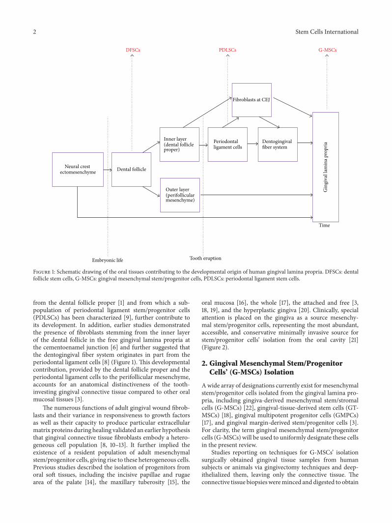
Figure 1: Schematic drawing of the oral tissues contributing to the developmental origin of human gingival lamina propria. DFSCs: dentalfollicle stem cells, G-MSCs: gingival mesenchymal stem/progenitor cells, PDLSCs: periodontal ligament stem cells.
from the dental follicle proper [1] and from which a sub-population of periodontal ligament stem/progenitor cells(PDLSCs) has been characterized [9], further contribute toits development. In addition, earlier studies demonstratedthe presence of fibroblasts stemming from the inner layerof the dental follicle in the free gingival lamina propria atthe cementoenamel junction [6] and further suggested thatthe dentogingival fiber system originates in part from theperiodontal ligament cells [8] (Figure 1). This developmentalcontribution, provided by the dental follicle proper and theperiodontal ligament cells to the perifollicular mesenchyme,accounts for an anatomical distinctiveness of the tooth-investing gingival connective tissue compared to other oralmucosal tissues [3].
The numerous functions of adult gingival wound fibrob-lasts and their variance in responsiveness to growth factorsas well as their capacity to produce particular extracellularmatrix proteins during healing validated an earlier hypothesisthat gingival connective tissue fibroblasts embody a hetero-geneous cell population [8, 10–13]. It further implied theexistence of a resident population of adult mesenchymalstem/progenitor cells, giving rise to these heterogeneous cells.Previous studies described the isolation of progenitors fromoral soft tissues, including the incisive papillae and rugaearea of the palate [14], the maxillary tuberosity [15], the
oral mucosa [16], the whole [17], the attached and free [3,18, 19], and the hyperplastic gingiva [20]. Clinically, specialattention is placed on the gingiva as a source mesenchy-mal stem/progenitor cells, representing the most abundant,accessible, and conservative minimally invasive source forstem/progenitor cells’ isolation from the oral cavity [21](Figure 2).
2. Gingival Mesenchymal Stem/ProgenitorCells’ (G-MSCs) Isolation
Awide array of designations currently exist for mesenchymalstem/progenitor cells isolated from the gingival lamina pro-pria, including gingiva-derived mesenchymal stem/stromalcells (G-MSCs) [22], gingival-tissue-derived stem cells (GT-MSCs) [18], gingival multipotent progenitor cells (GMPCs)[17], and gingival margin-derived stem/progenitor cells [3].For clarity, the term gingival mesenchymal stem/progenitorcells (G-MSCs) will be used to uniformly designate these cellsin the present review.
Studies reporting on techniques for G-MSCs’ isolationsurgically obtained gingival tissue samples from humansubjects or animals via gingivectomy techniques and deep-ithelialized them, leaving only the connective tissue. Theconnective tissue biopsieswereminced anddigested to obtain

Stem Cells International 3
(1) DPSCs and SHEDs (2) Alveolar bone proper-derived stem cells(3) PDLSCs
(7) G-MSCs(6) DFSCs(5) SCAP
(1)
(2)
(3)
(4)
(5)
(6)
(7)
(4) BM-MSCs
Figure 2: Sources of oral stem/progenitor cells isolated. DFSCs:dental follicle stem cells, G-MSCs: gingival mesenchymal stem/pro-genitor cells, PDLSCs: periodontal ligament stem cells, SHEDs: stemcells from the human exfoliated deciduous teeth,DPSCs: dental pulpstem cells, BM-MSCs: bone marrow mesenchymal stem cells, andSCAP: stem cells from the apical papilla.
single-cell suspensions [18, 20, 23–25] or kept intact and thetissue explants culture method was used to grow out theadherent connective tissue cells [3, 15, 26, 27]. The obtainedcells were subsequently cultured and expanded in vitro for 3-4 weeks.
Diverse G-MSCs’ isolation and expansion protocols wereproposed (Table 1). Some of the outlined protocols, except forI, III, IV, andV, did not attempt to select stem/progenitor cells’population from the heterogeneous gingival connective tissuecells via single-cell cloning [23, 28, 29] or magnetic activatedcell sorting (MACS) techniques [3, 30].This raises a questionabout whether thereafter characterized cultures would rep-resent enriched mesenchymal stem/progenitor cell culturesor merely mixed gingival connective tissue cell cultures,encompassing stem/progenitor cells in their original lowpercentages, usually present in the gingival lamina propria.A recent study relying on a STRO-1/MACS scheme for G-MSCs’ isolation underlined the importance of the utilizationof a cell selection/sorting technique for G-MSCs’ isolation,pointing out that two cell populations, a STRO-1/MACS+ anda STRO-1/MACS−, with distinctive properties and markerexpression profiles exist in the human gingival connectivetissue. The study demonstrated that the STRO-1/MACS+-cell population, in contrast to the STRO-1/MACS− one,harbored the cells with stem/progenitor cells’ characteristicsand distinctive osteogenic marker expression and validated
thereby the effectiveness of the STRO-1/MACS technique inthe field of G-MSCs’ isolation [3].
3. G-MSCs’ Characterization
To characterize G-MSCs and compare their properties tobone marrow mesenchymal stromal cells (BM-MSCs), theforerunner and gold standard in the field mesenchymal stro-mal cells’ (MSCs) isolation, characterization, and research[31], most studies referenced the minimal criteria proposedby the International Society for Cellular Therapy (ISCT)for MSCs’ characterization [32]. MSCs should show self-renewal capabilities and plastic adherence under standardculture conditions. More than 95% of the alleged MSCs’population should express the surface markers CD73, CD90,and CD105, as measured by flow cytometry, and these cellsmust lack the expression (less than 2%) of the surfacemarkersCD11b, CD14, CD19, CD34, CD45, CD79𝛼, and HLA-DR.Finally, the cells should show the ability to differentiate intoat least three tissue lineages (e.g., osteoblastic, adipocytic, andchondroblastic) under standard in vitro inductive conditions.
3.1. Self-Renewal. Self-renewal ability is one of the basic cellu-lar characteristics of stem/progenitor cells. MSCs may divideasymmetrically, giving rise to two distinct daughter cells, oneMSC and a second daughter programmed to differentiate intoa committed lineage, or divide symmetrically, producing twoidentical copies of the original MSC [44]. Similarly, humanG-MSCs demonstrated this ability through the formation ofcolony forming units (CFUs) [3, 15, 17, 18, 20, 22, 25].
As compared to BM-MSCs, G-MSCs show a fasterproliferation rate (the population doubling time remainingconstant in the range of 30–50 hours from primary to long-term cultures, whereas in BM-MSCs it increases from 50–60 hours in primary to up to 160–180 hours in long-termcultures) [18, 20, 25]. This significant property was primarilyascribed to a continuous activation of the telomerase enzymeeven in long-term cultures [25]. Unlike BM-MSCs, whichdemonstrate abnormalities typical of the Hayflick model ofcellular aging [45] at 8–10 passages, G-MSCs retain a stablemorphology, maintain normal karyotype, do not lose MSCs’characteristics at higher passages, and are not tumorigenic[15, 18], despite their origin from healthy [3] or hyperplas-tic/inflamed gingival tissue [20, 23].
3.2. Multilineage Differentiation Potential. Similar to pre-vious investigations on MSCs from other tissue sources,several studies reported on a multilineage differentiationability ofG-MSCs into osteoblastic, adipocytic, chondrocytic,endothelial, and neural directions, when incubated in in vitroinductive culture conditions (Table 2) [15, 17, 20, 22, 25, 40].
Osteogenic differentiation was demonstrated by theformation of calcified Alizarin-Red positive deposits [3,15, 17, 18, 20, 22, 25] and through transmission electronmicroscopic (TEM) ultrastructural examinations, showingcellular features of mature osteoblasts, including the pres-ence of two or three extended nucleoli, mitochondria withextended morphology, vacuoles in the process of exocytosis,extracellular granular and nongranular matrix, collagen

4 Stem Cells International
Table 1: Human G-MSCs isolation protocols.
Protocolnumber Tissue culture method Study
(I)
(1) Collected tissue incubated overnight with 2mg/mL dispase at 4∘C overnight to separate epithelium(2) The minced tissues are digested in 4mg/mL collagenase IV for 2 h at 37∘C(3) Cell filtered through 70 𝜇m strainer(4) Cells seeded out(5) Single-cell cloning
[25]
(II)
(1) Tissue mincing(2) Tissue digestion in 0.1% collagenase and 0.2% dispase for 15 min at 37∘C(3) Discarding of the first cell fraction containing some epithelial cells(4) Tissues are that further incubated with enzyme solution for 5, 10, and 15min and all cell fractions that are pooled(5) Cells seeded out in tissue culture flasks
[18]
(III)(1) Tissues digested with 0.4% dispase for 30min at 37∘C followed by collagenase type I (0.66mg/mL) for 50min(2) Cell filtered through 70 𝜇m strainer to single-cell suspensions(3) Single-cell cloning
[20]
(IV)
(1) The minced tissues are digested in 3mg/mL collagenase and 4mg/mL dispase for 2 hours at 37∘C(2) Cell filtered through 70 𝜇m strainer(3) Single-cell suspension plated at a concentration of 60 cells/cm2(4) Selection of single-cell-derived colonies
[23]
(V)
(1) Tissue deepithelized under magnification and cut in small pieces (2 × 2mm) and rinsed(2) Tissue placed in dry culture flasks to adhere for 30min then medium slowly added(3) Flasks incubated for cells to grow out(4) STRO-1 magnetic cell sorting
[19]
(VI)
(1) The minced tissues are digested in 2mg/mL collagenase and 1mg/mL dispase for 30min(2) Discarding of the first cell fraction containing some epithelial cells(3) Tissues that are further incubated with same enzyme solution for 90min at 37∘C(4) Cell filtered through 70 𝜇m strainer(5) Cells seeded out
[24]
Table 2: Multilineage induction protocols.
Differentiationdirection Inductive medium composition
Osteogenic 𝛼-MEM, 15% FCS, 100𝜇g/mL streptomycin, 1% amphotericin, 0.1 𝜇M dexamethasone, 10mM 𝛽-glycerophosphate,and 50 𝜇g/mL ascorbic acid
Adipogenic 𝛼-MEM, 15% FCS, 100𝜇g/mL streptomycin and 1% amphotericin, 1 𝜇M dexamethasone, 10 𝜇g/mL insulin,100 𝜇g/mL 1-methyl-3-isobutylxanthin, 60𝜇M indomethacin, and 4mM L-glutamine
Chondrogenic 𝛼-MEM, 100 𝜇g/mL streptomycin and 1% amphotericin, 10 ng/mL TgF-𝛽, 0.1 𝜇M dexamethasone, 50 𝜇g/mLascorbic acid, 10 𝜇g/mL insulin, and 1% ITS 100x
Neuronal
(I) Cells cultured on chamber slides coated with poly-D-lysine/laminin, cultured in DMEM/F12 with 10% FBS, 1 ×N-2 supplement, 100U/mL penicillin and 100 𝜇g/mL streptomycin, 10 ng/mL fibroblast growth factor 2, and10 ng/mL epidermal growth factor(II) Cells cultured on chamber slides coated with poly-D-lysine/laminin, cultured in DMEM/F12 with 125 ng/mLbasic fibroblast growth factor (bFGF), 1000 unit/mL leukemia inhibitory factor, and 4mM forskolin
Endothelial Cells cultured in 8-well chamber slides precoated with fibronectin and cultivated in the presence or absence ofendothelial growth medium 2
fibers, and areas of early mineralization [46]. Osteogenicdifferentiation was further demonstrated on the mRNA levelthrough the expression of bone specific markers, includ-ing Runx2, collagen I, collagen III, alkaline phosphatase(ALP), osteonectin (ON), osteopontin (OP), and osterix[3, 25, 28, 39]. G-MSCs with suitable carriers implantedsubcutaneously into immunocompromised mice generatedconnective tissue-like structures [20, 25], bonematrix [17, 22,47], or mineralized tissues that exhibited certain similaritiesto cementum and bone, positively staining for collagen (Col),
Ca, cementum attachment protein (CAP), cementum protein1 (CP-1), bone sialoprotein (BSP), ALP, and osteocalcin (OC)[48].
Adipogenic differentiation was demonstrated by Oil-Red-O staining and the expression of the adipogenic markersperoxisome proliferator-activated receptor gamma (PPAR𝛾),fatty acid synthase, and lipoprotein lipase (LPL) [3, 18, 25,28]. Alginate-encapsulated G-MSCs were able to be differ-entiated into osteogenic and adipogenic tissues in vitro andthrough scanning electron microscopic (SEM) examinations

Stem Cells International 5
demonstrated the formation of hydroxyapatite-like crys-talline structures [47].
Chondrogenic differentiation was evident by Toluidine-Blue staining and the expression of Sox-9, aggrecan, and Col-II [18] or by Alcian blue staining and aggrecan expression[3, 49] in 3D micromasses of G-MSCs. In a study, G-MSCscultured for 3 weeks in chondrogenic inductive medium,followed by 2 weeks of hypoxic conditioning to inducecellular hypertrophy, further demonstrated Sox-9-dependentdifferentiation into chondrocyte and synoviocyte lineages inself-organized distinct areas that resembled native cartilagetemplates. Nonhypoxic conditions induced the expression ofSox-9, aggrecan, and Col-IIA1. With hypoxia cellular hyper-trophy was induced, with downregulation of Sox-9, aggrecan,and Col-IIA1 and upregulation of Indian Hedgehog (IHH),Col-XA1, vascular endothelial growth factor a (VEGFA),matrix metalloproteinase 13 (MMP13), Runx2, and Col-IA1.Peripheral cells in the micromass cultures were organizedin layers of cuboidal cells with villous structures facing theinductive medium and were strongly positive for cadherin-11, a marker of synoviocytes. Inhibition of cadherin-11 bysiRNA transfection showed inhibition of the formation of thisperipheral cell lining [49].
A further study reported on the ability of G-MSCs forneuronal and endothelial differentiation [25]. This remainshowever to be a controversial issue in the scientific com-munity, regarding the minimal evidence provided to supportthe differentiation results. The study reported that neuronaldifferentiation was evident by the immunohistochemicalstaining of glial fibrillary acidic protein (GFAP), neurofil-ament 160/200 (NF-M), and 𝛽-tubulin III in the neuronalinduced cultures. Here it should be noted that GFAP is notspecific for neuronal differentiation, as the protein filament,aside of being expressed by astrocytes [50] and ependymalcells [51], is present in many cell types including glomeruliand peritubular fibroblasts in rat kidneys [52], Leydig cellsof the testis in humans [53], and human osteocytes andchondrocytes [54]. For endothelial differentiation, the studyrelied solely on the expression of CD31 [25]. Apart from theimmunohistochemical staining, no quantification of specificgene expressions for neuronal or endothelial differentiationwas undertaken.
3.3. MSCs’ Associated Markers. Currently, no explicit surfacemarker constellation exists for MSCs’ characterization. Forstandardization purposes, studies commonly refer to themarker arrangement proposed by the ISCT [32] for G-MSCs’ identification (see the above). Many studies furtheraugmented the ISCT’s list by additional markers, includingCD13, CD38, CD44, CD54, CD117, CD144, CD146, CD166,Sca-1, STRO-1, SSEA-4, Oct-3/4, Oct-4A, Nanog, nestin,integrin 𝛽1, and vimentin [3, 24, 26, 36, 37, 43] (mostcommonly explored markers listed in Table 3).
Marker expression was shown to be altered by culturingconditions, where G-MSCs cultured as 3D spheroids demon-strated elevated expression Stro-1, CXC chemokine receptor 4(CXCR-4), Oct-4, and Nanog, important transcriptional fac-tors relevant to stem cell properties, and decreased expressionof other MSCs-associated markers, including CD29, CD90,
and CD105 [34]. Ascorbic acid (vitamin C) primed G-MSCssignificantly elevated the expression of SSEA-3, Sox-2, Oct-3/4, Nanog, and TRA-1-60 [27]. Oct-3/4, Nanog, and Sox-2 expression are vital for maintaining a progenitor statuswith an unlimited stem cells’ division, without affecting theirself-renewal or differentiation capacity [55, 56]. Nanog isfurther a key gene for maintaining the cells’ pluripotency[55, 57]. The expression of pluripotency markers, includ-ing Oct-3/4, Nanog, and Sox-2, by G-MSCs, similar tothe expression described in a population of dental pulppluripotent-like stem cells (DPPSCs) [55], presents an inter-esting finding and questions the true potential of G-MSCs.A proposed explanation, similar to previously describedstem/progenitor cell sources [58, 59] could be that the humangingiva harbors subpopulations of stem/progenitor cells withpluripotent characteristics. However, another and in our viewvery interesting explanation is that pluripotency could bemaintained/induced through specific culture conditions orbiomolecules. DPPSCs cultured in a cell culture mediumcontaining LIF (leukemia inhibitory factor), EGF (epidermalgrowth factor), and PDGF (platelets derived growth factor)expressed the pluripotency markers [55]. Similarly, G-MSCs’incubation in ascorbic acid (see the following) significantlyelevated their pluripotency markers [27].
This varied expression of multi- as well as pluripotentmarkers byG-MSCs under different settings/culturing condi-tions, their remarkable differentiation potential (even appar-ently breaching endodermal and neuroectodermal barriers),and their long-term telomerase expression [25], similar toembryonic stem cells, raise the question about whether thetrue potential of G-MSCs has been elucidated yet. Furtherextensive research is needed in this area to precisely definethe genuine potential of G-MSCs, the possible presence ofsubpopulations with diverse differentiation potentials, andthe development of culture techniques and settings that couldpositively influence/direct their cellular properties prior totransplantation.
4. G-MSCs’ Subpopulations
A study demonstrated the existence of G-MSCs in inflamedgingival tissues, exhibiting a phenotypic profile, an in vitrodifferentiation capacity and an in vivo developmental poten-tial similar to G-MSCs obtained from healthy gingival tissues[23].This finding is of prime importance, as G-MSCs isolatedfrom the gingival tissues usually reside in a field of con-stant bacterial challenge, with resultant tissue inflammatorychanges, in the oral cavity. It further underlines their positiveattributes. Their resistance to inflammatory stimuli whileretaining their MSCs’ properties makes them a promisingcellular source for tissue engineering therapeutic applicationsin vivo, where they could be exposed to similar inflammatoryconditions.
It was further demonstrated that the gingival lamina pro-pria contains two subpopulations of G-MSCs: 90% neural-crest-derived G-MSCs (N-GMSCs) and 10% mesoderm-derived G-MSCs (M-GMSCs) with distinctive stem cellproperties. Compared to M-GMSCs, N-GMSCs showedan elevated aptitude to differentiate into neural cells, as was

6 Stem Cells International
Table3:Major
surfa
cemarkersexpressedon
G-M
SCs.
Stud
yCD
13%
CD14 %
CD29 %
CD31
%CD
34 %CD
38 %CD
44 %CD
45 %CD
54 %CD
73 %CD
90 %CD
105
%CD
117 %CD
146
%CD
166
%
SSEA
-4 %
STRO
-1%
HLA
-DR %
Oct-4
Nanog
Zhangetal.,2009
[25]
99.8
0.1
99.9
100
29.9
7.136.9
18.3
+
Tomar
etal.,2010
[18]
78.74
3.37
95.25
3.21
98.03
98.32
97.16
Fournier
etal.,
2010
[17]
100
0100
0100
100
100
3–17
350
Mitranoetal.,2010
[15]
99.48
0.95
0.25
99.4
0.85
0.80
98.98
99.52
96.1
Tang
etal.,2011
[20]
82.4
0.3
900.5
76.4
92.5
93.3
75.6
Wangetal.,2011
[22]
99.98
0.01
0.41
92.87
34.75
17.89
Zhangetal.,2012
[33]
94.7
1.23
79.0
80.4
98.3
41.1
10.8
14.2
13+
+
Zhangetal.,2012
Spherio
d[34]
67.5
0.6
44.8
65.4
80.0
10.8
9.514.7
25.2
++
Yang
etal.,2013
[35]
100
1.31.9
99.9
9755.2
16.3
Xuetal.,2013
[36]
0.03
98.03
0.14
47.47
90.06
73.59
0.08
El-Bialyetal.,2014
[26]
∼2
∼1
∼1
∼65∼50∼45
El-Sayed
etal.,2015
[19]
0.1
0.07
0.12
95.11
99.33
97.71
8.47
31.64
Gao
etal.,2
014[28]
13.4
1.81.6
99.4
99.5
8.5
10.0
Gay
etal.,2014
[37]
++
++
Moshaverin
iaetal.,
2014
[38]
−+
+
Moshaverin
iaetal.,
2014
[39]
−+
+
Wuetal.,2014
[40]
2.28
1.67
0.53
99.76
99.44
99.02
8.05
0.19
Xuetal.,2014
[41]
98.17
99.43
96.71
90.85
20.31
Jiang
etal.,2015
[42]
94.5
3.9
9225.5
Jinetal.,2015
[24]
0.05
0.07
99.94
0.02
99.47
99.84
94.96
++
++
vanPh
ametal.,
2016
[27]
−−
+−
++
+−
Yinetal.,2016
[43]
++

Stem Cells International 7
evident by an increase in nestin, neurofilament M (NF-09),and 𝛽-tubulin III expression, as well as chondrocytes, as wasevident by Col-II and Sox-9 expression, and demonstratedenhanced immunomodulatory properties, inducing activatedT-cell apoptosis, elevation of Tregs, and downregulation ofTh-17. It appeared that theN-GMSCsmediated immunomod-ulation is associatedwith an elevated expression of Fas Ligand(FasL). However, both subpopulations showed no differencein their aptitude for osteogenic and adipogenic differentiation[36]. Further studies are needed to investigate the presenceand properties of additional G-MSCs’ subpopulations.
5. Gingiva-Derived iPSCs
The encouraging therapeutic prospective/potential of MSCsin the field of tissue engineering and regenerative approacheshas highlighted the need for identifying easily accessiblesources to obtain them in large quantities. A proposedsource for obtaining large populations of MSCs is throughthe controlled induction of pluripotent stem cells (iPSCs)from the abundant and readily accessible human gingivalfibroblasts (GFs).
Initially, iPSCs were generated from human and mouseGFs via genomic insertion of reprogramming factors carriedon retroviral vectors [60, 61]. Although currently retrovi-ral vectors provide the highest transfection efficiency, thetechnique harbors a high risk of cellular genetic mutationand viral genomic transmission [62, 63]. Retroviral-inducediPSCs from the GFs showed fast proliferation with a typicalfibroblastic morphology and, unlike classical iPSCs cultures,the capacity to proliferate on standard culture flasks in theabsence of a feeder cell layer. The gingival iPSCs, generatedthrough transduction of Oct-3/4, Sox-2, Klf4, and c-mycand subsequently cultured for two weeks and passaged (upto 5–10 passages) in a MSCs’ medium, consisting of mini-mum essential medium eagle-alpha modified (𝛼-MEM) with10% fetal calf serum (FCS), penicillin/streptomycin, sodiumpyruvate, L-ascorbate-2-phosphate, L-glutamine, nonessen-tial amino acids and HEPES, expressed MSCs-associatedmarkers (CD73, CD90, CD105, CD146, and CD166), lackedthe expression of the pluripotent (TRA160, TRA181, andALP) and hematopoietic markers (CD14, CD34, and CD45),and showed a multilineage differentiation potential intoosteoblastic, adipocytic, and chondrocytic directions [64].The lack of pluripotent markers’ expression however ques-tions whether the described cells are true iPSCs or if theyhave undergone differentiation under the MSCs’ cultureconditions into a mesenchymal stromal cell type and qualifythem, therefore, to be designated “iPSCs-like G-MSCs.”
In a second study, true iPSCs were generated from GFsthrough a virus/integration-free and feeder-free approach,delivering the reprogramming factors of Oct-4, Sox-2, Klf4,L-myc, Lin28, and TP53 shRNA on episomal plasmid vec-tors. The generated gingival iPSCs presented morphologyand proliferation characteristics similar to embryonic stemcells (ESCs), expressed, in contrast to the earlier study[64], pluripotent markers including Oct-4, Tra181, Nanog,and SSEA-4, maintained a normal karyotype, and showeddecreased CpG methylation ratio in the promoter regions
of Oct-4 and Nanog. In vivo teratoma formation assaydemonstrated the development of tissues representative of thethree germ layers, confirming their pluripotency [43].
A further study demonstrated in an opposite directionthe successful differentiation of GFs integration-free episomal plasmid vectors-derived iPSCs intoCD44+CD73+CD90+CD105+ G-MSCs-like cells, withosteogenic, adipogenic, and chondrogenic differentiationcapabilities [65]. A recent study tested the osteogenicdifferentiation of iPSCs from GFs seeded on a nanohydrox-yapatite/chitosan gelatin (nHA/CG) porous scaffold withtwo shapes (rod and sphere) in vitro and in vivo. Resultsrevealed that sphere-nHA/CG significantly increased iPSCsproliferation and their osteogenic differentiation aptitudein vitro. iPSCs which were cultured on sphere-nHA/CGproduced large, while iPSCs which were grown on rod-nHA/CG showed tiny bone in-vivo [66]. These resultspoint clearly again at the influential effect of culturingconditions/matrix properties on the cellular differentiationpotential.
6. Immunomodulatory Properties of G-MSCs
Besides the well-established self-renewal, multipotent differ-entiation, and tissue regeneration capabilities, G-MSCs, simi-lar to otherMSCs sources, possess outstanding immunomod-ulatory properties, which could be of great therapeuticinterest. Generally, MSCs are nonimmunogenic and holdimmunomodulatory capability, allowing for their allogenictransplantation without host immunosuppression.The inter-action that occurs between G-MSCs and the surroundinginflammatory cells is thereby very complex (Figure 3). Theseimmunomodulatory properties allow G-MSCs to amelio-rate inflammatory diseases therapeutically, through theirinfluence on the local microenvironment [33]. The cellularand molecular mechanisms, by which G-MSCs exert theirimmunomodulatory effects, are currently a matter of intenseresearch, representing a potentially promising tool in cellulartherapy [15].
7. Effects of G-MSCs on the InnateImmune System
The innate immune system is the first line of the host’s defenseand is comprised of several types of immune molecules andcells [67], particularly toll-like receptors (TLRs), dendriticcells (DCs), macrophages, and mast cells (MCs). Multiplestudies revealed how G-MSCs exhibit potent immunomod-ulatory effects on these cells [29, 68].
7.1. Toll-Like Receptors (TLRs). Toll-like receptors (TLRs),major molecules linking the innate and adaptive immunity,are germ line-encoded pattern-recognition receptors (PRRs),detecting specific pathogen-associated molecular patterns(PAMPs) and thereby promoting immune cells’ activation[69, 70]. G-MSCs may interact with their inflammatoryenvironment via toll-like receptors (TLRs). A recent studyoutlined a distinctive G-MSCs’ TLRs expression profile [71].

8 Stem Cells International
T-cells
Tregs
Activated dendritic
cells
Dendriticcells
Mast cells MonocytesM1
M2
Activated T-cells
Activated mast cells
ProliferationActivation Apoptosis
UptakeINF-𝛾LPS
IL-4IL-13
INF-𝛾TGF-𝛽 FASL
G-MSCs
TNF-𝛼
↓ Tryptophan
↑ IL-10
↑ IL-10
↑ IL-10
↑ IL-6,↑ GM-CSF,↑
↑ COX-2
↑ IDO
↑ IL-23↑ IL-10
PGE2,↑ PGE2
↑ PGE2
Figure 3: Immunomodulatory “crosstalk” between G-MSCs and mast cells, macrophages (with their M1 and M2 phenotypes), dendriticcells, and T-cells. COX-2: cyclooxygenase-2; PGE
2: prostaglandin E
2; GM-CSF: granulocyte-macrophage colony-stimulating factor; INF:
interferon; IL: interleukin; TGF: transforming growth factor; IDO: indoleamine 2,3-dioxygenase; LPS: lipopolysaccharide.
In basic medium, G-MSCs expressed TLRs 1, 2, 3, 4, 5, 6, 7,and 10. The inflammatory medium significantly upregulatedTLRs 1, 2, 4, 5, 7, and 10 and diminished TLR 6 expression.Whether this differential up/downregulation of the TLRs isreflective of an increased/decreased ability to respond to therespective ligands remains to be explored. The describedTLRs’ expression profile of G-MSCs in inflamed and unin-flamed conditions could impact their therapeutic potential ininflammatory environments in vivo [72].
7.2. Dendritic Cells. Dendritic cells (DCs) are major antigen-presenting cells, linking the innate and adaptive immu-nity [73]. Prostaglandin E
2(PGE2), a lipid mediator pro-
duced from arachidonic acid by cyclooxygenase (COX), actson four cellular receptor subtypes (EP1–EP4), encoded byPtger1−Ptger4 genes, causing diverse physiological actions,including pyrexia, pain sensation, and inflammation. PGE
2
may further exert an anti-inflammatory effect, especiallywhen binding with EP3 receptors usually present onmast cells (discussed in detail below) [74]. DCs expressEP4 and its binding to PGE
2normally induces an IL-
23 mediated proinflammatory reaction with Th-17 activa-tion [74]. However, through PGE
2production, G-MSCs
were reported to significantly arrest the maturation andactivation of DCs, reducing their antigen presentationcapacity and attenuating the inflammatory response [68].This could be explained by an elevation/activation ofthe anti-inflammatory cytokine IL-10, through a PGE
2-
mediated activation of the system E prostanoid (EP)
receptor/cAMP/protein-kinase-A (PKA), which phosphory-lates S133-cAMP response element-binding protein (CREB),to create a docking site for the coactivator CREB-bindingprotein and the initiation of IL-10 transactivation. PKAactivity also inhibits salt-induced kinases (SIKs), which allowthe cytoplasmic retention of CREB coactivators transducerof regulated CREB activity, (TORC)/CREB-regulated tran-scriptional coactivator (CRTC) 2, and TORC/CRTC3, andthereby elevates IL-10 levels [75]. PGE
2further represses the
TLR-induced cytokine induction inDCs in the absence of IL-10 [75], thereby contributing to the anti-inflammatory effect.This PGE
2-mediated attenuation effect may be reversed
through indomethacin, an inhibitor of cyclooxygenases[68].
7.3. Macrophages. Macrophages, essential cellular compo-nents of the innate immune response [73], can generallybe categorized into M1 (proinflammatory) and M2 (anti-inflammatory) subpopulations. M2 macrophages are con-sidered to possess anti-inflammatory properties in light oftheir increased production of anti-inflammatory cytokines,including IL-10, and TGF-𝛽 [76], which could affect T-cells (see the following). G-MSCs demonstrated an abilityfor the polarization of macrophages into the M2 pheno-type via enhanced secretion of IL-6, IL-10, GM-CSF, andPGE2[29, 33]. The immunomodulatory effect exerted by
PGE2is expected to be the same as described above.
This in turn reduces the inflammatory response in thetissues.

Stem Cells International 9
7.4. Mast Cells. Mast cells (MCs), key cells of the innateimmunity, are critical in allergic and inflammatory disorders[77]. G-MSCs demonstrate suppressive effects on specificfunctions of MCs in vitro and in vivo, including de novoproduction of the major proinflammatory cytokine TNF-𝛼, from activated human mast cells (HMC-1) in a cell-cell contact-independent manner. The outlined G-MSCs-induced blockage of the de novo production of proinflam-matory cytokines by MCs is alleged to be partly medi-ated by the tumor necrosis factor-alpha/prostaglandin E
2
(TNF-𝛼/PGE2) feedback axis. However, G-MSCs demon-
strated no obvious inhibitory effects on MCs’ degranula-tion in vitro. In vivo, however, G-MSCs’ administrationsuppressed MCs’ degranulation. The described inhibitoryeffects were dependent on the COX
2/PGE2pathway and
mediated by PGE2-EP3 receptors [78], suggesting collec-
tively that the TNF-𝛼/COX2/PGE2axis constitutes a negative
feedback loop in the crosstalk between G-MSCs and MCs[68].
8. Effects of G-MSCs on the AcquiredImmune System
Effects of G-MSCs on T-Cells. G-MSCs have been shownto exhibit a powerful dose dependent suppressive effect onthe cellular proliferation and activation of human periph-eral blood mononuclear cells (PBMC) stimulated either byphytohemagglutinin (PHA) [25] or by allogenic lympho-cytes in a mixed lymphocyte reaction (MLR) [15, 20]. G-MSCs appear to possess the ability to suppress the prolif-eration of mitogen-activated lymphocytes in vitro [18, 20,29].TheG-MSCs’ suppressed PHA-dependent T-lymphocyteproliferation and activation occur via upregulation in IL-10 and downregulation tryptophan secretion in a cell-cellcontact dependent and independent manner, seeminglymediated via indoleamine 2, 3-dioxygenase. (IDO) [25, 33].The inflammatory cytokine INF-𝛾, secreted by activated T-lymphocytes in the coculture system, is assumed to acthereby as a feedback signal between G-MSCs and T-cells[25]. Additionally, findings from both in vitro and in vivostudies showed that G-MSCs could significantly inhibitTh17 cells and simultaneously promote the expansion ofCD4+CD25+FoxP3+ regulatory T-cells (Tregs), a cell typethat has been recognized to play an important role in con-trolling autoimmunity [79–82]. The mechanism underlyingit is believed to be mediated through a TGF-𝛽 dependentmechanism, involvingM2macrophages, following the uptakeof apoptotic T-cells. The latter effect is induced throughthe Fas-Ligand (FasL) secreted by the G-MSCs, a type-IItransmembrane protein, belonging to the TNF family, whichthrough binding with its receptor induces T-cell apoptosis[42].
Collectively, G-MSCs’ induced immunomodulation [20,29, 68, 83, 84], through a complex interplay with variousinflammatory cells and molecules, represents a promisingand an effective treatment perspective for various inflamma-tory and autoimmune diseases.
9. G-MSCs’ Cell Delivery Strategies
Providing a suitable microenvironment for MSCs’ delivery,proliferation, anddifferentiation in the presence of exogenousstimuli and growth factors is a critical step toward successfulclinical applications [47, 85]. As a fundamental part of thetissue engineering triad, consisting of cells, biomolecules,and scaffolds, cell delivery vehicles or scaffolds play animportant role in the in vivo performance of MSCs andcould influence the outcome of any regenerative therapy[86]. A variety of cell delivery approaches currently exist forG-MSCs’ application, including scaffold-free direct local orsystemic injection for homing [30, 34, 41], cell sheet engineer-ing [87], and scaffold-augmented G-MSCs’ transplantation[22, 38, 39, 47, 88, 89].
For mandibular and calvarial critical size defect recon-struction, G-MSCs were seeded in a collagen gel scaffold[22]. A periodontal regeneration study, seeding G-MSCson collagen and inorganic bovine bone matrix, demon-strated that the cells attached and spread on both scaf-fold types prior to their transplantation into the experi-mental animals [89]. Multiple studies outlined the posi-tive regenerative effect of a RGD (arginine-glycine-asparticacid) tripeptide, vital peptides for cellular recognition, andattachment via integrins, enclosing alginate scaffold. Thescaffold provided inward flux of nutrients and sufficient levelsof oxygen, mimicked the natural cell-interactive functionof the extracellular matrix (ECM), and provided a favor-able physiochemical microenvironment with ligands, whichspecifically bind with G-MSCs’ receptors. Encapsulated G-MSCs differentiated into osteogenic and adipogenic tissuesin vitro, demonstrating that the encapsulation process didnot negatively affect their stem/progenitor cells properties[38, 39, 47].
A study incorporated G-MSCs together with interleukin-1 receptor antagonist (IL-1ra) in a hyaluronic acid basedsynthetic hydrogel extracellular matrix (HA-sECM) anddemonstrated successful cell inclusion, via SEMexamination,as well as a controlled short-term IL-1ra release prior totransplantation into an experimental periodontitis modelin vivo. On transplantation G-MSCs/HA-sECM constructdemonstrated a remarkable periodontal regenerative poten-tial [88].
Recently, G-MSCs were seeded on tetracycline-loadedsilk fibroin membranes (TC-SFMs). Significantly highercell viability was noted with 1% and 5% TC-SFMs. Themorphology of G-MSCs on 0% and 1% TC-SFMs showedspindle shaped cells and at 10% TC-SFMs G-MSCs appearedspheroidal. G-MSCs cultured on 1% and 5% TC-SFMsshowed higher proliferation and osteogenic potential andosteogenic gene expression for Runx2, Col-I, and BSP thanG-MSCs on 10% TC-SFM [90].
The further developments of suitable G-MSCs’ deliveryvehicles/scaffolds, of their mechanical properties, their con-sistency, and their controlled resorption/tissue replacement,and of the incorporation and controlled release of biologicalmolecules in a biomimeticmanner remain all aspects for vitalfuture improvement and research in the field of G-MSCs’transplantation.

10 Stem Cells International
10. G-MSCs’ In Vitro Preconditioning
Numerous innovative and traditional biological agents as wellas culturing conditions, including enamel matrix derivative(EMD), traditional oriental herbal medicines, vitamin C,Risedronate, and hypoxia, have recently been tested for theirpreconditioning effect in vitro in an attempt to improve thecellular properties and regenerative treatment outcome of G-MSCs in vivo.
10.1. Enamel Matrix Derivative (EMD). Emdogain is a com-mercially available enamel matrix derivative (EMD) [91],comprised of a mixture of hydrophobic enamel matrixproteins, nearly 90% of which is amelogenin, along withother enamel matrix proteins, such as amelin, ameloblastin,enamelin, and tuftelin [92], in a antimicrobial propylene-glycol-alginate (PGA) carrier. During tooth germ develop-ment, EMD is produced by the epithelial root sheath ofHertwig and plays a crucial role during root cementogenesisand during the development of the periodontal apparatusanchoring the root cementum to the surrounding alveolarbone via Sharpey’s fibers [93]. In vitro studies reported onthe aptitude of EMD to induce proliferation, migration,adhesion, mineralization, and differentiation as well as theincreased collagen and protein production in periodontalligament, dental follicle, and alveolar bone proper-derivedstem/progenitor cells [94–97]. In vitro EMD preconditioningenhanced G-MSCs’ proliferation. EMD further induced theirosteogenic differentiation, with an amplified mRNA expres-sion of Cbf𝛼-l (a transcription factor of the runt-domain genefamily), ALP (the early marker of osteogenic differentiation),and OC (the specific late marker of osteogenic differentiationand the major noncollagenic protein of the bone matrix) aswell as an increased calcified nodule formation [40].
10.2. Traditional Oriental Herbal Medicines. Traditional ori-ental herbal medicines used in China, Japan, and Korea asAsiasari radix (A. radix), Cimicifugae rhizoma, and Angelicaedahuricae radix have been tested for their effect on G-MSCs in vitro. A. radix, commonly used in the treatmentof dental diseases, including toothache and aphthous stom-atitis, negatively influenced the viability and altered themorphology of G-MSCs in vitro [98]. Similarly, Cimicifugaerhizoma, commonly used as an anti-inflammatory, analgesic,and antipyretic remedy, negatively influenced the viabilityof the G-MSCs, especially at high concentrations, reducingcell number and CCK-8 values as well as altering theirmorphology from spindle to round shaped [99]. In contrast,Angelicae dahuricae radix, also an anti-inflammatory, anal-gesic, antipyretic, and antioxidant remedy, showed no effecton cell viability or morphology of G-MSCs [100]. Studies onthese agents are still at an early stage, making it hard to drawa conclusion on the mechanism of action, the feasibility, andvalue of these herbal remedies in G-MSCs’ preconditioning.
10.3. Vitamin C (Ascorbic Acid). Vitamin C (ascorbic acid(AA)) is a commonly used vitamin with antioxidant prop-erties. Earlier studies confirmed that AA, an essential agentin stem/progenitor cells’ proliferation, is characterized by its
ability to trigger pluripotentmarkers’ expression in both adultand embryonic stem cells [101, 102]. G-MSCs cultured invarious concentrations of AA (10–250 𝜇M) showed increasedcell proliferation, significantly reducing the S and G2/Mcell cycle time in a dose dependent manner. However, withAA concentrations higher than 250𝜇M (the cell-toxicitythreshold), AA could intoxicate G-MSCs and drive them toapoptosis [27]. The increased cell proliferation effect couldbe attributed to the fact that AA upregulates the expressionofmultiple proliferation-related genes, comprising Fos, E2F2,Ier2, Mybl1, Cdc45, JunB, FosB, and Cdca5 as well as themRNA expression of HGF, IGFBP6, VEGF, bFGF, and KGF[101].
AA-treated G-MSCs at concentrations below the definedcell-toxicity threshold showed significantly higher expressionof the regenerative markers SSEA-3, Sox-2, Oct-3/4, Nanog,and TRA-1-60 andmaintained the G-MSCs’ phenotype, theirmarker expression, and their cell differentiation capacity[27]. Similar reports showed that AA plays a crucial rolein inducing a pluripotent state in mouse embryonic stemcells through themodulation ofmicro-RNA expression [103].Further reports suggested that AA can enhance somaticreprogramming to produce pluripotent stem cells [102].The underlying mechanism is postulated to be related tothe increase of promoter activity of pluripotent genes andenhancer protein levels [28].
Interestingly, despite the demonstrated pluripotency-inductive effect in vitro, AA preconditioned G-MSCs showedno tumor formation when transplanted in athymic mice invivo [27]. The potential of AA and other biomolecules toaffect theMSCs’ potency opens a newperspective inG-MSCs’research.
10.4. Risedronate. G-MSCs were cultured in the presenceof Risedronate (1–10𝜇M), a nitrogen-containing bisphos-phonate commonly used for the prevention and treatmentof postmenopausal and corticosteroid-induced osteoporosis.The drug is reported to reduce bone turnover and decreaseresorption, chiefly through its effects on osteoclasts, with noundesirable effect on cortical porosity, thickness, or cancel-lous bone volume [104]. G-MSCs treated with Risedronateshowed notable negative alterations in the morphology of thecells with fewer, rounder cells, alterations in the cytoskeletalorganization, and reduced viability with decreased CCK-8values [105].
10.5. Hypoxia. Hypoxia may be a promising preconditioningagent to promote the regenerative/reparative potential ofG-MSCs in cell-based therapies. 2% hypoxic stimulationpromoted the immunomodulatory properties of G-MSCs,through enhancing their suppressive effects on peripheralbloodmononuclear cells (PBMCs), inhibiting their prolifera-tion and increasing their apoptosis. This effect was attributedto the expression of FasL, which through its binding with itsreceptor induces cell apoptosis, by G-MSCs in the hypoxicenvironment [42].
Systemically infused G-MSCs enhanced skin woundrepair in vivo and a 24-hour hypoxic preinfusion stimu-lation significantly supported their reparative capacity. The

Stem Cells International 11
delivered G-MSCs inhibited the local inflammation of theinjured skin through inflammatory cells’ suppression, reduc-ing TNF-𝛼 and increasing the anti-inflammatory cytokineIL-10. These effects were reinforced by hypoxia [42]. Theresults point at the positive potential of possible hypoxicpreconditioning of G-MSCs, prior to their therapeutic appli-cation. Further studies are needed to validate these effectsand develop, in light of the obtained results, enhancedstandardized G-MSCs’ culturing protocols.
11. Experimental TherapeuticApplications of G-MSCs
11.1. Skin Wound Repair. Considering the characteristicallyobserved scarless gingival intraoral wound healing prop-erties, G-MSCs have become an exciting alternative fortissue engineering approaches, aiming at enhanced woundrepair in extraoral tissues, originally branded, in secondaryhealing intentions, by scar formation [11, 13]. The utility oftreating wounds with G-MSCs has recently been demon-strated through their systemic infusion for wound repairin a mouse model [106]. Besides a local enrichment inmultipotent and self-renewing G-MSCs at the wound site,one of the mechanisms by which G-MSCs were assumedto improve repair is via their modulation of the localinflammatory response. As discussed above, G-MSCs areproposed to promote polarization of macrophages towardthe regenerative (M2) phenotype, causing a rise in the levelof anti-inflammatory IL-10 and a concomitant decrease inthe expression of M1-cytokines (TNF-𝛼 and IL-6), therebyattenuating the local inflammation, promoting angiogenesis,and significantly enhancing wound repair [106]. The pre-viously described immunomodulatory, in addition to thetissue-regenerative effect of G-MSCs, could bring about theobserved outstanding wound repair attributes.
11.2. Tendon Regeneration. Tendon injuries are common insports and in everyday life. The successful repair or regener-ation of the injured tendon remains a clinically challengingtask, especially in light of the reduced blood supply andcellular activity in the tendon areas of the humanbody. Earlierstudies reported on the positive effect of the application ofMSCs in tendon repair and regeneration [107, 108]. G-MSCsencapsulated in an injectable and biodegradable TGF-𝛽3-loadedRGD-coupled alginate hydrogelmicrospheres scaffold(see scaffolds description above) were tested as an alternativetreatment modality for tendon regeneration. Following asubcutaneous encapsulated G-MSCs’ transplantation intoimmunocompromised mice, ectopic de novo tendon regen-eration was observed, comparable to that induced by BM-MSCs. The results were evident by a positive immunohisto-chemical staining of the tissues using antibodies against thespecific tendon markers Tenomodulin (Tnmd), Eya1, Eya2,and Scleraxis (Scx), confirming the regenerative capacity ofthe encapsulated G-MSCs [38]. Further studies are needed tovalidate the observed tendon repair/regeneration effect.
11.3. Bone Defects Regeneration. Multiple studies outlined thepositive potential of G-MSCs in the field of MSCs-based
bone reconstruction [18, 22]. eGFP-labelled G-MSCs seededon Col-I gel implanted into mandibular (5 × 2 × 1mm) aswell as critical size calvarial defects (5mm in diameter) inrats showed bone reconstruction potential over 2 months[22]. Transplanted G-MSCs encapsulated in a RGD-coupledalginate microencapsulation system were tested for theirregenerative ability in 5mm diameter critical size calvarialdefects in immunocompromised mice. G-MSCs, despiteshowing reduced osteogenic differentiation capability, wereable to repair the critical size defects. These newly formedbony tissues were immune-positive for Runx2 and OCantibodies [39]. G-MSCs preconditioned in an osteogenicdifferentiation medium showed induction of Runx2, ALP,and osterix expression, with mineralized nodules formation.When transplanted into C57BL/6J mice with mandibularbony defects via the tail vein, G-MSCs homed to the bonedefects and promoted bone regeneration [41]. All of theseresults combined confirm a clear bone regenerative capacityby G-MSCs.
11.4. Periodontal Regeneration. G-MSCs are considered apromising and readily available cell source for periodontaltissue regeneration, including the reestablishment of func-tional tooth cementum, periodontal ligament, and alveolarbone. In an earlier study, porcine free gingivalmargin derivedstem/progenitor cells isolated via a minimally invasive proce-dure and magnetically sorted, employing anti-STRO-1 anti-bodies and delivered on collagen or inorganic bovine bonematrix, showed a remarkable periodontal regenerative capac-ity in vivo [89]. This result evidently challenged the classicalperiodontal compartmentalization theory, declaring that thegingiva does not contribute to periodontal regeneration andthat it should be excluded via guided tissue regeneration(GTR) barriers [109], showing that its connective tissueharboredmultipotent stem/progenitor cells with a significantperiodontal regenerative potential.
In a further study, GFP-labelled G-MSCs’ cell sheetscultured in the medium supplemented with 100mg/mLAA were employed for periodontal regeneration in a classIII furcation defects dog model. The transplanted G-MSCssignificantly enhanced the regeneration of the damagedperiodontal tissues, including the alveolar bone, cementum,and periodontal ligament [87].
Recently, periodontal regenerative potential of G-MSCscombined with a short-term releasing IL-1ra hyaluronic acidbased hydrogel synthetic extracellular matrix demonstrateda remarkable periodontal regenerative potential in a porcineexperimental periodontitis model in vivo, with newly formedbone, cementum, and periodontal ligament fibers [88].
11.5. Peri-Implantitis. Peri-implantitis, one of the most seri-ous medium- and long-term complications following dentalimplants oral rehabilitation, is characterized by bacterialdestructive inflammatory changes in the tissues surroundingand supporting the dental implant [110]. G-MSCs encap-sulated in a silver lactate- (SL-) containing RGD-coupledalginate hydrogel scaffold demonstrated antimicrobial prop-erties againstAggregatibacter actinomycetemcomitans (Aa) onthe surface of titanium disc, mimicking a peri-implantitis

12 Stem Cells International
model in vitro, while maintaining the G-MSCs’ proliferationand osteogenic differentiation capacity. Silver ions, effectivelyreleased from the SL-loaded alginate microspheres for up totwo weeks, were responsible for the antibacterial activity andthe effect was dose dependent [111]. This in addition to thepreviously described G-MSCs’ anti-inflammatory potential(see the above) could make them attractive agents in peri-implantitis treatment. Further studies are needed to explorethis promising therapeutic potential in vivo.
11.6. Antitumor Effect. Tongue squamous cell carcinoma(TSCC) is presently the most prevalent type of oral cancer[112]. It clearly affects the life quality of the affected patientswith malfunction of mastication, speech, and deglutition.Despite recent improvements in diagnostic techniques andtherapeutic approaches, the number of deaths linked toTSCCincreased by over 10% during the past 5 years [113]. G-MSCstherapeutic application could provide a new hope for itsmanagement. G-MSCs showed the ability to migrate towardsTSCC cell lines (Tca8113 and Cal27) in an in vitro tran-swell cell-migration-assay, inducing tumor cell necrosis andapoptosis. Tumor necrosis factor-related apoptosis-inducingligand (TRAIL), a member of the TNF superfamily, is atype 2 transmembrane death ligand that causes apoptosisof transformed cells, but not in most of the normal cells[114]. TRAIL-transducedG-MSCswere administered to nudemice locally and systemically (mixed injection with tumorcells and tail vein injection). The transduced cells migratedtoward TSCC in a large quantity and homed efficiently,reducing or even inhibiting TSCC growth, especially whenthe ratio of TRAIL-transduced G-MSCs to tumor cells was1 : 1 [30]. Taking into account the clinical difficulties com-monly encountered, as the unexposed tumor sites and thedifficulty of topical administration of drugs, the proposedapproach could present a future promising solution for localtherapeutic delivery of biomolecules and cell.
11.7. Oral Mucositis. One of the major side effects of head andneck anticancer radio- and chemotherapy, affecting patients’life quality, is the resultant oral mucositis, secondary to basalcell layers damage and the subsequent impaired regenerativecapacity of the oral epithelium. Anticancer therapy-inducedoral mucositis represents a challenging and painful clinicalsituation showing a persistent oral wound characterized byatrophy, erythema, ulceration, and, eventually, loss of themucosal barrier functions [115].
Employing an in vivo murine model of chemotherapy-induced oral mucositis, spheroid-derived G-MSCs deliveredsystemically reserved body weight loss and promoted theregeneration of disrupted epithelial lining of the murinemucositic tongue. 3D spheroid cultures of G-MSCs expressedhigh levels of reactive oxygen species, hypoxia-induciblefactor- (HIF-) 1 and -2a, superoxide dismutase-2 (SOD2),and manganese superoxide dismutase, which improved theirresistance to oxidative stress-induced apoptosis. Spheroidcultures derived G-MSCs displayed improved cell plasticityand aptitudes to home to mucositic lesions. The relativelysmaller cell sizes and increased expression of CXCR-4 byspheroid cultures derived G-MSCs facilitated their faster
trafficking through the lung microvasculature and moreefficient distribution into mucositis affected tissues. Theseeffects ameliorated the chemotherapy-induced oral mucositislesions [34] and hold a promising therapeutic potential,warranting further in-depth research.
11.8. Experimental Colitis. G-MSCs ameliorated dextran sul-fate sodium- (DSS-) induced colitis in a mouse model.Systemic infusion of G-MSCs in experimental colitis signifi-cantly improved both clinical and histopathological severityof the colonic inflammation, refurbished the injured gastroin-testinal mucosal tissues, reversed diarrhea and weight loss,and suppressed the overall disease activity. The therapeuticeffect of G-MSCs was suggested to be mediated, in part, bythe suppression of inflammatory infiltrates and inflammatorycytokines/mediators, the increased infiltration of regulatoryT-cells, and the expression of anti-inflammatory cytokine IL-10 at the colonic sites [25]. The immunomodulatory effectof G-MSCs was further hypothesized to be associated withupregulated expression of the FasL, which plays an importantrole inMSCs-based immunomodulation (see the above) [36].Additional studies are needed to further elucidate the exactmechanism underlying the described colitis-amelioratingtherapeutic effect.
11.9. Collagen-Induced Arthritis (CIA). G-MSCs may pro-vide a promising therapeutic approach for the treat-ment of patients suffering from rheumatoid arthritis andother autoimmune diseases. G-MSCs significantly attenu-ated inflammatory arthritis in a collagen-induced arthritis(CIA) model. The therapeutic effects of G-MSCs dependedmainly uponCD39/CD73-induced signals and partially uponthe induction and expansion of Tregs (see the above). G-MSCs may suppress CIA directly in a CD39 or CD73dependent manner. However, G-MSCs may also exert anindirect suppressing effect via promoting Tregs’ productionthrough CD39 and CD73 signaling, as was demonstratedby the fact, that G-MSCs pretreatment with CD39 or CD73inhibitors abolished G-MSC-mediated Tregs’ upregulation[116].
11.10. Contact Hypersensitivity. Systemic infusion of G-MSCsprior to sensitization and challenge phase dramaticallysuppressed hapten-induced murine contact hypersensitivity(CHS), an experimental model for human allergic con-tact dermatitis (ACD), one of the prevalent skin diseasesworldwide. G-MSCs’ infusion modulated the function ofmultiple innate and adaptive immune cells through theCOX/PGE
2pathway, resulting in a decreased infiltration
of DCs, CD81 T-cells, Th-17, and MCs, a suppression ofa variety of inflammatory cytokines, a reciprocal increasedinfiltration of Tregs, and an expression of IL-10 at theregional lymph nodes and the allergic contact areas. G-MSCs further blocked de novo synthesis of proinflam-matory cytokines by MCs via PGE
2-dependent mecha-
nisms [68] (see the above). All of these effects com-bined account for the hypersensitivity ameliorating effect ofG-MSCs.

Stem Cells International 13
12. Conclusion and Outlook
The human gingival connective tissue provides a readilyaccessible as well as easily obtainable and renewable sourceof multipotent postnatal stem/progenitor cells for cellularapproaches in different tissue repair/engineering/regenera-tion performances. The striking positive attributes of G-MSCs make them attractive cellular sources in the field oftissue engineering. G-MSCs show remarkable tissue repara-tive/regenerative potential, noteworthy immunomodulatoryproperties, and primary experimental therapeutic applica-tions of G-MSCs are very promising, pointing at futurebiologically based therapeutic techniques, being potentiallysuperior to conventional clinical treatment modalities.
However, numerous biological and technical challengesneed to be addressed prior to considering transplantationapproaches of G-MSCs a clinical reality in humans. Of primeimportance remain the further optimization of techniquesfor cellular integration and propagation in apt biocompat-ible scaffolds and the improvement of their properties forclinical handling. Potential ex vivo karyotypic instability withpossible genemutations in prolonged cell-expansion-culturesremains currently hazardous outcome possibilities. Presently,the different inductive/differentiation/growth factors and cel-lular processes activated during stem/progenitor cells’ self-renewal and differentiation are not satisfactorily illuminated.Most of our present understanding and elucidation modelsstem from in vitro cell culture and in vivo animal models,which do not entirely translate to human clinical situations.Finally, in view of our current knowledge gaps of tissuedevelopment processes, deeper understanding of biologicalprocesses is required, before reliable biologically based regen-erative therapies become a clinical reality.
Competing Interests
The authors declare that they have no competing interests.
References
[1] M.-I. Cho andP. R.Garant, “Development and general structureof the periodontium,” Periodontology 2000, vol. 24, no. 1, pp. 9–27, 2000.
[2] H. Larjava, C.Wiebe, C.Gallant-Behm,D.A.Hart, J. Heino, andL. Hakkinen, “Exploring scarless healing of oral soft tissues,”Journal of the Canadian Dental Association, vol. 77, article 18,2011.
[3] K. M. El-Sayed, S. Paris, C. Graetz et al., “Isolation and charac-terisation of human gingival margin-derived STRO-1/MACS+and MACS− cell populations,” International Journal of OralScience, vol. 7, no. 2, pp. 80–88, 2014.
[4] Y. Chai, X. Jiang, Y. Ito et al., “Fate of the mammalian cranialneural crest during tooth and mandibular morphogenesis,”Development, vol. 127, no. 8, pp. 1671–1679, 2000.
[5] Y. Chai and R. E. Maxson Jr., “Recent advances in craniofacialmorphogenesis,” Developmental Dynamics, vol. 235, no. 9, pp.2353–2375, 2006.
[6] A. R. Ten Cate, C. Mills, and G. Solomon, “The developmentof the periodontium. A transplantation and autoradiographicstudy,” Anatomical Record, vol. 170, no. 3, pp. 365–379, 1971.
[7] C. Morsczeck, W. Gotz, J. Schierholz et al., “Isolation ofprecursor cells (PCs) from human dental follicle of wisdomteeth,”Matrix Biology, vol. 24, no. 2, pp. 155–165, 2005.
[8] S. Pitaru, C. A. McCulloch, and S. A. Narayanan, “Cellular ori-gins and differentiation control mechanisms during periodon-tal development and wound healing,” Journal of PeriodontalResearch, vol. 29, no. 2, pp. 81–94, 1994.
[9] B.-M. Seo, M. Miura, S. Gronthos et al., “Investigation of multi-potent postnatal stem cells from human periodontal ligament,”The Lancet, vol. 364, no. 9429, pp. 149–155, 2004.
[10] G.D. Sempowski,M.A. Borrello, T.M. Blieden, R. K. Barth, andR. P. Phipps, “Fibroblast heterogeneity in the healing wound,”Wound Repair and Regeneration, vol. 3, no. 2, pp. 120–131, 1995.
[11] S. L. Schor, I. Ellis, C. R. Irwin et al., “Subpopulations of fetal-like gingival fibroblasts: characterisation and potential signif-icance for wound healing and the progression of periodontaldisease,” Oral Diseases, vol. 2, no. 2, pp. 155–166, 1996.
[12] R. P. Phipps, M. A. Borrello, and T. M. Blieden, “Fibroblastheterogeneity in the periodontium and other tissues,” Journalof Periodontal Research, vol. 32, no. 1, pp. 159–165, 1997.
[13] L. Hakkinen, V.-J. Uitto, and H. Larjava, “Cell biology ofgingival wound healing,” Periodontology 2000, vol. 24, no. 1, pp.127–152, 2000.
[14] D. Widera, C. Zander, M. Heidbreder et al., “Adult palatum as anovel source of neural crest-related stem cells,” Stem Cells, vol.27, no. 8, pp. 1899–1910, 2009.
[15] T. I. Mitrano, M. S. Grob, F. Carrion et al., “Culture and char-acterization of mesenchymal stem cells from human gingivaltissue,” Journal of Periodontology, vol. 81, no. 6, pp. 917–925,2010.
[16] K. Marynka-Kalmani, S. Treves, M. Yafee et al., “The laminapropria of adult human oral mucosa harbors a novel stem cellpopulation,” Stem Cells, vol. 28, no. 5, pp. 984–995, 2010.
[17] B. P. J. Fournier, F. C. Ferre, L. Couty et al., “Multipotent progen-itor cells in gingival connective tissue,” Tissue Engineering PartA, vol. 16, no. 9, pp. 2891–2899, 2010.
[18] G. B. Tomar, R. K. Srivastava, N. Gupta et al., “Humangingiva-derived mesenchymal stem cells are superior to bonemarrow-derived mesenchymal stem cells for cell therapy inregenerative medicine,” Biochemical and Biophysical ResearchCommunications, vol. 393, no. 3, pp. 377–383, 2010.
[19] K. M. El-Sayed, S. Paris, C. Graetz et al., “Isolation and charac-terisation of human gingival margin-derived STRO-1/MACS+and MACS− cell populations,” International Journal of OralScience, vol. 7, no. 2, pp. 80–88, 2015.
[20] L. Tang,N. Li,H.Xie, andY. Jin, “Characterization ofmesenchy-mal stem cells from human normal and hyperplastic gingiva,”Journal of Cellular Physiology, vol. 226, no. 3, pp. 832–842, 2011.
[21] K. M. Fawzy El-Sayed, C. Dorfer, F. Fandrich, F. Gieseler, M.H. Moustafa, and H. Ungefroren, “Adult mesenchymal stemcells explored in the dental field,” Advances in BiochemicalEngineering/Biotechnology, vol. 130, pp. 89–103, 2013.
[22] F. Wang, M. Yu, X. Yan et al., “Gingiva-derived mesenchymalstem cell-mediated therapeutic approach for bone tissue regen-eration,” Stem Cells and Development, vol. 20, no. 12, pp. 2093–2102, 2011.
[23] S. H. Ge, K. M. Mrozik, D. Menicanin, S. Gronthos, and P.M. Bartold, “Isolation and characterization of mesenchymalstem cell-like cells from healthy and inflamed gingival tissue:potential use for clinical therapy,” Regenerative Medicine, vol. 7,no. 6, pp. 819–832, 2012.

14 Stem Cells International
[24] S. H. Jin, J. E. Lee, J.-H. Yun, I. Kim, Y. Ko, and J. B. Park,“Isolation and characterization of human mesenchymal stemcells from gingival connective tissue,” Journal of PeriodontalResearch, vol. 50, no. 4, pp. 461–467, 2015.
[25] Q. Zhang, S. Shi, Y. Liu et al., “Mesenchymal stem cells derivedfrom human gingiva are capable of immunomodulatory func-tions and ameliorate inflammation-related tissue destruction inexperimental colitis,”The Journal of Immunology, vol. 183, no. 12,pp. 7787–7798, 2009.
[26] T. El-Bialy, A. Alhadlaq, B. Wong, and C. Kucharski, “Ultra-sound effect on neural differentiation of gingival stem/progen-itor cells,” Annals of Biomedical Engineering, vol. 42, no. 7, pp.1406–1412, 2014.
[27] P. van Pham,N. Y. Tran, N. L.-C. Phan,N. B. Vu, andN. K. Phan,“Vitamin C stimulates human gingival stem cell proliferationand expression of pluripotent markers,” In Vitro Cellular andDevelopmental Biology—Animal, vol. 52, no. 2, pp. 218–227,2016.
[28] Y. Gao, G. Zhao, D. Li, X. Chen, J. Pang, and J. Ke, “Isolation andmultiple differentiation potential assessment of human gingivalmesenchymal stem cells,” International Journal of MolecularSciences, vol. 15, no. 11, pp. 20982–20996, 2014.
[29] Q. Z. Zhang, S. Shi, Y. Liu et al., “Mesenchymal stem cellsderived fromhuman gingiva are capable of immunomodulatoryfunctions and ameliorate inflammation-related tissue destruc-tion in experimental colitis,” Journal of Immunology, vol. 183, no.12, pp. 7787–7798, 2009.
[30] L. Xia, R. Peng, W. Leng et al., “TRAIL-expressing gingival-derived mesenchymal stem cells inhibit tumorigenesis oftongue squamous cell carcinoma,” Journal of Dental Research,vol. 94, no. 1, pp. 219–228, 2015.
[31] A. J. Friedenstein, R. K. Chailakhyan, N. V. Latsinik, A. F.Panasyvk, and I. V. Keiliss-Borok, “Stromal cells responsible fortransferring the microenvironment of the hemopoietic tissues:cloning in vitro and retransplantation in vivo,” Transplantation,vol. 17, no. 4, pp. 331–340, 1974.
[32] M. Dominici, K. Le Blanc, I. Mueller et al., “Minimal crite-ria for defining multipotent mesenchymal stromal cells. TheInternational Society for Cellular Therapy position statement,”Cytotherapy, vol. 8, no. 4, pp. 315–317, 2006.
[33] Q. Z. Zhang, A. L. Nguyen,W.H. Yu, andA. D. Le, “Human oralmucosa and gingiva: a unique reservoir for mesenchymal stemcells,” Journal of Dental Research, vol. 91, no. 11, pp. 1011–1018,2012.
[34] Q. Z. Zhang, A. L. Nguyen, S. Shi et al., “Three-dimensionalspheroid culture of human gingiva-derived mesenchymalstem cells enhances mitigation of chemotherapy-induced oralmucositis,” Stem Cells and Development, vol. 21, no. 6, pp. 937–947, 2012.
[35] H. Yang, L.-N. Gao, Y. An et al., “Comparison of mesenchymalstem cells derived fromgingival tissue and periodontal ligamentin different incubation conditions,” Biomaterials, vol. 34, no. 29,pp. 7033–7047, 2013.
[36] X. Xu, C. Chen, K. Akiyama et al., “Gingivae contain neural-crest- andmesoderm-derivedmesenchymal stem cells,” Journalof Dental Research, vol. 92, no. 9, pp. 825–832, 2013.
[37] I. Gay, A. Cavender, D. Peto et al., “Differentiation of humandental stem cells reveals a role for microRNA-218,” Journal ofPeriodontal Research, vol. 49, no. 1, pp. 110–120, 2014.
[38] A.Moshaverinia, X. Xu, C. Chen et al., “Application of stem cellsderived from the periodontal ligament or gingival tissue sources
for tendon tissue regeneration,” Biomaterials, vol. 35, no. 9, pp.2642–2650, 2014.
[39] A. Moshaverinia, C. Chen, X. Xu et al., “Bone regenerationpotential of stem cells derived from periodontal ligament orgingival tissue sources encapsulated in RGD-modified alginatescaffold,” Tissue Engineering Part A, vol. 20, no. 3-4, pp. 611–621,2014.
[40] S.-M. Wu, H.-C. Chiu, Y.-T. Chin et al., “Effects of enamelmatrix derivative on the proliferation and osteogenic differen-tiation of human gingival mesenchymal stem cells,” Stem CellResearch andTherapy, vol. 5, no. 2, article 52, 2014.
[41] Q.-C. Xu, Z.-G. Wang, Q.-X. Ji et al., “Systemically transplantedhuman gingiva-derivedmesenchymal stem cells contributing tobone tissue regeneration,” International Journal of Clinical andExperimental Pathology, vol. 7, no. 8, pp. 4922–4929, 2014.
[42] C. M. Jiang, J. Liu, J. Y. Zhao et al., “Effects of hypoxia onthe immunomodulatory properties of human gingiva-derivedmesenchymal stem cells,” Journal of Dental Research, vol. 94, no.1, pp. 69–77, 2015.
[43] X. Yin, Y. Li, J. Li et al., “Generation and periodontal differen-tiation of human gingival fibroblasts-derived integration-freeinduced pluripotent stem cells,” Biochemical and BiophysicalResearch Communications, vol. 473, no. 3, pp. 726–732, 2016.
[44] S. J. Morrison and J. Kimble, “Asymmetric and symmetric stem-cell divisions in development and cancer,” Nature, vol. 441, no.7097, pp. 1068–1074, 2006.
[45] L.Hayflick and P. S.Moorhead, “The serial cultivation of humandiploid cell strains,” Experimental Cell Research, vol. 25, no. 3,pp. 585–621, 1961.
[46] A. R. Sanz, F. S. Carrion, and A. P. Chaparro, “Mesenchymalstem cells from the oral cavity and their potential value in tissueengineering,” Periodontology 2000, vol. 67, no. 1, pp. 251–267,2015.
[47] A.Moshaverinia, C. Chen, K. Akiyama et al., “Alginate hydrogelas a promising scaffold for dental-derived stem cells: an in vitrostudy,” Journal of Materials Science: Materials in Medicine, vol.23, no. 12, pp. 3041–3051, 2012.
[48] S. Treves-Manusevitz, L. Hoz, H. Rachima et al., “Stem cellsof the lamina propria of human oral mucosa and gingivadevelop into mineralized tissues in vivo,” Journal of ClinicalPeriodontology, vol. 40, no. 1, pp. 73–81, 2013.
[49] F. C. Ferre, H. Larjava, L.-S. Loison-Robert et al., “formation ofcartilage and synovial tissue by human gingival stem cells,” StemCells and Development, vol. 23, no. 23, pp. 2895–2907, 2014.
[50] C. M. Jacque, C. Vinner, M. Kujas, M. Raoul, J. Racadot, andN. A. Baumann, “Determination of glial fibrillary acidic protein(GFAP) in human brain tumors,” Journal of the NeurologicalSciences, vol. 35, no. 1, pp. 147–155, 1978.
[51] U. Roessmann, M. E. Velasco, S. D. Sindely, and P. Gambetti,“Glial fibrillary acidic protein (GFAP) in ependymal cellsduring development. An immunocytochemical study,” BrainResearch, vol. 200, no. 1, pp. 13–21, 1980.
[52] G. Buniatian, P. Traub,M.Albinus et al., “The immunoreactivityof glial fibrillary acidic protein inmesangial cells and podocytesof the glomeruli of rat kidney in vivo and in culture,” Biology ofthe Cell, vol. 90, no. 1, pp. 53–61, 1998.
[53] M. S. Davidoff, R. Middendorff, E. Kofuncu, D. Muller, D.Jezek, and A.-F. Holstein, “Leydig cells of the human testispossess astrocyte and oligodendrocyte marker molecules,” ActaHistochemica, vol. 104, no. 1, pp. 39–49, 2002.

Stem Cells International 15
[54] V. Kasantikul and S. Shuangshoti, “Positivity to glial fibrillaryacidic protein in bone, cartilage, and chordoma,” Journal ofSurgical Oncology, vol. 41, no. 1, pp. 22–26, 1989.
[55] M. Atari, C. Gil-Recio, M. Fabregat et al., “Dental pulp of thethirdmolar: a new source of pluripotent-like stem cells,” Journalof Cell Science, vol. 125, no. 14, pp. 3343–3356, 2012.
[56] J. H.Hanna, K. Saha, andR. Jaenisch, “Pluripotency and cellularreprogramming: facts, hypotheses, unresolved issues,” Cell, vol.143, no. 4, pp. 508–525, 2010.
[57] K. Takahashi and S. Yamanaka, “Induction of pluripotent stemcells from mouse embryonic and adult fibroblast cultures bydefined factors,” Cell, vol. 126, no. 4, pp. 663–676, 2006.
[58] M. Z. Ratajczak, E. K. Zuba-Surma, B. MacHalinski, J. Rata-jczak, and M. Kucia, “Very small embryonic-like (VSEL) stemcells: purification from adult organs, characterization, andbiological significance,” Stem Cell Reviews, vol. 4, no. 2, pp. 89–99, 2008.
[59] E. K. Zuba-Surma, M. Kucia, J. Ratajczak, and M. Z. Ratajczak,“‘Small stem’ cells in adult tissues: very small embryonic-likestem cells stand up!,” Cytometry Part A, vol. 75, no. 1, pp. 4–13,2009.
[60] H. Egusa, K. Okita, H. Kayashima et al., “Gingival fibroblastsas a promising source of induced pluripotent stem cells,” PLoSONE, vol. 5, no. 9, Article ID e12743, 2010.
[61] N. Wada, B. Wang, N.-H. Lin, A. L. Laslett, S. Gronthos, andP. M. Bartold, “Induced pluripotent stem cell lines derivedfrom human gingival fibroblasts and periodontal ligamentfibroblasts,” Journal of Periodontal Research, vol. 46, no. 4, pp.438–447, 2011.
[62] S. Yamanaka, “Induced pluripotent stem cells: past, present, andfuture,” Cell Stem Cell, vol. 10, no. 6, pp. 678–684, 2012.
[63] S. M. I. Hussein, K. Nagy, and A. Nagy, “Human inducedpluripotent stem cells: the past, present, and future,” ClinicalPharmacology &Therapeutics, vol. 89, no. 5, pp. 741–745, 2011.
[64] K. Hynes, D. Menicanin, K. Mrozik, S. Gronthos, and P. M.Bartold, “Generation of functional mesenchymal stem cellsfrom different induced pluripotent stem cell lines,” Stem Cellsand Development, vol. 23, no. 10, pp. 1084–1096, 2014.
[65] Y. Umezaki, Y. Hashimoto, N. Nishishita, S. Kawamata, andS. Baba, “Human gingival integration-free iPSCs; a source forMSC-like cells,” International Journal of Molecular Sciences, vol.16, no. 6, pp. 13633–13648, 2015.
[66] J. Ji, X. Tong, X. Huang et al., “Sphere-shaped nano-hydroxyapatite/chitosan/gelatin 3D porous scaffolds increaseproliferation and osteogenic differentiation of human inducedpluripotent stem cells from gingival fibroblasts,” BiomedicalMaterials, vol. 10, no. 4, Article ID 045005, 2015.
[67] R. Clark and T. Kupper, “Old meets new: the interactionbetween innate and adaptive immunity,” Journal of InvestigativeDermatology, vol. 125, no. 4, pp. 629–637, 2005.
[68] W.-R. Su, Q.-Z. Zhang, S.-H. Shi, A. L. Nguyen, and A. D. Le,“Human gingiva-derived mesenchymal stromal cells attenuatecontact hypersensitivity via prostaglandin E2-dependentmech-anisms,” Stem Cells, vol. 29, no. 11, pp. 1849–1860, 2011.
[69] T. Kawai and S. Akira, “The role of pattern-recognition recep-tors in innate immunity: update on Toll-like receptors,” NatureImmunology, vol. 11, no. 5, pp. 373–384, 2010.
[70] D. R. Getts, E. M. L. Chastain, R. L. Terry, and S. D.Miller, “Virus infection, antiviral immunity, and autoimmu-nity,” Immunological Reviews, vol. 255, no. 1, pp. 197–209, 2013.
[71] K. Fawzy-El-Sayed,M.Mekhemar, S. Adam-Klages, D. Kabelitz,and C. Dorfer, “TlR expression profile of human gingivalmargin-derived stem progenitor cells,”Medicina Oral PatologıaOral y Cirugia Bucal, vol. 21, no. 1, pp. e30–e38, 2016.
[72] O.DelaRosa and E. Lombardo, “Modulation of adultmesenchy-mal stem cells activity by toll-like receptors: implications ontherapeutic potential,” Mediators of Inflammation, vol. 2010,Article ID 865601, 9 pages, 2010.
[73] S. J. Galli, N. Borregaard, and T. A. Wynn, “Phenotypic andfunctional plasticity of cells of innate immunity: macrophages,mast cells and neutrophils,” Nature Immunology, vol. 12, no. 11,pp. 1035–1044, 2011.
[74] K. Kawahara, H. Hohjoh, T. Inazumi, S. Tsuchiya, and Y.Sugimoto, “Prostaglandin E2-induced inflammation: relevanceof prostaglandin e receptors,” Biochimica et Biophysica Acta—Molecular and Cell Biology of Lipids, vol. 1851, no. 4, pp. 414–421,2015.
[75] M. Rodrıguez, E. Domingo, C. Municio et al., “Polarization ofthe innate immune response by prostaglandin E2: a puzzle ofreceptors and signals,” Molecular Pharmacology, vol. 85, no. 1,pp. 187–197, 2014.
[76] W. J. Lee, S. Tateya, A. M. Cheng et al., “M2 macrophagepolarization mediates anti-inflammatory effects of endothelialnitric oxide signaling,” Diabetes, vol. 64, no. 8, pp. 2836–2846,2015.
[77] B. A. Sayed, A. Christy, M. R. Quirion, and M. A. Brown,“The master switch: the role of mast cells in autoimmunity andtolerance,” Annual Review of Immunology, vol. 26, pp. 705–739,2008.
[78] T. Kunikata, H. Yamane, E. Segi et al., “Suppression of allergicinflammation by the prostaglandin E receptor subtype EP3,”Nature Immunology, vol. 6, no. 5, pp. 524–531, 2005.
[79] S. Sakaguchi, T. Yamaguchi, T. Nomura, and M. Ono, “Regu-latory T cells and immune tolerance,” Cell, vol. 133, no. 5, pp.775–787, 2008.
[80] S. G. Zheng, J. H. Wang, J. D. Gray, H. Soucier, and D.A. Horwitz, “Natural and induced CD4+CD25+ cells educateCD4+CD25− cells to develop suppressive activity: the role of IL-2, TGF-𝛽, and IL-10,” Journal of Immunology, vol. 172, no. 9, pp.5213–5221, 2004.
[81] S. G. Zheng, J. H. Wang, M. N. Koss, F. Quismorio Jr., J. D.Gray, and D. A. Horwitz, “CD4+ and CD8+ regulatory T cellsgenerated ex vivo with IL-2 and TGF-𝛽 suppress a stimulatorygraft-versus-host disease with a lupus-like syndrome,” TheJournal of Immunology, vol. 172, no. 3, pp. 1531–1539, 2004.
[82] Q. Lan, H. Fan, V. Quesniaux, B. Ryffel, Z. Liu, and S. G. Zheng,“Induced Foxp3+ regulatory T cells: a potential new weaponto treat autoimmune and inflammatory diseases?” Journal ofMolecular Cell Biology, vol. 4, no. 1, pp. 22–28, 2012.
[83] X. Chen, C. Hu, G. Wang et al., “Nuclear factor-𝜅B modulatesosteogenesis of periodontal ligament stem cells through compe-tition with 𝛽-catenin signaling in inflammatory microenviron-ments,” Cell Death and Disease, vol. 4, no. 2, article e510, 2013.
[84] M. Chen, W. Su, X. Lin et al., “Adoptive transfer of humangingiva-derived mesenchymal stem cells ameliorates collagen-induced arthritis via suppression of Th1 and Th17 cells andenhancement of regulatory T cell differentiation,” Arthritis andRheumatism, vol. 65, no. 5, pp. 1181–1193, 2013.
[85] G. Tour, M. Wendel, and I. Tcacencu, “Cell-derived matrixenhances osteogenic properties of hydroxyapatite,” Tissue Engi-neering Part A, vol. 17, no. 1-2, pp. 127–137, 2011.

16 Stem Cells International
[86] T. Kaully, K. Kaufman-Francis, A. Lesman, and S. Levenberg,“Vascularization—the conduit to viable engineered tissues,”Tissue Engineering—Part B: Reviews, vol. 15, no. 2, pp. 159–169,2009.
[87] X. Yu, S. Ge, S. Chen et al., “Human gingiva-derived mes-enchymal stromal cells contribute to periodontal regenerationin beagle dogs,”Cells Tissues Organs, vol. 198, no. 6, pp. 428–437,2013.
[88] K.M. Fawzy El-Sayed,M. K.Mekhemar, B. E. Beck-Broichsitteret al., “Periodontal regeneration employing gingival margin-derived stem/progenitor cells in conjunction with IL-1ra-hydrogel synthetic extracellular matrix,” Journal of ClinicalPeriodontology, vol. 42, no. 5, pp. 448–457, 2015.
[89] K. M. Fawzy El-Sayed, S. Paris, S. T. Becker et al., “Per-iodontal regeneration employing gingival margin-derivedstem/progenitor cells: an animal study,” Journal of ClinicalPeriodontology, vol. 39, no. 9, pp. 861–870, 2012.
[90] S.-H. Jin, H. Kweon, J.-B. Park, and C.-H. Kim, “The effectsof tetracycline-loaded silk fibroin membrane on proliferationand osteogenic potential of mesenchymal stem cells,” Journal ofSurgical Research, vol. 192, no. 2, pp. E1–E9, 2014.
[91] L. Trombelli and R. Farina, “Clinical outcomes with bioactiveagents alone or in combination with grafting or guided tissueregeneration,” Journal of Clinical Periodontology, vol. 35, no. 8,supplement, pp. 117–135, 2008.
[92] C. Robinson, S. J. Brookes, R. C. Shore, and J. Kirkham, “Thedeveloping enamel matrix: nature and function,” EuropeanJournal of Oral Sciences, vol. 106, no. 1, pp. 282–291, 1998.
[93] L. Hammarstrom, “Periodontal regeneration in a buccal dehis-cence model in monkeys after application of enamel matrixproteins,” Journal of Clinical Periodontology, vol. 24, no. 9, pp.669–677, 1997.
[94] M. T. Van Der Pauw, T. Van Den Bos, V. Everts, and W.Beertsen, “Enamel matrix-derived protein stimulates attach-ment of periodontal ligament fibroblasts and enhances alkalinephosphatase activity and transforming growth factor 𝛽1 releaseof periodontal liqament and ginqival fibroblasts,” Journal ofPeriodontology, vol. 71, no. 1, pp. 31–43, 2000.
[95] S. Gestrelius, C. Andersson,D. Lidstrom, L.Hammarstrom, andM. Somerman, “In vitro studies on periodontal ligament cellsand enamel matrix derivative,” Journal of Clinical Periodontol-ogy, vol. 24, no. 9, pp. 685–692, 1997.
[96] S. S. Hakki, J. E. Berry, and M. J. Somerman, “The effectof enamel matrix protein derivative on follicle cells in vitro,”Journal of Periodontology, vol. 72, no. 5, pp. 679–687, 2001.
[97] K. M. Fawzy El-Sayed, C. Dorfer, H. Ungefroren, N. Kassem,J. Wiltfang, and S. Paris, “Effect of Emdogain enamel matrixderivative and BMP-2 on the gene expression and min-eralized nodule formation of alveolar bone proper-derivedstem/progenitor cells,” Journal of Cranio-Maxillofacial Surgery,vol. 42, no. 5, pp. 568–576, 2014.
[98] S.-H. Jeong, J.-E. Lee, S.-H. Jin, Y. Ko, and J.-B. Park, “Effectsof Asiasari radix on the morphology and viability of mesenchy-mal stem cells derived from the gingiva,” Molecular MedicineReports, vol. 10, no. 6, pp. 3315–3319, 2014.
[99] S.-H. Jeong, J.-E. Lee, B.-B. Kim, Y. Ko, and J.-B. Park, “Evalu-ation of the effects of cimicifugae Rhizoma on the morphologyand viability of mesenchymal stem cells,” Experimental andTherapeutic Medicine, vol. 10, no. 2, pp. 629–634, 2015.
[100] S.-H. Jeong, B.-B. Kim, J.-E. Lee, Y. Ko, and J.-B. Park,“Evaluation of the effects of Angelicae dahuricae radix on
the morphology and viability of mesenchymal stem cells,”Molecular Medicine Reports, vol. 12, no. 1, pp. 1556–1560, 2015.
[101] J. H. Kim, W.-K. Kim, Y. K. Sung et al., “The molecularmechanism underlying the proliferating and preconditioningeffect of vitamin C on adipose-derived stem cells,” Stem Cellsand Development, vol. 23, no. 12, pp. 1364–1376, 2014.
[102] M. A. Esteban and D. Pei, “Vitamin C improves the quality ofsomatic cell reprogramming,”Nature Genetics, vol. 44, no. 4, pp.366–367, 2012.
[103] Y. Gao, Z. Han, Q. Li et al., “Vitamin C induces a pluripotentstate in mouse embryonic stem cells by modulating microRNAexpression,” FEBS Journal, vol. 282, no. 4, pp. 685–699, 2015.
[104] C. J. Dunn and K. L. Goa, “Risedronate: a review of itspharmacological properties and clinical use in resorptive bonedisease,” Drugs, vol. 61, no. 5, pp. 685–712, 2001.
[105] B. B. Kim, Y. Ko, and J. B. Park, “Effects of risedronate onthe morphology and viability of gingiva-derived mesenchymalstem cells,” Biomedical Reports, vol. 3, no. 6, pp. 845–848, 2015.
[106] Q.-Z. Zhang,W.-R. Su, S.-H. Shi et al., “Human gingiva-derivedmesenchymal stem cells elicit polarization of M2 macrophagesand enhance cutaneous wound healing,” Stem Cells, vol. 28, no.10, pp. 1856–1868, 2010.
[107] H. A. Awad, D. L. Butler, G. P. Boivin et al., “Autologousmesenchymal stem cell-mediated repair of tendon,” TissueEngineering, vol. 5, no. 3, pp. 267–277, 1999.
[108] A. K. S. Chong, J. Chang, and J. C. H. Go, “Mesenchymal stemcells and tendon healing,” Frontiers in Bioscience, vol. 14, no. 12,pp. 4598–4605, 2009.
[109] J. Gottlow, S. Nyman, T. Karring, and J. Lindhe, “New attach-ment formation as the result of controlled tissue regeneration,”Journal of Clinical Periodontology, vol. 11, no. 8, pp. 494–503,1984.
[110] G. Tabanella, H. Nowzari, and J. Slots, “Clinical and microbio-logical determinants of ailing dental implants,” Clinical ImplantDentistry and Related Research, vol. 11, no. 1, pp. 24–36, 2009.
[111] I. M. A. Diniz, C. Chen, S. Ansari et al., “Gingival mesenchymalstem cell (GMSC) delivery system based on RGD-coupled algi-nate hydrogel with antimicrobial properties: a novel treatmentmodality for peri-implantitis,” Journal of Prosthodontics, vol. 25,no. 2, pp. 105–115, 2016.
[112] X. Yu and Z. Li, “MicroRNA expression and its implicationsfor diagnosis and therapy of tongue squamous cell carcinoma,”Journal of Cellular and Molecular Medicine, vol. 20, no. 1, pp.10–16, 2016.
[113] J. S. Hwa, O. J. Kwon, J. J. Park et al., “The prognostic value ofimmunohistochemical markers for oral tongue squamous cellcarcinoma,” European Archives of Oto-Rhino-Laryngology, vol.272, no. 10, pp. 2953–2959, 2015.
[114] A. Ashkenazi, R. C. Pai, S. Fong et al., “Safety and antitumoractivity of recombinant soluble Apo2 ligand,” Journal of ClinicalInvestigation, vol. 104, no. 2, pp. 155–162, 1999.
[115] S. Al-Ansari, J. A. Zecha, A. Barasch, J. de Lange, F. R.Rozema, and J. E. Raber-Durlacher, “Oral mucositis induced byanticancer therapies,” Current Oral Health Reports, vol. 2, no. 4,pp. 202–211, 2015.
[116] M. G. Chen, W. Su, X. Lin et al., “Adoptive transfer of humangingiva-derived mesenchymal stem cells ameliorates collagen-induced arthritis via suppression of Th1 and Th17 cells andenhancement of regulatory T cell differentiation,” Arthritis &Rheumatism, vol. 65, no. 5, pp. 1181–1193, 2013.

Submit your manuscripts athttp://www.hindawi.com
Hindawi Publishing Corporationhttp://www.hindawi.com Volume 2014
Anatomy Research International
PeptidesInternational Journal of
Hindawi Publishing Corporationhttp://www.hindawi.com Volume 2014
Hindawi Publishing Corporation http://www.hindawi.com
International Journal of
Volume 2014
Zoology
Hindawi Publishing Corporationhttp://www.hindawi.com Volume 2014
Molecular Biology International
GenomicsInternational Journal of
Hindawi Publishing Corporationhttp://www.hindawi.com Volume 2014
The Scientific World JournalHindawi Publishing Corporation http://www.hindawi.com Volume 2014
Hindawi Publishing Corporationhttp://www.hindawi.com Volume 2014
BioinformaticsAdvances in
Marine BiologyJournal of
Hindawi Publishing Corporationhttp://www.hindawi.com Volume 2014
Hindawi Publishing Corporationhttp://www.hindawi.com Volume 2014
Signal TransductionJournal of
Hindawi Publishing Corporationhttp://www.hindawi.com Volume 2014
BioMed Research International
Evolutionary BiologyInternational Journal of
Hindawi Publishing Corporationhttp://www.hindawi.com Volume 2014
Hindawi Publishing Corporationhttp://www.hindawi.com Volume 2014
Biochemistry Research International
ArchaeaHindawi Publishing Corporationhttp://www.hindawi.com Volume 2014
Hindawi Publishing Corporationhttp://www.hindawi.com Volume 2014
Genetics Research International
Hindawi Publishing Corporationhttp://www.hindawi.com Volume 2014
Advances in
Virolog y
Hindawi Publishing Corporationhttp://www.hindawi.com
Nucleic AcidsJournal of
Volume 2014
Stem CellsInternational
Hindawi Publishing Corporationhttp://www.hindawi.com Volume 2014
Hindawi Publishing Corporationhttp://www.hindawi.com Volume 2014
Enzyme Research
Hindawi Publishing Corporationhttp://www.hindawi.com Volume 2014
International Journal of
Microbiology