review article...
TRANSCRIPT

SAGE-Hindawi Access to ResearchStroke Research and TreatmentVolume 2011, Article ID 282845, 13 pagesdoi:10.4061/2011/282845
Review Article
Intracranial Atherosclerotic Disease
Maria Khan,1 Imama Naqvi,2 Asha Bansari,2 and Ayeesha Kamran Kamal3
1 Fellow International Cerebrovascular Translational Clinical Research Training Program, Stroke Service,Aga Khan University Hospital, Karachi 74800, Pakistan
2 Research Officers, Medical College, Aga Khan University Hospital, Karachi 74800, Pakistan3 Director Stroke Service and Vascular Fellowship Program, Section of Neurology, Department of Medicine,Aga Khan University Hospital, Karachi 74800, Pakistan
Correspondence should be addressed to Ayeesha Kamran Kamal, [email protected]
Received 9 December 2010; Revised 13 April 2011; Accepted 2 May 2011
Academic Editor: Scott E. Kasner
Copyright © 2011 Maria Khan et al. This is an open access article distributed under the Creative Commons Attribution License,which permits unrestricted use, distribution, and reproduction in any medium, provided the original work is properly cited.
Intracranial atherosclerotic disease (ICAD) is the most common proximate mechanism of ischemic stroke worldwide. Approxi-mately half of those affected are Asians. For diagnosis of ICAD, intra-arterial angiography is the gold standard to identify extent ofstenosis. However, noninvasive techniques including transcranial ultrasound and MRA are now emerging as reliable modalities toexclude moderate to severe (50%–99%) stenosis. Little is known about measures for primary prevention of the disease. In terms ofsecondary prevention of stroke due to intracranial atherosclerotic stenosis, aspirin continues to be the preferred antiplatelet agentalthough clopidogrel along with aspirin has shown promise in the acute phase. Among Asians, cilostazol has shown a favorableeffect on symptomatic stenosis and is of benefit in terms of fewer bleeds. Moreover, aggressive risk factor management alone and incombination with dual antiplatelets been shown to be most effective in this group of patients. Interventional trials on intracranialatherosclerotic stenosis have so far only been carried out among Caucasians and have not yielded consistent results. Since the Asianpopulation is known to be preferentially effected, focused trials need to be performed to establish treatment modalities that aremost effective in this population.
1. Introduction
1.1. Epidemiology. Intracranial atherosclerotic stenosis of themajor arteries (intracranial internal carotid artery, middlecerebral artery, vertebral artery, and basilar artery) is themost common proximate mechanism of ischemic strokeworldwide [1]. It causes 30% to 50% of strokes in Asians [2]and 8% to 10% of strokes in North American Caucasians [3].
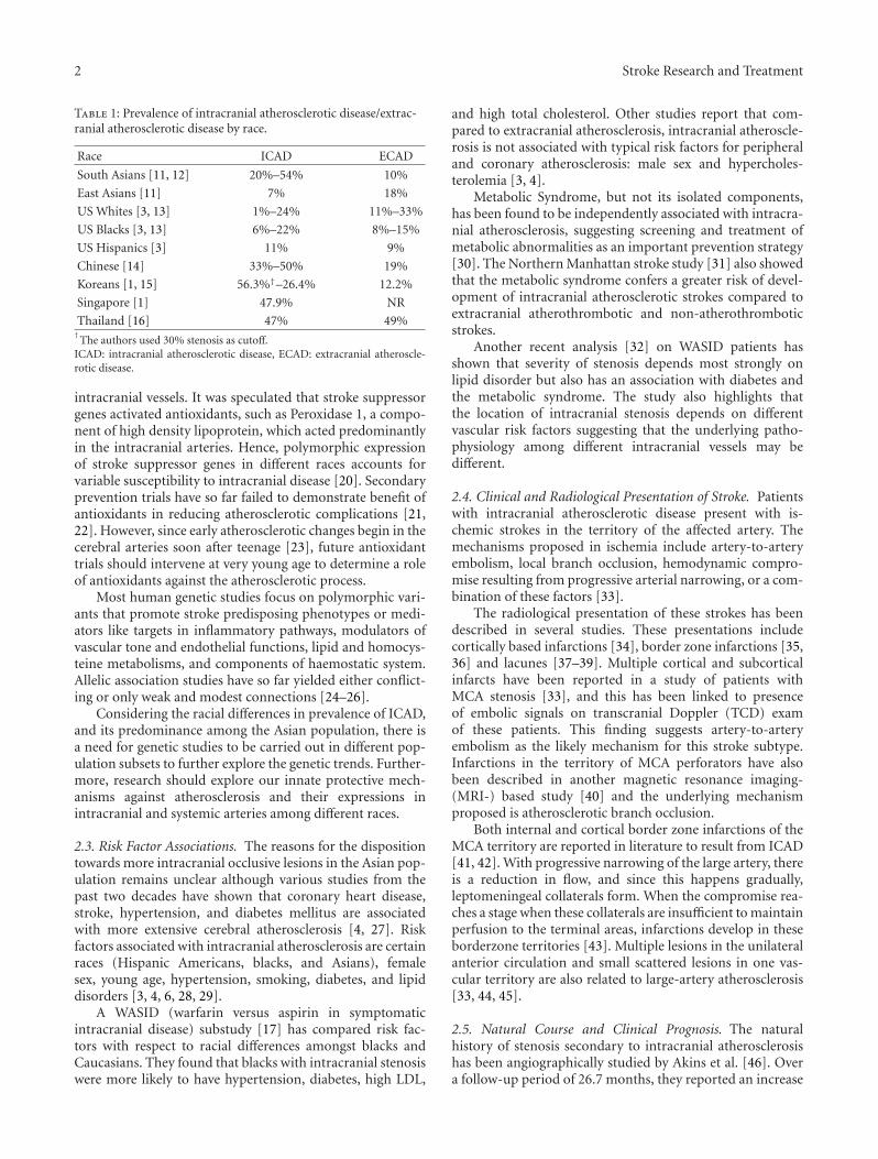
Intracranial atherosclerotic disease, ICAD, defined asatherosclerosis of the large arteries at the base of the brain,preferentially affects Asians, Hispanics, Far East Asians, andBlacks as compared to carotid bifurcation disease [3–6]. Also,about 20%–45% of non-Caucasians with large artery diseasehave combined extracranial and intracranial lesions [7–10].The prevalence of atherosclerotic stenosis by subtype andrace is further reported in Table 1.
2. Predisposing Factors for ICAD inSusceptible Populations
2.1. Racial Associations. Sacco et al. [3] found no differencebetween races in the proportion of patients with extracranial
atherosclerotic stroke, while intracranial atherosclerosis wasseen more frequently in African American and Hispanic sub-jects than in Caucasian subjects. As per this study the greaterprevalence of Diabetes Mellitus in African American andHispanic subjects accounted for the increased frequency ofICAD.
Waddy et al. [17] report racial differences between blacksand Caucasians with respect to intracranial stenosis. In thisstudy, risk of recurrence of stroke was higher in blacks andrisk factor profiles were also different.
Among Chinese populations, ICAD-related strokes ac-count for 33%–37% of all ischemic strokes and an evenhigher prevalence is reported from Korea, Thailand, and Sin-gapore [15, 16]. Hence, there is evidence of racial associationwith incidence of ICAD.
2.2. Genetic Associations. Several studies suggest a positivecorrelation between race and cerebrovascular disease [18,19]. A study conducted on subjects of European ancestryuncovered a genetic trait that increased their resistance toatherosclerosis, though protection was confined to large
2 Stroke Research and Treatment
Table 1: Prevalence of intracranial atherosclerotic disease/extrac-ranial atherosclerotic disease by race.
Race ICAD ECAD
South Asians [11, 12] 20%–54% 10%
East Asians [11] 7% 18%
US Whites [3, 13] 1%–24% 11%–33%
US Blacks [3, 13] 6%–22% 8%–15%
US Hispanics [3] 11% 9%
Chinese [14] 33%–50% 19%
Koreans [1, 15] 56.3%†–26.4% 12.2%
Singapore [1] 47.9% NR
Thailand [16] 47% 49%†
The authors used 30% stenosis as cutoff.ICAD: intracranial atherosclerotic disease, ECAD: extracranial atheroscle-rotic disease.
intracranial vessels. It was speculated that stroke suppressorgenes activated antioxidants, such as Peroxidase 1, a compo-nent of high density lipoprotein, which acted predominantlyin the intracranial arteries. Hence, polymorphic expressionof stroke suppressor genes in different races accounts forvariable susceptibility to intracranial disease [20]. Secondaryprevention trials have so far failed to demonstrate benefit ofantioxidants in reducing atherosclerotic complications [21,22]. However, since early atherosclerotic changes begin in thecerebral arteries soon after teenage [23], future antioxidanttrials should intervene at very young age to determine a roleof antioxidants against the atherosclerotic process.
Most human genetic studies focus on polymorphic vari-ants that promote stroke predisposing phenotypes or medi-ators like targets in inflammatory pathways, modulators ofvascular tone and endothelial functions, lipid and homocys-teine metabolisms, and components of haemostatic system.Allelic association studies have so far yielded either conflict-ing or only weak and modest connections [24–26].
Considering the racial differences in prevalence of ICAD,and its predominance among the Asian population, there isa need for genetic studies to be carried out in different pop-ulation subsets to further explore the genetic trends. Further-more, research should explore our innate protective mech-anisms against atherosclerosis and their expressions inintracranial and systemic arteries among different races.
2.3. Risk Factor Associations. The reasons for the dispositiontowards more intracranial occlusive lesions in the Asian pop-ulation remains unclear although various studies from thepast two decades have shown that coronary heart disease,stroke, hypertension, and diabetes mellitus are associatedwith more extensive cerebral atherosclerosis [4, 27]. Riskfactors associated with intracranial atherosclerosis are certainraces (Hispanic Americans, blacks, and Asians), femalesex, young age, hypertension, smoking, diabetes, and lipiddisorders [3, 4, 6, 28, 29].
A WASID (warfarin versus aspirin in symptomaticintracranial disease) substudy [17] has compared risk fac-tors with respect to racial differences amongst blacks andCaucasians. They found that blacks with intracranial stenosiswere more likely to have hypertension, diabetes, high LDL,
and high total cholesterol. Other studies report that com-pared to extracranial atherosclerosis, intracranial atheroscle-rosis is not associated with typical risk factors for peripheraland coronary atherosclerosis: male sex and hypercholes-terolemia [3, 4].
Metabolic Syndrome, but not its isolated components,has been found to be independently associated with intracra-nial atherosclerosis, suggesting screening and treatment ofmetabolic abnormalities as an important prevention strategy[30]. The Northern Manhattan stroke study [31] also showedthat the metabolic syndrome confers a greater risk of devel-opment of intracranial atherosclerotic strokes compared toextracranial atherothrombotic and non-atherothromboticstrokes.
Another recent analysis [32] on WASID patients hasshown that severity of stenosis depends most strongly onlipid disorder but also has an association with diabetes andthe metabolic syndrome. The study also highlights thatthe location of intracranial stenosis depends on differentvascular risk factors suggesting that the underlying patho-physiology among different intracranial vessels may bedifferent.
2.4. Clinical and Radiological Presentation of Stroke. Patientswith intracranial atherosclerotic disease present with is-chemic strokes in the territory of the affected artery. Themechanisms proposed in ischemia include artery-to-arteryembolism, local branch occlusion, hemodynamic compro-mise resulting from progressive arterial narrowing, or a com-bination of these factors [33].
The radiological presentation of these strokes has beendescribed in several studies. These presentations includecortically based infarctions [34], border zone infarctions [35,36] and lacunes [37–39]. Multiple cortical and subcorticalinfarcts have been reported in a study of patients withMCA stenosis [33], and this has been linked to presenceof embolic signals on transcranial Doppler (TCD) examof these patients. This finding suggests artery-to-arteryembolism as the likely mechanism for this stroke subtype.Infarctions in the territory of MCA perforators have alsobeen described in another magnetic resonance imaging-(MRI-) based study [40] and the underlying mechanismproposed is atherosclerotic branch occlusion.
Both internal and cortical border zone infarctions of theMCA territory are reported in literature to result from ICAD[41, 42]. With progressive narrowing of the large artery, thereis a reduction in flow, and since this happens gradually,leptomeningeal collaterals form. When the compromise rea-ches a stage when these collaterals are insufficient to maintainperfusion to the terminal areas, infarctions develop in theseborderzone territories [43]. Multiple lesions in the unilateralanterior circulation and small scattered lesions in one vas-cular territory are also related to large-artery atherosclerosis[33, 44, 45].
2.5. Natural Course and Clinical Prognosis. The naturalhistory of stenosis secondary to intracranial atherosclerosishas been angiographically studied by Akins et al. [46]. Overa follow-up period of 26.7 months, they reported an increase

Stroke Research and Treatment 3
in the degree of stenosis in the MCA-ACA-PCA group,whereas intracranial ICA stenoses remained stable. Theyalso reported a 14% regression in intracranial ICA stenosisand a 28% regression in the MCA-ACA-PCA group. Theyconcluded that intracranial lesions are dynamic and mayprogress or regress with time.
A study by Kwon et al. [47], evaluating the role of cilosta-zol in ICAD, describes both progression and regression inthese lesions. In a more recent study [48] evaluating the roleof cilostazol and clopidogrel in intracranial atherosclerosis,a total of 14% patients demonstrated lesion progression.
ICAD has also been evaluated in terms of developmentof stroke in the territory of the diseased vessel. The riskof future ischemic strokes depends on whether the vessel isasymptomatic or symptomatic. This difference has been wellstudied in the WASID trial [49]. Patients recruited in thetrial, though symptomatic, also had asymptomatic stenosesin other arteries. When these patients were followed up, the1-year risk of developing a stroke in the territory of theseasymptomatic vessels was 3.5% (CI 0.8%–9%). In contrastto this, patients with ≥70% stenosis had a risk of 14% at oneyear for a stroke in the same territory and 19% for stroke inany vascular territory. Apart from severity of stenosis, recentsymptoms and female gender also predicted risk of strokerecurrence in this study [50]. A similar risk rate of 14% wasreported from the GESICA (Groupe d’Etude des StenosesIntra-Craniennes Atheromateuses symptomatiques) study[51].
A WASID substudy [52] has identified that the presenceof collaterals is a strong predictor of subsequent stroke incase of moderate-to-severe stenosis, but milder stenoses aremore unstable and presence of collaterals in these predicts anincreased risk of subsequent stroke.
2.6. Diagnosis. In patients with clinical suspicion of intracra-nial steno-occlusive lesions, accurate assessment of intracra-nial arteries is essential for optimal therapeutic decisions.The options for imaging patients with intracranial steno-sis include noninvasive techniques such as transcranialDoppler or magnetic resonance angiography (MRA), such ascontrast-enhanced (CE) MRA and CE computerized tomog-raphy CT.
Catheter cerebral angiography, though invasive, is con-sidered to be the gold standard for diagnosis of ICAD. Thecomplication rates associated with the procedure performedby trained neurointerventionists have gone down signifi-cantly. A recent review [53] of six-year data based on 363diagnostic angiographies reports a low complication rate of0.3% and an even lower risk of stroke (0.03%).
MRA is being proposed as a replacement for the goldstandard, intra-arterial angiography. 3D time-of-flight(TOF) MRA is currently the most commonly used pulsesequence in the MR evaluation of intracranial arteries. Itdetects clinically significant stenotic lesions in intracranialvessels. The other potential field of application is to monitorthe response of a stenotic lesion to antistenosis medications[47].
Both TOF MRA and CE MRA have shown [54] highaccuracy for the detection of high-grade ICA stenosis and
occlusions. CE MRA has some edge over TOF MRA. How-ever, for moderately severe stenosis, both had only poor(TOF-MRA) to fair (CE MRA) sensitivity. Also, 3D TOF-MRA can be restricted by factors such as susceptibility arti-facts near the sphenoid sinus, limited scan range, limitedspatial resolution, and flow signal intensity loss due to satura-tion or phase dispersion [55, 56]. The portion of intracranialvessels near the skull base and especially the paracavernousand supraclinoid segments of the internal carotid arteriesare areas of frequent over and underestimation of stenosisdue to the presence of dephasing artifacts. Some of theselimitations can be overcome by use of CE MRA which is notflow dependant.
When 3 Tesla MRI is used, the sensitivity of TOF-MRAfor >50% stenosis is reported to be 78%–85% with a positivepredictive value 75%–79%, and for complete occlusions, thesensitivity and positive predictive values are in the range of100% and 87%, respectively [57].
A recent paper by Arenillas [58] has highlighted a newconcept of intracranial plaque imaging using high resolution(3T) 3D TOF-MRA. This gives the advantage of charac-terization of plaque, detection of nonstenotic intracranialatheroma, and detection of intraplaque hemorrhage. Thisis yet to come into widespread clinical practice due tononavailability and limited clinical value. However, it isinteresting, because it characterizes lesser degrees of stenosis.
In a comparative study [59], the ability of helical CTangiography (CTA) to help detect and quantify intracranialstenosis and occlusion compared with DSA (digital subtrac-tion angiography) and MRA was evaluated. CTA revealedhigher sensitivity than MRA for intracranial stenosis (98%versus 70%, P < .001) and occlusion (100% versus 87%,P = .02).
Transcranial Doppler, TCD is another noninvasive andeasy to perform modality used for evaluation of ICAD.The stroke outcomes and neuroimaging of intracranialatherosclerosis (SONIA) trial [60] showed that both TCDand MRA identify 50% to 99% intracranial large vessel steno-sis with a significant negative predictive value. Therefore,both can reliably exclude the presence of intracranial stenosisthough abnormal findings would require a confirmatory testsuch as angiography to reliably identify stenosis.
Yet, another study [61] shows that advanced ultra-sonographic techniques like power-flow imaging and colorDoppler-assisted duplex imaging with and without contrasthave a better yield for near-occlusion and complete occlusiondetection.
Available noninvasive imaging modalities and theirsensitivity and specificity of detection with respect to degreeof stenosis is enlisted in Table 2. Overall, noninvasive imagingmodalities have a high negative predictive value in detectingintracranial atherosclerosis. The gold standard for confirma-tion of the diagnosis remains intra-arterial angiography.
3. Treatment of AtheroscleroticIntracranial Stenosis
3.1. Medical Management. The medical management of int-racranial atherosclerotic disease has been evaluated in several

4 Stroke Research and Treatment
Table 2: Comparison of nonimaging modalities in the detection of intracranial stenosis.
Modality Degree of stenosis Sensitivity Specificity Limitations
Digital subtractionangiography
Invasive test: Procedure risk rate: 0.3% for allcomplications, 0.03% for stroke [56]
MRA (TOF) [54] forICA disease
50%–69% 37.9% 92.1%Limited spatial resolution, flow signal intensity loss as aresult of saturation or phase dispersion, susceptibilityartifacts near sphenoid sinus, and over- andunderestimation of stenosis due to dephasing artifacts
>70%–99% 91.2% 88.3%
ICA occlusion 94.5% 99.3%
MRA (TOF) 3T [57]50%–99% stenosis 78%–85% 95%
Occlusion 100% 99%
MRA (CE) [54]50%–69% 65.9% 93.5%
>70%–99% 94.6% 91.9%
ICA occlusion 99.4% 99.6%
CTA∗ [59]Stenosis‡ 98% 99%
Occlusion 100% 100%
Transcranial DopplerUltrasound [62]
>50% stenosis orocclusion
High level of technical and procedural skill is requiredto obtain the best quality images. Reliable insonation ofthe posterior circulation is particularly difficult
For MCA stem (M1) 90%–99% 90%–99%
For intracranialsegment (V4) of
vertebral and basilarartery
70%–80% 90%–99%
CDDI [61]
Atheromatouspseudo-occlusion
Unenhanced† 70% 92% False negative rate 30%
Echo-enhanced PFI 83% 92% False negative rate 17%
Unenhanced 95% 92% False negative rate 5%
Echo-enhanced 94% 100% False negative rate 6%
CDDI: Color Doppler-assisted duplex imaging, PFI: power-flow imaging.∗Data are percentages using DSA as the reference standard.‡North American Symptomatic Carotid Endarterectomy Trial (NASCET) criteria were used for stenosis calculations: [(Dn − Ds)/Dn] × 100, where Dn isnormal diameter and Ds is stenosed diameter. NASCET stenoses were grouped according to the following grading scale: normal (0%–9%), mild (10%–29%),moderate (30%–69%), severe (70%–99%), or occluded (no flow detected). Normal (0%–9%) and mild (10%–29%) stenosis were not considered diseasedvessel segments and were excluded from analysis.†Ultrasound emission energy and gain cannot be increased high enough without the appearance of disturbing acoustic noise that diminishes the reliabledepiction of orthograde flow signals.
trials over the past decade. There is no data on primary pre-vention of strokes in patients with asymptomatic stenosis.
The effectiveness of Aspirin in secondary prevention ofischemic strokes is irrefutable. Since international stroketrial—IST and chinese acute stroke trial—CAST [63, 64], it isthe standard of care. For intracranial atherosclerotic strokes,it has never been tested in isolation, but extrapolating fromthis data, the benefit spans across all stroke subtypes.
For secondary prevention, anticoagulation with warfarinwas shown to be less safe and equal in efficacy to aspirin inthe WASID (warfarin versus aspirin in symptomatic intra-cranial disease) trial [49]. WASID was stopped early aftera mean followup of 1.8 years because of higher rates of deathand major hemorrhage in the warfarin group. The rates ofmyocardial infarction or sudden death were also higher in thewarfarin group. The primary end point of ischemic stroke,brain hemorrhage or vascular death, occurred in 22.1%of patients in aspirin and 21.8% of those in the warfaringroup. Before WASID, it was thought that patients withvertebrobasilar disease might benefit from warfarin, but the
study failed to show a significantly lower rate of primary endpoint or stroke in the group on warfarin [65], suggesting thatthere is no clear evidence for supremacy of warfarin overaspirin for patients with vertebrobasilar stenosis either.
Efficacy of other antiplatelet agents has been evaluatedin several other trials, and aspirin/extended release dipyri-damole [66] is recommended over aspirin for secondaryprevention of all ischemic strokes. Clopidogrel [67] was alsoshown to be superior to aspirin for composite vascular endpoints. Therefore even though not subtype specific, there isa role for these agents in ischemic strokes. Another morerecent study (prevention regimen for effectively avoiding sec-ond strokes—PRoFESS) has shown similar stroke recurrencerates in patients with various underlying causes of strokeand in a subset of patients with large artery atherosclerosiswhen treated with clopidogrel alone versus a combination ofaspirin/extended release dipyridamole [68].
Therefore, as per the American Stroke Association rec-ommendation [69], aspirin alone, aspirin/extended releasedipyridamole, and clopidogrel alone are all acceptable

Stroke Research and Treatment 5
options for secondary stroke prevention after a non-cardi-oembolic ischemic stroke including large artery atheroscle-rotic stroke.
A recent trial [70] investigating the role of combinationantiplatelet (aspirin+clopidogrel) in acute management ofstroke secondary to large artery atherosclerosis has shownpromise. The study demonstrated that early combinationtherapy (within 7 days of symptom onset) was more effectivethan aspirin alone in reducing microembolic signals insymptomatic arteries of these patients. Whether this trans-lates into clinical benefit is yet to be evaluated.
Cilostazol is a newer antiplatelet agent being investigatedfor intracranial stenosis. It is a phosphodiesterase 3 inhibitorthat inhibits smooth muscle cell growth in vitro and has anantiatherogenic and antiproliferative action in addition toantiplatelet effects. The first study [47] evaluating cilostazolin a randomized fashion came out in 2005. In this study,during a 6-month follow-up period, there were no strokes incilostazol+aspirin or placebo+aspirin arm. However, pro-gression of the intracranial stenosis was significantly less inthe cilostazol group (6.7% versus 28.8%; P = .008).
This finding led to a multicenter study of cilosta-zol+aspirin versus clopidogrel+aspirin in patients with sym-ptomatic intracranial stenosis, the trial of cilostazol in symp-tomatic intracranial arterial stenosis II (TOSS II) [48]. TOSSII enrolled 456 acute ischemic stroke patients with symp-tomatic intracranial arterial stenosis. After 7 months of treat-ment, follow-up MRA showed a slightly lower but significantrate of progression (9.90% versus 15.46%) and a higherrate of regression (30.20% versus 23.67%) in symptomaticstenosis with cilostazol versus clopidogrel group, respectively.More patients in the cilostazol group had new asymptomaticischemic lesions at the follow-up MRA than those receivingclopidogrel (18.68% versus 12.04%), and in the territoryof the symptomatic intracranial stenosis (12.09% versus8.90%), but this difference was not statistically significant.There was no statistically significant difference in theoccurrence of clinical events by treatment group althoughevents tended to be more frequent in the cilostazol. Bleedingcomplications were nonsignificantly higher with clopidogrel.Therefore, cilostazol combination therapy had a favorableeffect on the overall change in symptomatic intracranialatherosclerotic stenosis, but in this study, it did not translateinto better clinical outcomes.
A pilot study [71] of Chinese patients with ischemicstrokes has compared aspirin to cilostazol in a randomizedfashion. There were fewer ischemic and hemorrhagic strokesin the cilostazol group, suggesting that it might be safer andmore effective compared to aspirin. Cilostazol stroke pre-vention study 2—CSPS-2 [72] is a more recent randomizedtrial that has shown that cilostazol is noninferior to and maybe superior to aspirin in prevention of recurrent stroke inpatients with noncardioembolic ischemic strokes. One thirdof the patients in this trial had large vessel atherosclerosis.
Management of Risk Factors. WASID presented additionaldata supporting aggressive risk-factor control in patientswith intracranial stenosis. In WASID, vascular risk factors
were managed by following national guidelines [48]. How-ever, in many patients risk-factor control was not optimal,and patients with poor control of risk factors had higher ratesof recurrent vascular events [73, 74].
A WASID-substudy [74] demonstrated that elevatedblood pressure was associated with an increased risk of is-chemic stroke and other major vascular events. Contrary tothe common practice of permissive hypertension in high-grade stenosis, this study showed no increased risk withmaintaining blood pressures in the normal range. Thefindings cannot be generalized to acute, unstable patients,but the rest the guidelines for blood pressure control shouldbe followed.
Elevated LDL also conferred a high risk of subsequentevents in the WASID study [73]. Although the differencefailed to reach statistical significance, there were fewer vas-cular events in patients with LDL <70 compared to thosewith levels ≥70. Based on this data and the SPARCL (strokeprevention by aggressive reduction in cholesterol levels) trial[75], the recommendation is for aggressive lipid loweringwith statin in patients with atherosclerotic ischemic stroke.
Another WASID substudy [76] has demonstrated thatmetabolic syndrome is associated with intracranial ather-osclerosis and confers a higher risk of major vascular eventsin these patients. It is, therefore, an important additionaltarget for primary as well as secondary prevention of intra-cranial atherosclerotic strokes.
In short, better control of risk factors that promote ath-erosclerosis like diabetes, tobacco use, and particularly hyp-ertension, dyslipidemia, and metabolic syndrome is war-ranted in patients with intracranial atherosclerosis.
3.2. Endovascular Therapies. In earlier reports, intracranialangioplasty had a high risk of complications, and the proce-dure was abandoned [77]. Since then, several factors have ledto renewed attention in intracranial angioplasty and stent-ing. These include advances in microcatheter and balloontechnology, high risk of recurrent stroke in patients withintracranial stenosis despite medical management in WASID,and success of endovascular treatments for coronary arterydisease [78].
3.3. Angioplasty. Data on angioplasty without stenting isrestricted to retrospective studies only. These studies reporta high technical success rate (with reduction of stenosis toless than 50%). However, restenosis rates of up to 50% havealso been reported [79]. This study reports a periproceduraldeath and stroke rate of 8.3% and an annual stroke risk of4.4%. The procedure is associated with complications likeintimal dissection, thrombus formation, and vessel ruptureas well [80]. Long-term outcome after balloon angioplasty isalso yet to be prospectively studied.
3.4. Stenting. Stenting can be used as adjunct to balloonangioplasty to prevent plaque recoiling and to cover a plaqueor an intimal flap. It may also reduce the chances of vesseldissection.

6 Stroke Research and Treatment
The first prospective trial on stenting was SSYLVIA(stenting of symptomatic atherosclerotic lesions in the ver-tebral or intracranial arteries) [81] performed in 2004. It wasa multicenter trial of use of bare metal stent for symptomaticICAD. It reported a technical success rate of 95%, 30-daystroke risk of 6.6%, and a 1-year stroke risk of 8.5%. Therewere no deaths reported. Although a restenosis rate of 35%was seen, most of these were asymptomatic.
The next big trial on stenting used Wingspan [82] whichis a flexible, self-expanding, microcatheter deployed stent.This study was also a prospective multicenter trial on 45patients refractory to medical therapy. Authors reporteda technical success rate of 97.7%, a 30-day stroke or death rateof 4.5%, and a 1-year stroke rate of 9.3%. Their restenosisrates were better at 7.5% at 6 months and all were asymp-tomatic. Long-term follow-up data is also now available forWingspan [83], and they report restenosis rates in the rangeof 25%–32% although most is still asymptomatic.
Smaller case series report use of bare metal balloon-mounted stents and drug eluting stents. The former are lim-ited by their rigidity and the latter by nonavailability of long-term safety data. Qureshi et al. [84] and Gupta et al. [85]report use of drug coated stents in small number of patientswith good short term results in terms of restenosis. Thesepatients need to be on long-term antiplatelet therapy for atleast 12 months due to risk of subacute and late thrombosis.Therefore, although technically safe and feasible, there isa need for a long-term study.
A comparative study [86] of primary angioplasty andstenting reveals no difference in terms of stroke free survivalat 2 years. Another systematic review [87], however, reportsa greater 1-year rate of stroke and death in the angioplastytreated group compared to stent treated patients (20 versus14%).
Comparison has also been done between aggressive med-ical management and endovascular treatment with Wing-span by Jarvis et al. [88]. They report a 13% rate of strokeor death at 6 months with Wingspan and 16% with medicalmanagement.
Based on this evidence, a prospective, randomized study,stenting versus aggressive medical management for prevent-ing recurrent stroke in intracranial stenosis (SAMMPRIS)[89] was started in November 2008 to compare intensivemedical therapy alone with stenting plus intensive medicaltherapy. The trial has very recently been halted with only59% of the planned number of patients recruited. The majorreason for this early termination is an unacceptably highcomplication rate in the stenting arm (14% patients hada stroke or died in the 30-day period after stenting comparedto only 5.8% in the medical arm) [90]. One importantfinding coming out from this trial is a much lower strokerecurrence rate than that previously reported. Interestingly,these patients were on combination antiplatelet therapy.Hence, aggressive medical management may well be the wayforward for most patients.
3.5. Surgical Treatment. Similar to coronary artery bypassgrafting, it is being speculated that a subgroup of patientswith intracranial stenosis may benefit from External to
Internal carotid bypass. These patients are the ones whohave poor hemodynamic reserve. For assessment of hemo-dynamic, several modalities are coming up including PET,SPECT, TCD, CT, and MR perfusion. With better use of thesemodalities, it might be easier to identify patients who maybenefit from such a bypass procedure. Two trials are currentlyunderway to determine the effectiveness of such a bypasssurgery, one in the US (carotid occlusion surgery study—COSS) and the other in Japan (Japanese EC-IC bypass trial—JET) [91, 92]. Before the final verdict is out, the procedurecannot be widely recommended (Table 3).
4. Summary of Therapeutic Options
At present, medical management with antiplatelets is themainstay of therapy for symptomatic intracranial stenosis.Aspirin is the best studied, but in the acute phase, doubleantiplatelet agents aspirin/extended release dipyridamole oraspirin plus clopidogrel may be used. For long-term use,combination of Aspirin and clopidogrel may not be as safeand clopidogrel alone should be used. Cilostazol due to itspleiotropic effects has shown promise in Asians.
Aggressive risk factor control is recommended both forsymptomatic and asymptomatic disease. This includes agg-ressive control of blood pressures and LDL cholesterol inparticular and of other atherogenic risk factors like diabetesand tobacco use in general.
Endovascular management with angioplasty plus stent-ing is emerging as a promising modality for high-gradestenosis in patients with failure of medical management.SAMMPRIS has been halted due to adverse effects in theintervention arm [90]. Long-term outcomes of the procedureare yet unknown and restenosis is common, and hence, itcannot be widely recommended.
Surgical treatment with EC-IC bypass is also being evalu-ated in two randomized trials. Pending results, this modalityalso has limited application.
5. Conclusion
5.1. Known Facts about ICAD. Intracranial atheroscleroticstenosis may be the most common cause of stroke worldwide.Asians are predominantly affected, relative to other races.Various genetic and environmental factors have been impli-cated as predisposing factors. Although intra-arterial angiog-raphy is the gold standard to identify extent of stenosis,noninvasive techniques including TCD ultrasound and TOFMRA have been established as reliable modalities to excludemoderate-to-severe (50%–99%) stenosis. Also, CTA and PFIcan be used to correctly identify degree of severe stenosis.
In terms of secondary prevention of stroke due to int-racranial atherosclerotic stenosis, aspirin continues to be thepreferred antiplatelet agent due to its effectiveness compara-tive to newer antiplatelet therapies. However, among Asians,cilostazol has shown a favorable effect on symptomatic sten-osis and is of benefit in terms of fewer bleeds. Combinationtherapy has shown promise. Moreover, aggressive risk factormanagement, that is, lowering blood pressure and LDL levelslowers risk of vascular events. Endovascular therapy is still

Stroke Research and Treatment 7
Ta
ble
3:O
utl
ine
ofin
terv
enti
onal
tria
lsas
sess
ing
trea
tmen
tm
odal
itie
sfo
rin
trac
ran
ials
ten
osis
.
Stu
dies
/tri
als
Inte
rven
tion
sPo
pula
tion
Follo
wu
pD
egre
eof
sten
osis
Rec
urr
ence
Fata
litie
sP
rim
ary
and
seco
nda
ryen
dp
oin
tW
ASI
D20
05[4
9]M
edic
alA
spir
in56
9pa
tien
ts(2
80in
Asp
irin
grou
pan
d28
9in
War
fari
ngr
oup)
1.8
yrs
Mea
n50
%–9
9%(i
n50
%–6
9%st
enos
is,1
year
stro
keri
skw
as6%
and
in70
%–9
9%st
enos
is19
%)
12%
at1
year
and
19.7
%at
2ye
ars
4.3%
,3.2
%∞
,1.
1%∗∗
,3.2
%�
,2.
5%�
�
22.1
%† ,
20.7
%�
,20
.4%∧ ,
15%††
,8.9
%¶ ,
23.6
%∗∗∗
War
fari
n11
%at
1ye
aran
d17
.2%
at2
year
s
9.7%
,5.9
%∞
,3.
8%∗∗
,8.3
%�
,4.
2%�
�
21.8
%,1
7.6%
�,1
7%∧ ,
12.1
%††
,6.2
%¶ ,
24.6
%∗∗∗
TO
SS20
05[4
7]C
ilost
azol
+A
spir
in
135
(67
pati
ents
inci
lost
azol
grou
pan
d68
pati
ents
inpl
aceb
ogr
oup)
6m
onth
s5
grad
esN
orm
alM
ild(s
ign
alre
duct
ion<
50%
)M
oder
ate
(sig
nal
redu
ctio
n>
50%
)Se
vere
(foc
alsi
gnal
loss
)
No
stro
kes
1su
bjec
tP
rogr
essi
on6.
7%µ
Reg
ress
ion
24.4
%
Pla
cebo
+A
spir
in1
subj
ect
Pro
gres
sion
28.8
%R
egre
ssio
n15
.4%
Mar
kset
al.
2006
[79]
An
giop
last
y12
0pa
tien
ts42
.3m
onth
sM
ean
50%
–95%
pre-
angi
opla
sty
An
nu
alst
roke
rate
3%fo
rte
rrit
ory
oftr
eatm
ent
4.3%
for
any
terr
itor
y
Du
rin
gfo
llow
up
no
deat
hs
attr
ibu
tabl
eto
isch
emic
orh
emor
rhag
icst
roke
occu
rred
3p
eri-
proc
edu
ral
stro
kes
and
4de
ath
s:5.
8%
0%–9
0%po
stan
giop
last
yR
esid
ual
sten
osis
59.3
%h
ad<
50%
,32.
4%h
ad50
%–6
9%an
d8.
3%h
ad>
70%
sten
osis
At
mea
nfo
llow
-up
20.5±
22.7
mon
ths
26.9
%im
prov
emen
tin
sten
osis
com
pare
dto
post
angi
opla
sty
angi
ogra
m,4
9.3%
un
chan
ged,
23.9
%di
spla
yed
wor
sen
ing
sten
osis
SSY
LVIA
2004
[81]
Bar
em
etal
sten
t(t
ech
nic
alsu
cces
sra
te95
%)
61(4
3in
tra-
cran
iala
nd
18ex
trac
ran
ialv
erte
bral
sten
osis
12m
onth
s>
50%
>50
%st
enos
isat
6m
onth
s32
.4%
inin
trac
ran
ial
42.9
%in
extr
acra
nia
lst
ents
Non
eat
30da
ys
30da
yst
roke
rate
7.2%
1ye
arst
roke
rate
13.1
%D
iabe
tes,
pos
tpr
oced
ure
>30
%st
enos
isan
dpr
etre
atm
ent
vess
eldi
amet
erca
npr
edic
tst
enos
isat
6m
onth
s

8 Stroke Research and Treatment
Ta
ble
3:C
onti
nu
ed.
Stu
dies
/tri
als
Inte
rven
tion
sPo
pula
tion
Follo
wu
pD
egre
eof
sten
osis
Rec
urr
ence
Fata
litie
sP
rim
ary
and
seco
nda
ryen
dp
oin
t
Win
gspa
n(H
um
anit
ar-
ian
devi
ceex
empt
ion
)st
udy
2007
[82]
Win
gspa
nst
ent
(flex
ible
,se
lf-e
xpan
din
g)te
chn
ical
succ
ess
rate
97.7
%
45(1
2in
tern
atio
nal
cen
ters
)13
mon
ths
50%
–99%
Bas
elin
em
ean
sten
osis
74.9
%po
st-s
ten
tin
g31
.9%
At
6m
onth
s28
%R
este
nos
isra
te7.
5%at
6m
onth
s
Cau
sem
orta
lity
2.3%
30da
yst
roke
orde
ath
rate
4.5%
Dea
thor
ipsi
late
ral
stro
kera
te7.
1%at
6m
onth
s1
year
rate
ofip
sila
tera
lst
roke
and
deat
h9.
3%
Nat
ion
alIn
stit
ute
ofH
ealt
hre
gist
ry20
08[8
3]
Win
gspa
nst
ent
(pos
tm
arke
tst
udy
)te
chn
ical
succ
ess
rate
97%
129
(17
cen
ters
)5.
4m
onth
sm
edia
n70
%–9
9%In
-ste
nt
rest
enos
isra
te25
%
Cu
mu
lati
ve6
mon
thst
roke
deat
han
dde
laye
dip
sila
tera
lstr
oke
rate
14%
Peri
proc
edu
ral
com
plic
atio
nra
tes
7.5%
Bar
em
etal
ballo
onm
oun
ted
sten
ts
Apo
lost
ent
(tec
hn
ical
succ
ess
rate
91.7
%)
2007
[93]
4623
.9m
onth
s>
50%
28%
atm
edia
n7.
4m
onth
sC
um
ula
tive
prob
abili
tyof
isch
emic
stro
kes
inta
rget
arte
ryte
rrit
ory,
incl
udi
ng
any
stro
kean
dde
ath
wit
hin
30da
ys,
was
8.8%
atyr
s1-
2.A
mon
gel
ecti
veca
ses
no
proc
edu
rere
late
dde
ath
s
Isch
emic
stro
kera
tew
as4.
3p
er10
0pa
tien
tye
ars.
1pa
tien
t(2
.2%
,1/4
6)de
velo
ped
min
oris
chem
icst
roke
inth
eta
rget
-les
ion
arte
ryte
rrit
ory
at6.
7m
onth
s,w
hic
hw
asre
late
dto
angi
ogra
phic
ally
veri
fied
rest
enos
is
Ph
aros
intr
acra
nia
lst
ent
(tec
hn
ical
succ
ess
rate
85.7
5am
ong
non
emer
gen
tly
trea
ted
14pa
tien
ts)
2008
[94]
217.
3m
onth
sm
edia
n
>50
%w
ith
recu
rren
tsy
mpt
oms
onan
tith
rom
boti
cs>
70%
post
sten
tst
enos
isde
crea
sed
from
med
ian
85%
to20
%
‡ No
pati
ents
trea
ted
elec
tive
lyh
adre
curr
ent
sym
ptom
s
Maj
orst
roke
(in
-ste
nt
thro
mbo
sis
2da
ysaf
ter
disc
onti
nu
atio
nof
aspi
rin
)in
4pa
tien
tsin
30da
ysM
inor
stro
ke(r
ever
sibl
edy
sart
hri
aan
dpa
resi
sof
the
righ
tle
g)in
1pa
tien
tat
disc
har
ge

Stroke Research and Treatment 9
Ta
ble
3:C
onti
nu
ed.
Stu
dies
/tri
als
Inte
rven
tion
sPo
pula
tion
Follo
wu
pD
egre
eof
sten
osis
Rec
urr
ence
Fata
litie
sP
rim
ary
and
seco
nda
ryen
dp
oin
tIn
tern
atio
nal
ran
dom
ized
tria
l198
5[9
5]
Med
ical
ther
apy
(Asp
irin
)13
77(7
14as
sign
edto
best
med
ical
care
and
663
wit
hm
edic
alca
re+
Byp
ass)
55.8
mon
ths
14%
conv
ersi
onfr
omst
enos
isto
occl
usi
onof
MC
A7%
–10%
∗30
day
surg
ical
mor
talit
yra
te0.
6%
Peri
oper
ativ
est
roke
sin
med
ical
grou
p1.
3%
Med
ical
ther
apy+
EC
-IC
Byp
ass
Post
oper
ativ
eby
pass
pate
ncy
rate
atm
edia
n32
days
96%
Peri
-op
erat
ive
mor
talit
yra
te1.
1%
4.5%
insu
rgic
algr
oup
30da
ym
ajor
stro
kem
orbi
dity
rate
2.5%
† Pri
mar
yen
dpo
ints
:dea
th,i
sch
emic
stro
ke,m
ajor
hem
orrh
age,
orde
ath
from
vasc
ula
rca
use
sot
her
than
stro
ke.S
econ
dary
end
poi
nts
:isc
hem
icst
roke
orbr
ain
hem
orrh
age,
isch
emic
stro
ke,I
sch
emic
stro
kein
terr
itor
yof
sten
otic
arte
ry,d
isab
ling
orfa
tali
sch
emic
stro
ke,i
sch
emic
stro
ke,m
yoca
rdia
lin
farc
tion
,or
deat
hfr
omva
scu
lar
cau
ses
oth
erth
anst
roke
.‡ E
xcep
tin
one
case
wh
ere
66%
resi
dual
sten
osis
was
left
and
ipsi
late
rals
trok
eoc
curr
edaf
ter
7m
onth
s.µP
rogr
essi
on:w
orse
nin
gof
sten
osis
by1
orm
ore
grad
eon
fin
alM
RA
com
pare
dto
the
base
line
MR
A.
Reg
ress
ion
:im
prov
emen
tof
sten
osis
by1
orm
ore
grad
e.∗ R
ate
ofst
roke
inpa
tien
tsw
ith
caro
tid
siph
onor
MC
Ast
enos
is.
�Is
chem
icst
roke
orbr
ain
hem
orrh
age.
∧ Isc
hem
icst
roke
.††
Isch
emic
stro
kein
the
terr
itor
yof
sten
otic
arte
ry.
¶ Dis
ablin
gor
fata
lisc
hem
icst
roke
.∗∗∗ I
sch
emic
stro
ke,m
yoca
rdia
lin
farc
tion
,or
deat
hfr
omva
scu
lar
cau
ses
oth
erth
anst
roke
.∞
Dea
thfr
omva
scu
lar
cau
ses.
∗∗D
eath
from
non
vasc
ula
rca
use
s.�
Maj
orh
emor
rhag
e.�
�M
yoca
rdia
lin
farc
tion
.

10 Stroke Research and Treatment
investigational. Surgical treatment, that is, EC-IC bypass, hasfailed to show any benefit but may be helpful in selectedpatients with poor hemodynamic reserve.
5.2. Unknown Facts about ICAD. Since Asians are at partic-ular risk of stroke secondary to intracranial atheroscleroticstenosis, studies to locate genetic markers responsible forthis racial predominance in lesion distribution would be ofinterest. Implications of early detection and treatment ofasymptomatic intracranial stenosis need to be explored interms of benefit as a stroke prevention modality. Noninvasivediagnostic techniques that can reliably gauge extent of mildand moderate intracranial stenosis have yet to be identifiedand are needed. The role of antiplatelet agents other thanaspirin, in combination or as single therapy can still befurther investigated to advance current medical treatmentoptions. Endovascular therapy is still investigational and isstill associated with adverse effects.
5.3. Future Directions. Conducted interventional trials onintracranial atherosclerotic stenosis have so far only beencarried out among Caucasians. Since the Asian population isknown to be preferentially effected, focused trials need to becarried out to establish treatment modalities that are mosteffective in this population. Additionally, these may focuson prevention, since intervention is expensive and requirestechnical expertise in low- and middle-income countrieswhere these resources are scant. Screening for intracranialstenosis needs to be further explored. Noninvasive diagnosticmodalities that can reliably identify all degrees of stenosesare needed. For symptomatic patients, this will help inmanagement decisions, and for asymptomatic patients, theymay help explore the role of preventive therapy.
Acknowledgments
M. Khan is a neurovascular fellow whose training is currentlyfunded by Award no. D43TW008660 from the Fogarty Inter-national Center. The content is solely the responsibility of theauthors and does not necessarily represent the official viewsof the Fogarty International Center or the National Institutesof Health. A. Kamal is the Principal Investigator for theKarachi Intracranial Stenosis Study (KISS) funded by theHigher Education Commission Government of Pakistan.
References
[1] P. B. Gorelick, K. S. Wong, H. J. Bae, and D. K. Pandey, “Largeartery intracranial occlusive disease: a large worldwide burdenbut a relatively neglected frontier,” Stroke, vol. 39, no. 8, pp.2396–2399, 2008.
[2] L. K. Wong, “Global burden of intracranial atherosclerosis,”International Journal of Stroke, vol. 1, no. 3, pp. 158–159, 2006.
[3] R. L. Sacco, D. Kargman, Q. Gu, and M. C. Zamanillo, “Race-ethnicity and determinants of intracranial atherosclerotic cer-ebral infarction: the Northern Manhattan Stroke Study,”Stroke, vol. 26, no. 1, pp. 14–20, 1995.
[4] L. R. Caplan, P. B. Gorelick, and D. B. Hier, “Race, sex andocclusive cerebrovascular disease: a review,” Stroke, vol. 17, no.4, pp. 648–655, 1986.
[5] K. Nishimaru, L. C. McHenry Jr., and J. F. Toole, “Cere-bral angiographic and clinical differences in carotid systemtransient ischemic attacks between American Caucasian andJapanese patients,” Stroke, vol. 15, no. 1, pp. 56–59, 1984.
[6] E. Feldmann, N. Daneault, E. Kwan et al., “Chinese-whitedifferences in the distribution of occlusive cerebrovascular dis-ease,” Neurology, vol. 40, no. 10, pp. 1541–1545, 1990.
[7] P. McGarry, L. A. Solberg, M. A. Guzman, and J. P. Strong,“Cerebral atherosclerosis in New Orleans. Comparisons of le-sions by age, sex, and race,” Laboratory Investigation, vol. 52,no. 5, pp. 533–539, 1985.
[8] M. T. Garcia-Rondon and Y. Reyes-Iglesias, “Cerebral angio-graphic findings and risk factor profile in a hispanic male pop-ulation,” Annals of Neurology, vol. 42, p. 440, 1997.
[9] S. Y. Leung, T. H. K. Ng, S. T. Yuen, I. J. Lauder, and F. C. S. Ho,“Pattern of cerebral atherosclerosis in Hong Kong Chinese:severity in intracranial and extracranial vessels,” Stroke, vol. 24,no. 6, pp. 779–786, 1993.
[10] S. J. Lee, S. J. Cho, H. S. Moon et al., “Combined extracranialand intracranial atherosclerosis in Korean patients,” Archivesof Neurology, vol. 60, no. 11, pp. 1561–1564, 2003.
[11] M. Moussouttas, L. Aguilar, K. Fuentes et al., “Cerebrovasculardisease among patients from the Indian subcontinent,” Neu-rology, vol. 67, no. 5, pp. 894–896, 2006.
[12] D. A. de Silva, F.-P. Woon, M.-P. Lee, C. P. L. H. Chen, H.-M.Chang, and M.-C. Wong, “South Asian patients with ischemicstroke: intracranial large arteries are the predominant site ofdisease,” Stroke, vol. 38, no. 9, pp. 2592–2594, 2007.
[13] R. J. Wityk, D. Lehman, M. Klag, J. Coresh, H. Ahn, and B.Litt, “Race and sex differences in the distribution of cerebralatherosclerosis,” Stroke, vol. 27, no. 11, pp. 1974–1980, 1996.
[14] Y. N. Huang, S. Gao, S. W. Li et al., “Vascular lesions in Chinesepatients with transient ischemic attacks,” Neurology, vol. 48,no. 2, pp. 524–525, 1997.
[15] Y. D. Kim, H. Y. Choi, H. J. Cho et al., “Increasing frequencyand burden of cerebral artery atherosclerosis in Korean strokepatients,” Yonsei Medical Journal, vol. 51, no. 3, pp. 318–325,2010.
[16] N. C. Suwanwela and A. Chutinetr, “Risk factors for ath-erosclerosis of cervicocerebral arteries: intracranial versusextracranial,” Neuroepidemiology, vol. 22, no. 1, pp. 37–40,2003.
[17] S. P. Waddy, G. Cotsonis, M. J. Lynn et al., “Racial differencesin vascular risk factors and outcomes of patients with intracra-nial atherosclerotic arterial stenosis,” Stroke, vol. 40, no. 3, pp.719–725, 2009.
[18] T. Uehara, M. Tabuchi, T. Hayashi, H. Kurogane, and A.Yamadori, “Asymptomatic occlusive lesions of carotid andintracranial arteries in Japanese patients with ischemic heartdisease: evaluation by brain magnetic resonance angiography,”Stroke, vol. 27, no. 3, pp. 393–397, 1996.
[19] L. A. Solberg and P. A. McGarry, “Cerebral atherosclerosis inNegroes and Caucasians,” Atherosclerosis, vol. 16, no. 2, pp.141–154, 1972.
[20] W. Mak, T. S. Cheng, K. H. Chan, R. T. F. Cheung, and S. L. Ho,“A possible explanation for the racial difference in distributionof large-arterial cerebrovascular disease: ancestral Europeansettlers evolved genetic resistance to atherosclerosis, but con-fined to the intracranial arteries,” Medical Hypotheses, vol. 65,no. 4, pp. 637–648, 2005.
[21] K. Asplund, “Antioxidant vitamins in the prevention of car-diovascular disease: a systematic review,” Journal of InternalMedicine, vol. 251, no. 5, pp. 372–392, 2002.

Stroke Research and Treatment 11
[22] D. P. Vivekananthan, M. S. Penn, S. K. Sapp et al., “Use ofantioxidant vitamins for the prevention of cardiovascular dis-ease: metaanalysis of randomized trials,” The Lancet, vol. 361,no. 9374, pp. 2017–2023, 2003.
[23] J. Moossy, “Development of cerebral atherosclerosis in variousage groups,” Neurology, vol. 9, pp. 569–574, 1959.
[24] A. Elbaz and P. Amarenco, “Genetic susceptibility and is-chemic stroke,” Current Opinion in Neurology, vol. 12, no. 1,pp. 47–55, 1999.
[25] A. Hassan and H. S. Markus, “Genetics and ischaemic stroke,”Brain, vol. 123, pp. 1784–1812, 2000.
[26] F. J. Carr, M. W. McBride, H. V. O. Carswell et al., “Geneticaspects of stroke: human and experimental studies,” Journal ofCerebral Blood Flow and Metabolism, vol. 22, no. 7, pp. 767–773, 2002.
[27] S. Kunitz, C. Gross, A. Heyman et al., “The pilot stroke databank: definition, design, and data,” Stroke, vol. 15, no. 4, pp.740–746, 1984.
[28] I. Holme, S. C. Enger, A. Helgeland et al., “Risk factors andraised atherosclerotic lesions in coronary and cerebral arteries.Statistical analysis from the Oslo study,” Arteriosclerosis, vol. 1,no. 4, pp. 250–256, 1981.
[29] P. B. Gorelick, L. R. Caplan, D. B. Hier, S. L. Parker, andD. Patel, “Racial differences in the distribution of anteriorcirculation occlusive disease,” Neurology, vol. 34, no. 1, pp. 54–59, 1984.
[30] O. Y. Bang, J. W. Kim, J. H. Lee et al., “Association of themetabolic syndrome with intracranial atherosclerotic stroke,”Neurology, vol. 65, no. 2, pp. 296–298, 2005.
[31] F. Rincon, R. L. Sacco, G. Kranwinkel et al., “Incidence andrisk factors of intracranial atherosclerotic stroke: the NorthernManhattan Stroke Study,” Cerebrovascular Diseases, vol. 28, no.1, pp. 65–71, 2009.
[32] T. N. Turan, A. A. Makki, S. Tsappidi et al., “Risk factors ass-ociated with severity and location of intracranial arterial sten-osis,” Stroke, vol. 41, no. 8, pp. 1636–1640, 2010.
[33] K. S. Wong, S. Gao, Y. L. Chan et al., “Mechanisms of acutecerebral infarctions in patients with middle cerebral arterystenosis: a diffusion-weighted imaging and microemboli mon-itoring study,” Annals of Neurology, vol. 52, no. 1, pp. 74–81,2002.
[34] A. E. Baird, K. O. Lovblad, G. Schlaug, R. R. Edelman, and S.Warach, “Multiple acute stroke syndrome: marker of embolicdisease?” Neurology, vol. 54, no. 3, pp. 674–678, 2000.
[35] J. Bogousslavsky and F. Regli, “Borderzone infarctions distalto internal carotid artery occlusion: prognostic implications,”Annals of Neurology, vol. 20, no. 3, pp. 346–350, 1986.
[36] M. del Sette, M. Eliasziw, J. Y. Streifler, V. C. Hachinski, A.J. Fox, and H. J. Barnett, “Internal borderzone infarction: amarker for severe stenosis in patients with symptomatic inter-nal carotid artery disease,” Stroke, vol. 31, no. 3, pp. 631–636,2000.
[37] L. R. Caplan, “Intracranial branch atheromatous disease: aneglected, understudied, and underused concept,” Neurology,vol. 39, no. 9, pp. 1246–1250, 1989.
[38] H. P. Adams, H. C. Damasio, S. F. Putman, and A. R. Damasio,“Middle cerebral artery occlusion as a cause of isolated sub-cortical infarction,” Stroke, vol. 14, no. 6, pp. 948–952, 1983.
[39] C. M. Fisher, “Capsular infarcts. the underlying vascularlesions,” Archives of Neurology, vol. 36, no. 2, pp. 65–73, 1979.
[40] D. K. Lee, J. S. Kim, S. U. Kwon, S.-H. Yoo, and D.-W.Kang, “Lesion patterns and stroke mechanism in atheroscle-rotic middle cerebral artery disease: early diffusion-weightedimaging study,” Stroke, vol. 36, no. 12, pp. 2583–2588, 2005.
[41] L. C. Turtzo, R. F. Gottesman, and R. H. Llinas, “Diffusion-weighted imaging showing “Pearls” predicts large-vessel dis-ease as stroke etiology,” Cerebrovascular Diseases, vol. 28, no.1, pp. 49–54, 2009.
[42] H. Yamauchi, R. Nish, T. Higashi, S. Kagawa, and H. Fuk-uyama, “Hemodynamic compromise as a cause of internalborder-zone infarction and cortical neuronal damage in ath-erosclerotic middle cerebral artery disease,” Stroke, vol. 40, no.12, pp. 3730–3735, 2009.
[43] C. P. Derdeyn, W. J. Powers, and R. L. Grubb Jr., “Hemody-namic effects of middle cerebral artery stenosis and occlusion,”American Journal of Neuroradiology, vol. 19, no. 8, pp. 1463–1469, 1998.
[44] D. W. Kang, J. A. Chalela, M. A. Ezzeddine, and S. Warach,“Association of ischemic lesion patterns on early diffusion-weighted imaging with TOAST stroke subtypes,” Archives ofNeurology, vol. 60, no. 12, pp. 1730–1734, 2003.
[45] J. Bogousslavsky, “Double infarction in one cerebral hemi-sphere,” Annals of Neurology, vol. 30, no. 1, pp. 12–18, 1991.
[46] P. T. Akins, T. K. Pilgram, D. T. Cross III, and C. J. Moran,“Natural history of stenosis from intracranial atherosclerosisby serial angiography,” Stroke, vol. 29, no. 2, pp. 433–438, 1998.
[47] S. U. Kwon, Y. J. Cho, J. S. Koo et al., “Cilostazol preventsthe progression of the symptomatic intracranial arterial steno-sis: the multicenter double-blind placebo-controlled trial ofcilostazol symptomatic intracranial arterial stenosis,” Stroke,vol. 36, no. 4, pp. 782–786, 2005.
[48] S. U. Kwon, D. W. Kang, J. M. Park et al., “Trial of efficacyand safety of cilostazol on the progression of symptomaticintracranial stenosis comparing clopidogrel: trial of cilostazolin symptomatic intracranial stenosis-2 (TOSS-2),” Cerebrovas-cular Diseases, vol. 27, supplement 6, pp. 10–11, 2009.
[49] M. I. Chimowitz, M. J. Lynn, H. Howlett-Smith et al., “Com-parison of warfarin and aspirin for symptomatic intracranialarterial stenosis,” The New England Journal of Medicine, vol.352, no. 13, pp. 1305–1316, 2005.
[50] S. E. Kasner, M. I. Chimowitz, M. J. Lynn et al., “Predictors ofischemic stroke in the territory of a symptomatic intracranialarterial stenosis,” Circulation, vol. 113, no. 4, pp. 555–563,2006.
[51] M. Mazighi, R. Tanasescu, X. Ducrocq et al., “Prospectivestudy of symptomatic atherothrombotic intracranial stenoses:the GESICA study,” Neurology, vol. 66, no. 8, pp. 1187–1191,2006.
[52] D. S. Liebeskind, G. A. Cotsonis, J. L. Saver et al., “Collateraldramatically alter stroke risk in intracranial atherosclerosis,”Annals of Neurology, vol. 69, no. 6, pp. 963–974, 2011.
[53] J. T. Fifi, P. M. Meyers, S. D. Lavine et al., “Complications ofmodern diagnostic cerebral angiography in an academic med-ical center,” Journal of Vascular and Interventional Radiology,vol. 20, no. 4, pp. 442–447, 2009.
[54] S. M. Debrey, H. Yu, J. K. Lynch et al., “Diagnostic accuracyof magnetic resonance angiography for internal carotid arterydisease: a systematic review and meta-analysis,” Stroke, vol. 39,no. 8, pp. 2237–2248, 2008.
[55] J. E. Heiserman, B. P. Drayer, P. J. Keller et al., “Intracranialvascular stenosis and occlusion: evaluation with three-dim-ensional time-of-flight MR angiography,” Radiology, vol. 185,no. 3, pp. 667–673, 1992.
[56] Y. Korogi, M. Takahashi, N. Mabuchi et al., “Intracranial vas-cular stenosis and occlusion: diagnostic accuracy of three-dimensional, fourier transform, time-of-flight MR angiogra-phy,” Radiology, vol. 193, no. 1, pp. 187–193, 1994.

12 Stroke Research and Treatment
[57] C. G. Choi, D. H. Lee, J. H. Lee et al., “Detection of intracranialatherosclerotic steno-occlusive disease with 3D time-of-flightmagnetic resonance angiography with sensitivity encoding at3T,” American Journal of Neuroradiology, vol. 28, no. 3, pp.439–446, 2007.
[58] J. F. Arenillas, “Intracranial atherosclerosis: current concepts,”Stroke, vol. 42, supplement 1, pp. S20–S23, 2011.
[59] S. Bash, J. P. Villablanca, R. Jahan et al., “Intracranial vascularstenosis and occlusive disease: evaluation with CT angiogra-phy, MR angiography, and digital subtraction angiography,”American Journal of Neuroradiology, vol. 26, no. 5, pp. 1012–1021, 2005.
[60] E. Feldmann, J. L. Wilterdink, A. Kosinski et al., “The strokeoutcomes and neuroimaging of intracranial atherosclerosis(SONIA) trial,” Neurology, vol. 68, no. 24, pp. 2099–2106,2007.
[61] G. Furst, A. Saleh, F. Wenserski et al., “Reliability and validityof noninvasive imaging of internal carotid artery pseudo-occlusion,” Stroke, vol. 30, no. 7, pp. 1444–1449, 1999.
[62] A. M. Demchuk, I. Christou, T. H. Wein et al., “Accuracyand criteria for localizing arterial occlusion with transcranialdoppler,” Journal of Neuroimaging, vol. 10, no. 1, pp. 1–12,2000.
[63] International Stroke Trial Collaborative Group, “The Interna-tional Stroke Trial (IST): a randomised trial of aspirin, subcu-taneous heparin, both, or neither among 19435 patients withacute ischaemic stroke,” The Lancet, vol. 349, no. 9065, pp.1569–1581, 1997.
[64] Z.-M. Chen, “CAST: randomised placebo-controlled trial ofearly aspirin use in 20,000 patients with acute ischaemicstroke,” The Lancet, vol. 349, no. 9066, pp. 1641–1649, 1997.
[65] S. E. Kasner, M. J. Lynn, M. I. Chimowitz et al., “War-farin aspirin symptomatic intracranial disease (WASID) trialinvestigators. Warfarin vs aspirin for symptomatic intracranialstenosis: subgroup analyses from WASID,” Neurology, vol. 67,no. 7, pp. 1275–1278, 2006.
[66] H. C. Diener, L. Cunha, C. Forbes, J. Sivenius, P. Smets, andA. Lowenthal, “European stroke prevention study 2. Dipyri-damole and acetylsalicylic acid in the secondary prevention ofstroke,” Journal of the Neurological Sciences, vol. 143, no. 1-2,pp. 1–13, 1996.
[67] CAPRIE Steering Committee, “A randomised, blinded, trialof clopidogrel versus aspirin in patients at risk of ischaemicevents (CAPRIE),” The Lancet, vol. 348, no. 9038, pp. 1329–1339, 1996.
[68] R. L. Sacco, H. C. Diener, S. Yusuf et al., “Aspirin andextended-release dipyridamole versus clopidogrel for recur-rent stroke,” The New England Journal of Medicine, vol. 359,no. 12, pp. 1238–1251, 2008.
[69] R. J. Adams, G. Albers, M. J. Alberts et al., “Update to theAHA/ASA recommendations for the prevention of stroke inpatients with stroke and transient ischemic attack,” Stroke, vol.39, no. 5, pp. 1647–1652, 2008.
[70] K. Wong, C. Chen, J. Fu et al., “Clopidogrel plus aspirinversus aspirin alone for reducing embolisation in patients withacute symptomatic cerebral or carotid artery stenosis (CLAIRstudy): a randomised, open-label, blinded-endpoint trial,” TheLancet Neurology, vol. 9, no. 5, pp. 489–497, 2010.
[71] Y. Huang, Y. Cheng, J. Wu et al., “Cilostazol as an alternativeto aspirin after ischaemic stroke: a randomised, double-blind,pilot study,” The Lancet Neurology, vol. 7, no. 6, pp. 494–499,2008.
[72] Y. Shinohara, Y. Katayama, S. Uchiyama et al., “Cilostazolfor prevention of secondary stroke (CSPS 2): an aspirin-controlled, double-blind, randomised non-inferiority trial,”The Lancet Neurology, vol. 9, no. 10, pp. 959–968, 2010.
[73] S. Chaturvedi, T. N. Turan, M. J. Lynn et al., “Risk factor statusand vascular events in patients with symptomatic intracranialstenosis,” Neurology, vol. 69, no. 22, pp. 2063–2068, 2007.
[74] T. N. Turan, G. Cotsonis, M. J. Lynn, S. Chaturvedi, and M.Chimowitz, “Relationship between blood pressure and strokerecurrence in patients with intracranial arterial stenosis,”Circulation, vol. 115, no. 23, pp. 2969–2975, 2007.
[75] P. Amarenco, J. Bogousslavsky, A. Callahan III et al., “High-dose atorvastatin after stroke or transient ischemic attack,” TheNew England Journal of Medicine, vol. 355, no. 6, pp. 549–559,2006.
[76] B. Ovbiagele, J. L. Saver, M. J. Lynn, and M. Chimowitz,“Impact of metabolic syndrome on prognosis of symptomaticintracranial atherostenosis,” Neurology, vol. 66, no. 9, pp.1344–1349, 2006.
[77] T. M. Sundt Jr., H. C. Smith, J. K. Campbell et al., “Translu-minal angioplasty for basilar artery stenosis,” Mayo Clinic Pro-ceedings, vol. 55, no. 11, pp. 673–680, 1980.
[78] T. N. Turan, C. P. Derdeyn, D. Fiorella, and M. I. Chimowitz,“Treatment of atherosclerotic intracranial arterial stenosis,”Stroke, vol. 40, no. 6, pp. 2257–2261, 2009.
[79] M. P. Marks, J. C. Wojak, F. Al-Ali et al., “Angioplasty forsymptomatic intracranial stenosis: clinical outcome,” Stroke,vol. 37, no. 4, pp. 1016–1020, 2006.
[80] M. Hartmann and O. Jansen, “Angioplasty and stenting of int-racranial stenosis,” Current Opinion in Neurology, vol. 18, no.1, pp. 39–45, 2005.
[81] SSYLVIA Study Investigators, “Stenting of symptomatic ath-erosclerotic lesions in the vertebral or intracranial arteries(SSYLVIA): study results,” Stroke, vol. 35, no. 6, pp. 1388–1392,2004.
[82] A. Bose, M. Hartmann, H. Henkes et al., “A novel, self-exp-anding, nitinol stent in medically refractory intracranial ath-erosclerotic stenoses: the wingspan study,” Stroke, vol. 38, no.5, pp. 1531–1537, 2007.
[83] O. O. Zaidat, R. Klucznik, M. J. Alexander et al., “The NIHregistry on use of the wingspan stent for symptomatic 70–99%intracranial arterial stenosis,” Neurology, vol. 70, no. 17, pp.1518–1524, 2008.
[84] A. I. Qureshi, J. F. Kirmani, H. M. Hussein et al., “Early andintermediate-term outcomes with drug-eluting stents in high-risk patients with symptomatic intracranial stenosis,” Neuro-surgery, vol. 59, no. 5, pp. 1044–1051, 2006.
[85] R. Gupta, F. Al-Ali, A. J. Thomas et al., “Safety, feasibility, andshort-term follow-up of drug-eluting stent placement in theintracranial and extracranial circulation,” Stroke, vol. 37, no.10, pp. 2562–2566, 2006.
[86] F. Siddiq, G. Vazquez, M. Z. Memon et al., “Comparison ofprimary angioplasty with stent placement for treating symp-tomatic intracranial atherosclerotic diseases: a multicenterstudy,” Stroke, vol. 39, no. 9, pp. 2505–2510, 2008.
[87] F. Siddiq, M. Z. Memon, G. Vazquez, A. Safdar, and A.I. Qureshi, “Comparison between primary angioplasty andstent placement for symptomatic intracranial atheroscleroticdisease: meta-analysis of case series,” Neurosurgery, vol. 65, no.6, pp. 1024–1033, 2009.
[88] A. L. Jarvis, M. Chimowitz, M. J. Lynn et al., “Outcome ofpatients with 50–99% intracranial stenosis and TIA or strokeon antithrombotic therapy treated medically vs. stenting,” inProceedings of the 60th Annual Meeting of the AmericanAcademy of Neurology, Chicago, Ill, USA, April 2008.

Stroke Research and Treatment 13
[89] C. P. Derdeyn and M. I. Chimowitz, “Angioplasty and stentingfor atherosclerotic intracranial stenosis: rationale for a ran-domized clinical trial,” Neuroimaging Clinics of North America,vol. 17, no. 3, pp. 355–363, 2007.
[90] K. Dmyterko Jr., “SAMMPRIS trial halted after high risk ofstroke/death found,” Industry News, April 2011.
[91] R. L. Grubb Jr., W. J. Powers, C. P. Derdeyn, H. P. AdamsJr., and W. R. Clarke, “The carotid occlusion surgery study,”Neurosurgical Focus, vol. 14, no. 3, p. e9, 2003.
[92] S. Mizumura, J. Nakagawara, M. Takahashi et al., “Three-dimensional display in staging hemodynamic brain ischemiafor JET study: objective evaluation using SEE analysis and 3D-SSP display,” Annals of Nuclear Medicine, vol. 18, no. 1, pp.13–21, 2004.
[93] W.-J. Jiang, X.-T. Xu, M. Jin, B. Du, K.-H. Dong, and J.-P.Dai, “Apollo stent for symptomatic atherosclerotic intracranialstenosis: study results,” American Journal of Neuroradiology,vol. 28, no. 5, pp. 830–834, 2007.
[94] W. Kurre, J. Berkefeld, M. Sitzer, T. Neumann-Haefelin, andR. du Mesnil de Rochemont, “Treatment of symptomatichigh-grade intracranial stenoses with the balloon-expandablePharos stent: initial experience,” Neuroradiology, vol. 50, no. 8,pp. 701–708, 2008.
[95] The EC/IC Bypass Study Group, “Failure of extracranial-int-racranial arterial bypass to reduce the risk of ischemic Stroke.Results of an international randomized trial,” The New Eng-land Journal of Medicine, vol. 313, no. 19, pp. 1191–1200, 1985.

Submit your manuscripts athttp://www.hindawi.com
Stem CellsInternational
Hindawi Publishing Corporationhttp://www.hindawi.com Volume 2014
Hindawi Publishing Corporationhttp://www.hindawi.com Volume 2014
MEDIATORSINFLAMMATION
of
Hindawi Publishing Corporationhttp://www.hindawi.com Volume 2014
Behavioural Neurology
EndocrinologyInternational Journal of
Hindawi Publishing Corporationhttp://www.hindawi.com Volume 2014
Hindawi Publishing Corporationhttp://www.hindawi.com Volume 2014
Disease Markers
Hindawi Publishing Corporationhttp://www.hindawi.com Volume 2014
BioMed Research International
OncologyJournal of
Hindawi Publishing Corporationhttp://www.hindawi.com Volume 2014
Hindawi Publishing Corporationhttp://www.hindawi.com Volume 2014
Oxidative Medicine and Cellular Longevity
Hindawi Publishing Corporationhttp://www.hindawi.com Volume 2014
PPAR Research
The Scientific World JournalHindawi Publishing Corporation http://www.hindawi.com Volume 2014
Immunology ResearchHindawi Publishing Corporationhttp://www.hindawi.com Volume 2014
Journal of
ObesityJournal of
Hindawi Publishing Corporationhttp://www.hindawi.com Volume 2014
Hindawi Publishing Corporationhttp://www.hindawi.com Volume 2014
Computational and Mathematical Methods in Medicine
OphthalmologyJournal of
Hindawi Publishing Corporationhttp://www.hindawi.com Volume 2014
Diabetes ResearchJournal of
Hindawi Publishing Corporationhttp://www.hindawi.com Volume 2014
Hindawi Publishing Corporationhttp://www.hindawi.com Volume 2014
Research and TreatmentAIDS
Hindawi Publishing Corporationhttp://www.hindawi.com Volume 2014
Gastroenterology Research and Practice
Hindawi Publishing Corporationhttp://www.hindawi.com Volume 2014
Parkinson’s Disease
Evidence-Based Complementary and Alternative Medicine
Volume 2014Hindawi Publishing Corporationhttp://www.hindawi.com