review article molecularregulationofstriataldevelopment…orca.cf.ac.uk/42638/1/evans 2012.pdf ·...
TRANSCRIPT

Hindawi Publishing CorporationAnatomy Research InternationalVolume 2012, Article ID 106529, 14 pagesdoi:10.1155/2012/106529
Review Article
Molecular Regulation of Striatal Development: A Review
A. E. Evans, C. M. Kelly, S. V. Precious, and A. E. Rosser
Brain Repair Group, Cardiff University, Museum Avenue, Cardiff CF10 3AX, UK
Correspondence should be addressed to A. E. Evans, [email protected]
Received 16 June 2011; Accepted 7 October 2011
Academic Editor: David Bueno
Copyright © 2012 A. E. Evans et al. This is an open access article distributed under the Creative Commons Attribution License,which permits unrestricted use, distribution, and reproduction in any medium, provided the original work is properly cited.
The central nervous system is composed of the brain and the spinal cord. The brain is a complex organ that processes andcoordinates activities of the body in bilaterian, higher-order animals. The development of the brain mirrors its complex functionas it requires intricate genetic signalling at specific times, and deviations from this can lead to brain malformations such asanencephaly. Research into how the CNS is specified and patterned has been studied extensively in chick, fish, frog, and mice, butfindings from the latter will be emphasised here as higher-order mammals show most similarity to the human brain. Specifically,we will focus on the embryonic development of an important forebrain structure, the striatum (also known as the dorsal striatumor neostriatum). Over the past decade, research on striatal development in mice has led to an influx of new information aboutthe genes involved, but the precise orchestration between the genes, signalling molecules, and transcription factors remainsunanswered. We aim to summarise what is known to date about the tightly controlled network of interacting genes that controlstriatal development. This paper will discuss early telencephalon patterning and dorsal ventral patterning with specific referenceto the genes involved in striatal development.
1. Striatum: An Overview
The striatum plays a vital role in the coordination of move-ment (primary motor control), emotions, and cognition [1–3]. In humans, the striatum is divided into two nuclei, thecaudate and the putamen, by the internal capsule, whereas inmice it is one structure. This is shown in Figure 1. The com-plexity and importance of the striatum is best highlightedwhen it is impaired. There are a number of diseases thatmay produce striatal damage, including acquired conditionssuch as a stroke and genetically inherited conditions suchas Huntington’s disease (HD). HD is a condition that ischaracterised by neuronal dysfunction and neuronal loss thatprincipally affects the medium spiny neurons (MSNs) ofthe striatum. MSNs are the major projection neuron andconstitute the vast majority of neurons in this structure.HD results in progressive deterioration of movement andcognition and, in many cases, additional behavioural deficitsover a period of 15–30 years, and eventually renders anindividual unable to care for themselves. To date there islittle in the way of symptomatic treatment and no disease-modifying agents available. A better understanding of striatal
development is likely to accelerate our understanding ofthe pathogenic processes underlying conditions such as HDand is central to the development of protocols to engineerstem cells to be suitable as donor tissue for cell replacementtherapy [3–5].
2. Neuronal Development
Development of the nervous system starts with neuralinduction, followed by neurulation that gives rise to theneural tube, and finally, patterning of this tube along theanterior-posterior (AP) axis. Following AP patterning, theneural tube folds and is subdivided into the prosencephalon(forebrain), the most anterior (rostral) part of the neuraltube, which consists of the telencephalon and diencephalon,the mesencephalon (midbrain), and the rhombencephalon(hindbrain) [6]. These major subdivisions are shown inFigure 2. Regional patterning of the putative brain regionsis then controlled by a series of interacting gene networks, ofwhich the ones controlling telencephalic development are themost complex.

2 Anatomy Research International
Putamen
Cortex
Caudate
(a)
Striatum
Cortex
(b)
Figure 1: (a) Coronal section of a human brain showing the cortex, the caudate, and the putamen separately that when combined make upthe striatum in comparison to (b) a caudal section of a mouse brain stained with cresyl violet showing the striatum as one structure and thecortex [1].
Posterior
Anterior
Telencephelon
Dience-Mesenc-
Rhombence-
Spinal cord
Dorsal
Ventral
Figure 2: Patterning of the neural tube. The neural plate foldsto form the neural tube, which comprises developing areas ofthe CNS. The prosencephalon is split into the telencephalon anddiencephalon and the mesencephalon and rhombencephalon.
3. Regional Patterning of theDeveloping Telencephalon
The embryonic telencephalon, which is located at the mostrostral end of the neural tube, is divided into the dorsaltelencephalon (also called pallium), which gives rise to theneocortex, and the ventral telencephalon (also called thesubpallium), which forms the striatum and is the originof cells that populate the olfactory bulb, globus pallidus(GP), and some cells that also populate the cortex [7]. This
paper will concentrate on the development of the ventraltelencephalon. Although the adult striatum is differentbetween all mammalian species, the initial subdivisionsobserved in the telencephalon are comparable [8, 9].
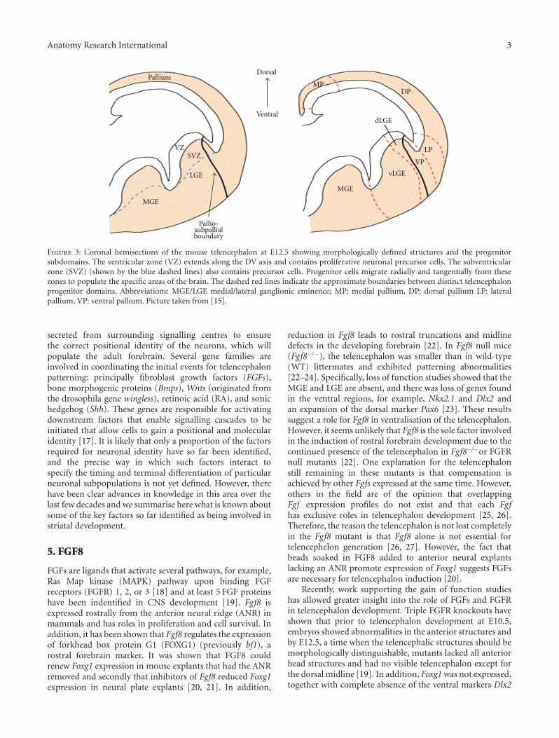
Due to the rapid migration of postmitotic neurons in thesubpallium, three prominent intraventricular bulges form;the septum, the medial, and lateral ganglionic eminences(MGE/LGE), collectively referred to as the whole ganglioniceminence (WGE), shown in Figure 3. The MGE, the mostventral eminence, gives rise to the amygdaloid body and theGP whilst the LGE, that is situated more dorsally, gives riseto the caudate and putamen [10, 11]. The LGE is furtherdivided into the dorsal LGE (dLGE) and the ventral LGE(vLGE) on the basis of regional gene expression, which isdiscussed later.
Within the surrounding neural epithelium of the devel-oping telencephalon, there are two proliferative zones, whichare shown in Figure 3, the ventricular zone (VZ), which ispositioned on the perimeter of the lateral ventricles, and thesubventricular zone (SVZ) (unique to the telencephalon),which extends from the basal region of the VZ [12]. It is inthe proliferative zones of the ventral telencephelon that bothprojection neurons (including MSNs) and γ-aminobutyricacid (GABAergic) interneurons are born before migratingto the regions they populate in the adult brain. Specifically,striatal projection neurons are born in the LGE and make upnearly 90% of LGE neurons. Interneurons originating fromthe LGE populate the cortex, olfactory bulb, and striatum,whereas those from the MGE migrate to the cortex, GP, andalso the striatum [13–16].
4. Unzipping Telencephalon Development
The telencephalon is the most complex region of the mam-malian brain and shows vast heterogeneity in terms ofits various neuronal populations, structures, and function.Telencephalon development requires a variety of signals
Anatomy Research International 3
DP
LP
MP
VP
dLGE
Pallio-subpallialboundary
MGE
MGE
LGE
SVZVZ
Ventral
DorsalPallium
vLGE
Figure 3: Coronal hemisections of the mouse telencephalon at E12.5 showing morphologically defined structures and the progenitorsubdomains. The ventricular zone (VZ) extends along the DV axis and contains proliferative neuronal precursor cells. The subventricularzone (SVZ) (shown by the blue dashed lines) also contains precursor cells. Progenitor cells migrate radially and tangentially from thesezones to populate the specific areas of the brain. The dashed red lines indicate the approximate boundaries between distinct telencephalonprogenitor domains. Abbreviations: MGE/LGE medial/lateral ganglionic eminence; MP: medial pallium, DP: dorsal pallium LP: lateralpallium, VP: ventral pallium. Picture taken from [15].
secreted from surrounding signalling centres to ensurethe correct positional identity of the neurons, which willpopulate the adult forebrain. Several gene families areinvolved in coordinating the initial events for telencephalonpatterning: principally fibroblast growth factors (FGFs),bone morphogenic proteins (Bmps), Wnts (originated fromthe drosophila gene wingless), retinoic acid (RA), and sonichedgehog (Shh). These genes are responsible for activatingdownstream factors that enable signalling cascades to beinitiated that allow cells to gain a positional and molecularidentity [17]. It is likely that only a proportion of the factorsrequired for neuronal identity have so far been identified,and the precise way in which such factors interact tospecify the timing and terminal differentiation of particularneuronal subpopulations is not yet defined. However, therehave been clear advances in knowledge in this area over thelast few decades and we summarise here what is known aboutsome of the key factors so far identified as being involved instriatal development.
5. FGF8
FGFs are ligands that activate several pathways, for example,Ras Map kinase (MAPK) pathway upon binding FGFreceptors (FGFR) 1, 2, or 3 [18] and at least 5 FGF proteinshave been indentified in CNS development [19]. Fgf8 isexpressed rostrally from the anterior neural ridge (ANR) inmammals and has roles in proliferation and cell survival. Inaddition, it has been shown that Fgf8 regulates the expressionof forkhead box protein G1 (FOXG1) (previously bf1), arostral forebrain marker. It was shown that FGF8 couldrenew Foxg1 expression in mouse explants that had the ANRremoved and secondly that inhibitors of Fgf8 reduced Foxg1expression in neural plate explants [20, 21]. In addition,
reduction in Fgf8 leads to rostral truncations and midlinedefects in the developing forebrain [22]. In Fgf8 null mice(Fgf8−/−), the telencephalon was smaller than in wild-type(WT) littermates and exhibited patterning abnormalities[22–24]. Specifically, loss of function studies showed that theMGE and LGE are absent, and there was loss of genes foundin the ventral regions, for example, Nkx2.1 and Dlx2 andan expansion of the dorsal marker Pax6 [23]. These resultssuggest a role for Fgf8 in ventralisation of the telencephalon.However, it seems unlikely that Fgf8 is the sole factor involvedin the induction of rostral forebrain development due to thecontinued presence of the telencephalon in Fgf8−/−or FGFRnull mutants [22]. One explanation for the telencephalonstill remaining in these mutants is that compensation isachieved by other Fgfs expressed at the same time. However,others in the field are of the opinion that overlappingFgf expression profiles do not exist and that each Fgfhas exclusive roles in telencephalon development [25, 26].Therefore, the reason the telencephalon is not lost completelyin the Fgf8 mutant is that Fgf8 alone is not essential fortelencephelon generation [26, 27]. However, the fact thatbeads soaked in FGF8 added to anterior neural explantslacking an ANR promote expression of Foxg1 suggests FGFsare necessary for telencephalon induction [20].
Recently, work supporting the gain of function studieshas allowed greater insight into the role of FGFs and FGFRin telencephalon development. Triple FGFR knockouts haveshown that prior to telencephalon development at E10.5,embryos showed abnormalities in the anterior structures andby E12.5, a time when the telencephalic structures should bemorphologically distinguishable, mutants lacked all anteriorhead structures and had no visible telencephalon except forthe dorsal midline [19]. In addition, Foxg1 was not expressed,together with complete absence of the ventral markers Dlx2
4 Anatomy Research International
and Nkx2.1. Unexpectedly, the dorsal marker Emx1 was alsoabsent suggesting that FGF has a role in forming the dorsaltelencephalon in addition to the ventral telencephalon [19].
The phenotype of the FGFR triple knockout wasmarkedly more severe than the mild phenotype observed insingle or double receptor mutants [18]. This supports theargument that these receptors do compensate for each otherand do not have exclusive roles. However, this compensationis not absolute given that a mild phenotype was still evidentin single or double mutants. This suggests the compensatingreceptors can function, but as they are not the first choicefor the ligand, they produce signalling cascades at lowerefficiencies [19]. Therefore, results from the different combi-nations of FGFR mutants suggest that the FGFR1 receptor isresponsible for the majority of signalling, but it is the overalllevels of FGF signalling that operate to initiate, pattern, andsustain early telencephalon development as a whole ratherthan specific ligands patterning different areas [19]. What isleft to be answered is if the FGFs function as morphogens orindependent to a concentration dependant gradient.
Foxg1 and Fgf8 are both expressed at approximately thesame time in the ANR (∼E8) and function through a tightlylinked positive feedback loop [19, 20]. FGF signalling issufficient and necessary for Foxg1 expression, and Foxg1 isnecessary for Fgf8 expression [20]. However, the differentphenotypes between Foxg1 mutants and the FGFR triplemutant suggest that both genes must function at leastpartially through independent genetic pathways, the detailsof which are still unknown [19].
6. SHH
SHH is a member of the hedgehog (Hh) family of secretedproteins and acts as a morphogen that is first secreted fromthe notochord, which underlies the posterior structures ofthe brain, following which expression is from the overlyingneural plate [6, 27, 28]. By E9.5 Shh is expressed inneural epithelium of the ventral telencephelon [29], and byE12 it is expressed in the mantle zone and is no longerdetectable in the neuroepithelium [30]. Shh operates througha concentration gradient that spans the DV axis at differenttime points to confer different neuronal identities on thedeveloping precursors, and expression is first seen in theventral telencenphelon from E11.5 [31, 32]. SHH expressiondirects neural progenitors to a ventral fate and importantlyis both necessary and sufficient to induce specific ventralforebrain markers [32–34]. Shh is expressed in the ventraltelencephalon and is thought to maintain Fgf8 expression[32, 35] as well as induce expression of forebrain markers.Specifically, SHH activates several TFs including Nkx2.1 [36,37], Gsx2 (formerly Gsh2) [38–40], and Pax6 [41].
SHH acts as a ligand for a pathway involving twotransmembrane proteins, patched (Ptc) and smoothened(Smo). Normally, Ptc is bound to Smo and the pathway isinactive as Smo is not free to activate the Glioma-associatedoncogene homolog 3 (Gli3). This is illustrated in Figure 4.However, when SHH binds Ptc, Smo is de-repressed whichresults in the Gli repressor (GliR) becoming activated (GliA)
and being able to translocate to the nucleus and activate geneexpression, as shown in Figure 4.
There are three members of the Gli family of zinc-finger TFs, Gli1, Gli2, and Gli3 and all have been shownto regulate SHH-dependant gene expression. Gli proteinshave both activator and repressor activities, the N-terminalencodes a repressor function, and the C-terminal regionis required for positive activity [43]. It is believed thatthe Gli3 protein functions principally in its repressor formand it appears that its activity is negatively regulated byShh [44, 45], whereas Gli1 and 2 function primarily astranscriptional activators [46, 47]. The negative regulationby Shh on Gli3 is observed in the limb, where Shh inhibitsGli3 from processing into its repressor form [43]. Analysisof mouse mutants for each of the Gli genes (Gli1−/−),Gli2−/−, and Gli3−/−) has shown that mice lacking Gli3 orGli2 show only slight defects in telencephelon development[48], whereas mice lacking Gli3 have strong defects in dorsaltelencephelon patterning [49–51]. At the dorsal region of thetelencephelon, where the concentration of SHH is limited,the Gli3 protein is cleaved from an activator into a repressorform and promotes dorsal patterning [43]. It is the inhibitionof the Gli3 repressor complex in the ventral region thatfacilities telencephalon development; therefore, the primaryfunction of Shh is to prevent the production of excessive Glirepressors. However, as Gli3 has been shown to be able tofunction as a weak activator of Shh in vivo [52], the questionrecently investigated by Yu and colleagues [53] is whether thisprotein (or either Gli1 or 2) has an activating role in ventraltelencephalon [53]. Recent work by this group suggests Gliactivators do have a role in specification, differentiation, andpositioning of some subgroups of telencephalon neuronalprogenitors that arise from the interganglionic sulcus [53],but further experiments need to be carried out to learn moreabout the role of Gli proteins as activators, and therefore thispaper will concentrate on the role of Gli3 as a repressor inDV patterning.
The relationship between Shh and Gli3 has been shownfunctionally through varying combinations of mutants. InShh−/− mutants, the ventral markers Dlx2, and Gsx2 werereduced, whereas in Gli3−/− mutants the expression patternof these genes was extended into more dorsal regions [43].Overall the Shh−/− mutant shows more severe telencephelonabnormalities than the Gli3−/− mutant [49, 50]. In accordwith the loss of ventral markers in Shh mutants (Shh−/−),there is a loss of ventral telencephalic cells leading to analtered morphology of the ventral telencephalon togetherwith the ectopic expression of dorsal forebrain markers [33–35, 43]. Specifically, the mutants lack any MGE developmentas shown by the absence of Nkx2.1 [43]. In vitro, cultures oftelencephalon explants treated with SHH result in expressionof ventral markers such as Nkx2.1 [33]. The complimentarygain of function experiments carried out in both fish andmice has shown that SHH promotes ventral identity indorsal telencephalic cells in vivo with induction of theventral forebrain markers Gsx2, Dlx2 and Nkx2.1 [38,54]. Importantly, conditional knockouts using a FoxG1-Cre to knock-out Shh have allowed the optimal window ofsignalling in telencephalon development to be investigated.

Anatomy Research International 5
Smo
Gli complex
Ptc
Shh
Target gene notexpressed
Smo
Target gene expressed
GliR GliA
GliA
Figure 4: The Shh pathway for target gene expression: (a) repressed pathway—when SHH cannot bind Ptc, ptc represses gene expression bybeing bound to Smo. Smo cannot then activate the Gli complex meaning the target gene is repressed. (b) Induction pathway—when SHHbinds ptc, smo is released which allows the GliA to bind the DNA and activate gene expression. Abbreviations: SHH: sonic hedgehog, Ptc:patched, Smo: smoothened, GliA: Gli Activator.
Fuccillo et al. [55] showed that if Shh is knocked-out at E8.5,there are severe defects of all ventral telencephalic regions[55]. However, in knockouts at E10-12 using a Nestin-Cre,there are limited defects in ventral telencephalic patterningand cortical interneurons are affected rather than grosspatterning deficits [56].
In the Shh−/− and Gli3−/+ mutants, telencephalonmorphology is largely restored to WT, but regional geneexpression is not fully restored. The ventral marker Nkx2.1is not rescued unless both copies of the Gli3 proteinare removed, suggesting Nkx2.1 is very sensitive to theantagonism between Shh and Gli3 [43]. Also, in Gli3−/−mutants, the expression of Nkx2.1 is not extended dorsallylike Dlx2 and Gsx2, nor can it be expressed ectopicallyin the cortex when exogenous Shh is added. From thisRallu and colleagues suggested that the Gli2 protein has acomplimentary role to Gli3. It is thought that Gli2 functionsas a weaker repressor and can repress the expansion ofNkx2.1 in Gli3−/− mutants but is not strong enough toprevent the expansion of the more ventral genes Dlx2and Gsx2 [43]. Moreover, in the Shh−/− and Gli3−/−,double mutant ventral markers such as Gsx2 and Dlx againwere largely restored to WT levels [43, 57]. In support ofthis, Nkx2.1 expression was expanded in Ptc mutant mice(Ptc−/−). In this mutant, Smo should be persistently de-repressed and Gli proteins should remain in their activatedform, thus having a repressive effect on Nkx2.1 expressionin the MGE [58]. However, the fact that Dlx2 and Gsh2were restored in the absence of Shh signalling in thismutant suggests that other genes and signalling pathways,independent to Shh signalling, have a role in DV patterningof the telencephalon [43].
In summary between E9 and E12.5, SHH acts mainlyby inhibiting the formation of the Gli3 repressor [43] andcontributes to the establishment of DV patterning [34, 55].Secondly, SHH signalling also supports the expansion of
progenitors of the ventral telencephalon by inducing andmaintaining the expression of Nkx2.1 until at least E14 andlater into neurogenesis [56].
7. Retinoic Acid
RA is the biologically active form of vitamin A and hasbeen implicated in survival, specification, proliferation, anddifferentiation during forebrain development [58–60]. Twooxidation events occur to ensure RA is successfully derivedto function as a ligand for either RA receptors (RARs)(RARα, RARβ and RARγ) or retinoid X receptors (RXRα,RXRβ, and RXRγ) that belong to the steroid/thyroid receptorsuperfamily [61]. Initially retinol dehydrogenases oxidateretinol to retinaldehyde and then the rate-limiting enzymes,retinaldehyde dehydrogenases (Raldh), are required to oxi-date retinaldehyde to RA [62].
The first known source of RA in the developing striatumis in the LGE at approximately E12.5 and is produced fromreactions mainly catalysed by Raldh3 [63]. It is not until E14that RA and Raldh3 are obviously expressed in the LGE andpromote GABAergic neuronal differentiation by inducingGad67, an enzyme needed for GABA synthesis [64]. Raldh3continues to promote GABAergic differentiation at E18.5,and RA continues to be expressed into adulthood [64]. It hasalso been reported that in vitro LGE-derived neurospheresand human embryonic stem cells (hESCs) induce GABAergicdifferentiation once RA was added to the media [64]. How-ever, it is thought that the RA in the LGE is not only a productfrom Raldh3-mediated reactions as Raldh3−/− mutant micedo not show an obvious telencephalic phenotype [65]. It hasbeen shown that retinoids from the glia found in the LGEare also functioning in striatal neuronal differentiation [66];therefore, it is possible that these, and other sources not yetknown could be compensating for Raldh3.

6 Anatomy Research International
Furthermore, at this time the RARs α and β are presentin the ventral telencephalon, with RARβ preferentially ex-pressed in LGE together with RXRγ [67]. In RAR β−/−
mutant mice, there is a loss of striatal-enriched tyrosinephosphatase mRNA, a gene regulated by RA in the striatum[67], and a reduction of striatal dopamine and cAMP-regulated protein (DARPP-32) positive neurons togetherwith dynophin, μ-opioid receptor (MOR1), and tyrosinehydroxylase (TH) compared to WT mice [59, 67]. It has alsobeen shown that when chick explants of LGE are treatedwith RAR antagonists, LGE specification is prevented [58].The complimentary experiment shows that when exogenousRA was added to dorsal explants, LGE was evident insteadof MGE [58]. Additionally, supplementation of RA to LGEcultures showed an increase in DARPP-32 positive neurons,whilst there was no effect seen in the MGE relevant toincreasing doses of RA [66]. Moreover, blocking RA inchick embryos prevents the expression of Meis2 which isexpressed in progenitor cells of the intermediate zone of thetelencephalon and is the earliest known marker of striatalprecursors [66]. Taken together, these results confirm theimportance of RA in LGE specification.
As well as being important in embryonic development,RA expression remains in the forebrain throughout adultlife and has been shown to maintain the expression ofFgf8 and Shh in this region, as when RA is removed, Fgf8and Shh expression is lost [59, 60]. It has recently beenproposed that Nolz1, a zinc finger TF that is expressed inthe proliferative SVZ in LGE precursor cells, is implicatedin RA signalling [68]. At E12.5 Nolz1-induced neurogenesispartially depends on RA signalling as it has been shownthat this TF activates the RAR β receptor in LGE-derivedneural precursor cells and that this effect was inhibited whenRA was removed [68]. However, Nolz1 expression was notaffected in Raldh3 mutant mice (Raldh3−/−), which lacks RAin the LGE, or when a vitamin A deficient diet was fed tothe mothers [63, 69] suggesting RA is not essential to Nolz1expression throughout development and is only needed toinduce early expression. RA activates Nolz1 to induce initialneurogenesis during early striatal development at E12.5 butis not sufficient for its maintenance beyond this time [68].Also, it has also been shown that Nolz1 contributes to laterstriatal development by working downstream of Gsx2 toactivate the RARβ receptor.
8. Wnt Signalling
Wnts belong to the wingless protein family and are a classof ligands that are crucial in embryogenesis and have beenimplicated in CNS development. Wnts can signal throughthree different pathways: the canonical pathway, the planarcell polarity pathway, and the calcium pathway and it isthe canonical pathway that is important in telencephalondevelopment. In the canonical pathway, β-catenin is indi-rectly activated by a WNT ligand binding to the cell surfacereceptor, Frizzled. Upon binding, frizzled activates its intra-cellular component dishevelled (Dsh) that dephosphorylatesβ-catenin preventing its degradation by the axin-glycogen
synthase kinas 3β (GSK3β) complex (in the absence ofWNT signalling, β-catenin is phosphorylated by the GSK3βcomplex and degraded). β-catenin then translocates to thenucleus where it can activate the transcription of Wnt targetgenes such as T-cell factors (TCF), which in turn regulategenes such as c-myc. This pathway is shown in Figure 5.
Wnts are part of the cohort of caudalizing factors thatare involved in the initial AP orientation of the neuralplate and are crucial for the generation of the dorsaltelencephelon [70]. Specific concentrations of WNTs areneeded to further refine regional patterning and to inducethe expression of Pax6, a dorsal telencephelon marker [71].A reporter line carrying a LacZ reporter gene under thecontrol of β-catenin/TCF response elements showed thatWNT signalling is active in the pallium at E11.5 and E16.5but not in the subpallium [72, 73]. In the absence ofcanonical signalling, there was ectopic expression of Gsx2,Dlx2, and Ascl1 (formerly Mash1) in dorsal telencephelontogether with downregulation of the dorsal markers Emx1, 2and 3 [73]. This ectopic expression of ventral genes facilitatedthe cells of the dorsal telencephelon to adopt a ventral fate,therefore allowing these cells to have the potential to becomeGABAergic projection neurons [73]. Therefore, this workin mice has shown that WNT signalling is necessary forensuring the correct molecular characterisation, and thusmorphology, of the dorsal telencephelon before the onsetof neurogenesis and that inhibition of WNT signalling isnecessary for subpallidal development [73]. In addition, gainof function experiments carried out in chicks has shown theimportance of Wnt gene expression in dorsal telencephelonpatterning. Using chick explant cultures, Gunhaga et al. [71]showed that Wnt3a or Wnt8 expression can convert theventral telencephalic cells into Pax6 and Ngn2 positive cellsat the expense of Ascl1 and Nkx2.1 [71].
9. BMPs
Although BMP inhibition is required for neuronal devel-opment, graded concentrations are necessary in the neuralplate to establish medial-to-lateral patterning [42]. BMPsbelong to the TGFβ family of secreted proteins. It is thoughtthat BMPs are also needed to dorsalize the telencephalonand restrict ventral telencephalic development. Forebrainpatterning was repressed in forebrain explant cultures whenBMPs were added as shown by inhibition of Foxg1, Nkx2.1,and Dlx2 [74]. Similarly beads soaked in BMP4 or BMP5that were implanted into the neural tube of a chick forebraininduced dorsal markers, for example, Wnt4 and repressedventral markers [75]. Additionally, when the telencephalicroof plate (a source of BMPs) was ablated, there was areduction in cortical size and a decrease of one of the mostdorsal cortical markers, Lhx2 [76]. BMPs are inhibited byseveral factors including chordin and noggin. In mice thatlacked both copies of the chordin gene (chordin−/−) and onecopy of the noggin gene (noggin+/−), a dorsal, rather thanventral telencephalon was evident. However, this effect maynot be direct because of an increase in BMP and may be in

Anatomy Research International 7
TCF
AxinAPC
P
Degraded
Target gene not expressed
Dsh
Frizzled
LRP5/6
Gsk3β
β-catenin
(a)
WntLRP5/6
TCF
Axin
Gsk3β
APC
P
Target gene expressed
Dsh
Frizzled
β-catenin
(b)
Figure 5: The canonical Wnt pathway for target gene expression. (a) When a WNT ligand is absent, β-catenin is phosphorylated by theGsk3β complex and is targeted for degradation and there is no gene expression. (b) When a Wnt ligand binds Frizzled, Dsh is activatedand inhibits the axin-Gsk3β complex phosphorylating β-catenin enabling it to translocate to the nucleus and can activate TCF which canactivate gene expression. Abbreviations: APC: adenomatous polyposis coli, Dsh: Dishevelled; GSK3β: glycogen synthase kinase 3 β; TCF:T-cell factor.
part due to the decreased levels of Shh and Fgf8 expression inthe forebrain caused by increased BMP levels [77].
10. Foxg1
Foxg1 is a member of the winged helix family of TFs and isthe earliest recognised marker of the telencephalon [78]. ThisTF was first identified in the rat brain, where it was shownthat its expression was restricted to the telencephelon [78].By E8.5 Foxg1 is expressed in the neural tube, specificallyin the anterior plate cells that are fated to contribute tothe telencephalon [20, 79], and functions to establish andsubdivide the telencephalon.
Foxg1−/− mutant mice show no morphological differ-ences in the size of the developing telencephalon at E10.5.However, the ventral markers Ascl1, Nkx2.1, Gsx2, andDlx1/2 are absent, and instead the dorsal markers, Emx2 andPax6, are expressed throughout the telencephelon [80, 81].It has also been shown that Fgf8 was reduced in the mutanttelencephalon at E10.5 [81]. By E12.5 there were considerablemorphological differences in the ventral telencephalon ofthe mutant when compared to WT; notably the GEs wereabsent but there were no defects in the dorsal area [80,81]. A reason for the absence of the ventral telencephalonin these mutants is the loss of proliferating cells in thisregion (shown through BrdU staining) with telencephalicproliferation being restricted to the dorsal region [80]. Thisdecrease in proliferation could be a direct consequence of thelack of Foxg1 expression or could be due to effectors of Foxg1not being able to function optimally.
It has also been shown that Foxg1 coordinates signallingpathways of SHH and WNTs, which are required for the
development of the subpallial and pallial telencephalon,respectively, and have been described above [82]. Dorsalidentity is prevented by Foxg1 inhibiting the Wnt pathway,further confirming the role of Foxg1 in ventral telencephaloninduction in an independent way to SHH. Manuel et al.[17] cultured cells from Foxg1−/− mice and showed thatthe addition of SHH and FGF8 alone could not inducethe expression of other ventral telencephalon genes. Also,further experiments where Foxg1−/− cells were grafted intoFoxg1−/−/Foxg1+/+chimeras, showed that the mutant cells,were specified abnormally, did not integrate with WT cellsand expressed dorsal rather than ventral markers [17].Recently, Manuel et al. [83] have also shown that thereason for mutant cells behaving differently is due to themhaving an increased cell cycle, something initially reportedby Martynoga et al. [81]. Specifically, Manuel and colleagueshave shown that this is due to a decrease in Pax6 expression,a cell cycle organiser. Upon addition of PAX6, the mutantphenotype was partially rescued [83]. This work suggests thatFoxg1 not only promotes the production of ventralising cues,but has a cell autonomous role in regulating Pax6 [83]. Foxg1is crucial in forebrain development and is absolutely requiredfor the regulation of telencephalic identity.
11. Dorsoventral (DV) Organisation of theDeveloping Telencephalon
As discussed already, the developing telencephalon is dividedinto the dorsal region, and the ventral region and these areascan be defined on a morphological and genetic basis. Inthe dorsal telencephalon Pax6, Neurogenin (Ngn) 1/2 and

8 Anatomy Research International
LP
VP
dLGE
MGE(pallidum)
vLGE(striatum)
Ngn1/2
Exm1/2
Pax6Lhx2
Nkx2.1
Dlx1/2
Gsx2
Gsx1
Mash1
Figure 6: Schematic coronal section through the developing telencephalon at E12.5—the dorsal and ventral subdomains are shown anddefined by unique gene expression patterns. Dorsal telencephalic markers shown are Emx1/2, Ngn1/1, and Pax6. The ventral telencephalicmarkers shown can be split into identifying the LGE or MGE. Mash1(Ascl1), Gsx1/2, and Dlx1/2 are associated with the LGE (specifically thevLGE), and Nkx2.1 is associated with the MGE. Some of the gene interactions are shown on the diagram. One of the important interactions isbetween Pax6 and Gsx2; these genes work together to ensure the subpallalial/pallial border is maintained. Arrows denote positive interactions;T-bars denote inhibitory control. The green arrows represent the genetic signalling that has been unravelled in more recent data and occursat later time points. Figure adapted from [42].
Emx1/2 are expressed; in the ventral telencephalon, Gsx2,Asc1, Dlx1/2, and Nkx2.1 are expressed, as shown in Figure 6[84]. The pallial genes will only be discussed briefly in thecontext of them as markers, not to fully elucidate theirrole in cortical development. Emx1/2 expression profiles arerestricted to the most dorsal region of the cortex with noexpression seen in the ventral cortical region. Ngn 1 and 2are basic helix loop helix (bHLH) TFs which are expressedthroughout the cortex together with Pax6. In the absence ofNgn expression, Ascl1 is ectopically expressed in the dorsaltelencephalon thus priming these cells to adopt a ventralfate and becoming GABAergic rather than glutamatergicneurons. Therefore, the role of Ngn1/2 is to maintain theDV boundary in the developing telencephalon and to inhibitventral gene expression such as Ascl1 [24, 85].
Homeodomain gene interactions are crucial in mediatingDV patterning and importantly, in setting up regionalsubdivisions within the developing telencephalon, as wellas within the ventral telencephelon, and are principallyregulated through Shh [36–41]. Pax6 and Gsx2 are twomembers of this homeodomain family [86]. These proteins
have overlapping expression profiles and work in synergy toensure the subpallium-pallium border is maintained [39].Their expression profiles mirror each other; Pax6 is expressedin a dorsal (high) to ventral (low) gradient and Gsx2 isexpressed in a ventral (high) to dorsal (low) gradient [87].
The embryonic patterning role of Pax6 was initially iden-tified through genetic mapping of the classical “small eye”(sey) mouse mutant [88]. Pax6 is needed for corticaldevelopment and to establish the subpallial-pallial borderand is initially detected in the developing forebrain at E8[89]. It is not until the neural tube stage that expressionis downregulated in ventral regions simultaneous with theup regulation of Nkx2.1 in this region, thus instantaneouslysetting up the DV ventral border on the basis of differentialgene expression [37, 38, 90]. In Pax6 mutant mice (Pax6−/−),there is a shift in the cortical-striatal boundary [91] andthe cortical markers Ngn1/2 and Emx1 are downregulated,and Dlx1/2 [39, 41, 92], Ascl11 [39, 41] and Gsx2 [39] areectopically expressed in dorsal regions of the telencephalon.Nkx2.1 also expands dorsally into the LGE shifting the LGE-MGE border [41].

Anatomy Research International 9
Gsx2 is first detected in the developing forebrain betweenE9 and E10 and is expressed in the LGE. Gsx2 mutants(Gsx2−/−) have the opposite phenotype to that seen in Pax6mutants; there is ectopic expression of Pax6 and Ngn2 inthe LGE accompanied by the loss of Ascl1 and Dlx2 [38–40]. On the whole, there was a reduction in the size of theLGE at E12 [93], which by E18.5 led to a reduction in thesize of the striatum [94] and reduced expression of striatalprojection neurons confirmed through a marked decrease inDARPP-32 and the earlier MSN marker, Forkhead box P1(FoxP1) [38–40, 95]. However, there was a slight increasein the striatal-matrix marker calbindin [95]. These resultssuggest that Gsx2 is a crucial inducer of Ascl1, Dlx1, andDlx2 whilst repressing dorsal character and is implicated inthe differentiation of calbindin positive neurons. However, inmice that lack both Gsx2 and Pax6 (Gsx2−/− and Pax6−/−),the phenotype observed was more subtle than the singlemutations as is the case in Shh−/−Gli3−/− double mutants[38]. This suggests that other genes are also important inmediating DV patterning and positioning of the pallial-subpallial boundary.
Gsx1, a gene closely related to Gsx2, is also expressedin the ventral telencephalon but unlike Gsx2 its expressionis restricted to the ventral most region of the LGE [96].It is thought that Gsx1 can partially compensate for thephenotype observed in Gsx2−/− mutants [95, 97, 98]. In theGsx2−/− mutant, Gsx1 expression spreads throughout theLGE between E11 and E14.5 and shows similar expressionpatterns to Ascl1 in the LGE [95]. DARPP-32 expression islost in both Gsx2−/− /Ascl1−/− and Gsx2−/− Gsx1−/− doublemutants suggesting Ascl1 has a role in regulating Gsx1 tocompensate for Gsx2 in Gsx2−/− mutants [95]. Until recentlythe role of Gsx1 has remained elusive as no phenotype hasbeen discovered through using knockouts. Pei et al. [99] haveshown that Gsx1 and Gsx2 differentially regulate the matura-tion of LGE progenitors. Gain-of-function experiments werecarried out to distinguish if these two closely linked genes doindeed carry out different roles in the LGE or if Gsx1 is simplya “backup” for Gsx2. Results showed that Gsx2 maintainsLGE progenitors in an undifferentiated position before Gsx1,in part through the downregulation of Gsx2, directs theprogenitors to acquire a mature neuronal phenotype [99].This is shown in Figure 6. These novel results indicatethat the Gsx genes regulate LGE patterning through acontrolled balance of signalling allowing proliferation anddifferentiation of neuronal progenitors [99].
Ascl1 is also a member of the bHLH family of TFs. Ithas a primary role in the correct development of the ventraltelencephalon and relies on Gsx2 for normal expression [38–40, 100]. In contrast to Ngn1/2 that is expressed in the dorsaltelencephelon, Ascl1 is expressed throughout the ventraltelencephalon and ASCL1 protein is seen in the VZ and SVZ,where neuronal precursor cells reside [101]. When Ascl1was ectopically expressed in the dorsal telencephalon, it wasable to induce neurons to express Dlx1/2 at the expense ofcortical markers [24]. Ascl1 interacts with Dlx1/2 that in turnactivates GAD/67, the rate-limiting enzyme for GABAergicsynthesis, and the two combined function to facilitate
GABAergic differentiation in the telencephalon [85, 100].However, in Ascl1 knockout experiments (Ascl1−/−), Dlxand Gad/67 are still expressed in the ventral telencephelon[100]. Together with the fact that these developing neuronscan still acquire a GABAergic phenotype in the absence ofDlx1 and 2, this suggests an element of redundancy in thissignalling pathway and/or the involvement of other genesnot yet identified. Expression of Gsx2 in the Ascl1−/− mutantis the same at E12.5, but by E18.5 there is an increase inGsx2 expressing cells suggesting that Ascl1 has the additionalrole of repressing Gsx2 function later in development. Thisis shown in Figure 6 [94]. Ascl1−/− mutants also show areduction in the number of early born striatal (cholinergic)and cortical (GABAergic) interneurons and a reduction insize of the MGE [100]. This phenotype can be explained bythe initial loss of precursor cells in the SVZ, subsequentlyleads to a decrease in neurons populating the mantle zone[100, 102]. Conversely, TH, D2R, and enkephalin positiveneurons are only slightly reduced in this mutant. This isexpected considering the LGE is only partially reduced [102].However, Wang et al. [94] suggest that the reason for theAscl1 mutant not giving a more severe phenotype is that Gsx2signals through another bHLH TF.
From these experiments, it can be concluded that Ascl1has the dual role of specifying precursors and controllingthe timing of their differentiation, principally in the MGEand possibly has a role in the LGE, although it is not crucialin this later eminence [98, 102]. Recent experiments byCastro and colleagues have looked more closely into the pre-cise mechanisms by which Ascl1 controls proliferation ofneuronal precursors [103]. Gene expression analysis carriedout from embryonic brains and neural stem cell culturesshowed that Ascl1 has a role in regulating genes concernedwith cell cycle progression and ultimately showed thatthere was a direct association between neural progenitorexpansion and the corresponding phases of cell cycle exitand neuronal differentiation [103]. What is clear is thatAscl1 is autonomously involved in patterning of earlytelencephalic progenitors (∼E10.5) and nonautonomouslyinvolved in repressing the differentiation of adjacent progen-itors through Notch signalling [100]. After Ascl1 has beenexpressed to aid neurogenesis, Dlx1 and 2 repress Ascl1 andsubsequent notch signalling to promote terminal neuronaldifferentiation [104].
The Dlx family bears homology to the drosophila distalless-homeobox gene family of which there are 6 murinemembers, 4 of which are expressed in the developing MGEand LGE [105]. Dlx1 and Dlx2 are expressed by subsetsof cells in the VZ and by most cells in the SVZ andswitch off as cells start to differentiate [101, 106]. Dlx5and Dlx6 are expressed in the SVZ and mantle zone only[13]. Single mutations of Dlx1 or 2 show no noticeableforebrain defects; although in the absence of both Dlx1 and2; there is arrested migration of matrix neurons within theSVZ [13, 106]. However, striatal development is not stoppedcompletely, and this phenotype suggests other genes areinvolved in neuronal migration or that other genes suchas Gsx2 and Ascl1 are compensating somehow by directlyactivating the targets of Dlx1 and 2. A series of experiments
10 Anatomy Research International
from Rubenstein’s lab have sought to identify other genes andTFs that could function downstream and upstream of Dlx1/2to control LGE specification and differentiation through aseries of gene expression arrays and in situ hybridisations[107]. It is likely that further work will continue to findrelationships between these genes and Dlx1 and 2.
As mentioned above, Dlx1 and 2 activate GAD67, anenzyme needed for GABA synthesis and found in neuronalprecursors of the SVZ and neurons of the mantle zone inthe ventral telencephalon [100], and in Dlx1/2−/− there aredecreased levels of GAD67 in the dLGE [108]. It is hasbeen suggested that Dlx1/2 indirectly activate GAD67 andthat cooperation with other proteins is needed to promotea GABA neuronal phenotype [109]. Necidin is a maternallyimprinted gene and is only expressed in the paternal allele[110, 111]. Mutant mice that lacked the paternal necidinallele showed a significant decrease in the differentiation ofGABAergic neurons in vivo and in vitro, therefore suggestingthat necidin facilitates the specification of GABAergic neu-rons in cooperation with Dlx proteins [109].
Nkx2.1 is expressed exclusively in the MGE and is anotherhomedomain protein. The primary role of Nkx2.1 is in ven-tral specification of the telencephalon where it acts to repressLGE identity, and it is also important in the developmentof striatal interneurons [7, 37]. Nkx2.1 is induced by Shh atE8 [33], and as earlier mentioned, inhibition of Shh leads toreduced expression of Nkx2.1 and dorsalisation of the ventralembryo [34]. In the Nkx2.1 mutant mouse (Nkx2.1−/−),there are a lack of MGE derivatives and a DV switch of theMGE as it shows properties similar to the LGE rather thanthe MGE, for example some cells have been shown to expressDARPP-32 [37]. The loss of Nkx2.1 also showed a reductionof GABA and calbindin positive neurons from the cortex[37]. Additionally, generation of an (Nkx2.1−/−) mouse withthe addition of a DlxtauLacZ reporter gene showed loss ofearly migration of Dlx2-expressing progenitors [30].
It is clear that DV patterning of the telencephelonrequires the precise orchestration of several genes and TFsthat work together to ensure the correct development of thestriatum. It is likely that further experiments using the everexpanding range of genetic tools will further disclose theroles of genes already known, further elucidate the signallingpathways they operate through, and identify novel genes thathave a functional role in striatal development and that can beused as specific regional markers [112–114].
12. Conclusion
This paper has aimed to summarise and organise researchthat is being carried out to understand the genetic mecha-nisms controlling striatal development. However, it is clearthat there are still many pieces of the jigsaw to be foundand fitted into the gaps of this puzzle. The more thatis known about the development of the striatum, and,importantly, the development and differentiation of striatalMSN neurons, the more precise the protocols can be todirect the fate of renewable cell sources, such as embryonicstem cells, to a functional MSN phenotype for use incell replacement therapy for HD. An additional aspiration
is to identify specific genes to detect MSN precursorsrather than relying on markers of terminally differentiatedMSNs, such as DARPP-32. Earlier markers of putative MSNscould be used to facilitate the generation and refinementof neuronal differentiation protocols as well as trackingneuronal differentiation in grafts.
References
[1] G. Bates, P. S. Harper, and L. Jones, Huntigton’s Disease, 2002.[2] R. A. Hauser, S. Furtado, C. R. Cimino et al., “Bilateral
human fetal striatal transplantation in Huntington’s disease,”Neurology, vol. 58, no. 5, pp. 687–695, 2002.
[3] C. R. Gerfen, “The neostriatal mosaic: multiple levels of com-partmental organization,” Trends in Neurosciences, vol. 15,no. 4, pp. 133–139, 1992.
[4] C. M. Kelly, S. B. Dunnett, and A. E. Rosser, “Mediumspiny neurons for transplantation in Huntington’s disease,”Biochemical Society Transactions, vol. 37, no. 1, pp. 323–328,2009.
[5] A. C. Bachoud-Levi, V. Gaura, P. Brugieres et al., “Effect offetal neural transplants in patients with Huntington’s disease6 years after surgery: a long-term follow-up study,” LancetNeurology, vol. 5, no. 4, pp. 303–309, 2006.
[6] J. L. R. Rubenstein, K. Shimamura, S. Martinez, and L.Puelles, “Regionalization of the prosencephalic neural plate,”Annual Review of Neuroscience, vol. 21, pp. 445–477, 1998.
[7] M. Jain, R. J. E. Armstrong, R. A. Barker, and A. E. Rosser,“Cellular and molecular aspects of striatal development,”Brain Research Bulletin, vol. 55, no. 4, pp. 533–540, 2001.
[8] A. S. Fernandez, C. Pieau, J. Reperant, E. Boncinelli, andM. Wassef, “Expression of the Emx-1 and Dlx-1 homeoboxgenes define three molecularly distinct domains in thetelencephalon of mouse, chick, turtle and frog embryos:implications for the evolution of telencephalic subdivisionsin amniotes,” Development, vol. 125, no. 11, pp. 2099–2111,1998.
[9] L. Puelles, E. Kuwana, E. Puelles et al., “Pallial and subpallialderivatives in the embryonic chick and mouse telencephalon,traced by the expression of the genes Dlx-2, Emx-1, Nkx-2.1, Pax-6, and Tbr-1,” Journal of Comparative Neurology, vol.424, no. 3, pp. 409–438, 2000.
[10] R. R. Sturrock, “A comparative quantitative and morpho-logical study of ageing in the mouse neostriatum, indusiumgriseum and anterior commissure,” Neuropathology andApplied Neurobiology, vol. 6, no. 1, pp. 51–68, 1980.
[11] T. W. Deacon, P. Pakzaban, and O. Isacson, “The lateralganglionic eminence is the origin of cells committed to stri-atal phenotypes: neural transplantation and developmentalevidence,” Brain Research, vol. 668, no. 1-2, pp. 211–219,1994.
[12] K. Campbell, “Dorsal-ventral patterning in the mammaliantelencephalon,” Current Opinion in Neurobiology, vol. 13, no.1, pp. 50–56, 2003.
[13] S. A. Anderson, M. Qiu, A. Bulfone et al., “Mutations ofthe homeobox genes Dlx-1 and Dlx-2 disrupt the striatalsubventricular zone and differentiation of late born striatalneurons,” Neuron, vol. 19, no. 1, pp. 27–37, 1997.
[14] K. Campbell, M. Olsson, and A. Bjorklund, “Regionalincorporation and site-specific differentiation of striatal pre-cursors transplanted to the embryonic forebrain ventricle,”Neuron, vol. 15, no. 6, pp. 1259–1273, 1995.

Anatomy Research International 11
[15] M. Olsson, A. Bjorklund, and K. Campbell, “Early spec-ification of striatal projection neurons and interneuronalsubtypes in the lateral and medial ganglionic eminence,”Neuroscience, vol. 84, no. 3, pp. 867–876, 1998.
[16] R. O’Rahilly and F. Muller, The Embryonic Human Brain: AnAtlas of Development Stages, Wiley-Liss, New York, NY, USA,1999.
[17] M. Manuel, B. Martynoga, T. Yu, J. D. West, J. O. Mason,and D. J. Price, “The transcription factor Foxg1 regulates thecompetence of telencephalic cells to adopt subpallial fates inmice,” Development, vol. 137, no. 3, pp. 487–497, 2010.
[18] I. Mason, “Initiation to end point: the multiple roles offibroblast growth factors in neural development,” NatureReviews Neuroscience, vol. 8, no. 8, pp. 583–596, 2007.
[19] H. Paek, G. Gutin, and J. M. Hebert, “FGF signaling isstrictly required to maintain early telencephalic precursor cellsurvival,” Development, vol. 136, no. 14, pp. 2457–2465, 2009.
[20] K. Shimamura and J. L. R. Rubenstein, “Inductive interac-tions direct early regionalization of the mouse forebrain,”Development, vol. 124, no. 14, pp. 2709–2718, 1997.
[21] W. Ye, K. Shimamura, J. L. R. Rubenstein, M. A. Hynes, andA. Rosenthal, “FGF and Shh signals control dopaminergicand serotonergic cell fate in the anterior neural plate,” Cell,vol. 93, no. 5, pp. 755–766, 1998.
[22] S. Shanmugalingam, C. Houart, A. Picker et al., “Ace/Fgf8 isrequired for forebrain commissure formation and patterningof the telencephalon,” Development, vol. 127, no. 12, pp.2549–2561, 2000.
[23] E. E. Storm, S. Garel, U. Borello et al., “Dose-dependentfunctions fo Fgf8 in regulating telencephalic patterningcenters,” Development, vol. 133, no. 9, pp. 1831–1844, 2006.
[24] S. W. Wilson and J. L. R. Rubenstein, “Induction anddorsoventral patterning of the telencephalon,” Neuron, vol.28, no. 3, pp. 641–651, 2000.
[25] J. A. Cholfin and J. L. R. Rubenstein, “Frontal cortexsubdivision patterning is coordinately regulated by Fgf8,Fgf17, and Emx2,” Journal of Comparative Neurology, vol.509, no. 2, pp. 144–155, 2008.
[26] U. Borello, I. Cobos, J. E. Long, C. Murre, and J. L. R.Rubenstein, “FGF15 promotes neurogenesis and opposesFGF8 function during neocortical development,” NeuralDevelopment, vol. 3, no. 1, article 17, 2008.
[27] Y. Echelard, D. J. Epstein, B. St-Jacques et al., “Sonic hedge-hog, a member of a family of putative signaling molecules,is implicated in the regulation of CNS polarity,” Cell, vol. 75,no. 7, pp. 1417–1430, 1993.
[28] H. Roelink, J. A. Porter, C. Chiang et al., “Floor plateand motor neuron induction by different concentrationsof the amino-terminal cleavage product of sonic hedgehogautoproteolysis,” Cell, vol. 81, no. 3, pp. 445–455, 1995.
[29] K. Shimamura, D. J. Hartigan, S. Martinez, L. Puelles, and J.L. R. Rubenstein, “Longitudinal organization of the anteriorneural plate and neural tube,” Development, vol. 121, no. 12,pp. 3923–3933, 1995.
[30] S. Nery, J. G. Corbin, and G. Fishell, “Dlx2 progenitormigration in wild type and Nkx2.1 Mutant telencephalon,”Cerebral Cortex, vol. 13, no. 9, pp. 895–903, 2003.
[31] T. M. Jessell, “Neuronal specification in the spinal cord:inductive signals and transcriptional codes,” Nature ReviewsGenetics, vol. 1, no. 1, pp. 20–29, 2000.
[32] J. D. Kohtz, D. P. Baker, G. Corte, and G. Fishell, “Regional-ization within the mammalian telencephalon is mediated by
changes in responsiveness to Sonic Hedgehog,” Development,vol. 125, no. 24, pp. 5079–5089, 1998.
[33] J. Ericson, J. Muhr, M. Placzek, T. Lints, T. M. Jessell, andT. Edlund, “Sonic hedgehog induces the differentiation ofventral forebrain neurons: a common signal for ventralpatterning within the neural tube,” Cell, vol. 81, no. 5, pp.747–756, 1995.
[34] C. Chiang, Y. Litingtung, E. Lee et al., “Cyclopia anddefective axial patterning in mice lacking Sonic hedgehoggene function,” Nature, vol. 383, no. 6599, pp. 407–413, 1996.
[35] Y. Ohkubo, C. Chiang, and J. L. R. Rubenstein, “Coordinateregulation and synergistic actions of BMP4, SHH and FGF8in the rostral prosencephalon regulate morphogenesis of thetelencephalic and optic vesicles,” Neuroscience, vol. 111, no. 1,pp. 1–17, 2002.
[36] S. Kimura, Y. Hara, T. Pineau et al., “The T/ebp null mouse:thyroid-specific enhancer-binding protein is essential for theorganogenesis of the thyroid, lung, ventral forebrain, andpituitary,” Genes and Development, vol. 10, no. 1, pp. 60–69,1996.
[37] L. Sussel, O. Marin, S. Kimura, and J. L. R. Rubenstein,“Loss of Nkx2.1 homeobox gene function results in a ventralto dorsal molecular respecification within the basal telen-cephalon: evidence for a transformation of the pallidum intothe striatum,” Development, vol. 126, no. 15, pp. 3359–3370,1999.
[38] J. G. Corbin, N. Gaiano, R. P. Machold, A. Langston, andG. Fishell, “The Gsh2 homeodomain gene controls multipleaspects of telencephalic development,” Development, vol. 127,no. 23, pp. 5007–5020, 2000.
[39] H. Toresson, S. S. Potter, and K. Campbell, “Genetic controlof dorsal-ventral identity in the telencephalon: opposingroles for Pax6 and Gsh2,” Development, vol. 127, no. 20, pp.4361–4371, 2000.
[40] K. Yun, S. Potter, and J. L. R. Rubenstein, “Gsh2 and Pax6play complementary roles in dorsoventral patterning of themammalian telencephalon,” Development, vol. 128, no. 2, pp.193–205, 2001.
[41] A. Stoykova, D. Treichel, M. Hallonet, and P. Gruss, “Pax6modulates the dorsoventral patterning of the mammaliantelencephalon,” Journal of Neuroscience, vol. 20, no. 21, pp.8042–8050, 2000.
[42] S. W. Wilson and C. Houart, “Early steps in the developmentof the forebrain,” Developmental Cell, vol. 6, no. 2, pp. 167–181, 2004.
[43] M. Rallu, R. Machold, N. Gaiano, J. G. Corbin, A. P. McMa-hon, and G. Fishell, “Dorsoventral patterning is establishedin the telencephalon of mutants lacking both Gli3 andhedgehog signaling,” Development, vol. 129, no. 21, pp. 4963–4974, 2002.
[44] V. Marigo, R. L. Johnson, A. Vortkamp, and C. J. Tabin,“Sonic hedgehog differentially regulates expression of GLIand GLI3 during limb development,” Developmental Biology,vol. 180, no. 1, pp. 273–283, 1996.
[45] B. Wang, J. F. Fallon, and P. A. Beachy, “Hedgehog-regulatedprocessing of Gli3 produces an anterior/posterior repressorgradient in the developing vertebrate limb,” Cell, vol. 100, no.4, pp. 423–434, 2000.
[46] C. B. Bai and A. L. Joyner, “Gli1 can rescue the in vivofunction of Gli2,” Development, vol. 128, no. 24, pp. 5161–5172, 2001.
[47] P. Dai, H. Akimaru, Y. Tanaka, T. Maekawa, M. Nakafuku,and S. Ishii, “Sonic hedgehog-induced activation of the

12 Anatomy Research International
Gli1 promoter is mediated by GLI3,” Journal of BiologicalChemistry, vol. 274, no. 12, pp. 8143–8152, 1999.
[48] H. L. Park, C. Bai, K. A. Platt et al., “Mouse Gli1 mutantsare viable but have defects in SHH signaling in combinationwith a Gli2 mutation,” Development, vol. 127, no. 8, pp. 1593–1605, 2000.
[49] E. A. Grove, S. Tole, J. Limon, L. W. Yip, and C. W. Ragsdale,“The hem of the embryonic cerebral cortex is defined by theexpression of multiple Wnt genes and is compromised inGli3-deficient mice,” Development, vol. 125, no. 12, pp. 2315–2325, 1998.
[50] T. Theil, G. Alvarez-Bolado, A. Walter, and U. Ruther,“Gli3 is required for Emx gene expression during dorsaltelencephalon development,” Development, vol. 126, no. 16,pp. 3561–3571, 1999.
[51] S. Tole, C. W. Ragsdale, and E. A. Grove, “Dorsoventralpatterning of the telencephalon is disrupted in the mousemutant extra-toes,” Developmental Biology, vol. 217, no. 2,pp. 254–265, 2000.
[52] C. B. Bai, D. Stephen, and A. L. Joyner, “All mouse ventralspinal cord patterning by Hedgehog is Gli dependent andinvolves an activator function of Gli3,” Developmental Cell,vol. 6, no. 1, pp. 103–115, 2004.
[53] W. Yu, Y. Wang, K. McDonnell, D. Stephen, and C. B.Bai, “Patterning of ventral telencephalon requires positivefunction of Gli transcription factors,” Developmental Biology,vol. 334, no. 1, pp. 264–275, 2009.
[54] N. Gaiano, J. D. Kohtz, D. H. Turnbull, and G. Fishell,“A method for rapid gain-of-function studies in the mouseembryonic nervous system,” Nature Neuroscience, vol. 2, no.9, pp. 812–819, 1999.
[55] M. Fuccillo, M. Rallu, A. P. McMahon, and G. Fishell, “Tem-poral requirement for hedgehog signaling in ventral telen-cephalic patterning,” Development, vol. 131, no. 20, pp. 5031–5040, 2004.
[56] Q. Xu, C. P. Wonders, and S. A. Anderson, “Sonic hedgehogmaintains the identity of cortical interneuron progenitors inthe ventral telencephalon,” Development, vol. 132, no. 22, pp.4987–4998, 2005.
[57] A. Gulacsi and S. A. Anderson, “Shh maintains Nkx2.1 in theMGE by a Gli3-independent mechanism,” Cerebral Cortex,vol. 16, pp. i89–95, 2006.
[58] M. Marklund, M. Sjodal, B. C. Beehler, T. M. Jessell, T.Edlund, and L. Gunhaga, “Retinoic acid signalling specifiesintermediate character in the developing telencephalon,”Development, vol. 131, no. 17, pp. 4323–4332, 2004.
[59] R. A. Schneider, D. Hu, J. L. R. Rubenstein, M. Maden, andJ. A. Helms, “Local retinoid signaling coordinates forebrainand facial morphogenesis by maintaining FGF8 and SHH,”Development, vol. 128, no. 14, pp. 2755–2767, 2001.
[60] G. T. Haskell and A. S. LaMantia, “Retinoic acid signalingidentifies a distinct precursor population in the developingand adult forebrain,” Journal of Neuroscience, vol. 25, no. 33,pp. 7636–7647, 2005.
[61] M. Mark, N. B. Ghyselinck, and P. Chambon, “Functionof retinoid nuclear receptors: lessons from genetic andpharmacological dissections of the retinoic acid signalingpathway during mouse embryogenesis,” Annual Review ofPharmacology and Toxicology, vol. 46, pp. 451–480, 2006.
[62] G. Duester, “Retinoic acid synthesis and signaling duringearly organogenesis,” Cell, vol. 134, no. 6, pp. 921–931, 2008.
[63] N. Molotkova, A. Molotkov, and G. Duester, “Role of retinoicacid during forebrain development begins late when Raldh3
generates retinoic acid in the ventral subventricular zone,”Developmental Biology, vol. 303, no. 2, pp. 601–610, 2007.
[64] C. Chatzi, T. Brade, and G. Duester, “Retinoic acid functionsas a key gabaergic differentiation signal in the basal ganglia,”PLoS Biology, vol. 9, no. 4, Article ID e1000609, 2011.
[65] V. Dupe, N. Matt, J. M. Garnier, P. Chambon, M. Mark, andN. B. Ghyselinck, “A newborn lethal defect due to inactiva-tion of retinaldehyde dehydrogenase type 3 is prevented bymaternal retinoic acid treatment,” Proceedings of the NationalAcademy of Sciences of the United States of America, vol. 100,no. 2, pp. 14036–14041, 2003.
[66] H. Toresson, A. M. De Urquiza, C. Fagerstrom, T. Perlmann,and K. Campbell, “Retinoids are produced by glia in thelateral ganglionic eminence and regulate striatal neurondifferentiation,” Development, vol. 126, no. 6, pp. 1317–1326,1999.
[67] W. L. Liao and F. C. Liu, “RARβ isoform-specific regulationof DARPP-32 gene expression: an ectopic expression studyin the developing rat telencephalon,” European Journal ofNeuroscience, vol. 21, no. 12, pp. 3262–3268, 2005.
[68] N. Urban, R. Martın-Ibanez, C. Herranz et al., “Nolz1promotes striatal neurogenesis through the regulation ofretinoic acid signaling,” Neural Development, vol. 5, no. 1,article 21, 2010.
[69] A. K. Verma, A. Shoemaker, R. Simsiman, M. Denning,and R. D. Zachman, “Expression of retinoic acid nuclearreceptors and tissue transglutaminase is altered in varioustissues of rats fed a vitamin A-deficient diet,” Journal ofNutrition, vol. 122, no. 11, pp. 2144–2152, 1992.
[70] C. Houart, L. Caneparo, C. P. Heisenberg, K. A. Barth,M. Take-Uchi, and S. W. Wilson, “Establishment of thetelencephalon during gastrulation by local antagonism ofWnt signaling,” Neuron, vol. 35, no. 2, pp. 255–265, 2002.
[71] L. Gunhaga, M. Marklund, M. Sjodal, J. C. Hsieh, T. M.Jessell, and T. Edlund, “Specification of dorsal telencephaliccharacter by sequential Wnt and FGF signaling,” NatureNeuroscience, vol. 6, no. 7, pp. 701–707, 2003.
[72] S. Maretto, M. Cordenonsi, S. Dupont et al., “MappingWnt/β-catenin signaling during mouse development and incolorectal tumors,” Proceedings of the National Academy ofSciences of the United States of America, vol. 100, no. 6, pp.3299–3304, 2003.
[73] M. Backman, O. Machon, L. Mygland et al., “Effects of ca-nonical Wnt signaling on dorso-ventral specification of themouse telencephalon,” Developmental Biology, vol. 279, no.1, pp. 155–168, 2005.
[74] Y. Furuta, D. W. Piston, and B. L. M. Hogan, “Bone mor-phogenetic proteins (BMPs) as regulators of dorsal forebraindevelopment,” Development, vol. 124, no. 11, pp. 2203–2212,1997.
[75] J. A. Golden, A. Bracilovic, K. A. Mcfadden, J. S. Beesley,J. L. R. Rubenstein, and J. B. Grinspan, “Ectopic bonemorphogenetic proteins 5 and 4 in the chicken forebrainlead to cyclopia and holoprosencephaly,” Proceedings of theNational Academy of Sciences of the United States of America,vol. 96, no. 5, pp. 2439–2444, 1999.
[76] E. S. Monuki, F. D. Porter, and C. A. Walsh, “Patterning of thedorsal telencephalon and cerebral cortex by a roof plate-lhx2pathway,” Neuron, vol. 32, no. 4, pp. 591–604, 2001.
[77] R. M. Anderson, A. R. Lawrence, R. W. Stottmann, D.Bachiller, and J. Klingensmith, “Chordin and noggin pro-mote organizing centers of forebrain development in themouse,” Development, vol. 129, no. 21, pp. 4975–4987, 2002.

Anatomy Research International 13
[78] W. Tao and E. Lai, “Telencephalon-restricted expression ofBF-1, a new member of the HNF- 3/fork head gene family, inthe developing rat brain,” Neuron, vol. 8, no. 5, pp. 957–966,1992.
[79] J. M. Hebert and S. K. McConnell, “Targeting of cre tothe Foxg1 (BF-1) locus mediates loxP recombination inthe telencephalon and other developing head structures,”Developmental Biology, vol. 222, no. 2, pp. 296–306, 2000.
[80] S. Xuan, C. A. Baptista, G. Balas, W. Tao, V. C. Soares, and E.Lai, “Winged helix transcription factor BF-1 is essential forthe development of the cerebral hemispheres,” Neuron, vol.14, no. 6, pp. 1141–1152, 1995.
[81] B. Martynoga, H. Morrison, D. J. Price, and J. O. Mason,“Foxg1 is required for specification of ventral telencephalonand region-specific regulation of dorsal telencephalic precur-sor proliferation and apoptosis,” Developmental Biology, vol.283, no. 1, pp. 113–127, 2005.
[82] C. Danesin, J. N. Peres, M. Johansson et al., “Integration oftelencephalic Wnt and hedgehog signaling center activities byFoxg1,” Developmental Cell, vol. 16, no. 4, pp. 576–587, 2009.
[83] M. N. Manuel, B. Martynoga, M. D. Molinek et al., “Thetranscription factor Foxg1 regulates telencephalic progenitorproliferation cell autonomously, in part by controlling Pax6expression levels,” Neural Development, vol. 6, no. 1, article 9,2011.
[84] C. Schuurmans and F. Guillemot, “Molecular mechanismsunderlying cell fate specification in the developing telen-cephalon,” Current Opinion in Neurobiology, vol. 12, no. 1,pp. 26–34, 2002.
[85] C. Fode, Q. Ma, S. Casarosa, S. L. Ang, D. J. Anderson,and F. Guillemot, “A role for neural determination genesin specifying the dorsoventral identity of telencephalicneurons,” Genes and Development, vol. 14, no. 1, pp. 67–80,2000.
[86] H. M. Hsieh-Li, D. P. Witte, J. C. Szucsik, M. Weinstein, H. Li,and S. S. Potter, “Gsh-2, a murine homeobox gene expressedin the developing brain,” Mechanisms of Development, vol. 50,no. 2-3, pp. 177–186, 1995.
[87] J. M. Hebert and G. Fishell, “The genetics of early te-lencephalon patterning: some assembly required,” NatureReviews Neuroscience, vol. 9, no. 9, pp. 678–685, 2008.
[88] R. E. Hill, J. Favor, B. L. M. Hogan et al., “Mouse smalleye results from mutations in a paired-like homeobox-containing gene,” Nature, vol. 354, no. 6354, pp. 522–525,1991.
[89] A. Stoykova and P. Gruss, “Roles of Pax-genes in developingand adult brain as suggested by expression patterns,” Journalof Neuroscience, vol. 14, no. 3, pp. 1395–1412, 1994.
[90] P. H. Crossley, S. Martinez, Y. Ohkubo, and J. L. R.Rubenstein, “Coordinate expression of Fgf8, Otx2, Bmp4,and Shh in the rostral prosencephalon during developmentof the telencephalic and optic vesicles,” Neuroscience, vol. 108,no. 2, pp. 183–206, 2001.
[91] A. Stoykova, M. Gotz, P. Gruss, and J. Price, “Pax6-dependentregulation of adhesive patterning, R-cadherin expression andboundary formation in developing forebrain,” Development,vol. 124, no. 19, pp. 3765–3777, 1997.
[92] A. Stoykova, R. Fritsch, C. Walther, and P. Gruss, “Forebrainpatterning defects in small eye mutant mice,” Development,vol. 122, no. 11, pp. 3453–3465, 1996.
[93] J. C. Szucsik, D. P. Witte, H. Li, S. K. Pixley, K. M. Small, andS. S. Potter, “Altered forebrain and hindbrain development inmice mutant for the Gsh- 2 homeobox gene,” DevelopmentalBiology, vol. 191, no. 2, pp. 230–242, 1997.
[94] B. Wang, R. R. Waclaw, Z. J. Allen, F. Guillemot, andK. Campbell, “Ascl1 is a required downstream effector ofGsx gene function in the embryonic mouse telencephalon,”Neural Development, vol. 4, no. 1, article 5, 2009.
[95] R. M. Wang, Q. G. Zhang, J. Li, L. C. Yang, F. Yang, and D.W. Brann, “The ERK5-MEF2C transcription factor pathwaycontributes to anti-apoptotic effect of cerebral ischemiapreconditioning in the hippocampal CA1 region of rats,”Brain Research, vol. 1255, no. C, pp. 32–41, 2009.
[96] M. T. Valerius, H. Li, J. L. Stock et al., “Gsh-l: a novel murinehomeobox gene expressed in the central nervous system,”Developmental Dynamics, vol. 203, no. 3, pp. 337–351, 1995.
[97] H. Toresson and K. Campbell, “A role Gsh1 in the developingstriatum and olfactory bulb of Gsh2 mutant mice,” Develop-ment, vol. 128, no. 23, pp. 4769–4780, 2001.
[98] K. Yun, S. Garel, S. Fischman, and J. L. R. Rubenstein,“Patterning of the lateral ganglionic eminence by the Gsh1and Gsh2 homeobox genes regulates striatal and olfactorybulb histogenesis and the growth of axons through the basalganglia,” Journal of Comparative Neurology, vol. 461, no. 2,pp. 151–165, 2003.
[99] Z. Pei, B. Wang, G. Chen, M. Nagao, M. Nakafuku, and K.Campbell, “Homeobox genes Gsx1 and Gsx2 differentiallyregulate telencephalic progenitor maturation,” Proceedingsof the National Academy of Sciences of the United States ofAmerica, vol. 108, no. 4, pp. 1675–1680, 2011.
[100] S. Casarosa, C. Fode, and F. Guillemot, “Mash1 regulatesneurogenesis in the ventral telencephalon,” Development, vol.126, no. 3, pp. 525–534, 1999.
[101] M. H. Porteus, A. Bulfone, J. K. Liu, L. Puelles, L. C. Lo, and J.L. R. Rubenstein, “DLX-2, MASH-1, and MAP-2 expressionand bromodeoxyuridine incorporation define molecularlydistinct cell populations in the embryonic mouse forebrain,”Journal of Neuroscience, vol. 14, no. 11 I, pp. 6370–6383, 1994.
[102] O. Marın, S. A. Anderson, and J. L. R. Rubenstein, “Originand molecular specification of striatal interneurons,” Journalof Neuroscience, vol. 20, no. 16, pp. 6063–6076, 2000.
[103] D. S. Castro, B. Martynoga, C. Parras et al., “A novel functionof the proneural factor Ascl1 in progenitor proliferationidentified by genome-wide characterization of its targets,”Genes and Development, vol. 25, no. 9, pp. 930–945, 2011.
[104] K. Yun, S. Fischman, J. Johnson, M. Hrabe de Angelis, G.Weinsmaster, and J. L. R. Rubenstein, “Modulation of thenotch signalling by Mash1 and Dlx1/2 regulates sequentialspecification and differentiation of progenitor cell types inthe subcortical telencephalon,” Development, vol. 129, no. 21,pp. 5029–5040, 2002.
[105] J. K. Liu, I. Ghattas, S. Liu, S. Chen, and J. L. R. Rubenstein,“Dlx genes encode DNA-binding proteins that are expressedin an overlapping and sequential pattern during basal gangliadifferentiation,” Developmental Dynamics, vol. 210, no. 4, pp.498–512, 1997.
[106] S. Nery, H. Wichterle, and G. Fishell, “Sonic hedgehog con-tributes to oligodendrocyte specification in the mammalianforebrain,” Development, vol. 128, no. 4, pp. 527–540, 2001.
[107] J. E. Long, C. Swan, W. S. Liang, I. Cobos, G. B. Potter,and J. L.R. Rubenstein, “Dlx1&2 and Mash1 transcriptionfactors control striatal patterning and differentiation throughparallel and overlapping pathways,” Journal of ComparativeNeurology, vol. 512, no. 4, pp. 556–572, 2009.
[108] J. E. Long, S. Garel, M. Alvarez-Dolado et al., “Dlx-dependentand -independent regulation of olfactory bulb interneurondifferentiation,” Journal of Neuroscience, vol. 27, no. 12, pp.3230–3243, 2007.

14 Anatomy Research International
[109] T. Kuwajima, I. Nishimura, and K. Yoshikawa, “Necdinpromotes GABAergic neuron differentiation in cooperationwith Dlx homeodomain proteins,” Journal of Neuroscience,vol. 26, no. 20, pp. 5383–5392, 2006.
[110] P. Jay, C. Rougeulle, A. Massacrier et al., “The humanNECDIN gene, NDN, is maternally imprinted and locatedin the Prader-Willi syndrome chromosomal region,” NatureGenetics, vol. 17, no. 3, pp. 357–361, 1997.
[111] H. R. MacDonald and R. Wevrick, “The necdin gene isdeleted in Prader-Willi syndrome and is imprinted in humanand mouse,” Human Molecular Genetics, vol. 6, no. 11, pp.1873–1878, 1997.
[112] P. Arlotta, B. J. Molyneaux, J. Chen, J. Inoue, R. Kominami,and J. D. MacKlis, “Neuronal subtype-specific genes thatcontrol corticospinal motor neuron development in vivo,”Neuron, vol. 45, no. 2, pp. 207–221, 2005.
[113] R. Martın-Ibanez, E. Crespo, N. Urban et al., “Ikaros-1couples cell cycle arrest of late striatal precursors with neuro-genesis of enkephalinergic neurons,” Journal of ComparativeNeurology, vol. 518, no. 3, pp. 329–351, 2010.
[114] S. Tamura, Y. Morikawa, H. Iwanishi, T. Hisaoka, and E.Senba, “Foxp1 gene expression in projection neurons of themouse striatum,” Neuroscience, vol. 124, no. 2, pp. 261–267,2004.

Submit your manuscripts athttp://www.hindawi.com
Hindawi Publishing Corporationhttp://www.hindawi.com Volume 2014
Anatomy Research International
PeptidesInternational Journal of
Hindawi Publishing Corporationhttp://www.hindawi.com Volume 2014
Hindawi Publishing Corporation http://www.hindawi.com
International Journal of
Volume 2014
Zoology
Hindawi Publishing Corporationhttp://www.hindawi.com Volume 2014
Molecular Biology International
Hindawi Publishing Corporationhttp://www.hindawi.com
GenomicsInternational Journal of
Volume 2014
The Scientific World JournalHindawi Publishing Corporation http://www.hindawi.com Volume 2014
Hindawi Publishing Corporationhttp://www.hindawi.com Volume 2014
BioinformaticsAdvances in
Marine BiologyJournal of
Hindawi Publishing Corporationhttp://www.hindawi.com Volume 2014
Hindawi Publishing Corporationhttp://www.hindawi.com Volume 2014
Signal TransductionJournal of
BioMed Research International
Hindawi Publishing Corporationhttp://www.hindawi.com Volume 2014
Evolutionary BiologyInternational Journal of
Hindawi Publishing Corporationhttp://www.hindawi.com Volume 2014
Hindawi Publishing Corporationhttp://www.hindawi.com Volume 2014
Biochemistry Research International
ArchaeaHindawi Publishing Corporationhttp://www.hindawi.com Volume 2014
Hindawi Publishing Corporationhttp://www.hindawi.com Volume 2014
Genetics Research International
Hindawi Publishing Corporationhttp://www.hindawi.com Volume 2014
Advances in
Virolog y
Hindawi Publishing Corporationhttp://www.hindawi.com
Nucleic AcidsJournal of
Volume 2014
Stem CellsInternational
Hindawi Publishing Corporationhttp://www.hindawi.com Volume 2014
Hindawi Publishing Corporationhttp://www.hindawi.com Volume 2014
Enzyme Research
Hindawi Publishing Corporationhttp://www.hindawi.com Volume 2014
International Journal of
Microbiology