review article transcranial alternating current and random
TRANSCRIPT

Review ArticleTranscranial Alternating Current and Random NoiseStimulation: Possible Mechanisms
Andrea Antal1 and Christoph S. Herrmann2,3
1Department of Clinical Neurophysiology, University Medical Center, 37073 Gottingen, Germany2Experimental Psychology Lab, Department of Psychology, Center for Excellence “Hearing4all”, European Medical School,Carl von Ossietzky University, 26111 Oldenburg, Germany3Research Center Neurosensory Science, University of Oldenburg, Oldenburg, Germany
Correspondence should be addressed to Christoph S. Herrmann; [email protected]
Received 4 January 2016; Accepted 3 April 2016
Academic Editor: Volker Tronnier
Copyright © 2016 A. Antal and C. S. Herrmann.This is an open access article distributed under the Creative CommonsAttributionLicense, which permits unrestricted use, distribution, and reproduction in anymedium, provided the originalwork is properly cited.
Background. Transcranial alternating current stimulation (tACS) is a relatively recent method suited to noninvasively modulatebrain oscillations. Technically the method is similar but not identical to transcranial direct current stimulation (tDCS). Whiledecades of research in animals and humans has revealed the main physiological mechanisms of tDCS, less is known about thephysiological mechanisms of tACS.Method. Here, we review recent interdisciplinary research that has furthered our understandingof how tACS affects brain oscillations and by what means transcranial random noise stimulation (tRNS) that is a special form oftACS can modulate cortical functions. Results. Animal experiments have demonstrated in what way neurons react to invasivelyand transcranially applied alternating currents. Such findings are further supported by neural network simulations and knowledgefrom physics on entraining physical oscillators in the human brain. As a result, fine-grained models of the human skull and brainallow the prediction of the exact pattern of current flow during tDCS and tACS. Finally, recent studies on human physiology andbehavior complete the picture of noninvasivemodulation of brain oscillations.Conclusion. In future, themethodsmay be applicablein therapy of neurological and psychiatric disorders that are due to malfunctioning brain oscillations.
1. Introduction
Brain oscillations have proven to play important roles inmultiple perceptual, motor, and cognitive functions [1, 2].In case of disturbed brain oscillations, neurological andpsychiatric diseases can be the consequences [3, 4]. Inorder to establish a causal relationship between oscillationsand cognitive functions as well as try to restore disturbedoscillatory activity, it is desirable to develop newmethods thatcan externallymodulate and control them.Noninvasive brainstimulation (NiBS) methods such a repetitive transcranialmagnetic stimulation (rTMS) have been reported to modifybrain oscillations (e.g., [5]). Indeed, it is well documentedthat the periodic electromagnetic force generated duringrTMS can result in local entrainment of biologically relevantrhythms, mimicking frequency specific oscillatory activitiestriggered, for example, by cognitive tasks. More recently, twotypes of transcranial electric stimulation (tES) methods have
been introduced that also could serve this purpose: transcra-nial alternating current stimulation (tACS) and transcranialrandom noise stimulation (tRNS) [6]. The aim of our paperis to review some of the relevant results that shed light on thephysiological mechanisms behind these two methods.
When considering the physiological mechanism under-lying tES, we have to introduce some basic terms at first.If the effect of tES is measured during the interval ofelectrical stimulation, it is considered an online effect andis most probably due to alterations of membrane voltagethat facilitate or inhibit neural firing. If, on the other hand,an effect is measured after the end of the stimulation, it isconsidered an offline effect or after-effect. These are mostprobably induced by changes in synaptic activity/plasticity[7–9]. With regard to the measurements, in animal studiesthe outcomes are usually either neural spike rates or someform of coherence between the tES signal (e.g., a sine wave incase of tACS) and the neural signal. Typically, the stimulation
Hindawi Publishing CorporationNeural PlasticityVolume 2016, Article ID 3616807, 12 pageshttp://dx.doi.org/10.1155/2016/3616807
2 Neural Plasticity
intensity required to modulate a spike rate is higher than thatrequired to modulate coherence [10]. In human studies, themeasures are commonly either behavioral (reaction times,performance, etc.) or physiological, that is, obtained fromthe electroencephalogram (EEG), magnetoencephalogram(MEG), or electromyogram (EMG).A standard procedure forquantifying the effects of a tES technique is to measure themagnitude and time course of its effects on primary motorcortex (M1) excitability, by quantifying single pulse TMS-elicited motor evoked potentials (MEPs) in EMG recordings[11]. Since this is a very straightforward procedure relyingon an objective outcome parameter, a high percentage of thetES studies, including tACS and tRNS applications in healthysubjects, were carried out using M1 as a model.
2. Transcranial AlternatingCurrent Stimulation
2.1. Shifting the Resting Potential of the Membrane Voltage:Comparison to Direct Current (DC) Stimulation. With regardto DC stimulation one of the questions that was firstaddressed in early animal studies was whether externally(transcranially or subdurally) applied electric fields are ableto modulate neuronal activity. It was shown that anodalstimulation could enhance neural firing rates while cathodalstimulation resulted in a reduction of the firing rate [12, 13].The mechanism behind this differential modulation of corti-cal excitability is believed to rely on up- or downregulationof the resting potential of the neurons’ membrane voltagein response to anodal or cathodal stimulation, respectively(Figure 1) [14–16]. The effects of transcranial direct currentstimulation (tDCS) are always subthreshold, meaning thatthe polarization induced when a neuron is silent will not beable to drive the generation of action potentials. Evidently,weak polarizations can modulate the probability of actionpotentials when a neuron is activated by other neuronalinput.
Neuronal compartments are polarized in different waysduring stimulation, with the soma typically polarized inopposite way than dendrites [16, 17]. Thus, a stimulationconsidered anodal (depolarizing) for the neuronal soma(which can control the generation of action potentials verydirectly) is cathodal (hyperpolarizing) for the dendrites andvice versa. In the case of DC stimulation the somatic polar-ization probably plays the dominant role in the developmentof the online effects. During AC stimulation, the dendriticpolarization could also be relevant, since brain oscillationscan determine how neurons process the sometimes opposingsynaptic inputs.
In human studies, a bipolar stimulation of neuronalnetworks can be induced by placing both anodal and cathodalelectrodes on the scalp. However, the current flow is notalways maximal directly beneath the stimulation electrodesbut rather at their rims under the connectors and betweenelectrodes [18]. In case only one of the twomechanisms (exci-tation or inhibition) is required, one of the electrodes can beplaced extracranially [19]. Alternatively, a small stimulationelectrode can be combined with a larger “passive” returnelectrode. This procedure leads to strong current densities
under the small stimulation electrode and weaker currentdensities under the large return electrode [20].
One of the main technical differences between the moreestablished tDCS and the more recent tACS is the functionalinterpretation of the two or more electrodes that have to bemounted on the scalp. In case of tDCS, as it was mentionedabove, these two electrodes are referred to as anode andcathode and result in excitation or inhibition of the targetedunderlying cortex, respectively. For tACS the concept ofhow to modulate brain oscillations is quite different due tothe applied alternating current. During one half cycle of atACS oscillation, one electrode will serve as anode and theother one as cathode and current strength will increase anddecrease following a half sine wave. During the other halfcycle, the patternwill reverse and the former anodemust nowbe considered the cathode and vice versa. Thus, on average,the membrane potential is not affected and therefore tACS isprobably not so well suited to enhance or decrease excitabilityof cortex over sustained intervals of time as is tDCS.However,the effects during the brief depolarizing or hyperpolarizingphase of each half cycle could already be strong enough toinduce online effects via entrainment, an effect discussedbelow.
3. How Can tACS Influence Cortical Activity?
3.1. Computer Models Can Predict Intracranial Current Den-sities. A common question about tES is whether a weakextracranially applied current is able to influence the activityof cortical neurons in the human brain, which in turn alterscognitive functions. We will review a series of studies thathave addressed this issue in modelling tDCS current flow.In principle, many of the basic tDCS findings should alsohold for tACS [21]. The maximum current flow is, of course,only reached during the peak of the alternating currentand changes polarity during the two half-waves, that is,the direction of current flow changes by 180∘. Computersimulations of the current flow during tDCS using humanhead models have revealed that a significant amount of thecurrent is shunted by the well-conducting skin (∼90%), whilemuch less of the current actually reaches the brain [22]. In thecase of tACS, the different frequency response of each typeof conducting element between the electrodes and the brainshould also be taken into account [23].
Miranda et al. [24] demonstrated in an isotropic sphericalhead model that 2mA of tDCS results in an intracranialcurrent density of 0.1 A/m2 (Ampere per squaremeter). Usinga value of 0.450 S/m (Siemens per meter) for the conductivityof the brain, the authors argued that this results in an electricfield gradient of 0.22V/m (Volt per meter). In a more recentarticle these authors used amore realistic headmodel derivedfrom magnetic resonance imaging (MRI) measurementsresulting in amaximum electric field of 0.38V/musing a graymatter conductivity of 0.33 S/m [24].
Neuling et al. [25] used a very fine-grained anisotropicfinite element model of the human head to show that1mA of tDCS/tACS applied to human visual cortex resultsin intracranial current densities of 0.05 to 0.15 A/m2. Thisamounts to a cortical electric field of 0.417V/m when using

Neural Plasticity 3
No stimulation
4002000Time (ms)
−100
−90
−80
−70
−60
−50
−40
−30
−20
−10
0
Mem
bran
e vol
tage
(mV
)
(a)
Anodal tDCS
4002000Time (ms)
−100
−90
−80
−70
−60
−50
−40
−30
−20
−10
0
Mem
bran
e vol
tage
(mV
)
(b)
Cathodal tDCS
4002000Time (ms)
−100
−90
−80
−70
−60
−50
−40
−30
−20
−10
0
Mem
bran
e vol
tage
(mV
)
(c)
Figure 1: Assumed neural mechanism of tDCS. (a) Without tDCS, the resting potential of the cell is at −70mV and an incoming excitatorypostsynaptic potential (EPSP) arriving 100ms after onset of the experiment does not reach the threshold for firing at −50mV (dashed line).(b) If the neuron is close to an anode, the positive voltage from the anode will raise the resting potential towards a more positive voltage andthe same EPSP will exceed the threshold and result in a neural spike. (c) If the neuron is close to a cathode, the negative voltage from thecathode will lower the resting potential towards a more negative voltage and the same EPSP will not exceed the threshold.
the mean current density of 0.1 A/m2 if a gray matterconductivity of 0.24 S/m is assumed (Figure 2). Note that inthis paper we assume gray matter conductivities in the rangefrom 0.333 to 0.352 S/m, while the above-mentioned articlesused values in a wider range from 0.24 to 0.45 S/m.
The above-mentioned studies used two conventionallarge stimulation electrodes with an area of approx. 35 cm2per electrode. Large area under the electrode is desirableto reduce the current density in the skin. Using multiplesmaller electrodes yields stronger intracranial current den-sities (Figure 3). Together with an optimized placement ofthe electrodes derived from simulations this can significantlyenhance intracranial current densities and result in a morefocal stimulation [26].
The above-mentioned studies compute intracranial cur-rent densities that allow the prediction of the location andintensity of tES-effects. However, this is not sufficient toanswer the question, whether or not the computed intensityis below or above the threshold to elicit neuronal spiking inneurons that would not spike without these extracraniallyapplied currents. In order to come to a conclusion about thistopic, we need to consider animal studies with intracranialrecordings.
3.2. Animal Studies. As was mentioned above, the appli-cation of very weak electric fields applied to the humanbrain extracranially leads to the question, what intensity ofstimulation is required to see an effect upon neural activity.In animal studies this issue is frequently addressed; however,
it is still not clear how these data can be transferred tohuman studies. Indeed, the stimulation protocols appliedin animal studies are often quite different from those usedin humans. Therefore, these data should be handled withcaution. Furthermore, knowing the magnitude of the electricfield inside the brain is not enough to estimate the effects onneurons. The geometry and orientation of neurons relativeto the electric field determine the amount of polarization[28]. Neurons oriented parallel to the electric field polar-ize maximally while neurons oriented perpendicularly areideally not polarized at all. Currents in the dendrites-to-axon direction depolarize the soma, while axon-to-dendritescurrents hyperpolarize it. Considering the convolution of thecortex, currents flowing into a gyrus are expected to polarizeneurons on one side with a given polarity but neurons ofthe other side of the gyrus might react in the oppositeway. Therefore, at the single neuron level, the definition of“anodal” versus “cathodal” stimulation is less obvious if theorientation relative to the electric field and geometry of theneuron is not specified. The fact that anodal/cathodal stimu-lation usually increases/decreases excitability is probably dueto a higher number of depolarized/hyperpolarized neurons.Furthermore, the dynamics of the stimulated populationsof neurons can rectify the effects of electric fields as wasshown using a computational model [29]. Nevertheless, aclear experimental validation of this issue is still lacking.
Francis et al. [30] were able to demonstrate that electricpulses of 140 𝜇V/mm root mean square or 295 𝜇V/mm peakamplitude (0.295mV/mm) are sufficient to increase the firing

4 Neural Plasticity
PatchesSkinSkull compactaSkull spongiosa
CSFGray matterWhite matter
(a)
0.139
0.105
0.071
0.037
0.003
(A/m
2 )
(b)
Figure 2: (a) Anisotropic finite element model for simulations of intracranial current flow. Stimulation electrodes are at the EEG electrodepositions Cz and Oz of the international 10–20 system for electrode placement (red and blue, resp.). (b) Current density simulations revealstrongest current flow is in the posterior part of the brain underneath and between the stimulation electrodes. Reprinted with permission ofthe authors from [25].
rate of single neurons in hippocampal slice preparationsfrom the rat. However, these pulses resemble neither tDCS(constant current typically for multiple minutes) nor tACS(sine wave for multiple minutes) closely. Reato et al. [31]performed electrical stimulation experiments in slices of rathippocampus and network simulations. Both experimentsrevealed a threshold of 0.2mV/mm before an electrical ACwas able to modulate ongoing neural activity.
A more direct comparison of animal findings to tACS ispossible for the application of electric fields of alternating cur-rents (AC) to cortex. Frohlich and McCormick [10] appliedsuch AC fields to the cortical slices of ferrets. They were ableto demonstrate that AC fields as low as 0.5mV/mm weresufficient tomodulate ongoing neural activity. In a simulationstudy with a spiking neuronal network, the authors showedthat at low stimulation intensities only spike timing wasmodulated but spike rate was not altered [31]. In this case,spikes would align to certain phases of the externally appliedsine wave. At higher intensities, also the spike rate wasmodulated.
A further question is how much of an extracraniallyapplied electric current actually arrives at the cortex. Ozenet al. [32] attached stainless steel wires to the skull ofanesthetized rats, stimulated them electrically with AC, andsimultaneously recorded intracranial neural activity. Theywere able to demonstrate an entrainment of ongoingneuronalactivity at frequencies mimicking the frequency of corticalslow oscillations (0.8–1.7Hz) in multiple cortical areas. Theauthors reported that membrane potentials as well as unitactivity were modulated by AC stimulation.The experimentsrevealed that voltage gradients of 1mV/mm in the extracel-lular space were sufficient to affect discharge probability ofneurons.
3.3. Tissue Resistivity/Conductivity. A last step that isrequired before tES intensity can be compared to neuralfiring or phase-locking thresholds is to determine the graymatter resistivity or conductivity, which is the inverse ofresistivity.The intensity of tES is specified in current strengthin the unit A (Ampere) or mA (milliAmpere, i.e., 1/1000Ampere). As we have seen, modelling studies can predictintracranial current densities from these extracranial currentstrengths. Current density is given in A/m2 (Ampere persquare meter). Animal studies apply a voltage across thecortex in order to determine the threshold for modulatingneural activity. Two electrodes are required to do so; thereforethe resulting unit is a voltage gradient specified in V/m (Voltper meter); that is, 500𝜇V could be applied at two electrodesspaced 1mm apart resulting in 500 𝜇V/mm which convertsinto 0.5mV/mm or 0.5 V/m. In order to convert currentdensity into voltage gradients, the resistance of the brainis required according to Ohms law: 𝑈 = 𝑅 ∗ 𝐼 where 𝑈 isvoltage, 𝑅 is resistance, and 𝐼 is current flow. Table 1 gives anexample of such a conversion for 1mA of tES intensity.
The resistivity of tissue is typically measured in animalsand it is assumed that it is of equal value in humans.Measurements in rabbits and cats resulted in values fortissue resistivity of 2.84Ωm (Ohm meter, corresponding toa conductivity of 0.352 S/m) for gray matter at low frequencyand body temperature [33]. However, many researchers usevalues that slightly differ, for example, 3.03Ωm (conductivity0.333 S/m), for inverse modelling [34].
With this value we finally have all the information neededto convert a tES intensity into an intracranial voltage gradient.Figure 4 demonstrates this procedure; for example, Neulinget al. [25] applied tACS at an intensity of 1mA.Theywere ableto show that this stimulation intensity results in intracranial

Neural Plasticity 5
HD
-tACS
Emax0
(a)
Con
vent
iona
l tAC
S
Emax0
(b)
Figure 3: Different montages result in different patterns of current densities. (a) tACS with two 4 × 1 electrode montages (high density, HD)was used in order to achieve a more focal current density. (b) Trying to achieve a similar pattern of current flow with conventional largeelectrodes results in a more widespread distribution of currents. Reprinted with permission of the authors from [27].
Table 1: Translating a current strength of tDCS/tACS (left column) into a voltage gradient (right column) that is used in animal studies. Atfirst, an intracranial current density must be computed in a simulation. If this is multiplied by the gray matter resistivity it yields the voltagegradient.
tDCS/tACS intensity Intracranial current density Gray matter resistivity Intracranial voltage gradientUnit mA A/m2 Ωm V/mValue 1 0.1 3.03 0.303

6 Neural Plasticity
tACS intensity Current density Resistivity Voltage gradient(V/m)(mA)
0.3030.284
0.5
0.295
0.21
0.1 3.032.84Neuling et al., 2013
Reato et al., 2010
Francis et al., 2003
(A/m2)
(0.333S/m)(0.352S/m)
(Ωm)
Frohlich and McCormick, 2010
Figure 4: Translation of tACS intensity to intracranial voltage gradients allows a comparison to thresholds for eliciting spikes in animalresearch. The left axis represents tACS intensity. Neuling et al. [35] applied 1mA intensity (peak-to-peak value: translates to a sine wave of0.5mA amplitude). In their FEM, they could show that this intensity results in a number of current densities in different parts of the brainwith a maximum of 0.1 A/m2 (axis: current density). The third axis represents the tissue resistivity of gray matter (tissue conductivity givenon the left of the axis in brackets). Values in the range from 2.84 to 3.03Ωm result in voltage gradients from 0.284 to 0.303V/m being in therange of thresholds for neural firing or phase-locking (axis: voltage gradient). Note that for voltage gradients 1mV/mm is equal to 1 V/m.
current densities up to 0.1 A/m2 [21]. Note, however, thata range of current densities was found in the brain asrepresented by the gray area in the figure. Multiplying thiscurrent density with the gray matter resistivity of 2.84Ωmresults in an intracranial voltage gradient of 0.284V/m.Using 3.03Ωm instead would result in a voltage gradient of0.303V/m.
3.4. Comparing tES to Neural Thresholds. Comparing theabove-mentioned values for voltage gradients resulting fromtES to the above-mentioned thresholds for neural firing orphase-locking demonstrates that 1mA of tES would be abovethe lowest threshold determined by, for example, Reato et al.[31].However, for the other thresholds that have been defined,tES seems to be near or below threshold (Figure 4). Due to thewide range of thresholds observed in animal experiments, itcan be concluded that tES seems to result in voltage gradientsthat are roughly within the range of thresholds as obtainedfrom animal experiments. However, despite the differencesof the experimental preparations, some forms of entrainmentusing electric fields were reported in many AC studies,suggesting the entrainment may be a generic way electricfields can affect neuronal networks. The idea of entrainmentof neuronal oscillations by weak electrical alternating currentstimulation was first introduced by Deans et al. [36] forinduced gamma oscillations. At the single neuron level [37]showed how weak AC polarization entrained neuronal firingand modulated spike timing. Modulation of spontaneouslyactive Purkinje cells by AC fields was also already shownby Chan et al. [15]. In later studies Frohlich, Ozen, andReato extended these concepts for slow-waves and gammaoscillations. The values of the coupling constant (how manymV a neuron polarizes per V/m electric field) measuredin many of the studies were highly comparable. A valueof 0.1–0.2mV/V/m for the somatic polarization has beenmeasured in [10, 16, 28, 36, 37].The existence of such a reliablevalue suggests that the sensitivity of single neurons is alwaysmore or less the same across experimental preparations and
maybe species. The different thresholds found in animalpreparations could be explained by the differences in thedynamics underling the neuronal networks of interests.
The fact that behavioral results are obtained in well-controlled human experiments suggests that probably thelower of the thresholds of Figure 3 is to be taken intoconsideration for tES. Future experiments in humans arerequired, for example in patients with intra-cranial recordingelectrodes.
3.5. The Most Relevant Factors for Applying tACS in HumanStudies. As mentioned above, compared to tDCS a differentmechanism is at work during tACS, requiring a differentrationale for designing an experiment. At first, experimentersshould identify a cognitive process that is characterized bya specific brain oscillation or combination of oscillations.Next, what parameter of this brain oscillation is responsiblefor which aspect of the cognitive process has to be defined.In principle, three key parameters as well as higher orderinteractions can be taken into consideration.
(1) The amplitude of brain oscillations has been shownto be correlated with cognitive functions. For example,EEG alpha oscillations have been closely linked to visualperception [38] and different amplitudes of this oscillationdetermine whether or not a near-threshold visual stimuluswill be consciously perceived by human subjects [39].
(2) The frequency of brain oscillations is also crucialfor cognitive functions. For example, the number of gammacycles (30–80Hz, 25ms cycle length at 40Hz) that fit into onetheta cycle (4–8Hz, 125ms cycle length at 8Hz) is believedto determine the number of items that human subjects cankeep in short term memory, that is, 5 items (5 ∗ 25ms) at afrequency relation of 40Hz/8Hz [40].
(3) The phase of brain oscillations affects cognitive func-tions, too. For example, the phase of delta oscillations isimportant for recognition of speech [41]. In addition, phase-locking or phase coherence between cortical sites is supposedto reflect neural teamwork. In line with this assumption,
Neural Plasticity 7
for example, phase-locking in the gamma frequency rangebetween hemispheres is enhanced during perception of hor-izontal motion which requires interhemispheric integration[42].
In addition, higher order interactions between tACS andbrain oscillations have to be taken into account. If we want tomodulate brain oscillations by tACS, it will not be a linearprocess and the effect will not be limited to the frequencyof stimulation. First, linear increases in stimulation intensitymay have nonlinear effects on the affected neural tissue.Second, entraining oscillations does affect oscillations notonly at the frequency of stimulation but also at harmonicmultiples as well as subharmonics. In addition, it is knownthat certain frequencies interact with each other duringcognitive processes, a process referred to as cross-frequencycoupling [43].Therefore, it has to be assumed that entrainingone frequency may have effects also at other frequencies.
In the following, we do not intend to give a completereview of all tACS studies that have been carried out untilnow. That has been done recently (e.g., [44]). Instead, ourgoal is to focus on experiments that exemplify the above-mentioned parameters of modulating brain oscillations.
3.6.Modulating the Amplitude of BrainOscillations with tACS.The most prominent oscillation in the human EEG is thealpha rhythmwith a frequency in the range from 8 to 12Hz. Ifthe posterior part of the brain is stimulated with 10Hz tACSthis results in an enhancement of the EEG alpha amplitudeduring stimulation [45]. This after-effect can be seen forat least 30 minutes after 10 minutes of stimulation [35].Such elevations of amplitudes of EEG oscillations have alsobehavioral relevance. For example, elevating the naturallyoccurring EEG delta oscillations (0–4Hz) during sleep with0.75Hz tACS resulted in enhanced retention of declarativememories from the previous day [46]. Along the same lines,amplifying gamma oscillations (30–80Hz) with 40Hz tACSduring sleep leads to the induction of lucid dreaming [47].
3.7. Modulating the Frequency of Brain Oscillations with tACS.Animal experiments (see above) have shown that stimulatingcortical tissue with AC currents at a stimulation frequencybelow the frequency of intrinsic oscillations can slow downthese brain oscillations [10]. When the stimulation wasapplied at a frequency above that of the intrinsic oscillations,the frequency of the brain oscillations was speeded up.This resembles the so-called entrainment of an oscillator,known from physical systems [48–51]. A similar effect wasobserved recently by Helfrich and coworkers [45]. Theseauthors found a sharpening of the EEG alpha peak in thefrequency spectrum during 10Hz tACS over the visual cortexin humans. Along similar lines, tACS was used to slow downthe frequency of human theta oscillations in order to improveshort termmemory capacity, since according to the model ofLisman and Spruston [52] slower theta oscillations can hostmore cycles of faster gamma oscillations [53].
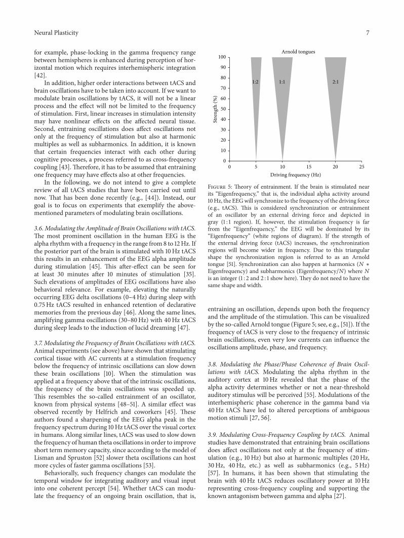
Behaviorally, such frequency changes can modulate thetemporal window for integrating auditory and visual inputinto one coherent percept [54]. Whether tACS can modu-late the frequency of an ongoing brain oscillation, that is,
Arnold tongues
5 10 15 20 250Driving frequency (Hz)
0
10
20
30
40
50
60
70
80
90
100
Stre
ngth
(%)
1:2 1:1 2:1
Figure 5: Theory of entrainment. If the brain is stimulated nearits “Eigenfrequency,” that is, the individual alpha activity around10Hz, the EEGwill synchronize to the frequency of the driving force(e.g., tACS). This is considered synchronization or entrainmentof an oscillator by an external driving force and depicted ingray (1 : 1 region). If, however, the stimulation frequency is farfrom the “Eigenfrequency,” the EEG will be dominated by its“Eigenfrequency” (white regions of diagram). If the strength ofthe external driving force (tACS) increases, the synchronizationregions will become wider in frequency. Due to this triangularshape the synchronization region is referred to as an Arnoldtongue [51]. Synchronization can also happen at harmonics (𝑁 ∗Eigenfrequency) and subharmonics (Eigenfrequency/𝑁) where 𝑁is an integer (1 : 2 and 2 : 1 show here). They do not need to have thesame shape and width.
entraining an oscillation, depends upon both the frequencyand the amplitude of the stimulation. This can be visualizedby the so-called Arnold tongue (Figure 5; see, e.g., [51]). If thefrequency of tACS is very close to the frequency of intrinsicbrain oscillations, even very low currents can influence theoscillations amplitude, phase, and frequency.
3.8. Modulating the Phase/Phase Coherence of Brain Oscil-lations with tACS. Modulating the alpha rhythm in theauditory cortex at 10Hz revealed that the phase of thealpha activity determines whether or not a near-thresholdauditory stimulus will be perceived [55]. Modulations of theinterhemispheric phase coherence in the gamma band via40Hz tACS have led to altered perceptions of ambiguousmotion stimuli [27, 56].
3.9. Modulating Cross-Frequency Coupling by tACS. Animalstudies have demonstrated that entraining brain oscillationsdoes affect oscillations not only at the frequency of stim-ulation (e.g., 10Hz) but also at harmonic multiples (20Hz,30Hz, 40Hz, etc.) as well as subharmonics (e.g., 5Hz)[57]. In humans, it has been shown that stimulating thebrain with 40Hz tACS reduces oscillatory power at 10Hzrepresenting cross-frequency coupling and supporting theknown antagonism between gamma and alpha [27].

8 Neural Plasticity
Due to the above-mentioned mechanism of tACS, werecommend a list of questions and suggestions for goodpractice in designing tACS experiments as follows.
Good Scientific Practice for Planning a tACS Experiment.Consider the following
Which cognitive or motor process shall be modu-lated?Which brain oscillation is associated with this cogni-tive or motor process?Which parameter of the brain oscillation (ampli-tude, frequency, phase angle, coherence between tworegions, etc.) is to bemodulated to achieve the desiredchange in cognitive or motor processing?Ideally, the observed or desired effect should bemodelled in a neural network. While it is relativelystraightforward to model whether an incoming sen-sory stimuluswill exceed the firing threshold of a neu-ron with a sinusoidally modulated resting potential, itis harder to find goodmodels that can explain humanreaction times or hit rates in models composed ofmultiple firing neurons.Which brain region should be targeted and whichelectrode montage is suited to achieve it?Model the intracranial current densities that resultfrom tACS or refer to existing modelling results.Demonstrate both behavioral and physiologicalchanges to ascertain the correlation of the two.Choose a plausible control condition to demonstratethat the observed effects are due to stimulation of aspecific brain region at a specific frequency and soforth.Good care has to be taken in order to rule out thatsubjects can differentiate between stimulation andcontrol conditions.
4. Transcranial Random Noise Stimulation
tRNS is a special form of tACS. During tRNS a low intensityalternating current is applied where intensity and frequencyof the current vary in a randomized manner.The stimulationis biphasic. Like with tACS, various forms of noise may beapplied, depending on the frequency ranges. In most of thestudies using tRNS, a frequency spectrum between 0.1Hzand 640Hz (full spectrum) or 101–640Hz (high-frequencystimulation) were used [58, 59]. The probability function ofthe noisy current stimulation follows a Gaussian or bell-shaped curve with zero mean and a variance, for which99% of all generated current levels are within ±1mA. In thefrequency domain all coefficients of the random sequencehave a similar amplitude (“white noise”).
tRNS over M1 had an effect comparable to the previouslydetermined effect of anodal tDCS on the development ofMEPs over time, enhancing the cortical excitability of thiscortical area [59–62]. For example, Terney and colleagues [59]
have shown that 10 minutes of tRNS applied over M1 with1mA intensity can induce facilitatory after-effects lasting upto 1–1.5 hours and is capable of improving the performance inan implicit motor learning task. It was also reported that thehigh-frequency subdivision between 100 and 640Hz of thewhole spectrum is functionally responsible for alteration ofexcitability in M1, superiorly to low frequency (0.1–100Hz)stimulation. In another study M1 stimulation using tRNSenhancedmotor skill learning compared to sham stimulation[63]. Compared to the time course of anodal tDCS, tRNSexerted more gradual effects, while tDCS resulted in largeskill gains immediately following the onset of stimulation.Interestingly, the after-effect of tRNS is intensity depen-dent. Lower intensity stimulation at 0.4mA tRNS leads toinhibitory after-effects comparable towhat has been observedwith cathodal tDCS using 1mA or 140Hz tACS using 0.4mA[61]. This suggests that inhibitory neurons in M1 might havelower thresholds, at least for this kind of stimulation. Onthe behavioral level, effects of high-frequency tRNS werealso demonstrated, for example, by Fertonani et al. [58]. Inthis study the authors applied tRNS to the visual cortices ofhealthy subjects during the performance of an orientation-discrimination task. A significant enhancement in visualperceptual learning during the application of high-frequencytRNSwas observed compared to anodal and cathodal tDCS aswell as sham stimulation. Interestingly, anodal tDCS induceda larger facilitation if it was applied before task execution andtRNS if it was applied during the task [64], suggesting that theideal timing of application of different electrical stimulationmethods varies and depends on the stimulation type. tRNSover the lateral occipital cortex facilitated facial identityperception [65]. In contrast, tRNS to the right dorsolateralprefrontal cortex (DLPFC) impaired categorical learning ina prototype distortion task [66]. These results demonstratethat depending on the involved cortical area and the typeof protocols, tRNS can induce long-term positive but alsonegative changes of cognitive and brain functions. However,a neutral effect was also reported. With regard to the effect oftRNS on working memory performance, a study showed noeffect of stimulation over the DLPFC on performance [67].
The physiological mechanisms of tRNS are not clarifiedcompletely yet. Animal studies on tRNS that could elucidatethe effects of this technique are completelymissing. Althoughhigher frequencies (e.g., 140Hz) have been shown to modu-late brain activity, the neuronal membrane acts as a low-passfilter; therefore, high frequencies that are applied by tRNSare supposed to polarize neurons by a very small amount.Deans et al. [36] measured the polarization of neuronsduring AC stimulation and estimated the coupling constantbetween electric field and induced polarization (mV per V/mapplied). They found that 100Hz AC stimulation gives acoupling constant of 0.050mV/V/m. Therefore, 1 V/m (max)in the brain at 100Hz can polarize a neuron by only 50 𝜇V.This value appears very small with respect to significantlymodulated brain function. However, as suggested by otherstudies, the stimulation ofmany synaptically connected activeneurons can provide an amplification mechanism [10, 31].
One potential online effect of tRNS might be associatedwith repetitive opening of Na+ channels, as was observed

Neural Plasticity 9
in a study investigating the application of AC stimulationto rat hippocampal slices [68]. Along this line, in a recentpilot study the Na+ channel blocker carbamazepine showeda tendency towards inhibiting MEPs 5–60 minutes afterstimulation [69]. Interestingly, the partial NMDA receptoragonist D-cycloserine, the NMDA receptor antagonist dex-tromethorphan that could block the effect of tDCS, had nosignificant effect on the excitability increases seen with tRNS.
Besides this, the effects of tRNS might be based onother mechanisms, such as stochastic resonance [70]. Briefly,stochastic resonance refers to the phenomenon that a signalthat is too weak to exceed a threshold is amplified by addingnoise, for example, when a neural oscillation in the brainis subthreshold. These, probably synaptically operated sub-threshold activities, driven by oscillatory inputs that neuronsreceive from other brain regions, are not strong enoughto induce action potential generation. If random noise isadded, the sum of the two signals exceeds the threshold atcertain times. The frequency of the suprathreshold signalis determined by the existing subthreshold neural oscilla-tion. It was suggested that tRNS may increase synchroniza-tion of neural firing through amplification of subthresholdoscillatory activity, which in turn reduces the amount ofendogenous noise. The improvement of the signal-to-noiseratio in the central nervous system and the sensitizationof sensory processing can lead to enhanced perception orcognitive performance [71–73]. However, it is not clear howthis process can induce long-term changes in the humanbrain [74, 75]. A study reported that bifrontal application oftRNS for 5 days enhanced the speed of both calculation- andmemory-recall-based arithmetic learning [74]. Six monthslater the behavioral and physiological modifications in thestimulated group relative to sham controls were still present.Similarly, in another study repeated bilateral parietal stimu-lation increased numerosity discrimination ability [75] withan after-effect for several weeks.
5. Summary
Both subtypes of tES, tACS, and tRNS are effective at mod-ulating neural brain activity and result in behavioral effectsin animals and human subjects.They are increasingly used inthe research and also in phase II clinical studies; for example,for the reduction of the symptoms in tinnitus patients it hasbeen shown that tRNS is more effective than either tDCS ortACS [76]. They have a better blinding potential with regardto the cutaneous sensations, such as itching, tingling, orburning, compared to tDCS applications [77]. Nevertheless,phosphene perception during tACS in awide frequency range(6–70Hz) might affect the interpretation of results (e.g., byinducing shifts in attentional state/arousal).
Unfortunately, it is not completely clarified how thesestimulation methods act on the neuronal level. The regularsinusoidal ups and downs of tACS result in a weak mod-ulation of the membrane voltage. If neuronal input fromother cells is just below threshold, this regular sinusoidalmodulation may be sufficient to drive the neuronal inputto exceed the threshold at the frequency of tACS. Similarly,the random fluctuations of external voltage in case of tRNS
may be sufficient to help an otherwise subthreshold neuraloscillation to exceed the threshold for firing. tRNS mightonly amplify neural activity that was already present beforestimulation, while tACS can interfere with ongoing neuraloscillations and change their frequency. However, it is asimplified picture, and recent experiments have revealed thateffects of tES are nonlinear and cross-frequency interactionsoccur. Furthermore, please note that oscillations in thelocal field potential at the network level can be visible asrhythmic postsynaptic potentials in single neurons. Thesepostsynaptic potentials can already drive neuronal firing andtherefore keep the neuron already in a suprathreshold statethat might not be affected by external stimulation. In thefuture, intracranial recordings of neural activity in patientsduring tES could shed light on many open questions. Inaddition, a direct comparison of transcranial stimulationmethods is desirable, for example, comparing rTMS to tESbut also comparing different tES methods with each other(e.g., [62]).
Additional Points
Coherence. Generally, coherence is ameasure of the variabilityof time differences between two time series. “EEG coherence”is often interpreted as a measure of the functional interplaybetween two brain regions. This can be defined by the directrelationship between the time and frequency domains of thebrain oscillations. If the time difference as a phase difference(or angle) is constant over time, the coherence is close to1.0, and if time difference varies in time from moment tomoment then the coherence is closer to 0. Phase-LockedActivity. Phase-locked activities contain evoked oscillationsthat are rigidly time-locked to the moment of stimulusdelivery. Neuronal Entrainment. It is a term used to describethe property of brain oscillations, how they synchronize theirperiodicity and rhythm through interaction(s). During thisprocess the given frequency of oscillations resulting from thesynchronous electrical activity of neuronal ensembles couldbe synchronized to the periodic activity of, for example, aninternal or external stimulus or event. Stochastic Resonance.It is a process that results in an improvement of detectionfor subthreshold signals by application of noise. It plays avery important role in nonlinear systems, especially in theinformation processing in the brain.
Competing Interests
The authors declare that they have no competing interests.
Acknowledgments
Christoph S. Herrmann was supported by the DeutscheForschungsgemeinschaft (DFG, Grant SPP 1665).
References
[1] C. S. Herrmann, M. H. J. Munk, and A. K. Engel, “Cognitivefunctions of gamma-band activity: memory match and utiliza-tion,” Trends in Cognitive Sciences, vol. 8, no. 8, pp. 347–355,2004.

10 Neural Plasticity
[2] A. K. Engel, P. Fries, and W. Singer, “Dynamic predictions:oscillations and synchrony in top-down processing,” NatureReviews Neuroscience, vol. 2, no. 10, pp. 704–716, 2001.
[3] C. S. Herrmann and T. Demiralp, “Human EEG gamma oscil-lations in neuropsychiatric disorders,”Clinical Neurophysiology,vol. 116, no. 12, pp. 2719–2733, 2005.
[4] P. J. Uhlhaas and W. Singer, “Neural synchrony in braindisorders: relevance for cognitive dysfunctions and pathophys-iology,” Neuron, vol. 52, no. 1, pp. 155–168, 2006.
[5] G. Thut and C. Miniussi, “New insights into rhythmic brainactivity from TMS-EEG studies,” Trends in Cognitive Sciences,vol. 13, no. 4, pp. 182–189, 2009.
[6] W. Paulus, “Transcranial electrical stimulation (tES—tDCS;tRNS, tACS) methods,” Neuropsychological Rehabilitation, vol.21, pp. 602–617, 2011.
[7] T. Zaehle, P. Sandmann, J. D. Thorne, L. Jancke, and C. S.Herrmann, “Transcranial direct current stimulation of theprefrontal cortex modulates working memory performance:combined behavioural and electrophysiological evidence,”BMCNeuroscience, vol. 12, article 2, 2011.
[8] A. Vossen, J. Gross, and G. Thut, “Alpha power increase aftertranscranial alternating current stimulation at alpha frequency(𝛼-tACS) reflects plastic changes rather than entrainment,”Brain Stimulation, vol. 8, no. 3, pp. 499–508, 2015.
[9] D. Struber, S. Rach, T. Neuling, and C. S. Herrmann, “On thepossible role of stimulation duration for after-effects of tran-scranial alternating current stimulation,” Frontiers in CellularNeuroscience, vol. 9, article 311, 2015.
[10] F. Frohlich and D. A. McCormick, “Endogenous electric fieldsmay guide neocortical network activity,” Neuron, vol. 67, no. 1,pp. 129–143, 2010.
[11] M. A. Nitsche and W. Paulus, “Excitability changes induced inthe human motor cortex by weak transcranial direct currentstimulation,” Journal of Physiology, vol. 527, no. 3, pp. 633–639,2000.
[12] L. J. Bindman, O. C. Lippold, and J. W. Redfearn, “The actionof brief polarizing currents on the cerebral cortex of the rat (1)during current flow and (2) in the production of long-lastingafter-effects,” The Journal of Physiology, vol. 172, pp. 369–382,1964.
[13] O. D. Creutzfeldt, G. H. Fromm, and H. Kapp, “Influenceof transcortical d-c currents on cortical neuronal activity,”Experimental Neurology, vol. 5, no. 6, pp. 436–452, 1962.
[14] J. G. R. Jefferys, “Influence of electric fields on the excitabilityof granule cells in guinea-pig hippocampal slices,” Journal ofPhysiology, vol. 319, pp. 143–152, 1981.
[15] C. Y. Chan, J. Hounsgaard, and C. Nicholson, “Effects of electricfields on transmembrane potential and excitability of turtlecerebellar Purkinje cells in vitro,” Journal of Physiology, vol. 402,pp. 751–771, 1988.
[16] M. Bikson, M. Inoue, H. Akiyama et al., “Effects of uniformextracellular DC electric fields on excitability in rat hippocam-pal slices in vitro,” The Journal of Physiology, vol. 557, no. 1, pp.175–190, 2004.
[17] A. Rahman, D. Reato, M. Arlotti et al., “Cellular effects ofacute direct current stimulation: somatic and synaptic terminaleffects,”The Journal of Physiology, vol. 591, no. 10, pp. 2563–2578,2013.
[18] A. Opitz, W. Paulus, S. Will, A. Antunes, and A. Thielscher,“Determinants of the electric field during transcranial directcurrent stimulation,” NeuroImage, vol. 109, pp. 140–150, 2015.
[19] V. Moliadze, A. Antal, and W. Paulus, “Electrode-distancedependent after-effects of transcranial direct and random noisestimulation with extracephalic reference electrodes,” ClinicalNeurophysiology, vol. 121, no. 12, pp. 2165–2171, 2010.
[20] M. A. Nitsche, S. Doemkes, T. Karakose et al., “Shaping theeffects of transcranial direct current stimulation of the humanmotor cortex,” Journal of Neurophysiology, vol. 97, no. 4, pp.3109–3117, 2007.
[21] S. Wagner, S. M. Rampersad, U. Aydin et al., “Investigationof tDCS volume conduction effects in a highly realistic headmodel,” Journal of Neural Engineering, vol. 11, no. 1, Article ID016002, 2014.
[22] R. N. Holdefer, R. Sadleir, and M. J. Russell, “Predicted currentdensities in the brain during transcranial electrical stimulation,”Clinical Neurophysiology, vol. 117, no. 6, pp. 1388–1397, 2006.
[23] M. Akhtari, H. C. Bryant, D. Emin et al., “Amodel for frequencydependence of conductivities of the live human skull,” BrainTopography, vol. 16, no. 1, pp. 39–55, 2003.
[24] P. C. Miranda, A. Mekonnen, R. Salvador, and G. Ruffini,“The electric field in the cortex during transcranial currentstimulation,” NeuroImage, vol. 70, pp. 48–58, 2013.
[25] T. Neuling, S. Wagner, C. H. Wolters, T. Zaehle, and C. S.Herrmann, “Finite-element model predicts current density dis-tribution for clinical applications of tDCS and tACS,” Frontiersin Psychiatry, vol. 3, article 83, 2012.
[26] J. P. Dmochowski, A. Datta, M. Bikson, Y. Su, and L. C. Parra,“Optimized multi-electrode stimulation increases focality andintensity at target,” Journal of Neural Engineering, vol. 8, no. 4,Article ID 046011, 2011.
[27] R. F. Helfrich, H. Knepper, G. Nolte et al., “Selective modula-tion of interhemispheric functional connectivity by HD-tACSshapes perception,” PLoS Biology, vol. 12, no. 12, Article IDe1002031, 2014.
[28] T. Radman, R. L. Ramos, J. C. Brumberg, and M. Bikson,“Role of cortical cell type and morphology in subthreshold andsuprathreshold uniform electric field stimulation in vitro,”BrainStimulation, vol. 2, no. 4, pp. 215.e1–228.e3, 2009.
[29] D. Reato, F. Gasca, A. Datta, M. Bikson, L. Marshall, and L.C. Parra, “Transcranial electrical stimulation accelerates humansleep homeostasis,” PLoS Computational Biology, vol. 9, no. 2,Article ID e1002898, 2013.
[30] J. T. Francis, B. J. Gluckman, and S. J. Schiff, “Sensitivity ofneurons to weak electric fields,”The Journal of Neuroscience, vol.23, no. 19, pp. 7255–7261, 2003.
[31] D. Reato, A. Rahman, M. Bikson, and L. C. Parra, “Low-intensity electrical stimulation affects network dynamics bymodulating population rate and spike timing,” Journal of Neu-roscience, vol. 30, no. 45, pp. 15067–15079, 2010.
[32] S. Ozen, A. Sirota, M. A. Belluscio et al., “Transcranial electricstimulation entrains cortical neuronal populations in rats,”TheJournal of Neuroscience, vol. 30, no. 34, pp. 11476–11485, 2010.
[33] L. A. Geddes and L. E. Baker, “The specific resistance ofbiological material—a compendium of data for the biomedicalengineer and physiologist,”Medical and Biological Engineering,vol. 5, no. 3, pp. 271–293, 1967.
[34] J. Haueisen, C. Ramon, P. Czapski, and M. Eiselt, “On theinfluence of volume currents and extended sources on neu-romagnetic fields: a simulation study,” Annals of BiomedicalEngineering, vol. 23, no. 6, pp. 728–739, 1995.

Neural Plasticity 11
[35] T. Neuling, S. Rach, and C. S. Herrmann, “Orchestratingneuronal networks: sustained after-effects of transcranial alter-nating current stimulation depend upon brain states,” Frontiersin Human Neuroscience, vol. 7, article 161, 2013.
[36] J. K. Deans, A. D. Powell, and J. G. R. Jefferys, “Sensitivity ofcoherent oscillations in rat hippocampus to AC electric fields,”The Journal of Physiology, vol. 583, no. 2, pp. 555–565, 2007.
[37] T. Radman, Y. Su, H. A. Je, L. C. Parra, and M. Bikson, “Spiketiming amplifies the effect of electric fields on neurons: implica-tions for endogenous field effects,” Journal of Neuroscience, vol.27, no. 11, pp. 3030–3036, 2007.
[38] M. Schurmann and E. Basar, “Functional aspects of alpha oscil-lations in the EEG,” International Journal of Psychophysiology,vol. 39, no. 2-3, pp. 151–158, 2001.
[39] S. Hanslmayr, A. Aslan, T. Staudigl, W. Klimesch, C. S. Her-rmann, and K.-H. Bauml, “Prestimulus oscillations predictvisual perception performance between and within subjects,”NeuroImage, vol. 37, no. 4, pp. 1465–1473, 2007.
[40] J. E. Lisman and M. A. P. Idiart, “Storage of 7 +/− 2 short-termmemories in oscillatory subcycles,” Science, vol. 267, no. 5203,pp. 1512–1515, 1995.
[41] M. J. Henry and J. Obleser, “Frequency modulation entrainsslow neural oscillations and optimizes human listening behav-ior,” Proceedings of the National Academy of Sciences of theUnited States of America, vol. 109, no. 49, pp. 20095–20100, 2012.
[42] M. Rose and C. Buchel, “Neural coupling binds visual tokensto moving stimuli,” Journal of Neuroscience, vol. 25, no. 44, pp.10101–10104, 2005.
[43] O. Jensen and L. L. Colgin, “Cross-frequency coupling betweenneuronal oscillations,” Trends in Cognitive Sciences, vol. 11, no. 7,pp. 267–269, 2007.
[44] C. S. Herrmann, S. Rach, T. Neuling, and D. Struber, “Transcra-nial alternating current stimulation: a review of the underlyingmechanisms and modulation of cognitive processes,” Frontiersin Human Neuroscience, vol. 7, article 279, 2013.
[45] R. F. Helfrich, T. R. Schneider, S. Rach, S. A. Trautmann-Lengsfeld, A. K. Engel, and C. S. Herrmann, “Entrainment ofbrain oscillations by transcranial alternating current stimula-tion,” Current Biology, vol. 24, no. 3, pp. 333–339, 2014.
[46] L. Marshall, H. Helgadottir, M. Molle, and J. Born, “Boostingslow oscillations during sleep potentiates memory,”Nature, vol.444, no. 7119, pp. 610–613, 2006.
[47] U. Voss, R. Holzmann, A. Hobson et al., “Induction of selfawareness in dreams through frontal low current stimulation ofgamma activity,”Nature Neuroscience, vol. 17, no. 6, pp. 810–812,2014.
[48] A. Pikovsky and M. Rosenblum, “Partially integrable dynamicsof hierarchical populations of coupled oscillators,” PhysicalReview Letters, vol. 101, no. 26, Article ID 264103, 2008.
[49] M. Rosenblum, L. Cimponeriu, and A. Pikovsky, “Coupledoscillators approach in analysis of physiological data,” in Pro-ceedings of the 28th Annual International Conference of the IEEEEngineering in Medicine and Biology Society (EMBS ’06), pp.441–444, New York, NY, USA, September 2006.
[50] M. G. Rosenblum and A. S. Pikovsky, “Controlling synchro-nization in an ensemble of globally coupled oscillators,” PhysicalReview Letters, vol. 92, Article ID 114102, 2004.
[51] A. Pikovsky, M. Rosenblum, and J. Kurths, Synchronization: AUniversal Concept in Nonlinear Sciences, Cambridge UniversityPress, 2003.
[52] J. Lisman and N. Spruston, “Postsynaptic depolarizationrequirements for LTP and LTD: a critique of spike timing-dependent plasticity,”Nature Neuroscience, vol. 8, no. 7, pp. 839–841, 2005.
[53] J. Vosskuhl, R. J. Huster, and C. S. Herrmann, “Increasein short-term memory capacity induced by down-regulatingindividual theta frequency via transcranial alternating currentstimulation,”Frontiers inHumanNeuroscience, vol. 9, article 257,2015.
[54] R. Cecere, G. Rees, and V. Romei, “Individual differences inalpha frequency drive crossmodal illusory perception,” CurrentBiology, vol. 25, no. 2, pp. 231–235, 2015.
[55] T. Neuling, S. Rach, S. Wagner, C. H. Wolters, and C. S. Her-rmann, “Good vibrations: oscillatory phase shapes perception,”NeuroImage, vol. 63, no. 2, pp. 771–778, 2012.
[56] D. Struber, S. Rach, S. A. Trautmann-Lengsfeld, A. K. Engel, andC. S. Herrmann, “Antiphasic 40Hz oscillatory current stimula-tion affects bistable motion perception,” Brain Topography, vol.27, no. 1, pp. 158–171, 2014.
[57] D. Reato, A. Rahman, M. Bikson, and L. C. Parra, “Effectsof weak transcranial alternating current stimulation on brainactivity-a review of known mechanisms from animal studies,”Frontiers in Human Neuroscience, vol. 7, article 687, 2013.
[58] A. Fertonani, C. Pirulli, and C. Miniussi, “Random noisestimulation improves neuroplasticity in perceptual learning,”The Journal ofNeuroscience, vol. 31, no. 43, pp. 15416–15423, 2011.
[59] D. Terney, L. Chaieb, V. Moliadze, A. Antal, and W. Paulus,“Increasing human brain excitability by transcranial high-frequency random noise stimulation,” The Journal of Neuro-science, vol. 28, no. 52, pp. 14147–14155, 2008.
[60] L. Chaieb, W. Paulus, and A. Antal, “Evaluating aftereffectsof short-duration transcranial random noise stimulation oncortical excitability,” Neural Plasticity, vol. 2011, Article ID105927, 5 pages, 2011.
[61] V. Moliadze, D. Atalay, A. Antal, and W. Paulus, “Close tothreshold transcranial electrical stimulation preferentially acti-vates inhibitory networks before switching to excitation withhigher intensities,” Brain Stimulation, vol. 5, no. 4, pp. 505–511,2012.
[62] V. Moliadze, G. Fritzsche, and A. Antal, “Comparing the effi-cacy of excitatory transcranial stimulation methods measuringmotor evoked potentials,”Neural Plasticity, vol. 2014, Article ID837141, 6 pages, 2014.
[63] G. Prichard, C. Weiller, B. Fritsch, and J. Reis, “Effects of differ-ent electrical brain stimulation protocols on subcomponents ofmotor skill learning,” Brain Stimulation, vol. 7, no. 4, pp. 532–540, 2014.
[64] C. Pirulli, A. Fertonani, and C. Miniussi, “The role of timingin the induction of neuromodulation in perceptual learning bytranscranial electric stimulation,” Brain Stimulation, vol. 6, no.4, pp. 683–689, 2013.
[65] A. Romanska, C. Rezlescu, T. Susilo, B. Duchaine, and M. J.Banissy, “High-frequency transcranial random noise stimula-tion enhances perception of facial identity,”Cerebral Cortex, vol.25, no. 11, pp. 4334–4340, 2015.
[66] G. G. Ambrus, M. Zimmer, Z. T. Kincses et al., “The enhance-ment of cortical excitability over the DLPFC before and duringtraining impairs categorization in the prototype distortion task,”Neuropsychologia, vol. 49, no. 7, pp. 1974–1980, 2011.
[67] P. G.Mulquiney, K. E. Hoy, Z. J. Daskalakis, and P. B. Fitzgerald,“Improving working memory: exploring the effect of transcra-nial random noise stimulation and transcranial direct current

12 Neural Plasticity
stimulation on the dorsolateral prefrontal cortex,” ClinicalNeurophysiology, vol. 122, no. 12, pp. 2384–2389, 2011.
[68] I. Schoen and P. Fromherz, “Extracellular stimulation of mam-malian neurons through repetitive activation of Na+ channelsby weak capacitive currents on a silicon chip,” Journal ofNeurophysiology, vol. 100, no. 1, pp. 346–357, 2008.
[69] L. Chaieb, A. Antal, andW. Paulus, “Transcranial random noisestimulation-induced plasticity is NMDA-receptor independentbut sodium-channel blocker and benzodiazepines sensitive,”Frontiers in Neuroscience, vol. 9, article 125, 2015.
[70] W. C. Stacey andD.M.Durand, “Stochastic resonance improvessignal detection in hippocampal CA1 neurons,” Journal ofNeurophysiology, vol. 83, no. 3, pp. 1394–1402, 2000.
[71] C. Miniussi, J. A. Harris, and M. Ruzzoli, “Modelling non-invasive brain stimulation in cognitive neuroscience,” Neuro-science and Biobehavioral Reviews, vol. 37, no. 8, pp. 1702–1712,2013.
[72] C. Miniussi and M. Ruzzoli, “Transcranial stimulation andcognition,” Handbook of Clinical Neurology, vol. 116, pp. 739–750, 2013.
[73] F. Moss, L. M. Ward, and W. G. Sannita, “Stochastic resonanceand sensory information processing: a tutorial and review ofapplication,” Clinical Neurophysiology, vol. 115, no. 2, pp. 267–281, 2004.
[74] A. Snowball, I. Tachtsidis, T. Popescu et al., “Long-termenhancement of brain function and cognition using cognitivetraining and brain stimulation,” Current Biology, vol. 23, no. 11,pp. 987–992, 2013.
[75] M. Cappelletti, E. Gessaroli, R. Hithersay et al., “Transfer ofcognitive training across magnitude dimensions achieved withconcurrent brain stimulation of the parietal lobe,” The Journalof Neuroscience, vol. 33, no. 37, pp. 14899–14907, 2013.
[76] S. Vanneste, F. Fregni, and D. De Ridder, “Head-to-head com-parison of transcranial random noise stimulation, transcranialAC stimulation, and transcranial DC stimulation for tinnitus,”Frontiers in Psychiatry, vol. 4, article 158, 2013.
[77] G. G. Ambrus, A. Antal, andW. Paulus, “Comparing cutaneousperception induced by electrical stimulation using rectangularand round shaped electrodes,” Clinical Neurophysiology, vol.122, no. 4, pp. 803–807, 2011.

Submit your manuscripts athttp://www.hindawi.com
Neurology Research International
Hindawi Publishing Corporationhttp://www.hindawi.com Volume 2014
Alzheimer’s DiseaseHindawi Publishing Corporationhttp://www.hindawi.com Volume 2014
International Journal of
ScientificaHindawi Publishing Corporationhttp://www.hindawi.com Volume 2014
Hindawi Publishing Corporationhttp://www.hindawi.com Volume 2014
BioMed Research International
Hindawi Publishing Corporationhttp://www.hindawi.com Volume 2014
Research and TreatmentSchizophrenia
The Scientific World JournalHindawi Publishing Corporation http://www.hindawi.com Volume 2014
Hindawi Publishing Corporationhttp://www.hindawi.com Volume 2014
Neural Plasticity
Hindawi Publishing Corporationhttp://www.hindawi.com Volume 2014
Parkinson’s Disease
Hindawi Publishing Corporationhttp://www.hindawi.com Volume 2014
Research and TreatmentAutism
Sleep DisordersHindawi Publishing Corporationhttp://www.hindawi.com Volume 2014
Hindawi Publishing Corporationhttp://www.hindawi.com Volume 2014
Neuroscience Journal
Epilepsy Research and TreatmentHindawi Publishing Corporationhttp://www.hindawi.com Volume 2014
Hindawi Publishing Corporationhttp://www.hindawi.com Volume 2014
Psychiatry Journal
Hindawi Publishing Corporationhttp://www.hindawi.com Volume 2014
Computational and Mathematical Methods in Medicine
Depression Research and TreatmentHindawi Publishing Corporationhttp://www.hindawi.com Volume 2014
Hindawi Publishing Corporationhttp://www.hindawi.com Volume 2014
Brain ScienceInternational Journal of
StrokeResearch and TreatmentHindawi Publishing Corporationhttp://www.hindawi.com Volume 2014
Neurodegenerative Diseases
Hindawi Publishing Corporationhttp://www.hindawi.com Volume 2014
Journal of
Cardiovascular Psychiatry and NeurologyHindawi Publishing Corporationhttp://www.hindawi.com Volume 2014