review article volatile anaesthetic depression of the...
TRANSCRIPT

Review ArticleVolatile Anaesthetic Depression of the Carotid BodyChemoreflex-Mediated Ventilatory Response to Hypoxia:Directions for Future Research
J. J. Pandit
Nuffield Department of Anaesthetics, John Radcliffe Hospital, Oxford OX3 9DU, UK
Correspondence should be addressed to J. J. Pandit; [email protected]
Received 22 December 2013; Accepted 6 March 2014; Published 6 April 2014
Academic Editors: K. M. Ho, P. M. Lalley, and A. Vanin
Copyright © 2014 J. J. Pandit. This is an open access article distributed under the Creative Commons Attribution License, whichpermits unrestricted use, distribution, and reproduction in any medium, provided the original work is properly cited.
In assessing whether volatile anaesthetics directly depress the carotid body response to hypoxia it is necessary to combine inmeta-analysis studies of when it is “functionally isolated” (e.g., recordings are made from its afferent nerve). Key articles wereretrieved (full papers in English) and subjected to quantitative analysis to yield an aggregate estimate of effect. Results from articlesthat did not use such methodology were assessed separately from this quantitative approach, to see what could be learned alsofrom a nonquantitative overview. Just 7 articles met the inclusion criteria for hypoxia and just 6 articles for hypercapnia. Withinthese articles, the anaesthetic (mean dose 0.75, standard deviation (SD) 0.40 minimum alveolar concentration, MAC) statisticallysignificantly depressed carotid body hypoxic response by 24% (𝑃 = 0.041), but a similar dose (mean 0.81 (0.42)MAC) did not affectthe hypercapnic response. The articles not included in the quantitative analysis (31 articles), assessed qualitatively, also indicatedthat anaesthetics depress carotid body function. This conclusion helps direct future research on the anaesthetic effects on putativecellular/molecular processes that underlie the transduction of hypoxia in the carotid body.
1. Introduction
Hypoxia is damaging to the body and for any sustainableperiod if severe, incompatible with life. Many if not all ofthe technical and monitoring aspects of the clinical practiceof anaesthesia are dedicated to preventing hypoxia. Approxi-mately 3 million general anaesthetics are delivered each yearin theUnitedKingdomalone [1]. Postoperative complicationsthat render patients vulnerable to hypoxaemia are common,such as atelectasis [2] and airway obstruction [3]. Normallyacute hypoxaemia is detected by the carotid bodies, gener-ating neural afferent signals to the central nervous systemrespiratory control mechanisms. The result of this reflexloop is an increase in minute ventilation; the acute hypoxicventilatory response (AHVR). However, volatile anaestheticagents depress the hypoxic response at doses that persist wellinto the postoperative phase of anaesthesia [4].
Herein lies the clinical problem: a commonly encoun-tered complication (hypoxia) coincides with the normallyprotective mechanisms being obtunded. Even at very low
doses (<0.2 minimum alveolar concentration, MAC) thedegree of depression is ∼50%; at higher doses of ∼1MACthe ventilatory hypoxic response is virtually abolished [5–14]. Thus, even at sedative doses (i.e., levels that prevail forsome hours after surgery) [4], patients have severely bluntedprotective chemoreflex responses, and this is clearly of clinicalimportance as patients are at risk of perioperative hypox-aemia. What is unclear is the precise site in the chemoreflexpathway (from carotid body glomus cell to integrative sites inthe brain) at which anaesthetics might exert this depressiveaction.
Such questions are important because they concern thewider issue of “oxygen sensing.” Most body tissues sufferimpaired function or harm during hypoxic exposure, but thecarotid body is among the few organs that shows an adaptiveresponse (with the other organs being pulmonary arteri-oles and the juxtaglomerular apparatus of the kidney) [15].For a comprehensive review of the role of the carotid bodiesin chemoreflex control, see Whipp and Wasserman [16]. Inotherwords, whereas themetabolic activity of all other tissues
Hindawi Publishing CorporationScientificaVolume 2014, Article ID 394270, 15 pageshttp://dx.doi.org/10.1155/2014/394270

2 Scientifica
such as cardiac and neuronal, is reduced by exposure tohypoxia, the activity of the carotid body and of the othertwo tissues mentioned above increases such that the carotidbody glomus cells generate an intracellular calcium, Ca2+,transient [15]. Thus, glomus cells can be safely cultured in ahypoxic environment (e.g., 2% O
2) for several days, an insult
which would kill other tissues [17]. Insight into these adaptivemechanisms might enable their exploitation for therapeu-tic benefit, in terms of protection against hypoxia. Morespecifically, discovering the basicmechanisms bywhich someanaesthetics are less depressive on the hypoxic responsewould help define the favourable properties of these agentsat cellular/molecular level.
Although it is self-evident that anaesthetics have actionson the brain and this is how they cause hypnosis (narcosis), infact much evidence suggests that, with respect to the hypoxicchemoreflex, their main effect may instead be at carotidbody level. In humans, anaesthetics at low dose selectivelydepress the hypoxic but not the hypercapnic ventilatoryresponse, implying (as discussed in Section 4) an action inthe chemoreflex pathway at a site before the two stimuli—hypoxia and hypercapnia—have integrated (i.e., at the carotidbody) [9, 18–20]. At a cellular level, Buckler et al. havedescribed a potassium (K+) channel in the carotid bodyglomus cell which is sensitive to both hypoxia and halothane,offering a plausible singlemechanismwithin the carotid bodyfor the human effects described [21].
There are many relevant questions we might pose in rela-tion to this topic: does the experimental method of inducinghypoxia in research studies (i.e., rapidly as a step input usingcomputer-controlled technologies or more slowly as a rampinput using older methods such as rebreathing) influence theresults that are obtained in studying anaesthetic effect on thechemoreflex? Does the volunteer’s or patient’s state of arousal(i.e., awake, sedated, stimulated by noise, etc.) influence theresults? Where in the chemoreflex pathway do anaestheticsact (i.e., do they influence the hypoxic chemoreflex periph-erally at the carotid body or more centrally in the brain)?Can we explain in cellular ormolecular terms themechanismby which anaesthetics depress the chemoreflex? Do differentanaesthetic agents depress the hypoxic response differentially,or equally? Are there clinically relevant consequences of theanaesthetic depression of chemoreflex? In this review, we willfocus on the last two and ask primarily where anaesthetics actwithin the chemoreflex pathway.
Perhaps the most direct experimental means of estab-lishing the site of action is simply to measure the responseof functionally isolated animal carotid bodies to hypoxia/hypercapnia. This can only be undertaken in animal studiesand several such studies have been undertaken. Yet the lastmajor review into this specific question drew an unambigu-ous conclusion, in which “the chemosensitivity of the carotidbody chemoreceptor seems not to be influenced by volatileinhaled anesthetics” [22]. This assertion implies that, ratherthan a primary effect at the carotid body, any depressive effectof anaesthetics occurs instead at a site more centrally in thehypoxic chemoreflex loop (i.e., in the brain)—as that reviewargued, “The depression of hypoxic ventilatory response duringanesthesia must therefore originate within the central nervous
system” [22]. If this conclusion was true, then there wouldbe little reason to explore volatile anaesthetic action on thecarotid body. But this would seem to be at odds with theaforementioned lines of evidence [9, 18–23].
Before accepting that earlier review’s conclusions uncrit-ically, it is important to appreciate some of its limitations.First, it was not systematic; that is, it did not define explicitsearch strategies or specific criteria for accepting or rejectingretrieved papers. Rather it was a subjective assessment of thedata in which personal as opposed to objective weighting wasgiven to the evidence. It is now recommended that any reviewshould be objective and systematic, rather than subjective [23,24]. Second, the review referred to just 9 animal reports and itincluded one abstract.Thismay indeed have reflected the truepaucity of data in the field, or it may have indicated that thenonsystematic approach had failed to retrieve all the availableevidence and further inclusion of an abstract is not recom-mended for quantitative reviews. Third, the synthesis of thedata from the retrieved reports was not quantitative: the con-clusion that the carotid bodies are “minimally affected” is verymuch a qualitative statement and does not tell the reader themagnitude of effect. The available data might have been usedto estimate the likely degree of depression of hypoxic responsein more quantitative terms. Finally, Eriksson did not discussany evidence related to carotid body hypercapnic response[22].
One method of resolving these contradictions is bysystematic search of the literature for relevant material andemploying a quantitative synthesis of the results retrieved.
The starting point of this review is therefore to addressthe very simple question: what is the evidence that volatileanaesthetics influence the hypoxic ventilatory response byan action at the carotid body? In providing an answer, wewill also discuss the wider importance of studies of oxygensensing on anaesthetic practice and research.
2. Methods
Using the Bodleian Library services at Oxford University(EMBASE, MEDLINE/PubMed, Ovid), an electronic searchwas conducted (1950–2012) using a combination of appropri-ate search terms (e.g., “an(a)esthetics”, “ventilation”, “controlof breathing”, “carotid body”, “chemoreflex”).Thiswas supple-mented by a manual search of the reference lists of initiallyretrieved papers. The reference lists from the publishedpapers, reference articles, and correspondence retrieved bythis initial search were used to search manually the papers inthe listed journals. Each of the authors involved in the pub-lications retrieved was also searched electronically using theabove databases to assess whether they had been involved inother relevant publications. The journals manually searchedwere: British Journal of Anaesthesia, Anesthesiology, Anaesthe-sia, Anesthesia and Analgesia, Canadian Journal of Anaesthe-sia, Acta Anesthesiologica Scandinavica, Journal of Physiology,Journal of Applied Physiology, and Japanese Journal of Physiol-ogy and Respiration Physiology and Neurobiology. In addition,abstracts from the following meetings were searched: Amer-ican Society of Anesthesiology Annual Meeting (publishedon-line inAnesthesiology); International Anesthetic Research

Scientifica 3
Society Meeting (published online in Anesthesia and Analge-sia); Anaesthetic Research Society (UK) Meeting (publishedas symposium proceedings and in Anaesthesia); TriennialInternational Meeting on Modelling and Control of Breath-ing (published in Advances in Experimental Medicine andBiology); Symposium on the Neural and Chemical Controlof Breathing, Leiden University, The Netherlands (publishedas a monograph). Finally, the following doctoral theses werealso searched: Pandit (1993) The Effects of Exercise on theChemical Control of Breathing in Man, and Nagyova (1997)Ventilatory Response to Hypoxia in Humans (both at theBodleian Library, Oxford); Dahan (1990) The VentilatoryResponse to Carbon Dioxide and Oxygen in Man, and vanden Elsen (1997) Influence of Low Dose Anaesthetic Agentson Ventilatory Control in Man (both at Leiden University,The Netherlands). The Cochrane Controlled Trials Regis-ter (http://www.thecochranelibrary.com/view/0/index.html)was also searched in which, although is a register for humantrials, relevant sourcesmay have been found in reference lists.
2.1. Inclusion Criteria and Assessment of Quality. Articlesthat met the following criteria were considered relevant:(a) the article was published as a full paper: abstracts anddata in letters or case reports were excluded; (b) the articlewas published in English (or an English cotranslation wasprovided in the text); (c) the article concerned animal inves-tigation (i.e., not human physiology); (d) volatile anestheticscommonly used in recent clinical practice were used in thearticle (i.e., halothane, enflurane, isoflurane, sevoflurane, anddesflurane).
2.2. Quantitative Analysis. In the quantitative analysis, theplan was to include only those articles whose experimentalprotocol functionally isolated the carotid body and so enabledany anaesthetic effect to be located to this organ. For example,if a study exposed the whole animal to anaesthetic but simplythen recorded from the phrenic nerve during hypoxia orhypercapnia, this did not functionally isolate the carotidbody, since in such a protocol any observed anaestheticeffect might be primarily on the brain. However, a study thatexposed the whole animal to anaesthetic but recorded fromthe carotid sinus nerve did measure the direct drug effect onthe carotid body. The following general experimental prepa-rations are suitable.
(a) Any study in which recordings were taken fromthe carotid sinus nerve (the afferent nerve of the carotidbody; anaesthetic being administered either to an isolated,separately perfused carotid body or to the whole animal).
(b) Any study in which the carotid body was physicallyisolated and so could be separately perfused and so separatelyexposed to anaesthetic and/or hypoxia and/or hypercapnia.In this experimental procedure, recordings might be takeneither from the carotid sinus nerve or from the phrenic nerveor from the minute ventilation of the animal. Such methodsof isolation might include methods that separately perfusedthe carotid body from the systemic circulation, or artificialbrainstem perfusion techniques that separated the brain’s
circulation from that of the body (including from that of thecarotid body).
It is important to exclude from quantitative analysis allabstracts, letters, case reports, non-English work, and studieson historical agents (e.g., ether, chloroform, methoxyflurane,and cyclopropane). Also, because the focus of enquiry is onvolatile agents, also excluded are reports on nitrous oxide andxenon (both gases) or any intravenous agents (in fact, nonecame to light).
For articles included in the analysis, the following wasrecorded: the animal species (and number) studied; whetherneuromuscular blockadewas used during the experiment; theanaesthetic used and its dose. If an article presented only arange of doses and did not specify the exact dose adminis-tered, the middle of the range of doses was used. Where apublished article contained data from more than one agent,or the same agent at more than one dose, or had examinedmore than one species of animal, the data for each agent,each dose, or each species was regarded as a separate studyfor analysis.
The outcome measure of interest was simply the propor-tion by which anaesthetic depressed the measured responseto hypoxia or hypercapnia in the selected study. For example,in studies measuring activity of the carotid sinus or phrenicnerves, the responsewas in impulses⋅s−1 of the relevant nerve.For studies measuring the animal’s minute ventilation, theresponse was in L⋅min−1. In combining standardised anaes-thetic effects for hypoxia or hypercapnia of more than onestudy,weweighted by the size of the study (i.e., number of ani-mals used), and statistical analysis was undertaken on theseweighted means [25].
In summary, each published article yielded one or moreseparate studies. Each study yielded an anaesthetic effect forhypoxic response and/or an anaesthetic effect for hypercapnicresponse. The standardised anaesthetic effects for all studieswere averaged (with weighting) and analysis was undertakenon these weighted means [25].
Analysis of variance (ANOVA, SPSS forWindows version10.0, SPSS Inc., Chicago, IL, USA) was performed on thedata [26]. The values for standardised anaesthetic effects forhypoxic or hypercapnic response, respectively, were used asthe “response” term for the ANOVA. There were three fixedfactors: “agent” (three levels, one for each agent), species (twolevels, one for each species studied), and “neuromuscularblockade” (two levels, indicatingwhether or not blockadewasused in the experiment). If ANOVAsuggested any statisticallysignificant effects, these were further explored using post hoc𝑡-tests with Bonferroni correction at the appropriate level. Avalue of 𝑃 < 0.05 was taken as statistically significant.
2.3. Qualitative Analysis. In addition to quantifying anaes-thetic effect, it was appreciated that any papers rejectedfrom the quantitative part of the study may contain usefulinformation. It is important to include these in a qualitativecommentary to assess if their conclusions supported or notany conclusions of quantitative analysis.

4 Scientifica
3,801 papers identified by initial searches
3,754 articles excluded based on title/abstract
47 papers potentially acceptable
20 articles excluded because
(a) no hypoxia/hypercapnia,
(b) carotid body not functionally isolated,
(c) no control protocol
14 articles excluded because studies in humans
33 articles in animals
13 articles in animal carotid body, using anaesthetic and hypoxic or hypercapnic stimulus
5 articles excluded because anaesthetic
agents not in inclusion criteria
8 suitable papers in animal carotid using appropriate anaesthetics (7 which examined hypoxia; 5 of these also examined hypercapnia +1 which examined hypercapnia alone)
Figure 1: Flow diagram showing the selection of articles for analysis.
3. Results
Figure 1 indicates the flowchart for retrieval and inclusionof papers. After promptly excluding a number of articlesretrieved by the initial search, another 14 articles wereexcluded which were conducted in humans or had usedintravenous agents [5–15, 18–20, 27–30] and 20 articles thatdid not use hypoxia or hypercapnia as stimuli or had usedexperimental techniques that did not functionally isolate thecarotid body or used no control (i.e., without anaesthetic)protocols [31–50]. Five articles examined agents not in theoriginal inclusion criteria [51–55].This left just 7 articles fromwhich data could be used to assess hypoxic responses [56–62]and 6 to assess hypercapnic responses [56, 57, 60–63].
3.1. Anaesthetic Effect onHypoxic Responses. From 1968whenone study examined one animal (at two doses of halothane)[56], there was no publication in this field until 1982 [57].The7 articles yielded 16 studies for analysis (Table 1). Six of thesehave come from just one publication [60]. The most recentpublication in this field was ∼14 years ago [59]. Only three
agents have been examined: halothane (10 studies), isoflurane(4 studies), and enflurane (2 studies). Only the cat or rabbithas been used, and all but three studies used neuromuscularblockade during the experiment.
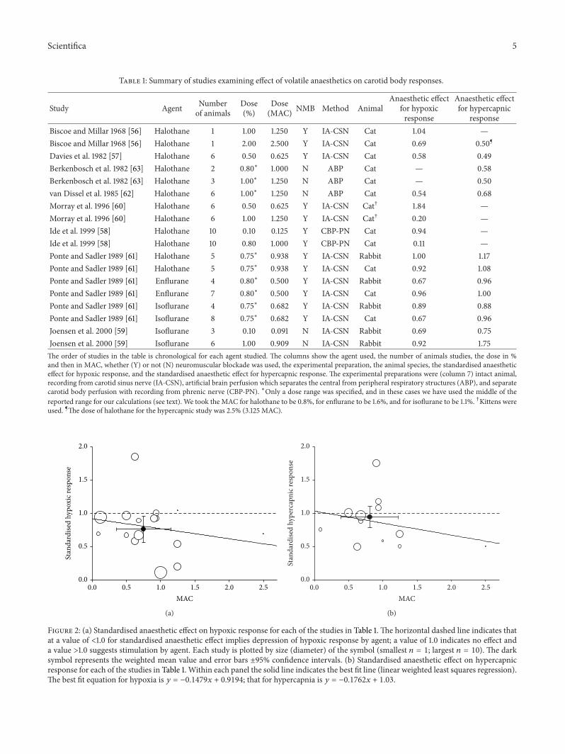
The mean (standard deviation, SD) dose of anaestheticsused in all studies combined was 0.75 (0.40)MAC. Theweightedmean anaesthetic effects for the agents were similar:halothane 0.73 (0.53), enflurane 0.85 (0.23), and isoflurane0.79 (0.11), 𝑃 = 0.96. For all agents combined, the weightedanaesthetic effect was 0.76 (0.42), 95% confidence interval0.56–0.96, indicating that on average anaesthetics signifi-cantly depressed carotid body hypoxic response by 24% (𝑃 =0.041) ( Figure 2).
Figure 2 also shows the effects of increasing anaestheticdose on responses to hypoxia showing a modest dose-dependent decline in response.
For completeness we conducted a subgroup analysis toexplore the potential effect of neuromuscular blockade andspecies on the effect of anaesthetic, However, neither wassignificant (ANOVA: 𝑃 = 0.620 and 𝑃 = 0.296 resp., whenfactors considered random).Thus, notwithstanding the small
Scientifica 5
Table 1: Summary of studies examining effect of volatile anaesthetics on carotid body responses.
Study Agent Numberof animals
Dose(%)
Dose(MAC) NMB Method Animal
Anaesthetic effectfor hypoxicresponse
Anaesthetic effectfor hypercapnic
responseBiscoe and Millar 1968 [56] Halothane 1 1.00 1.250 Y IA-CSN Cat 1.04 —Biscoe and Millar 1968 [56] Halothane 1 2.00 2.500 Y IA-CSN Cat 0.69 0.50¶
Davies et al. 1982 [57] Halothane 6 0.50 0.625 Y IA-CSN Cat 0.58 0.49Berkenbosch et al. 1982 [63] Halothane 2 0.80∗ 1.000 N ABP Cat — 0.58Berkenbosch et al. 1982 [63] Halothane 3 1.00∗ 1.250 N ABP Cat — 0.50van Dissel et al. 1985 [62] Halothane 6 1.00∗ 1.250 N ABP Cat 0.54 0.68Morray et al. 1996 [60] Halothane 6 0.50 0.625 Y IA-CSN Cat† 1.84 —Morray et al. 1996 [60] Halothane 6 1.00 1.250 Y IA-CSN Cat† 0.20 —Ide et al. 1999 [58] Halothane 10 0.10 0.125 Y CBP-PN Cat 0.94 —Ide et al. 1999 [58] Halothane 10 0.80 1.000 Y CBP-PN Cat 0.11 —Ponte and Sadler 1989 [61] Halothane 5 0.75∗ 0.938 Y IA-CSN Rabbit 1.00 1.17Ponte and Sadler 1989 [61] Halothane 5 0.75∗ 0.938 Y IA-CSN Cat 0.92 1.08Ponte and Sadler 1989 [61] Enflurane 4 0.80∗ 0.500 Y IA-CSN Rabbit 0.67 0.96Ponte and Sadler 1989 [61] Enflurane 7 0.80∗ 0.500 Y IA-CSN Cat 0.96 1.00Ponte and Sadler 1989 [61] Isoflurane 4 0.75∗ 0.682 Y IA-CSN Rabbit 0.89 0.88Ponte and Sadler 1989 [61] Isoflurane 8 0.75∗ 0.682 Y IA-CSN Cat 0.67 0.96Joensen et al. 2000 [59] Isoflurane 3 0.10 0.091 N IA-CSN Rabbit 0.69 0.75Joensen et al. 2000 [59] Isoflurane 6 1.00 0.909 N IA-CSN Rabbit 0.92 1.75The order of studies in the table is chronological for each agent studied. The columns show the agent used, the number of animals studies, the dose in %and then in MAC, whether (Y) or not (N) neuromuscular blockade was used, the experimental preparation, the animal species, the standardised anaestheticeffect for hypoxic response, and the standardised anaesthetic effect for hypercapnic response. The experimental preparations were (column 7) intact animal,recording from carotid sinus nerve (IA-CSN), artificial brain perfusion which separates the central from peripheral respiratory structures (ABP), and separatecarotid body perfusion with recording from phrenic nerve (CBP-PN). ∗Only a dose range was specified, and in these cases we have used the middle of thereported range for our calculations (see text). We took the MAC for halothane to be 0.8%, for enflurane to be 1.6%, and for isoflurane to be 1.1%. †Kittens wereused. ¶The dose of halothane for the hypercapnic study was 2.5% (3.125 MAC).
MAC0.0 0.5 1.0 1.5 2.0 2.5
Stan
dard
ised
hypo
xic r
espo
nse
0.0
0.5
1.0
1.5
2.0
(a)
MAC0.0 0.5 1.0 1.5 2.0 2.5
Stan
dard
ised
hype
rcap
nic r
espo
nse
0.0
0.5
1.0
1.5
2.0
(b)
Figure 2: (a) Standardised anaesthetic effect on hypoxic response for each of the studies in Table 1. The horizontal dashed line indicates thatat a value of <1.0 for standardised anaesthetic effect implies depression of hypoxic response by agent; a value of 1.0 indicates no effect anda value >1.0 suggests stimulation by agent. Each study is plotted by size (diameter) of the symbol (smallest 𝑛 = 1; largest 𝑛 = 10). The darksymbol represents the weighted mean value and error bars ±95% confidence intervals. (b) Standardised anaesthetic effect on hypercapnicresponse for each of the studies in Table 1. Within each panel the solid line indicates the best fit line (linear weighted least squares regression).The best fit equation for hypoxia is 𝑦 = −0.1479𝑥 + 0.9194; that for hypercapnia is 𝑦 = −0.1762𝑥 + 1.03.

6 Scientifica
size of subgroups, the mean result of studies using neuro-muscular blockade (0.72 (0.23), 𝑛 = 3) was similar to that ofstudies using no blockade (0.76 (0.47), 𝑛 = 13); and the meanresult of studies in cats (0.77 (0.47), 𝑛 = 11) was similar tothat of studies in rabbits (0.83 (0.15), 𝑛 = 5).
3.2. Anaesthetic Effect on Hypercapnic Responses. For hyper-capnia, our search yielded the same papers as for hypoxia andwe additionally located one article that examined peripheralCO2(but not hypoxic) sensitivity using an artificial brain
perfusion technique at two doses of halothane in cats [57, 58].Table 1 also presents the results for the 13 studies (from
6 articles) in hypercapnia. The overall result was that anaes-thetic at mean dose 0.81 (0.42)MAC did not significantlyaffect the hypercapnic response (mean anaesthetic effect0.94 (0.35), 95% confidence interval 0.76–1.14, 𝑃 = 0.165)(Figure 2). There was insufficient data to undertake anysubgroup analyses on the hypercapnic responses. Figure 2also shows a modest dose-dependent decline in response.
4. Discussion
In answer to the primary question addressed, the mainconclusion of the quantitative review is that volatile anaes-thetics moderately but significantly depress the carotid bodyresponse to hypoxia but they do not, however, influencecarotid body response to hypercapnia (although there is adose-dependent depression of the CO
2response such that
at high doses this may be suppressed, an effect which couldoccur at the carotid bodies (Figure 2)). These results areconsistent with a large body of human literature [5–15], butat odds with the conclusion of the last major review in thefield [22].
The finding that volatile anaesthetics impair carotid bodysensing of hypoxia is important. Undoubtedly, the primarysite of the hypnotic (narcotic) action of anaesthetics is thebrain. It is therefore tempting to assume that therefore the siteof anaesthetic depression of the hypoxic chemoreflex mustalso be centrally in the brain. However, since the brain isupstream of the carotid body, anaesthetic agents can onlysuppress the chemoreflex centrally if information is arisingperipherally from the carotid bodies and transmitted tothe brain. If, as is shown in this review, anaesthetics haveobtunded the carotid body response then this is one situationwhere, perhaps counter-intuitively, the peripheral action ofagents is more important than any central action.The impor-tance of this is arguably further underlined by Smith et al.[64] who argue that the peripheral and central chemorecep-tors are not independent, but that the degree of peripheralstimulation critically influences central sensitivity.Thenotionof peripheral-central interaction has also been assessed inhumans [65, 66]. If this is the case, then the peripheraldepressive effect of volatile anaesthetics as shown in thisreview becomes a more important phenomenon for respira-tory control. However, an assumption has been made in thisreview that all or most of the ventilatory response to hypoxiais confined to the carotid bodies. Although that appearscorrect for all the species examined in this review, Curran et
al. [67] have reported the finding that central neurones aresensitive to hypoxia in dogs. The extent to which this is awider phenomenon that is unclear.
We discuss below the wider implications of the mainresult.
4.1. Limitations of Nonsystematic Reviews. A previous reviewincluded consideration of studies whose methodology didnot functionally isolate the carotid body, yet conclusions werenonetheless drawn about anaesthetic-carotid body interac-tion [22]. For example, the studies of Gaudy et al. [35, 36],Koh and Severinghaus [42], and Stuth et al. [45, 46] wereexplicitly quoted in support of a conclusion that there mustbe no anaesthetic effect on the peripheral chemoreceptor. Incontrast, the systematic approach conducted here excludedthese studies from quantitative analysis because in thesereports, anaesthetic had been administered to the wholeanimal while, respectively, recording phrenic nerve activity,minute ventilation, or blood gases (none of which function-ally isolate the carotid body). These methodologies are nottherefore able to robustly distinguish an anaesthetic effect oncarotid body function as opposed to an effect at another,morecentral site.
In the earlier review [22], it was stated that results ofall animal studies in this field were “uniform”; that is, allpapers had hitherto suggested little or no anesthetic effecton the peripheral chemoreceptor. The results here do notsupport this contention. Table 1 shows a tenfold difference inthe crude standardised hypoxic responses (range 0.11–1.04),which superficially indicates considerable heterogeneity, andFigure 2 confirms this graphically. Nevertheless, the confi-dence intervals of the analysis are sufficiently narrow to reflecta statistically significant anaesthetic effect when the data werecombined. One possible reason for some relatively disparateresults may be if the “true” dose-response relationship foranesthetic effect on hypoxic response is quite steep for dosesbetween ∼0.5 and 2MAC. Then, small errors in dosing ormeasurement could have quite large effects on anaestheticeffect.
The “uniformity” for lack of effect previously suggestedby Eriksson [22] is challenged by the comments made byauthors in individual papers. While Joensen et al. statedthat “1MAC isoflurane does not depress the hypoxic responseof rabbit carotid body chemoreceptors” [59], Ide et al. incontrast stated: “the hypoxic ventilatory response is depressedby halothane through a peripheral effect at the carotid body”[58]. Davies et al. found that “halothane has a direct effecton carotid body chemoreceptor mechanisms” [57]. Making asomewhat less decisive statement, Ponte and Sadler opined:“it is most likely that volatile anaesthetics exert a direct actionupon the chemoreceptor mechanism” [61], van Dissel et al.“could not exclude” an effect of halothane on the peripheralchemoreceptor [62], and Morray et al. concluded that theanesthetic effect was “at least partially explained by an effecton peripheral chemoreceptors” [60]. Thus, the consensus ofthe majority of authors actually favors the notion that anaes-thetics depress carotid body function (Table 1). Furthermore,as discussed below, this is also consistent with more recentstudies that find depressive effects of anesthetic on molecular

Scientifica 7
structures within the carotid body putatively involved in thetransduction process.
4.2. Strengths of a Quantitative Review. The quantitativenature of the analysis presented here enables a more preciseestimate of anaesthetic effect to be described than can a moresubjective review of the literature. As compared with theprevious nonsystematic approach [22], the systematic searchstrategy was also able to locate an additional study byMorrayet al. [60] and by Ide et al. [58] both of which had founddepression of carotid body function by anaesthetic (Table 1).
It is pertinent here to comment on the article of Ponteand Sadler [61]. They reported that a “chemodepressant effectof anaesthetics was absent at PaO2 values <5.3 kPa.” Thisstatement appears to have been taken at face value andrepeated by others in review [22], but in fact there appear tobe no data in the paper of Ponte and Sadler to support thisassertion. On the contrary, their graphical data suggests thatthe greatest magnitude of depression by anaesthetic occurredat the lowest level of hypoxia (∼5.3 kPa) [61].
Similar reappraisal of published data applies also to thearticle of Joensen et al., who concluded in their study thatcarotid body chemoreceptors “were not depressed by isoflu-rane” [59]. However, their data indicate that 0.1% isofluranedepressed hypoxic response by 30% (Table 1). Even if thecontrol data for the 3 cats in their study that were exposed to0.1% isoflurane are used alone (rather than the control datafor all 6 cats combined), the hypoxic response is depressedby 13%, but this does not alter the overall weighted meanvalue, which remains 0.76 ± 0.42 (24% depression), norchanges the statistics of Table 1. So, regardless of the methodof calculation used for their published data, these authors’stated conclusion is at odds with their own data, which findsa clear depression of the carotid body hypoxic response byanaesthetic. Furthermore, the authors found a higher dose of1% isoflurane in the same article that was paradoxically lessdepressive (just 8% depression) than 0.1% (whichever calcu-lation is used for the latter). The authors left this paradoxunresolved. It is only with a formal quantitativemethodologythat these intricacies are revealed (Table 1).
While a quantitative review arguably offers a perhapsmore objective analysis than does a subjective review, itsultimate utility depends almost entirely on the quality ofthe original studies included in the analysis. The method ofweighting studies by size and statisticalmethodology can onlyin part compensate for any shortcomings in the original work.
By excluding agents such as chloroform, cyclopropane,and ether, important scientific data may be missed, but theclinical relevance of such agents is now very limited.
One limitation to quantitative analysis is that the truerelationship between ventilation (or carotid sinus nerveor phrenic nerve discharge) and partial pressure (PO
2) is
nonlinear [68]. So the proper way of assessing the hypoxicresponse is using a range of values for PO
2. However, none
of the original papers has been so comprehensive. With thisshortcoming of the original studies in mind, it is appropriateto choose to use data froma single point (i.e., the response to ahypoxic stimulus of PO
2∼6.6 kPa, with background PCO
2∼
Table 2: Ranking of studies by quality (the Berkenbosch study wasnot ranked as it examines hypercapnia).
Study Rank quality of studyBiscoe and Millar 1968 [56] 7
Biscoe and Millar 1968 [56] 7Davies et al. 1982 [57] 2Berkenbosch et al. 1982 [63] —Berkenbosch et al. 1982 [63] —van Dissel et al. 1985 [62] 3Morray et al. 1996 [60] 5Morray et al. 1996 [60] 5Ide et al. 1999 [58] 1Ide et al. 1999 [58] 1Ponte and Sadler 1989 [61] 4Ponte and Sadler 1989 [61] 4Ponte and Sadler 1989 [61] 4Ponte and Sadler 1989 [61] 4Ponte and Sadler 1989 [61] 4Ponte and Sadler 1989 [61] 4Joensen et al. 2000 [59] 6Joensen et al. 2000 [59] 6
5.3 kPa) for all articles. Justification for this is that this PO2
stimulus is a common or even “standard” hypoxic stimulus inhuman studies [9], and all animal studies retrieved by us usedat least this level of hypoxia. Nonetheless, if experimentallyonewished to delineate the precise estimate of effect, then oneshould properly attempt to map responses over a wider rangeof hypoxic stimuli.
4.3. Quality Ranking of the Articles Retrieved. Themethodol-ogy used in this review makes it possible to rank the articlesaccording to their quality (Table 1, last column). Note thatthis ranking relates specifically to the question addressed inthis review, and not necessarily to the wider value of thearticle as a contribution to science, or indeed, to the originalpurpose of the article when it waswritten [69, 70]. Articles areranked lower in the order if they used very few or potentiallyimmature animals (Biscoe andMillar [56],Morray et al. [60]),if their results were internally inconsistent (Joensen et al.[59]), or if they did not control or specify the anaestheticdose (Ponte and Sadler [61]) (Table 2). The methodologies ofIde et al. [58] and Davies et al. [57] are very persuasive (andthe results of these two studies themselves seemed consistentwith each other), and of van Dissel et al. [62], although thislast is ranked a little lower than Ide et al. orDavies et al. simplybecause it did not specify an exact anaesthetic dose.
4.4. The Effects of Neuromuscular Blockade. Nondepolaris-ing neuromuscular blocking drugs can blunt the carotidbody response to hypoxia [22, 71–73]. Acetylcholine releasedwithin the carotid body, acting on nicotinic receptors, isdirectly implicated in the hypoxic transduction process butwe were unable to demonstrate any independent influence ofneuromuscular blockade on the aggregate results.
8 Scientifica
Joensen et al. [59] and Eriksson [22] argued that in animalstudies where neuromuscular blockade was used, this—andnot the anaesthetic—had caused the depression of hypoxicresponse, thus confounding the proper interpretation ofresults. However, we found that since studies normallyemploy a control protocol without anaesthetic, any separatedepressive effect of neuromuscular blockade on hypoxicresponse was presumably constant for both control (i.e.,without-anaesthetic) and test (i.e., with-anaesthetic) proto-cols. In other words, the majority of studies that demonstratedepression of carotid body hypoxic response by anaesthetic,despite the presence of neuromuscular blockade, are clearlyshowing some anaesthetic effect per se. Furthermore, studieslike that of van Dissel et al. [62] avoided neuromuscularblockade (the animals breathed spontaneously) but theseauthors nonetheless found a large depressive effect of anaes-thetic on the peripheral chemoreceptor of ∼50% (Table 1).We conclude that while neuromuscular blocking drugsmightindependently inhibit the carotid body, they may not neces-sarily mask the action of anaesthetic.
4.5. Species Differences. Only two species, cat and rabbit, havebeen studied and this is too few tomake generalisations about“animals” in general. Furthermore, it is now apparent thateven within a species, genetic factors can play an importantrole. Weil et al. demonstrated that the ventilatory hypoxicresponse of spontaneously hypertensive (SHR) rats was seventimes higher than that of the Fischer 344 (F344) strain [74].Similarly in mice, Yamaguchi et al. described structural andfunctional differences in the carotid bodies of two strains(DBA/2J and A/J) [75]. However, any additional interactionsof these genetic influences with anaesthetic effect remainunexplored. Similar genetic effectsmay underlie the observeddifferences in human hypoxic ventilatory sensitivity [76, 77],and it is unclear if overall any between-species variation is lessthan or greater than any within-species variation.
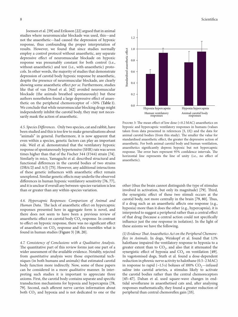
4.6. Hypercapnic Responses: Comparison of Animal andHuman Data. The lack of anaesthetic effect on hypercapnicresponses presented here in aggregate form is novel, andthere does not seem to have been a previous review ofanaesthetic effect on carotid body CO
2response. In contrast
to effect on hypoxic response, there was no significant effectof anaesthetic on CO
2response and this resembles what is
found in human studies (Figure 3) [18, 20].
4.7. Consistency of Conclusions with a Qualitative Analysis.The quantitative part of this review forms just one part of awider assessment of the available evidence. Notably, rejectedfrom quantitative analysis were those experimental tech-niques (in both humans and animals) that estimated carotidbody function more indirectly. Now, some of these paperscan be considered in a more qualitative manner. In inter-preting such studies it is important to appreciate threeaxioms. First, the carotid body contains separate and specifictransduction mechanisms for hypoxia and hypercapnia [78,79]. Second, each afferent nerve carries information aboutboth CO
2and hypoxia and is not dedicated to one or the
1.2
1.0
0.8
0.6
0.4
0.2
0.0
Stan
dard
ised
anae
sthe
tic eff
ect
Hypoxia hypercapnia Hypoxia hypercapniaHuman ventilatory
responses responsesAnimal carotid body
Figure 3: The mean effect of low dose (<0.2MAC) anaesthetics onhypoxic and hypercapnic ventilatory responses in humans (valuestaken from data presented in references [5, 13]) and the data foranimal carotid bodies (from this study). The smaller the value forstandardised anaesthetic effect, the greater the depressive action ofanaesthetic. For both animal carotid body and human ventilation,anaesthetics significantly depress hypoxic but not hypercapnicresponse. The error bars represent 95% confidence intervals. Thehorizontal line represents the line of unity (i.e., no effect ofanaesthetic).
other (thus the brain cannot distinguish the type of stimulusinvolved in activation, but only its magnitude) [79]. Third,the synergistic effect of these two stimuli occurs at thecarotid body, not more centrally in the brain [79, 80]. Thus,if a drug such as an anaesthetic affects one response (e.g.,hypoxia) more than it does the other (e.g., hypercapnia), it isinterpreted to suggest a peripheral rather than a central effectof that drug (because a central action could not specificallyinfluence just the one response in isolation). In the light ofthese axioms we have the following.
(1) EvidenceThat Anaesthetics Act on the Peripheral Chemore-flex in Animals. In dogs, Weiskopf et al. found that 1.1%halothane impaired the ventilatory response to hypoxia to agreater extent than to CO
2, and also that it attenuated the
synergistic effect of hypoxia and CO2on ventilation [49].
In vagotomised dogs, Stuth et al. found a dose-dependentreduction in phrenic nerve activity to halothane (0.5–2MAC)in response to rapid (∼1.5 s) boluses of 100% CO
2—infused
saline into carotid arteries, a stimulus likely to activatethe carotid bodies rather than the central chemoreceptors[45–47]. Dahan et al. used square-wave changes in end-tidal sevoflurane in anaesthetised cats and, after analysingresponses mathematically, they found a greater reduction ofperipheral than central chemoreflex gain [33].
Scientifica 9
(2) EvidenceThat Anaesthetics Act on the Peripheral Chemore-flex in Humans. First, hypoxia-driven ventilation decreaseswithin 30 s of exposure to subanaesthetic concentrations ofboth halothane [5–14] and isoflurane [5–15], a time intervaltoo short to affect the brain, but adequate to influence theperipheral chemoreceptor. Second, acute metabolic acidosisselectively activates peripheral, not central, chemoreceptors,and its effect is abolished by halothane (0.1MAC) [5–14].Third, if the ventilatory response to hypercapnia is analysedmathematically into fast (peripheral) and slow (central)components, it is the former that is preferentially reducedby volatile anaesthetics [23, 29]. Finally, combining resultsof many human studies indicates that low dose, subanaes-thetic (<0.2MAC) volatile anaesthetics blunt the ventilatoryresponse to hypoxia [9] but not hypercapnia [18] (Figure 3),a result which is more consistent with the notion that theireffect is on the peripheral chemoreceptor rather than oncentral mechanisms.
(3) Evidence of Anaesthetic Action on Cellular/Molecular Pro-cesses in Carotid Body. Buckler et al. reported that halothaneblunted the response of a background K+ channel in isolatedrat glomus cells to hypoxia [21]. Very recently, Pandit et al.have extended these observations to show that halothane andsevoflurane inhibit the intracellular calcium transient of thesecells to hypoxia [81, 82], and that halothane and isofluraneantagonise the action of hypoxia on background rat glomuscell K+ channels measured using voltage (patch) clamptechniques [83]. Halothane, sevoflurane, and isofluraneall influence activity of human background K+ channelsexpressed in oocytes [84] and halothane, enflurane, anddesflurane all influence K+ channel activity of Fisher rat thy-roid epithelial monolayer preparations [85]. Closure of back-ground K+ channels is probably the key step in the transduc-tion of hypoxia by the carotid body and anatagonism of thisby anaesthetics is therefore a very plausiblemechanismwhichexplains the blunting of human hypoxic ventilatory responseby these agents. Finally, Karanovic et al. have reproduced in arat model the predictions made by Pandit of human studies;namely, reporting that (a) volatile agents act on the peripheralchemoreceptor and (b) exhibit a specific order of potency forthis effect as found in humans (i.e., halothane > enflurane >isoflurane > sevoflurane) [86].
In this review, two animal studies supported a primarycentral rather than peripheral effect of anaesthetics (neitherof these functionally isolated the carotid body, so theywere excluded from quantitative analysis). Stuth et al. foundthat halothane reduced hypoxic and hypercapnic phrenicnerve responses in vagotomised dogs equally rather thandifferentially (i.e., contrary to the result shown in Figure 3)[45–47].This result was unusual in that it rather contradictedtheir own results of a contemporaneous paper [45–47] thathad indicated a more peripheral halothane effect. Also, itwas surprising that in all papers Stuth et al. found even invery high doses of halothane (2MAC), hypoxic/hypercapnicresponses were present: usually, responses are abolished atthis very high dose. In goats, Koh and Severinghaus foundthat hypoxic and hypercapnic responses were similarly
0.0 0.2 0.4 0.6 0.8 1.0 1.2 1.40
20
40
60
80
100
Carotid body (animals)
Humans
Dose (MAC)
Hyp
oxic
resp
onse
inta
ct aft
er an
aest
hetic
(% o
f con
trol)
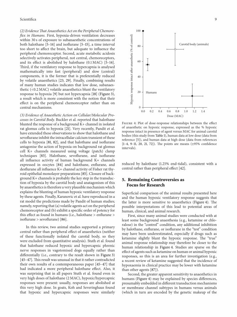
Figure 4: Plot of dose-response relationships between the effectof anaesthetic on hypoxic response, expressed as the % hypoxicresponse intact in presence of agent versus MAC for animal carotidbodies (this study from Table 1), human data at low dose (data fromreference [5]), and human data at high dose (data from references[1–4, 9–11, 20, 21, 72]). The points are means (±95% confidenceintervals).
reduced by halothane (1.25% end-tidal), consistent with acentral rather than peripheral effect [42].
5. Remaining Controversies asFocus for Research
Superficial comparison of the animal results presented hereand the human hypoxic ventilatory response suggests thatthe latter is more sensitive to anaesthetics (Figure 4). Thepossible interpretations of this lead to potential areas ofhuman, clinical, and animal research.
First, since many animal studies were conducted with atleast some background anaesthesia (e.g., ketamine or chlo-ralose) in the “control” condition, any additional inhibitionby halothane, enflurane, or isoflurane in the “test” conditionmay have been underestimated, especially if drugs such asketamine slightly blunt the hypoxic response. The “true”animal response relationship may therefore be closer to thehuman relationship in Figure 4. Studies are sparse on theeffect of agents such as ketamine on human or animal hypoxicresponses, so this is an area for further investigation (e.g.,a recent review of ketamine suggested that the incidence ofhypoxaemia in clinical practice may be lower with ketaminethan other agents [87]).
Second, the greater apparent sensitivity to anaesthetics inhumans (Figure 4) may be explained by species differences,presumably embedded in different transductionmechanismsor membrane channel subtypes in humans versus animals(which in turn are encoded by the genetic makeup of the

10 Scientifica
species). Although there are recognised to be genetically-based differences between human volunteers in their hypoxicresponses, Weil has observed that researchers tend only tostudy those volunteers with robust responses [74]. So itremains unknown if humans with more modest responsesexhibit different sensitivity to the depressive effect of anaes-thetics.
Third, the greater apparent sensitivity to anaesthetics inhumans (Figure 4) may be explained by an additional central(i.e., brain) depressive effects of anaesthetic in human studies,of course absent from the functionally isolated carotid bodystudies examined here in animals. Whether different anaes-thetics interact with the hypoxic stimulus in different ways atbrain level and in which parts of the brain these interactionsmay occur are questions amenable to investigation usingtechniques such as functional magnetic resonance imaging(as has been suggested elsewhere [88]).
The mechanisms of oxygen (and CO2) sensing in the
carotid body remain poorly understood but most probablyinvolve a type 1 cell membrane channel and/or cytoplasmic,non- ormitochondrial heme containing enzymes [16, 89, 90].Recent work suggests that one (or more) K+ channels (ofthe two-pore/four transmembrane segement family, termedTREK, TASK, TWIK, TRAAK, Kv, maxi-K) may initiatethe O
2-sensing cascade [91, 92]. There may be some species
differences here, with perhaps TASK-like channels implicatedin rats [17]; Kv channels implicated in rabbit [93].
The exact mechanism by which hypoxia closes K+ chan-nels is unknown but may be related to their sensitivity toreactive oxygen species (ROS) and/or changes in their redoxstate [94]. In hypoxic conditions, halothane produces ROS,an effect which mediates its adverse effects on the liverthrough lipid peroxidation [95]. Halothane also increasesK+ channel (especially TASK channel) conductance [21].These two observations raised the possibility that halothanemight reduce carotid body/ventilatory response to hypoxiaby producing ROS. Franks and Lieb tested this hypothesis bypretreating human subjects with an antioxidant cocktail ofascorbic acid and 𝛼-tocopherol: consistent with the notionthat halothane acts via ROS, this reversed its inhibitionof the hypoxic response [92]. Teppema et al. also showedthat antioxidants reverse isoflurane-induced depression ofhypoxic response [96]. Furthermore, they observed a rela-tionship between the degree to which an anesthetic is metab-olized and produces ROS to the degree to which it blunts thehypoxic response [97, 98]. Thus, metabolism is the highestfor halothane > enflurane > isoflurane > desflurane, anddegree of depression of hypoxic response is also halothane> enflurane > isoflurane > desflurane [9]. Perhaps, thoughsevoflurane alone breaks this trend since its metabolism ishigher than isoflurane, it is less depressive of the hypoxicresponse [9].
These observations relating to ROS may be extendedto animals and thereby also explain some of the variabilitynoted in this review. Dahan and Teppema have noted thatascorbic acid (an antioxidant) production varies in speciesand, for example, is high in the goat > rabbit > cat >human [99]. The “ROS-K+ channel hypothesis” predicts thatthe hypoxic response of the goat should therefore be more
resistant to the depressive effects of halothane than the humanand, if we accept the results of Koh and Severinghaus forgoats referred to above, this does indeed seem to be thecase [42]. However, the quantitative analysis presented hereindicates that there are as yet insufficient data to commenton differences between cat and rabbit (Table 1). Althoughthis “ROS-K+ channel” hypothesis is perhaps attractive forbeing a potential “unifying mechanism” for some of theeffects described in this review, Cotten and Miller identifieda number of serious problems with the theory [85]. Mostnotable was the fundamental deficiency that it is still notestablished whether hypoxia increases or decreases ROSproduction [95].
Furthermore, this is not the only mechanism proposed:others include that, for example, complex 1 (nicotine adeninedehydrogenase:ubiquinone oxidoreductase) and complex II(succinate ubiquinone dehydrogenase) are possibly involvedin the O
2sensing cascade and suggestedmolecular targets for
anesthetics [100, 101].
6. Conclusions
Operative and perioperative hypoxia remain a leading causeof anaesthesia-related morbidity and mortality, whether thisarises from postoperative complications or airway manage-ment problems [102–104]. Hypoxia-linked problems are alsoa leading cause of litigation [103]. Millions of anaestheticsare administered worldwide every year (∼3 million per yearin the United Kingdom alone [1]) and postoperative hypox-aemia (especially nocturnal hypoxaemia) is recognised as aserious and independent risk factor formyocardial infarction,thromboembolic disease, brain injury, and confusion [105].Patients with preexisting cardiorespiratory and neurologicaldisease aremost susceptible to the harmful effects of hypoxia,and more of these patients are presenting for surgery [106].Because so many healthy patients undergo surgery, even themodest risk reduction will translate to large reductions inthe burden on health services. Since low concentrations ofvolatile anaesthetics long after surgery and depress the venti-latory response to hypoxia, (an effect magnified by coadmin-istered drugs like opiates), any intervention or combination ofdrugs which maintains the ventilatory response will logicallybe protective against hypoxaemia [107]. The provision ofadequate analgesia and prevention of postoperative respira-tory complications is highly relevant in hospital care, so ourresults will help inform strategies designed to achieve optimaloutcomes.
This review underlines the importance of a system-atic approach to the evidence, as opposed to a subjectiveapproach. The main conclusions of this review can be sum-marised as follows.
(1) Volatile anaesthetics depress the ventilatory responseto hypoxia primarily by an action on oxygen sensingmechanisms within carotid body (rather than actionsmore centrally in the brain).
(2) This is important for understanding the mechanismsof perioperative hypoxaemia, as a situation where the

Scientifica 11
patient’s natural defence mechanisms against hypoxia(e.g., a robust ventilatory response) are blunted.
(3) Future research can fruitfully focus upon (a) themechanisms of oxygen sensing within this organ and(b) developing agents that retain an anaesthetic actionwhilst avoiding actions that interfere with oxygensensing.
This systematic review reaches a conclusion differentfrom that of a previous analysis [22]. If the conclusion ofthat earlier analysis was correct—namely, that anaestheticshave aminimal effect on the carotid body—there would seemlittle justification for further investigating any anaestheticeffects on this organ. The consequence of the very differentconclusion proposed in this review is that it becomes impor-tant to identify the cellular/molecular processes that underlieanaesthetic depression of carotid body hypoxic responses.This will in turn require further elaboration of the processesby which the carotid body senses hypoxia at cellular level.This is fundamental to understanding how the body as awhole protects itself from the harmful effects of hypoxia. Thecomplementary human and animal studies suggested by ourreview will offer insights into this question.
7. Clinical Implications
Goodman had previously argued that reduction of AHVR byanaesthetics would not result in harm, because at anaestheticdoses of drug, an anaesthetist was always on hand to providecare. He suggested it was mechanical airway obstruction,rather than reduced ventilatory response to hypoxia, thatcauses harm [108]. This view has not stood the test oftime. Postoperative hypoxia is in fact multifactorial, causedby synergism with other drugs such as opiates, diffusionhypoxia, and ventilation-perfusion mismatch. In all theseother cases, a strong AHVR is very important to mitigateagainst hypoxaemia. Furthermore, it is now appreciated thatrelevant concentrations of volatile agents remain in the bodyfor many hours after surgery. Exposure to ∼1-2% enflurane orisoflurane for just an hour results in the brain concentrationsof these agents remaining >0.1MAC for several hours [109].This is concentration which can halve AHVR and at a timewhen patients are back on the general surgical ward, relativelyunsupervised. Part of the hypoxic response also consists ofarousal, which itself is a defence mechanism against hypoxia,including that caused by mechanical airway obstruction.In summary, it is for those patients likely at greatest riskof postoperative hypoxemia, such as those with chroniccardiac or lung disease, who we need to adapt techniquesto increase safety. It would seem sensible to employ drugswith weakly depressive actions on AHVR (e.g., sevoflurane),rather than those with more strongly depressive actions(e.g., halothane or enflurane). This observation is relevant indiverse situations: isoflurane and sevoflurane have both beenproposed as additions to nitrous oxide to enhance analgesia inlabour [110, 111]. Pregnant women are clearly at particular riskof hypoxia, so optimal drug combinations with respect toAHVR become very relevant.
Nocturnal hypoxemia can be episodic and associatedwith tachycardia, myocardial ischaemia, and postoperativemyocardial infarction [112]. It is an important researchquestion to establish which anaesthetic combinations worsenor help relieve these episodes [113]. Furthermore, there is apossible interaction of anaesthetics, hypoxia, and thrombosis,with subsequent risks of postoperative deep vein thrombosisor pulmonary embolism [114].
Hypoxia is routinely monitored using pulse oximetry butin fact a pulse oximeter cannot measure minute ventilation.Therefore, Dahan and Teppema proposed that monitoringof PCO
2(e.g., using portable, easily applied transcutaneous
devices) should become standard practice in recovery roomsand general wards [115].
There is considerable scope for clinical work in the areasindicated, especially to assess whether changes in anaestheticdrugs used or techniques can minimise side effects or indeedpostoperative mortality. There remain, however, importantgaps in the scientific literature on this subject.This reviewhas,intentionally, focussed upon the role of volatile anaestheticsand their effect on the acute hypoxic ventilatory response.Perhaps of equal importance is the role of episodic orintermittent hypoxia, sustained hypoxia. It is possible thatintravenous anaesthetics have a very different effect onAHVR[116]. Because they were commonly used only for induc-tion, with anaesthesia subsequently maintained using volatileagents, the practical consequences of intravenous agenteffects on postoperative respiratory control were somewhatlimited. However, now total intravenous anaesthesia (usuallyusing propofol) is now very common [117].
8. Directions for Future Research
The interaction of subject arousal, hypoxia, and anaestheticagents can be investigated by brain imaging techniques (e.g.,functional magnetic resonance imaging, fMRI) which couldreveal the sites of action for anaesthetics, and where theymight interact with the hypoxic chemoreflex and/or thearousal pathways.
The basic science research focussing on isolated carotidbody function can be extended to molecular investigations ofhow the carotid body transduces the hypoxic signal and howanaesthetics interact with this process.
These researches into actions of anesthetics on cellu-lar/molecular components of the carotid body responsemight reveal insights that could be exploited for harnessedto therapeutic benefit. For example, if a particular proteinchannel is discovered responsible for conferring susceptibilityto depressive effects of anaesthetic, then identifying it (orits associated gene) might also reveal those patients atgreater risk of the depressive response to anaesthetic. Or, thisknowledge might enable drug development such as specificanaesthetics that avoid the risk of respiratory depression.
Conflict of Interests
The author declares that there is no conflict of interestsregarding the publication of this paper.

12 Scientifica
Acknowledgments
This review overviews several studies in respiratory phys-iology supported by the National Institute of AcademicAnaesthesia, the Higher Education Funding Council (UK),and the Medical Research Council (UK). The author thanksDr. Helen Doll, Statistician, and Dr. Tim Lancaster, formerlyReader in Statistics, of the Health Services Research Unit,Department of Public Health, University of Oxford, and Dr.Carrol Preston, Statistician, Centre for Statistics in Medicine,Institute of Health Sciences, and K. O’Gallagher, ResearchStudent, Oxford, for their statistical advice in the preparationof this paper and with those referenced in [9, 18].
References
[1] T. M. Cook, N. Woodall, J. Harper, and J. Benger, “Majorcomplications of airway management in the UK: results of theFourth National Audit Project of the Royal College of Anaes-thetists and the Difficult Airway Society. Part 2: intensive careand emergency departments,”British Journal of Anaesthesia, vol.106, no. 5, pp. 632–642, 2011.
[2] P. S. Myles, K. Leslie, M. T. V. Chan et al., “Avoidance of nitrousoxide for patients undergoing major surgery: a randomizedcontrolled trial,”Anesthesiology, vol. 107, no. 2, pp. 221–231, 2007.
[3] R. Hines, P. G. Barash, G. Watrous, and T. O’Connor, “Com-plications occurring in the postanesthesia care unit: a survey,”Anesthesia and Analgesia, vol. 74, no. 4, pp. 503–509, 1992.
[4] G. Lockwood, “Theoretical context-sensitive elimination timesfor inhalation anaesthetics,” British Journal of Anaesthesia, vol.104, no. 5, pp. 648–655, 2010.
[5] A. W. Gelb and R. L. Knill, “Subanaesthetic halothane: its effecton regulation of ventilation and relevance to the recoveryroom,”Canadian Anaesthetists Society Journal, vol. 25, no. 6, pp.488–494, 1978.
[6] R. L. Knill and A. W. Gelb, “Ventilatory responses to hypoxiaand hypercapnia during halothane sedation and anesthesia inman,” Anesthesiology, vol. 49, no. 4, pp. 244–251, 1978.
[7] R. L. Knill, P. H. Manninen, and J. L. Clement, “Ventilationand chemoreflexes during enflurane sedation and anaesthesiain man,” Canadian Anaesthetists Society Journal, vol. 26, no. 5,pp. 353–360, 1979.
[8] R. L. Knill and J. L. Clement, “Site of selective action ofhalothane on the peripheral chemoreflex pathway in humans,”Anesthesiology, vol. 61, no. 2, pp. 121–126, 1984.
[9] J. J. Pandit, “The variable effect of low-dose volatile anaestheticson the acute ventilatory response to hypoxia in humans: aquantitative review,” Anaesthesia, vol. 57, no. 7, pp. 632–643,2002.
[10] J. J. Pandit, J. Manning-Fox, K. L. Dorrington, and P. A.Robbins, “Effects of subanaesthetic sevoflurane on ventilation.2: response to acute and sustained hypoxia in humans,” BritishJournal of Anaesthesia, vol. 83, no. 2, pp. 210–216, 1999.
[11] J. J. Pandit, B. Moreau, S. Donoghue, and P. A. Robbins,“Effect of pain and audiovisual stimulation on the depressionof acute hypoxic ventilatory response by low-dose halothane inhumans,” Anesthesiology, vol. 101, no. 6, pp. 1409–1416, 2004.
[12] M. van den Elsen, A.Dahan, J. DeGoede, A. Berkenbosch, and J.van Kleef, “Influences of subanesthetic isoflurane on ventilatorycontrol in humans,” Anesthesiology, vol. 83, no. 3, pp. 478–490,1995.
[13] R. L. Knill, H. T. Kieraszewicz, B. G.Dodgson, and J. L. Clement,“Chemical regulation of ventilation during isoflurane sedationand anaesthesia in humans,” Canadian Anaesthetists SocietyJournal, vol. 30, no. 6, pp. 607–614, 1983.
[14] R. L. Knill and J. L. Clement, “Ventilatory responses to acutemetabolic acidemia in humans awake, sedated, and anes-thetized with halothane,” Anesthesiology, vol. 62, no. 6, pp. 745–753, 1985.
[15] N. R. Prabhakar, “O2sensing at the mammalian carotid body:
why multiple O2sensors and multiple transmitters?” Experi-
mental Physiology, vol. 91, no. 1, pp. 17–23, 2006.[16] B. J. Whipp and K.Wasserman, “Carotid bodies and ventilatory
control dynamics in man,” Federation Proceedings, vol. 39, no. 9,pp. 2668–2673, 1980.
[17] A. Jackson and C. Nurse, “Plasticity in cultured carotid bodychemoreceptors: environmental modulation of GAP-43 andneurofilament,” Journal of Neurobiology, vol. 26, no. 4, pp. 485–496, 1995.
[18] J. J. Pandit, “Effect of low dose inhaled anaesthetic agentson the ventilatory response to carbon dioxide in humans: aquantitative review,” Anaesthesia, vol. 60, no. 5, pp. 461–469,2005.
[19] J. J. Pandit, J.Manning-Fox, K. L.Dorrington, and P. A. Robbins,“Effects of subanaesthetic sevoflurane on ventilation. 1: responseto acute and sustained hypercapnia in humans,” British Journalof Anaesthesia, vol. 83, no. 2, pp. 204–209, 1999.
[20] J. J. Pandit and B. Moreau, “Interaction of arousal states and lowdose halothane on the acute hypercapnic ventilatory responsein humans,” Anaesthesia, vol. 60, no. 2, pp. 139–145, 2005.
[21] K. J. Buckler, B. A. Williams, and E. Honore, “An oxygen-,acid- and anaesthetic-sensitive TASK-like background potas-sium channel in rat arterial chemoreceptor cells,” Journal ofPhysiology, vol. 525, no. 1, pp. 135–142, 2000.
[22] L. I. Eriksson, “The effects of residual neuromuscular blockadeand volatile anesthetics on the control of ventilation,”Anesthesiaand Analgesia, vol. 89, no. 1, pp. 243–251, 1999.
[23] A. D. Oxman and G. H. Guyatt, “Validation of an index of thequality of review articles,” Journal of Clinical Epidemiology, vol.44, no. 11, pp. 1271–1278, 1991.
[24] L. Manchikanti, J. E. Heavner, G. B. Racz et al., “Methods forevidence synthesis in interventional pain management,” PainPhysician, vol. 6, no. 1, pp. 89–111, 2003.
[25] J. M. Bland and S.M. Kerry, “Statistics Notes: weighted compar-ison of means,” British Medical Journal, vol. 316, no. 7125, p. 129,1998.
[26] J. J. Pandit, “The analysis of variance in anaesthetic research:statistics, biography and history,”Anaesthesia, vol. 65, no. 12, pp.1212–1220, 2010.
[27] A. Dahan, M. J. L. J. van den Elsen, A. Berkenbosch et al.,“Effects of subanesthetic halothane on the ventilatory responsesto hypercapnia and acute hypoxia in healthy volunteers,” Anes-thesiology, vol. 80, no. 4, pp. 727–738, 1994.
[28] D. Sjogren, S. G. E. Lindahl, C. Gottlieb, and A. Sollevi, “Venti-latory responses to acute and sustained hypoxia during sevoflu-rane anesthesia in women,” Anesthesia and Analgesia, vol.89, no. 1, pp. 209–214, 1999.
[29] D. Sjogren, S. G. E. Lindahl, and A. Sollevi, “Ventilatoryresponses to acute and sustained hypoxia during isofluraneanesthesia,” Anesthesia and Analgesia, vol. 86, no. 2, pp. 403–409, 1998.

Scientifica 13
[30] M. J. L. J. van den Elsen, A. Dahan, A. Berkenbosch, J. DeGoede,J. W. van Kleef, and I. C. W. Olievier, “Does subanestheticisoflurane affect the ventilatory response to acute isocapnichypoxia in healthy volunteers?”Anesthesiology, vol. 81, no. 4, pp.860–867, 1994.
[31] A. Beck,M. Zimpfer, andG. Raberger, “Inhibition of the carotidchemoreceptor reflex by enflurane in chronically instrumenteddogs,” Naunyn-Schmiedeberg’s Archives of Pharmacology, vol.321, no. 2, pp. 145–148, 1982.
[32] D. J. Cullen and E. I. Eger, “The effects of halothane on respi-ratory and cardiovascular responses to hypoxia in dogs: a dose-response study,”Anesthesiology, vol. 33, no. 5, pp. 487–496, 1970.
[33] A. Dahan, E. Olofsen, L. Teppema, E. Sarton, and C. Olievier,“Speed of onset and offset and mechanisms of ventilatorydepression from sevoflurane: an experimental study in the cat,”Anesthesiology, vol. 90, no. 4, pp. 1119–1128, 1999.
[34] J.-H. Gaudy, M. Quignon, J.-F. Sicard, and R. Maneglia, “Effectsof halothane on ventilation and arterial blood gases in ratswith or without diaphragmatic paralysis,” Canadian Journal ofAnaesthesia, vol. 42, no. 3, pp. 249–255, 1995.
[35] J.-H. Gaudy, J.-F. Sicard, R. Maneglia, and M. Quignon Atos,“The effects of halothane on themodifications of the PaCO
2, the
acid-base equilibrium and the ventilation caused by hypoxia inthe rat,” Canadian Journal of Anaesthesia, vol. 41, no. 4, pp. 347–352, 1994.
[36] J.-H. Gaudy, J.-F. Sicard, R. Maneglia, and M. Quignon, “Theeffects of halothane on arterial blood gas and acid-base balancein intact rats and chemodenervated rats,” Canadian Journal ofAnaesthesia, vol. 40, no. 9, pp. 883–890, 1993.
[37] H.Gautier, “Pattern of breathing during hypoxia or hypercapniaof the awake or anesthetized cat,” Respiration Physiology, vol. 27,no. 2, pp. 193–206, 1976.
[38] H.Gautier,M. Bonora, andD. Zaoui, “Influence of halothane oncontrol of breathing in intact and decerebrated cats,” Journal ofApplied Physiology, vol. 63, no. 2, pp. 546–553, 1987.
[39] H. Gautier, J. H. Gaudy, and M. Bonora, “Effects of anesthesiaon breathing pattern,” Advances in Experimental Medicine andBiology, vol. 99, pp. 93–103, 1978.
[40] H. Groeben, S. Meier, C. G. Tankersley, W. Mitzner, and R. H.Brown, “Heritable differences in respiratory drive and breathingpattern in mice during anaesthesia and emergence,” BritishJournal of Anaesthesia, vol. 91, no. 4, pp. 541–545, 2003.
[41] C. A. Hirshman, R. E. McCullough, P. J. Cohen, and J. V.Weil, “Depression of hypoxic ventilatory response by halothane,enflurane and isoflurane in dogs,” British Journal of Anaesthesia,vol. 49, no. 10, pp. 957–963, 1977.
[42] S. O. Koh and J. W. Severinghaus, “Effect of halothane onhypoxic and hypercapnic ventilatory responses of goats,” BritishJournal of Anaesthesia, vol. 65, no. 5, pp. 713–717, 1990.
[43] R. Maruyama and Y. Fukuda, “Ventilation- and carotid chem-oreceptor discharge-response to hypoxia during inducedhypothermia in halothane anesthetized rat,” Japanese Journal ofPhysiology, vol. 50, no. 1, pp. 91–99, 2000.
[44] C. G. Morrill, J. R. Meyer, and J. V. Weil, “Hypoxic ventilatorydepression in dogs,” Journal of Applied Physiology, vol. 38, no. 1,pp. 143–146, 1975.
[45] E. A. E. Stuth, Z. Dogas, M. Krolo, J. P. Kampine, F. A. Hopp,and E. J. Zuperku, “Dose-dependent effects of halothane on thephrenic nerve responses to acute hypoxia in vagotomized dogs,”Anesthesiology, vol. 87, no. 6, pp. 1428–1439, 1997.
[46] E. A. E. Stuth, Z. Dogas, M. Krolo, J. P. Kampine, F. A.Hopp, and E. J. Zuperku, “Effects of halothane on the phrenicnerve responses to carbon dioxide mediated by carotid bodychemoreceptors in vagotomized dogs,” Anesthesiology, vol. 87,no. 6, pp. 1440–1449, 1997.
[47] E. A. E. Stuth, M. Tonkovic-Capin, J. P. Kampine, and E. J.Zuperku, “Dose-dependent effects of isoflurane on the CO
2
responses of expiratory medullary neurons and the phrenicnerve activities in dogs,” Anesthesiology, vol. 76, no. 5, pp. 763–774, 1992.
[48] E. A. E. Stuth, M. Tonkovic-Capin, J. P. Kampine, J. Bajic,and E. J. Zuperku, “Dose-dependent effects of halothane onthe carbon dioxide responses of expiratory and inspiratorybulbospinal neurons and the phrenic nerve activities in dogs,”Anesthesiology, vol. 81, no. 6, pp. 1470–1483, 1994.
[49] R. B.Weiskopf, L.W. Raymond, and J.W. Severinghaus, “Effectsof halothane on canine respiratory responses to hypoxia withandwithout hypercarbia,”Anesthesiology, vol. 41, no. 4, pp. 350–360, 1974.
[50] M. Zimpfer, S. P. Sit, and S. F. Vatner, “Effects of anesthesia onthe canine carotid chemoreceptor reflex,” Circulation Research,vol. 48, no. 3, pp. 400–406, 1981.
[51] R. D. Dripps and P. R. Dumke, “The effect of narcotics on thebalance between central and chemoreceptor control of respira-tion,” Journal of Pharmacology and Experimental Therapeutics,vol. 77, pp. 290–306, 1943.
[52] J. H. Gaudy, S. Bergeret, J. F. Boitier, and F. Ferracci, “Hypoxicventilatory drive in the dog under althesin anaesthesia,” BritishJournal of Anaesthesia, vol. 56, no. 6, pp. 631–636, 1984.
[53] J. H. Gaudy, S. Bergeret, J. F. Boitier, and F. Ferracci, “Ventilatoryeffects of oxygen-enriched mixtures in the dog under althesinanaesthesia,”British Journal of Anaesthesia, vol. 58, no. 1, pp. 99–102, 1986.
[54] S. Landgren, G. Liljestrand, and Y. Zotterman, “Wirkung vonalcohol, aceton, ather und chloroformauf dieChemoreceptorendes glomus caroticum,” Archiv fur Experimentelle Pathologieund Pharmakologie, vol. 219, no. 3, pp. 185–191, 1953.
[55] H. L. Price and J. Widdicombe, “Actions of cyclopropane oncarotid sinus baroreceptors and carotid body,” The Journal ofpharmacology and experimental therapeutics, vol. 135, pp. 233–239, 1962.
[56] T. J. Biscoe and R. A. Millar, “Effects of inhalation anaestheticson carotid body chemorceptor activity,” British Journal ofAnaesthesia, vol. 40, no. 1, pp. 2–12, 1968.
[57] R. O. Davies, M. W. Edwards, and S. Lahiri, “Halothanedepresses the response of carotid body chemoreceptors tohypoxia and hypercapnia in the cat,” Anesthesiology, vol. 57, no.3, pp. 153–159, 1982.
[58] T. Ide, Y. Sakurai, M. Aono, and T. Nishino, “Contribution ofperipheral chemoreception to the depression of the hypoxicventilatory response during halothane anesthesia in cats,”Anes-thesiology, vol. 90, no. 4, pp. 1084–1091, 1999.
[59] H. Joensen, C. L. Sadler, J. Ponte, Y. Yamamoto, S. G. E. Lindahl,and L. I. Eriksson, “Isoflourane does, not depress the hypoxicresponse of rabbit carotid body chemoreceptors,” Anesthesiaand Analgesia, vol. 91, no. 2, pp. 480–485, 2000.
[60] J. P. Morray, R. Nobel, L. Bennet, and M. A. Hanson, “Theeffect of halothane on phrenic and chemoreceptor responses tohypoxia in anesthetized kittens,” Anesthesia and Analgesia, vol.83, no. 2, pp. 329–335, 1996.
[61] J. Ponte and C. L. Sadler, “Effect of halothane, enflurane andisoflurane on carotid body chemoreceptor activity in the rabbit

14 Scientifica
and the cat,” British Journal of Anaesthesia, vol. 62, no. 1, pp. 33–40, 1989.
[62] J. T. van Dissel, A. Berkenbosch, and C. N. Olievier, “Effectsof halothane on the ventilatory response to hypoxia andhypercapnia in cats,”Anesthesiology, vol. 62, no. 4, pp. 448–456,1985.
[63] A. Berkenbosch, J. de Goede, C. N. Olievier, and H. Quanjer,“Sites of action of halothane on respiratory pattern and ventila-tory response to CO
2in cats,” Anesthesiology, vol. 57, no. 5, pp.
389–398, 1982.[64] C. A. Smith, H. V. Forster, G. M. Blain, and J. A. Dempsey, “An
interdependent model of central/peripheral chemoreception:evidence and implications for ventilatory control,” RespiratoryPhysiology and Neurobiology, vol. 173, no. 3, pp. 288–297, 2010.
[65] I. D. Clement, J. J. Pandit, D. A. Bascom, K. L. Dorrington,D. F. O’Connor, and P. A. Robbins, “An assessment of central-peripheral ventilatory chemoreflex interaction using acid andbicarbonate infusions in humans,” Journal of Physiology, vol.485, no. 2, pp. 561–570, 1995.
[66] I. D. Clement, J. J. Pandit, D. A. Bascom, and P. A. Robbins,“Ventilatory chemoreflexes at rest following a brief period ofheavy exercise in man,” Journal of Physiology, vol. 495, no. 3,pp. 875–884, 1996.
[67] A. K. Curran, J. R. Rodman, P. R. Eastwood, K. S. Henderson, J.A. Dempsey, and C. A. Smith, “Ventilatory responses to specificCNS hypoxia in sleeping dogs,” Journal of Applied Physiology,vol. 88, no. 5, pp. 1840–1852, 2000.
[68] D. A. Bascom, J. J. Pandit, I. D. Clement, and P. A. Robbins,“Effects of different levels of end-tidal P(O
2) on ventilation
during isocapnia in humans,”Respiration Physiology, vol. 88, no.3, pp. 299–311, 1992.
[69] J. J. Pandit and S. M. Yentis, “All that glisters... How to assessthe “value” of a scientific paper,” Anaesthesia, vol. 60, no. 4, pp.373–383, 2005.
[70] A. R. Jadad, R. A. Moore, D. Carroll et al., “Assessing the qualityof reports of randomized clinical trials: is blinding necessary?”Controlled Clinical Trials, vol. 17, no. 1, pp. 1–12, 1996.
[71] L. I. Eriksson, “Reduced hypoxic chemosensitivity in partiallyparalysed man. A new property of muscle relaxants?” ActaAnaesthesiologica Scandinavica, vol. 40, no. 5, pp. 520–523, 1996.
[72] L. I. Eriksson, M. Sato, and J. W. Severinghaus, “Effect of avecuronium-induced partial neuromuscular block on hypoxicventilatory response,”Anesthesiology, vol. 78, no. 4, pp. 693–699,1993.
[73] N. Wyon, H. Joensen, Y. Yamamoto, S. G. E. Lindahl, and L. I.Eriksson, “Carotid body chemoreceptor function is impaired byvecuronium during hypoxia,” Anesthesiology, vol. 89, no. 6, pp.1471–1479, 1998.
[74] J. V. Weil, T. Stevens, C. K. Pickett et al., “Strain-associateddifferences in hypoxic chemosensitivity of the carotid bodyin rats,” American Journal of Physiology—Lung Cellular andMolecular Physiology, vol. 274, no. 5, pp. L767–L774, 1998.
[75] S. Yamaguchi, A. Balbir, B. Schofield et al., “Structural andfunctional differences of the carotid body between DBA/2J andA/J strains of mice,” Journal of Applied Physiology, vol. 94, no. 4,pp. 1536–1542, 2003.
[76] D. A. Thomas, S. Swaminathan, C. S. Beardsmore et al., “Com-parison of peripheral chemoreceptor responses in monozygoticand dizygotic twin infants,” American Review of RespiratoryDisease, vol. 148, no. 6, pp. 1605–1609, 1993.
[77] J. V. Weil, “Variation in human ventilatory control—geneticinfluence on the hypoxic ventilatory response,” RespiratoryPhysiology and Neurobiology, vol. 135, no. 2-3, pp. 239–246,2003.
[78] C. Gonzalez, L. Almaraz, A. Obeso, and R. Rigual, “Carotidbody chemoreceptors: from natural stimuli to sensory dis-charges,” Physiological Reviews, vol. 74, no. 4, pp. 829–898, 1994.
[79] S. Lahiri and R. G. DeLaney, “Stimulus interaction in theresponses of carotid body chemoreceptor single afferent fibers,”Respiration Physiology, vol. 24, no. 3, pp. 249–266, 1975.
[80] J. J. Pandit and K. J. Buckler, “Differential effects of halothaneand sevoflurane on hypoxia-induced intracellular calcium tran-sients of neonatal rat carotid body type i cells,” British Journal ofAnaesthesia, vol. 103, no. 5, pp. 701–710, 2009.
[81] R. S. Fitzgerald andG.A.Dehghani, “Neural responses of the catcarotid and aortic bodies to hypercapnia and hypoxia,” Journalof Applied Physiology Respiratory Environmental and ExercisePhysiology, vol. 52, no. 3, pp. 596–601, 1982.
[82] J. J. Pandit and K. J. Buckler, “Halothane and sevoflurane exertdifferent degrees of inhibition on carotid body glomus cell intra-cellular Ca2+ response to hypoxia,” Advances in ExperimentalMedicine and Biology, vol. 669, pp. 201–204, 2010.
[83] J. J. Pandit, V. Winter, R. Bayliss, and K. J. Buckler, “Differentialeffects of halothane and isoflurane on carotid body glomus cellintracellular Ca2+ and background K+ channel responses tohypoxia,” Advances in Experimental Medicine and Biology, vol.669, pp. 205–208, 2010.
[84] C. Putzke, P. J. Hanley, G. Schlichthor et al., “Differential effectsof volatile and intravenous anesthetics on the activity of humanTASK-1,” American Journal of Physiology—Cell Physiology, vol.293, no. 4, pp. C1319–C1326, 2007.
[85] J. F. Cotten and K. W. Miller, “Volatile anesthetic regulation ofTASK tandem pore potassium channels,” Anesthesiology, vol.105, article A170, 2006.
[86] N. Karanovic, R. Pecotic,M.Valic et al., “The acute hypoxic ven-tilatory response under halothane, isoflurane, and sevofluraneanaesthesia in rats,”Anaesthesia, vol. 65, no. 3, pp. 227–234, 2010.
[87] C. Morris, A. Perris, J. Klein, and P. Mahoney, “Anaesthesiain haemodynamically compromised emergency patients: doesketamine represent the best choice of induction agent?” Anaes-thesia, vol. 64, no. 5, pp. 532–539, 2009.
[88] J. J. Pandit, “Volatile anesthetics and the hypoxic ventilatoryresponse: effects, clinical implications, and future research,”Seminars in Anesthesia, PerioperativeMedicine and Pain, vol. 26,no. 2, pp. 49–57, 2007.
[89] W. Heinke and C. Schwarzbauer, “Subanesthetic isofluraneaffects task-induced brain activation in a highly specific man-ner,” Anesthesiology, vol. 94, no. 6, pp. 973–981, 2001.
[90] J. Lopez-Barneo, R. Pardal, and P. Ortega-Saenz, “Cellularmechanisms of oxygen sensing,” Annual Review of Physiology,vol. 63, pp. 259–287, 2001.
[91] C. Gonzalez, L. Almaraz, A. Obeso, and R. Rigual, “Carotidbody chemoreceptors: from natural stimuli to sensory dis-charges,” Physiological Reviews, vol. 74, no. 4, pp. 829–898, 1994.
[92] N. P. Franks and W. R. Lieb, “Background K+ channels: animportant target for volatile anesthetics?” Nature Neuroscience,vol. 2, no. 5, pp. 395–396, 1999.
[93] M. T. Perez-Garcia and J. R. Lopez-Lopez, “Are Kv channels theessence of O
2sensing?” Circulation Research, vol. 86, pp. 490–
491, 2000.

Scientifica 15
[94] D. Sanchez, J. R. Lopez-Lopez, M. T. Perez-Garcıa et al.,“Molecular identification of Kv𝛼 subunits that contribute to theoxygen-sensitive K+ current of chemoreceptor cells of the rabbitcarotid body,” Journal of Physiology, vol. 542, no. 2, pp. 369–382,2002.
[95] C. Gonzalez, G. Sanz-Alfayate, M. T. Agapito, A. Gomez-Nino,A. Rocher, and A. Obeso, “Significance of ROS in oxygensensing in cell systemswith sensitivity to physiological hypoxia,”Respiratory Physiology and Neurobiology, vol. 132, no. 1, pp. 17–41, 2002.
[96] L. J. Teppema, D. Nieuwenhuijs, E. Sarton et al., “Antioxidantsprevent depression of the acute hypoxic ventilatory responseby subanaesthetic halothane in men,” Journal of Physiology, vol.544, no. 3, pp. 931–938, 2002.
[97] L. Teppema, R. Romberg, E. Sarton, and A. Dahan, “Antiox-idants prevent depression of the acute hypoxic response bysubanaesthetic isoflurane,”Anesthesiology, vol. 102, pp. 747–753,2005.
[98] A. Dahan, R. Romberg, E. Sarton, and L. Teppema, “Theinfluence of inhalational anesthetics on carotid body mediatedresponses,” in Pharmacology and Pathophysiology of the Controlof Breathing, D. S. Ward, A. Dahan, and L. Teppema, Eds., vol.202, Boca Raton, Tayler and Francis, 2005.
[99] A. Dahan and L. J. Teppema, “Influence of anaesthesia and anal-gesia on the control of breathing,” British Journal of Anaesthesia,vol. 91, no. 1, pp. 40–49, 2003.
[100] P. J. Hanley, J. Ray, U. Brandt, and J. Daut, “Halothane, isofluraneand sevolfurane inhibit NADH: ubiquinone oxidoreductase(complex I) of cardiac mitochondria,” Journal of Physiology, vol.544, no. 3, pp. 687–693, 2002.
[101] A. Dahan, P. E. M. Taschner, J. C. Jansen, A. van der Mey, L. J.Teppema, andC. J. Cornelisse, “Carotid body tumors in humanscaused by amutation in the gene for succinate dehydrogenase D(SDHD),” Advances in Experimental Medicine and Biology, vol.551, pp. 71–76, 2004.
[102] K. Gannon, “Mortality associated with anaesthesia. A casereview study,” Anaesthesia, vol. 46, no. 11, pp. 962–966, 1991.
[103] T. M. Cook, S. Scott, and R. Mihai, “Litigation related to airwayand respiratory complications of anaesthesia: an analysis ofclaims against theNHS in England 1995–2007,”Anaesthesia, vol.65, no. 6, pp. 556–563, 2010.
[104] N. M. Woodall and T. M. Cook, “National census of airwaymanagement techniques used for anaesthesia in the UK: firstphase of the Fourth National Audit Project at the Royal Collegeof Anaesthetists,” British Journal of Anaesthesia, vol. 106, no. 2,pp. 266–271, 2011.
[105] N. P. Gill, B. Wright, and C. S. Reilly, “Relationship betweenhypoxaemic and cardiac ischaemic events in the perioperativeperiod,”British Journal of Anaesthesia, vol. 68, no. 5, pp. 471–473,1992.
[106] W.C.Wilson andB. Shapiro, “Perioperative hypoxia: the clinicalspectrum and current oxygen monitoring methodology,” Anes-thesiology Clinics of North America, vol. 19, no. 4, pp. 769–812,2001.
[107] A. Dahan, M. Roozekrans, R. van der Schrier, T. Smith, and L.Aarts, “Primum non nocere or how to resolve drug-inducedrespiratory depression,” Anesthesiology, vol. 118, pp. 1261–1263,2013.
[108] N. W. Goodman, “Volatile agents and the ventilatory responseto hypoxia,”British Journal of Anaesthesia, vol. 72, no. 5, pp. 503–505, 1994.
[109] J. M. Bailey, “Context-sensitive half-times and other decrementtimes of inhaled anesthetics,” Anesthesia and Analgesia, vol. 85,no. 3, pp. 681–686, 1997.
[110] M. Y. K. Wee, M. A. Hasan, and T. A. Thomas, “Isoflurane inlabour,” Anaesthesia, vol. 138, pp. 610–616, 1993.
[111] S. T. Yeo, A. Holdcroft, S. M. Yentis, and A. Stewart, “Analgesiawith sevoflurane during labour: I. Determination of the opti-mum concentration,” British Journal of Anaesthesia, vol. 98, no.1, pp. 105–109, 2007.
[112] I. Gogenur, S. Rosenberg-Adamsen, C. Lie, M. Carstensen, V.Rasmussen, and J. Rosenberg, “Relationship between nocturnalhypoxaemia, tachycardia and myocardial ischaemia after majorabdominal surgery,” British Journal of Anaesthesia, vol. 93, no.3, pp. 333–338, 2004.
[113] G. Shirakami, Y. Teratani, and K. Fukuda, “Nocturnal episodichypoxemia after ambulatory breast cancer surgery: comparisonof sevoflurane and propofol-fentanyl anesthesia,” Journal ofAnesthesia, vol. 20, no. 2, pp. 78–85, 2006.
[114] W. Schobersberger, B. Schobersberger, M. Mittermayr, D. Fries,andW. Streif, “Air travel, hypobaric hypoxia, and prothromboticchanges,” Journal of the American Medical Association, vol. 296,no. 19, pp. 2313–2314, 2006.
[115] A. Dahan and L. Teppema, “Influence of low-dose anaestheticagents on ventilatory control: where do we stand?” BritishJournal of Anaesthesia, vol. 83, no. 2, pp. 199–201, 1999.
[116] B. Nagyova, K. L. Dorrington, E. W. Gill, and P. A. Robbins,“Comparison of the effects of sub-hypnotic concentrations ofpropofol and halothane on the acute ventilatory response tohypoxia,” British Journal of Anaesthesia, vol. 75, no. 6, pp. 713–718, 1995.
[117] S. Karan, W. Voter, L. Palmer, and D. S. Ward, “Effects of painand audiovisual stimulation on the opioid-induced depressionof the hypoxic ventilatory response,”Anesthesiology, vol. 103, no.2, pp. 384–390, 2005.

Submit your manuscripts athttp://www.hindawi.com
Stem CellsInternational
Hindawi Publishing Corporationhttp://www.hindawi.com Volume 2014
Hindawi Publishing Corporationhttp://www.hindawi.com Volume 2014
MEDIATORSINFLAMMATION
of
Hindawi Publishing Corporationhttp://www.hindawi.com Volume 2014
Behavioural Neurology
EndocrinologyInternational Journal of
Hindawi Publishing Corporationhttp://www.hindawi.com Volume 2014
Hindawi Publishing Corporationhttp://www.hindawi.com Volume 2014
Disease Markers
Hindawi Publishing Corporationhttp://www.hindawi.com Volume 2014
BioMed Research International
OncologyJournal of
Hindawi Publishing Corporationhttp://www.hindawi.com Volume 2014
Hindawi Publishing Corporationhttp://www.hindawi.com Volume 2014
Oxidative Medicine and Cellular Longevity
Hindawi Publishing Corporationhttp://www.hindawi.com Volume 2014
PPAR Research
The Scientific World JournalHindawi Publishing Corporation http://www.hindawi.com Volume 2014
Immunology ResearchHindawi Publishing Corporationhttp://www.hindawi.com Volume 2014
Journal of
ObesityJournal of
Hindawi Publishing Corporationhttp://www.hindawi.com Volume 2014
Hindawi Publishing Corporationhttp://www.hindawi.com Volume 2014
Computational and Mathematical Methods in Medicine
OphthalmologyJournal of
Hindawi Publishing Corporationhttp://www.hindawi.com Volume 2014
Diabetes ResearchJournal of
Hindawi Publishing Corporationhttp://www.hindawi.com Volume 2014
Hindawi Publishing Corporationhttp://www.hindawi.com Volume 2014
Research and TreatmentAIDS
Hindawi Publishing Corporationhttp://www.hindawi.com Volume 2014
Gastroenterology Research and Practice
Hindawi Publishing Corporationhttp://www.hindawi.com Volume 2014
Parkinson’s Disease
Evidence-Based Complementary and Alternative Medicine
Volume 2014Hindawi Publishing Corporationhttp://www.hindawi.com