review - embor.embopress.orgembor.embopress.org/content/embor/6/1/22.full.pdf · proteins that...
TRANSCRIPT
EMBO reports VOL 6 | NO 1 | 2005 ©2005 EUROPEAN MOLECULAR BIOLOGY ORGANIZATION22
reviewreviewPostsynaptic scaffold proteins at non-synaptic sites
The role of postsynaptic scaffold proteins in motor-protein–receptor complexesMatthias KneusselUniversität Hamburg, Hamburg, Germany
Synapse-associated proteins that are located at the postsynapticdensity (PSD) have recently been shown to have a structural role atnon-synaptic locations. Here, they act as adaptor proteins betweenneurotransmitter receptors and the microtubule- or microfilament-based motor-protein complexes that are responsible for transportto the PSD. The use of a common set of proteins that contain multi-ple domains for protein–protein interactions as both intracellulartransport adaptors and synaptic scaffold proteins might contributeto the transport specificity and postsynaptic integration of receptors that underlie synapse formation and plasticity.Keywords: motor protein; neurotransmitter receptor; postsynapticdensity; synapse; transportEMBO reports (2005) 6, 22–27. doi:10.1038/sj.embor.7400319
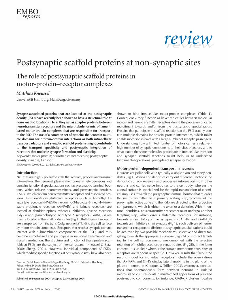
IntroductionNeurons are highly polarized cells that receive, process and transmitinformation. The neuronal plasma membrane is heterogeneous andcontains functional specializations such as presynaptic terminal bou-tons, which release neurotransmitters, and postsynaptic densities(PSDs), which contain neurotransmitter receptors and associated pro-teins. Most excitatory glutamate receptors (such as N-methyl D-aspartate receptors (NMDARs), α-amino-3-hydroxy-5-methyl-4-isox-azole propionate receptors (AMPARs) and kainate receptors) arelocated at dendritic spines, whereas inhibitory glycine receptors(GlyRs) and γ-aminobutyric acid type A receptors (GABAARs) aremainly located at the shaft of dendrites (Fig 1). Both types of receptorare transported from the trans-Golgi network (TGN) to the cell surfaceby motor protein complexes. Receptors that reach a synaptic contactinteract with submembrane components of the PSD, and thusbecome immobilized and participate in neuronal transmission andsignal transduction. The structure and function of these protein scaf-folds at PSDs are the subject of intense research (Kneussel & Betz,2000; Sheng, 2001). However, individual components of PSDs,which mediate specific functions at postsynaptic sites, have also been
shown to bind intracellular motor-protein complexes (Table 1).Consequently, they function as linker molecules between molecularmotors and neurotransmitter receptors during the processes of cargorecruitment towards and/or from the postsynaptic specialization.Proteins that participate in scaffold reactions at the PSD usually con-tain multiple domains for protein–protein interactions, which mightenable motors to interact with a large number of synaptic passengers.Understanding how a limited number of motors carries a relativelyhigh number of synaptic components to their sites of action, and towhat extent the same molecules participate in intracellular transportand synaptic scaffold reactions might help us to understand fundamental operational principles of synapse formation.
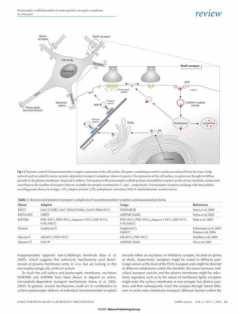
Motor-protein-dependent transport in neuronsNeurons are polar cells with typically a single axon and many den-drites (Fig 1). Axons and dendrites carry out different functions: thedendritic surface receives and processes information from otherneurons and carries nerve impulses to the cell body, whereas theaxonal surface is specialized for the rapid transmission of electri-cal impulses towards the presynaptic terminal bouton that releasesthe neurotransmitter. In a primary sorting step, proteins of thepresynaptic active zone and the PSD are directed to the respectivecompartment, which is either the axon or a dendrite. Within neu-ronal dendrites, neurotransmitter receptors must undergo anothertargeting step, which directs glutamate receptors, for instance,towards an excitatory spine synapse and GlyRs and GABAARstowards an inhibitory shaft synapse (Fig 1). Such delivery of neuro-transmitter receptors to distinct postsynaptic specializations couldbe achieved by two possible mechanisms: selective and direct tar-geting towards the appropriate synapse (Fig 2A) or indirect target-ing to the cell surface membrane combined with the selectiveretention of mobile receptors at synaptic sites (Fig 2B). In the lattercontext, it is unclear whether the surface membrane entry sites ofreceptors are random or specific. However, results that favour thesecond model for individual receptors include the observationsthat AMPARs and GlyRs display lateral mobility in the plane of theplasma membrane (Choquet & Triller, 2003). Moreover, connec-tions that spontaneously form between neurons in isolated micro-island cultures contain mismatched appositions of pre- andpostsynaptic components; for instance, GABAAR clusters localize
Zentrum für Molekulare Neurobiologie Hamburg, ZMNH, Universität Hamburg,Falkenried 94, D-20251 Hamburg, GermanyTel: +49 40 42803 6275; Fax: +49 40 42803 7700;E-mail: [email protected]
Submitted 30 September 2004; accepted 22 November 2004
Nature Publishing Group© 2005
review
©2005 EUROPEAN MOLECULAR BIOLOGY ORGANIZATION EMBO reports VOL 6 | NO 1 | 2005
Postsynaptic scaffold proteins in motor-protein–receptor complexesM. Kneussel
23
inappropriately opposite non-GABAergic terminals (Rao et al,2000), which suggests that selectivity mechanisms exist down-stream of plasma membrane entry in vivo, but are lacking in thiselectrophysiologically artificial system.
To reach the cell surface and postsynaptic membrane, excitatoryNMDARs and AMPARs have been shown to depend on active,microtubule-dependent, transport mechanisms (Setou et al, 2000,2002). In general, several mechanisms could act in combination toachieve postsynaptic delivery of individual neurotransmitter receptors
towards either an excitatory or inhibitory synapse, located on spinesor shafts, respectively: receptors might be sorted to different post-Golgi carriers at the level of the TGN; transport units might be directedto different subdomains within the dendrite; the fusion between indi-vidual transport vesicles and the plasma membrane might be selec-tively regulated, such as by the nature of membrane lipids; receptorsmight enter the surface membrane at non-synaptic but distinct posi-tions and then subsequently reach the synapse through lateral diffu-sion or active intra-membrane transport; diffusion barriers within the
Presynapticterminal bouton
Dendritic spine
Spine synapse
Shaft synapse
Shaft synapse
Nucleus
ER
Endosome Lysosome
Clathrin-coatedvesicle
Clathrin
AP2
Golgi
NSF
Micr
otub
ule
Transportadaptor
Transportvesicle
Postsynapticscaffold
Mot
or
Mot
or
+ –Nucleus
Dendrite
Dendrite
Axon
Cell body
Fig 1 | Dynamic control of neurotransmitter receptor expression at the cell surface. Receptor-containing secretory vesicles are released from the trans-Golgi
network and recruited by motor-protein-dependent transport complexes (shown in green). On expression at the cell surface, receptors are thought to diffuse
laterally in the plasma membrane (depicted in yellow). Interactions with postsynaptic scaffold proteins immobilize receptors at sites of axo-dendritic contact and
contribute to the number of receptors that are available for synaptic transmission (+ and –, respectively). Extrasynaptic receptors exchange with intracellular
recycling pools (shown in orange). AP2, adaptor protein 2; ER, endoplasmic reticulum; NSF, N-ethylmaleimide-sensitive factor.
Table 1 | Known and putative transport complexes of neurotransmitter receptors and associated proteins
Motor Adaptor Cargo References
KIF17 Lin2 (CASK), Lin7 (MALS/Velis), Lin10 (Mint/X11) NMDAR2B Setou et al, 2000
KIF5A/B/C GRIP1 AMPAR GluR2 Setou et al, 2002
KIF1Bα PSD-95(?), PSD-93(?), chapsyn 110(?), SAP-97(?), PSD-95(?), PSD-93(?), chapsyn 110(?), SAP-97(?), Mok et al, 2002S-SCAM(?) S-SCAM(?)
Dynein Gephyrin(?) Gephyrin(?), Fuhrmann et al, 2002GlyR(?) Hanus et al, 2004
Myosin V GKAP(?), PSD-95(?) GKAP(?), PSD-95(?) Naisbitt et al, 2000
Myosin VI SAP-97 AMPAR GluR1 Wu et al, 2002
Nature Publishing Group© 2005
review
EMBO reports VOL 6 | NO 1 | 2005 ©2005 EUROPEAN MOLECULAR BIOLOGY ORGANIZATION
Postsynaptic scaffold proteins in motor-protein–receptor complexesM. Kneussel
24
plasma membrane might hinder individual receptors to enter or leavespecific compartments; and finally, mechanisms that differentiallyregulate receptor endocytosis and receptor turnover might contributeto receptor densities at a given time and location.
In terms of motor-protein-dependent transport of componentstowards postsynaptic specializations, it is also important to considerthat the number of cargo molecules to be transported within a givenneuron is much higher than the number of motors available for therecruitment process. Therefore, adaptor proteins have been postulatedand subsequently identified (Setou et al, 2000, 2002) that not onlyregulate binding affinities but also mediate transport specificity andcargo identity. The latter might be achieved through the combinatorial
use of several polypeptides within the transport complex, many ofwhich have different protein–protein interaction domains.
Microtubules: tracks for polarized transportThe nature of the tracks along which motors move is thought to be acrucial factor in dendritic transport, especially as cytoskeletal poly-mers display polarity and associate with several other proteins.Microtubules generally have a radial organization in many celltypes, with their plus-ends typically orientated to the cell peripheryand their minus-ends anchored in a microtubule-organizing centre(MTOC). With respect to neurons, this uniformity of microtubulepolarity is found in axons but not in dendrites: axonal microtubulesare directed with their plus-ends towards the growth cone, whereasdendritic microtubules show a mixed orientation. In proximal den-dritic regions, about 75 μm from the cell body, roughly equal pro-portions of microtubules are orientated with their plus-ends directedtowards both the growth cone and the cell body. However, in distaldendritic regions, within about 15 μm of the growth cone, micro-tubule polarity orientation is similar to that in axons (Baas et al,1988). Microtubules often do not reach the cellular cortex at thecytoplasmic face of the plasma membrane; instead this region isrich in actin filaments, which are thought to represent the tracks forthe final stages of delivery of many surface molecules. This morpho-logical characteristic is also found in dendritic spines, which contain actin filaments but lack or contain few microtubules.
A recent study has suggested that microtubules themselvesmight provide directional information for polarized transport, as thekinesin-family motor KIF5 has a preference for microtubules in theinitial segment of the axon and KIF5-driven post-Golgi axonal carri-ers move directly from the TGN towards the axonal compartment(Nakata & Hirokawa, 2003). According to this model, not only thepolarity of the track, but also the microtubule-associated proteins(MAPs) could provide a cue for transport. Alternatively, regulationat the level of the interaction between the microtubule track and theindividual motor head might account for such preferences.Regulatory mechanisms could in this context include phosphoryla-tion, acetylation or methylation of target proteins within the system.It is therefore important that future research investigates to whatextent directionality of polarized transport is encoded by the micro-tubule, the MAPs, the structure of the motor head, the different regulatory states of the respective components or by a combinationof these different factors.
Motor–cargo interactions in excitatory synapse remodellingAll motors are enzymes that convert the chemical energy that isstored in ATP into molecular motion, thereby producing a forceon the associated cytoskeletal polymer. The main binding char-acteristic of molecular motor complexes is their simultaneousaffinity for cytoskeletal polymers and cargo elements. Typically,the ATPase function is mediated by the heavy chain of the respec-tive motor protein complex, whereas accessory intermediate andlight chains are specialized for self-assembly or interaction withmolecular cargo. However, individual heavy chains have alsobeen shown to interact directly with cargo molecules. Three fam-ilies of motor-protein complexes—kinesin, dynein and myosin—are known, each of which consists of many domains or accessorysubunits. Kinesins and dyneins use microtubules as transporttracks, whereas myosins represent actin-based motors that movealong microfilaments.
Micr
otub
ule
Micr
otub
ule
Transportvesicle
Transportvesicle
Postsynapticscaffold
Postsynapticscaffold
Receptor
Receptor
Mot
or
Mot
or
Mot
or
A
B
Fig 2 | Proteins with dual functions in motor-protein-dependent transport and
at postsynaptic densities (shown in red) contribute to the post-Golgi surface
membrane delivery of neurotransmitter receptors. (A,B) Two possible
transport mechanisms are shown. The available data favour the mechanism
shown in (B), which suggests that the surface membrane entry of receptors
occurs at extrasynaptic locations. It is unknown whether the interactions of the
proteins (red) with receptors persist during the process of surface delivery and
lateral diffusion into postsynaptic sites.
Nature Publishing Group© 2005

review
©2005 EUROPEAN MOLECULAR BIOLOGY ORGANIZATION EMBO reports VOL 6 | NO 1 | 2005
Postsynaptic scaffold proteins in motor-protein–receptor complexesM. Kneussel
25
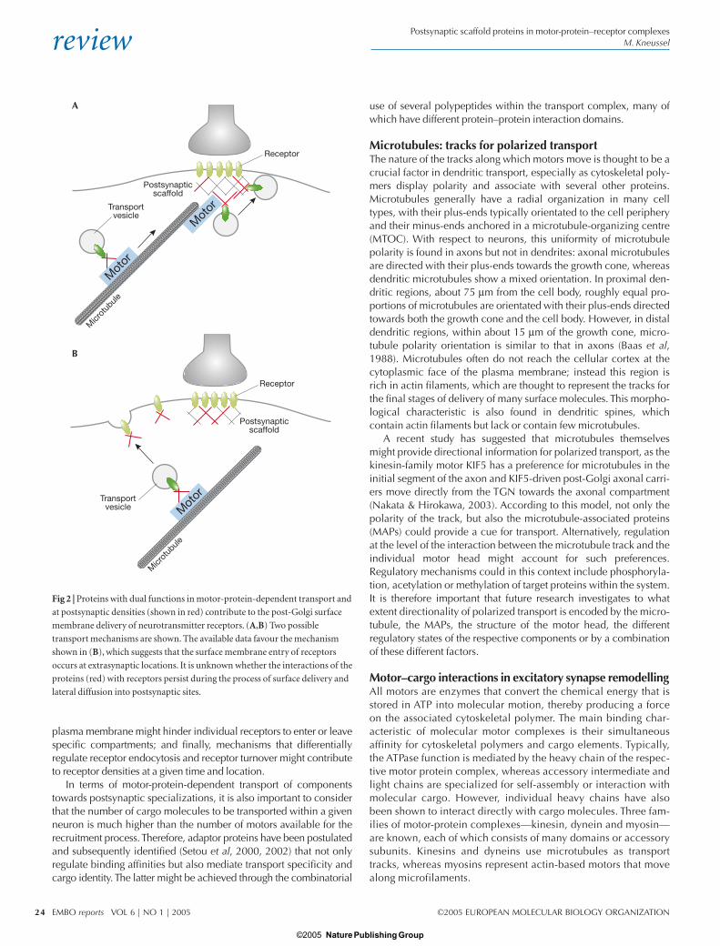
For the anterograde recruitment of glutamate receptors of both theNMDA- and AMPA-type, kinesin-family motors have been identifiedas the driving force of a cargo complex that consists of an individualreceptor subunit, one or more adaptor proteins and the motor-proteincomponents (Setou et al, 2000, 2002; Fig 3). For both receptor types,the same molecules that link the receptor complex to the motor com-plex have previously been shown to locate to and mediate specificfunctions within the PSD. The NMDAR NR2B subunit interacts withthe kinesin-family motor Kif17 through a protein complex that consistsof mouse Lin7 (also known as MALS/Velis), Lin2 (CASK) and Lin10(Mint1/X11; Fig 3A). The microtubule-associated motor binds to thecytoplasmic Lin10 through a PDZ-domain-mediated interaction. Thevesicular NR2B subunit interacts through its carboxy-terminal tail withcytoplasmic Lin7. To connect the cargo vesicle with its transport track,Lin2 then functions as a linker between the Lin10-motor complex andthe Lin7-receptor-vesicle complex, thereby generating a large com-plex that transports NR2B-containing NMDARs towards postsynapticsites (Setou et al, 2000). Notably, the protein Lin2, which represents amembrane-associated guanylate kinase (MAGUK), not only functionsas a transport adaptor at intracellular transport complexes but also rep-resents a component of the PSD. Transgenic overexpression of themotor protein Kif17 enhances NR2B-mediated spatial and workingmemory in mice, which confirms the physiological relevance of thisform of microtubule-based NMDAR transport (Wong et al, 2002).
With respect to the use of PSD components as transport adap-tors, a similar observation has been made for an AMPAR transportcomplex. Here, the kinesin-family motors KIF5A, B and C bind tothe AMPAR GluR2 subunit through glutamate-receptor-interactingprotein 1 (GRIP1)-mediated interactions (Setou et al, 2002; Fig 3B).The expression patterns of KIF5 family members in different neu-ronal cell types (Kanai et al, 2000) are consistent with the distribu-tion of the GRIP1 protein (Dong et al, 1999) and AMPAR GluR2subunits in brain and spinal cord (Jakowec et al, 1995; Petralia &Wenthold, 1992). GRIP1 also represents a component of the post-synaptic scaffold, where it binds to synaptically localized AMPARs(Sheng, 2001). In the context of KIF5-mediated transport, not onlydoes GRIP1 form a bridge between the motor and its cargo recep-tor, but also the minimal kinesin-binding domain of GRIP1 delocal-ized kinesin predominantly to the somatodendritic compartmentand not significantly to axons. Thus, GRIP1 steers the KIF5 familyheavy chains to dendritic compartments, despite the fact that thesemotors also mediate axonal transport reactions. These studies indi-cate that binding proteins can determine the direction of transportof a motor protein (Setou et al, 2002).
It is not clear whether the observed interactions represent thegeneral anterograde microtubule motors for glutamate receptortransport or whether these motor–cargo interactions account for thespecific delivery of certain receptor subtypes. Neurotransmitterreceptors are known to be expressed and assembled in a spatio-temporal-dependent manner in the mammalian central nervous sys-tem. During development for instance, there is a shift in synthesisfrom NR2B-containing to NR2A-containing NMDARs. Moreover,NMDARs locate to and mediate specific functions at both synapticand extrasynaptic locations (Tovar & Westbrook, 1999). Therefore,individual receptor subtypes could either use different molecularmotors for postsynaptic delivery or, alternatively, use the samemotors but different sets of adaptor and/or accessory proteins. Thiscould account for the differing subunit composition or for transporttowards a specific surface membrane microdomain. A candidate set
of proteins for the regulated delivery of individual subunit composi-tions is known as the transmembrane AMPAR regulatory proteins(TARPs). The TARP stargazin/γ2 interacts with AMPARs in the den-dritic cytoplasm before reaching the PSD (Greger et al, 2002; Tomitaet al, 2003) and individual AMPAR complexes contain only oneTARP isoform, which suggests that TARP–AMPAR complexes arestrictly segregated (Tomita et al, 2003). Whether TARPs are compo-nents of KIF5–GRIP1–AMPAR complexes remains to be determined.However, although TARPs bind both AMPARs and the postsynapticscaffold protein PSD-95, they precisely colocalize with AMPARsand are absent from excitatory PSD-95-positive synapses that lackAMPARs (Tomita et al, 2003). It is therefore likely that TARPs are fundamental to the control of AMPAR localization.
Further evidence for the view that different motors participate inthe delivery of excitatory postsynaptic components has been provided
Microtubule
Microtubule
mLin10
GRIP1
KIF5 A,B,C
AMPA-R
KIF17
mLin2
NMDA-R
mLin7
+–
+–
A
B
PDZ PDZ
PDZ
PDZ
PDZ
PDZPDZ
PDZ PDZ PDZ
Fig 3 | Kinesin-mediated transport of NMDA receptors (A) and AMPA
receptors (B). Schematic representation of microtubule-dependent transport
complexes that consist of secretory vesicles and associated proteins. These
contain receptor (shown in red) and adaptor proteins, which link receptors to
kinesin motors (in blue) through PDZ-domain-mediated interactions. Note
that proteins depicted in yellow represent polypeptides with dual functions at
both the intracellular transport pathway and the postsynaptic membrane
specialization. AMPA, α-amino-3-hydroxy-5-methyl-4-isoxazole propionate;
GRIP1, glutamate-receptor-interacting protein 1; KIF, kinesin family; NMDA,
N-methyl D-aspartate; PDZ, postsynaptic density/discs large/zonula adherens.
Nature Publishing Group© 2005

review
EMBO reports VOL 6 | NO 1 | 2005 ©2005 EUROPEAN MOLECULAR BIOLOGY ORGANIZATION
Postsynaptic scaffold proteins in motor-protein–receptor complexesM. Kneussel
26
by a yeast two-hybrid screen that used the C-terminal PDZ-domain-binding motif of the kinesin-family motor protein KIF1Bα , as bait.This screen identified several PSD components as direct KIF1Bαinteractors, including PSD-95, PSD-93, chapsyn 110, synapse-associated protein 97 (SAP97) and the synaptic scaffolding molecule(S-SCAM; Mok et al, 2002). In addition, PSD-95 has been shown toform part of a protein complex containing guanylate-kinase-associatedprotein (GKAP), dynein light chain (Dlc) and the actin-based motorprotein myosin V (Naisbitt et al, 2000). Although it remains unclearwhether these interactions (Table 1) represent cargo, cargo adaptorsor both, it is an appealing hypothesis that these factors, many ofwhich contain several domains for protein–protein interactions, indicate the existence of larger transport complexes that includetransmembrane components of the postsynaptic specialization.
With respect to the endocytosis of AMPARs, the actin-based motormyosin VI might be involved. Myosin VI has been implicated in endo-cytic mechanisms in different cell types (Hasson, 2003) and interactswith the postsynaptic scaffold protein SAP97 and the AMPAR GluR1subunit (Wu et al, 2002; Table 1). Although there is as yet no evidencethat these three binding partners colocalize at postsynaptic sites, therespective trimeric complex co-immunoprecipitates in light mem-brane fractions prepared from brain tissue (Wu et al, 2002).Moreover, AMPAR GluR2 subunits contain overlapping binding sitesfor the ATPase N-ethylmaleimide-sensitive factor (NSF) and theclathrin-binding adaptor protein 2 (AP2). NSF function is required tomaintain AMPARs at postsynaptic sites, whereas AP2–clathrin inter-actions are involved in receptor internalization (Lee et al, 2002).Whether a molecular motor system contributes to the recruitment ofAMPARs that are bound to NSF or AP2 is not known; however, bothcomponents are involved in transport and PSD-mediated reactions(Husi et al, 2000; Peng et al, 2004). Remarkably, as considered for theplasma membrane entry of receptors, the endocytosis of AMPARsthrough clathrin-coated pits also mainly occurs at extrasynaptic sites(Ashby et al, 2004; Petralia et al, 2003; Fig.1), which suggests thatsurface delivery and removal might be an extrasynaptic phenomenon.
Motor–cargo interactions in inhibitory synapse remodellingThe intracellular transport of GlyRs and GABAARs to and frominhibitory postsynaptic sites is barely understood. However, thepostsynaptic scaffold component gephyrin, which is essential for theclustering of GlyRs and individual GABAARs at synaptic sites, mightalso function as a motor–cargo adaptor at intracellular locations.Evidence that gephyrin binds to components of motor protein com-plexes has been obtained from a screen that identified Dlc1 andDlc2 as direct binding partners of the gephyrin molecule (Fuhrmannet al, 2002). As Dlc proteins are components of both the micro-tubule-based dynein motor complex and the microfilament-basedmyosin Va motor complex, this interaction could lead to differenttransport pathways. In neurons derived from gephyrin-deficientmice, the expression of a gephyrin deletion mutant that can nolonger interact with Dlc1 and -2 does not alter the synaptic localiza-tion of gephyrin. It is therefore likely that gephyrin–Dlc interactionscontribute to retrograde neuronal transport, which is consistent withthe dynein motor moving towards the minus-end of microtubules.
In a recent study, Hanus and coworkers analysed heterologouslyexpressed GlyRs and gephyrin at intracellular locations of fibroblastcells (Hanus et al, 2004). A chimeric GlyR-α receptor subunit, whichcontains the gephyrin-binding motif of the GlyR-β subunit, localizedgephyrin to intracellular structures. The movement of these putative
GlyR–gephyrin aggregates was affected in the presence of nocoda-zole, a microtubule-depolymerizing drug, which suggests thatrecruitment of the colocalized structures is microtubule-dependent.Moreover, the presence of the gephyrin-binding motif in GlyR-αaccelerated the accumulation of GlyR at the cell surface. Althoughthis study was not performed in neuronal cells, it suggests that someGlyRs associate with gephyrin during their transport to the cell sur-face. Consistent with this model, gephyrin transport complexes arerecruited within neuronal dendrites over time and enter or leaveputative inhibitory postsynaptic scaffolds (M.K., unpublished data).Clearly, a transport complex remains to be identified that connectsthe vesicular GlyR with an anterograde dendritic motor for plasmamembrane delivery. However, these observations suggest that thescaffold protein gephyrin also contributes to transport reactions atintracellular sites (Table 1). This view is further supported by theobservation that gephyrin interacts with the GABAAR-associated pro-tein (GABARAP; Kneussel et al, 2000), which is a putative post-Golgitransport factor that in turn binds to γ2-subunit-containing GABAARtransport complexes (Wang et al, 1999) and NSF (Kittler et al, 2001).
Common principles and future directionsEach motor-protein–cargo interaction encodes the identity of a cer-tain transport complex and thereby contributes to the regulated andspecific delivery of cargo to its cellular destination. It is becomingincreasingly clear that molecular motors associate with their cargothrough intermediate components, which include adaptor, scaffoldand transmembrane proteins, as well as GTPases and other motors(Klopfenstein et al, 2000). With respect to the transport of neuro-transmitter receptors in neurons, additional questions arise: what arethe reasons and consequences for the use of certain proteins in bothtransport and membrane scaffold reactions? At which subcellularlocation do these interactions initially occur? Do the same interac-tions that regulate, for instance, NMDAR (Lin2/CASK) or AMPAR(GRIP1) transport, persist at postsynaptic sites and could the mainte-nance of such interactions beyond transport represent a generalprinciple in synaptogenesis?
At present, it is not clear at which membrane positions neuro-transmitter receptors enter the plasma membrane (Fig 2). Receptorscould be incorporated into the plane of the plasma membrane at anyposition of the cell and final transport reactions could exclusivelydepend on lateral movements either through diffusion and/or incombination with actin-based motor systems (Fig 2B). A mode ofactive intramembrane transport is known for the endocytic receptormegalin in membranes of epithelial cells (Christensen & Birn, 2002).Alternatively, receptors could enter the surface membrane at distinctextrasynaptic sites, possibly encoded by microenvironments withdefined lipid compositions, and reach the postsynaptic specializa-tion as preformed extrasynaptic clusters. This mode of synapticdevelopment has been reported for NMDAR clusters in culturedneurons (Rao et al, 1998). Another possible mechanism could posi-tion the transport complex near the PSD with direct incorporation ofindividual receptors into existing synapses (Fig 2A); however, a studyperformed in cultured neurons that contradicts this view for certaintypes of NMDAR has been reported (Guillaud et al, 2003). Here,NR2B subunit clusters either colocalize with the motor KIF17 or thepre- and postsynaptic markers synaptophysin and PSD-95, respec-tively. By contrast, no colocalization was observed between KIF17and synaptic markers, which suggests that the motor complex doesnot reach the vicinity of the PSD.
Nature Publishing Group© 2005

review
©2005 EUROPEAN MOLECULAR BIOLOGY ORGANIZATION EMBO reports VOL 6 | NO 1 | 2005
Postsynaptic scaffold proteins in motor-protein–receptor complexesM. Kneussel
27
Whether the surface expression of receptors depends on a singleaforementioned mechanism or on a combination of different modesremains to be determined. For individual receptor subunits and asso-ciated proteins it is known that clusters appear at extrasynaptic loca-tions before synaptogenesis (Meier et al, 2000; Rao et al, 1998). It istherefore important to understand whether receptor-binding pro-teins, some of which carry out a dual role in both transport andsynaptic scaffolding reactions, contribute to surface delivery and lat-eral mobility of receptors. Polypeptides with such dual functionmight interact with receptors not only before, but also during theirsurface membrane entry; therefore, these interactions would persistuntil both binding partners reach the synapse. The incorporation ofnew receptors and their associated binding partners into extrasynapticor synaptic sites might promote subsequent receptor clustering.
In summary, additional transport complexes need to be identifiedand characterized, particularly to understand whether specific sub-units of the same neurotransmitter receptor type travel as passengers ofdistinct or identical motors. These questions require the combinationof biochemical and imaging approaches and will highly benefit fromloss-of-function experiments, including genetic ablation in mice.
ACKNOWLEDGEMENTSM.K. is supported by the University of Hamburg and grants from the DeutscheForschungsgemeinschaft (SFB 444/B7, Kn 556/1-1, Kn 556/1-2).
REFERENCESAshby MC, De La Rue SA, Ralph GS, Uney J, Collingridge GL, Henley JM (2004)
Removal of AMPA receptors (AMPARs) from synapses is preceded bytransient endocytosis of extrasynaptic AMPARs. J Neurosci 24: 5172–5176
Baas PW, Deitch JS, Black MM, Banker GA (1988) Polarity orientation ofmicrotubules in hippocampal neurons: uniformity in the axon andnonuniformity in the dendrite. Proc Natl Acad Sci USA 85: 8335–8339
Choquet D, Triller A (2003) The role of receptor diffusion in the organization ofthe postsynaptic membrane. Nat Rev Neurosci 4: 251–265
Christensen EI, Birn H (2002) Megalin and cubilin: multifunctional endocyticreceptors. Nat Rev Mol Cell Biol 3: 256–266
Dong H, Zhang P, Liao D, Huganir RL (1999) Characterization, expression, anddistribution of GRIP protein. Ann NY Acad Sci 868: 535–540
Fuhrmann JC, Kins S, Rostaing P, El Far O, Kirsch J, Sheng M, Triller A, Betz H,Kneussel M (2002) Gephyrin interacts with dynein light chains 1 and 2,components of motor protein complexes. J Neurosci 22: 5393–5402
Greger IH, Khatri L, Ziff EB (2002) RNA editing at arg607 controls AMPA receptorexit from the endoplasmic reticulum. Neuron 34: 759–772
Guillaud L, Setou M, Hirokawa N (2003) KIF17 dynamics and regulation ofNR2B trafficking in hippocampal neurons. J Neurosci 23: 131–140
Hanus C, Vannier C, Triller A (2004) Intracellular association of glycine receptorwith gephyrin increases its plasma membrane accumulation rate. J Neurosci24: 1119–1128
Hasson T (2003) Myosin VI: two distinct roles in endocytosis. J Cell Sci 116:3453–3461
Husi H, Ward MA, Choudhary JS, Blackstock WP, Grant SG (2000) Proteomicanalysis of NMDA receptor–adhesion protein signaling complexes. NatNeurosci 3: 661–669
Jakowec MW, Yen L, Kalb RG (1995) In situ hybridization analysis of AMPAreceptor subunit gene expression in the developing rat spinal cord.Neuroscience 67: 909–920
Kanai Y, Okada Y, Tanaka Y, Harada A, Terada S, Hirokawa N (2000) KIF5C, a novelneuronal kinesin enriched in motor neurons. J Neurosci 20: 6374–6384
Kittler JT, Rostaing P, Schiavo G, Fritschy JM, Olsen R, Triller A, Moss SJ (2001) The subcellular distribution of GABARAP and its ability to interact with NSFsuggest a role for this protein in the intracellular transport of GABA(A)receptors. Mol Cell Neurosci 18: 13–25
Klopfenstein DR, Vale RD, Rogers SL (2000) Motor protein receptors:moonlighting on other jobs. Cell 103: 537–540
Kneussel M, Betz H (2000) Clustering of inhibitory neurotransmitter receptors atdeveloping postsynaptic sites: the membrane activation model. TrendsNeurosci 23: 429–435
Kneussel M, Haverkamp S, Fuhrmann JC, Wang H, Wassle H, Olsen RW, Betz H(2000) The γ-aminobutyric acid type A receptor (GABAAR)-associatedprotein GABARAP interacts with gephyrin but is not involved in receptoranchoring at the synapse. Proc Natl Acad Sci USA 97: 8594–8599
Lee SH, Liu L, Wang YT, Sheng M (2002) Clathrin adaptor AP2 and NSF interactwith overlapping sites of GluR2 and play distinct roles in AMPA receptortrafficking and hippocampal LTD. Neuron 36: 661–674
Meier J, Meunier-Durmort C, Forest C, Triller A, Vannier C (2000) Formation ofglycine receptor clusters and their accumulation at synapses. J Cell Sci 113:2783–2795
Mok H, Shin H, Kim S, Lee JR, Yoon J, Kim E (2002) Association of the kinesinsuperfamily motor protein KIF1Bα with postsynaptic density-95 (PSD-95),synapse-associated protein-97, and synaptic scaffolding molecule PSD-95/discs large/zona occludens-1 proteins. J Neurosci 22: 5253–5258
Naisbitt S, Valtschanoff J, Allison DW, Sala C, Kim E, Craig AM, Weinberg RJ,Sheng M (2000) Interaction of the postsynaptic density-95/guanylate kinasedomain-associated protein complex with a light chain of myosin-V anddynein. J Neurosci 20: 4524–4534
Nakata T, Hirokawa N (2003) Microtubules provide directional cues forpolarized axonal transport through interaction with kinesin motor head. J Cell Biol 162: 1045–1055
Peng J, Kim MJ, Cheng D, Duong DM, Gygi SP, Sheng M (2004) Semiquantitativeproteomic analysis of rat forebrain postsynaptic density fractions by massspectrometry. J Biol Chem 279: 21003–21011
Petralia RS, Wenthold RJ (1992) Light and electron immunocytochemicallocalization of AMPA-selective glutamate receptors in the rat brain. J CompNeurol 318: 329–354
Petralia RS, Wang YX, Wenthold RJ (2003) Internalization at glutamatergicsynapses during development. Eur J Neurosci 18: 3207–3217
Rao A, Kim E, Sheng M, Craig AM (1998) Heterogeneity in the molecularcomposition of excitatory postsynaptic sites during development ofhippocampal neurons in culture. J Neurosci 18: 1217–1229
Rao A, Cha EM, Craig AM (2000) Mismatched appositions of presynaptic andpostsynaptic components in isolated hippocampal neurons. J Neurosci 20:8344–8353
Setou M, Nakagawa T, Seog DH, Hirokawa N (2000) Kinesin superfamily motorprotein KIF17 and mLin-10 in NMDA receptor-containing vesicle transport.Science 288: 1796–1802
Setou M, Seog DH, Tanaka Y, Kanai Y, Takei Y, Kawagishi M, Hirokawa N (2002)Glutamate-receptor-interacting protein GRIP1 directly steers kinesin todendrites. Nature 417: 83–87
Sheng M (2001) Molecular organization of the postsynaptic specialization. ProcNatl Acad Sci USA 98: 7058–7061
Tomita S, Chen L, Kawasaki Y, Petralia RS, Wenthold RJ, Nicoll RA, Bredt DS(2003) Functional studies and distribution define a family of transmembraneAMPA receptor regulatory proteins. J Cell Biol 161: 805–816
Tovar KR, Westbrook GL (1999) The incorporation of NMDA receptors with adistinct subunit composition at nascent hippocampal synapses in vitro. J Neurosci 19: 4180–4188
Wang H, Bedford FK, Brandon NJ, Moss SJ, Olsen RW (1999) GABA(A)-receptor-associated protein links GABA(A) receptors and the cytoskeleton. Nature397: 69–72
Wong RW, Setou M, Teng J, Takei Y, Hirokawa N (2002) Overexpression of motorprotein KIF17 enhances spatial and working memory in transgenic mice.Proc Natl Acad Sci USA 99: 14500–14505
Wu H, Nash JE, Zamorano P, Garner CC (2002) Interaction of SAP97 with minus-end-directed actin motor myosin VI. Implications for AMPA receptortrafficking. J Biol Chem 277: 30928–30934
Matthias Kneussel
Nature Publishing Group© 2005