review open access immunosuppressive/anti-inflammatory
TRANSCRIPT

JOURNAL OF HEMATOLOGY& ONCOLOGY
Shao et al. Journal of Hematology & Oncology 2014, 7:80http://www.jhoonline.org/content/7/1/80
REVIEW Open Access
Immunosuppressive/anti-inflammatory cytokinesdirectly and indirectly inhibit endothelialdysfunction- a novel mechanism for maintainingvascular functionYing Shao1, Zhongjian Cheng1, Xinyuan Li1, Valeria Chernaya1, Hong Wang1 and Xiao-feng Yang1,2*
Abstract
Endothelial dysfunction is a pathological status of the vascular system, which can be broadly defined as an imbalancebetween endothelium-dependent vasoconstriction and vasodilation. Endothelial dysfunction is a key event in theprogression of many pathological processes including atherosclerosis, type II diabetes and hypertension. Previousreports have demonstrated that pro-inflammatory/immunoeffector cytokines significantly promote endothelialdysfunction while numerous novel anti-inflammatory/immunosuppressive cytokines have recently been identifiedsuch as interleukin (IL)-35. However, the effects of anti-inflammatory cytokines on endothelial dysfunction have receivedmuch less attention. In this analytical review, we focus on the recent progress attained in characterizing the direct andindirect effects of anti-inflammatory/immunosuppressive cytokines in the inhibition of endothelial dysfunction.Our analyses are not only limited to the importance of endothelial dysfunction in cardiovascular disease progression,but also expand into the molecular mechanisms and pathways underlying the inhibition of endothelial dysfunction byanti-inflammatory/immunosuppressive cytokines. Our review suggests that anti-inflammatory/immunosuppressivecytokines serve as novel therapeutic targets for inhibiting endothelial dysfunction, vascular inflammation andcardio- and cerebro-vascular diseases.
Keywords: Anti-inflammatory cytokines, Endothelial dysfunction, Metabolic cardiovascular diseases
IntroductionThe endothelium has long been viewed not only as amonolayer of endothelial cells (EC) lining the lumen ofall blood vessels to function as a protective biocompat-ible barrier between tissues and circulating blood, butalso as a highly specialized, heterogeneous [1], dynamic,disseminated organ [2] with paracrine and autocrinefunctions which respond to alterations of hemodynamicforces and chemical stimuli. In its entirety, the endothe-lium is composed of 1 to 6 × 10¹³ EC covering a surfacearea of more than 1,000 square meters [3,4]. Endothelialdysfunction is a systemic pathological condition, which
* Correspondence: [email protected] of Pharmacology, Center for Metabolic Disease Research andCardiovascular Research Center, Temple University School of Medicine, MERB1059, 3500 North Broad Street, Philadelphia, PA 19140, USA2Department of Microbiology and Immunology, Temple University School ofMedicine, Philadelphia, PA 19140, USA
© 2014 Shao et al.; licensee BioMed Central LtCommons Attribution License (http://creativecreproduction in any medium, provided the orDedication waiver (http://creativecommons.orunless otherwise stated.
can be defined as an imbalance between endothelium-dependent vasoconstriction and vasodilation. Endothelialdysfunction initiates a number of events that trigger ECactivation, which predisposes the vessel wall to be stimu-lated by cardiovascular risk factors [5]. Specifically, endo-thelial dysfunction is associated with reduced nitric oxide(NO) production, increased reactive oxygen species (ROS)production, upregulation of cytokines and chemokines,decreased anticoagulant properties and enhanced plateletaggregation and leukocyte adherence. One related butmore specific term known as endothelial activation ischaracterized by the upregulation of EC adhesion mole-cules such as intercellular adhesion molecule-1 (ICAM-1),vascular cell adhesion molecule-1 (VCAM-1) and in-creased secretion of cytokines and chemokines, which fa-cilitates trans-EC migrations of monocytes, macrophages,dendritic cells, leukocytes, B cells, natural killer cells, andT cells [6]. Perturbations in EC functions play important
d. This is an Open Access article distributed under the terms of the Creativeommons.org/licenses/by/4.0), which permits unrestricted use, distribution, andiginal work is properly credited. The Creative Commons Public Domaing/publicdomain/zero/1.0/) applies to the data made available in this article,

Shao et al. Journal of Hematology & Oncology 2014, 7:80 Page 2 of 14http://www.jhoonline.org/content/7/1/80
roles in the development of several major diseases includ-ing cardiovascular diseases, metabolic syndrome, systemicinflammatory diseases, and sepsis [7].Cytokines, including lymphocyte-generated lympho-
kines, monocyte-produced monokines, chemokines [8],interferons, interleukins, adipocyte-secreted adipokines[9] and muscle-generated myokines [10] act by bindingto their specific receptors in concert with specific cyto-kine inhibitors and soluble cytokine receptors, to regu-late innate and adaptive immune responses. They areproduced by many types of cells such as vascular EC, in re-sponse to the stimulations caused by metabolite-relateddanger signal-associated molecular patterns (DAMPs)[11] and pathogen-associated molecular patterns (PAMPs)[12] that include bacterial endotoxins, injury, or inflam-matory mediators [13]. Cytokines can be divided intopro-inflammatory and anti-inflammatory (immunosuppres-sive) cytokines. Anti-inflammatory cytokines may eitherinhibit pro-inflammatory cytokine synthesis or control pro-inflammatory cytokine-mediated cellular activities [14,15].Numerous reviews have been published on the roles ofpro-inflammatory cytokines on eliciting endothelial dys-function [16-26] and many pro-inflammatory cytokineantagonists have been developed. These new cytokineanta-gonists include: a) etanercept, infliximab, adalimu-mab and certolizumab pegol as tumor necrosis factor-α(TNF-α) antagonists; b) Sant1, Sant5, and Sant7 as inter-leukin-6 (IL-6) receptor superantagonists; c) anakinra asIL-1 receptor antagonist; d) IL-1 receptor antagonist (IL-1Ra) as IL-1α and IL-1β antagonists; e) IL-18BP as IL-18 antagonist, and f ) soluble Endoglin as transforminggrowth factor-β (TGF-β) antagonist [27]. Potential the-rapeutic effects of these new inflammatory cytokine an-tagonists on endothelial dysfunction remain unclear. Inaddition, previous reports showed that anti-inflammatorycytokines IL-10, members of the TGF-β family, and pro-resolving lipid mediators (such as lipoxins, resolvins, andprotectins) may suppress pro-inflammatory signaling [28].Moreover, recent studies have shown that the spectrum ofanti-inflammatory cytokinesis expanding [29], which mayhold promise for developing potential novel therapeuticsand tools for the diagnosis and management of diseases[27]. However, the roles of anti-inflammatory cytokinesin endothelial dysfunction have not been extensively ana-lyzed. Therefore, the protective effects of anti-inflammatorycytokines against endothelial dysfunction are the focus ofthis review (Figure 1).
Endothelium-dependent regulation of vascular toneRecent studies have demonstrated that endothelial dys-function is a mechanistic link between atherosclerotic riskfactors and an early development of vascular diseases,which is an independent predictor of future cardiovascularpathologies in patients with atherogenic risk factors or
ischemic heart disease [30]. Although endothelial dysfunc-tion may have a more generalized definition, current prac-tical definition is related to the endothelial dysfunction ofvascular tone. A healthy endothelium modulates vasculartone by producing numerous vascular dilators and con-strictors. In clinical practices, flow-mediated dilation testis the standard tool used to assess endothelial function[31]. In addition, various reactivity tests, coupled withtechniques measuring skin blood flux, are used to non-invasively explore both endothelial and neurovascularmicrovascular functioning in humans [32].First described by Mulvany and Halpern, vascular tone
is measured by using myography in mouse models [33].This instrument is used for the examination of isolated ar-teries, with internal diameters between 100–400 μm thatare independent of homeostatic mechanisms such asblood flow or autonomic nervous control [34]. The condi-tions near the physiological setting obtained in myographallows in-depth characterization of intrinsic vascular tonereactions to physiological and pharmacological stimuli[35]. According to active tension-length relation, forceproduction and the sensitivity of arteries to different ago-nists are dependent on the extent of stretch [36]. Thereare two types of myographs for measuring vascular func-tion and studying vascular tones known as the pressuremyograph and wire myograph (also see DMT website foran introduction http://www.dmt.dk/default.asp?Action=-Details&Item=543). Pressure myograph may have someadvantages over wire myograph such like: 1) micro resist-ance arteries with internal diameters of less than 500 μm[37] can be studied whereas wire myograph is limited tolarge conduit arteries; 2) the risk to damage endotheliumby wire is reduced; 3) the nature morphology of the arter-ies is maintained; and 4) the potential effects of a widerange of pressures and shear stress on artery dimensioncan be studied [35]. In addition, studying microarteriescan be more informative than examining larger conduitarteries to understand the pathophysiological and molecu-lar mechanisms underlying altered vascular tone in certainmouse models of cardiovascular diseases such as hyper-tension [35]. For example, endothelial dysfunction can befound in 2nd order mesenteric arteries but not in theaortic rings of mice fed with a high fat diet for 8 weeks[38]. In vessel rings pre-contracted with phenylephrine(PE; 1 μM) [39], vascular tone reactivity can be groupedinto endothelium-dependent reactivity (to acetylcholine,ACh; 10 nM to 33 μM) and endothelium-independent re-activity (to sodium nitroprusside, SNP; 1 nM to 10 μM),therefore we focus our discussion mainly on endothelium-dependent pathways.Since high myocardial oxygen extraction occurs at basal
conditions, additional metabolic demands may be met byincreasing the myocardial blood supply. The coronary ar-tery is capable of increasing the basal flow by at least three

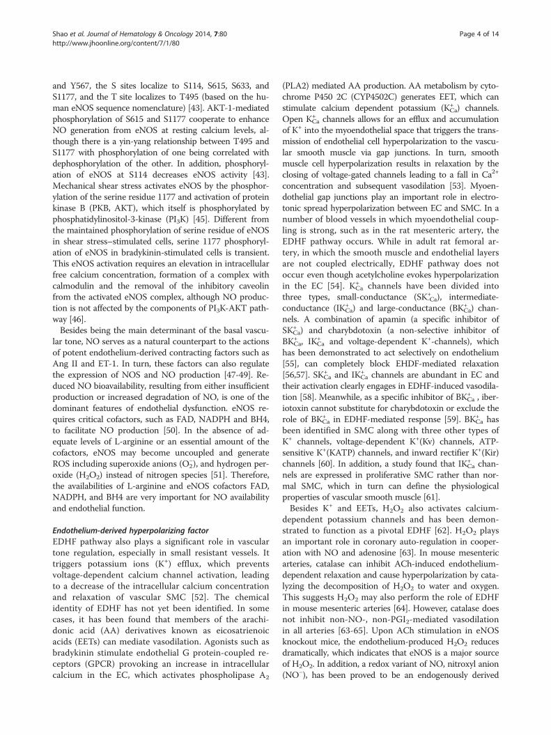
Figure 1 A new working model: Regulatory T cells and immunosuppressive/anti-inflammatory cytokines inhibit endothelialdysfunction. In physiological status, the interaction between endothelium-dependent vasoconstrictors (including Ang II, ET-1, ROS) and vasodilators(NO, EDHF and PGI2) maintain the endothelial function and equilibrium of vascular tone. The impairment of the balance between the vasoconstrictorsand vasodilators is generally defined as the endothelial dysfunction. Under cardiovascular diseases risk factors stimuli, when vasodilationpathways being impaired or vasoconstriction being activated, endothelial dysfunction occurs. Regulatory T cells (Tregs) act on endothelialcells via cell-cell-interaction and/or immunosuppressive/antiinflammatory cytokines to inhibit endothelial dysfunction and restore normalvascular tone.
Shao et al. Journal of Hematology & Oncology 2014, 7:80 Page 3 of 14http://www.jhoonline.org/content/7/1/80
folds. This maximum increase in coronary blood flow isdefined as “coronary artery reserve”. This remarkable fea-ture is regulated by various vasodilators and vasoconstric-tors produced by coronary artery endothelial cells [40].Not limited to coronary artery endothelium, endotheliumgenerally are capable of producing vasodilators such asNO, prostacyclin (PGI2), and endothelium-derived hyper-polarizing factor (EDHF) as well as vasoconstrictors suchas endothelin-1 (ET-1), angiotensin II (Ang II), and freeradicals [2,41] that maintain the physiologically balancedvascular tone.
Endothelium-derived vasodilatorsNitric oxideAmong several endothelium-dependent pathways thatcontrol vascular tone, the NO pathway is the most prom-inent in macrovasculature. NO is a key endothelium-derived relaxing factor, produced through the conversionof L-arginine to L-citrulline by the endothelial nitric oxidesynthase (eNOS). Once produced in EC, NO diffusesfreely into the vascular smooth muscle cells (SMC) whereit activates soluble guanylyl cyclase (sGC), which in turn
catalyzes the formation of cyclic guanosine monopho-sphate (cGMP) and finally leads to the relaxation of thevascular smooth muscle [42]. NO is synthesized by a fam-ily of NO synthases (NOS) that include neuronal NOS(nNOS), eNOS and inducible NOS (iNOS). These threeNOS share 50-60% homology at the amino acid sequenceand all have an N-terminal oxygenase domain with heme-,L-arginine- and tetrahydrobiopterin (BH4) binding do-mains, a central calmodulin (CaM)-binding domain, aC-terminal reductase domain with nicotinamide adeninedinucleotide phosphate (NADPH), flavin adenine dinucleo-tide (FAD), and a flavin mononucleotide (FMN) bindingsite [43]. Biochemical and mechanical regulations of eNOSactivity play an important role in physiological andpathophysiological responses. eNOS enzymatic activitycan be regulated at multiple levels including gene tran-scription, mRNA stability, substrate, co-factor availability,post-translational modifications, and protein-protein inter-action with heat shock protein 90(hsp90) and caveolins[44]. Phosphorylation of eNOS can take place at seven dif-ferent phosphorylation sites at specific tyrosine (Y), serine(S) and threonine (T). The identified Y sites localize to Y81
Shao et al. Journal of Hematology & Oncology 2014, 7:80 Page 4 of 14http://www.jhoonline.org/content/7/1/80
and Y567, the S sites localize to S114, S615, S633, andS1177, and the T site localizes to T495 (based on the hu-man eNOS sequence nomenclature) [43]. AKT-1-mediatedphosphorylation of S615 and S1177 cooperate to enhanceNO generation from eNOS at resting calcium levels, al-though there is a yin-yang relationship between T495 andS1177 with phosphorylation of one being correlated withdephosphorylation of the other. In addition, phosphoryl-ation of eNOS at S114 decreases eNOS activity [43].Mechanical shear stress activates eNOS by the phosphor-ylation of the serine residue 1177 and activation of proteinkinase B (PKB, AKT), which itself is phosphorylated byphosphatidylinositol-3-kinase (PI3K) [45]. Different fromthe maintained phosphorylation of serine residue of eNOSin shear stress–stimulated cells, serine 1177 phosphoryl-ation of eNOS in bradykinin-stimulated cells is transient.This eNOS activation requires an elevation in intracellularfree calcium concentration, formation of a complex withcalmodulin and the removal of the inhibitory caveolinfrom the activated eNOS complex, although NO produc-tion is not affected by the components of PI3K-AKT path-way [46].Besides being the main determinant of the basal vascu-
lar tone, NO serves as a natural counterpart to the actionsof potent endothelium-derived contracting factors such asAng II and ET-1. In turn, these factors can also regulatethe expression of NOS and NO production [47-49]. Re-duced NO bioavailability, resulting from either insufficientproduction or increased degradation of NO, is one of thedominant features of endothelial dysfunction. eNOS re-quires critical cofactors, such as FAD, NADPH and BH4,to facilitate NO production [50]. In the absence of ad-equate levels of L-arginine or an essential amount of thecofactors, eNOS may become uncoupled and generateROS including superoxide anions (O2
−), and hydrogen per-oxide (H2O2) instead of nitrogen species [51]. Therefore,the availabilities of L-arginine and eNOS cofactors FAD,NADPH, and BH4 are very important for NO availabilityand endothelial function.
Endothelium-derived hyperpolarizing factorEDHF pathway also plays a significant role in vasculartone regulation, especially in small resistant vessels. Ittriggers potassium ions (K+) efflux, which preventsvoltage-dependent calcium channel activation, leadingto a decrease of the intracellular calcium concentrationand relaxation of vascular SMC [52]. The chemicalidentity of EDHF has not yet been identified. In somecases, it has been found that members of the arachi-donic acid (AA) derivatives known as eicosatrienoicacids (EETs) can mediate vasodilation. Agonists such asbradykinin stimulate endothelial G protein-coupled re-ceptors (GPCR) provoking an increase in intracellularcalcium in the EC, which activates phospholipase A2
(PLA2) mediated AA production. AA metabolism by cyto-chrome P450 2C (CYP4502C) generates EET, which canstimulate calcium dependent potassium (KCa
+ ) channels.Open KCa
+ channels allows for an efflux and accumulationof K+ into the myoendothelial space that triggers the trans-mission of endothelial cell hyperpolarization to the vascu-lar smooth muscle via gap junctions. In turn, smoothmuscle cell hyperpolarization results in relaxation by theclosing of voltage-gated channels leading to a fall in Ca2+
concentration and subsequent vasodilation [53]. Myoen-dothelial gap junctions play an important role in electro-tonic spread hyperpolarization between EC and SMC. In anumber of blood vessels in which myoendothelial coup-ling is strong, such as in the rat mesenteric artery, theEDHF pathway occurs. While in adult rat femoral ar-tery, in which the smooth muscle and endothelial layersare not coupled electrically, EDHF pathway does notoccur even though acetylcholine evokes hyperpolarizationin the EC [54]. KCa
+ channels have been divided intothree types, small-conductance (SK-Ca
+ ), intermediate-conductance (IKCa
+ ) and large-conductance (BKCa+ ) chan-
nels. A combination of apamin (a specific inhibitor ofSKCa
+ ) and charybdotoxin (a non-selective inhibitor ofBKCa
+ , IKCa+ and voltage-dependent K+-channels), which
has been demonstrated to act selectively on endothelium[55], can completely block EHDF-mediated relaxation[56,57]. SKCa
+ and IKCa+ channels are abundant in EC and
their activation clearly engages in EDHF-induced vasodila-tion [58]. Meanwhile, as a specific inhibitor of BKCa
+ , iber-iotoxin cannot substitute for charybdotoxin or exclude therole of BKCa
+ in EDHF-mediated response [59]. BKCa+ has
been identified in SMC along with three other types ofK+ channels, voltage-dependent K+(Kv) channels, ATP-sensitive K+(KATP) channels, and inward rectifier K+(Kir)channels [60]. In addition, a study found that IKCa
+ chan-nels are expressed in proliferative SMC rather than nor-mal SMC, which in turn can define the physiologicalproperties of vascular smooth muscle [61].Besides K+ and EETs, H2O2 also activates calcium-
dependent potassium channels and has been demon-strated to function as a pivotal EDHF [62]. H2O2 playsan important role in coronary auto-regulation in cooper-ation with NO and adenosine [63]. In mouse mesentericarteries, catalase can inhibit ACh-induced endothelium-dependent relaxation and cause hyperpolarization by cata-lyzing the decomposition of H2O2 to water and oxygen.This suggests H2O2 may also perform the role of EDHFin mouse mesenteric arteries [64]. However, catalase doesnot inhibit non-NO-, non-PGI2-mediated vasodilationin all arteries [63-65]. Upon ACh stimulation in eNOSknockout mice, the endothelium-produced H2O2 reducesdramatically, which indicates that eNOS is a major sourceof H2O2. In addition, a redox variant of NO, nitroxyl anion(NO−), has been proved to be an endogenously derived

Shao et al. Journal of Hematology & Oncology 2014, 7:80 Page 5 of 14http://www.jhoonline.org/content/7/1/80
EDHF that can induce preserved vascular relaxation in theaorta from Ang II-treated hypertensive mice [66,67]. Re-cently, H2S has also been suggested to be an EDHF. H2Scauses vascular EC and SMC hyperpolarization and vaso-dilation by activating the KATP, SKCa
+ and IKCa+ channels
through cysteine S-sulfhydration [68].It has long been accepted that, unlike NO, which in-
duces vasodilation in large conductance arteries, EDHFregulates vascular tone and reactivity in small resistancevessels [69]. However, recent in vivo studies found thatcytochrome P-450-related EDHF is involved in the regu-lation of the peripheral conduit artery diameter at restbut not in the control of the basal vascular resistance inthe healthy human forearm [70]. After inhibition of NOand prostacyclin, the inhibition of EET synthesis furtherdecreases radial arterial blood flow and diameter. Thisindicates that EETs plays a potential compensatory rolein maintaining basal tone when NO availability is dimin-ished. In addition, it has been demonstrated that inhypercholesterolemia KCa
+ channel-mediated vasodilationcompensates for the reduced NO bioavailability [71].Furthermore, the activity of EDHF is much higher in in-dividuals with multiple risk factors than that in healthysubjects when compared to NO.
ProstacyclinProstacyclin, also termed as PGI2 is also an endothelium-released effective vasodilator, which functions by bindingto GPCRs(G-protein-coupled receptors) to stimulate theproduction of cyclic adenosine monophosphate (cAMP)that subsequently activates protein kinase A (PKA) andleads to smooth muscle relaxation and vasodilation. PGI2also inhibits vasoconstriction, platelet activation and fur-ther coagulation by counteracting the effects of the vaso-constrictor thromboxane A2 (TXA2). PGI2 and TXA2 areproduced from AA metabolism mediated by cyclooxygen-ase (COX). There are two isoforms of COX, which mainlydiffer in their pattern of expression. COX-1 is expressedin most tissues, whereas COX-2 is usually absent but in-duced by various physiologic stimuli. COX-1 mostly pro-duces TXA2 while the induction of COX-2 is associatedwith an increase in PGI2 production [72]. P38 and P44/42mitogen-activated protein kinase (MAPK) pathways medi-ate the induction of COX-2. In recent years, the wildly ac-cepted COX-2-dependent PGI2 production pathway hasbeen challenged by the direct measurement of circulatingPGI2 levels rather than by the use of urinary markers,which unveiled that it is COX-1, not COX-2, that is re-sponsible for PGI2 production in healthy individuals [73].The opposite conclusions engender controversy of PGI2testing [74], although specific COX-2 inhibitors increasethe risk of cardiovascular events, which supports that vas-cular COX-2 is an important protein for maintainingvascular hemostasis [72-74].
The three endothelium-dependent vasodilation path-ways, NO, EDHF, and PGI2 do not appear to be mutuallyexclusive but act synergistically in a complex manner tomaintain the vasculature health. In addition, the roles ofthese three components may vary among the vascularbeds in different sizes and tissue locations [69]. Moreover,different pathways vary in time course. Many studies haveshown that EDHF is more important during the earlierphase of endothelial dysfunction while NO and PGI2 areprimarily responsible for later and more sustained parts ofthe vasodilator in response to acetylcholine or bradykininin various arteries [75,76].
Endothelium-derived vasoconstrictorsReactive oxygen speciesAs analyzed in our recent review [77], ROS can be derivedfrom the transfer of electrons to molecular oxygen in themitochondrial respiratory systems [78,79] or produced bythe activity of NADPH oxidases, which is co-localizedwith eNOS in subcellular compartments within EC [80].This observation provides a direct link between NADPHoxidase and the endothelial function in humans. Smalland transient amounts of O2
− can be beneficial for theendothelial function, through activation of eNOS via Src(a homolog gene highly similar to the v-src gene of Roussarcoma virus)/PI3-kinase/Akt pathway [51]. High levelsof ROS generated in pathological situations will reduceNO bioavailability by the binding of O2
− to form peroxy-nitrite (ONOO−). This ONOO− coupling with NO re-sults in an imbalance of vascular tension. In addition,NADPH-derived H2O2 impairs endothelial function byamplifying itself in vascular disease [81]. Elevated levelsof ROS have been associated with endothelial dysfunctiondeveloped in diabetes mellitus, hypertension, hypercholes-terolemia, obesity, atherosclerosis [82-86] and sepsis [87].In addition, several inflammatory cytokines are inducedby oxidant stress [88,89] while cytokines themselves inturn lead to increased levels of ROS and reactive nitrogenspecies (RNS) in acute or chronic inflammation [90].
Angiotensin IIAng II is part of the renin-angiotensin system that causesvasoconstriction, which increases blood pressure. Ang IIstimulates Gq protein in vascular SMC, which increasesintracellular calcium levels by an IP3-dependent mechan-ism that consequently leads to contraction. In addition,Ang II enhances vascular arginase activity that impairsNO production by decreasing L- arginine availability[91,92]. It has been demonstrated that the p38 MAPKpathway participates in this arginase upregulation [93].Moreover, Ang II also increases superoxide produc-tion, which leads to eNOS uncoupling [48,94] in EC.Meanwhile, other studies demonstrated that the activa-tion of the COX-1 pathway is involved in Ang II-
Shao et al. Journal of Hematology & Oncology 2014, 7:80 Page 6 of 14http://www.jhoonline.org/content/7/1/80
induced development of endothelial dysfunction in smallresistance arteries [95,96].
Endothelin-1ET-1, a 21-aa peptide released by EC, is a natural counter-part of NO, which is normally kept in balance by manymechanisms. ET-1 and NO take part in a paracrine regu-lation of each other, and the release of ET-1 is blockedby endothelium-derived NO [97,98]. In pathological sit-uations, ET-1 upregulates the expression of caveolae-1,which appears to be a key negative regulator protein foreNOS activity which leads to eNOS inhibition [99]. Inaddition, ET-1 can also increase ROS production attrib-uting to NO degradation [100]. Selective endothelin re-ceptor type A (ETA) and dual (ETA + ETB) antagonistsimprove NO bioavailability and endothelial function inpathological situations. Transgenic mice that overex-press ET-1 specifically in EC (eET-1) have an increaseof endothelial dysfunction, vascular remodeling, oxidativestress, and inflammation [100,101]. Recently, a study dem-onstrated that ET-1-induced vascular hypertrophy andoxidative stress participate in innate immunity system ac-tivation [102,103]. Overexpression of ET-1 in EC causesan increase in monocytes/macrophage infiltration into ad-ventitia, which is similar to the findings in mice infusedwith Ang II [104]. Colony stimulation factor-1 (CSF1)deficiency, a gene mutation impairing monocyte andmacrophage production and maturation, can preventthis vascular damage. This finding provides evidence ofthe roles of monocyte/macrophage as well as innate im-munity in ET-1-induced vascular injury.
Role of anti-inflammatory cytokines in endothelialdysfunctionThe delicate balance between pro- and anti-inflammatorycytokines determines the net effect of an inflammatory re-sponse. Perturbations in this equilibrium can drive thehost defense immune response either towards chronicinflammation (pro-inflammatory) or towards healing(anti-inflammatory). Exposure of endothelial cells topro-inflammatory cytokines leads to transient and re-versible endothelial dysfunction [105,106]. A number ofanti-inflammatory treatment strategies improve endo-thelial function by preventing pro-inflammatory cyto-kine synthesis. Anti-inflammatory cytokines are a seriesof immune-regulatory molecules that control the pro-inflammatory cytokines response, which consequently re-duces inflammation and promotes healing. In addition, anelevation in the level of anti-inflammatory cytokinescan also be found in the development of vascular dis-ease [107], which reflects an early compensatory mech-anism and serves as an indicator of pro-inflammatoryreactions. Major anti-inflammatory cytokines includeIL-1Ra, IL-4, IL-10, IL-11, IL-13 and TGF-β. Several
newly found cytokines, such as IL-33, IL-35, and IL-37also participate in regulating the function of EC. Thefollowing will discuss two of these anti-inflammatorycytokines including IL-10 and TGF-β in detail.
Interleukin-10IL-10 is an anti-inflammatory cytokine produced by manytypes of immune cells, such as monocytes, macrophages,type 2 T helper cells (Th2), mast cells, natural killer (NK)cells, and CD4 +CD25 + Foxp3+ regulatory T cells (Tregs).Its primary biological function is to limit and terminate in-flammatory responses and regulate the differentiation andproliferation of several immune cells. IL-10 receptor 1(IL-10R1) and IL-10R2 are two subunits of the IL-10receptor that are expressed by hematopoietic and non-hematopoietic cells. The receptor expression has alsobeen observed in endothelial cells [108,109], which pro-vides the structural evidence for IL-10 to not onlycounteract pro-inflammatory cytokines but also poten-tially inhibit endothelial dysfunction directly. Many studieshave found that IL-10 is a key mediator of vascular pro-tection in atherosclerosis, type II diabetes and hyperten-sion [110-112]. IL-10 is shown to protect endothelialfunction by initiating the degradation of several cytokinemRNAs, inhibiting the production of monocyte/macro-phage- and neutrophil-derived cytokines [113] and attenu-ating induction of superoxide generation within thevascular wall [114,115]. Clinically, in patients with coron-ary artery disease, IL-10 serum level acts as an independ-ent predictor of the endothelium-mediated vasodilatorresponse of the forearm circulation [116]. Furthermore,IL-10 prevents the impairment of endothelial dysfunctioninduced by elevated levels of C-reactive proteins [116].IL-10 also attenuates inflammatory responses by its anti-
oxidant properties. It plays a protective role in blood ves-sels by inhibiting NADPH oxidase activity and ROSproduction [111,117]. In addition, IL-10 can restore AngII-induced endothelium-dependent relaxation impairmentmeasured by wire myographs in healthy murine aortarings [118]. Mechanistically, Ang II leads to endothelialdysfunction by increasing gp91phox (NOX2) expression,which is a subunit of NADPH oxidase, while IL-10 in-hibits this response by normalizing NADPH oxidaseprotein expression. In IL-10 knockout (IL-10−/−) mice,carotid arteries and thoracic aortas show a marked aug-mentation of vascular dysfunction after systemic treat-ment with Ang II that can be prevented by the treatmentwith superoxide dismutase-mimetic compound TEMPOL[112]. In Ang II-infused hypertensive mice, IL-10 that isreleased by transferred CD4+CD25+ natural Treg cellsfrom wild type mice significantly reduces NAPDH oxidaseactivity and systolic blood pressure; while the transfer ofTregs isolated from IL-10−/− mice has no effect on thehypertension mice. Collectively, these results suggest that

Shao et al. Journal of Hematology & Oncology 2014, 7:80 Page 7 of 14http://www.jhoonline.org/content/7/1/80
IL-10 generated by the immunosuppressive Treg cells pro-tects against Ang II-induced vascular dysfunction andhypertension development by suppressing oxidative stress[119].In recent studies on aging, old IL-10−/− mice are shown
to have stiffer vessels and more severe endothelium-dependent relaxation impairment than wild type mice,which can be reversed by a scavenger of superoxide [120].In addition, gp91phox is significantly induced in IL-10−/−
than that of wild type mice in aging vessels. Furthermore,it is demonstrated that IL-10 protects against age-relatedendothelial dysfunction by the inhibition of oxidativestress. Aside from oxidative stress, it was also found thatthere is an increase of COX-2 activity and consequently ac-tivation in thromboxane A2 receptor instead of PGI2 inolder IL-10−/− mice [121].In vitro, IL-10 suppresses the production of pro-
inflammatory cytokines such as interferon-γ (IFN-γ), IL-1β, TNF-α, and IL-6 by immune cells including T cells,monocytes, macrophages and dendritic cells to produce[113,122-124]. Meanwhile, IL-10 blocks the activity ofNF-κB [125], which is a key pro-inflammatory tran-scription factor [126-128]. In vivo, IL-10 dampens thepro-inflammatory effects of IL-1 and TNF by stimulat-ing IL-1Ra and soluble TNF receptors (sTNFR) produc-tion [129]. In TNF-α-treated mouse models, impairmentof vascular relaxation is accompanied with a reduction ofeNOS, which can be counteracted by IL-10 which induceseNOS expression and attenuates superoxide production[130]. In addition, by using mouse aortic rings in amyograph, TNF-α triggers a significant decrease in ACh-induced relaxation, which can be restored by IL-10.The Janus kinase/signal transducers and activators of
transcription (JAK/STAT) signaling pathway plays an es-sential role in mediating the anti-inflammatory actionsof IL-10 [131]. In human EC, IL-10 up-regulates eNOSexpression and activity mediated by the activation ofSTAT3 [132]. IL-10 and IL-10 receptor interaction in-volves the JAK family tyrosine kinases Jak1 and Tyk3that induce tyrosine phosphorylation and the activationof latent transcription factors STAT3, STAT1, andSTAT5 [133,134]. IL-10 inhibits pro-inflammatory cyto-kine production in the macrophages via a JAK/STAT3-dependent pathway [135]. In IL-10−/− mice, STAT3phosphorylation induction does not occur.Aside from the JAK/STAT pathway, recent studies sug-
gest that IL-10 also confers endothelial protection throughseveral other signaling pathways. IL-10 inhibits the IL-1-induced inhibitor of kappa B (IκB) expression, decreasesIκB phosphorylation and causes an increase in the eNOSactivity [136]. In addition, IL-10 is associated with the in-hibition of extracellular signal-regulated protein kinases 1and 2 (ERK1/2) activity and the MAPK kinase (MEK)/ERK pathway [137-139]. In TNF-α-infused IL-10−/− mice
there is an increase of total and phosphorylated ERK1/2[140], and the aorta and mesenteric arteries isolated fromthose mice display increased contractile responses to ET-1through the ETA receptor, which can be abrogated by theERK1/2 inhibitor PD-98059. This result demonstrates thatIL-10 attenuates ET-1 induced vascular injury through theinhibition of the ERK1/2 pathway.Emerging evidence also suggests that IL-10 plays a
major role in suppressing endothelial dysfunction inlipopolysaccharide (LPS)-induced endotoxemia. In LPS-induced endotoxemia, activated ERK1/2 can induce higherexpression of IL-10. IL-10 in turn restores eNOS-mediatedrelaxation by inhibiting production of ROS in monocytesand neutrophils. Meanwhile, it also decreases the pro-duction of pro-inflammatory cytokines such as TNF-αand IL-6, leading to an attenuation of septic shock[141]. Mechanistically, IL-10 inhibits the transcriptionof several inflammatory genes that are induced by theToll-like receptor (TLR) signaling, such as COX-2, IL-8,and IL-1 [142,143]. This response can be completely or par-tially abrogated by the PI3K or Akt1/2 inhibitor. The PI3K-Akt-glycogen synthase kinase 3 (GSK3) pathway regulatesIL-10-induced gene expression and controls the ability ofIL-10 to suppress a set of inflammatory genes [144].In summary, as a cytokine synthesis inhibitor factor
(CSIF), IL-10 inhibits a broad spectrum of the functionsof activated monocytes/macrophages and T cells, includ-ing pro-inflammatory cytokine synthesis, and NO pro-duction. It also contributes to an essential part in thebalance between pro- and anti-inflammatory cytokines.
Transforming growth factor-βTGF-β is a multifunctional growth factor capable of in-ducing cell proliferation, differentiation, programmedcell death, and stimulating matrix deposition. TGF-βfamily members are also a set of pleiotropic secretedsignaling molecules with unique and potent immuno-regulatory properties that not only significantly contrib-ute to establishing and maintaining the vascular wallintegrity [145], but also participate in the process ofwound healing and tissue fibrosis [146,147]. Various typesof cells, such as all the immune cell lines, secrete TGF-β.There are at least three isoforms including TGF-β1, TGF-β2 and TGF-β3, in which TGF-β1 is predominantly ex-pressed in the immune system and has long been known tomediate remarkable actions in the pathogenesis of manyvascular diseases. TGF-β performs anti-inflammatory ef-fects and plays an important role in inhibiting vascular dis-ease. Three types of TGF-β receptors are highly expressedin endothelial cells, in which Type I and II receptors areexpressed by all the endothelial cells while Type III recep-tors are mainly located in microvascular endothelial cells[148]. The expressions of TGF-β receptors in endothelialcells provide the structural evidence for TGF-β to not only

Shao et al. Journal of Hematology & Oncology 2014, 7:80 Page 8 of 14http://www.jhoonline.org/content/7/1/80
counteract pro-inflammatory cytokines but also potentiallyinhibit endothelial dysfunction directly. Moreover, a signifi-cant increase in eNOS mRNA levels can be found in TGF-β treated EC [149]. In D-glucose stimulated humanumbilical vein endothelium cells (HUVEC), TGF-β bindsto type II TGF-β receptors and increases L-argininetransport and NO synthesis, which protects againsthyperglycemia-induced endothelial dysfunction [150]. Inaddition, TGF-β1 knockout mice develop multifocal in-flammatory disease associated with increased inflammatorycytokine production, which demonstrates that TGF-β is es-sential in immune suppression under physiological condi-tions [151].In EC, TGF-β increases eNOS expression by activating
Smad2, a transcription factor, which interacts with theeNOS promoter [149]. Meanwhile, NO inversely con-trols the transcription of TGF-β via the endothelial NO/cGMP/PKG pathway and interferes with TGF-β/Smad2signaling in vitro and in vivo [149,152]. With NO donortreatment, EC show a decreased response to TGF-β andsuppressed TGF-β target gene expression. Reversely, ineNOS deficient mice, the impairment of NO signalingleads to the upregulation and activation of TGF-β, whichaccelerates vascular injuries. These results show that theTGF-β dependent effects are complex and can bemodulated by NO bioavailability. In response to shearstress, TGF-β3 plays a protective role in maintainingEC homeostasis accompanied by eNOS phosphoryl-ation that leads to the release of NO [153]. In a highsalt intake mouse model, TGF-β induced by an excesssalt diet restores endothelial NO production via AKTactivation and NOS3 phosphorylation as well as allevi-ates arterial compliance impairment, which forms aninhibitory feedback loop during the vascular functionalteration [154].The inhibitory effects of TGF-β are also in association
with Tregs, which perform important immunosuppres-sive effects [155]. TGF-β converts CD4+CD25− effectorT cells into CD4+CD25+ Treg cells by inducing Foxp3expression. TGF-β signaling is not only required for thesurvival of peripheral Tregs, but also for the maintenanceof Treg suppressive function. Further studies found thatTGF-β1 secreted by effector T cells is dispensable for thedevelopment and maintenance of Treg cells, while localTGF-β1 production from infiltrating Treg cells appears tobe required for Treg immunosuppressive functions [156].It is well known that Treg cells play an active role in theprevention of cardiovascular diseases [157]. Treg adoptivetransfer prevents Ang II–induced hypertension and allevi-ates aldosterone-induced impairment of the vasodilatoryresponse of resistance mesenteric arteries to Ach [104].The NOS inhibitor significantly decreases the protectiveeffects of Treg cells, indicating that Treg cells confer pro-tection against the Ang II induced vasodilatory responses
through a NO-dependent pathway. In addition, Ang II–in-duced NADPH oxidase activity can be prevented by Tregadoptive transfer. Thus, the suppressive function in inhi-biting both macrophages and T lymphocytes from Tregsis associated with Tregs’ contribution to the inhibition ofAng II–induced oxidative stress.All together, there is a considerable interest in TGF-β
for its potential role to be used as a therapeutic target,which prevents endothelial dysfunction through either in-ducing NO production or counteracting oxidative stress.
Other anti-inflammatory cytokinesIL-37, previously known as IL-1 family member 7, hasbeen identified as a new anti-inflammatory cytokine. Itis expressed in several tissues and inflammatory cells.Mice with transgenic expressions of IL-37 are protectedfrom LPS-induced endotoxemia and show markedly re-duction of liver damage and improved lung and kidneyfunction [158]. Meanwhile, through the interaction withintracellular smad3, IL-37 significantly decreases the ex-pression of various pro-inflammatory cytokines, such asIL-6, IL-1β, IL-17 and IFN-γ both in plasma and organswhen compared with vehicle-treat control mice. It hasalso been observed that IL-37 suppresses the productionof pro-inflammatory cytokines in macrophages and epi-thelial cells, but its role in regards to EC is yet to beexplored.IL-33, also a member of the IL-1 cytokine family, is an-
other newly identified anti-inflammatory cytokine [159].IL-33 shows various protective effects in the cardiovascu-lar system when ligated with ST2 (IL-33 receptor), amember of the Toll-interleukin 1 receptor (TIR) super-family. IL-33 markedly decreases the aortic sinus athero-sclerotic lesion size in apolipoprotein E (ApoE)−/− micefed on a high fat diet, which is accompanied by the induc-tion of IL-4, IL-5, and IL-13 and reduction of IFN-γ in theserum [160]. In type II diabetes, IL-33 shows protectivemetabolic effects by improving insulin tolerance, as wellas reducing fasting glucose and adiposity [161]. So far, themolecular mechanisms underlying IL-33’s protective ef-fects are not fully understood. Recently, it was found thatIL-33 is widely expressed in normal human tissues such asbranched blood vessels, lymphoid tissues, adipocytes, car-diac fibroblasts, and even in human tumors. As a novelnuclear marker, IL-33 has been identified as an endogen-ous alarm in the immune system when the endotheliumgets injured during infection and stress [162].IL-4 and IL-13 both decrease the sensitivity of vascu-
lar EC to complement-mediated killing and apoptosisthrough the activation of a PI3K/Akt Pathway [163]. Inaddition, IL-4 protects EC from complement injury byupregulating claudin-5 through JAK/STAT6 and FoxO1activation [164].

Shao et al. Journal of Hematology & Oncology 2014, 7:80 Page 9 of 14http://www.jhoonline.org/content/7/1/80
As a member of the IL-12 family, IL-35 has been iden-tified as a novel anti-inflammatory/immunosuppressivecytokine generated by Tregs [165] and B cells [166,167].We found that unlike IL-10 and TGF- β, IL-35 is not con-stitutively expressed in tissues and is mainly produced byinflammatory stimuli in EC, SMC and monocytes [168].IL-35 also triggers CD4+CD25− T effector cell transform-ation into CD4+CD25+ independent of Foxp3 expression.However, the potential effect of IL-35 on endothelial dys-function remains unknown.
MicroRNA and endothelial dysfunctionMicroRNAs (miRNAs) are a recently discovered classof posttranscriptional modulators of gene expression thathave an essential role in vascular diseases [169,170]. Theyare a group of highly conserved, small, non-coding RNAsthat, after maturation and entry into the RNA interferencepathway, inhibit specific gene expression. Specific micro-RNAs can be regulated by inflammatory stimuli and cer-tain microRNAs can act as mediators of inflammatorystimuli. In a human acute monocytic leukemia cell line,LPS stimulates the expression of miR-146a, miR-132 andmiR-155. Overexpression of miR-146a inhibits not onlythe expression of interleukin-1 receptor-associated kinaseand TNF receptor-associated factor 6 s [171], but also theexpression of IL-6 and IL-8 [172,173]. In turn, the induc-tion of miR-155, which is mediated by NFκB and the acti-vator protein-1 (Ap-1) pathway, inhibits the expression ofIL-8 [174,175], which ultimately demonstrates a negativefeedback loop involving microRNAs in an inflammatoryresponse. Some miRNAs were proven to be highlyexpressed in EC in vitro [176]. TNF induced miR-31, miR-17-3p and miR-126 significantly increase the expression ofE-selectin, ICAM-1 and VCAM-1 in EC [177,178]. Trans-fections with mimics of these miRNAs decrease neutro-phil adhesion to EC [177]. miR-181b, which is decreasedby TNF, can down-regulate the NF-kB-responsive genessuch as VCAM-1 and E-selectin in EC in vitro and in vivo[179]. In miR-10a knockdown human aortic EC, MCP-1(monocyte chemotactic protein 1), IL-6, IL-8, VCAM-1and E-selectin are highly elevated, which contributes toathero-susceptible phenotypes in vivo [180], suggestingthat miR-10a suppresses atherogenesis.In the aspect of regulating vascular tone, a recent report
found that miR-222/221 controls eNOS protein levelsafter a member of the RNase III family know as dicer isknockdown [181]. Induced by shear stress, miR-21 canstimulate the phosphorylation of NOS and thereby in-crease NO production [182]. In contrast, miR-155 causesa reduction of NO by decreasing NOS expression [183].In addition to mediating NO-derived vasodilation, micro-RNAs also play a role in regulating vasoconstriction. Arecent report [184] found that miR-125a/b-5p can sup-press oxidized low density lipoprotein (oxLDL) induced
ET-1 expression by directly repressing prepro-ET-1 mRNAexpression.In summary, working together with the classical anti-
inflammatory cytokines, anti-inflammatory/anti-athero-genic microRNAs [169,170], as a new concept wediscussed, could be an important therapeutic target forprotecting against endothelial dysfunction and control-ling cardiovascular diseases.
ConclusionsEndothelial dysfunction has been known as a well-established response to cardiovascular risk factors andprecedes the development of cardiovascular diseases.Anti-inflammatory cytokines protect against the im-pairment of endothelial function by counteracting theeffects of pro-inflammatory cytokines and suppressingoxidative stress. Further studies performed on the in-hibitory properties of anti-inflammatory cytokines onendothelial dysfunction may provide novel promisingtherapeutic strategies for the treatment of cardiovas-cular diseases.
AbbreviationsAA: Arachidonic acid; Ach: Acetylcholine; Ang II: Angiotensin II;ATP: Adenosine triphosphate; BH4: Tetrahydrobiopterin; BKCa
+ :Large-conductance channels; cAMP: Cyclic adenosine monophosphate;cGMP: Cyclic guanosine monophosphate; COX: Cyclooxygenase;EC: Endothelial cells; EDHF: Endothelium-derived hyperpolarizing factor;EETs: Eicosatrienoic acids; eNOS: Endothelial nitric oxide synthase; ERK1/2: Extracellular signal-regulated protein kinases 1 and 2; ET-1: Endothelin-1;ETA: Endothelin receptor type A; FAD: Flavin adenine dinucleotide;GTP: Guanosine triphosphate; GPCRs: G-protein-coupled receptors;H2O2: Hydrogen peroxide; ICAM-1: Intercellular adhesion molecule-1;IFN-γ: Interferon-γ; IκB: Inhibitor of kappa B; IKCa
+ : Intermediate-conductancechannels; IL-1Ra: IL-1 receptor antagonist; IL-10−/−: IL-10 knockout;JAK: JANUS kinase; K+: Potassium ions; KATP: ATP-sensitive K+(KATP) channels;KCa+ : Calcium dependent potassium channels Kir, inward rectifier K+ channels;Kv: Voltage-dependent K+ channels; LPS: Lipopolysaccharide; MAPK:Mitogen-activated protein kinase; MiRNAs: MicroRNAs; NADPH: Nicotinamideadenine dinucleotide phosphate; NO: Nitric oxide; O2
−: Superoxide anions;ONOO−: Peroxynitrite; PI3K: Phosphatidylinositol-3-kinase; PGI2: Prostacyclin;ROS: Reactive oxygen species; STAT: Signal transducers and activators oftranscription; SKCa
+ : Small-conductance channels; SMC: Smooth muscle cells;TGF-β: Transforming growth factor-β; TNF-α: Tumor necrosis factor-α;Treg: Regulatory T cells; TXA2: Thromboxane A2; VCAM-1: Vascular celladhesion molecule-1.
Competing interestsThe authors declare that they have no competing interests.
Authors’ contributionYS carried out the primary literature search and drafted the manuscript. ZC,XL, and VC provided material input and helped revising the manuscript. HWand XFY supervised the manuscript writing and provided field expertise. Allauthors read and approved the final manuscript.
AcknowledgementsThis work is partially supported by NIH grants to Drs. XF. Yang and H. Wang.
Received: 30 July 2014 Accepted: 13 October 2014
References1. Aird WC: Endothelial cell heterogeneity. Cold Spring Harb Perspect Med
2012, 2(1):a006429.

Shao et al. Journal of Hematology & Oncology 2014, 7:80 Page 10 of 14http://www.jhoonline.org/content/7/1/80
2. Aird WC: Endothelium as an organ system. Crit Care Med 2004,32(5 Suppl):S271–S279.
3. Jaffe EA: Cell biology of endothelial cells. Hum Pathol 1987, 18(3):234–239.4. Mai J, Virtue A, Shen J, Wang H, Yang XF: An evolving new paradigm:
endothelial cells–conditional innate immune cells. J Hematol Oncol 2013,6:61.
5. Deanfield J, Donald A, Ferri C, Giannattasio C, Halcox J, Halligan S, LermanA, Mancia G, Oliver JJ, Pessina AC, Rizzoni D, Rossi GP, Salvetti A, Schiffrin EL,Taddei S, Webb DJ, Working Group on Endothelin and Endothelial Factorsof the European Society of Hypertension: Endothelial function anddysfunction. Part I: methodological issues for assessment in the differentvascular beds: a statement by the Working Group on Endothelin andEndothelial Factors of the European Society of Hypertension. J Hypertens2005, 23(1):7–17.
6. Yang XF, Yin Y, Wang H: Vascular inflammation and atherogenesis areactivated via receptors for pamps and suppressed by regulatory t cells.Drug Discov Today Ther Strateg 2008, 5(2):125–142.
7. Boisrame-Helms J, Kremer H, Schini-Kerth V, Meziani F: Endothelialdysfunction in sepsis. Curr Vasc Pharmacol 2013, 11(2):150–160.
8. Hansell C, Nibbs R: Professional and part-time chemokine decoys in theresolution of inflammation. Sci STKE 2007, 2007(384):e18.
9. Trayhurn P, Wood IS: Signalling role of adipose tissue: adipokines andinflammation in obesity. Biochem Soc Trans 2005, 33(Pt 5):1078–1081.
10. Pedersen BK, Febbraio MA: Muscles, exercise and obesity: skeletal muscleas a secretory organ. Nat Rev Endocrinol 2012, 8(8):457–465.
11. Miller YI, Choi SH, Wiesner P, Fang L, Harkewicz R, Hartvigsen K, Boullier A,Gonen A, Diehl CJ, Que X, Montano E, Shaw PX, Tsimikas S, Binder CJ,Witztum JL: Oxidation-specific epitopes are danger-associated molecularpatterns recognized by pattern recognition receptors of innateimmunity. Circ Res 2011, 108(2):235–248.
12. Jialal I, Kaur H, Devaraj S: Toll-like receptor status in obesity and metabolicsyndrome: a translational perspective. J Clin Endocrinol Metab 2014,99(1):39–48.
13. Lackie JM: A Dictionary of Biomedicine. 1st edition. Oxford: Oxford UniversityPress; 2010.
14. Munoz C, Carlet J, Fitting C, Misset B, Bleriot JP, Cavaillon JM: Dysregulationof in vitro cytokine production by monocytes during sepsis. J Clin Invest1991, 88(5):1747–1754.
15. Kasai T, Inada K, Takakuwa T, Yamada Y, Inoue Y, Shimamura T, Taniguchi S,Sato S, Wakabayashi G, Endo S: Anti-inflammatory cytokine levels inpatients with septic shock. Res Commun Mol Pathol Pharmacol 1997,98(1):34–42.
16. Murdaca G, Spano F, Cagnati P, Puppo F: Free radicals and endothelialdysfunction: potential positive effects of TNF-alpha inhibitors. Redox Rep2013, 18(3):95–99.
17. Kusuhara M, Isoda K, Ohsuzu F: Interleukin-1 and occlusive arterialdiseases. Cardiovasc Hematol Agents Med Chem 2006, 4(3):229–235.
18. Aroor AR, McKarns S, Demarco VG, Jia G, Sowers JR: Maladaptive immuneand inflammatory pathways lead to cardiovascular insulin resistance.Metabolism 2013, 62(11):1543–1552.
19. Deanfield JE, Halcox JP, Rabelink TJ: Endothelial function and dysfunction:testing and clinical relevance. Circulation 2007, 115(10):1285–1295.
20. Pober JS, Sessa WC: Evolving functions of endothelial cells ininflammation. Nat Rev Immunol 2007, 7(10):803–815.
21. Bautista LE: Inflammation, endothelial dysfunction, and the risk of highblood pressure: epidemiologic and biological evidence. J Hum Hypertens2003, 17(4):223–230.
22. Hadi HA, Carr CS, Al Suwaidi J: Endothelial dysfunction: cardiovascular riskfactors, therapy, and outcome. Vasc Health Risk Manag 2005,1(3):183–198.
23. van den Oever IA, Raterman HG, Nurmohamed MT, Simsek S: Endothelialdysfunction, inflammation, and apoptosis in diabetes mellitus.Mediators Inflamm 2010, 2010:792393.
24. Cai H, Harrison DG: Endothelial dysfunction in cardiovascular diseases:the role of oxidant stress. Circ Res 2000, 87(10):840–844.
25. Cai H: Hydrogen peroxide regulation of endothelial function: origins,mechanisms, and consequences. Cardiovasc Res 2005, 68(1):26–36.
26. Xu J, Zou MH: Molecular insights and therapeutic targets for diabeticendothelial dysfunction. Circulation 2009, 120(13):1266–1286.
27. Sprague AH, Khalil RA: Inflammatory cytokines in vascular dysfunctionand vascular disease. Biochem Pharmacol 2009, 78(6):539–552.
28. Frangogiannis NG: Regulation of the inflammatory response in cardiacrepair. Circ Res 2012, 110(1):159–173.
29. Banchereau J, Pascual V, O'Garra A: From IL-2 to IL-37: the expandingspectrum of anti-inflammatory cytokines. Nat Immunol 2012,13(10):925–931.
30. Sitia S, Tomasoni L, Atzeni F, Ambrosio G, Cordiano C, Catapano A,Tramontana S, Perticone F, Naccarato P, Camici P, Picano E, Cortigiani L,Bevilacqua M, Milazzo L, Cusi D, Barlassina C, Sarzi-Puttini P, Turiel M: Fromendothelial dysfunction to atherosclerosis. Autoimmun Rev 2010,9(12):830–834.
31. Stoner L, Sabatier MJ: Use of ultrasound for non-invasive assessment offlow-mediated dilation. J Atheroscler Thromb 2012, 19(5):407–421.
32. Roustit M, Cracowski JL: Assessment of endothelial and neurovascularfunction in human skin microcirculation. Trends Pharmacol Sci 2013,34(7):373–384.
33. Mulvany MJ, Halpern W: Mechanical properties of vascular smoothmuscle cells in situ. Nature 1976, 260(5552):617–619.
34. Spiers A, Padmanabhan N: A guide to wire myography. Methods Mol Med2005, 108:91–104.
35. Shahid M, Buys ES: Assessing murine resistance artery function usingpressure myography. J Vis Exp 2013, (76):e50328.
36. Bridges LE, Williams CL, Pointer MA, Awumey EM: Mesenteric arterycontraction and relaxation studies using automated wire myography.J Vis Exp 2011, (55):e3119.
37. Mulvany MJ, Aalkjaer C: Structure and function of small arteries.Physiol Rev 1990, 70(4):921–961.
38. Lei C, Yu B, Shahid M, Beloiartsev A, Bloch KD, Zapol WM: Inhaled nitricoxide attenuates the adverse effects of transfusing stored syngeneicerythrocytes in mice with endothelial dysfunction after hemorrhagicshock. Anesthesiology 2012, 117(6):1190–1202.
39. Cheng Z, Jiang X, Kruger WD, Pratico D, Gupta S, Mallilankaraman K,Madesh M, Schafer AI, Durante W, Yang X, Wang H:Hyperhomocysteinemia impairs endothelium-derived hyperpolarizingfactor-mediated vasorelaxation in transgenic cystathionine betasynthase-deficient mice. Blood 2011, 118(7):1998–2006.
40. Gutierrez E, Flammer AJ, Lerman LO, Elizaga J, Lerman A, Fernandez-AvilesF: Endothelial dysfunction over the course of coronary artery disease.Eur Heart J 2013, 34(41):3175–3181.
41. Cheng Z, Yang X, Wang H: Hyperhomocysteinemia and endothelialdysfunction. Curr Hypertens Rev 2009, 5(2):158–165.
42. Pacher P, Beckman JS, Liaudet L: Nitric oxide and peroxynitrite in healthand disease. Physiol Rev 2007, 87(1):315–424.
43. Rafikov R, Fonseca FV, Kumar S, Pardo D, Darragh C, Elms S, Fulton D, BlackSM: eNOS activation and NO function: structural motifs responsible forthe posttranslational control of endothelial nitric oxide synthase activity.J Endocrinol 2011, 210(3):271–284.
44. Kashiwagi S, Atochin DN, Li Q, Schleicher M, Pong T, Sessa WC, Huang PL:eNOS phosphorylation on serine 1176 affects insulin sensitivity andadiposity. Biochem Biophys Res Commun 2013, 431(2):284–290.
45. Dimmeler S, Fleming I, Fisslthaler B, Hermann C, Busse R, Zeiher AM:Activation of nitric oxide synthase in endothelial cells by Akt-dependentphosphorylation. Nature 1999, 399(6736):601–605.
46. Fleming I, Fisslthaler B, Dimmeler S, Kemp BE, Busse R: Phosphorylation ofThr(495) regulates Ca(2+)/calmodulin-dependent endothelial nitric oxidesynthase activity. Circ Res 2001, 88(11):E68–E75.
47. Olson SC, Dowds TA, Pino PA, Barry MT, Burke-Wolin T: ANG II stimulatesendothelial nitric oxide synthase expression in bovine pulmonary arteryendothelium. Am J Physiol 1997, 273(2 Pt 1):L315–L321.
48. Marasciulo FL, Montagnani M, Potenza MA: Endothelin-1: the yin and yangon vascular function. Curr Med Chem 2006, 13(14):1655–1665.
49. Salvemini D, Misko TP, Masferrer JL, Seibert K, Currie MG, Needleman P:Nitric oxide activates cyclooxygenase enzymes. Proc Natl Acad Sci U S A1993, 90(15):7240–7244.
50. Andrew PJ, Mayer B: Enzymatic function of nitric oxide synthases.Cardiovasc Res 1999, 43(3):521–531.
51. Anselm E, Chataigneau M, Ndiaye M, Chataigneau T, Schini-Kerth VB: Grapejuice causes endothelium-dependent relaxation via a redox-sensitiveSrc- and Akt-dependent activation of eNOS. Cardiovasc Res 2007,73(2):404–413.
52. Feletou M, Vanhoutte PM: EDHF: an update. Clin Sci (Lond) 2009,117(4):139–155.

Shao et al. Journal of Hematology & Oncology 2014, 7:80 Page 11 of 14http://www.jhoonline.org/content/7/1/80
53. Ozkor MA, Quyyumi AA: Endothelium-derived hyperpolarizing factor andvascular function. Cardiol Res Pract 2011, 2011:156146.
54. Sandow SL, Tare M, Coleman HA, Hill CE, Parkington HC: Involvement ofmyoendothelial gap junctions in the actions of endothelium-derivedhyperpolarizing factor. Circ Res 2002, 90(10):1108–1113.
55. Doughty JM, Plane F, Langton PD: Charybdotoxin and apamin block EDHFin rat mesenteric artery if selectively applied to the endothelium. Am JPhysiol 1999, 276(3 Pt 2):H1107–H1112.
56. Corriu C, Feletou M, Canet E, Vanhoutte PM: Endothelium-derived factorsand hyperpolarization of the carotid artery of the guinea-pig. Br JPharmacol 1996, 119(5):959–964.
57. Murphy ME, Brayden JE: Apamin-sensitive K + channels mediate anendothelium-dependent hyperpolarization in rabbit mesenteric arteries.J Physiol 1995, 489(Pt 3):723–734.
58. Hannah RM, Dunn KM, Bonev AD, Nelson MT: Endothelial SK(Ca) and IK(Ca) channels regulate brain parenchymal arteriolar diameter andcortical cerebral blood flow. J Cereb Blood Flow Metab 2011,31(5):1175–1186.
59. Chataigneau T, Feletou M, Duhault J, Vanhoutte PM: Epoxyeicosatrienoicacids, potassium channel blockers and endothelium-dependenthyperpolarization in the guinea-pig carotid artery. Br J Pharmacol 1998,123(3):574–580.
60. Nelson MT, Quayle JM: Physiological roles and properties of potassiumchannels in arterial smooth muscle. Am J Physiol 1995,268(4 Pt 1):C799–C822.
61. Neylon CB, Lang RJ, Fu Y, Bobik A, Reinhart PH: Molecular cloning andcharacterization of the intermediate-conductance Ca(2+)-activated K(+)channel in vascular smooth muscle: relationship between K(Ca)channel diversity and smooth muscle cell function. Circ Res 1999,85(9):e33–e43.
62. Larsen BT, Gutterman DD, Sato A, Toyama K, Campbell WB, Zeldin DC,Manthati VL, Falck JR, Miura H: Hydrogen peroxide inhibits cytochromep450 epoxygenases: interaction between two endothelium-derivedhyperpolarizing factors. Circ Res 2008, 102(1):59–67.
63. Yada T, Shimokawa H, Hiramatsu O, Kajita T, Shigeto F, Goto M, OgasawaraY, Kajiya F: Hydrogen peroxide, an endogenous endothelium-derivedhyperpolarizing factor, plays an important role in coronaryautoregulation in vivo. Circulation 2003, 107(7):1040–1045.
64. Matoba T, Shimokawa H, Nakashima M, Hirakawa Y, Mukai Y, Hirano K,Kanaide H, Takeshita A: Hydrogen peroxide is an endothelium-derivedhyperpolarizing factor in mice. J Clin Invest 2000, 106(12):1521–1530.
65. Morikawa K, Shimokawa H, Matoba T, Kubota H, Akaike T, Talukder MA,Hatanaka M, Fujiki T, Maeda H, Takahashi S, Takeshita A: Pivotal role of Cu,Zn-superoxide dismutase in endothelium-dependent hyperpolarization.J Clin Invest 2003, 112(12):1871–1879.
66. Wynne BM, Labazi H, Tostes RC, Webb RC: Aorta from angiotensin IIhypertensive mice exhibit preserved nitroxyl anion mediated relaxationresponses. Pharmacol Res 2012, 65(1):41–47.
67. Andrews KL, Irvine JC, Tare M, Apostolopoulos J, Favaloro JL, Triggle CR,Kemp-Harper BK: A role for nitroxyl (HNO) as an endothelium-derivedrelaxing and hyperpolarizing factor in resistance arteries. Br J Pharmacol2009, 157(4):540–550.
68. Mustafa AK, Sikka G, Gazi SK, Steppan J, Jung SM, Bhunia AK, Barodka VM,Gazi FK, Barrow RK, Wang R, Amzel LM, Berkowitz DE, Snyder SH: Hydrogensulfide as endothelium-derived hyperpolarizing factor sulfhydratespotassium channels. Circ Res 2011, 109(11):1259–1268.
69. Shimokawa H, Yasutake H, Fujii K, Owada MK, Nakaike R, Fukumoto Y,Takayanagi T, Nagao T, Egashira K, Fujishima M, Takeshita A: Theimportance of the hyperpolarizing mechanism increases as the vesselsize decreases in endothelium-dependent relaxations in rat mesentericcirculation. J Cardiovasc Pharmacol 1996, 28(5):703–711.
70. Bellien J, Joannides R, Iacob M, Arnaud P, Thuillez C: Evidence for a basalrelease of a cytochrome-related endothelium-derived hyperpolarizingfactor in the radial artery in humans. Am J Physiol Heart Circ Physiol 2006,290(4):H1347–H1352.
71. Ozkor MA, Murrow JR, Rahman AM, Kavtaradze N, Lin J, Manatunga A,Quyyumi AA: Endothelium-derived hyperpolarizing factor determinesresting and stimulated forearm vasodilator tone in health and indisease. Circulation 2011, 123(20):2244–2253.
72. Caughey GE, Cleland LG, Penglis PS, Gamble JR, James MJ: Roles ofcyclooxygenase (COX)-1 and COX-2 in prostanoid production by human
endothelial cells: selective up-regulation of prostacyclin synthesis byCOX-2. J Immunol 2001, 167(5):2831–2838.
73. Kirkby NS, Lundberg MH, Harrington LS, Leadbeater PD, Milne GL, PotterCM, Al-Yamani M, Adeyemi O, Warner TD, Mitchell JA: Cyclooxygenase-1,not cyclooxygenase-2, is responsible for physiological production ofprostacyclin in the cardiovascular system. Proc Natl Acad Sci U S A 2012,109(43):17597–17602.
74. Ricciotti E, Yu Y, Grosser T, Fitzgerald GA: COX-2, the dominant source ofprostacyclin. Proc Natl Acad Sci U S A 2013, 110(3):E183.
75. Tare M, Parkington HC, Coleman HA: EDHF, NO and a prostanoid:hyperpolarization-dependent and -independent relaxation in guinea-pigarteries. Br J Pharmacol 2000, 130(3):605–618.
76. Dautzenberg M, Just A: Temporal characteristics of nitric oxide-,prostaglandin-, and EDHF-mediated components of endothelium-dependent vasodilation in the kidney. Am J Physiol Regul Integr CompPhysiol 2013, 305(9):R987–R998.
77. Li X, Fang P, Mai J, Choi ET, Wang H, Yang XF: Targeting mitochondrialreactive oxygen species as novel therapy for inflammatory diseases andcancers. J Hematol Oncol 2013, 6:19.
78. Addabbo F, Montagnani M, Goligorsky MS: Mitochondria and reactiveoxygen species. Hypertension 2009, 53(6):885–892.
79. Zhang JG, Nicholls-Grzemski FA, Tirmenstein MA, Fariss MW: Vitamin Esuccinate protects hepatocytes against the toxic effect of reactiveoxygen species generated at mitochondrial complexes I and III byalkylating agents. Chem Biol Interact 2001, 138(3):267–284.
80. Yang B, Rizzo V: TNF-alpha potentiates protein-tyrosine nitration throughactivation of NADPH oxidase and eNOS localized in membrane rafts andcaveolae of bovine aortic endothelial cells. Am J Physiol Heart Circ Physiol2007, 292(2):H954–H962.
81. Cai H: NAD(P)H oxidase-dependent self-propagation of hydrogenperoxide and vascular disease. Circ Res 2005, 96(8):818–822.
82. Houstis N, Rosen ED, Lander ES: Reactive oxygen species have a causalrole in multiple forms of insulin resistance. Nature 2006,440(7086):944–948.
83. Mugge A, Elwell JH, Peterson TE, Hofmeyer TG, Heistad DD, Harrison DG:Chronic treatment with polyethylene-glycolated superoxide dismutasepartially restores endothelium-dependent vascular relaxations incholesterol-fed rabbits. Circ Res 1991, 69(5):1293–1300.
84. Miller FJ Jr, Gutterman DD, Rios CD, Heistad DD, Davidson BL: Superoxideproduction in vascular smooth muscle contributes to oxidative stressand impaired relaxation in atherosclerosis. Circ Res 1998,82(12):1298–1305.
85. Langenstroer P, Pieper GM: Regulation of spontaneous EDRF release indiabetic rat aorta by oxygen free radicals. Am J Physiol 1992,263(1 Pt 2):H257–H265.
86. Oak JH, Cai H: Attenuation of angiotensin II signaling recouples eNOSand inhibits nonendothelial NOX activity in diabetic mice. Diabetes 2007,56(1):118–126.
87. Bulua AC, Simon A, Maddipati R, Pelletier M, Park H, Kim KY, Sack MN,Kastner DL, Siegel RM: Mitochondrial reactive oxygen species promoteproduction of proinflammatory cytokines and are elevated in TNFR1-associated periodic syndrome (TRAPS). J Exp Med 2011, 208(3):519–533.
88. David F, Farley J, Huang H, Lavoie JP, Laverty S: Cytokine and chemokinegene expression of IL-1beta stimulated equine articular chondrocytes.Vet Surg 2007, 36(3):221–227.
89. Vlahopoulos S, Boldogh I, Casola A, Brasier AR: Nuclear factor-kappaB-dependent induction of interleukin-8 gene expression by tumor necrosisfactor alpha: evidence for an antioxidant sensitive activating pathwaydistinct from nuclear translocation. Blood 1999, 94(6):1878–1889.
90. Zhang C: The role of inflammatory cytokines in endothelial dysfunction.Basic Res Cardiol 2008, 103(5):398–406.
91. Bagnost T, Berthelot A, Bouhaddi M, Laurant P, Andre C, Guillaume Y,Demougeot C: Treatment with the arginase inhibitor N(omega)-hydroxy-nor-L-arginine improves vascular function and lowers blood pressure inadult spontaneously hypertensive rat. J Hypertens 2008, 26(6):1110–1118.
92. Demougeot C, Prigent-Tessier A, Marie C, Berthelot A: Arginase inhibitionreduces endothelial dysfunction and blood pressure rising inspontaneously hypertensive rats. J Hypertens 2005, 23(5):971–978.
93. Toque HA, Romero MJ, Tostes RC, Shatanawi A, Chandra S, Carneiro ZN,Inscho EW, Webb RC, Caldwell RB, Caldwell RW: p38 Mitogen-activatedprotein kinase (MAPK) increases arginase activity and contributes to

Shao et al. Journal of Hematology & Oncology 2014, 7:80 Page 12 of 14http://www.jhoonline.org/content/7/1/80
endothelial dysfunction in corpora cavernosa from angiotensin-II-treatedmice. J Sex Med 2010, 7(12):3857–3867.
94. Satoh M, Fujimoto S, Arakawa S, Yada T, Namikoshi T, Haruna Y, Horike H,Sasaki T, Kashihara N: Angiotensin II type 1 receptor blocker amelioratesuncoupled endothelial nitric oxide synthase in rats withexperimental diabetic nephropathy. Nephrol Dial Transplant 2008,23(12):3806–3813.
95. Virdis A, Colucci R, Neves MF, Rugani I, Aydinoglu F, Fornai M, Ippolito C,Antonioli L, Duranti E, Solini A, Bernardini N, Blandizzi C, Taddei S:Resistance artery mechanics and composition in angiotensin II-infusedmice: effects of cyclooxygenase-1 inhibition. Eur Heart J 2012,33(17):2225–2234.
96. Cheng ZJ, Vapaatalo H, Mervaala E: Angiotensin II and vascularinflammation. Med Sci Monit 2005, 11(6):RA194–RA205.
97. Kourembanas S, McQuillan LP, Leung GK, Faller DV: Nitric oxide regulatesthe expression of vasoconstrictors and growth factors by vascularendothelium under both normoxia and hypoxia. J Clin Invest 1993,92(1):99–104.
98. Wiley KE, Davenport AP: Nitric oxide-mediated modulation of theendothelin-1 signalling pathway in the human cardiovascular system.Br J Pharmacol 2001, 132(1):213–220.
99. Minshall RD, Sessa WC, Stan RV, Anderson RG, Malik AB: Caveolinregulation of endothelial function. Am J Physiol Lung Cell Mol Physiol 2003,285(6):L1179–L1183.
100. Amiri F, Virdis A, Neves MF, Iglarz M, Seidah NG, Touyz RM, Reudelhuber TL,Schiffrin EL: Endothelium-restricted overexpression of human endothelin-1causes vascular remodeling and endothelial dysfunction.Circulation 2004, 110(15):2233–2240.
101. Amiri F, Ko EA, Javeshghani D, Reudelhuber TL, Schiffrin EL: Deleteriouscombined effects of salt-loading and endothelial cell restrictedendothelin-1 overexpression on blood pressure and vascular function inmice. J Hypertens 2010, 28(6):1243–1251.
102. Javeshghani D, Barhoumi T, Idris-Khodja N, Paradis P, Schiffrin EL: Reducedmacrophage-dependent inflammation improves endothelin-1-inducedvascular injury. Hypertension 2013, 62(1):112–117.
103. De Ciuceis C, Amiri F, Brassard P, Endemann DH, Touyz RM, Schiffrin EL:Reduced vascular remodeling, endothelial dysfunction, and oxidativestress in resistance arteries of angiotensin II-infused macrophage colony-stimulating factor-deficient mice: evidence for a role in inflammation inangiotensin-induced vascular injury. Arterioscler Thromb Vasc Biol 2005,25(10):2106–2113.
104. Barhoumi T, Kasal DA, Li MW, Shbat L, Laurant P, Neves MF, Paradis P,Schiffrin EL: T regulatory lymphocytes prevent angiotensin II-inducedhypertension and vascular injury. Hypertension 2011, 57(3):469–476.
105. Bhagat K, Vallance P: Inflammatory cytokines impair endothelium-dependent dilatation in human veins in vivo. Circulation 1997,96(9):3042–3047.
106. Stenvinkel P: Endothelial dysfunction and inflammation-is there a link?Nephrol Dial Transplant 2001, 16(10):1968–1971.
107. Herder C, Carstensen M, Ouwens DM: Anti-inflammatory cytokines andrisk of type 2 diabetes. Diabetes Obes Metab 2013, 15(Suppl 3):39–50.
108. Gleissner CA, Zastrow A, Klingenberg R, Kluger MS, Konstandin M, Celik S,Haemmerling S, Shankar V, Giese T, Katus HA, Dengler TJ: IL-10 inhibitsendothelium-dependent T cell costimulation by up-regulation ofILT3/4 in human vascular endothelial cells. Eur J Immunol 2007,37(1):177–192.
109. Moore KW, de Waal MR, Coffman RL, O'Garra A: Interleukin-10 and theinterleukin-10 receptor. Annu Rev Immunol 2001, 19:683–765.
110. Caligiuri G, Rudling M, Ollivier V, Jacob MP, Michel JB, Hansson GK, NicolettiA: Interleukin-10 deficiency increases atherosclerosis, thrombosis, andlow-density lipoproteins in apolipoprotein E knockout mice. Mol Med2003, 9(1–2):10–17.
111. Gunnett CA, Heistad DD, Faraci FM: Interleukin-10 protects nitric oxide-dependent relaxation during diabetes: role of superoxide. Diabetes 2002,51(6):1931–1937.
112. Didion SP, Kinzenbaw DA, Schrader LI, Chu Y, Faraci FM: Endogenousinterleukin-10 inhibits angiotensin II-induced vascular dysfunction.Hypertension 2009, 54(3):619–624.
113. Oberholzer A, Oberholzer C, Moldawer LL: Interleukin-10: a complex rolein the pathogenesis of sepsis syndromes and its potential as ananti-inflammatory drug. Crit Care Med 2002, 30(1 Supp):S58–S63.
114. Gunnett CA, Heistad DD, Berg DJ, Faraci FM: IL-10 deficiency increasessuperoxide and endothelial dysfunction during inflammation. Am JPhysiol Heart Circ Physiol 2000, 279(4):H1555–H1562.
115. Haddad JJ, Fahlman CS: Redox- and oxidant-mediated regulation ofinterleukin-10: an anti-inflammatory, antioxidant cytokine? BiochemBiophys Res Commun 2002, 297(2):163–176.
116. Fichtlscherer S, Breuer S, Heeschen C, Dimmeler S, Zeiher AM: Interleukin-10serum levels and systemic endothelial vasoreactivity in patients withcoronary artery disease. J Am Coll Cardiol 2004, 44(1):44–49.
117. Dang PM, Elbim C, Marie JC, Chiandotto M, Gougerot-Pocidalo MA, El-Benna J: Anti-inflammatory effect of interleukin-10 on human neutrophilrespiratory burst involves inhibition of GM-CSF-induced p47PHOXphosphorylation through a decrease in ERK1/2 activity. FASEB J 2006,20(9):1504–1506.
118. Zemse SM, Hilgers RH, Webb RC: Interleukin-10 counteracts impairedendothelium-dependent relaxation induced by ANG II in murine aorticrings. Am J Physiol Heart Circ Physiol 2007, 292(6):H3103–H3108.
119. Kassan M, Galan M, Partyka M, Trebak M, Matrougui K: Interleukin-10released by CD4(+)CD25(+) natural regulatory T cells improvesmicrovascular endothelial function through inhibition of NADPH oxidaseactivity in hypertensive mice. Arterioscler Thromb Vasc Biol 2011,31(11):2534–2542.
120. Kinzenbaw DA, Chu Y, Pena Silva RA, Didion SP, Faraci FM: Interleukin-10protects against aging-induced endothelial dysfunction. Physiol Rep 2013,1(6):e00149.
121. Sikka G, Miller KL, Steppan J, Pandey D, Jung SM, Fraser CD 3rd, Ellis C, RossD, Vandegaer K, Bedja D, Gabrielson K, Walston JD, Berkowitz DE, BarouchLA: Interleukin 10 knockout frail mice develop cardiac and vasculardysfunction with increased age. Exp Gerontol 2013, 48(2):128–135.
122. Fiorentino DF, Zlotnik A, Mosmann TR, Howard M, O'Garra A: IL-10 inhibitscytokine production by activated macrophages. J Immunol 1991,147(11):3815–3822.
123. Fiorentino DF, Zlotnik A, Vieira P, Mosmann TR, Howard M, Moore KW,O'Garra A: IL-10 acts on the antigen-presenting cell to inhibit cytokineproduction by Th1 cells. J Immunol 1991, 146(10):3444–3451.
124. de Waal MR, Abrams J, Bennett B, Figdor CG, de Vries JE: Interleukin 10(IL-10) inhibits cytokine synthesis by human monocytes: an autoregulatoryrole of IL-10 produced by monocytes. J Exp Med 1991, 174(5):1209–1220.
125. Asadullah K, Sterry W, Volk HD: Interleukin-10 therapy–review of a newapproach. Pharmacol Rev 2003, 55(2):241–269.
126. Csiszar A, Wang M, Lakatta EG, Ungvari Z: Inflammation and endothelialdysfunction during aging: role of NF-kappaB. J Appl Physiol (1985) 2008,105(4):1333–1341.
127. Marchesi C, Paradis P, Schiffrin EL: Role of the renin-angiotensin system invascular inflammation. Trends Pharmacol Sci 2008, 29(7):367–374.
128. Huang X, Gong R, Li X, Virtue A, Yang F, Yang IH, Tran AH, Yang XF, WangH: Identification of novel pretranslational regulatory mechanisms forNF-kappaB activation. J Biol Chem 2013, 288(22):15628–15640.
129. Seitz M, Loetscher P, Dewald B, Towbin H, Gallati H, Baggiolini M:Interleukin-10 differentially regulates cytokine inhibitor and chemokinerelease from blood mononuclear cells and fibroblasts. Eur J Immunol1995, 25(4):1129–1132.
130. Zemse SM, Chiao CW, Hilgers RH, Webb RC: Interleukin-10 inhibits thein vivo and in vitro adverse effects of TNF-alpha on the endothelium ofmurine aorta. Am J Physiol Heart Circ Physiol 2010, 299(4):H1160–H1167.
131. Riley JK, Takeda K, Akira S, Schreiber RD: Interleukin-10 receptor signalingthrough the JAK-STAT pathway. Requirement for two distinctreceptor-derived signals for anti-inflammatory action. J Biol Chem 1999,274(23):16513–16521.
132. Cattaruzza M, Slodowski W, Stojakovic M, Krzesz R, Hecker M: Interleukin-10induction of nitric-oxide synthase expression attenuates CD40-mediatedinterleukin-12 synthesis in human endothelial cells. J Biol Chem 2003,278(39):37874–37880.
133. Wehinger J, Gouilleux F, Groner B, Finke J, Mertelsmann R, Weber-Nordt RM:IL-10 induces DNA binding activity of three STAT proteins (Stat1, Stat3,and Stat5) and their distinct combinatorial assembly in the promoters ofselected genes. FEBS Lett 1996, 394(3):365–370.
134. Finbloom DS, Winestock KD: IL-10 induces the tyrosine phosphorylationof tyk2 and Jak1 and the differential assembly of STAT1 alpha andSTAT3 complexes in human T cells and monocytes. J Immunol 1995,155(3):1079–1090.

Shao et al. Journal of Hematology & Oncology 2014, 7:80 Page 13 of 14http://www.jhoonline.org/content/7/1/80
135. Pattison MJ, Mackenzie KF, Arthur JS: Inhibition of JAKs in macrophagesincreases lipopolysaccharide-induced cytokine production by blockingIL-10-mediated feedback. J Immunol 2012, 189(6):2784–2792.
136. Jojima T, Suzuki K, Hirama N, Uchida K, Hattori Y: Glimepiride upregulateseNOS activity and inhibits cytokine-induced NF-kappaB activationthrough a phosphoinoside 3-kinase-Akt-dependent pathway.Diabetes Obes Metab 2009, 11(2):143–149.
137. D'Angelo G, Adam LP: Inhibition of ERK attenuates force development bylowering myosin light chain phosphorylation. Am J Physiol Heart CircPhysiol 2002, 282(2):H602–H610.
138. Stumpf C, Lehner C, Yilmaz A, Daniel WG, Garlichs CD: Decrease of serumlevels of the anti-inflammatory cytokine interleukin-10 in patients withadvanced chronic heart failure. Clin Sci (Lond) 2003, 105(1):45–50.
139. Suttles J, Milhorn DM, Miller RW, Poe JC, Wahl LM, Stout RD: CD40signaling of monocyte inflammatory cytokine synthesis through anERK1/2-dependent pathway. A target of interleukin (il)-4 and il-10anti-inflammatory action. J Biol Chem 1999, 274(9):5835–5842.
140. Giachini FR, Zemse SM, Carneiro FS, Lima VV, Carneiro ZN, Callera GE, ErgulA, Webb RC, Tostes RC: Interleukin-10 attenuates vascular responses toendothelin-1 via effects on ERK1/2-dependent pathway. Am J PhysiolHeart Circ Physiol 2009, 296(2):H489–H496.
141. Noh KT, Son KH, Jung ID, Kang HK, Hwang SA, Lee WS, You JC, Park YM:Protein kinase C delta (PKCdelta)-extracellular signal-regulated kinase1/2 (ERK1/2) signaling cascade regulates glycogen synthase kinase-3(GSK-3) inhibition-mediated interleukin-10 (IL-10) expression inlipopolysaccharide (LPS)-induced endotoxemia. J Biol Chem 2012,287(17):14226–14233.
142. Williams L, Bradley L, Smith A, Foxwell B: Signal transducer and activatorof transcription 3 is the dominant mediator of the anti-inflammatoryeffects of IL-10 in human macrophages. J Immunol 2004, 172(1):567–576.
143. Lang R, Patel D, Morris JJ, Rutschman RL, Murray PJ: Shaping geneexpression in activated and resting primary macrophages by IL-10.J Immunol 2002, 169(5):2253–2263.
144. Antoniv TT, Ivashkiv LB: Interleukin-10-induced gene expression andsuppressive function are selectively modulated by the PI3K-Akt-GSK3pathway. Immunology 2011, 132(4):567–577.
145. Pepper MS: Transforming growth factor-beta: vasculogenesis,angiogenesis, and vessel wall integrity. Cytokine Growth Factor Rev 1997,8(1):21–43.
146. Massague J: The transforming growth factor-beta family. Annu Rev CellBiol 1990, 6:597–641.
147. Roberts AB: Molecular and cell biology of TGF-beta. Miner ElectrolyteMetab 1998, 24(2–3):111–119.
148. Morello JP, Plamondon J, Meyrick B, Hoover R, O'Connor-McCourt MD:Transforming growth factor-beta receptor expression on endothelialcells: heterogeneity of type III receptor expression. J Cell Physiol 1995,165(1):201–211.
149. Saura M, Zaragoza C, Cao W, Bao C, Rodriguez-Puyol M, Rodriguez-Puyol D,Lowenstein CJ: Smad2 mediates transforming growth factor-betainduction of endothelial nitric oxide synthase expression. Circ Res 2002,91(9):806–813.
150. Vasquez R, Farias M, Vega JL, Martin RS, Vecchiola A, Casanello P, Sobrevia L:D-glucose stimulation of L-arginine transport and nitric oxide synthesisresults from activation of mitogen-activated protein kinases p42/44 andSmad2 requiring functional type II TGF-beta receptors in humanumbilical vein endothelium. J Cell Physiol 2007, 212(3):626–632.
151. Kehrl JH, Wakefield LM, Roberts AB, Jakowlew S, Alvarez-Mon M, Derynck R,Sporn MB, Fauci AS: Production of transforming growth factor beta byhuman T lymphocytes and its potential role in the regulation of T cellgrowth. J Exp Med 1986, 163(5):1037–1050.
152. Saura M, Zaragoza C, Herranz B, Griera M, Diez-Marques L, Rodriguez-Puyol D,Rodriguez-Puyol M: Nitric oxide regulates transforming growth factor-betasignaling in endothelial cells. Circ Res 2005, 97(11):1115–1123.
153. Walshe TE, dela Paz NG, D’Amore PA: The role of shear-inducedtransforming growth factor-beta signaling in the endothelium.Arterioscler Thromb Vasc Biol 2013, 33(11):2608–2617.
154. Ying WZ, Aaron KJ, Sanders PW: Transforming growth factor-betaregulates endothelial function during high salt intake in rats.Hypertension 2013, 62(5):951–956.
155. Letterio JJ, Roberts AB: Regulation of immune responses by TGF-beta.Annu Rev Immunol 1998, 16:137–161.
156. Li MO, Wan YY, Flavell RA: T cell-produced transforming growth factor-beta1controls T cell tolerance and regulates Th1- and Th17-celldifferentiation. Immunity 2007, 26(5):579–591.
157. Pastrana JL, Sha X, Virtue A, Mai J, Cueto R, Lee IA, Wang H, Yang XF:Regulatory T cells and Atherosclerosis. J Clin Exp Cardiolog 2012,2012(Suppl 12):2.
158. Nold MF, Nold-Petry CA, Zepp JA, Palmer BE, Bufler P, Dinarello CA: IL-37 isa fundamental inhibitor of innate immunity. Nat Immunol 2010,11(11):1014–1022.
159. Miller AM: Role of IL-33 in inflammation and disease. J Inflamm (Lond)2011, 8(1):22.
160. Miller AM, Xu D, Asquith DL, Denby L, Li Y, Sattar N, Baker AH, McInnes IB,Liew FY: IL-33 reduces the development of atherosclerosis. J Exp Med2008, 205(2):339–346.
161. Miller AM, Asquith DL, Hueber AJ, Anderson LA, Holmes WM, McKenzie AN,Xu D, Sattar N, McInnes IB, Liew FY: Interleukin-33 induces protectiveeffects in adipose tissue inflammation during obesity in mice. Circ Res2010, 107(5):650–658.
162. Moussion C, Ortega N, Girard JP: The IL-1-like cytokine IL-33 isconstitutively expressed in the nucleus of endothelial cells andepithelial cells in vivo: a novel 'alarmin'? PLoS One 2008,3(10):e3331.
163. Grehan JF, Levay-Young BK, Fogelson JL, Francois-Bongarcon V, Benson BA,Dalmasso AP: IL-4 and IL-13 induce protection of porcine endothelialcells from killing by human complement and from apoptosis throughactivation of a phosphatidylinositide 3-kinase/Akt pathway. J Immunol2005, 175(3):1903–1910.
164. Dalmasso AP, Goldish D, Benson BA, Tsai AK, Wasiluk KR, Vercellotti GM:Interleukin-4 induces up-regulation of endothelial cell claudin-5 throughactivation of FoxO1: role in protection from complement-mediatedinjury. J Biol Chem 2014, 289(2):838–847.
165. Collison LW, Workman CJ, Kuo TT, Boyd K, Wang Y, Vignali KM, Cross R,Sehy D, Blumberg RS, Vignali DA: The inhibitory cytokine IL-35 contributesto regulatory T-cell function. Nature 2007, 450(7169):566–569.
166. Wang RX, Yu CR, Dambuza IM, Mahdi RM, Dolinska MB, Sergeev YV,Wingfield PT, Kim SH, Egwuagu CE: Interleukin-35 induces regulatory Bcells that suppress autoimmune disease. Nat Med 2014, 20(6):633–641.
167. Shen P, Roch T, Lampropoulou V, O'Connor RA, Stervbo U, Hilgenberg E,Ries S, Dang VD, Jaimes Y, Daridon C, Li R, Jouneau L, Boudinot P, Wilantri S,Sakwa I, Miyazaki Y, Leech MD, McPherson RC, Wirtz S, Neurath M, HoehligK, Meinl E, Grützkau A, Grün JR, Horn K, Kühl AA, Dörner T, Bar-Or A,Kaufmann SH, Anderton SM, et al: IL-35-producing B cells are criticalregulators of immunity during autoimmune and infectious diseases.Nature 2014, 507(7492):366–370.
168. Li X, Mai J, Virtue A, Yin Y, Gong R, Sha X, Gutchigian S, Frisch A, Hodge I,Jiang X, Wang H, Yang XF: IL-35 is a novel responsive anti-inflammatorycytokine–a new system of categorizing anti-inflammatory cytokines.PLoS One 2012, 7(3):e33628.
169. Virtue A, Wang H, Yang XF: MicroRNAs and toll-like receptor/interleukin-1receptor signaling. J Hematol Oncol 2012, 5:66.
170. Virtue A, Mai J, Yin Y, Meng S, Tran T, Jiang X, Wang H, Yang XF: Structuralevidence of anti-atherogenic microRNAs. Front Biosci (Landmark Ed) 2011,16:3133–3145.
171. Taganov KD, Boldin MP, Chang KJ, Baltimore D: NF-kappaB-dependentinduction of microRNA miR-146, an inhibitor targeted to signalingproteins of innate immune responses. Proc Natl Acad Sci U S A 2006,103(33):12481–12486.
172. Bhaumik D, Patil CK, Campisi J: MicroRNAs: an important player inmaintaining a balance between inflammation and tumor suppression.Cell Cycle 2009, 8(12):1822.
173. Perry MM, Moschos SA, Williams AE, Shepherd NJ, Larner-Svensson HM,Lindsay MA: Rapid changes in microRNA-146a expression negativelyregulate the IL-1beta-induced inflammatory response in human lungalveolar epithelial cells. J Immunol 2008, 180(8):5689–5698.
174. Xiao B, Liu Z, Li BS, Tang B, Li W, Guo G, Shi Y, Wang F, Wu Y, Tong WD,Guo H, Mao XH, Zou QM: Induction of microRNA-155 during Helicobacterpylori infection and its negative regulatory role in the inflammatoryresponse. J Infect Dis 2009, 200(6):916–925.
175. Worm J, Stenvang J, Petri A, Frederiksen KS, Obad S, Elmen J, Hedtjarn M,Straarup EM, Hansen JB, Kauppinen S: Silencing of microRNA-155 in miceduring acute inflammatory response leads to derepression of c/ebp

Shao et al. Journal of Hematology & Oncology 2014, 7:80 Page 14 of 14http://www.jhoonline.org/content/7/1/80
Beta and down-regulation of G-CSF. Nucleic Acids Res 2009,37(17):5784–5792.
176. Matsui J, Wakabayashi T, Asada M, Yoshimatsu K, Okada M: Stem cellfactor/c-kit signaling promotes the survival, migration, and capillary tubeformation of human umbilical vein endothelial cells. J Biol Chem 2004,279(18):18600–18607.
177. Suarez Y, Wang C, Manes TD, Pober JS: Cutting edge: TNF-inducedmicroRNAs regulate TNF-induced expression of E-selectin andintercellular adhesion molecule-1 on human endothelial cells: feedbackcontrol of inflammation. J Immunol 2010, 184(1):21–25.
178. Harris TA, Yamakuchi M, Ferlito M, Mendell JT, Lowenstein CJ: MicroRNA-126regulates endothelial expression of vascular cell adhesion molecule 1.Proc Natl Acad Sci U S A 2008, 105(5):1516–1521.
179. Sun X, Icli B, Wara AK, Belkin N, He S, Kobzik L, Hunninghake GM, Vera MP,Blackwell TS, Baron RM, Feinberg MW: MicroRNA-181b regulatesNF-kappaB-mediated vascular inflammation. J Clin Invest 2012,122(6):1973–1990.
180. Fang Y, Shi C, Manduchi E, Civelek M, Davies PF: MicroRNA-10a regulationof proinflammatory phenotype in athero-susceptible endotheliumin vivo and in vitro. Proc Natl Acad Sci U S A 2010, 107(30):13450–13455.
181. Suarez Y, Fernandez-Hernando C, Pober JS, Sessa WC: Dicer dependentmicroRNAs regulate gene expression and functions in humanendothelial cells. Circ Res 2007, 100(8):1164–1173.
182. Hagiwara S, McClelland A, Kantharidis P: MicroRNA in DiabeticNephropathy: Renin Angiotensin, AGE/RAGE, and Oxidative StressPathway. J Diabetes Res 2013, 2013:173783.
183. Sun HX, Zeng DY, Li RT, Pang RP, Yang H, Hu YL, Zhang Q, Jiang Y, HuangLY, Tang YB, Yan GJ, Zhou JG: Essential role of microRNA-155 in regulatingendothelium-dependent vasorelaxation by targeting endothelial nitricoxide synthase. Hypertension 2012, 60(6):1407–1414.
184. Li D, Yang P, Xiong Q, Song X, Yang X, Liu L, Yuan W, Rui YC: MicroRNA-125a/b-5p inhibits endothelin-1 expression in vascular endothelial cells.J Hypertens 2010, 28(8):1646–1654.
doi:10.1186/s13045-014-0080-6Cite this article as: Shao et al.: Immunosuppressive/anti-inflammatorycytokines directly and indirectly inhibit endothelial dysfunction- a novelmechanism for maintaining vascular function. Journal of Hematology &Oncology 2014 7:80.
Submit your next manuscript to BioMed Centraland take full advantage of:
• Convenient online submission
• Thorough peer review
• No space constraints or color figure charges
• Immediate publication on acceptance
• Inclusion in PubMed, CAS, Scopus and Google Scholar
• Research which is freely available for redistribution
Submit your manuscript at www.biomedcentral.com/submit