review open access the role of open abdomen in … · 2018-01-16 · the open abdomen (oa) is...
TRANSCRIPT

REVIEW Open Access
The role of open abdomen in non-traumapatient: WSES Consensus PaperFederico Coccolini1*, Giulia Montori1, Marco Ceresoli1, Fausto Catena2, Ernest E. Moore3, Rao Ivatury4, Walter Biffl5,Andrew Peitzman6, Raul Coimbra7, Sandro Rizoli8, Yoram Kluger9, Fikri M. Abu-Zidan10, Massimo Sartelli11,Marc De Moya12, George Velmahos12, Gustavo Pereira Fraga13, Bruno M. Pereira13, Ari Leppaniemi14,Marja A. Boermeester15, Andrew W. Kirkpatrick16, Ron Maier17, Miklosh Bala18, Boris Sakakushev19,Vladimir Khokha20, Manu Malbrain21, Vanni Agnoletti22, Ignacio Martin-Loeches23, Michael Sugrue24,Salomone Di Saverio25, Ewen Griffiths26, Kjetil Soreide27,28, John E. Mazuski29, Addison K. May30,Philippe Montravers31, Rita Maria Melotti32, Michele Pisano1, Francesco Salvetti1, Gianmariano Marchesi33,Tino M. Valetti33, Thomas Scalea34, Osvaldo Chiara35, Jeffry L. Kashuk36 and Luca Ansaloni1
Abstract
The open abdomen (OA) is defined as intentional decision to leave the fascial edges of the abdomen un-approximatedafter laparotomy (laparostomy). The abdominal contents are potentially exposed and therefore must be protected witha temporary coverage, which is referred to as temporal abdominal closure (TAC). OA use remains widely debatedwith many specific details deserving detailed assessment and clarification. To date, in patients with intra-abdominalemergencies, the OA has not been formally endorsed for routine utilization; although, utilization is seemingly increasing.Therefore, the World Society of Emergency Surgery (WSES), Abdominal Compartment Society (WSACS) and the DonegalResearch Academy united a worldwide group of experts in an international consensus conference to review andthereafter propose the basis for evidence-directed utilization of OA management in non-trauma emergency surgeryand critically ill patients. In addition to utilization recommendations, questions with insufficient evidence urgentlyrequiring future study were identified.
Keywords: Open abdomen, Laparostomy, Non-trauma, Peritonitis, Pancreatitis, Vascular emergencies, Fistula, Nutrition,Re-exploration, Re-intervention, Closure, Biological, Synthetic, Mesh, Technique, Timing
BackgroundThe decision by a surgeon to utilize the open abdomen(OA) technique is a dramatically non-anatomic situationthat dramatically increases resource utilization and haspotential severe side effects. It is, however, often dramat-ically effective at countering the drastically impairedphysiology of critical illness when no other perceivedoptions exist. There are both mandatory and relativeindications for OA use, which are heavily influenced bythe primary pathophysiologic insults and responses tointra-abdominal sepsis and inflammation, both inherentto the patient and induced through medical treatments.
The abdominal compartment is dramatically affected inboth its contents and the characteristics of the abdom-inal wall. Several factors as systemic inflammatory re-sponse syndrome, increased vascular permeability, andaggressive crystalloid resuscitation predispose to fluidsequestration leading to peritoneal fluid formation.Patients with severe sepsis and septic shock commonlyreceive large amounts of resuscitation fluids and maydevelop excessive gut edema and diminished contractil-ity and motility. These changes in combination withsequestration of second and third space fluids andforced closure of an abdominal wall with alteredcompliance may result in increased intra-abdominalpressure (IAP) ultimately leading to intra-abdominalhypertension (IAH) or even abdominal compartmentsyndrome (ACS) [1, 2].
* Correspondence: [email protected], Emergency and Trauma Surgery dept., Papa Giovanni XXIII Hospital,Piazza OMS 1, 24127 Bergamo, ItalyFull list of author information is available at the end of the article
© The Author(s). 2017 Open Access This article is distributed under the terms of the Creative Commons Attribution 4.0International License (http://creativecommons.org/licenses/by/4.0/), which permits unrestricted use, distribution, andreproduction in any medium, provided you give appropriate credit to the original author(s) and the source, provide a link tothe Creative Commons license, and indicate if changes were made. The Creative Commons Public Domain Dedication waiver(http://creativecommons.org/publicdomain/zero/1.0/) applies to the data made available in this article, unless otherwise stated.
Coccolini et al. World Journal of Emergency Surgery (2017) 12:39 DOI 10.1186/s13017-017-0146-1
The pathophysiologic implications of elevated IAPhave been restarted to be studied in deep during the last20 years [2–4]. In 2013, The Abdominal CompartmentSociety (WSACS) updated the previously published def-inition and guidelines for the management of intra-abdominal hypertension [5]. Elevated IAP constitutesIAH and was classified into four grades: (1) grade IIAP 12–15 mmHg, (2) grade II IAP 16–20 mmHg,(3) grade III IAP 21–25 mmHg, and (4) grade IV IAP>25 mmHg. Elevated IAP commonly causes markeddeficits in loco-regional and whole body perfusionthat may result in organ failure [5]. An uncontrolledIAH, with an IAP exceeding 20 mmHg and new onsetorgan failure, is defined as an abdominal compart-ment syndrome (ACS) [2, 5]. ACS is a syndrome andnot a disease, as such, it can have many causes and itcan occur in many disease processes, it is an all ornothing phenomenon, while IAH is a more gradedcontinuum. ACS in turn has further effects on intra-abdominal organs, as well as indirect effects on theother organ(s) and system(s). The ACS is a potentiallyand frequently lethal complication characterized byeffects on splanchnic, cardiovascular, pulmonary,renal, and central nervous systems [2, 5]. While me-dical therapies should be attempted, the ACS is ra-pidly lethal and opening of the abdominal cavityconducted promptly if medical interventions do notquickly alleviate or temporize the situation. If surgeryhas been undertaken for the index disease, leaving theabdomen temporarily open is often required to prevent in-ducing ACS in a critically ill pro-inflammatory patientwith visceral edema and ongoing resuscitation. Whetherleaving the abdomen open will primarily influence theseptic response is also intriguing but unproven at thepresent time.The OA procedure is defined as intentionally leaving
the fascial edges of the abdomen un-approximated(laparostomy). The abdominal contents are exposed andthus must be protected with a temporary coverage,which is itself termed a temporary abdominal coverage(TAC) [2, 6]. The OA technique, when used appropri-ately, may be useful in the management of surgicalpatients with compromised general conditions due toany critical illness/injury but most frequently cases ofintra-abdominal sepsis and severe pancreatitis are seenrecently [7]. Despite many serious potential complica-tions, the OA is perceived to be a life-saving interven-tion in catastrophically injured patients [2]. Comparedto trauma patients, however, patients undergoing OAmanagement for intra-abdominal non-trauma emergen-cies have greater risks subsequent to OA utilization, in-cluding entero-atmospheric fistula (EAF) and a “frozenabdomen”, intra-abdominal abscesses, and lower rates ofdefinitive fascial closure [8, 9] with resultant large ventral
hernia defects. This discrepancy in risks and benefits,along with economic considerations [10], was the primaryreason the WSACS suggested not routinely using the OAfor septic cases versus traumatic cases [5]. Thus, every ef-fort should be exerted to attempt abdominal closure assoon as the patient can physiologically tolerate it.
MethodsThe recommendations are formulated and graded accor-ding to the modified Grading of Recommendations Assess-ment, Development and Evaluation (GRADE) hierarchy ofevidence from the GRADE Group, summarized in theTable 1 [11].The WSES and Abdominal Compartment Society
together with the Donegal Research Academy united agroup of subject-matter experts coordinated by a centralcoordinator to review and summarize the evidence andthereafter to express their evidence-based opinion onimportant issues concerning OA utilization in non-trauma patients:Which non-trauma patients can benefit from OA
techniques and for which specific critical conditionsis indicated (example, peritonitis, vascular emergen-cies, and severe pancreatitis)?What is the optimum TAC technique for use in non-
trauma patients?Is there a role for fluid instillation?What is the optimum timing of re-exploration before
definitive closure in non-trauma patients?What is the optimum timing to definitively close an
OA in non-trauma patients?What are the optimum adjunctive techniques to defini-
tively close an OA in non-trauma patients consideringboth non-mesh-mediated techniques and mesh-mediatedtechniques?What is the optimum treatment to treat frozen abdomen
and enteral fistulas?What nutritional support is indicated in OA?A central project coordinator compiled the answers and
statements derived from the first round of presentationsand discussions. The statements were discussed duringthe Consensus Conference held in Dublin (Ireland) in July2016. Once an agreement was reached within the expertsgroups, a final round of discussion among a larger groupof experts led to the final version of recommenda-tions reflecting the final expert-consensus document(Table 2).
Open abdomen in peritonitisThe open abdomen is an option for emergency surgerypatients with severe peritonitis and septic shock underthe following circumstances: abbreviated laparotomy dueto the severe physiological derangement, or the need for adeferred intestinal anastomosis or a planned second look
Coccolini et al. World Journal of Emergency Surgery (2017) 12:39 Page 2 of 17

for intestinal ischemia, or persistent source of peritonitis(failure of source control), or extensive visceral edemawith the concern for development of abdominal compart-ment syndrome (grade 2C).In severe secondary peritonitis, some patients may ex-
perience a disease progression to severe sepsis and septicshock experiencing progressive organ dysfunction,hypotension, myocardial depression, and coagulopathyand a staged approach may be required [12]. These areoften hemodynamically unstable and unfit for immediatecomplex surgical interventions [12]. If the patient is notin a condition to be undergone to a definitive repairand/or abdominal wall closure, the intervention shouldbe abbreviated due to suboptimal local conditions forhealing and global susceptibility to spiraling organ fail-ure. For instance, intestinal continuity restoration can bedeferred to a subsequent surgical intervention, which isparticularly important in hypotensive patients who arereceiving inotropes [13]. In facing the impossibility tocompletely obtain a source control of the contamination
in a single operation or if extensive visceral edema anddecreased abdominal wall compliance increases the riskof ACS development, primary fascial closure should notbe attempted and the abdomen should be left open [14].The rationale for using the OA is to leave the abdomenopen and to treat the infected peritoneal cavity like an“open abscess” with subsequent re-operations involvinggenerous irrigations and potentially active TAC techniques[15] to definitively control the contamination while alsopreventing IAH progression to ACS. No definitive dataexist about the management of severe peritonitis with theopen abdomen. Robledo et al. compared open versusclosed abdomen procedures in 40 patients with severe sec-ondary peritonitis; no significant differences in mortalityrates were found (55% open vs. 30% closed). The studywas interrupted at the first interim analysis for high rela-tive risk and odds ratios for death in the open group (1.83and 2.85, respectively) [16]. However, the TAC techniquethat was selected as the “intervention” would be relativelycontraindicated in current OA management. Some other
Table 1 “Modified Grading of Recommendations Assessment, Development and Evaluation (GRADE)” hierarchy of evidence from theAmerican College of Chest Physicians task force by Guyatt and colleagues [11]
Grade of recommendation Clarity of risk/benefit Quality of supporting evidence Implications
1A
Strongrecommendation,highquality evidence
Benefits clearly outweigh riskand burdens, or vice versa
RCTs without important limitationsor overwhelming evidence fromobservational studies
Strong recommendation,applies to most patients inmost circumstances withoutreservation
1B
Strongrecommendation,moderate-qualityevidence
Benefits clearly outweigh riskand burdens, or vice versa
RCTs with important limitations(inconsistent results, methodologicalflaws, indirect analyses or impreciseconclusions) or exceptionally strongevidence from observational studies
Strong recommendation,applies to most patients inmost circumstances withoutreservation
1C
Strongrecommendation,lowquality or verylowquality evidence
Benefits clearly outweigh riskand burdens, or vice versa
Observational studies or case series Strong recommendation butsubject to change whenhigher quality evidencebecomes available
2A
Weakrecommendation,high-quality evidence
Benefits closely balancedwith risks and burden
RCTs without important limitationsor overwhelming evidence fromobservational studies
Weak recommendation, bestaction may differ dependingon the patient, treatmentcircumstances, or social values
2B
Weakrecommendation,moderate-qualityevidence
Benefits closely balancedwith risks and burden
RCTs with important limitations(inconsistent results, methodologicalflaws, indirect or imprecise) orexceptionally strong evidence fromobservational studies
Weak recommendation, bestaction may differ dependingon the patient, treatmentcircumstances, or social values
2C
Weakrecommendation,Low-quality or verylowquality evidence
Uncertainty in the estimatesof benefits, risks, and burden;benefits, risk, and burden maybe closely balanced
Observational studies or case series Very weak recommendation;alternative treatments may beequally reasonable and meritconsideration
Coccolini et al. World Journal of Emergency Surgery (2017) 12:39 Page 3 of 17

Table 2 Statement Grid
Statements
Open Abdomen indication:
➢ Peritonitis The open abdomen is an option for emergency surgery patients with severe peritonitis and septicshock under the following circumstances: abbreviated laparotomy due to the severe physiologicalderangement, or the need for a deferred intestinal anastomosis or a planned second look forintestinal ischemia, or persistent source of peritonitis (failure of source control), or extensive visceraledema with the concern for development of abdominal compartment syndrome (Grade 2C).
➢ Vascular Emergencies The open abdomen should be strongly considered following management of hemorrhagic vascularcatastrophes such as ruptured abdominal aortic aneurysm (Grade 1C)The open abdomen should be considered following surgical management of acute mesentericischemic insults (Grade 2C).
➢ Pancreatitis In patients with severe acute pancreatitis unresponsive to step-up conservative management surgicaldecompression and leaving the abdomen open is effective in treating abdominal compartmentsyndrome (Grade 2C)Leaving the abdomen open after surgical necrosectomy for infected pancreatic necrosis is notrecommended excepted in those situation at high risk of abdominal compartment syndrome (Grade 1C)
Optimal technique for temporary abdominalclosure
Negative pressure wound therapy with continuous fascial traction is suggested as the preferredtechnique for temporary abdominal closure (Grade 1B).Temporary Abdominal Closure without Negative pressure wound therapy (e.g., mesh alone, Bogotabag) whenever possible should NOT be applied for the purpose of temporary abdominal closure,because of low delayed fascial closure rate and being accompanied by a significant intestinal fistularate (Grade 1B).
Is there a role for NPWT with Fluid Instillation? There is inadequate evidence to make a recommendation regarding use of negative pressure woundtherapy in combination with fluid instillation in patients with temporary abdominal closure (NOTGRADED).
Planning re-exploration before definitiveclosure
- In critically ill non-trauma patients with open abdomen, once any requirements for on-goingresuscitation have ameliorated, early re-operation with the intention of closing the abdomen shouldbe given a high priority (Grade 1C).
- In critically ill patients with open abdomen, re-laparotomy with concern for ongoing ischemia/contamination reoperation should be conducted no later than 24–48 h after the index operation,with the duration from the index operation shortening with increasing degrees of patient non-improvement and hemodynamic instability (Grade 1C).
Best timing to definitively close an openabdomen
- Fascia should be closed as soon as possible (Grade 1C).- Acidosis (pH <7.25), hypothermia (temperature < 34 °C) and coagulopathy (TEG, INR) are notpredictive of the need for maintaining the open abdomen in non-trauma patients (Grade 2A).
- The abdomen should be maintained open in non-trauma patients if the source of contaminationpersists, if a condition of haemodynamic instability persists meaning in presence of on-going fluidresuscitation or vasopressor support necessity, if a deferred intestinal anastomosis is needed, if thereis the necessity for a planned second look for ischemic intestine and lastly if there are concernsabout abdominal compartment syndrome development (Grade 2C).
- Early fascia closure (within 7 days) should be the strategy for management of the open abdomenonce the source control has been reached, the severe sepsis has been controlled meaning that thepatient is haemodynamically stable and the hypoperfusion has been definitively corrected, nofurther surgical re-exploration is needed and there are no concerns for abdominal compartmentsyndrome (Grade 2C).
Best solution to definitively close an open abdomen
➢ Non-mesh mediated techniques - Primary fascia closure is the ideal solution to restore the abdominal closure (2A).- Component separation is an effective technique; however, it’s early use is NOT recommended infascial temporary closure. It should be considered only for definitive closure or reconstructiveinterventions (Grade 2C)
- Planned ventral hernia (skin graft or skin closure only) remains an option for complicated openabdomen (i.e. in the presence of entero-atmospheric fistula or in cases with a protracted openabdomen due to underlying diseases) or in those low resource setting where no other facilities arepresent (Grade 2C)
➢ Mesh mediated techniques - A fascial bridge using prosthetic mesh (polypropylene, polytetrafluoruroethylene (PTFE) andpolyester products) should NOTt be recommended to achieve definitive fascial closure in patientswith open abdomen and should be placed only in patients without other alternatives (Grade 1B).
- Biologic meshes are reliable for definitive abdominal wall reconstruction in the presence of a largewall defect, bacterial contamination, comorbidities and difficult wound healing. NPWT can be usedcombined with biologic mesh to facilitate granulation and skin closure (Grade 2B).
- Non–cross-linked biologic meshes seem to be preferred in sublay position when the linea alba canbe reconstructed. Non–cross-linked biologic mesh is easily integrated, with reduced fibrotic reactionand lesser infection and removal rate (Grade 2B).
Coccolini et al. World Journal of Emergency Surgery (2017) 12:39 Page 4 of 17

cohort studies showed the effectiveness of OA techniquein treating severe peritonitis. At present, however, no de-finitive data from randomized trials exist.
Open abdomen in vascular emergenciesThe open abdomen should be strongly considered follow-ing management of hemorrhagic vascular catastrophessuch as ruptured abdominal aortic aneurysm (grade 1C).The open abdomen should be considered following sur-
gical management of acute mesenteric ischemic insults(grade 2C).The ACS has been well described in the setting of
ruptured abdominal aortic aneurysm (rAAA) [17].Rupture of aortic as well as iliac or visceral aneurysmoften results in life-threatening hemorrhagic shock.The combination of severe shock and massive resusci-tation contributes to retroperitoneal, mesenteric, andbowel wall edema and production of ascites that canincrease abdominal pressure and lead to ACS. Intra-abdominal hypertension occurs in up to 50% ofpatients following AAA repair, and ACS occurs in 8–20%.Mortality after rAAA is as high as 30–50%; of note,mortality is generally twice as high among patients whodevelop ACS compared with those who do not [18].Consequently, prevention of ACS, if possible, would
be of tremendous benefit to the patient.
In prospective non-randomized studies, the incidenceof ACS is reduced when prophylactic OA is employed[19]. Unfortunately, selection criteria for employing OAare not well defined; the surgeon might considerinability to close the fascia without tension; use ofaortic balloon occlusion catheter; and preoperativeblood loss >5 L [19, 20]. Such criteria should promptthe surgeon to consider temporary OA utilization. Whenthe abdomen is closed primarily, postoperative monitoringof IAP is recommended, with vigilance for ACS asreflected by elevated airway pressures, reduced cardiacoutput, or oliguria. Concerns for infection of aortic graftswith OA are allayed by existing data, indicating a relativelylow rate [21]. Patients are often selected for endovascularrepair (EVAR) of rAAA if they have less hemodynamiccompromise. Although it is less common, ACS still occursafter EVAR [17]. The major risk factor appears to bemassive resuscitation. These patients should have vigilantmonitoring for elevated IAP and the onset of ACS.Mesenteric ischemia may result from arterial (throm-
botic, embolic, or low perfusion) or venous (venousthrombosis) insults. Fundamental principles of manage-ment include making the diagnosis, restoration of intes-tinal perfusion, and assessment of bowel viability withresection as necessary. The bowel ischemia leads tobowel wall and mesenteric edema, as well as ascites
Table 2 Statement Grid (Continued)
- The long-term outcome of a bridging non–cross-linked biologic mesh is laxity of the abdominal walland a high rate of recurrent ventral hernia. In the bridge position (no linea alba closure), cross-linkedbiologic meshes maybe associated with less ventral hernia recurrence (Grade 2B).
Best treatment for open abdomen andentero-atmospheric fistulas
- Several clinical circumstances may contribute to the development of entero-atmospheric fistula andfew risk factors may predict its development. Awareness of this complication and avoidance ofcontributing conditions for its development are mandatory; moreover preemptive measures areimperative (Grade 1C).
- The management of entero-atmospheric fistula should be personalized according to standardclassification and grading system. Current different classification schemes echo the problematic andchallenging issues related to their management (Grade 1C)
- The caloric intake and protein demands of patients with entero-atmospheric fistula increase; theNitrogen balance should be corrected and protein supplemented. Nutrition should be startedimmediately upon recognition of entero-atmospheric fistula (Grade 1C)
- Entero-atmospheric fistula effluent isolation is essential for proper wound healing. Separating thewound into different compartments in order to facilitate the collection of fistula output is ofparamount importance (Grade 2A).
- Many methods for wound care exist; however in the presence of entero-atmospheric fistula in openabdomen, negative pressure wound therapy makes effluent isolation feasible and wound healingconceivable (Grade 2A).
Definitive management of entero-atmospheric fistula should be delayed to after the patient hasrecovered and the wound completely healed (Grade 1C).
Nutritional support - Open abdomen patients are in a hyper-metabolic condition; an immediate and adequate nutritionalsupport is mandatory (Grade 1C).
- Open abdomen techniques result in a significant nitrogen loss that must be replaced with abalanced nutrition regimen (Grade 1C).
- Early enteral nutrition should be started as soon as possible if the gastrointestinal tract allows (Grade 1C).- Enteral nutrition should be delayed in patients with high output fistula with no possibility to obtainfeeding access distal to the fistula (Grade 2C)
- Oral feeding is not contraindicated; whenever it’s possible it could be started as soon as the patientis able to eat (Grade 2C).
Patient Mobilization - To date, no recommendations can be made about early mobilization of patients with open abdomen.
Coccolini et al. World Journal of Emergency Surgery (2017) 12:39 Page 5 of 17

production; reperfusion of the bowel can exacerbatebowel edema and ascites and thus increase risk of ACS.For this reason, OA use should be considered followingrestoration of perfusion in a patient with acute mesen-teric ischemia. As there are no reliable independentpredictors of ACS in this setting, the surgeon should as-sess bowel swelling and the patient’s physiology to makethis decision [22, 23]. Another reason to consider tem-porary OA following mesenteric ischemia is to facilitatesecond-look laparotomy to assess bowel viability andperform bowel anastomosis as needed [24]. Bowel resec-tion is much less common in the setting of venousthrombosis than arterial occlusion, so the patients withmesenteric venous thrombosis probably do not requireOA as often as those with acute arterial occlusion [25];although, IAP should be followed.
Open abdomen in pancreatitisIn patients with severe acute pancreatitis unresponsive tostep-up conservative management, surgical decompressionand leaving the abdomen open is effective in treating ab-dominal compartment syndrome (grade 2C).Leaving the abdomen open after surgical necrosectomy
for infected pancreatic necrosis is not recommended ex-cept in those situations at high risk of abdominal com-partment syndrome (grade 1C).Acute pancreatitis (AP) is a mild self-limiting disease
in the majority of cases, even though the 15% of patientswith AP progress to severe disease identified by develop-ment of persistent organ failure [26]. Multiple organ fail-ure (MOF) is the factor mainly associated to mortality inAP, as a counterpart in absence of organ dysfunction orif it transient the risk of dying is very low [27–29]. How-ever, in those with severe AP, MOF develops generallyearly, with over half of the patients exhibiting organdysfunction’s signs at hospital admission and in any case,most part of them develops within the first 4 days afteradmission [28, 30]. More than half of the deaths happenwithin the first week from onset of AP and generally withina week after MOF first symptoms [31]. Principal treatmentsof MOF are support therapies: vasopressors, fluid replace-ment, and renal replacement therapy and mechanical venti-lation if indicated. During AP, IAH/ACS may aggravateMOF, and therefore, constant IAP measurements are cru-cial to identify patients with high risk of developing ACS[32]. ACS should be prevented and treated, whenever pos-sible, with non-operative management. Surgical decom-pression is the last but the most effective tool to decreasethe IAP, and it should not be postponed if the patient pre-sents ACS manifestation [5, 33].In the event of AP, the risk to develop subsequent infec-
tions (i.e., bacteremia, pneumonia and infection of pancre-atic or peripancreatic necrosis) is increased. The first weekof illness is crucial for the extra-pancreatic infection
occurrence, whereas pancreatic necrosis usually becomesinfected later [34]. Some factors are associated to an in-creased risk of infected necrosis: the presence of organfailure, early bacteremia, and the extent of pancreatic ne-crosis [34]. Surgical necrosectomy is the last resort if moreconservative management including percutaneous drain-age failure [35]. Patients with persistent organ failure com-plicated with infected pancreatic necrosis face a very highmortality risk [36].
Optimal technique for temporary abdominal closureNegative pressure wound therapy with continuous fascialtraction is suggested as the preferred technique fortemporary abdominal closure (grade 1B).Temporary abdominal closure without negative pres-
sure wound therapy (e.g., mesh alone, Bogota bag) when-ever possible should NOT be applied for the purpose oftemporary abdominal closure, because of low delayedfascial closure rate and being accompanied by a signifi-cant intestinal fistula rate (grade 1B).There is inadequate evidence to make a recommenda-
tion regarding use of negative pressure wound therapy incombination with fluid instillation in patients withtemporary abdominal closure (NOT GRADED).The perceived indications and subsequent treatment
choices in managing OA differ among surgeons. Theexisting techniques result in different risk of entero-atmospheric fistula (EAF) and the different rate of delayedfascial closure. Overall, 74 relevant studies exist for a totalof 4358 patients: 3461 (79%) with peritonitis. The de-scribed OA indications are considerably different. Thirty-eight out of 78 series described negative pressure woundtherapy (NPWT) TAC systems. NPWT with a dynamiccomponent (mesh-mediated fascial traction or dynamicsutures) gives the best results in terms of delayed fascialclosure, but dynamic sutures result more often in fistula.NPWT without a dynamic component (Barker’s VAC orcommercial products) for the use of temporary fascialclosure has a moderate delayed fascial closure rate and afistula rate similar to mesh closure without NPWT.Several TAC techniques exist that could be used
alone or combined together. Six-eight series reportedabout one TAC technique. Ten series described pa-tients managed with combined TAC systems. NPWTwas used alone in 32 studies [37–68], and in 6 stud-ies, NWPT is combined with fascial traction (mesh orsutures) [69–74] and eight series described the use ofmeshes (non-absorbable and/or absorbable) [75–81].Six series reported about the Bogota-bag use [75, 82–86];five, about Zipper [87–91]; and other five, about dynamicretention sutures [92–96]. Two more series describedloose packing [97, 98]. Lastly, the Wittmann patch wasused in one series [99]. The remnant three series applied
Coccolini et al. World Journal of Emergency Surgery (2017) 12:39 Page 6 of 17

different TAC systems [82, 100, 101]. The delayed fascialclosure rate ranged from 3.2 to 100%.Twenty-two series were prospective, and ten out of
them described NPWT (608 patients) showing aweighted fascial closure rate of 53.9% and an EAF rateof 9.8%. The four prospective series on NPWT withfascial traction (411 patients) showed a weighted fascialclosure rate of 77.8% and an EAF rate of 4.3%. Includingretrospective studies data per closure type are in linewith the aforementioned results. With the highestweighted fascial closure rate for NPWT with fascial trac-tion (73.1%) and dynamic retention sutures (73.6%).TAC using a mesh or zipper showed the lowest delayedclosure rates (34.2 and 34.0% respectively). Nine serieswere not exhaustive in describing eventual fascialclosure attempts [16, 45, 75, 81, 87, 89, 98, 102, 103].
Is there a role for NPWT with fluid instillation?There are no series published on the use of NPWT withinstillation in situations of TAC in non-trauma patientsor in trauma patients. Recently, a systematic reviewperformed by an expert consensus group has been pub-lished underlining the need of more evidence to supportthe fluid instillation and giving no recommendation ofits use in abdominal wound [104].
Planning re-exploration before definitive closureIn critically ill non-trauma patients with open abdomen,once any requirements for on-going resuscitation have ame-liorated, early re-operation with the intention of closing theabdomen should be given a high priority (grade 1C).In critically ill patients with open abdomen, re-laparotomy
with concern for ongoing ischemia/contamination re-operation should be conducted no later than 24–48 h afterthe index operation, with the duration from the indexoperation shortening with increasing degrees of patient non-improvement and hemodynamic instability (grade 1C).A related question for clinicians is when to re-operate
(if ever) for the sole purpose of “revise” when there is rec-ognition that closing an abdomen will not be possible.This question may be further conceptually complicated inan attempt to distinguish indications to re-operate be-cause the patient is not improving or deteriorating andthere is fear that contamination or ischemia is ongoingand those cases of non-improvement or only modest im-provement in whom there is operation intention to “wash”the peritoneal cavity and to “change” the TAC dressing ordevice. No RCTs or meta-analyses examining the timingof re-operation in OA patients exist. Guidelines andreview papers did not generally discuss timing of re-operation [8, 105]. In the position paper of the WSES, it isrecommended that as a general principle, patients shouldbe taken back to the operating room at 24–48 h after theinitial surgery [2]. Other expert opinions come from the
survey of Trauma Association of Canada in 2006, and themajority of responders indicated the best timing includedbetween 24 and 72 h [106, 107]. Pommerening et al. uti-lized the American Association for the Surgery of Trauma(AAST) Open Abdomen Registry to evaluate time to thefirst re-operation on trauma OA patients as a predictor ofprimary fascial closure using a hierarchical multivariate lo-gistic regression analysis [108]. Adjusting for other factors,including resuscitation volumes, increasing delay to thefirst re-operation was associated with a decreasedlikelihood of primary fascial closure (PFC), with a 1.1% de-crease in PFC rates for every hour after 24 h from theindex operation [108]. Further, there was a trend (95% CI1.0–3.25 OR) of increased complications in patientshaving the first re-operation after 48 h [108].It should be clearly understood however that
extrapolation of these findings regarding the timing ofre-operation in trauma patients might not be directlyapplicable to non-trauma patients with OA. It isbecoming apparent that infected and non-infected pa-tients with auto-activation of the immune responsesleading to multi-organ dysfunction syndrome (MODS)and MOF have more fundamental differences than previ-ously appreciated [109]. Fundamental evidences frombasic science are emerging justifying the OA in criticallyill/injured patents in order to manipulate the systemic im-mune response and ameliorate the bio mediator burdensof catastrophic illness [110–113]. There are also newly de-scribed populations of fully mature indwelling peritonealmacrophages that migrate locally within the peritonealcavity within an hour of injury [114]. Whether mechanic-ally removing such cell populations through scheduled“wash-outs” is beneficial or harmful is a completely un-studied question. Thus, the timing of re-operation is morecomplex in non-trauma patients and urgently requiresfurther study. Lastly, in critically ill patients with an OA,re-laparotomy with the intention of cleaning or “washing-out” the abdomen has an unknown priority and should besubjected to future randomized study.
Best timing to definitively close an open abdomenFascia should be closed as soon as possible (grade 1C).Acidosis (pH <7.25), hypothermia (temperature <34 °C),
and coagulopathy (TEG, INR) are not predictive of theneed for maintaining the open abdomen in non-traumapatients (grade 2A).The abdomen should be maintained open in non-
trauma patients if the source of contamination persists, ifa condition of hemodynamic instability persists meaningin the presence of an on-going fluid resuscitation or vaso-pressor support necessity, if a deferred intestinal anasto-mosis is needed, if there is the necessity for a plannedsecond look for ischemic intestine, and lastly if there are
Coccolini et al. World Journal of Emergency Surgery (2017) 12:39 Page 7 of 17

concerns about abdominal compartment syndrome devel-opment (grade 2C).Early fascia closure (within 7 days) should be the strat-
egy for management of the open abdomen once the sourcecontrol has been reached, the severe sepsis has been con-trolled meaning that the patient is hemodynamicallystable and the hypoperfusion has been definitively cor-rected, no further surgical re-exploration is needed, andthere are no concerns for abdominal compartment syn-drome (grade 2C).The early definitive abdominal closure is the first goal
to achieve in order to reduce the OA complications rate[115], (i.e., EAF, fascial retraction with loss of abdominalwall domain, and incisional hernias) [115, 116]. The pri-mary closure rates have a bimodal distribution, withearly closure depending on postoperative intensive caremanagement and delayed closure depending on thechoice of the TAC technique [117]. Mortality, complica-tions, and length of stay were compared between earlyand delayed fascial closure in a meta-analysis [118]. 3125patients were included and 1942 (62%) successfullyachieved early fascial closure. Early fascial closure is afactor significantly associated with a reduced mortality(12.3 versus 24.8%, RR 0.53, P < 0.0001) and complica-tion rate (RR, 0.68, P < 0.0001). Early fascial closure iscommonly performed within 4–7 days of the initiallaparostomy [13]. No major technical difficulties are de-scribed to obtain primary fascial closure within few daysfrom the index operation. Patients having abdominalsepsis are less likely to achieve an early fascial closure[119] and therefore should have closure attempts per-formed as soon as possible after severe abdominal sepsisis controlled [120].
Best solution to definitively close an open abdomenOften the OA, particularly if prolonged, results infascia retraction and consequently in large abdominalwall defects that require complex abdominal wall re-construction. Moreover, the situation is often compli-cated by a contaminated field [121] with high risk ofinfections and wound complications, such as woundinfections, seromas, fistula formation, recurrence ofthe defect, and mortality [122–124].
Non-mesh-mediated techniquesPrimary fascia closure is the ideal solution to restore theabdominal closure (grade 2A).Component separation is an effective technique; however,
its early use is NOT recommended in fascial temporaryclosure. It should be considered only for definitive closureor reconstructive interventions (grade 2C).Planned ventral hernia (skin graft or skin closure only)
remains an option for complicated open abdomen (i.e., inthe presence of entero-atmospheric fistula or in cases with
a protracted open abdomen due to underlying diseases) orin those low-resource setting where no other facilities arepresent (grade 2C).Abdominal component separation is most commonly
considered an elective procedure for ventral hernia re-pair [118]. One important technique described for thereconstruction of the abdominal wall is the componentseparation. The technique of anterior component separ-ation consists in a relaxing incision made in the apo-neurosis of the external oblique muscle, a separation ofthe external and internal oblique muscle and the incisionof the rectus fascia to achieve the advancement of theabdominal wall to cover the defect. This technique hasbeen well studied and described in elective giant ventralhernia repair, and it provides an effective technique witha recurrence rate of 16% [125, 126] but a very relevantcomplication rate of 50%. Other surgical techniques thathave been described include the posterior componentseparation: the rectus sheath is opened and the posteriorrectus fascia and rectus muscle are separated. At thelateral margin of the rectus muscle, the aponeurosis ofthe transverse abdominis muscle is incised with theseparation of the internal oblique muscle from the trans-verse abdominis muscle.However, the use of abdominal component separation
technique was recently described in acute fascia closureafter open abdomen in a small case series by Rasilainen etal. [127] with 75% of primary fascia closure. At present,there is not enough evidence to support componentseparation in the acute setting due to the related highmorbidity and the fact that these techniques can only beperformed on a patient once, so that if ill timed, future op-tions are not available. Therefore, a valuable alternativeoption for closure of the open abdomen remains theplanned ventral hernia: its main goal is to cover abdominalviscera to prevent complications such as EAF. The ab-dominal wall defect could be closed only with skin sutureand or a skin graft put on the underlying granulating tis-sue creating a planned laxity. After physiologic recoveryand a significant period of scar and adhesion maturation,the complete restoration of the patient’s abdominal wallthrough reconstructive techniques can be undertaken asan elective procedure.
Mesh-mediated techniquesA fascial bridge using prosthetic mesh (polypropylene, poly-tetrafluoruroethylene (PTFE) and polyester products) shouldnot be recommended to achieve definitive fascial closure inpatients with open abdomen and should be placed only inpatients without other alternatives (grade 1B).Biologic meshes are reliable for definitive abdominal
wall reconstruction in the presence of a large wall defect,bacterial contamination, comorbidities, and difficultwound healing. NPWT can be used combined with
Coccolini et al. World Journal of Emergency Surgery (2017) 12:39 Page 8 of 17

biologic mesh to facilitate granulation and skin closure(grade 2B).Non-cross-linked biologic meshes seem to be preferred in
sublay position when the linea alba can be reconstructed.Non-cross-linked biologic mesh is easily integrated, with re-duced fibrotic reaction and lesser infection and removalrate (grade 2B).The long-term outcome of a bridging non-cross-linked
biologic mesh is laxity of the abdominal wall and a highrate of recurrent ventral hernia. In the bridge position(no linea alba closure), cross-linked biologic meshesmaybe associated with less ventral hernia recurrence(grade 2B).Two meta-analyses exist on BP in abdominal wall de-
fect. The first, by Sharrock et al. investigated the man-agement and closure of OA in trauma patients [128].Among the included studies, the point estimate recur-rence rate of ventral hernia after 1 year of BP position-ing was 51%. However, the authors highlighted thesmall number of included studies and their poor qual-ity; moreover, as above mentioned, great differencesexist between trauma and septic patients and great cau-tion should be addressed in interpretation of this result.A systematic review and meta-analysis by Atema et al.[129] investigated the utilization of BP in abdominalwall reconstruction. They clearly stated that the poorquantity and quality of available data strongly limitstaking a clear message from the results. Biological materialin infected fields had a recurrence rate of 30% comparedwith 7% of synthetic material, but data were derived froma single study and does not justify the use of synthetic ma-terials, especially as a bridge position after OA.The “bridging” technique refers to using some mesh (ei-
ther prosthetic or biologic) to physically interpose betweennative abdominal wall fascia that either cannot or shouldnot be primarily opposed. Thus, such fascial defects canbe closed with a mesh in a bridging position. In general,non-absorbable synthetic materials (i.e., polypropylenemesh) reinforce any fascial repair through a combinationof mechanical tension and intense inflammatory reaction,resulting in the entrapment of the mesh into scar tissue.However, in a bridging position, there is no native tissueto protect viscera from the mesh and thus, the persistentinflammatory response combined with the contaminatedfield may induce local side effects such as adhesions, ero-sions, and fistula formation [130–135]. Internationalguidelines on emergency repair of abdominal wall herniatherefore do not recommend the use of synthetic meshesin contaminated fields [136].Biological prosthesis (BP) has been designed to perform
as permanent surgical prosthesis in the abdominal wall re-pair, minimizing mesh-related complications [137]. Therationale of their usage in OA is based on the premise thatthe implantation of a biologic material triggers a cascade
of events leading to new healthy tissue deposition andprosthesis remodeling. The presence of vital tissue there-fore allows for perfusion and a native immune responsepreventing mesh infection and abscess formations. Theideal BP will also maintain mechanical characteristics of asynthetic mesh with a sufficient mechanical strength towithstand the physiological and anatomic stresses of thehuman abdominal wall. Such an ideal BP should also tol-erate adjunctive NPWT to facilitate wound healing,granulation, and skin closure [100, 138].Discordant data have been published about the use of
BP to bridge a wide defect of the abdominal wall. Theevidence is limited with few studies, all non-randomized,and with an overall small number of cases. Furtheramong heterogeneous patients reported, recurrence rateshave ranged between 0 and 100% [139–152]. When usedas a bridge to close the fascia defect, the reported recur-rence rate in a large retrospective series was >80% [153].Another study by Booth and colleagues compared pri-mary fascia closure with mesh reinforcement with theuse of the mesh as a bridge and demonstrated a higherrecurrence rate in the mesh in a bridge position (8 vs.56%, p < 0.001) [154].Several studies investigated the best anatomical pos-
ition in terms of BP function, but were not specificallyfocused on OA reconstruction. Nonetheless, evidence,including that from randomized trials, suggest thatimplanting the BP in the sublay position results in alower recurrence and complication rate [155–157].However, it should be stressed that the data includedwas not specific for the OA situation and the heterogen-eity among patients and indications was very high,resulting in a poor level of evidence.Two meta-analyses exist on BP in abdominal wall de-
fect. The first, by Sharrock et al. investigated the manage-ment and closure of OA in trauma patients [128]. Amongthe included studies, the point estimate recurrence rate ofventral hernia after 1 year of BP positioning was 51%.However, the authors highlighted the small number of in-cluded studies and their poor quality; moreover, as abovementioned, great differences exists between trauma andseptic patients and great caution should be addressed ininterpretation of this result.A systematic review and meta-analysis by Atema et
al. [129] investigated the utilization of BP in abdom-inal wall reconstruction; the poor quantity and qualityof available data strongly limits the results. Biologicalmaterial in infected fields had a recurrence rate of30% compared with 7% of synthetic material, but datawere derived from a single study and does not justifythe use of synthetic materials, especially as a bridgeposition after OA.In conclusion, no definitive evidence-based conclusions
could be obtained currently from the literature. The
Coccolini et al. World Journal of Emergency Surgery (2017) 12:39 Page 9 of 17

available evidence is really weak: most of the cited meta-analysis included rather poor quality retrospective caseseries. There is also great heterogeneity among the indica-tions for mesh implantation, the anatomic positioning ofthe mesh, and the type of mesh. This further weakens thequality of the evidences. Thus, well-performed random-ized trials comparing different type of meshes and thetechniques of mesh positioning are urgently required.
Best treatment for open abdomen and entero-atmospheric fistulasSeveral clinical circumstances may contribute to thedevelopment of entero-atmospheric fistula and few riskfactors may predict its development. Awareness of thiscomplication and avoidance of contributing conditionsfor its development are mandatory; moreover, preemptivemeasures are imperative (grade 1C).The management of entero-atmospheric fistula should
be personalized according to standard classification andgrading system. Current different classification schemesecho the problematic and challenging issues related totheir management (grade 1C).The caloric intake and protein demands of patients
with entero-atmospheric fistula increase; the nitrogenbalance should be corrected and protein supplemented.Nutrition should be started immediately upon recogni-tion of entero-atmospheric fistula (grade 1C).Entero-atmospheric fistula effluent isolation is essential
for proper wound healing. Separating the wound into dif-ferent compartments in order to facilitate the collection offistula output is of paramount importance (grade 2A).
Many methods for wound care exist; however, in thepresence of entero-atmospheric fistula in an open abdo-men, negative pressure wound therapy makes effluent iso-lation feasible and wound healing conceivable (grade 2A).Definitive management of entero-atmospheric fistula
should be delayed to after the patient has recovered andthe wound completely healed (grade 1C).Enteric fistula is a severe complication following ab-
dominal surgery. The opening of a fistula onto dehiscedwound therefore exposing and communicating the boweland its effluent to the atmosphere is defined as EAF.The incidence of EAF varies from 4.5 to 25% in thetrauma setting [158] and from 5.7 and 17.2% in non-trauma patients [105]. The presence of this complicationdramatically increases considerably mortality, length ofstays, and costs [159].Many factors may contribute to the development of
EAF. All linked as a “vicious cycle”: the lack of overlyingsoft tissue, with its blood supply, precludes spontaneoushealing and the exposed viscera predispose to additionaldisruptions in the gastrointestinal tract. EAFs may resultfrom various etiologies: anastomotic dehiscence or dis-ruption, iatrogenic injury during dissection or inappro-priate handling, and presence of synthetic prostheticmaterial (i.e., mesh) and from the prolonged exposure ofbowel [160–163]. ACS and severe IAH may result in re-duced bowel blood supply and therefore contribute toEAF development [68]. A prospective analysis of 517trauma emergency laparotomies showed that large bowelresections, large volume fluid resuscitation (>5 L/24 h),and increased number of re-explorations were signifi-cantly associated with an increased incidence of EAF
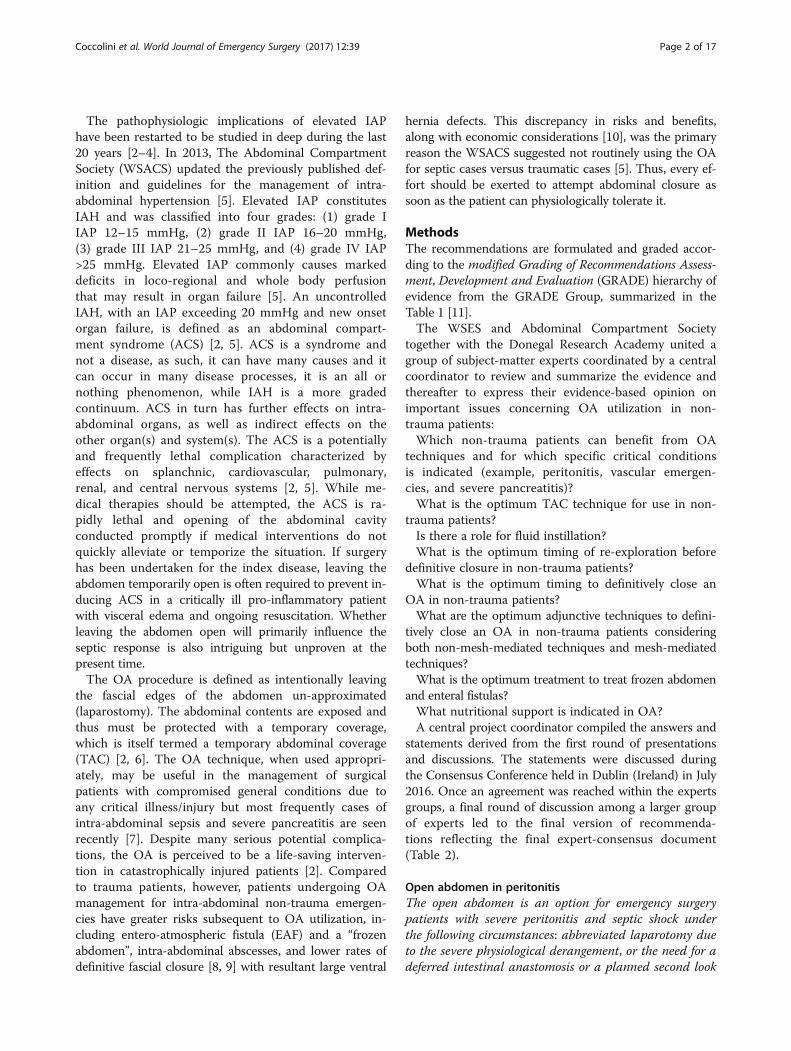
Fig. 1 Open Abdomen classification according to Bjork et al. [168]
Coccolini et al. World Journal of Emergency Surgery (2017) 12:39 Page 10 of 17

[158]. Preemptive measures could be undertaken inorder to prevent this complication: early abdominal wallclosure, bowel coverage with omentum or skin, and nodirect application of NPWT on the viscera are some ofthese measures [112, 164, 165].Several classifications and grading systems of EAF
exist. Schein and Decker proposed in 1991 a grading sys-tem based on the fistula location. Grade IV indicates afistula related to large abdominal wall defects withgrades IVa and IVb indicating the site of the fistula inregards to its location [166]. EAF can be classified basedon the fistula effluent output: low (<200 ml/day), moder-ate (200-500 ml/day), and high (>500 ml/day) [167].Bjork et al. proposed a classification based on the pres-ence of adhesions of the bowel in the setup of the openabdomen as well as the association to the fistula forma-tion (Fig. 1), and this was later adapted by WSACS[168]. Di Saverio et al. proposed a comprehensive classi-fication based on the combination of different criteria asanatomical location, output, exposure, and number offistulas [169]. As a general principle, a single, superficialfistula located in the lower GI tract with a low outputhas a higher probability of spontaneous closure ratherthan multiple fistulas deep in the wound with high out-put [169, 170]. According to this principle, the manage-ment should be tailored to each clinical situation andindividualized accordingly. In conclusion, the presenceof several different classifications represents the true dif-ficulties in the management of EAF in OA. Level of evi-dence is poor and many recommendations are based onexpert opinion suggestions.EAF is a poorly predictable and, above all, avoidable
complication. When patients develop EAF, an accurateand tailored management scheme should be adopted.Nutrition plays a key role in the management of thesepatients and should be always kept in mind as a funda-mental part of the treatment. The open abdomen strat-egy may result in fluid and electrolytes loss resulting inacid-base derangements [8]. The anatomy and the char-acteristics of the EAF(s) should be defined in order toplan the best treatment option [171]. Parenteral nutri-tion (TPN) should be started immediately after the pa-tient resuscitation. Enteral nutrition in OA patients hasbeen well studied demonstrating a reduction in infec-tious complications preserving the intestinal mucosalbarrier and its immunological function [172–174]. En-teral nutrition in patients with an EAF is has but mayincrease fistula output. Only small series of patientswith EAF treated with EN exists; therefore, no strongevidence can support these treatments and furtherstudies are needed [175, 176]. The use of octreotide an-alogs is controversial. No evidence exists about the useof somatostatin and octreotide in managing of EAF.Few studies suggest that octreotide may reduce fistula
output by diminishing GI secretions [177] while othersargue their benefit due to this agents’ reduction insplanchnic blood flow and reduction in immunefunction [178, 179].The main goal in the management of EAF should be
the closure of the fistula. Differently from common GIfistulas, the EAF is not a true fistula since a fistula tractdoes not exist. The lack of surrounding tissues preventsthe spontaneous closure. The goal of the treatmentshould be focused on trying to isolate the fistula effluentand enhancing the formation of granulation tissues sur-rounding it. Several different techniques were describedand proposed in the literature to control and treat EAF,and some attempts to standardize its management exist[169, 170]. A patient diagnosed with EAF in the setup ofOA should be treated by medical personnel familiar withthis complication and its consequences.Accurate fistula definition and anatomy should be made.
Sepsis control and management is important. Diversion ofthe fistula output in order to maintain clean the peritonealcavity is mandatory. Fistula effluent should be measuredin order to facilitate fluid balance and to ensure skin pro-tection from its digestive nature on the skin. This will en-hance and allow better patient care and mobility.Several different dressing and techniques were de-
scribed for the management of EAF, each one with rela-tively small case series and discordant results with aconsequent poor level of evidence [162, 170, 180–183].Proposed treatments vary from primary suture and fibringlue for small exposed distal fistula to a fistula suspen-sion fixating the fistula edges to the skin. Severalvariants of NPWT with devices for fistula isolation anddiversion were described with promising outcomes.The several techniques are described in detail else-
where and are not in the scope of the current positionpaper [170]. The described method to manage NPWT inpatients with EAF in the setup of OA should be applieddepending on surgeon preference, skills, and expertiseand according to hospital facilities and material availabi-lity. Generally, negative pressure wound therapy, withspecifically described variants, is the most accepted tech-nique. EAF isolation and proper wound managementwill enable skin grafting and converting EAF to a morecontrollable one with ease of applying effluent collectionbag. The definitive treatment, i.e., closure of the fistulaand repairing the abdominal wall defect should be post-poned at least 6 months and only after the patient andthe wound healed completely.
Nutritional supportOpen abdomen patients are in a hyper-metabolic condi-tion; an immediate and adequate nutritional support ismandatory (grade 1C).
Coccolini et al. World Journal of Emergency Surgery (2017) 12:39 Page 11 of 17

Open abdomen techniques result in a significant nitro-gen loss that must be replaced with a balanced nutritionregimen (grade 1C).Early enteral nutrition should be started as soon as
possible if the gastrointestinal tract allows (grade 1C).Enteral nutrition should be delayed in patients with
high output fistula with no possibility to obtain feedingaccess distal to the fistula (grade 2C).Oral feeding is not contraindicated; whenever its
possible, it could be started as soon as the patient isable to eat (grade 2C).The hyper-catabolic state of critically ill patients is asso-
ciated with muscle proteolysis, acute protein malnutrition,immune function impairment, and subclinical develop-ment of MOF. Several studies clearly demonstrated mal-nutrition as a fundamental risk factor associated to pooroutcomes during hospital stay [184]. Furthermore, in acritically ill patient, OA leads to significant nitrogen lossestimated to be 2 g per liter of abdominal fluid output.This issue requires adequate consideration and an ad-justed integration [185]. For this reason, the measurementof the abdominal fluid loss is mandatory [185]. This lossin nitrogen and protein is ulterior greatly increased in thepresence of EAF. A particular attention must be given tothis critical aspect because patients with OA are the sick-est, most inflamed, and subsequently most hyper-metabolic among surgical patients. During the OA patientmanagement, once the resuscitation is almost completedand the GI tract allows it, EN should be started as soon aspossible. Thus, it will bring beneficial effects for thepatient as faster fascia closure and lower pneumonia andfistula rate [173, 186, 187]. If malnutrition occurs, mucosalatrophy and malabsorption are among the earliest conse-quences. Gut-associated lymphoid tissue seems to be di-minished, and as a consequence, it can increase the riskfor disseminated infection due to bacterial translocationthrough the intestinal wall [188]. EN helps in maintaininggut mucosal barrier in good shape and function; as a con-sequence, it has been demonstrated to enhance immunityand IgA secretion, to prevent muscle atrophy, and lastlyto decreases systemic inflammation and oxidative injury[188, 189]. Early EN within the first 24–48 h is demon-strated to improve wound healing, decrease catabolism,preserve GI tract integrity, and finally, it reduces compli-cations, length of hospital stay, and costs. Compared toTPN early EN decreases septic complications especially inabdominal trauma and traumatic brain injuries. A retro-spective, single-institution study comparing DCS interven-tions with open abdomen performed to treat ACS, 43patients underwent early (<4 days) and 35 late (>4 days)EN. Early EN significantly increased primary closure (74%vs. 49%), reduced the fistula rate (9% vs. 26%) with nodifference in infections and but with a significant reduc-tions in hospitalization costs [186].
Patient mobilizationTo date, no recommendations can be made about earlymobilization of patients with open abdomen.Patients with an open abdomen generally should not be
mobilized out of bed until their abdomens are definitivelyclosed, for risk of evisceration [190]. This statement wasextrapolated from trauma literature [191]. However, pro-longed bed rest is associated with significant increase incomplication rate. More recent attention has been focusedon intensive care unit (ICU)-acquired weakness and thelong-term adverse functional sequelae for ICU survivors,particularly in the physical domain and this has led to anincreased interest in early mobilization in the ICU as apotential means of prevention [192–196]. The optimaltiming for initiation of mobilization of patients with OAhas yet to be defined. Early mobilization is currentlydefined as occurring within the first 2 to 5 days of ICUadmission [197].Patients with open abdomen managed with NPWT
however, may be mobilized by active or passive transfer.Further research must occur to provide the rationale toearly mobilization prior to definitive abdominal closure.
ConclusionsManagement of the open abdomen remains a verycontroversial domain, in which many techniques are stilldebated. Many important issues remain to be addressedthrough carefully designed and rigorously conducted stud-ies. Until better data is available, the use of the OA shouldbe carefully tailored to each single patient taking care tonot overuse this effective tool. Every effort should beexerted to attempt abdominal closure as soon as the patientcan physiologically tolerate it. Finally, all the precautionsshould be considered to minimize the complication rate.
AbbreviationsAAST: American Association for the Surgery of Trauma; ACS: Abdominalcompartment syndrome; AP: Acute pancreatitis; BP: Biological prosthesis;EAF: Entero-atmospheric fistula; EN: Enteral nutrition; EVAR: Endovascularrepair; GRADE: Grading of Recommendations Assessment, Development andEvaluation; IAH: Intra-abdominal hypertension; IAP: Intra-abdominal pressure;INR: International Normalized Ratio; MODS: Multi-organ dysfunctionsyndrome; MOF: Multiple organ failure; NPWT: Negative pressure woundtherapy; OA: Open abdomen procedure; PTFE: Polytetrafluoruroethylene;rAAA: Ruptured abdominal aortic aneurysm; RCT: Randomized controlledtrial; TAC: Temporal abdominal closure; TEG: Thromboelastography;TPN: Parenteral nutrition; WSACS: The Abdominal Compartment Syndrome;WSES: World Society of Emergency Surgery
AcknowledgementsSpecial thanks to Ms. Franca Boschini (Bibliographer, Medical Library, PapaGiovanni XXIII Hospital, Bergamo, Italy) for the precious bibliographical work.
FundingNone.
Availability of data and materialsNot applicable
Coccolini et al. World Journal of Emergency Surgery (2017) 12:39 Page 12 of 17

Authors’ contributionsFCo, GMo, MC, FCa, EEM, RI, WB, AP, RC, SR, YK, FMA-Z, MSa, MDM, GV, GPF,BMP, AL, MAB, AK, RM, MB, BS, VK, MM, VA, MIL, MSu, SDS, EG, KS, JEM, AKM,PM, RMM, MP, FS, GMa, TMV, TS, OC, JLK, and LA did the manuscript conceptionand draft, critically revised the manuscript, and contributed with importantscientific knowledge giving the final approval. All authors read and approvedthe final manuscript.
Ethics approval and consent to participateNot applicable
Consent for publicationNot applicable
Competing interestManu LNG Malbrain is Founding President and current Treasurer of theAbdominal Compartment Society (WSACS, www.wsacs.org). He is also amember of the executive committee of the International Fluid Academy(IFA). The IFA is integrated within the not-for-profit charitable organizationiMERiT (International Medical Education and Research Initiative) underBelgian Law. The IFA website (http://www.fluidacademy.org) is now anofficial SMACC (Social Media and Critical Care) affiliated site, and its contentis based on the philosophy of FOAM (Free Open Access Medical Education—#-FOAMed). He is a member of the medical advisory board of Pulsion MedicalSystems (Maquet Getinge group) and consults for Acelity, ConvaTec,Spiegelberg and Holtech Medical.Andrew Kirkpatrick has consulted for the Innovative Trauma Corporative,Acelity Corp., and Cook Medical Corp. The authors declare that they have nocompeting interests.
Publisher’s NoteSpringer Nature remains neutral with regard to jurisdictional claims inpublished maps and institutional affiliations.
Author details1General, Emergency and Trauma Surgery dept., Papa Giovanni XXIII Hospital,Piazza OMS 1, 24127 Bergamo, Italy. 2Emergency and Trauma Surgery, ParmaMaggiore hospital, Parma, Italy. 3Denver Health, Denver, CO 80204, USA.4Trauma Surgery, Virginia Commonwealth University, Richmond, VA 23284,USA. 5Acute Care Surgery, The Queen’s Medical Center, Honolulu, HI 96813,USA. 6Department of Surgery, Trauma and Surgical Services, University ofPittsburgh School of Medicine, Pittsburgh 15213, USA. 7Department ofSurgery, UC San Diego Health System, San Diego 92103, USA. 8Trauma &Acute Care Service, St Michael’s Hospital, Toronto, ON, Canada. 9Division ofGeneral Surgery Rambam Health Care Campus, Haifa, Israel. 10Department ofSurgery, College of Medicine and Health Sciences, UAE University, Al-Ain,United Arab Emirates. 11Department of Surgery, Macerata Hospital, Macerata,Italy. 12Department of Trauma, Emergency Surgery and Surgical Critical Care,Massachusetts General Hospital, Boston, MA 02114, USA. 13Faculdade deCiências Médicas (FCM) – Unicamp Campinas, São Paulo, Brazil. 14SecondDepartment of Surgery, Meilahti Hospital, Helsinki, Finland. 15AcademicMedical Center Amsterdam, Amsterdam, The Netherlands. 16Department ofSurgery, Foothills Medical Centre, Calgary, Canada. 17Department of Surgery,Harborview Medical Centre, Seattle 98104, USA. 18General SurgeryDepartment, Hadassah Medical Centre, Jerusalem, Israel. 19First Clinic ofGeneral Surgery, University Hospital/UMBAL/St George Plovdiv, Plovdiv,Bulgaria. 20General Surgery, Mozir Hospital, Mozir City, Belarus. 21ICU andHigh Care Burn Unit, Ziekenhius Netwerk Antwerpen, Antwerpen, Belgium.22ICU Department, Bufalini Hospital, Cesena, Italy. 23Critical Care Centre,Corporasiò Sanitaria Park Tauli, Sabdel, Spain. 24General Surgery Department,Letterkenny Hospital, Letterkenny, Ireland. 25General and Trauma SurgeryDepartment, Maggiore Hospital, Bologna, Italy. 26Upper GatrointestinalSurgery, Birmigham Hospital, Birmigham, UK. 27Department ofGastrointestinal Surgery, Stavanger University Hospital, Stavanger, Norway.28Department of Clinical Medicine, University of Bergen, Bergen, Norway.29Department of Surgery, School of Medicine, Washington University, SaintLouis, MO 63130, USA. 30Departments of Surgery and Anesthesiology,Division of Trauma and Surgical Critical Care, Vanderbilt University MedicalCenter, Nashville, TN 37232, USA. 31Département d’Anesthésie-Réanimation,CHU Bichat Claude-Bernard-HUPNVS, Assistance Publique-Hôpitaux de Paris,University Denis Diderot, Paris, France. 32ICU department
Sant’Orsola-Malpighi University Hospital, Bologna, Italy. 33ICU Department,Papa Giovanni XXIII Hospital, Bergamo, Italy. 34Trauma Surgery department,University of Maryland School of Medicine, Baltimore, MD 21201, USA.35Emergency and Trauma Surgery department, Niguarda Hospital, Milan, Italy.36General Surgery department, Assuta Medical Centers, Tel Aviv, Israel.
Received: 22 February 2017 Accepted: 25 July 2017
References1. Bailey J, Shapiro MJ. Abdominal compartment syndrome. Crit Care.
2000;4:23–9.2. Sartelli M, Abu-Zidan FM, Ansaloni L, Bala M, Beltrán MA, Biffl WL, et al. The
role of the open abdomen procedure in managing severe abdominalsepsis: WSES position paper. World J Emerg Surg. 2015;10:35.
3. Van Hee R. Historical highlights in concept and treatment of abdominalcompartment syndrome. Acta Clin Belg. 2007;62:9–15.
4. Demetriades D. Total management of the open abdomen. Int Wound J.2012;9:17–24.
5. Kirkpatrick AW, Roberts DJ, De Waele J, Jaeschke R, Malbrain MLNG, DeKeulenaer B, et al. Intra-abdominal hypertension and the abdominalcompartment syndrome: updated consensus definitions and clinicalpractice guidelines from the World Society of the Abdominal CompartmentSyndrome. Intensive Care Med. 2013;39:1190–206.
6. Leppäniemi AK. Laparostomy: why and when? Crit Care. 2010;14:216.7. Ivatury RR. Update on open abdomen management: achievements and
challenges. World J Surg. 2009;33:1150–3.8. Coccolini F, Biffl W, Catena F, Ceresoli M, Chiara O, Cimbanassi S, et al. The
open abdomen, indications, management and definitive closure. World JEmerg Surg. 2015;10:32.
9. Sartelli M, Catena F, Ansaloni L, Coccolini F, Corbella D, Moore EE, et al.Complicated intra-abdominal infections worldwide: the definitive data ofthe CIAOW Study. World J Emerg Surg. 2014;9:37.
10. van Ruler O, Mahler CW, Boer KR, Reuland EA, Gooszen HG, Opmeer BC, etal. Comparison of on-demand vs planned relaparotomy strategy in patientswith severe peritonitis: a randomized trial. JAMA. 2007;298:865–72.
11. Guyatt G, Gutterman D, Baumann MH, Addrizzo-Harris D, Hylek EM,Phillips B, et al. Grading strength of recommendations and quality ofevidence in clinical guidelines: report from an american college ofchest physicians task force. Chest. 2006;129:174–81.
12. Moore LJ, Moore FA. Epidemiology of sepsis in surgical patients. Surg ClinNorth Am. 2012;92:1425–43.
13. Ordóñez CA, Sánchez Á́I, Pineda JA, Badiel M, Mesa R, Cardona U, et al.Deferred primary anastomosis versus diversion in patients with severesecondary peritonitis managed with staged laparotomies. World J Surg.2010;34:169–76.
14. Plantefeve G, Hellmann R, Pajot O, Thirion M, Bleichner G, Mentec H.Abdominal compartment syndrome and intraabdominal sepsis: two of thesame kind? Acta Clin Belg. 2007;62:162–7.
15. Schein M. Surgical management of intra-abdominal infection: is there anyevidence? Langenbeck's Arch Surg. 2002;387:1–7.
16. Robledo FA, Luque-de-León E, Suárez R, Sánchez P, De-la-Fuente M,Vargas A, et al. Open versus closed management of the abdomen inthe surgical treatment of severe secondary peritonitis: a randomizedclinical trial. Surg Infect. 2007;8:63–72.
17. Rubenstein C, Bietz G, Davenport DL, Winkler M, Endean ED. Abdominalcompartment syndrome associated with endovascular and open repair ofruptured abdominal aortic aneurysms. J Vasc Surg. 2015;61:648–54.
18. Reite A, Soreide K, Ellingsen CL, Kvaløy JT, Vetrhus M. Epidemiology ofruptured abdominal aortic aneurysms in a well-defined Norwegianpopulation with trends in incidence, intervention rate, and mortality. J VascSurg. 2015;61:1168–74.
19. Ersryd S, Djavani-Gidlund K, Wanhainen A, Björck M. Abdominal compartmentsyndrome after surgery for abdominal aortic aneurysm: a NationwidePopulation Based Study. Eur J Vasc Endovasc Surg. 2016;52:158–65.
20. Björck M. Management of the tense abdomen or difficult abdominal closureafter operation for ruptured abdominal aortic aneurysms. Semin Vasc Surg.2012;25:35–8.
21. Acosta S, Wanhainen A, Bjorck M. Temporary abdominal closure afterabdominal aortic aneurysm repair: a systematic review of contemporaryobservational studies. Eur J Vasc Endovasc Surg. 2016;51:371–8.
Coccolini et al. World Journal of Emergency Surgery (2017) 12:39 Page 13 of 17

22. Kougias P, Lau D, El Sayed HF, Zhou W, Huynh TT, Lin PH. Determinants ofmortality and treatment outcome following surgical interventions for acutemesenteric ischemia. J Vasc Surg Off Publ Soc Vasc Surg [and] Int SocCardiovasc Surgery, North Am Chapter 2007;46:467–474.
23. Tilsed JVT, Casamassima A, Kurihara H, Mariani D, Martinez I, Pereira J, et al.ESTES guidelines: acute mesenteric ischaemia. Eur J Trauma Emerg Surg.2016;42:253–70.
24. Bruns BR, Ahmad SA, OʼMeara L, Tesoriero R, Lauerman M, Klyushnenkova E,et al. Nontrauma open abdomens: a prospective observational study.J Trauma Acute Care Surg. 2016;80:631–6.
25. Schermerhorn ML, Giles KA, Hamdan AD, Wyers MC, Pomposelli FB.Mesenteric revascularization: management and outcomes in the UnitedStates, 1988–2006. J Vasc Surg. NIH Public Access; 2009;50:341–348.e1.
26. Banks PA, Bollen TL, Dervenis C, Gooszen HG, Johnson CD, Sarr MG, et al.Classification of acute pancreatitis—2012: revision of the Atlanta classificationand definitions by international consensus. Gut. 2013;62:102–11.
27. Halonen KI, Pettilä V, Leppäniemi AK, Kemppainen EA, Puolakkainen PA,Haapiainen RK. Multiple organ dysfunction associated with severe acutepancreatitis. Crit Care Med. 2002;30:1274–9.
28. Buter A, Imrie CW, Carter CR, Evans S, McKay CJ. Dynamic nature of earlyorgan dysfunction determines outcome in acute pancreatitis. Br J Surg.2002;89:298–302.
29. Mofidi R, Duff MD, Wigmore SJ, Madhavan KK, Garden OJ, Parks RW.Association between early systemic inflammatory response, severity ofmultiorgan dysfunction and death in acute pancreatitis. Br J Surg. 2006;93:738–44.
30. Mentula P, Kylänpää-Bäck M-L, Kemppainen E, Takala A, Jansson S,Kautiainen H, et al. Decreased HLA (human leucocyte antigen)-DRexpression on peripheral blood monocytes predicts the development oforgan failure in patients with acute pancreatitis. Clin Sci (Lond).2003;105:409–17.
31. Mole DJ, Olabi B, Robinson V, Garden OJ, Parks RW. Incidence of individualorgan dysfunction in fatal acute pancreatitis: analysis of 1024 death records.HPB. 2009;11:166–70.
32. De Waele JJ, Leppäniemi AK. Intra-abdominal hypertension in acutepancreatitis. World J Surg. 2009;33:1128–33.
33. Mentula P, Hienonen P, Kemppainen E, Puolakkainen P, Leppäniemi A.Surgical decompression for abdominal compartment syndrome in severeacute pancreatitis. Arch Surg. 2010;145:764–9.
34. Besselink MG, Van Santvoort HC, Boermeester MA, Nieuweohuijs VB,Van Goor H, Dejong CHC, et al. Timing and impact of infections inacute pancreatitis. Br J Surg. 2009;96:267–73.
35. van Santvoort HC, Besselink MG, Bakker OJ, Hofker HS, Boermeester MA,Dejong CH, et al. A step-up approach or open necrosectomy for necrotizingpancreatitis. N Engl J Med. 2010;362:1491–502.
36. Petrov MS, Shanbhag S, Chakraborty M, Phillips ARJ, Windsor JA. Organfailure and infection of pancreatic necrosis as determinants of mortality inpatients with acute pancreatitis. Gastroenterology. 2010;139:813–20.
37. Tsuei BJ, Skinner JC, Bernard AC, Kearney PA, Boulanger BR. The openperitoneal cavity: etiology correlates with the likelihood of fascial closure.Am Surg. 2004;70:652–6.
38. Rao M, Burke D, Finan PJ, Sagar PM. The use of vacuum-assisted closure ofabdominal wounds: a word of caution. Color Dis. 2007;9:266–8.
39. Carlson GL, Patrick H, Amin AI, McPherson G, MacLennan G, Afolabi E, et al.Management of the open abdomen: a national study of clinical outcomeand safety of negative pressure wound therapy. Ann Surg. 2013;257:1154–9.
40. Pliakos I, Papavramidis TS, Michalopoulos N, Deligiannidis N, Kesisoglou I,Sapalidis K, et al. The value of vacuum-assisted closure in septic patientstreated with laparostomy. Am Surg. 2012;78:957–61.
41. Barker DE, Green JM, Maxwell RA, Smith PW, Mejia VA, Dart BW, et al.Experience with vacuum-pack temporary abdominal wound closure in258 trauma and general and vascular surgical patients. J Am Coll Surg.2007;204:784–92.
42. Oetting P, Rau B, Schlag PM. Abdominal vacuum device with openabdomen. Chirurg. 2006;77(586):588–93.
43. Kritayakirana K, Maggio PM, Brundage S, Purtill MA, Staudenmayer K, Spain DA.Outcomes and complications of open abdomen technique for managingnon-trauma patients. J Emerg Trauma Shock. 2010;3:118–22.
44. Prichayudh S, Sriussadaporn S, Samorn P, Pak-Art R, Sriussadaporn S,Kritayakirana K, et al. Management of open abdomen with an absorbablemesh closure. Surg Today. 2011;41:72–8.
45. Adkins AL, Robbins J, Villalba M, Bendick P, Shanley CJ. Open abdomenmanagement of intra-abdominal sepsis. Am Surg. 2004;70:137–40.
46. Amin AI, Shaikh IA. Topical negative pressure in managing severeperitonitis: a positive contribution? World J Gastroenterol. 2009;15:3394–7.
47. Bosscha K, Hulstaert PF, Visser MR, van Vroonhoven TJ, van der Werken C.Open management of the abdomen and planned reoperations in severebacterial peritonitis. Eur J Surg. 2000;166:44–9.
48. Cipolla J, Stawicki SP, Hoff WS, McQuay N, Hoey BA, Wainwright G, et al. Aproposed algorithm for managing the open abdomen. Am Surg.2005;71:202–7.
49. López-Quintero L, Evaristo-Méndez G, Fuentes-Flores F, Ventura-González F,Sepúlveda-Castro R. Treatment of open abdomen in patients withabdominal sepsis using the vacuum pack system. Cir Cir. 2010;78:317–21.
50. Padalino P, Dionigi G, Minoja G, Carcano G, Rovera F, Boni LDR. Fascia-to-fascia closure with abdominal topical negative pressure for severeabdominal infections: preliminary results in a department of general surgeryand intensive care unit. Surg Infect. 2010;11:523–52.
51. Schmelzle M, Alldinger I, Matthaei H, Aydin F, Wallert I, Eisenberger CF, et al.Long-term vacuum-assisted closure in open abdomen due to secondaryperitonitis: a retrospective evaluation of a selected group of patients. DigSurg. 2010:329–35.
52. Pliakos I, Michalopoulos N, Papavramidis TS, Arampatzi S, Diza-Mataftsi E,Papavramidis S. The effect of vacuum-assisted closure in bacterial clearanceof the infected abdomen. Surg Infect. 2014;15:18–23.
53. Wondberg D, Larusson HJ, Metzger U, Platz A, Zingg U. Treatment of theopen abdomen with the commercially available vacuum-assisted closuresystem in patients with abdominal sepsis: low primary closure rate. World JSurg. 2008;32:2724–9.
54. Khan A, Hsee L, Mathur S, Civil I. Damage-control laparotomy in nontraumapatients: review of indications and outcomes. J Trauma Acute Care Surg.2013;75:365–8.
55. Brock WB, Barker DE, Burns RP. Temporary closure of open abdominalwounds: the vacuum pack. Am Surg. 1995;61:30–5.
56. Caro A, Olona C, Jimenez A, Vadillo J, Feliu F, Vicente V. Treatment of theopen abdomen with topical negative pressure therapy: a retrospectivestudy of 46 cases. Int Wound J. 2011;8:274–9.
57. Fieger AJ, Schwatlo F, Mündel DF-X, Schenk M, Hemminger F, Kirchdorfer B,et al. Abdominal vacuum therapy for the open abdomen—a retrospectiveanalysis of 82 consecutive patients. Zentralbl Chir. 2011;136:56–60.
58. Horwood J, Akbar F, Maw A. Initial experience of laparostomy withimmediate vacuum therapy in patients with severe peritonitis. Ann R CollSurg Engl. 2009;91:681–7.
59. Ozguc H, Paksoy E, Ozturk E. Temporary abdominal closure with the vacuumpack technique: a 5-year experience. Acta Chir Belg. 2008;108:414–9.
60. Perez D, Wildi S, Demartines N, Bramkamp M, Koehler C, Clavien PA. Prospectiveevaluation of vacuum-assisted closure in abdominal compartment syndromeand severe abdominal sepsis. J Am Coll Surg. 2007;205:586–92.
61. Pérez Domínguez L, Pardellas Rivera H, Cáceres Alvarado N, López Saco Á, RivoVázquez Á, Casal NE. Vacuum assisted closure: utilidad en el abdomen abiertoy cierre diferido. Experiencia en 23 pacientes. Cir Esp. 2012;90:506–12.
62. Plaudis H, Rudzats A, Melberga L, Kazaka I, Suba O, Pupelis G. Abdominalnegative-pressure therapy: a new method in countering abdominalcompartment and peritonitis—prospective study and critical review ofliterature. Ann Intensive Care. 2012;2(Suppl 1):S23.
63. Shaikh IA, Ballard-Wilson A, Yalamarthi S, Amin AI. Use of topical negativepressure in assisted abdominal closure does not lead to high incidence ofenteric fistulae. Color Dis. 2010;12:931–4.
64. Smith LA, Barker DE, Chase CW, Somberg LB, Bradford Brock W, Phillip BR.Vacuum pack technique of temporary abdominal closure: a four-yearexperience. Am Surg. 1997;63:1102–8.
65. Wilde JW, Loudon MA. Modified opsite® sandwich for temporary abdominalclosure: a non-traumatic experience. Ann R Coll Surg Engl. 2007;89:57–61.
66. Zielinski MD, Goussous N, Schiller HJ, Jenkins D. Chemical componentsseparation with botulinum toxin A: a novel technique to improve primaryfascial closure rates of the open abdomen. Hernia. 2013;17:101–7.
67. Bertelsen CA, Fabricius R, Kleif J, Kristensen B, Gögenur I. Outcome ofnegative-pressure wound therapy for open abdomen treatment afternontraumatic lower gastrointestinal surgery: analysis of factors affectingdelayed fascial closure in 101 patients. World J Surg. 2014;38:774–81.
68. Richter S, Dold S, Doberauer JP, Mai P, Schuld J. Negative pressurewound therapy for the treatment of the open abdomen and incidence
Coccolini et al. World Journal of Emergency Surgery (2017) 12:39 Page 14 of 17

of enteral fistulas: a retrospective bicentre analysis. Gastroenterol ResPract. 2013;2013:6–11.
69. Fortelny RH, Hofmann A, Gruber-Blum S, Petter-Puchner AH, Glaser KS.Delayed closure of open abdomen in septic patients is facilitated bycombined negative pressure wound therapy and dynamic fascial suture.Surg Endosc Other Interv Tech. 2014;28:735–40.
70. Kafka-Ritsch R, Birkfellner F, Perathoner A, Raab H, Nehoda H, Pratschke J, etal. Damage control surgery with abdominal vacuum and delayed bowelreconstruction in patients with perforated diverticulitis Hinchey III/IV. JGastrointest Surg. 2012;16:1915–22.
71. Kleif J, Fabricius R, Bertelsen CA, Bruun J, Gögenur I. Promising results aftervacuum-assisted wound closure and mesh-mediated fascial traction. DanMed J. 2012;59
72. Kafka-Ritsch R, Zitt M, Schorn N, Stroemmer S, Schneeberger S, Pratschke J, et al.Open abdomen treatment with dynamic sutures and topical negative pressureresulting in a high primary fascia closure rate. World J Surg. 2012;36:1765–71.
73. Acosta S, Bjarnason T, Petersson U, Pålsson B, Wanhainen A, Svensson M, et al.Multicentre prospective study of fascial closure rate after open abdomen withvacuum and mesh-mediated fascial traction. Br J Surg. 2011;98:735–43.
74. Huang Q, Zhao R, Yue C, Wang W, Zhao Y, Ren J, et al. Fluid volumeoverload negatively influences delayed primary facial closure in openabdomen management. J Surg Res. 2014;187:122–7.
75. García Iñiguez JA, Orozco CF, Muciño Hernández MI, Ortega AL, Trabaldo SS,Cortés Flores AO, Hermosillo Sandoval JMOA. Complications of themanagement of secondary peritonitis with contained-open abdomen.Comparison of the Bogota’s bag vs polypropylene mesh. Rev GastroenterolMex. 2004;69:147–55.
76. Schein M. Planned reoperations and open management in critical intra-abdominal infections: prospective experience in 52 cases. World J Surg.1991;15:537–45.
77. Gentile AT, Feliciano PD, Mullins RJ, Crass RA, Eidemiller LR, Sheppard BC.The utility of polyglycolic acid mesh for abdominal access in patients withnecrotizing pancreatitis. J Am Coll Surg. 1998;186:313–8.
78. Losanoff JE, Kjossev KT. Mesh-foil laparostomy. J Am Coll Surg. 1997;185:89–92.79. Töns C, Schachtrupp A, Rau M, Mumme T, Schumpelick V. Abdominal
compartment syndrome: prevention and treatment. Chirurg. 2000;71:918–26.80. Schachtrupp A, Höer J, Töns C, Klinge U, Reckord U, Schumpelick V. Intra-
abdominal pressure: a reliable criterion for laparostomy closure? Hernia.2002;6:102–7.
81. Sökmen S, Atila K, Bora S, AstarcIoǧlu H, Çoker A, Füzün M. Evaluation ofprosthetic mesh closure in semiopen-abdomen patients. Hernia. 2002;6:124–9.
82. Martinez-Ordaz JL, Cruz-Olivo PA, Chacon-Moya E, de la Fuente-Lira M,Chavelas-Lluck M, Blanco-Benavides R. Management of the abdominal wall insepsis. Comparison of two techniques. Rev Gastroenterol Mex. 2004;69:88–93.
83. Gönüllü D, Köksoy FN, Demiray O, Özkan SG, Yücel T, Yücel O. Laparostomyin patients with severe secondary peritonitis. Ulus Travma ve Acil CerrahiDerg. 2009;15:52–7.
84. Kirshtein B, Roy-Shapira A, Lantsberg L, Mizrahi S. Use of the “Bogota bag”for temporary abdominal closure in patients with secondary peritonitis. AmSurg. 2007;73:249–52.
85. Manterola C, Moraga J, Urrutia S. Contained laparostomy with a Bogota bag.Results of case series. Cir Esp. 2011;89:379–85.
86. Doyon A, Devroede G, Viens D, Saito S, Rioux A, Echavé V, et al. A simple,inexpensive, life-saving way to perform iterative laparotomy in patients withsevere intra-abdominal sepsis. Color Dis. 2001;3:115–21.
87. Hakkiluoto A, Hannukainen J. Open management with mesh and zipper ofpatients with intra-abdominal abscesses or diffuse peritonitis. Eur J Surg.1992;158:403–5.
88. Hubens G, Lafaire C, De Praeter M, Ysebaert D, Vaneerdeweg W, Heytens L,et al. Staged peritoneal lavages with the aid of a Zipper system in thetreatment of diffuse peritonitis. Acta Chir Belg. 1994;94:176–9.
89. Hedderich GS, Wexler MJ, McLean AP, Meakins JL. The septic abdomen: openmanagement with Marlex mesh with a zipper. Surgery. 1986;99:399–408.
90. Zingales F, Moschino P, Carniato S, Fabris G, Vittadello F, Corsini A.Laparostomy in the treatment of severe peritonitis: a review of 60 cases.Chir Ital. 2001;53:821–6.
91. Ercan F, Korkmaz A, Aras N. The zipper-mesh method for treating delayedgeneralized peritonitis. Surg Today. 1993;23:205–14.
92. Koniaris LG, Hendrickson RJ, Drugas G, Abt P, Schoeniger LO. Dynamicretention: a technique for closure of the complex abdomen in critically illpatients. Arch Surg. 2001;136:1359–62. discussion 1363
93. Reimer MW, Yelle JD, Reitsma B, Doumit G, Allen MA, Bell MS. Managementof open abdominal wounds with a dynamic fascial closure system.Can J Surg. 2008;51:209–14.
94. Salman AE, Yetisir F, Aksoy M, Tokac M, Yildirim MB, Kilic M. Use of dynamicwound closure system in conjunction with vacuum-assisted closure therapyin delayed closure of open abdomen. Hernia. 2014;18:99–104.
95. Verdam FJ, Dolmans DEJGJ, Loos MJ, Raber MH, De Wit RJ, Charbon JA, etal. Delayed primary closure of the septic open abdomen with a dynamicclosure system. World J Surg. 2011;35:2348–55.
96. Haddock C, Konkin DE, Blair NP. Management of the open abdomen withthe abdominal reapproximation anchor dynamic fascial closure system. AmJ Surg. 2013;205:528–33.
97. Anderson ED, Mandelbaum DM, Ellison EC, Carey LC, Cooperman M. Openpacking of the peritoneal cavity in generalized bacterial peritonitis.Am J Surg. 1983;145:131–5.
98. Hollender LF, Bur F, Schwenck D, Pigache P. The “left-open abdomen”.Technic, indication and results. Chirurg. 1983;54:316–9.
99. Wittmann DH. Staged abdominal repair: development and current practiceof an advanced operative technique for diffuse suppurative peritonitis. ActaChir Austriaca. 2000;32:171–8.
100. Dietz UA, Wichelmann C, Wunder C, Kauczok J, Spor L, Strauß A, et al. Earlyrepair of open abdomen with a tailored two-component mesh andconditioning vacuum packing: a safe alternative to the planned giantventral hernia. Hernia. 2012;16:451–60.
101. Losanoff J, Kjossev K. Palisade dorsoventral lavage for neglected peritonitis.Am J Surg. 1997;173:134–5.
102. Ivatury RR, Nallathambi M, Rao PM, Rohman M, Stahl WM. Openmanagement of the septic abdomen: therapeutic and prognosticconsiderations based on APACHE II. Crit Care Med. 1989;17:511–7.
103. Wittmann DH, Aprahamian C, Bergstein JM. Etappenlavage: advanceddiffuse peritonitis managed by planned multiple laparotomies utilizingzippers, slide fastener, and Velcro analogue for temporary abdominalclosure. World J Surg. 1990;14:218–26.
104. Kim PJ, Attinger CE, Olawoye O, Crist BD, Gabriel A, Galiano RD, et al.Negative pressure wound therapy with instillation: review of evidence andrecommendations. Wounds a Compend Clin Res Pract. 2015;27:S2–19.
105. Atema JJ, Gans SL, Boermeester MA. Systematic review and meta-analysis ofthe open abdomen and temporary abdominal closure techniques in non-trauma patients. World J Surg. 2015;39:912–25.
106. Karmali S, Evans D, Laupland KB, Findlay C, Ball CG, Bergeron E, et al. Toclose or not to close, that is one of the questions? Perceptions of TraumaAssociation of Canada surgical members on the management of the openabdomen. J Trauma. 2006;60:287–93.
107. Kirkpatrick AW, Laupland KB, Karmali S, Bergeron E, Stewart TC, Findlay C, etal. Spill your guts! Perceptions of Trauma Association of Canada membersurgeons regarding the open abdomen and the abdominal compartmentsyndrome. J Trauma. 2006;60:279–86.
108. Pommerening MJ, Dubose JJ, Zielinski MD, Phelan HA, Scalea TM, Inaba K,et al. Time to first take-back operation predicts successful primary fascialclosure in patients undergoing damage control laparotomy. Surg (UnitedStates). 2014;156:431–8.
109. Singer M, Deutschman CS, Seymour CW, Shankar-Hari M, Annane D, BauerM, et al. The Third International Consensus Definitions for Sepsis and SepticShock (Sepsis-3). JAMA. 2016;315:801–10.
110. Emr B, Sadowsky D, Azhar N, Gatto LA, An G, Nieman GF, et al. Removal ofinflammatory ascites is associated with dynamic modification of local andsystemic inflammation along with prevention of acute lung injury: in vivoand in silico studies. Shock. 2014;41:317–23.
111. Kubiak BD, Albert SP, Gatto LA, Snyder KP, Maier KG, Vieau CJ, et al. Peritonealnegative pressure therapy prevents multiple organ injury in a chronic porcinesepsis and ischemia/reperfusion model. Shock. 2010;34:525–34.
112. Cheatham ML, Demetriades D, Fabian TC, Kaplan MJ, Miles WS, SchreiberMA, et al. Prospective study examining clinical outcomes associated with anegative pressure wound therapy system and Barker’s vacuum packingtechnique. World J Surg. 2013;37:2018–30.
113. Kirkpatrick AW, Roberts DJ, Faris PD, Ball CG, Kubes P, Tiruta C, et al.Active negative pressure peritoneal therapy after abbreviatedlaparotomy: the intraperitoneal vacuum randomized controlled trial.Ann Surg. 2015;262:38–46.
114. Wang J, Kubes P. A reservoir of mature cavity macrophages that can rapidlyinvade visceral organs to affect tissue repair. Cell. 2016;165:668–78.
Coccolini et al. World Journal of Emergency Surgery (2017) 12:39 Page 15 of 17

115. Demetriades D, Salim A. Management of the open abdomen. Surg ClinNorth Am. 2014;94:131–53.
116. Regner JL, Kobayashi L, Coimbra R. Surgical strategies for management ofthe open abdomen. World J Surg. 2012;36:497–510.
117. Godat L, Kobayashi L, Costantini T, Coimbra R. Abdominal damage controlsurgery and reconstruction: world society of emergency surgery positionpaper. World J Emerg Surg. 2013;8
118. Ramirez OM, Ruas E, Dellon AL. “Components separation” method forclosure of abdominal-wall defects: an anatomic and clinical study. PlastReconstr Surg. 1990;86:519–26.
119. Paul JS, Ridolfi TJ. A case study in intra-abdominal sepsis. Surg Clin NorthAm. 2012;92:1661–77.
120. Lambertz A, Mihatsch C, Röth A, Kalverkamp S, Eickhoff R, Neumann UP, et al.Fascial closure after open abdomen: initial indication and early revisions aredecisive factors—a retrospective cohort study. Int J Surg. 2015;13:12–6.
121. Rasilainen SK, Juhani MP, Kalevi LA. Microbial colonization of openabdomen in critically ill surgical patients. World J Emerg Surg. 2015;10:25.
122. Leber GE, Garb JL, Alexander AI, Reed WP. Long-term complications associatedwith prosthetic repair of incisional hernias. Arch Surg. 1998;133:378–82.
123. Mathes SJ, Steinwald PM, Foster RD, Hoffman WY, Anthony JP. Complexabdominal wall reconstruction: a comparison of flap and mesh closure. AnnSurg. 2000;232:586–96.
124. Choi JJ, Palaniappa NC, Dallas KB, Rudich TB, Colon MJ, Divino CM. Use ofmesh during ventral hernia repair in clean-contaminated and contaminatedcases: outcomes of 33,832 cases. Ann Surg. 2012;255:176–80.
125. de Vries Reilingh TS, van Goor H, Charbon JA, Rosman C, Hesselink EJ, vander Wilt GJ, et al. Repair of giant midline abdominal wall hernias:“components separation technique” versus prosthetic repair : interimanalysis of a randomized controlled trial. World J Surg Springer. 2007;31:756–63.
126. Yegiyants S, Tam M, Lee DJ, Abbas MA. Outcome of components separationfor contaminated complex abdominal wall defects. Hernia. 2012;16:41–5.
127. Rasilainen SK, Mentula PJ, Leppäniemi AK. Components separationtechnique is feasible for assisting delayed primary fascial closure of openabdomen. Scand J Surg. 2016;105:17–21.
128. Sharrock AE, Barker T, Yuen HM, Rickard R, Tai N. Management andclosure of the open abdomen after damage control laparotomy fortrauma. A systematic review and meta-analysis. Injury. Elsevier Ltd. 2015;47:296–306.
129. Atema JJ, de Vries FEE, Boermeester MA. Systematic review and meta-analysis of the repair of potentially contaminated and contaminatedabdominal wall defects. Am J Surg Elsevier Inc. 2016;212:982–95.
130. Dinsmore RC, Calton WC, Harvey SB, Blaney MW. Prevention ofadhesions to polypropylene mesh in a traumatized bowel model.J Am Coll Surg. 2000;191:131–6.
131. van ‘t Riet M, de Vos van Steenwijk PJ, Bonthuis F, Marquet RL,Steyerberg EW, Jeekel J, et al. Prevention of adhesion to prostheticmesh: comparison of different barriers using an incisional hernia model.Ann Surg. 2003;237:123–8.
132. Konstantinovic ML, Lagae P, Zheng F, Verbeken EK, De Ridder D, Deprest JA.Comparison of host response to polypropylene and non-cross-linkedporcine small intestine serosal-derived collagen implants in a rat model.BJOG An Int J Obstet Gynaecol. 2005;112:1554–60.
133. Fansler RF, Taheri P, Cullinane C, Sabates B, Flint LM. Polypropylene meshclosure of the complicated abdominal wound. Am J Surg. 1995;170:15–8.
134. Voyles CR, Richardson JD, Bland KI, Tobin GR, Flint LM, Polk HC. Emergencyabdominal wall reconstruction with polypropylene mesh: short-termbenefits versus long-term complications. Ann Surg. 1981;194:219–23.
135. Brown GL, Richardson JD, Malangoni MA, Tobin GR, Ackerman D, PolkHC. Comparison of prosthetic materials for abdominal wallreconstruction in the presence of contamination and infection. AnnSurg. 1985;201:705–11.
136. Sartelli M, Coccolini F, van Ramshorst GH, Campanelli G, Mandalà V,Ansaloni L, et al. WSES guidelines for emergency repair of complicatedabdominal wall hernias. World J Emerg Surg. 2013;8:50.
137. Cornwell KG, Landsman A, James KS. Extracellular matrix biomaterials forsoft tissue repair. Clin Podiatr Med Surg. 2009;26:507–23.
138. Caviggioli F, Klinger FM, Lisa A, Maione L, Forcellini D, Vinci V, et al.Matching biological mesh and negative pressure wound therapy inreconstructing an open abdomen defect. Case Rep Med. HindawiPublishing Corporation; 2014;2014:235930.
139. Badylak SF. Xenogeneic extracellular matrix as a scaffold for tissuereconstruction. Transpl Immunol. 2004;12:367–77.
140. Winters JC. InteXen tissue processing and laboratory study. Int Urogynecol JPelvic Floor Dysfunct. 2006;17
141. Petter-Puchner AH, Dietz UA. Biological implants in abdominal wall repair.Br J Surg. 2013;100:987–8.
142. Montori G, Coccolini F, Manfredi R, Ceresoli M, Campanati L, Magnone S, etal. One year experience of swine dermal non-crosslinked collagenprostheses for abdominal wall repairs in elective and emergency surgery.World J Emerg Surg. 2015;10:28–35.
143. Primus FE, Harris HW. A critical review of biologic mesh use in ventralhernia repairs under contaminated conditions. Hernia. 2013;17:21–30.
144. Gurrado A, Franco IF, Lissidini G, Greco G, De Fazio M, Pasculli A, et al.Impact of pericardium bovine patch (Tutomesh®) on incisional herniatreatment in contaminated or potentially contaminated fields: retrospectivecomparative study. Hernia. 2015;19:259–66.
145. de Moya MA, Dunham M, Inaba K, Bahouth H, Alam HB, Sultan B, et al.Long-term outcome of acellular dermal matrix when used for largetraumatic open abdomen. J Trauma Inj Infect Crit Care. 2008;65:349–53.
146. Ginting N, Tremblay L, Kortbeek JB. Surgisis® in the management of thecomplex abdominal wall in trauma: a case series and review of theliterature. Injury. 2010;41:970–3.
147. Patton JH, Berry S, Kralovich KA. Use of human acellular dermal matrix incomplex and contaminated abdominal wall reconstructions. Am J Surg.2007;193:360–3.
148. Maurice SM, Skeete DA. Use of human acellular dermal matrix forabdominal wall reconstructions. Am J Surg. 2009;197:35–42.
149. Lin HJ, Spoerke N, Deveney C, Martindale R. Reconstruction of complexabdominal wall hernias using acellular human dermal matrix: a singleinstitution experience. Am J Surg. 2009;197:599–603.
150. Diaz JJ, Conquest AM, Ferzoco SJ, Vargo D, Miller P, Wu Y-C, et al.Multi-institutional experience using human acellular dermal matrix forventral hernia repair in a compromised surgical field. Arch Surg. 2009;144:209–15.
151. Lee EI, Chike-Obi CJ, Gonzalez P, Garza R, Leong M, Subramanian A, et al.Abdominal wall repair using human acellular dermal matrix: a follow-upstudy. Am J Surg. 2009;198:650–7.
152. Pomahac B, Aflaki P. Use of a non-cross-linked porcine dermal scaffold inabdominal wall reconstruction. Am J Surg. Elsevier Inc; 2010;199:22–7.
153. Abdelfatah MM, Rostambeigi N, Podgaetz E, Sarr MG. Long-term outcomes(>5-year follow-up) with porcine acellular dermal matrix (Permacol) inincisional hernias at risk for infection. Hernia. 2015;19:135–40.
154. Booth JH, Garvey PB, Baumann DP, Selber JC, Nguyen AT, Clemens MW, etal. Primary fascial closure with mesh reinforcement is superior tobridged mesh repair for abdominal wall reconstruction. J Am Coll Surg.2013;217:999–1009.
155. Chand B, Indeck M, Needleman B, Finnegan M, Van Sickle KR, Ystgaard B, et al.A retrospective study evaluating the use of Permacol™ surgical implant inincisional and ventral hernia repair. Int J Surg Elsevier Ltd. 2014;12:296–303.
156. Holihan JL, Nguyen DH, Nguyen MT, Mo J, Kao LS, Liang MK. Mesh locationin open ventral hernia repair: a systematic review and network meta-analysis. World J Surg. 2016;40:89–99.
157. Eriksson A, Rosenberg J, Bisgaard T. Surgical treatment for giant incisionalhernia: a qualitative systematic review. Hernia. 2014;18:31–8.
158. Bradley MJ, Dubose JJ, Scalea TM, Holcomb JB, Shrestha B, Okoye O, et al.Independent predictors of enteric fistula and abdominal sepsis afterdamage control laparotomy: results from the prospective AAST OpenAbdomen registry. JAMA Surg. 2013;148:947–54.
159. Teixeira PGR, Inaba K, Dubose J, Salim A, Brown C, Rhee P, et al.Enterocutaneous fistula complicating trauma laparotomy: a major resourceburden. Am Surg. 2009;75:30–2.
160. Martinez JL, Luque-De-Leon E, Mier J, Blanco-Benavides R, Robledo F.Systematic management of postoperative enterocutaneous fistulas: factorsrelated to outcomes. World J Surg. 2008;32:436–43.
161. Tavusbay C, Genc H, Cin N, Kar H, Kamer E, Atahan K, et al. Use of avacuum-assisted closure system for the management of enteroatmosphericfistulae. Surg Today. Springer Japan; 2015;45:1102–1111.
162. D’Hondt M, Devriendt D, Van Rooy F, Vansteenkiste F, D’Hoore A,Penninckx F, et al. Treatment of small-bowel fistulae in the openabdomen with topical negative-pressure therapy. Am J Surg. ElsevierInc; 2011;202:e20–4.
Coccolini et al. World Journal of Emergency Surgery (2017) 12:39 Page 16 of 17

163. Marinis A, Gkiokas G, Argyra E, Fragulidis G, Polymeneas G, Voros D.“Enteroatmospheric fistulae”-gastrointestinal openings in the openabdomen: a review and recent proposal of a surgical technique. Scand JSurg. 2013;102:61–8.
164. Schecter WP, Ivatury RR, Rotondo MF, Hirshberg A. Open abdomen aftertrauma and abdominal sepsis: a strategy for management. J Am Coll Surg.2006;203:390–6.
165. Carlson GL, Patrick H, Amin AI, McPherson G, MacLennan G, Afolabi E, et al.Management of the open abdomen. Ann Surg. 2013;257:1154–9.
166. Schein M, Decker GAG. Postoperative external alimentary tract fistulas. Am JSurg. Elsevier; 1991;161:435–438.
167. Schecter WP, Hirshberg A, Chang DS, Harris HW, Napolitano LM, Wexner SD,et al. Enteric fistulas: principles of management. J Am Coll Surg. Elsevier Inc;2009;209:484–91.
168. Björck M, Kirkpatrick AW, Cheatham M, Kaplan M, Leppäniemi A, de Waele JJ.Amended classification of the open abdomen. Scand J Surg. 2016;105:5–10.
169. Di Saverio S, Tarasconi A, Inaba K, Navsaria P, Coccolini F, Costa Navarro D,et al. Open abdomen with concomitant enteroatmospheric fistula: attemptto rationalize the approach to a surgical nightmare and proposal of aclinical algorithm. J Am Coll Surg. 2015;220:e23–33.
170. Di Saverio S, Tarasconi A, Walczak DA, Cirocchi R, Mandrioli M, Birindelli A,et al. Classification, prevention and management of entero-atmosphericfistula: a state-of-the-art review. Langenbeck's Arch Surg. 2016;401:1–13.
171. Polk TM, Schwab CW. Metabolic and nutritional support of the enterocutaneousfistula patient: a three-phase approach. World J Surg. 2012;36:524–33.
172. Moore FA, Feliciano D V, Andrassy RJ, McArdle AH, Booth F V, Morgenstein-Wagner TB, et al. Early enteral feeding, compared with parenteral, reducespostoperative septic complications. The results of a meta-analysis. Ann Surg.Lippincott, Williams, and Wilkins; 1992;216:172–83.
173. Dissanaike S, Pham T, Shalhub S, Warner K, Hennessy L, Moore EE, et al.Effect of immediate enteral feeding on trauma patients with an openabdomen: protection from nosocomial infections. J Am Coll Surg.2008;207:690–7.
174. Byrnes MC, Reicks P, Irwin E. Early enteral nutrition can be successfullyimplemented in trauma patients with an “open abdomen.”. Am J Surg.2010;199:359–63.
175. Reinisch A, Liese J, Woeste G, Bechstein W, Habbe N. A retrospective,observational study of enteral nutrition in patients with enteroatmosphericfistulas. Ostomy Wound Manage. 2016;62:36–47.
176. Yin J, Wang J, Yao D, Zhang S, Mao Q, Kong W, et al. Is it feasible toimplement enteral nutrition in patients with enteroatmospheric fistulae? Asingle-center experience. Nutr Clin Pract. 2014;29:656–661.
177. Nubiola P, Badia JM, Martinez-Rodenas F, Gil MJ, Segura M, Sancho J, et al.Treatment of 27 postoperative enterocutaneous fistulas with the long half-life somatostatin analogue SMS 201-995. Ann Surg. 1989;210:56–8.
178. Lattuada D, Casnici C, Crotta K, Mastrotto C, Franco P, Schmid HA, et al.Inhibitory effect of pasireotide and octreotide on lymphocyte activation.J Neuroimmunol. 2007;182:153–9.
179. Cooper AM, Braatvedt GD, Qamar MI, Brown H, Thomas DM, Halliwell M, etal. Fasting and post-prandial splanchnic blood flow is reduced by asomatostatin analogue (octreotide) in man. Clin Sci. 1991;81:169–75.
180. Navsaria PH, Bunting M, Omoshoro-Jones J, Nicol AJ, Kahn D. Temporaryclosure of open abdominal wounds by the modified sandwich-vacuumpack technique. Br J Surg. 2003;90:718–22.
181. Al-Khoury G, Kaufman D, Hirshberg A. Improved control of exposed fistulain the open abdomen. J Am Coll Surg. 2008;206:397–8.
182. Layton B, DuBose J, Nichols S, Connaughton J, Jones T, Pratt J. Pacifying theopen abdomen with concomitant intestinal fistula: a novel approach.Am J Surg. Elsevier Inc; 2010;199:e48–50.
183. Rekstad LC, Wasmuth HH, Ystgaard B, Stornes T, Seternes A. Topicalnegative-pressure therapy for small bowel leakage in a frozen abdomen: atechnical report. J Trauma Acute Care Surg. 2013;75:487–91.
184. Giner M, Laviano A, Meguid MM, Gleason JR. In 1995 a correlation betweenmalnutrition and poor outcome in critically ill patients still exists. Nutrition.1996;12:23–9.
185. Cheatham ML, Safcsak K, Brzezinski SJ, Lube MW. Nitrogen balance, proteinloss, and the open abdomen. Crit Care Med. 2007;35:127–31.
186. Collier B, Guillamondegui O, Cotton B, Donahue R, Conrad A, Groh K, et al.Feeding the open abdomen. JPEN J Parenter Enteral Nutr. 2007;31:410–5.
187. Cothren CC, Moore EE, Ciesla DJ, Johnson JL, Moore JB, Haenel JB, et al.Postinjury abdominal compartment syndrome does not preclude earlyenteral feeding after definitive closure. Am J Surg. 2004;188:653–8.
188. Marik PE, Zaloga GP. Meta-analysis of parenteral nutrition versus enteralnutrition in patients with acute pancreatitis. BMJ. 2004;328:1407–10.
189. McClave SA, Heyland DK. The physiologic response and associatedclinical benefits from provision of early enteral nutrition. Nutr Clin Pract.2009;24:305–15.
190. Panel OAA, Campbell A, Chang M, Fabian T, Franz M, Kaplan M, et al.Management of the open abdomen: from initial operation to definitiveclosure. Am Surg. 2009;75:S1–22.
191. Engels PT, Beckett AN, Rubenfeld GD, Kreder H, Finkelstein JA, da Costa L,et al. Physical rehabilitation of the critically ill trauma patient in the ICU. CritCare Med. 2013;41:1790–801.
192. Truong A, Fan E, Brower R, Needham D. Bench-to-bedside review:mobilizing patients in the intensive care unit—from pathophysiology toclinical trials. Crit Care. 2009;13:216.
193. Pavy-Le Traon A, Heer M, Narici MV, Rittweger J, Vernikos J. From space toEarth: advances in human physiology from 20 years of bed rest studies(1986-2006). Eur J Appl Physiol. 2007;101:143–94.
194. Herridge MS. Building consensus on ICU-acquired weakness. Intensive CareMed. 2009;35:1–3.
195. Cuthbertson BH, Roughton S, Jenkinson D, Maclennan G, Vale L. Qualityof life in the five years after intensive care: a cohort study. Crit Care.2010;14:R6.
196. Burtin C, Clerckx B, Robbeets C, Ferdinande P, Langer D, Troosters T, et al.Early exercise in critically ill patients enhances short-term functionalrecovery. Crit Care Med. 2009;37:2499–505.
197. Hodgson CL, Berney S, Harrold M, Saxena M. Clinical review: early patientmobilization in the ICU. Crit Care. 2013;17:207.
• We accept pre-submission inquiries
• Our selector tool helps you to find the most relevant journal
• We provide round the clock customer support
• Convenient online submission
• Thorough peer review
• Inclusion in PubMed and all major indexing services
• Maximum visibility for your research
Submit your manuscript atwww.biomedcentral.com/submit
Submit your next manuscript to BioMed Central and we will help you at every step:
Coccolini et al. World Journal of Emergency Surgery (2017) 12:39 Page 17 of 17