review role of cytokines and transcription factors in...
TRANSCRIPT

© 2015 Anne Carolina Eleutério Leite, Valéria Martins de Araújo Carneiro, Júlia Faria Nunes, André Cruz de Sousa, Maria
Imaculada Muniz-Junqueira and Maria do Carmo Machado Guimarães. This open access article is distributed under a
Creative Commons Attribution (CC-BY) 3.0 license.
American Journal of Immunology
Review
Role of Cytokines and Transcription Factors in Periodontitis:
A Review of Cellular and Molecular Mechanisms
1Anne Carolina Eleutério Leite,
1Valéria Martins de Araújo Carneiro,
2Júlia Faria Nunes,
2André Cruz de Sousa,
3Maria Imaculada Muniz-Junqueira and
1Maria do Carmo Machado Guimarães
1Department of Dentistry, Periodontics Division, University of Brasília, Brazil 2Department of Dentistry, Periodontics Division, Catholic University of Brasília, Brazil 3Faculty of Medicine, Pathology, Cellular Immunology Laboratory, University of Brasília, Brazil
Article history
Received: 29-04-2015
Revised: 09-10-2015
Accepted: 19-11-2015
Corresponding Author:
Anne Carolina Eleutério Leite,
Department of Dentistry,
Periodontics Division, University
of Brasília, Brazil Tel: +55 (61) 3107-3300
Email: [email protected]
Abstract: Periodontal Diseases (PD) are characterized by pathological
manifestation of host immune response to bacterial infection at the
tooth/gingival interface. Evidences points periodontitis as a risk factor for
pathological systemic conditions, such as, cardiovascular diseases and
diabetes. The identification of host factors that determine their susceptibility
to immune subversion can provide useful information in the pathogenesis of
periodontitis. Protective acute inflammatory response fails to remove
inflammatory cells, especially neutrophils, evolving to chronic, destructive
and pathological lesions. T and B cells actively participate in pathogenesis
of the disease. CD4+ naive T cells are activated by antigenic stimulation and
differentiate into subpopulations of distinct effector cells, characterized by
its specific cytokine production profiles and functions. In periodontal
infection, activated Th17 and regulatory T lymphocytes (Tregs) play
antagonistic roles as effector and suppressor cells, respectively. In presence
of Tregs, there is a decrease in the levels of pro-inflammatory cytokines,
such as, Interferon gamma (IFN-γ), Interleukin (IL) -17, Tumor Necrosis
Factor (TNF) and IL-1 β, at sites of disease. Absence of Tregs may cause a
variety of disorders, such as, periodontitis. The RANKL/RANK system and
Osteoprotegerin (OPG) are modulators of bone resorption in periodontitis.
The balance between periodontal bone resorption by osteoclasts and bone
formation by osteoblasts controls bone mass. RANKL induces osteoclast
differentiation and maturation and OPG inhibits RANK/RANKL interaction
and prevents bone resorption. RANKL mRNA and the RANKL/OPG
mRNA ratio were enhanced in chronic periodontitis. Furthermore, the role
of NF-kB, FoxP3 and T-bet transcription factors were explored. Therapies
for periodontitis involving cellular and molecular biology are not specific
and have many side effects. Current therapies to successfully control the PD
reduces clinical signs of inflammation at local sites of the disease; however,
new treatments for periodontitis should address the contribution of immune
cells in bone resorption, particularly in the natural course of the disease,
considering its periods of remission and progression.
Keywords: Transforming Growth Factor Beta, T Helper Cell, Periodontal
Diseases, RANKL/RANK
Introduction
Periodontal Diseases (PD) are characterized by
pathological manifestation of host immune response to
bacterial infection at the tooth/gingival interface. These
are mainly caused by Gram-negative bacteria, including
Porphyromonas gingivalis, Prevotella intermedia,
Aggregatibacter actinomycetemcomitans and Tannerella
forsythia. Differences in individual response can be
explained by the combination of factors that confer upon

Anne Carolina Eleutério Leite et al. / American Journal of Immunology 2015, 11 (4): 125.138
DOI: 10.3844/ajisp.2015.125.138
126
them a complex nature. Among these factors,
susceptibility becomes prominent, with special
attribution given to genetic polymorphisms,
environmental factors and pathogen virulence factors
(Ohlrich et al., 2009; Sanz and Winkelhoff, 2011).
Individuals affected by the disease share common
polymorphisms in specific genes that are important for
regulation of inflammatory response (D’Aiuto et al.,
2004d; Fitzsimmons et al., 2010). However,
determinants for susceptibility to periodontitis remain
still unclear (Linden et al., 2013).
Severe periodontitis affects up to 15% of most
populations (Papapanou, 1999). In Brazil, the most
recent National Survey of Oral Health reported a
prevalence of 19.4% and the most severe forms of
periodontal disease was more significant in adults
between 35-44 years of age (Leite et al., 2014).
Periodontitis comprises chronic forms of periodontal
disease, which are the result of a polymicrobial infection
and are characterized by loss of collagen fibers and
insertion in the cementum surface (mineralized tissue
that covers the root surface), apical migration of
junctional epithelium (epithelium continuous with the
oral epithelium, which promotes the insertion of the
gum to the tooth), periodontal pocket formation
(cementum surface devoid of periodontal fibers) and
alveolar bone reabsorption. Such damages impair
functions of periodontal tissues and may result in tooth
loss (Sanz and Winkelhoff, 2011).
There are more than 700 bacterial species in
periodontal pockets (Aas et al., 2005) and a combination
of aerobic and anaerobic microbiota is typically seen in
infection. Substantial tissue destruction in patients with
severe periodontitis is characterized, in many cases, by
deep periodontal pockets around many or all teeth. The
aggregate epithelial lesions are equal in size to an
ulcerated wound with an area of 8 to 20 cm2, in
accordance with clinical estimations (Slade et al., 2000).
Thus, the chronic and cyclical nature of periodontal
disease provides an opportunity for continuous
hematogenous dissemination of periodontal pathogens,
bacterial antigens and inflammatory mediators
(Offenbacher et al., 2005; Linden et al., 2013).
Since the beginning of the 1990s, evidence has
pointed to periodontitis as a risk factor for systemic
conditions, such as, Cardiovascular Disease (CVD),
adverse outcomes of pregnancy, diabetes, rheumatoid
arthritis and lung disease (Williams and Offenbacher,
2000; Hajishengallis, 2014).
In healthy gingiva, the sulcular epithelium and local
innate immunity act as a natural barrier that prevents
bacterial penetration. However, in PD, the epithelium
of ulcerated and inflamed gingiva is vulnerable to
bacteria and creates a gateway to subsequent tissues
(Linden et al., 2013). Bacteria and bacterial antigens that
are systemically dispersed cause significant systemic
inflammation. Leukocytes, epithelial and endothelial
cells and hepatocytes respond to virulence factors with
secretion of proinflammatory immune mediators, as
cytokines, chemokines (e.g., Monocyte Chemoattractant
Protein-1 (MCP-1) and Macrophage Inflammatory
Protein-1alpha (MIP-1alpha)), C-Reactive Protein
(CRP), an acute phase protein (Gemmell et al., 2001;
Maekawa et al., 2011). With continued exposure, soluble
antigens react with specific circulating antibodies to
form immune complexes that amplify inflammation in
places where there are immune complex deposition.
Similarly, pro-inflammatory mediators, such as
Interleukin (IL)-1β, IL-6, Tumor Necrosis Factor alpha
(TNF-α), Prostaglandin E2 (PGE2), among others,
locally produced in inflamed gingival tissues can spread
into the systemic circulation. The pro-inflammatory
cytokines in circulation induce leukocytosis and acute
phase proteins production (e.g., CRP, fibrinogen and
serum amyloid A) (Linden et al., 2013; Hajishengallis,
2014). In this context, the increase in the number of
White Blood Cells (WBC) is associated with the increased
risk of coronary heart disease, CVD, atherosclerosis,
thrombosis and myocardial ischemia. This increase may
be caused by the inflammatory nature of chronic
infections, such as, periodontitis (Loo et al., 2012).
It has been shown that the T CD4+ CD25+ regulatory
cells (Tregs) that express transcription factor FoxP3 may
prevent the overreactive adaptive immune response
associated with PD (de Brito Bezerra et al., 2012;
Kobayashi et al., 2011). Tregs may reduce Interferon
gamma (IFN-γ), IL-17, TNF and IL-1β levels at
periodontal disease sites (Glowacki et al., 2013). IL-10,
Transforming Growth Factor beta (TGF-β) and
Cytotoxic T-Lymphocyte-Associated protein 4 (CTLA-
4) are produced by Tregs to control inflammation,
slowing progression to periodontal disease (Linden et al.,
2013). TGF-β has also been studied as a form of
treatment, because it may heal and regenerate
periodontal bone (Glowacki et al., 2013).
This review aimed to report the cellular and
molecular mechanisms involved in periodontal
inflammation and alveolar bone loss, as well as the
systemic effects in periodontitis.
Classification of Periodontal Diseases and
Microbial Associations
In current clinical practice, two types of periodontitis
are recognized: Chronic Periodontitis (CP) and
Aggressive Periodontitis (AgP), as classified by the
Current Classification of PD by the American Academy
of Periodontology (AAP, 1999). CP has higher
prevalence in adults, but can also occur in children and
adolescents. In CP, there is correlation between the
severity of bone destruction and the quantity of dental

Anne Carolina Eleutério Leite et al. / American Journal of Immunology 2015, 11 (4): 125.138
DOI: 10.3844/ajisp.2015.125.138
127
biofilm and calculus, in addition to the moderate growth
rate in the majority of cases. Considering the extension
of lesions, CP is classified as localized when less than
30% of dental sites (mesiobuccal, buccal, distobuccal,
mesiolingual, lingual and distolingual) are affected and
as generalized when it exceeds this limit. Whereas, AgP
is characterized by the rapid loss of clinical attachment
and alveolar bone. The following characteristics are
considered for diagnosis of AgP: No contribution of
systemic diseases and familial aggregation. According to
the AAP, the diagnosis is based on clinical data, X-rays
and clinical history. Localized Aggressive Periodontitis
(LAgP) is characterized by destruction of the periodontal
tissues in the first molar/incisor with interproximal
insertion loss in at least two permanent teeth, one of
which is the first molar and involving no more than two
teeth beyond the first molars and incisors. It is usually
detected and diagnosed during puberty in systemically
healthy individuals. It also has a strong serum antibody
response to infectious agents. Generalized Aggressive
Periodontitis (GAgP) usually affects individuals under
age 30, but can affect older individuals. GAgP is
characterized as generalized when the loss of
interproximal insertion affects at least three permanent
teeth, in addition to the first molars and incisors.
Moreover, GAgP progresses in alternating periods of
activity and quiescence. In some period of activity,
pronounced destruction of tooth insertion, alveolar bone
destruction and insufficient response of serum antibodies
to infectious agents has been observed (Armitage, 1999;
Armitage and Cullinan, 2010).
Furthermore, it was recently suggested that CP is a
polygenic disease, in which multiple genes contribute
cumulatively to the overall disease risk (or protection) by
influencing the host immune response and the
composition and structure of the microbiota (Divaris et al.,
2013). Nonetheless, monogenic forms of the disease are
different, such as aggressive periodontitis, in young
patients with leukocyte adhesion deficiency, in which a
single gene (ITGB2; which encodes integrin β2)
invariably precipitates periodontal disease
(Moutsopoulos et al., 2014).
A systematic review concluded that there is no
evidence to support differences in the composition of
subgingival microbiota between the clinical forms CP
and AgP (Mombelli et al., 2002). Although no specific
systemic marker was associated with the severity of
periodontal destruction, it has been suggested that AgP
may show a hyper-responsive systemic profile associated
with genetic susceptibility (Cairo et al., 2010). It was
also suggested that high titers of A.
actinomycetemcomitans and P. gingivalis are suggestive
of generalized and severe PD (Papapanou et al., 2004;
Dye et al., 2005).
Recently, different serotypes of P. gingivalis have
been associated with the induction of a distinct type of
immune response, which suggests the role played by
capsule during activation of dendritic cells
(Hernández et al., 2011).
Inflammation is an important source of nutrients
(especially for asaccharolytic bacteria) and thus exerts a
powerful influence on the composition of periodontal
microbiota, favoring species that can use tissue
degradation products. Whereas, species that cannot
benefit from these environmental changes may have
disadvantage of fitness and thus are overcome. The
selective growth of bacteria, which act as pathogenic
bacteria, has the potential to trigger a self-feeding
vicious cycle for subsequent tissue destruction and
bacterial overgrowth (Hajishengallis, 2014).
Virulence factors such as cytotoxins, proteases and
hemagglutinins, structural molecules of the bacteria,
including Lipopolysaccharide (LPS) and Peptidoglycan
(PGN), lipoproteins, bacterial DNA and double-stranded
RNA interact with the host immune system. Most of
these molecules have conserved motifs known as
Pathogen-Associated Molecular Patterns (PAMPs),
which are recognized by host cell receptors called
Pattern Recognition Receptors (PRRs). The PRRs
detect PAMPs in the environment and activate
specific signaling pathways in host cells that initiate
inflammatory responses. Virulence factors of bacteria,
including PAMPs, are LPS, PGN, lipoteichoic acid
(LTA), fimbriae, proteases, Heat Shock Proteins
(HSPs), formyl-Methionyl-Leucyl-Phenylalanine
(fMLP) and toxins. PRRs include Toll-Like Receptors
(TLRs) and a variety of G-Protein Coupled Receptors
(GPCRs). However, it should be noted that the most
of these proposed interactions were only observed in
vitro or in animal models (Hans and Hans, 2011;
Linden et al., 2013).
The Microbial Challenge: Cellular and Molecular
Mechanisms in Periodontal Inflammation and Bone
Loss
During the last decade, increasing evidence supports
the idea that changes in subpopulations of immune cells
within the periodontal tissue may have an important
impact on the clinical phenotype and progression of
periodontal destruction. While Polymorphonuclear
Leukocytes (PMNs) are the first line defense (innate
immunity) in protecting the host from periodontal
pathogens, the increase in the number of macrophages in
subepithelial connective tissue has been involved in
progression of PD. They are present in a greater number
of active periodontal lesions compared to inactive sites.
The phenotype of hypersecretory macrophages
(hyperinflamatories) was hypothesized to account for the
increased amount of IL-1β and TNF-α in periodontal

Anne Carolina Eleutério Leite et al. / American Journal of Immunology 2015, 11 (4): 125.138
DOI: 10.3844/ajisp.2015.125.138
128
sites of disease in progression when compared to
inactive and healthy sites. In addition, the increased
number of monocytes in periodontal tissues may
promote the differentiation to bone-resorbing cells
(osteoclasts) under bacterial stimulation, which could be
responsible for bone loss that is observed in progressive
periodontitis sites (Hernández et al., 2011). The involvement of neutrophils in the pathogenesis
of chronic diseases, such as periodontitis, may seem
surprising, since they are usually associated with acute
host responses to infection. However, neutrophils play
an increasingly recognized role in chronic inflammatory
diseases, such as, rheumatoid arthritis and psoriasis. In
addition, it is uncertain whether the chronicity of
periodontitis is a constant pathological process or a
persistent series of brief acute bursts separated by
periods of remission (Hajishengallis, 2014).
Alongside with the epithelial barrier, the action of
phagocytes and the complement system, Natural Killer
(NK) cells play an important role on the front line in the
defense against antigens (Gemmell and Seymour, 1998).
TLRs in gingival epithelial cells are continuously
stimulated, resulting in production of cytokines and
defensins, which help to maintain oral health. If the
epithelial barrier is broken, allowing the invasion of
bacteria into the underlying connective tissue, the TLRs
in other resident cells (neutrophils, macrophages and
dendritic cells) and non-residents cells of the
periodontium are activated. This leads to an excessive
release of pro-inflammatory cytokines and other
biological mediators, which can cause destruction of host
tissues (Hans and Hans, 2011; Gonzalez et al., 2014).
After interacting with PAMPs, TLRs activate
innate immune cells through intracellular signaling
pathways. This innate immune response mediated by
TLRs is also essential to activate adaptive immunity
(Hans and Hans, 2011).
TLRs enhance expression of co-stimulatory
molecules and production of cytokines and chemokines,
which are critical for proliferation and differentiation of
T cells (Hans and Hans, 2011). It is now known that
immune cells release qualitatively and quantitatively
different cytokines when stimulated by Gram positive
and Gram-negative bacteria, indicating the specificity of
these responses (Hans and Hans, 2011).
The genetic regulation which leads to the secretion of
proinflammatory cytokines from a variety of cells
depends on activation of NF-κB. The NF-κB regulatory
pathways are activated by PAMPs, through TLR
pathway (Hanada et al., 2011; Linden et al., 2013;
Guerrini and Takayanagi, 2014).
Recent research showed that in homeostatic balance
between parasite and host, activation of TLRs by
commensal bacteria is critical to maintain oral health.
Gingival epithelial cells express TLRs 2, 3, 4, 5, 6 and 9
and recognize many microorganisms by these receptors.
Therefore, signaling by TLRs limit the microbial
invasion and prevent commensal microorganisms from
breaching the epithelial barrier, thus maintaining
gingival health (Hans and Hans, 2011).
Macrophages, phagocytic cells of the myeloid
lineage, efficiently ingest particulate antigens and
express Major Histocompatibility Complex molecules
(MHC) class II that induce T cells activation. Dendritic
cells also express MHC class II molecules and have co-
stimulatory activity. Clearly, the innate and adaptive
immune systems are coordinately involved in
inflammatory response and destruction of tissues,
although there is no complete understanding of the
mechanism of many inflammatory diseases, including
periodontitis, obesity, diabetes, rheumatoid arthritis and
CVD (Linden et al., 2013).
It is known that when the acute periodontal lesion
remains unresolved, bacterial antigens are processed and
presented to the adaptive immune system by
macrophages and dendritic cells. T lymphocytes
recognize intracellular pathogens presented by antigen-
presenting cells. The T cell antigen receptor is a
membrane-bound molecule that recognizes peptide
fragments of pathogens exposed in MHC molecules in
antigen-presenting cells. B-lymphocyte has
immunoglobulin molecules (Ig) on its surface, which is
its antigen receptor. Antibodies are soluble forms of Igs
secreted following activation of B cells and bind to
pathogens and external material in extracellular space
(humoral immunity) (Hajishengallis, 2015).
After antigenic stimulation, naïve CD4+ T cells are
activated, proliferate and differentiate into subpopulations
of distinct effector cells characterized by their specific
cytokine production profiles and specific functions
(Hernández et al., 2011). They may differentiate into
several subpopulations, among then, Th1 and Th2.
Increased T-bet expression, the transcription factors
controlling Th1 differentiation, has been detected in active
periodontal lesions and its over-expression correlated
positively with enhanced expression of IL-1β and
interferon-γ (Hernández et al, 2011).
Th1 cells secrete IL-2, IFN-γ and TNF-β and are
involved in control of intracellular pathogens and
inflammatory diseases. Whereas Th2 cells produce IL-4,
IL-5, IL-6, IL-10 and IL-13 (Linden et al., 2013).
Th1 cytokines (IL-2 and IFN-γ) boost cell-mediated
responses, whereas Th2 cytokines (IL-4) suppress cell-
mediated responses. T CD4+ lymphocytes
subpopulations also regulate B cells functions, directly
affecting immunoglobulin class switching by B cells
(Hajishengallis, 2015). Autoimmune reactions are evident
in periodontal lesions through the production of
autoantibodies against periodontal tissue components
(collagen, desmosomal proteins, neutrophils, phospholipids)

Anne Carolina Eleutério Leite et al. / American Journal of Immunology 2015, 11 (4): 125.138
DOI: 10.3844/ajisp.2015.125.138
129
(Berglundh et al., 2007; Houri-Haddad et al., 2007; Garlet,
2010; Hajishengallis, 2014).
It is known that Th cell responses provide the
necessary cytokines for proliferation of B cells and their
polyclonal activation, facilitated by the presence of LPS,
leading to high levels of antibodies and continuous
production of IL-1β, which contributes to bone
resorption (Amunulla et al., 2008; Gaffen and
Hajishengallis, 2008).
The discovery of the Th17 subpopulation propitiated
to re-examine the role of T cells in inflammatory
diseases (Hajishengallis, 2014). Two well-defined
subpopulations of CD4+ T cells, Th17 and Tregs, when
activated, perform antagonistic roles, as effector and
suppressor cells, respectively and both are important in
host response and pathogenesis of PD (Linden et al.,
2013; Hajishengallis, 2014).
Although Tregs and Th17 cells perform different
roles in the pathogenesis of infection, it has been shown
that they have a common path of development. Naïve T
cells exposed to TGF-β upregulate transcription factor
Foxp3 and become induced Treg, which suppress
inflammation. But when naïve cells are cultured with
TGF-β and IL-6, for instance, as occur during early
phase of periodontitis, Th17 cells are generated and
present pathological activities that are pro-inflammatory
and pro-resorptive (Hernández et al., 2011).
Recently, an anti-inflammatory protective role
mediated by IL-9 and IL-22, produced by Th9 and
Th22 lymphocytes, respectively, was suggested in
stable periapical lesions. IL-9 exerts proinflammatory
or anti-inflammatory activities through the modulation
of the development and function of Tregs and/or
Th17. IL-22 is also characterized as a pleiotropic
cytokine (Aranha et al., 2013).
Th17 cells act against extracellular bacteria and
fungi that were phagocytosed by antigen presenting
cells (APCs). Th17 cells also produce IL-6, IL-1β,
TNF-α, IL-21, IL-22, IL-23, IL-26 and Receptor
Activator of Nuclear factor Kappa B Ligand (RANKL)
(Linden et al., 2013; Hajishengallis, 2014). The
expansion of human Th17 cells is regulated by IL-23,
whereas IL-1, TGF-β1 and IL-6 initiate the
differentiation of Th17 (Konermann et al., 2012).
Moreover, IL-21, as an autocrine-regulating factor for
Th17 cells, plays an important role in inducing the
differentiation of Th17 cells and inhibition of Th1 and
Treg function (Zhao et al., 2011).
Another critical aspect in PD is the control of bone
homeostasis that depends on a dynamic balance between
bone-forming osteoblasts and bone-resorbing osteoclasts
that is tightly controlled by several regulatory
mechanisms, such as endocrine system and immune
response (Hernández et al., 2011). Osteoclasts induce
periodontal bone resorption and the balance between
bone resorption by osteoclasts and bone formation by
osteoblasts controls the level of bone mass (Chen et al.,
2014). The functional characterization of three new
members of the TNF superfamily (ligand and receptor)
has significantly contributed to the establishment of
osteoimmunology and their participation as key
modulators of physiology and pathology of bone
resorption: RANKL, its receptor RANK and the soluble
decoy receptor of RANKL, Osteoprotegerin (OPG)
(Srinivasan, 2013). RANKL is a polypeptide of 314 amino
acids expressed as membrane-bound protein or in secreted
form and the OPG inhibits RANK/RANKL interaction
(Chen et al., 2014). RANKL exerts its biological effects
by inducing osteoclast differentiation, maturation,
activation and inhibition of apoptosis (Hernández et al.,
2011). In presence of Macrophage Colony Stimulating
Factor (M-CSF), RANKL binds to RANK present in
osteoclasts and Osteoclast Precursors (OCP) and activate
these cells. In addition, many well-known osteotropic
factors, including TNF-α, PGE2, IL-1β, IL-6 and IL-17,
exert their osteoclastogenic activity through the induction
of RANKL expression in osteoblasts and CD4+ T cells
(Hernández et al., 2011; Hienz et al., 2015). The level of
RANKL mRNA and the RANKL/OPG mRNA ratio
were enhanced in chronic periodontitis, was related to
the number of P. gingivalis (Tuter et al., 2010;
Vernal et al., 2004; Wara-Aswapati et al., 2007) and by
inhibiting the RANKL/RANK signal there was a
decrease in bone resorption during experimental
periodontitis in rats (Jin et al., 2007).
A cytokine known as TRANCE (cytokine related to
TNF), previously identified in CD4+ T cells, may induce
differentiation and osteoclast activation and act directly
in OCP and mature osteoclasts, through RANKL
synthesis during diseases involving bone resorption
(Hernández et al., 2011).
Transcription factor NF-kB binds to DNA
sequences called kB sites, is essential for osteoclast
formation and survival and regulates bone homeostasis
by inducing the cytokines RANKL, TNF-α and IL-1
involved in periodontitis (Xu et al., 2009.
Porphyromonas gingivalis and Fusobacterium
nucleatum activate NF-κB transcription in epithelial
cells and upregulate proinflammatory cytokine gene
transcripts (Abu-Amer, 2013). Bacterial
lipopolysaccharides activate NF-κB, osteoclastogenesis
and osteolysis in mice (Abu-Amer et al., 1997).
Osteoblasts, T and B cells and fibroblasts express
RANKL. Osteoblasts express TLR 1, 2, 4 and 6 and
respond to TLR 2/6 and TLR 2/1 ligands, increasing
the transcription of NF-κB and RANKL expression.
OPG, the bone protection factor, produced by
osteoblasts and bone marrow stromal cells (fibroblasts
residing in the periodontum and endothelial cells),
inhibits RANK/RANKL interaction and prevents bone

Anne Carolina Eleutério Leite et al. / American Journal of Immunology 2015, 11 (4): 125.138
DOI: 10.3844/ajisp.2015.125.138
130
resorption (Kawai et al., 2006; Hans and Hans, 2011;
Yokoyama et al., 2011).
Additionally, chemotactic Macrophage Inflammatory
Protein (MIP)-3α is crucial for the chemotaxis and
maintenance of immune cells, especially Th17 cells in
inflamed tissues. It has been suggested a possible role
for periodontal ligament fibroblasts by upregulating the
chemotactic MIP-3α that attract Th17 cells into the
inflamed periodontal tissues (Konermann et al., 2012),
where these lymphocytes mediate tissue damage. Th17
lymphocyte may provide protection because they
attract neutrophils and induce the expression of
antimicrobial factors. However, they may also
contribute to pathogenesis of autoimmune inflammatory
disease, by causing osteoclastogenesis (IL-17 and
RANKL) (Konermann et al., 2012). Th17 cells are
observed at sites with CP and related Th17 cytokines are
produced in periodontal lesions (Linden et al., 2013;
Hajishengallis, 2014).
Recent studies have claimed that the bacteria P.
gingivalis is a host response handler rather than a potent
inducer of inflammation, i.e., it has activity usually
associated with another bacterium involved in
inflammatory disease. Specifically, by subverting the
innate immune response, including the interference in
complement pathway and TLRs, P. gingivalis can impair
host defenses in such a way that the growth and
development of the entire microbial community is
altered, thereby causing a destructive change in its
homeostatic relationship with the host. Furthermore, it is
suggested that P. gingivalis can modify the adaptive
immune response. The interaction of P. gingivalis with
dendritic cells induces a pattern of cytokines, which
promotes the polarization to Th17 lymphocytes, rather
than to the effective Th1 lineage, which promotes
immune clearance of P. gingivalis. P. gingivalis also
inhibits chemokine production by gingival epithelial
cells, which recruit Th1 cells, as well as IFN-γ
production by T cells (Hajishengallis, 2014).
IL-17 acts on cells of the innate and adaptive immune
system and connective tissue cells, such as neutrophils,
fibroblasts and osteoblasts. Through these interactions,
IL-17 induces the production of CXC chemokines
(cysteine-amino acid-cysteine, chemokine family, in
which the cysteine residues are separated by one amino
acid) that recruit neutrophils, Matrix Metalloproteinases
(MMPs) and other molecules related to tissue destruction
(Hajishengallis, 2014).
Thus, activated lymphocytes (B and T cells,
specifically Th1 and Th17) play an important role in
pathological bone resorption by the same mechanism
dependent on RANKL (Hajishengallis, 2014). In
addition, activated neutrophils express membrane-bound
RANKL and can stimulate osteoclastogenesis directly if
they are close enough to the bone. The anti-inflammatory
cytokine IL-10 produced by Tregs, as well as IFN-γ
produced by Th1 cells and IL-4 and IL-13 produced by
Th2 cells can suppress osteoclastogenesis
(Hajishengallis, 2014) (Fig. 1).
Recently, important hypotheses on the
pathophysiological role of IL-33 in PD were reported.
IL-33 is a recently discovered member of the IL-1
family. Endothelial and epithelial cells constitutively
produce this cytokine. IL-33 is a ligand ST2 receptor, a
member of the superfamily TLR/IL-1R. This cytokine
probably plays three roles in relation to PD: As an
alarm, a chemoattractant and a systemic cytokine. The
release of IL-33 when operating as an alarm, results in
the destruction of many cells, primarily fibroblasts
and epithelial cells, by necrosis. In the context of
inflammatory disease, IL-33 induces other responses
such as degranulation of mast cells and production of
pro-inflammatory cells (e.g., macrophages,
eosinophils and basophils). The release of
inflammatory mediators and IL-33 will induce
osteoblast activation, which leads to the production of
RANKL and diminishes OPG production and
subsequent activation of osteoclasts. Moreover, mast
cell degranulation, in addition to the inflammatory
state, will induce sensitization of circulating
monocytes, which will differentiate into osteoclasts in
this microenvironment (da Luz et al., 2014).
The role of B cell and plasma cell is not fully
understood in periodontitis. It is considered that the
antibody response is not protective. In fact, increased
deposition of immune complexes together with
complement fragments in the diseased gum suggest that
the antibodies secreted from plasma cells may be
involved in the inflammatory response. The ability of B
cells to produce inflammatory cytokines and MMPs can
further contribute to tissue damage. Perhaps most
importantly, B cells constitute, together with T cells, the
biggest source of RANKL secreted and bound to the
membrane in bone resorption lesions in periodontitis.
The postulated protective role of Th1 cells is consistent
with the negative correlation of Th1 cytokines (IFNγ and
IL-12) and the severity of periodontitis observed in some
studies, the ability of these cytokines to promote
immunity mediated by cells and to inhibit
osteoclastogenesis. However, other studies have
attributed the destructive effects to Th1 cells and IFNγ in
periodontitis, because the capacity of activated Th1 cells
also to express RANKL. Such discrepancies can
therefore be partly attributed to the opposing roles of the
same subpopulation of T cells in periodontitis. Likewise,
Th2 cells that promote destructive responses of B cells
can also secrete IL-4 and IL-13, which can inhibit
osteoclastogenesis (Hajishengallis, 2014).
Anne Carolina Eleutério Leite et al. / American Journal of Immunology 2015, 11 (4): 125.138
DOI: 10.3844/ajisp.2015.125.138
131
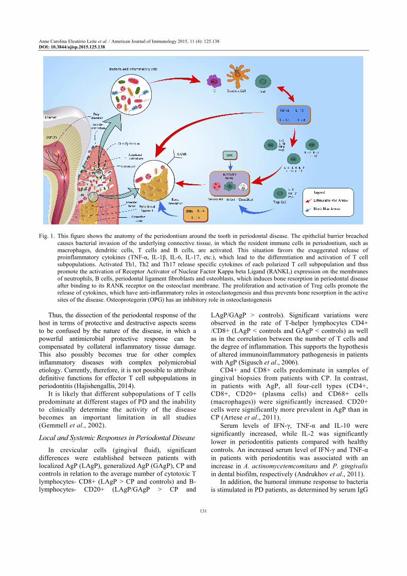
Fig. 1. This figure shows the anatomy of the periodontium around the tooth in periodontal disease. The epithelial barrier breached
causes bacterial invasion of the underlying connective tissue, in which the resident immune cells in periodontium, such as
macrophages, dendritic cells, T cells and B cells, are activated. This situation favors the exaggerated release of
proinflammatory cytokines (TNF-α, IL-1β, IL-6, IL-17, etc.), which lead to the differentiation and activation of T cell
subpopulations. Activated Th1, Th2 and Th17 release specific cytokines of each polarized T cell subpopulation and thus
promote the activation of Receptor Activator of Nuclear Factor Kappa beta Ligand (RANKL) expression on the membranes
of neutrophils, B cells, periodontal ligament fibroblasts and osteoblasts, which induces bone resorption in periodontal disease
after binding to its RANK receptor on the osteoclast membrane. The proliferation and activation of Treg cells promote the
release of cytokines, which have anti-inflammatory roles in osteoclastogenesis and thus prevents bone resorption in the active
sites of the disease. Osteoprotegerin (OPG) has an inhibitory role in osteoclastogenesis
Thus, the dissection of the periodontal response of the
host in terms of protective and destructive aspects seems
to be confused by the nature of the disease, in which a
powerful antimicrobial protective response can be
compensated by collateral inflammatory tissue damage.
This also possibly becomes true for other complex
inflammatory diseases with complex polymicrobial
etiology. Currently, therefore, it is not possible to attribute
definitive functions for effector T cell subpopulations in
periodontitis (Hajishengallis, 2014).
It is likely that different subpopulations of T cells
predominate at different stages of PD and the inability
to clinically determine the activity of the disease
becomes an important limitation in all studies
(Gemmell et al., 2002).
Local and Systemic Responses in Periodontal Disease
In crevicular cells (gingival fluid), significant
differences were established between patients with
localized AgP (LAgP), generalized AgP (GAgP), CP and
controls in relation to the average number of cytotoxic T
lymphocytes- CD8+ (LAgP > CP and controls) and B-
lymphocytes- CD20+ (LAgP/GAgP > CP and
LAgP/GAgP > controls). Significant variations were
observed in the rate of T-helper lymphocytes CD4+
/CD8+ (LAgP < controls and GAgP < controls) as well
as in the correlation between the number of T cells and
the degree of inflammation. This supports the hypothesis
of altered immunoinflammatory pathogenesis in patients
with AgP (Sigusch et al., 2006).
CD4+ and CD8+ cells predominate in samples of
gingival biopsies from patients with CP. In contrast,
in patients with AgP, all four-cell types (CD4+,
CD8+, CD20+ (plasma cells) and CD68+ cells
(macrophages)) were significantly increased. CD20+
cells were significantly more prevalent in AgP than in
CP (Artese et al., 2011).
Serum levels of IFN-γ, TNF-α and IL-10 were
significantly increased, while IL-2 was significantly
lower in periodontitis patients compared with healthy
controls. An increased serum level of IFN-γ and TNF-α
in patients with periodontitis was associated with an
increase in A. actinomycetemcomitans and P. gingivalis
in dental biofilm, respectively (Andrukhov et al., 2011). In addition, the humoral immune response to bacteria
is stimulated in PD patients, as determined by serum IgG

Anne Carolina Eleutério Leite et al. / American Journal of Immunology 2015, 11 (4): 125.138
DOI: 10.3844/ajisp.2015.125.138
132
antibodies to specific bacteria and/or their antigens
(Ebersole et al., 1987).
Although some studies have shown that severe forms
of PD have similar systemic inflammatory profiles
(Cairo et al., 2010; D’Aiuto et al., 2010; Picolos et al.,
2005; Lima et al., 2011) found different cell sources of
immunoregulatory cytokines in patients with AgP
compared to patients with CP, in which the frequency of
IL-10 in cells that express CD14+ was higher in CP but
not in AgP in comparison to healthy controls. The
amount of CD4+ T cells involved in IL-4 production was
higher in CP than in healthy controls.
Evidence that periodontitis constitutes a disease in
which an infectious and inflammatory factor is capable
of increasing the synthesis of inflammatory mediators is
shown by the relevant contribution that studies involving
periodontal therapy have provided. Nonsurgical
periodontal therapy reduced the levels of IL-17 and IL-
21 and increased the levels of IL-4 in gingival fluid;
however, there were no significant differences in the
levels of IFN-γ. Furthermore, the amount of Th17 cells
in peripheral blood was reduced, especially the IL-17+
and IFN-γ+ subpopulations. These results suggested that
Th17 cells play a destructive role in the immune balance
in periodontitis, the effect of Th1 cells was not
significant, while Th2 cells had a protective effect
(Zhao et al., 2011). In addition, it is known that
individuals with better response to periodontal therapy
decreased their inflammatory risk category (OR 4.8, 95%
CI 1.4 to 15.8) after correction for age, gender, ethnicity
and tobacco use (D’Aiuto et al., 2004a).
Note that the effectiveness clearly established by
periodontal therapy in the resolution of inflammation
and healing of periodontal tissues involves changing in
the microbiota, with recolonization by commensal
bacteria and restoration of homeostasis. This yields,
among other benefits, reduction in production of
inflammatory mediators and consequently a decrease or
absence of their systemic effects.
Discussion
In individuals susceptible to periodontitis, absence
of resolution of periodontal inflammation results in
chronic inflammation, which may have a systemic
impact (D’Aiuto et al., 2013; Dietrich et al., 2013; Ide and
Papapanou, 2013; Linden and Herzberg, 2013;
Linden et al., 2013; Taylor et al., 2013). The acute
inflammatory response is protective, but failure to
remove the inflammatory cells, especially neutrophils,
promotes chronic, pathological and destructive lesions.
Therefore, it is evident that the three aspects of the
pathogenesis of periodontitis (infection, inflammation
and adaptive immunity) have a potential role and impact
on the systemic immunoinflammatory response, which
either initiates or mediates a wide range of systemic
diseases (Linden et al., 2013).
In a previous study by our group, we found levels
of high-sensitivity C-reactive protein (hs-CRP) >0.3
mg/dL in individuals with severe periodontitis
compared to controls (60.87 versus 23.08,
respectively; p = 0.0216) (Leite et al., 2014) and
periodontal therapy was associated with a decrease in
hs-CRP levels circulating in serum and an increase in
High Density Lipoprotein (HDL).
Moreover, WBC count is characterized as a crude
marker of systemic inflammation and correlates to the
host response with respect to a variety of stimuli. This
marker has also been associated with a significant
prediction of future cardiovascular events and glucose
intolerance in different populations (D’Aiuto et al.,
2006; Graziani et al., 2010).
Earlier studies (do Vale et al., 2004; Figueira et al.,
2009) showed that peripheral blood mononuclear cells
from patients with periodontitis did not proliferate in
response to bacterial antigens. However, other studies
(Pejcic et al., 2011; Gaddale et al., 2014) have confirmed
an increased number of WBCs and neutrophils in the
peripheral blood of patients with moderate to severe
periodontitis, with a positive correlation between disease
severity and number of WBCs in the blood.
Additionally, rigorous therapeutic protocols and
periodontal maintenance contributed to the restoration of
phagocytic function in peripheral blood neutrophils
(Carneiro et al., 2012) and a reduction in hs-CRP levels
in patients with severe periodontitis (Leite et al.,
2014). These findings also support the conclusions of
D’Aiuto et al. (2004b; 2004c) and Kamil et al. (2011)
when reporting a greater reduction in the levels of
systemic inflammatory markers among those with better
clinical responses to periodontal therapy.
Conversely, serum levels of various cytokines in
patients with periodontitis were investigated in several
previous studies and the results were often controversial
(Duarte et al., 2010; Andrukhov et al., 2011; Zhao et al.,
2011). The contradictions in the existing data are
probably caused by the heterogeneity among the various
individuals, differences in susceptibility to periodontitis,
differences in the oral microbiota and low levels of
cytokines in the serum (Andrukhov et al., 2011).
Since Th1, Th2 and Th17 are mutually inhibitory, it
can be argued that the simultaneous action of T cells in
the development of periodontitis is unlikely and that
these subpopulations of T cells can lead to disease
progression independently. However, more studies are
necessary for the accurate determination of crosstalk
between T helper cytokines in periodontitis and its
impact on the evolution of the disease (Garlet, 2010). Different combinations of adhesion molecules and
their ligands, or the expression of different cytokines and

Anne Carolina Eleutério Leite et al. / American Journal of Immunology 2015, 11 (4): 125.138
DOI: 10.3844/ajisp.2015.125.138
133
chemokines in the microenvironment, contribute for
differences in the recirculation or selective recruitment
of lymphocyte subsets to sites of inflammation, which
in turn may influence the outcome of disease
(Seymour et al., 1993; Lima et al., 2011).
The unique feature of the biofilm in the oral cavity,
particularly the subgingival biofilm, is its close
proximity to highly vascularized tissues. Any disruption
of natural integrity of the subgingival epithelium, whose
thickness is at most 10 layers, can lead to bacteremia
(Parahitiyawa et al., 2009). Furthermore, in
periodontitis, the periodontal pocket epithelium is
typically thin and ulcerated and, therefore, often opens,
allowing access of pathogens to the connective tissue
and blood vessels. In patients with moderate to severe
periodontitis, the total area of the pocket epithelium in
direct contact with the subgingival biofilm is
surprisingly large, reaching to about the size of the palm
of the human hand or much larger in advanced cases
(Page, 1998). Therefore, both the access of
microorganisms to the bloodstream and the onset of
chronic inflammation with area and intensity sufficient
to elicit a significant host response provide the basis for
the study of the inter-relationship between periodontitis
and CVD and obesity, among others.
Thus, nonsurgical periodontal therapy acts to reduce
inflammation markers in the blood CRP, IL-6, etc.) and
reveals some important aspects of the systemic reach of
periodontal inflammation (Fitzsimmons et al., 2010;
Zhao et al., 2011). However, it is emphasized that the
mechanical removal of the dental biofilm by scaling and
root planning, with or without another supporting
modality, does not mean that, at any magnitude,
inflammation/infection does not persist. Sometimes the
removal of local factors is only achieved after repeated
specific actuations principally at sites in which there
still exist clinical signs of inflammation. Sites with
residual periodontal pockets that continue bleeding on
probing undoubtedly require new instrumentation
(scaling and root planning) and reinforcement of oral
hygiene instruction. Moreover, the need for
reinstrumentation and clinical response are unique to
each individual patient and for this reason, there is no
way to generically establish the completion of therapy
for all patients at the same time.
New treatments for periodontitis should address the
important contribution of immune cells in bone
resorption, particularly with regard to the natural course
of the disease (remission and progression periods)
(Hajishengallis, 2014). Current treatments for
periodontitis, despite of obtaining excellent local and
systemic clinical results, depend on mechanical
procedures (scaling and root planning), focusing mainly
in the dental biofilm that should be destroyed and
eliminated whenever possible, however it neglects, in the
most part, the immunological cells. However, based on
the current understanding of immune response, it is
essential to re-evaluate the forms of treatment currently
available, adding possible new therapeutic strategies to
control alveolar bone loss caused by the cell-mediated
immune response. As a possible target, inhibit the
production and/or expression of the RANKL (by acting
in transcription and/or translation) by activated immune
cells and/or block the RANKL-RANK physiological
interaction may be suggested. Although animal models
have shown promising results for several therapeutic
approaches, problems of side effects also arise.
It is also known that the absence of Tregs is an
indication of a great variety of disorders, such as
autoimmunity, dermatitis, periodontitis and even
transplant rejection. A potential treatment option for
these disorders revolves around increasing the number
of Tregs in local sites (Jhunjhunwala et al., 2012;
Gonzales et al., 2013). Current methods for in vivo
expansion of Tregs depend on biological therapies,
which are not specific for Tregs and are still associated
with many adverse side effects. Synthetic formulations
capable of inducing Tregs could be an alternative
strategy to achieve an in situ increase in the number of
Tregs. The in vitro tests of a synthetic formulation of
Treg inducers that consist of controlled release vehicles
of IL-2, TGF-β and rapamycin were already reported,
using cytokine and/or drug combination. IL-2, TGF-β
and rapamycin are released over three to four weeks
from these formulations. Furthermore, Tregs induced by
these formulations expressed the established markers for
Tregs (phenotype) and suppressed the proliferation and
function of naïve T cells to levels similar to that of the
natural soluble factor of Tregs, as well as naturally
occurring Tregs lymphocytes. These formulations were
also able to induce transcription factors FoxP3+ Tregs in
human cells in vitro. The authors concluded that the
combination of these microparticle formulations have the
potential to be used for the in vivo induction of Tregs at
local sites of rejected transplants or autoimmunity
(Jhunjhunwala et al., 2012).
Periodontitis is characterized by polymicrobial infection, so that, pathogens can modulate the T cell response to promote its own adaptability and the immune response itself becomes a mixture of immunological responses mediated by all the microorganisms represented in the biofilm (Garlet, 2010; Hajishengallis, 2014). Therefore, it may not be possible to reliably dissect patterns of dominant activities of Th1, Th2, Th17 or Tregs among the diseased periodontal tissue samples collected. Thus, it may be simpler and more productive to consider the roles played by each cytokine (Th1, Th2, Th17 or Tregs) in periodontal infection, since the performance of T cells in the maintenance of homeostasis between the biofilm microorganisms and host is remarkable.

Anne Carolina Eleutério Leite et al. / American Journal of Immunology 2015, 11 (4): 125.138
DOI: 10.3844/ajisp.2015.125.138
134
It is unclear which specific signaling pathways need
to be blocked or enhanced to mitigate the disease in
order to promote host defense. Moreover, the modulation
of the host immune system by drugs may result in
adverse side effects, requiring close monitoring of
patients for trying this approach (Jhunjhunwala et al.,
2012). Therefore, additional studies are needed to
understand the onset and progression of periodontitis and
to develop therapeutic interventions able to treat and
control the disease.
Conclusion
PD leads to an array of events involving the innate
and adaptive immunity in affected host, which in turn
brings the need for a growing understanding of the
immunopathogenesis of periodontitis in order to improve
the treatment and control of the disease.
New therapeutic approaches that include the current
understanding of the genetic factors and the host (genome,
proteomic analysis, transcription factors, cytokines,
immune cells) as well as the complex microbial
community (metagenomic, metatranscriptomic) should be
tested in animal models and epidemiological studies and
extended to clinical trials for the control of the overall
risk of periodontitis and associated systemic diseases.
Acknowledgement
The authors gratefully acknowledge Mr. Derrick
Mulder for reviewing the English language of the
manuscript. Maria Imaculada Muniz-Junqueira is an
investigator supported by the Conselho Nacional de
Desenvolvimento Científico e Tecnológico (CNPq),
Brazil (process number 308985/2013-3).
Author’s Contributions
All the authors contributed to the writing and revision
of the manuscript.
Ethics
This article is original and contains unpublished
material. The corresponding author confirms that all of
the other authors have read and approved the manuscript
and no ethical issues involved.
References
AAP, 1999. Consensus report: Chronic periodontitis. 1999
Ann. Periodontol., 4: 38-38.
DOI: 10.1902/annals.1999.4.1.38
Aas, J.A., B.J. Paster, L.N. Stokes, I. Olsen and F.E.
Dewhirst, 2005. Defining the normal bacterial flora of
the oral cavity. J. Clin. Periodontol., 43: 57215732.
DOI: 10.1128/JCM.43.11.5721-5732.2005
Abu-Amer, Y., F.P. Ross, J. Edwards and S.L. Teitelbaum, 1997. Lipopolysaccharide-stimulated osteoclastogenesis is mediated by tumor necrosis factor via its P55 receptor. J. Clin. Investigat., 100: 1557-1565. DOI: 10.1172/JCI119679
Abu-Amer, Y., 2013. NF-κB signaling and bone
resorption. Osteoporosis Int., 24: 2377-2386.
DOI: 10.1007/s00198-013-2313-x
Amunulla, A., R. Venkatesan, H. Ramakrishnan, K.V.
Arun and S. Sudarsan et al., 2008. Lymphocyte
subpopulation in healthy and diseased gingival
tissue. J. Ind. Society Periodontol., 12: 45-50.
DOI: 10.4103/0972-124X.44091
Andrukhov, O., C. Ulm, H. Reischl, P.Q. Nguyen and
M. Matejka et al., 2011. Serum cytokine levels in
periodontitis patients in relation to the bacterial load.
J. Periodontol., 82: 885-892.
DOI: 10.1902/jop.2010.100425
Aranha, A.M.F., C.E. Repeke, T.P. Garlet, A.E. Vieira
and A.P. Campanelli et al., 2013. Evidence
supporting a protective role for Th9 and Th22
cytokines in human and experimental periapical
lesions. J. Endodont., 39: 83-87.
DOI: 10.1016/j.joen.2012.10.015
Armitage, G.C., 1999. Development of a classification
system for periodontal diseases and conditions.
Annals Periodontol., 4: 1-6.
DOI: 10.1902/annals.1999.4.1.1
Armitage, G.C. and M.P. Cullinan, 2010. Comparison of
the clinical features of chronic and aggressive
periodontitis. Periodontology, 53: 12-27.
DOI: 10.1111/j.1600-0757.2010.00353.x
Artese, L., M.J. Simon, A. Piattelli, D.S. Ferrari and
L.A.G. Cardoso et al., 2011. Immunohistochemical
analysis of inflammatory infiltrate in aggressive and
chronic periodontitis: A comparative study. Clin.
Oral Investigat., 15: 233-240.
DOI: 10.1007/s00784-009-0374-1
Berglundh, T., M. Donati and N. Zitzmann, 2007. B cells
in periodontitis-friends or enemies? Periodontology,
45: 51-66. DOI: 10.1111/j.1600-0757.2007.00223.x
Cairo, F., M. Nieri, A.M. Gori, P. Tonelli and R. Branchi
et al., 2010. Markers of systemic inflammation in
periodontal patients: Chonic versus aggressive
periodontitis. An explorative crosss-sectional study.
Eur. J. Oral Implantol., 3: 147-153. PMID: 20623039
Carneiro, V.M., A.C. Bezerra, M. Guimarães and M.I.
Muniz-Junqueira, 2012. Effects of periodontal
therapy on phagocytic activity of peripheral blood
neutrophils-evidence for an extrinsic cellular defect.
Oral Health Preventive Dentistry, 10: 195-203.
DOI: 10.3290/j.ohpd.a28008 Chen, B., W. Wu, W. Sun, Q. Zhang and F. Yan et al.,
2014. RANKL expression in periodontal disease: Where does RANKL come from? BioMed Res. Int., 2014: 731039-731039. DOI: 10.1155/2014/731039

Anne Carolina Eleutério Leite et al. / American Journal of Immunology 2015, 11 (4): 125.138
DOI: 10.3844/ajisp.2015.125.138
135
D’Aiuto, F., L. Nibali, V. Mohamed-Ali, P. Vallance and
M.S. Tonetti, 2004a. Periodontal therapy: A novel
non-drug-induced experimental model to study
human inflammation. J. Periodontal Res., 39:
294-299. DOI: 10.1111/j.1600-0765.2004.00741.x
D’Aiuto, F., M. Parkar, G. Andreaou, P.M. Brett and D.
Ready et al., 2004b. Periodontitis and atherogenesis:
Causal association or simple coincidence? A pilot
intervention study. J. Clin. Periodontol., 31: 402-411.
DOI: 10.1111/j.1600-051X.2004.00580.x
D’Aiuto, F., M. Parkar, G. Andreou, J. Suvan and
P.M. Brett et al., 2004c. Periodontitis and systemic
inflammation: Control of the local infection is
associated with a reduction in serum inflammatory
markers. J. Dental Res., 83: 156-160.
DOI: 10.1177/154405910408300214
D’Aiuto, F., M. Parkar, P.M. Brett, D. Ready and M.S.
Tonetti, 2004d. Gene polymorphisms in pro-
inflammatory cytokines are associated with systemic
inflammation in patients with severe periodontal
infections. Cytokine, 28: 29-34.
DOI: 10.1016/j.cyto.2004.06.005
D’Aiuto, F., M. Parkar, L. Nibali, J. Suvan and J.
Lessem et al., 2006. Periodontal infections cause
changes in traditional and novel cardiovascular risk
factors: Results from a randomized controlled
clinical trial. Am. Heart J., 151: 977-984.
DOI: 10.1016/j.ahj.2005.06.018
D’Aiuto, F., L. Nibali, M. Parkar, K. Patel and
J. Suvan et al., 2010. Oxidative stress, systemic
inflammation and severe periodontitis. J. Dental
Res., 89: 1241-1246.
DOI: 10.1177/0022034510375830
D’Aiuto, F., M. Orlandi and J.C. Gunsolley, 2013.
Evidence that periodontal treatment improves
biomarkers and CVD outcomes. J. Clin.
Periodontol., 40: S85-S105.
DOI: 10.1902/jop.2013.134007
de Brito Bezerra, B.B., O. Andriankaja, J. Kang, S.
Pacios and H.J. Bae et al., 2012. A.
actinomycetemcomitans induced periodontal disease
promotes systemic and local responses in rat
periodontium. J. Clin. Periodontol., 39: 333-341.
DOI: 10.1111/j.1600-051X.2011.01847.x
da Luz, F.A., A.P. Oliveira, D. Borges, P.C. Brígido and
M.J. Silva, 2014. The physiopathological role of IL-
33: New highlights in bone biology and a proposed
role in periodontal disease. Mediators Inflammat.,
2014: 342410-342410. DOI: 10.1155/2014/342410
Dietrich, T., P. Sharma, C. Walter, P. Weston and J.
Beck, 2013. The epidemiological evidence behind
the association between periodontitis and incident
atherosclerotic cardiovascular disease. J. Clin.
Periodontol., 40: S70-S84. DOI: 10.1111/jcpe.12062
Divaris, K., K.L. Monda, K.E. North, A.F. Olshan and L.M. Reynolds et al., 2013. Exploring the genetic basis of chronic periodontitis: A genome-wide association study. Human Molecular Genet., 22: 2312-2324. DOI: 10.1093/hmg/ddt065
do Vale, C.H., L.A. de Oliveira Fraga, A.S. Costa, C.A. Tavares and O.A. Martins-Filho et al., 2004. Antiproliferative activity of Actinobacillus (Haemophilus) actinomycetemcomitans and Fusobacterium nucleatum in peripheral blood mononuclear cells. Res. Microbiol., 155: 731-740. DOI: 10.1016/j.resmic.2004.05.008
Duarte, P.M., M. da Rocha, E. Sampaio, M.J. Mestnik and M. Feres et al., 2010. Serum levels of cytokines in subjects with generalized chronic and agressive periodontitis before and after non-surgical periodontal therapy: A pilot study. J. Periodontol., 81: 1056-1063. DOI: 10.1902/jop.2010.090732
Dye, B.A., K. Choudhary, S. Shea and P.N. Papapanou, 2005. Serum antibodies to periodontal pathogens and markers of systemic inflammation. J. Clin. Periodontol., 32: 1189-1199.
DOI: 10.1111/j.1600-051X.2005.00856.x Ebersole, J.L., M.A. Taubman, D.J. Smith, D.E. Frey
and A.D. Haffajee et al., 1987. Human serum antibody responses to oral microorganisms. IV. Correlation with homologous infection. Oral Microbiol. Immunol., 2: 53-59.
DOI: 10.1111/j.1399-302X.1987.tb00290.x Figueira, E.A., M.L.R. de Rezende, S.A.Torres, G.P. Garlet
and V.S. Lara et al., 2009. Inhibitory signals mediated by programmed death-1 are involved with T-cell function in chronic periodontitis. J. Periodontol., 80: 1833-1844. DOI: 10.1902/jop.2009.090057
Fitzsimmons, T.R., A.E. Sanders, P.M. Bartold and G.D. Slade, 2010. Local and systemic biomarkers in gingival crevicular fluid increase odds of periodontitis. J. Clin. Periodontol., 37: 30-36.
DOI: 10.1111/j.1600-051X.2009.01506.x Gaddale, R., J.A. Mudda, I. Karthikeyan, S.R. Desai and H.
Shinde et al., 2014. Changes in cellular and molecular components of peripheral blood in patients with generalized aggressive periodontitis. J. Investigative Clin. Dentistry. DOI: 10.1111/jicd.12127
Gaffen, S.L. and G. Hajishengallis, 2008. A new inflammatory cytokine on the block: Re-thinking periodontal disease and the Th1/Th2 paradigm in the context of Th17 cells and Il-17. J. Dental Res., 87: 817-828. DOI: 10.1177/154405910808700908
Garlet, G.P., 2010. Destructive and protective roles of cytokines in periodontitis: A re-appraisal from host defense and tissue destruction viewpoints. J. Dental Res., 89: 1349-1363.
DOI: 10.1177/0022034510376402 Gemmell, E. and G.J. Seymour, 1998. Cytokine profiles
of cells extracted from humans with periodontal
diseases. J. Dental Res., 77: 16-26.
DOI: 10.1177/00220345980770010101

Anne Carolina Eleutério Leite et al. / American Journal of Immunology 2015, 11 (4): 125.138
DOI: 10.3844/ajisp.2015.125.138
136
Gemmell, E., C.L. Carter and G.J. Seymour, 2001.
Chemokines in human periodontal disease tissues.
Clin. Exp. Immunol., 125: 134-141.
DOI: 10.1046/j.1365-2249.2001.01511.x
Gemmell, E., K. Yamazaki and G.J. Seymour, 2002.
Destructive periodontitis lesions are determined by
the nature of the lymphocytic response. Critical Rev.
Oral Biol. Med., 13: 17-34.
DOI: 10.1177/154411130201300104
Glowacki, A.J., S. Yoshizawa, S. Jhunjhunwala, A.E.
Vieira and G.P. Garlet et al., 2013. Prevention of
inflammation-mediated bone loss in murine and
canine periodontal disease via recruitment of
regulatory lymphocytes. Proc. Nat. Acad. Sci. USA.,
110: 18525-18530. DOI: 10.1073/pnas.1302829110
Gonzales, J.R., S. Groeger, A. Johansson and J. Meyle,
2013. T helper cells from aggressive periodontitis
patients produce higher levels of interleukin-1 beta
and interleukin-6 in interaction with Porphyromonas
gingivalis. Clin. Oral Investig., 18: 1835-43.
DOI: 10.1007/s00784-013-1162-5
Gonzalez, O.A., M.J. Novak, S. Kirakodu, L. Orraca and
K.C. Chen et al., 2014. Comparative analysis of
gingival tissue antigen presentation pathways in
ageing and periodontitis. J. Clin. Periodontol., 41:
327-339. DOI: 10.1111/jcpe.12212
Graziani, F., S. Cei, M. Tonetti, M. Paolantonio and R.
Serio et al., 2010. Systemic inflammation following
non-surgical and surgical periodontal therapy. J.
Clin. Periodontol., 37: 848-854.
DOI: 10.1111/j.1600-051X.2010.01585.x
Guerrini, M.M. and H. Takayanagi, 2014. The immune
system, bone and RANKL. Archives Biochem.
Biophys., 561: 118-123.
DOI: 10.1016/j.abb.2014.06.003
Hajishengallis, G., 2014. Immunomicrobial pathogenesis
of periodontitis: Keystones, pathobionts and host
response. Trends Immunol., 35: 3-11.
DOI: 10.1016/j.it.2013.09.001
Hajishengallis, G., 2015. Periodontitis: From microbial
immune subversion to systemic inflammation.
Nature Rev. Immunol., 15: 30-44.
DOI: 10.1038/nri3785.
Hanada, R., T. Hanada, V. Sigl, D. Schramek and J.M.
Penninger, 2011. RANKL/RANK-beyond bones. J.
Molecular Med., 89: 647-656.
DOI: 10.1007/s00109-011-0749-z
Hans, M. and V.M. Hans, 2011. Toll-like receptors and
their dual role in periodontitis: A review. J. Oral
Sci., 53: 263-271. DOI: 10.2334/josnusd.53.263
Hernández, M., N. Dutzan, J. García-Sesnich, L.
Abusleme and A. Dezerega et al., 2011. Host-
pathogen interactions in progressive chronic
periodontitis. J. Dental Res., 90: 1164-1170.
DOI: 10.1177/0022034511401405
Hienz, S.A., S. Paliwal and S. Ivanovski, 2015.
Mechanisms of bone resorption in periodontitis. J.
Immunol. Res., 2015: 615486-615495.
DOI: 10.1155/2015/615486
Houri-Haddad, Y., A. Wilensky and L. Shapira, 2007. T-
cell phenotype as a risk factor for periodontal
disease. Periodontology, 45: 67-75.
DOI: 10.1111/j.1600-0757.2007.00227.x
Ide, M. and P.N. Papapanou, 2013. Epidemiology of
association between maternal periodontal disease
and adverse pregnancy outcomes-systematic review.
J. Clin. Periodontol., 40: S181-S194.
DOI: 10.1902/jop.2013.134009
Jhunjhunwala, S., S.C. Balmert, G. Raimondi, E. Dons
and E.E. Nichols et al., 2012. Controlled release
formulations of IL-2, TGF-β1 and rapamycin for the
induction of regulatory T cells. J. Controlled Release,
159: 78-84. DOI: 10.1016/j.jconrel.2012.01.013
Jin, Q., J.A. Cirelli, C.H. Park, J.V. Sugai and M. Taba
Jr. et al., 2007. RANKL inhibition through
osteoprotegerin blocks bone loss in experimental
periodontitis. J. Periodontol., 78: 1300-1308.
DOI: 10.1902/jop.2007.070073
Kamil, W., R. Al Habashneh, Y. Khader, L. Al Bayati
and D. Taani, 2011. Effects of nonsurgical
periodontal therapy on C-reactive protein and serum
lipids in Jordanian adults with advanced
periodontitis. J. Periodontal Res., 46: 616-621. DOI:
10.1111/j.1600-0765.2011.01380.x
Kawai, T., T. Matsuyama, Y. Hosokawa, S. Makihira
and M. Seki et al., 2006. B and T lymphocytes are
the primary sources of RANKL in the bone
resorptive lesion of periodontal disease. Am. J.
Pathol., 169: 987-998.
DOI: 10.2353/ajpath.2006.060180 Kobayashi, R., T. Kono, B.A. Bolerjack, Y. Fukuyama
and R.S. Gilbert et al., 2011. Induction of IL-10-producing CD4+ T-cells in chronic periodontitis. J. Dental Res., 90: 653-658.
DOI: 10.1177/0022034510397838
Konermann, A., M. Beyer, J. Deschner, J.P. Allam and N.
Novak et al., 2012. Human periodontal ligament cells
facilitate leukocyte recruitment and are influenced in
their immunomodulatory function by Th17 cytokine
release. Cellular Immunol., 272: 137-143.
DOI: 10.1016/j.cellimm.2011.10.020 Leite, A.C.E., V.M.A. Carneiro and M.C.M. Guimarães,
2014. Effects of periodontal therapy on C-reactive protein and HDL in serum of subjects with periodontitis. Revista Brasileira de Cirurgia Cardiovascular, 29: 69-77.
DOI: 10.5935/1678-9741.20140013
Pejcic, A., L. Kesic, Z. Pesic D. Mirkovic and M.
Stojanovic, 2011. White blood cell count in different
stages of chronic periodontitis. Acta Clinica Croata,
50: 159-167. DOI: 10.5935/1678-9741.20140013

Anne Carolina Eleutério Leite et al. / American Journal of Immunology 2015, 11 (4): 125.138
DOI: 10.3844/ajisp.2015.125.138
137
Lima, P.M.A., P.E.A. Souza, J.E.Costa, R.S. Gomez and K.J. Gollob et al., 2011. Aggressive and chronic periodontitis correlate with distinct cellular sources of key immunoregulatory cytokines. J. Periodontol., 82: 86-95. DOI: 10.1902/jop.2010.100248
Linden, G.J. and M.C. Herzberg, 2013. Periodontitis and systemic diseases: A record of discussions of working group 4 of the Joint EFP/AAP Workshop on Periodontitis and Systemic Diseases. J. Clin. Periodontol., 40: S20-S23. DOI: 10.1111/jcpe.12091
Linden, G.J., A. Lyons and F.A. Scannapieco, 2013. Periodontal systemic associations: Review of the evidence. J. Clin. Periodontol., 40: S8-S19.
DOI: 10.1111/jcpe.12064 Loo, W.T.Y., Y. Yue, C.B. Fan, L.J. Bai and Y.D.
Dou et al., 2012. Comparing serum levels of cardiac biomarkers in cancer patients receiving chemotherapy and subjects with chronic periodontitis. J. Translat. Med., 10: 1-7.
DOI: 10.1186/1479-5876-10-S1-S5 Maekawa, T., K. Tabeta, K. Kajita-Okui, T. Nakajima
and K. Yamazaki, 2011. Increased expression of C-reactive protein gene in inflamed gingival tissues could be derived from endothelial cells stimulated with interleukin-6. Archives Oral Biol., 56: 312-318. DOI: 10.1016/j.archoralbio.2011.04.010.
Mombelli, A., F. Casagni and P.N. Madianos, 2002. Can presence or absence of periodontal pathogens distinguish between subjects with chronic and aggressive periodontitis? A systematic review. J. Clin. Periodontol., 29: 10-21.
DOI: 10.1038/sj.ebd.6400204 Moutsopoulos, N.M., J. Konkel, M. Sarmadi, M.A.
Eskan and T. Wild et al., 2014. Defective neutrophil recruitment in leukocyte adhesion deficiency type I disease causes local IL-17-driven inflammatory bone loss. Sci. Translat. Med., 6: 229ra40-229ra40. DOI: 10.1126/scitranslmed.3007696.
Offenbacher, S., J.R. Elter, D. Lin and J.D. Beck, 2005. Evidence for periodontitis as a tertiary vascular infection. J. Int. Acad. Periodontol., 7: 39-48. PMID: 15912923
Ohlrich, E.J., M.P. Cullinan and G.J. Seymour, 2009. The immunopathogenesis of periodontal disease. Australian Dental J., 54: 2-10.
DOI: 10.1111/j.1834-7819.2009.01139.x Page, R.C., 1998. The pathobiology of periodontal
diseases may affect systemic diseases: Inversion of a paradigm. Ann. Periodontol., 3: 108-120.
DOI: 10.1902/annals.1998.3.1.108 Papapanou, P.N., 1999. Epidemiology of periodontal
disease: An update. J. Int. Acad. Periodontol., 1: 110-116. PMID: 12666955
Papapanou, P.N., A.M. Neiderud, E. Disick, E. Lalla and G.C. Miller et al., 2004. Longitudinal stability of
serum immunoglobulin G responses to periodontal bacteria. J. Clin. Periodontol., 31: 985-990.
DOI: 10.1111/j.1600-051X.2004.00599.x
Parahitiyawa, N.B., L.J. Jin, W.K. Leung, W.C. Yam and L.P. Samaranayake, 2009. Microbiology of odontogenic bacteremia: Beyond endocarditis. Clin. Microbiol. Rev., 22: 46-64.
DOI: 10.1128/CMR.00028-08 Picolos, D.K., J. Lerche-Sehm, A. Abron, F.B Fine and
P.N. Papapanou, 2005. Infection patterns in chronic and aggressive periodontitis. J. Clin. Periodontol., 32: 1055-1061.
DOI: 10.1111/j.1600-051X.2005.00828.x Sanz, M. and A.J.V. Winkelhoff, 2011. Periodontal
infections: Understanding the complexity-Consensus of the Seventh European Workshop on Periodontology. J. Clin. Periodontol., 38: 3-6.
DOI: 10.1111/j.1600-051X.2010.01681.x Seymour, G.J., E. Gemmell, R.A. Reinhardt, J. Eastcott
and M.A. Taubman, 1993. Immunopathogenesis of chronic inflammatory periodontal disease: Cellular and molecular mechanisms. J. Periodontal Res., 28: 478-486. DOI: 10.1111/j.1600-0765.1993.tb02108.x
Sigusch, B.W., A. Wutzler, T. Nietzsch and E. Glockmann, 2006. Evidence for a specific crevicular lymphocyte profile in aggressive periodontitis. J. Periodontal Res., 41: 391-396.
DOI: 10.1111/j.1600-0765.2006.00869.x Slade, G.D., S. Offenbacher, J.D. Beck, G. Heiss and
J.S. Pankow, 2000. Acute-phase inflammatory response to periodontal disease in the US population. J. Dental Res., 79: 49-57.
DOI: 10.1177/00220345000790010701 Srinivasan, P.C., 2013. The role of inflammatory
cytokines and the RANKL-RANK-OPG molecular triad in periodontal bone loss-A review. Clin. Cellular Immunol., S13: 007-007.
DOI: 10.4172/2155-9899.S13-007 Taylor, J.J., P.M. Preshaw and E. Lalla, 2013. A review
of the evidence for pathogenic mechanisms that may link periodontitis and diabetes. J. Clin. Periodontol., 40: S113-S134. DOI: 10.1111/jcpe.12059
Tuter, G., M. Serdar, B. Kurtis, S.G. Walker and A. Atak et al., 2010. Effects of scaling and root planing and subantimicrobial dose doxycycline on gingival crevicular fluid levels of matrix metalloproteinase-8, -13 and serum levels of HsCRP in patients with chronic periodontitis. J. Periodontol., 81: 1132-1139.
DOI: 10.1902/jop.2010.090694 Vernal, R., J. Diaz-Zúñiga, S. Melgar-Rodríguez, M. Pujol,
E. Diaz-Guerra et al., 2014. Activation of RANKL-induced osteoclasts and memory T lymphocytes by Porphyromonas gigingivalis is serotype dependant. J. Clin. Periodontol., 41: 451-459.
DOI: 10.1111/jcpe.12236 Xu, J., H.F. Wu, E.S.M. Ang, K. Yip and
M. Woloszyn et al., 2009. NF-kB modulators in osteolytic bone diseases. Cytokine Growth Factor Rev., 20: 7-17. DOI: 10.1016/j.cytogfr.2008.11.007

Anne Carolina Eleutério Leite et al. / American Journal of Immunology 2015, 11 (4): 125.138
DOI: 10.3844/ajisp.2015.125.138
138
Wara-Aswapati, N., R. Surarit, A. Chayasadom, J.A.
Boch and W. Pitiphat, 2007. RANKL upregulation
associated with periodontitis and porphyromonas
gingivalis. J. Periodontol., 78: 1062-1069.
DOI: 10.1902/jop.2007.060398
Williams, R.C. and S. Offenbacher, 2000. Periodontal
medicine: The emergence of a new branch of
periodontology. Periodontology, 23: 9-12.
DOI: 10.1034/j.1600-0757.2000.2230101.x
Yokoyama, M., T. Ukai, E.R. Ayon Haro, T. Kishimoto
and Y. Yoshinaga et al., 2011. Membrane-bound
CD40 ligand on T cells from mice injected with
lipopolysaccharide-induced osteoclastogenesis. J.
Periodontal Res., 46: 464-474.
DOI: 10.1111/j.1600-0765.2011.01362.x
Zhao, L., Y. Zhou, Y. Xu, Y. Sun and L. Li et al., 2011.
Effect of non-surgical periodontal therapy on the
levels of Th17/Th1/Th2 cytokines and their
transcription factors in Chinese chronic periodontitis
patients. J. Clin. Periodontol., 38: 509-516.
DOI: 10.1111/j.1600-051X.2011.01712.x