review vision in autism
TRANSCRIPT

Vision Research 49 (2009) 2705–2739
Contents lists available at ScienceDirect
Vision Research
journal homepage: www.elsevier .com/locate /v isres
Review
Vision in autism spectrum disorders
David R. Simmons *, Ashley E. Robertson, Lawrie S. McKay, Erin Toal, Phil McAleer, Frank E. PollickDepartment of Psychology, University of Glasgow, 58 Hillhead Street, Glasgow G12 8QB, Scotland, UK
a r t i c l e i n f o a b s t r a c t
Article history:Received 11 March 2009Received in revised form 4 August 2009
Keywords:AutismAutism spectrum disordersClinical vision
0042-6989/$ - see front matter � 2009 Elsevier Ltd. Adoi:10.1016/j.visres.2009.08.005
* Corresponding author. Fax: +44 (0)141 330 4606.E-mail address: [email protected] (D.R. Simmon
1 Pervasive Developmental Disorder-Not Otherwisediagnostic category used if the severity of the signs andlevel required for full autism or Asperger syndrome.
Autism spectrum disorders (ASDs) are developmental disorders which are thought primarily to affectsocial functioning. However, there is now a growing body of evidence that unusual sensory processingis at least a concomitant and possibly the cause of many of the behavioural signs and symptoms ofASD. A comprehensive and critical review of the phenomenological, empirical, neuroscientific and theo-retical literature pertaining to visual processing in ASD is presented, along with a brief justification of anew theory which may help to explain some of the data, and link it with other current hypotheses aboutthe genetic and neural aetiologies of this enigmatic condition.
� 2009 Elsevier Ltd. All rights reserved.
1. Introduction
Autism is a developmental disorder characterized by difficultieswith social interaction, social communication and an unusually re-stricted range of behaviours and interests (Frith, 2003). A diagnosisof autism also requires a clinically significant delay in languagedevelopment before the age of 3 years. Asperger Syndrome hassimilar signs and symptoms to autism except without the languagedelay. Together (with PDD-NOS)1 these two diagnostic groups havecome to be known as Autism spectrum disorders (ASDs), althoughthere is ongoing controversy about how distinct they really are(see Volkmar, State, & Klin, 2009).
A second controversy concerns whether or not the prevalence ofASDs (about 1% in western countries) is increasing (Charles, Car-penter, Jenner, & Nicholas, 2008). The prevalence controversy iscomplicated by two factors: the historical changes in diagnosticcriteria (see below) and the continuing debate over the causes ofASDs. Concerning the latter, those who favour a genetic cause pointto the strong evidence for a genetic component in the aetiology ofASD: chiefly the male/female differences in prevalence (about afactor of 4 higher in males) and the data indicating that ASD ismore prevalent in monozygotic than dyzygotic twins (Baileyet al., 1995; Baron-Cohen, 2003; Bourgeron, 2008). If the cause isgenetic (rather than epigenetic) then any increase in prevalencemust result from improved diagnosis. An environmental explana-tion centres on a genuine increase in the numbers of those whohave ASD arising from environmental stressors, such as pollution,
ll rights reserved.
s).Specified (PDD-NOS) is the
symptoms are just below the
diet or lifestyle (see Altevogt, Hanson, & Leshner, 2008; Rutter,2009; Thornton, 2006). It is likely that both genetic and environ-mental factors contribute to the aetiology of ASDs, although anumber of genetic disorders can result in similar symptoms (e.g.Fragile-X, Tuberous Sclerosis: see Gillberg & Coleman, 2000, formore).
2. The diagnosis of ASDs
The signs and symptoms of ASD amenable to diagnosis are al-most entirely behavioural, so a large variety of diagnostic instru-ments are based on either the direct or indirect observation ofindividual behaviour. These include the Autism Diagnostic Inter-view (ADI; Lord, Rutter, & Le Couteur, 1994), the Autism DiagnosticObservation Schedule (ADOS; Lord et al., 2000), the Developmen-tal, Dimensional and Diagnostic Interview (3Di; Skuse et al.,2004) and the Diagnostic Interview for Social and CommunicationDisorders (DISCO; Leekam, Libby, Wing, Gould, & Taylor, 2002).Variants of these tests are used depending on the age and/or lan-guage level of the individual concerned. Diagnostic instrumentsalso vary in the way that they gather data. Most instruments in-clude a history-taking procedure, particularly important for deter-mining early language development (e.g. ADI). However, othersrely largely on interviewing the individual concerned and/orobserving them directly (e.g. ADOS). It is generally thought that,for research purposes, a directly observed diagnosis is more reli-able because it does not utilize third-party information. However,the most reliable diagnoses probably involve a combination of ap-proaches (e.g. ADI together with ADOS). Within a clinical contextthe validity of research is frequently judged on the method andsource of diagnosis. Obtaining such reliable diagnoses requires atthe very least specialist training, and probably collaboration withclinical partners. In many studies that we will consider the ASD
2706 D.R. Simmons et al. / Vision Research 49 (2009) 2705–2739
population is characterized vaguely as ‘‘diagnosed by clinical spe-cialists” or may even rely solely on parental report of previousdiagnosis.2 Others will specify the use of standard diagnostic crite-ria, such as DSM-IV (American Psychiatric Association, 1994) orICD-10 (World Health Organisation, 1993). Sometimes furtherchecks will be made using screening instruments, either self-reportquestionnaires such as the Autism Spectrum Quotient (AQ; Baron-Cohen, Wheelwright, Skinner, Martin, & Clubley, 2001b), or third-party questionnaires, such as the Social Responsiveness Scale (SRS;Constantino et al., 2003). We shall try to assess the reliability of diag-noses used, whilst bearing in mind that important insights into vi-sual functioning in ASD may still be obtained even when the ‘‘goldstandard” of diagnosis is not (Dover & Le Couteur, 2007; McClure& Le Couteur, 2007). Note also that, for a study to be specific toASD, the known genetic syndromes which have similar symptomprofiles to ASD should be explicitly ruled out, as, ideally, shouldother forms of learning disability or neuropathology.
3. Other general issues with research into ASDs
The commonest design for an experiment in this area is tosource a population of individuals with ASD and compare theirperformance with a control group, which may be ‘‘typical” of thegeneral population, or be drawn from another clinical population,such as individuals with Attention Deficit Hyperactivity Disorder(ADHD) or Down’s syndrome. Normally these populations are‘‘matched”, using IQ (verbal (VIQ), non-verbal (NVIQ) or full-scale(FSIQ)), chronological age (CA) or mental age (MA), with the choicedepending upon the task concerned, such as whether or not itmakes high demands on language and whether performance mightbe more influenced by physical maturation or cognitive ability.These issues are typical of those encountered in clinical research,but there are some added difficulties when dealing with ASD. First,there is the question of which part of the autism spectrum to tar-get. Most studies use people with either ‘‘high-functioning” autism(HFA), or Asperger Syndrome (AS) who generally have average, oreven above-average, IQ and do not require an additional learn-ing-disabled control group for comparison. However, because theHFA/AS group will also have less profound symptoms, any perfor-mance differences between this group and controls will tend to besmaller. If a more severely affected autism group is chosen then alearning-disabled control group may be required, introducingproblems of how to convey instructions, maintain participantattention and record experimental responses. Furthermore, eventaking a population that is close in terms of IQ, age and positionon the autism spectrum does not guarantee homogeneity withinthe population. It is often suggested (only half jokingly) that theheterogeneity of performance in a given task is the only reliabledifference between data obtained from an ASD and a typical con-trol population! Finally, given that social reticence and mainte-nance of routine is one of the symptoms of ASD it can be hard torecruit volunteers. Thus, obtaining reliable and statistically signif-icant differences between ASD and control populations can be amajor challenge. Added to this is the care with which task designmust be undertaken. It is hard to determine whether poor perfor-mance on a task is due to an ASD-specific performance deficit, orwhether there is a more generalised issue, such as attention con-trol or a misunderstanding of the task instructions. Another poten-tial factor is the use of medication by the ASD group, particularly in
2 Given the significant changes in diagnostic criteria in the years since ASD was firstdescribed, very careful attention should be paid to the group characterization in anystudies which pre-date publication of DSM-IV in 1994. See Hincha-Ownby (2008) foran overview of these changes. Note also that these criteria will themselves shortly beupdated when DSM-V is published (see American Psychiatric Association web-site forthe latest developments: http://www.psych.org).
adults. The most convincing studies demonstrate differential per-formance on very similar tasks where these collateral demandsare the same (see Bertone & Faubert, 2006; Burack, Iarocci, Flana-gan, & Bowler, 2004; Jarrold & Brock, 2004).
A final important issue to consider is the fact that ASD is adevelopmental disorder, the signs and symptoms of which canchange dramatically over time in a given individual (Fecteau, Mot-tron, Berthiaume, & Burack, 2003). Hence, what is true for, say, 8–12 year old children on the autism spectrum may not apply toadults. This is a particular issue in ASD given that it is characterizedby what are sometimes called ‘‘asymmetric” developmentpatterns.
4. Sensory symptoms in ASDs
Given that ASDs are defined primarily in terms of social symp-toms it may seem odd that sensory processing in these conditionsis of interest at all. However, there is a large body of literaturewhich attests to the prevalence of sensory symptoms within ASDs.
Sensory symptoms were featured in the original descriptions ofASDs (Asperger, 1944; Kanner, 1943), and further explored byWing (1969) and Hermelin and O’Connor (1970). There are alsothe eloquent accounts written by people on the autism spectrum(e.g. Grandin, 1992, 2009; Grandin & Scariano, 1986; Jackson,2002; Williams, 1998) or by their parents/caregivers (e.g. Jackson,2003). Many of these accounts describe both hyper- and hypo-sen-sitivity to sensory stimuli – often fluctuating between the two ex-tremes (Jones, Quigney, & Huws, 2003).
Despite the obvious value of their insight, there are problemswith the interpretation of first-hand descriptions. Firstly, the re-ports tend to come from high-functioning individuals with ASD,so we do not know how well they apply to the autism spectrumas a whole. Secondly, an account may have been written in con-junction with someone who does not have ASD (e.g. Grandin &Scariano, 1986), risking the intrusion of interpreter bias. Lastly,O’Neill and Jones (1997) suggest that, for individuals with ASD,there can be confusion over ‘real’ versus ‘echoed’ memories and alack of insight into typical perceptual experiences. A helpful sum-mary of this literature is given by Bogdashina (2003).
A number of studies have collated this information by evaluat-ing parent/caregiver questionnaires (Baker, Lane, Angley, & Young,2008; Baranek, David, Poe, Stone, & Watson, 2006; Ben-Sassonet al., 2007; Robertson & Simmons, 2008; Rogers, Hepburn, & Weh-ner, 2003; Watling, Deitz, & White, 2001), sometimes combinedwith information from the individuals themselves (Kern et al.,2006, 2007; Leekam, Nieto, Libby, Wing, & Gould, 2007). These tellthe broadly similar story that (a) particular sensory symptoms ap-pear to be more common in ASD than in other developmental dis-orders; (b) these symptoms lessen with age; (c) these symptomsare correlated with the severity of social symptoms of ASD, at leastin children (Kern et al., 2007).
There are a number of problems with caregiver/parent reportdata. Methodological shortcomings (such as small sample sizesand variability across groups) have affected the degree to whichthese results can be generalized, reporters may attribute the child’sreaction to the wrong sensory stimulus and are open to bias (e.g.unwittingly over- or under-estimating the child’s sensory difficul-ties; see Nader, Oberlander, Chambers, & Craig, 2004). There arealso issues of recall bias, especially when recounting stressfulsituations.
Although it has been suggested that the importance of sensorysymptoms from the report literature has not been confirmed inlab-based studies (O’Neill & Jones, 1997; Rogers & Ozonoff,2005), we have recently found a strong correlation between sen-sory symptoms reported by members of the general public and

D.R. Simmons et al. / Vision Research 49 (2009) 2705–2739 2707
their score on the AQ which addresses some of the issues to dowith third-party report (Robertson & Simmons, 2009).
5. Visual symptoms in ASD
A non-exhaustive list of visual sensory symptoms is given byBogdashina (2003):
Hyper:
(1) focusing on tiny pieces of dust/particles,(2) dislike of the dark and bright lights,(3) dislike of sharp flashes of light,(4) looking down most of the time,(5) covering/closing eyes at bright lights.
Hypo:
(1) attracted to light,(2) looking intensely at objects or people,(3) moving fingers or objects in front of the eyes,(4) fascination with reflections and/or brightly coloured objects,(5) running hands around the edges of objects.
Another list is given in Leekam et al. (2007) and corresponds toquestions asked in the DISCO diagnostic interview (Leekam et al.,2002):
Quotes from individuals on the autism spectrum are also infor-mative, e.g.:
‘‘my bed was surrounded and totally encased by tiny spots which Icalled stars, like some kind of mystical glass coffin. I have sincelearned that they are actually air particles yet my vision was sohypersensitive that they often became a hypnotic foreground withthe rest of ’the world’ fading away.”
-Donna Williams, p. 15, ‘‘Nobody Nowhere” (Williams, 1998).There are also some commonly observed social symptoms
which may have a visual component such as unusual socially di-rected pointing, difficulties with interpretation of gestures, unu-sual eye contact, difficulty with the interpretation of facialexpressions, difficulty with following the gaze of others and diffi-culties with joint attention, as well as non-social signs like repeti-tive and stereotyped behaviours. Assessment of these symptoms isincluded in the ADOS (Lord et al., 2000).
6. Studies of visual processing in ASD
In this section of the review we shall deal with the full range ofpossible visual differences between ASD and typical controlpopulations.
6.1. Optometric issues
Although many studies report screening the vision of their par-ticipants, relatively few have focussed specifically on optometriccomparisons of ASD and typical populations. Scharre and Creedon(1992) reviewed previous findings and also evaluated 34 children(2–11 years) with autism for ocular alignment, refractive error, vi-sual acuity, oculomotility skills and stereopsis. Scharre and Cree-don (1992) noted a higher than average incidence of refractiveerrors (consistent with results from other developmental disabili-ties such as cerebral palsy), and also found that 21% of their chil-dren had strabismus, comparing this with figures of 3.7% fortypical children, 69% in cerebral palsy and 21.4% in developmentaldelay from other studies. They also reported that a ‘‘significantnumber” of the children had difficulty with voluntary pursuit eye
movements and had atypical OKN responses. They could not reporton the level of amblyopia in their sample because most of the chil-dren resisted ocular occlusion, although only 3 of the 34 failed theLang stereotest. Given that Sharre and Creedon (1992) pre-datesthe publication of DSM-IV and ICD-10 diagnostic criteria it is pos-sible that their data only apply to more severe cases of autism, asDSM-III criteria were much more restrictive (Hincha-Ownby,2008). A high prevalence of strabismus in ASD (50%) was also re-ported by Kaplan, Rimland, and Edelson (1999) from a sample ofolder children (7–19 years) along with a slightly lower prevalence(20%) taken from a questionnaire survey of 7640 parents. Neitherof these studies is definitive in terms of establishing optometricanomalies in ASD populations, since both relied on a previous clin-ical diagnosis by unspecified techniques, the numbers of partici-pants in the experimental studies were relatively small and therewere no control groups. The Kaplan et al. (1999) questionnairedata also suffer from the known problems with parental reports.Nevertheless, this finding would appear to be worthy of furtherinvestigation. It does seem surprising that the large number ofresearchers who have performed routine optometric screening oftheir participants have not noticed the high prevalence of strabis-mus in their ASD population (even if the conservative estimate of20% applies). Kaplan (2006) has recently embedded these optomet-ric data into his theory of visual dysfunction in autism.
6.2. Spatial vision
6.2.1. Visual acuityThe visual acuity of children and adults with ASD has been
routinely measured as part of the screening procedure in a num-ber of studies with nothing untoward being reported, but onevery recent study focused exclusively on visual acuity (Ashwin,Ashwin, Rhydderch, Howells, & Baron-Cohen, 2009). They mea-sured visual acuity in 15 individuals with ASD and 15 controlsusing the Freiburg Visual Acuity and Contrast Test (Bach, 1996)and reported a very high mean level of visual acuity in theirASD sample (20/7) compared to the 20/13 of the typical controlgroup. Unfortunately it appears that the spatial resolution ofthe display screen was not high enough to support acuity mea-surement at the viewing distance used, and some of the settingson the computerized test were inappropriate (Bach & Dakin, inpress). Consequently this apparently exciting result must be re-garded as unsound.
6.2.2. Static contrast sensitivityde Jonge, Kemner, de Haan, Coppens, and van den Berg (2007)
used the Vistech� contrast sensitivity charts to compare perfor-mance at a range of spatial frequencies (1.5–18 c/deg) between agroup of 29 people with ASD and age-matched controls. The clini-cal sample (7–33 years) was carefully diagnosed using multipleinstruments including the ADI-R and ADOS. De Jonge et al.(2007) found no significant differences between the two groups.Obviously the clinical sample was relatively small, and the testwas not as sensitive to subtle differences as a computer-based testwould be, but it does suggest that there are no large differences instatic contrast sensitivity between individuals with HFA andmatched controls. Behrmann et al. (2006a) and Milne, Scope, Pas-calis, Buckley, and Makeig (2009) have also reported no differencesin static contrast sensitivity for gratings of different spatial fre-quencies between ASD and matched control populations.
Bertone, Mottron, Jelenic, and Faubert (2005) compared con-trast thresholds for orientation identification between a group of13 HFA participants (11–31 years) and matched typical controls.Diagnosis was using ADI and ADOS. The stimuli were eitherluminance- (i.e. first-order) or contrast-modulated (i.e. second-order) grayscale noise. The sinusoidal modulations had a spatial

2708 D.R. Simmons et al. / Vision Research 49 (2009) 2705–2739
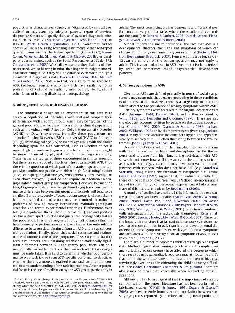
frequency of 0.75 c/deg in both cases and were presented for750 ms. The ASD participants were carefully guided through thetask, with the experimenter remaining in the room, remindingthe participants to fixate and initiating successive trials. This carewill have served to reduce any noise in the data due to attentionallapses. The result found was surprising in that the HFA group ob-tained significantly lower thresholds with the first-order stimulithan the matched controls, but significantly higher thresholds withthe second-order stimuli (see Fig. 1).
Sanchez-Marin and Padilla-Medina (2008) measured thedetectability of a static bright bar embedded in Gaussian noise insix participants with autism (7–17 years) and six controls. The chil-dren with autism were characterized using the Childhood AutismRating Scale (CARS; Schopler, Reichler, DeVellis, & Daly, 1980),although there was no attempt to match IQ with controls, whichis unfortunate as the children with autism had relatively severesymptoms. Sanchez-Marin and Padilla-Medina (2008) found thatchildren with autism performed significantly worse than controlsat a range of signal/noise ratios. They argued that these data couldreflect the influence of increased levels of internal noise in visualprocessing pathways in autism. Whilst this conclusion is veryinteresting (see Section 13 below), the experiment should haveused a more carefully controlled group of participants.
6.2.3. Dynamic contrast sensitivityBertone et al. (2005) also measured contrast thresholds for a
flickering grating stimulus using a temporal 2AFC paradigm. Theyused a conventional 0.5 c/deg grating counterphasing at 6 Hz(ostensibly to stimulate magnocellular pathways) and a 6 c/deggrating counterphasing at 1 Hz (ditto for parvocellular pathways).There were no significant differences in contrast sensitivity foreither of these stimuli between the HFA group and controls. A sim-ilar result was found by Pellicano, Gibson, Maybery, Durkin, andBadcock (2005) using similar procedures and carefully diagnosed
Fig. 1. Figure from Bertone et al. (2005) showing contrast thresholds for orientationidentification (horizontal/vertical) in noise for first-order and second-order gratingstimuli collected from children with high functioning autism (HFA) and typicallydeveloping controls (TD). The stimuli used are shown beneath the graph. Note thatthe units on the y-axis are contrast, not orientation. Reproduced with thepermission of Oxford University Press.
and matched participants, except that the stimulus was a Gaussianblob (3.15 deg sigma) flickering sinusoidally at 10 Hz.
Bertone, Mottron, Jelenic, and Faubert (2003) reported contrastsensitivities for drifting grating stimuli defined by either lumi-nance- (i.e. first-order) or contrast- (i.e. second-order) modulatedgrayscale noise. The gratings were either conventional verticalsinusoids, radially symmetric sinusoids or angled sinusoids in or-der to test translational, radial and rotational motion, respectively.For the translating and radial patterns the spatial frequency was1 c/deg with a temporal frequency of 2 Hz, giving a drift rate of2 deg/s. For the rotating grating the angular velocity was p/2 rad/s. The participants were a well diagnosed HFA sample (mean age13 years) and a typical control group (mean age 12 years). Bertoneet al. (2003) found no difference between contrast thresholds forfirst-order motion detection with these two groups, but did findsignificantly higher thresholds for second-order motion detectionwith the ASD group. There was no effect of motion type (seeFig. 2). Whilst this result has yet to be replicated, an interesting re-cent study by McCleery, Allman, Carver, and Dobkins (2007) doeslend qualified support. They measured contrast thresholds for thedetection of 0.27 c/deg gratings drifting upwards or downwardsat 15.6 deg/s. Their sample was 13 6-month-old infants who eachhad older siblings that had been diagnosed with ASD. The chanceof being diagnosed with ASD is 10–20 times more likely in this‘‘high-risk” group than in the general population (Dawson et al.,2002b; Plomin & McGuffin, 2003) and the study was aimed atdefining the characteristics of this population before ASD diagnosiswas possible. Being infants, a preferential-looking technique wasused to gather detection data. Curiously, they found that the con-trast thresholds of this ‘‘high-risk” group for the drifting gratingswere significantly lower than those of the control population. Thiswas true even when those infants who were subsequently diag-nosed with ASD (two) were removed from the analysis. McCleeryet al. (2007) found no performance difference with similarly drift-ing isoluminant chromatic red-green gratings and interpreted theirresults in terms of differential sensitivities of M and P pathways inthe high-risk and control groups.
Sanchez-Marin and Padilla-Medina (2008) also measuredthresholds for detection of their bright bar stimulus when it wasmoving across the display. As with the static version of their task,they found that the children with autism were significantly worseat detecting the stimulus at a range of signal-to-noise ratios. How-ever, the reservations about the methodology of this study pre-sented above apply here too.
6.2.4. Contrast sensitivity summaryTo summarise, no study with well-matched controls has dem-
onstrated poorer contrast sensitivity in an ASD group where thestimuli are defined by luminance contrast. One study (Bertoneet al., 2005) has demonstrated significantly lower thresholds inan ASD group for a static contrast sensitivity task, and this was alsotrue for a so-called ‘‘high-risk” infant population when the stimu-lus was dynamic (McCleery et al., 2007). In both studies where thestimulus was ‘‘second-order” (i.e. defined by contrast modula-tions), the modulation thresholds of the ASD group were signifi-cantly higher than those of controls (Bertone et al., 2003, 2005).
6.2.5. Spatial grouping/contour integrationSeveral studies have compared performance of ASD and typical
populations in contour integration tasks similar to those first em-ployed by Field, Hayes, and Hess (1993). These have been targetedat evaluating the ‘‘weak central coherence” (WCC) theory, first putforward by Frith (1989) and most recently elaborated by Happéand Frith (2006). WCC theory suggests that individuals with ASDhave difficulty integrating information, including visual informa-tion, from different spatial and or temporal sources. However,
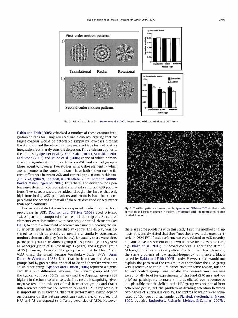
Fig. 3. The Glass pattern stimulus used by Spencer and O’Brien (2006) in their studyof motion and form coherence in autism. Reproduced with the permission of PionLimited, London.
Fig. 2. Stimuli and data from Bertone et al. (2003). Reproduced with permission of MIT Press.
D.R. Simmons et al. / Vision Research 49 (2009) 2705–2739 2709
Dakin and Frith (2005) criticized a number of these contour inte-gration studies for using oriented line elements, arguing that thetarget contour would be detectable simply by low-pass filteringthe stimulus, and therefore that they were not true tests of contourintegration, but merely contrast detection. This criticism applies tothe studies by Spencer et al. (2000), Blake, Turner, Smoski, Pozdol,and Stone (2003) and Milne et al. (2006) (none of which demon-strated a significant difference between ASD and control groups).More recently, however, two studies using Gabor elements – whichare not prone to the same criticism – have both shown no signifi-cant differences between ASD and control populations in this task(Del Viva, Igliozzi, Tancredi, & Brizzolara, 2006; Kemner, Lamme,Kovacs, & van Engelund, 2007). Thus there is no evidence for a per-formance deficit in contour integration tasks amongst ASD popula-tions. Two caveats should be added, though. The first is that onlyhigh-functioning ASD populations and controls have been com-pared and the second is that all of these studies used closed, ratherthan open contours.
Two recent related studies have reported a deficit in visual formprocessing in ASD. Spencer and O’Brien (2006) used oriented‘‘Glass” patterns composed of correlated dot triplets. Structuredelements were intermixed with randomly oriented elements (seeFig. 3) to obtain a threshold coherence measure for locating the cir-cular patch either side of the display centre. The display was de-signed to match as closely as possible a similarly constructedmotion coherence display (see below). Unusually there were threeparticipant groups: an autism group of 15 (mean age 13.5 years),an Asperger group of 10 (mean age 12 years) and a typical groupof 15 (mean age 12 years). The groups were matched for CA andVMA using the British Picture Vocabulary Scale (BPVS; Dunn,Dunn, & Whetton, 1982). Note that both autism and Aspergergroups had IQ greater than or equal to 70 and therefore were both‘‘high-functioning”. Spencer and O’Brien (2006) reported a signifi-cant threshold difference between their autism group and boththe typical controls (35.5% higher) and the Asperger group (26%higher) in the form coherence task. This result is surprising, givennegative results in this sort of task from other groups and that itdifferentiates performance between AS and HFA. If replicable, itis important in suggesting that task performance might dependon position on the autism spectrum (assuming, of course, thatHFA and AS correspond to differing severities of ASD). However,
there are some problems with this study. First, the method of diag-nosis: it is simply stated that they ‘‘met the relevant diagnostic cri-teria in DSM-IV”. If task performance were related to ASD severitya quantitative assessment of this would have been desirable (see,e.g., Blake et al., 2003). A second concern is about the stimuli.Although these were Glass patterns rather than line elements,the same problems of low spatial-frequency luminance artifactsraised by Dakin and Frith (2005) apply. However, this would notexplain the pattern of the results unless somehow the HFA groupwas insensitive to these luminance cues for some reason, but theAS and control group were. Finally, the presentation time wasexceptionally brief for experiments of this kind (250 ms), and toobrief for participants to make stimulus-elicited eye movements.It is plausible that the deficit in the HFA group was not one of formcoherence per se, but the problem of dividing attention betweentwo halves of a stimulus display, the centres of which were sepa-rated by 15.4 deg of visual angle (cf. Plaisted, Swettenham, & Rees,1999, but also Rutherford, Richards, Moldes, & Sekuler, 2007b).

2710 D.R. Simmons et al. / Vision Research 49 (2009) 2705–2739
This argument is reinforced by the fact that a similar size of deficitwas found in the matched motion coherence task (see below).Tsermentseli, O’Brien, and Spencer (2008) replicated the patternof Spencer and O’Brien’s (2006) data in a sample of adults, lendingforce to the argument for a form processing deficit specific to aut-ism diagnoses, but not Asperger Syndrome. Some of the same crit-icisms apply to this study. Tsermentseli et al. (2008) suggest thatthe presence of superior language skills in the Asperger groupmight be related to the differential performance which, as withSpencer and O’Brien (2006), seems to carry over to the motioncoherence task as well.
Further support for the view put forward by Spencer andO’Brien (2006) and Tsermentseli et al. (2008) is provided by Bros-nan, Scott, Fox, and Pye (2004). They looked at a variety of tasksin a group of 25 children with autism and 25 age- and VMA-matched controls. The mean CAs were about 10 years and themean VMAs about 5 years, meaning that the control group hadmoderate learning difficulties. The tasks utilized the gestaltgrouping rules of proximity, similarity and closure in simple lineand dot figures. Brosnan et al. (2004) found that their autismgroup were effectively at chance in these tasks, unlike the con-trols, and also impaired on identifying impossible figures,although this deficit was not so pronounced on having to drawreproductions of them (cf. Mottron & Belleville, 1993; Mottron,Belleville, & Menard, 1999a). A similar result has been found byBölte, Holtmann, Poustka, Scheurich, and Schmidt (2007) in agroup of adults with HFA.
6.2.6. Static shape perceptionDe Jonge et al. (2007) found normal shape discrimination in
their ASD population (squares vs. rectangles matched in area).However, Ropar and Mitchell (2002) found a curious failure ofshape constancy in their group of children with autism. The taskinvolved setting the shape of an ellipse on a computer screen tomatch the shape of a circle viewed in a special chamber, but tiltedaway from the viewer. Control participants typically set the ellipseto be more circular than it looks due to the knowledge that it isreally a circle, and this effect is strongest when the circle is pre-sented by itself and without any perspective cues. However, theautism group did not show this effect and set the shape of the el-lipse more accurately, suggesting that they were not so influencedby the knowledge that the shape was a circle. Ropar and Mitchell(2002) suggested that this result shows that individuals with aut-ism are less influenced by prior knowledge in visual judgementsand consequently visual processing may be less ‘top-down’ domi-nated in autism.
6.2.7. Susceptibility to visual illusionsHappé (1996) was the first to suggest that ASD populations
were less susceptible to these illusions (specifically the Ponzo, Pog-gendorff, Ebbinghaus/Titchener, Hering and Kanisza triangle, butnot the Müller-Lyer). These claims have been supported by Bölteet al. (2007). Happé’s (2006) original findings were, however, dis-puted by Ropar and Mitchell (1999, 2001). Commentators (e.g. Da-kin & Frith, 2005; Happé & Frith, 2006) have suggested that thisapparent discrepancy could be due to ‘‘methodological differences”between the studies. In particular, in Happé (1996) participantswere asked to judge whether the elements in an illusion display(normally perceived as different in size) were ‘‘the same” or ‘‘dif-ferent”. Many of the ASD participants reported ‘‘the same”, sug-gesting that they were not susceptible to the illusion. Ropar andMitchell (1999, 2001) used a computer-based method for adjustingthe relative sizes of the illusory-sized elements and found no sig-nificant differences between their ASD population and controls.No significant difference between ASD and control groups in illu-
sion susceptibility has also been reported by Hoy, Hatton, and Hare(2004) and Milne and Scope (2008).
An interesting recent study by Walter, Dassonville, and Bochsler(2009) may have clarified this discrepancy. Walter et al. (2009)used a large sample of 146 undergraduate students who performeda battery of tasks including psychophysical tests of a number ofstandard illusions and the Autism Spectrum, Empathising andSystemising Quotient questionnaires (AQ: Baron-Cohen et al.,2001b; EQ: Baron-Cohen & Wheelwright, 2004; SQ: Baron-Cohen,Richler, Bisarya, Gurunuthan, & Wheelwright, 2003, respectively),which measure traits associated with autism in the general popu-lation. Walter et al.’s (2009) key result was that susceptibility tothe Zöllner, Rod-and-frame, Roelofs, Ponzo and Poggendorff illu-sions related to score on the SQ, such that immunity to these illu-sions was associated with a high Systemizing Quotient. Thissuggests that previous results with populations on the autismspectrum (only one of the students scored high enough on theAQ to be considered a possible candidate for diagnosis) may havebeen affected by this trait, which is associated with ASD, ratherthan the diagnostic category in itself. Walter et al. (2009) alsomake a number of pertinent criticisms about methodology in pre-vious studies of illusory susceptibility.
Brown, Gruber, Boucher, Rippon, and Brock (2004) looked at adifferent illusion: the Kanisza triangle. They found no differencein behavioural performance between their six adolescents withautism and matched learning-disabled controls but there wereanomalies in the simultaneously recorded EEG responses. In partic-ular the induced activity in the gamma band (25–70 Hz) over pari-etal regions was different from that of the control group. A recenttechnical paper has cast doubt on the integrity of some inducedgamma band activity recorded with EEG, suggesting that if it hasthe profile illustrated by Brown et al. (2004) it is most likely anartifact due to small-amplitude saccadic eye movements (Yuval-Greenberg, Tomer, Keren, Nelken, & Deouell, 2008). Any studywhich does not control for these eye movements can no longerclaim to demonstrate neural gamma oscillations when measuredin this way, so Brown et al.’s (2004) results may well be reflectingdifferential fixational eye movement activity between the diagnos-tic groups, rather than anything significant about neural responses.Some ideas about how to control and interpret these artifacts haverecently been suggested by Melloni, Schwiedrzik, Rodriguez, andSinger (2009).
6.2.8. Visual completionA different approach to exploring context sensitivity effects
within ASD was taken by De Wit, Schlooz, Hulstijn, and van Lier(2007), who used a shape completion task. In shape completiontasks a shape (e.g. a circle) is initially presented partially occludedbehind another shape (e.g. a rectangle). After a short break the par-ticipant is then presented with two alternative shapes and has tochoose which one they think they saw (Sekuler & Palmer, 1992).De Wit et al.’s (2007) variation involved using a shape completionpriming task. The occluded shape was presented as a prime. Partic-ipants were then presented with two different (or identical) alter-native shapes and asked to speedily report whether they were thesame or different. Depending on the type of prime stimulus thisjudgement was performed either as fast or more quickly whenthe prime was in place than when it was not. The argument is thatpriming effects show evidence of different types of shape sensitiv-ity. De Wit et al.’s (2007) clinical sample was slightly unusual inthat they chose to focus on PDD-NOS, defined by the presence ofonly a subset of the usual symptoms of autism. In total there were19 participants in their clinical group (mean age 12 years): 16PDD-NOS and 3 AS. The control group was matched for CA andIQ. Their pattern of results was quite complicated. The clinicalgroup had largely similar reaction times to controls, but differed

D.R. Simmons et al. / Vision Research 49 (2009) 2705–2739 2711
in the pattern of priming stimuli that were effective. De Wit et al.(2007) argued that their results demonstrate that their PDD groupwas able to integrate context so as to complete partially occludedstimuli effectively, but they had somewhat more difficulty withunusual/unfamiliar or complex shapes. This may indicate a greaterdifficulty in learning novel shapes (or a greater sensitivity to differ-ences between them).
6.2.9. ReadingThe published data on reading ability in ASD show a large
amount of variability (Whitehouse & Harris, 1984). Recent studieshave suggested that the problems that people with ASD have withreading are dissociable from their symptoms (Ludlow, Wilkins, &Heaton, 2006; White et al., 2006).
6.3. Colour vision
There are a large number of anecdotal reports of unusual re-sponses to colour among people on the autism spectrum (e.g.Williams, 1999; see also Franklin, Sowden, Burley, Notman, &Alder, 2008; Ludlow et al., 2006). Individuals on the autism spec-trum can display strong affinities to, or aversions from, objects ofparticular colours (Ludlow & Wilkins, 2009; Moore, 2004). A fewstudies have reported incidental effects of colour: Brian, Tipper,Weaver, and Bryson (2003) found an unexpected facilitation effectof colour in their study of inhibitory mechanisms in ASD andGreenaway and Plaisted (2005) found a similar effect in a cueingtask, where invalid colour cues resulted in greater costs for individ-uals with ASD than for controls.
The first dedicated study of colour vision in ASD was Ludlowet al. (2006), who tested the efficacy of coloured overlays on thereading performance of a group of children on the autism spectrumand a gender-, age- and VIQ-matched control group. The ASD groupshowed a modest but significant average improvement in readingspeed of 13% with the overlays in place. Note that part of Ludlowet al’s (2006) screening process used the City and Ishihara colourvision tests, and nothing unusual was found, but this was too smalla sample to assert that colour vision is clinically normal in ASD.Ludlow et al. (2006) couch their explanation of the readingimprovements observed in the ASD group in terms of Wilkins’(2003) theory that appropriately chosen coloured filters can limitthe spread of activation in hyper-excitable areas of visual cortex(see also Ludlow & Wilkins, 2009).
Three very recent studies deal directly with the visual processingof chromatic information in ASD: Heaton, Ludlow, & Roberson, 2008;Franklin et al., 2008, in press. Heaton et al. (2008) used three groupsof 13 children and adolescents (mean age�11 years). One group wasfrom a school specializing in autism and thus was assumed to meetappropriate diagnostic criteria, one from a school for children withmoderate learning difficulties (MLD) and one group of typicallydeveloping (TD) children. Groups were all individually matched forchronological age and the MLD and autism groups were matchedfor non-verbal MA (assessed using Raven’s matrices). However,VMA (measured using the BPVS) was significantly different betweenall three groups, with the autism group having the lowest (mean�5.5 years). After checking that all groups could name and distin-guish the 11 basic colours correctly, they performed a discriminationtask: three coloured patches, two the same and one differing by asmall step in Munsell hue space were presented on a computerscreen and participants chose the odd one out. Both autism andMLD groups were impaired on this task relative to the TD controls,although not with respect to each other. Performance in this taskcorrelated with VMA. The second experiment tested colour memory.The same children were taught to associate pictures of familiar ani-mals with ‘‘focal” colours (i.e. red, green, blue and yellow) then sub-sequently tested by presenting the animal together with the four
colours, with the children being asked to choose which one matchedthe animal. In this phase of the experiment the autism and MLDgroups were just above chance, but the TD group performed quitewell. In the second phase of the experiment, after a memory refresh-ment, the participants were presented with three alternative coloursfor each animal picture, but this time each colour was drawn fromthe same colour category (e.g. three shades of red). In this phase ofthe task only the autism group scored above chance. In fact, whencalculated as a z-score, the performance of the autism group wasabout the same in both phases of the task. Subsequent analysisshowed that phase 1 performance correlated significantly with bothVMA and discrimination performance from the first experiment, butin phase 2 there was a significant negative correlation with VMA,suggesting that the children with lowest VMA performed best inthe task. Heaton et al. (2008) suggest that there is a profound link be-tween verbal ability and perceptual discrimination and that typi-cally developing children apply verbal labels to the colours, andtherefore are confused in the memory task when presented with col-ours which have the same verbal label. Children with language diffi-culties, on the other hand, are forced to remember the colourperceptually, and thus are less confused so long as the colours arediscriminably different. This differential performance is reminiscentof the performance of ASD groups in the Embedded Figures Task (Jol-liffe & Baron-Cohen, 1997; Shah & Frith, 1983) where a learnedgrouping of information disrupts performance in a TD group, butnot in a group with ASD.
Franklin et al. (2008) worked with 19 children with HFA(7–13 years) attending specialist schools and 14 CA- and NVIQ-(Raven’s Matrices) matched controls. Importantly, none of thechildren were diagnosed with ADHD, as it has been shown thatthese children tend to have blue-yellow colour deficiencies (Banas-chewski et al., 2006). Franklin et al. (2008) specified their stimuli inCIELAB colour space which were either coloured patches or ab-stract form stimuli, constructed from a standard set (Pick, 1965).The first experiment consisted of a visual search task and a visualmemory task. In the search task, participants were asked to spotthe odd coloured patch or the odd form in an array of distracters.The memory task was a delayed match-to-sample, with twochoices, one the same as the target and one foil, differing slightlyin colour or in form (curvature). Having checked that there wasno interaction, data from the two tasks were combined, and itwas found that the ASD group was impaired on the colour task,but not the form task. The second experiment was performed withtwo groups of 14 slightly older children (11–13 years). The exper-iment tested categorical perception of colour across the blue–greenboundary (Franklin, Pilling, & Davies, 2005). The 2AFC task was tolocate the presence (i.e. left or right of centre) of a coloured targeton a coloured background. The target was either drawn from thesame or different colour categories. Accuracy on the task did notdiffer between within- and between-category judgements, butthe ASD group performed significantly worse than the controls.Reaction times (RTs) showed a category boundary effect, withthe task being performed more quickly by both groups when thecolours were sampled from different categories. However, therewere no significant differences in RT between the groups.
Franklin et al. (in press) tested similarly characterized groupson the Farnsworth–Munsell 100-hue test (Farnsworth, 1943), anda conventional chromatic discrimination task which involveddetecting the orientation of the boundary between two isolumi-nant colours. In both experiments there were control tasks whichused stimuli differing only in luminance. As with Franklin et al.(2008) the ASD group performed significantly worse on the colourexperiments with higher error scores on the FM-100 hue test andhigher thresholds on the chromatic discrimination task. However,no significant differences were found in the luminance tasks. Notethat the chromatic discrimination difficulties of the ASD group

2712 D.R. Simmons et al. / Vision Research 49 (2009) 2705–2739
were not confined to a particular axis of colour space, such as thered–green or blue–yellow opponent axes.
The conclusion from this series of studies is that children withASD are challenged in a range of chromatic discrimination taskscompared to typical controls matched on non-verbal intelligenceand these difficulties do not transfer to similar luminance discrim-ination tasks. Franklin et al. (in press) estimate that performance ofthe ASD group in the FM-100 task is comparable to that of children3 years younger. It is unfortunate, however, that Franklin et al.(2008, in press) did not report VMA in their studies, given the po-tential importance of this factor (Heaton et al., 2008). Verbal abilityis known to be closely linked to colour naming performance inyoung children (Pitchford & Mullen, 2002) and a very recent ERPstudy in mono- and bi-lingual adults has suggested that colour lan-guage can affect the very earliest stages of perception (Thierry,Athanasopoulos, Wiggett, Dering, & Kuipers, 2009). Also, all threestudies (Franklin et al., 2008, in press; Heaton et al., 2008) failedto characterize fully the diagnostic status of their participants.Nevertheless, these results are exciting and, if they are found tobe specific to ASD, have profound implications for our understand-ing of early visual processing in this condition.
6.4. Depth perception and stereopsis
There are three lines of evidence which suggest that the percep-tion of depth in ASD merits further attention. First, there is a rangeof clinical and anecdotal reports about depth perception beingunusual in ASD. Kaplan (2006), for example, notes his observationsthat people with ASD often mis-judge inter-personal distance dur-ing social interaction and have difficulties with tasks such as ball-catching. Second, there is evidence for a higher incidence of stra-bismus in ASD populations (Kaplan et al., 1999; Scharre & Creedon,1992), which would suggest that binocular vision and stereopsismight also be affected. Third, because stereopsis requires a precisedevelopmental registration of information from each eye, it isdevelopmentally fragile (Atkinson, 2000). Depth perception andstereopsis in ASD would seem to be an area where at the very leasta screening study is warranted.
6.5. Motion perception
Motion perception is one of the most-studied and most contro-versial areas in the field of vision in ASD. Reviews by Dakin andFrith (2005) and Milne, Swettenham, and Campbell (2005; plusassociated peer commentaries) cover the literature up to 2005 inconsiderable detail, so we will concentrate on more recent studies.
6.5.1. Local motionSurprisingly, only one published study has examined low-level
local motion processing in an ASD population: the Bertone et al.(2003) study described in detail above (see Fig. 2). Bertone et al.(2003) found no significant difference in contrast thresholds forfirst-order motion direction identification between their ASD andcontrol groups, but there was a relative deficit in second-order mo-tion processing, with the ASD group exhibiting significantly highermodulation thresholds for the direction identification task. Pub-lished criticisms of Bertone et al. (2003) have tended to focus onthe explanation of the result in terms of the ‘‘complexity” of mo-tion processing, rather than questioning the result itself (see Dakin& Frith, 2005; Jarrold & Scott-Samuel, 2005; Mitchell, Ledgeway, &Landry, 2005). Interestingly, Kogan et al. (2004) measured localmotion processing in Fragile-X Syndrome (FXS) – a genetic disor-der which shares some of its symptomatology with ASD – usingsimilar techniques and stimuli to Bertone et al. (2003). Koganet al. (2004) found that modulation thresholds for local motiondirection identification were impaired for both first- and second-
order stimuli in this population, suggesting that the pattern of re-sults found by Bertone et al. (2003) is particularly specific to ASD(see also Bertone & Faubert, 2006).
Vandenbroucke, Scholte, van Engelund, Lamme, and Kemner(2008) recently examined two-grating plaid motion processing ina group of adults with HFA and typical controls, matched for CAand IQ. They found no significant differences between the partici-pant groups in the relative amount of time that the plaid was seenmoving as a coherent whole, rather than as two transparent com-ponents. There were also no significant differences between groupsin the rivalry rate. They concluded that there was no evidence for adifficulty with combining motion information in their ASD popula-tion, invoking an explanation in terms of the spatial frequency con-tent of their displays, which was predominantly low. They arguedthat individuals with ASD may have less difficulty processing mo-tion of low than high spatial frequency gratings, comparing theirdata with that of Bertone et al. (2003) and the motion coherencestudies considered below. Given this explanation it is unfortunatethat Vandenbroucke et al. (2008) did not use sinusoidal gratings astheir plaid components, as this would simplify the interpretation oftheir results, but they certainly suggest that the perception of high-contrast two-component motion is unaffected in adults with HFA.A further useful piece of information, given the results of Spencerand O’Brien (2006) and Tsermentseli et al. (2008) discussed below,would be how many of their HFA group could be characterized ashaving Asperger Syndrome.
6.5.2. Motion coherenceA typical motion coherence stimulus consists of a large number
of randomly moving dots of which a small proportion move coher-ently in a given direction and give a fleeting perception of motion(Newsome & Paré, 1988). Threshold for the task is defined as thepercentage of dots required to be moving coherently before the ob-server can reliably report their direction of motion. Usually this isrun as a 2AFC task, with the motions being up vs. down or left vs.right. Newsome and Paré (1988) originally used this stimulus as aprobe for investigating the efficacy of microscopic lesions in AreaV5/MT of macaque monkeys on the monkeys’ motion discrimina-tion abilities.
Wattam-Bell (1994) developed a slightly different version ofthis stimulus for use in preferential looking tasks with infants,which Atkinson and Braddick (2005) describe as a ‘‘road in thesnowstorm” stimulus. In this stimulus the signal dots oscillatebackwards and forwards in a horizontal direction and the noisedots appear transiently for the same duration (120 ms) in randomlocations. In one half of the display the signal dots all move in thesame direction. In the other half of the display a central strip con-tains dots moving in the exact opposite direction. This results in asegmented percept rather like looking at a road in a snowstorm.This was precisely the class of stimulus used by Spencer et al.(2000) to look at motion coherence thresholds in an ASD popula-tion. Their sample of 23 children with ‘‘autistic disorder” (diagnos-tic technique not specified) had significantly higher motioncoherence thresholds than the CA-matched controls (see Table 1).There was a smaller and non-significant difference between ASDand control performance in a similar form coherence task. Interest-ingly, motion coherence thresholds in the ASD group did decreasewith age (7–11 years), as did those of the controls, although the ra-tio of ASD/control performance remained about the same in all agegroups. Also, whereas control performance reached adult levels bythe age of 11, this was not true of the ASD group, although therewere no data on teenagers with ASD to test whether motion coher-ence thresholds were consistently higher through to adulthood orjust developmentally delayed.
Milne et al. (2002) criticized Spencer et al. (2000) for not match-ing their control population for IQ. Milne et al.’s (2002) sample

Table 1Stimulus parameters from studies on motion coherence. Note that �� denotes a significant difference and � a marginal difference between coherence thresholds.
Dot diameter(deg)
Coherent motion speed(deg/s)
Total dots perframe
Dot density(dots/deg2)
Display duration(ms)
Dot lifetime(ms)
Results (%) A = ASDC = CONTROL
Spencer et al. (2000) ?? 5.8 2000 4 330a 17 A: 25.5**C: 17.5
Milne et al. (2002) 1 pixel 8.8 150 0.3 1010 224 A: 25.0**(=0.1?) C: 15.3
Pellicano et al. (2005) 0.1 6.3 100 0.4 600 30 A: 22.4**C: 11.1
Milne et al. (2006) 0.1 7 300 2.1 85b 85 A: 17.2*C: 10.3
Spencer and O’Brien (2006) ?? 5.8 5655 4 166 50 Aut: 46.2**Asp: 28.7C: 24.6
Del Viva et al. (2006) 0.4 10 100c 0.4 160 66 A: 4.9Cl: 6.9C2: 5.5
a Before direction reversal. The total duration was self-limited.b Also before direction reversal. Total duration 2300 ms.c 50% black; 50% white.
D.R. Simmons et al. / Vision Research 49 (2009) 2705–2739 2713
consisted of 25 children with ASD (9.5–15.5 years) and 22 typicalcontrols matched for CA and NVIQ (which was in the normal rangefor both groups). The diagnostic method was ‘‘according to DSM-IVcriteria”. The children were also all attending specialist schools.Milne et al.’s (2002) stimulus was closer to the Newsome andParé (1988) style with a single display region and the task beingto identify the direction of coherent motion. The display dynamicswere also slightly different. A central fixation cross was presentthroughout stimulus presentation which the children were in-structed to fixate. Mean thresholds for the ASD and control groupsgave a similar performance ratio to that of Spencer et al. (2000). Aswith Spencer et al. (2000), the range of performance was greater inthe ASD group (6–64% rather than 6–29%), but the inter-group dif-ference was still significant when two of the ASD group with veryhigh thresholds were removed from the calculations.
The studies of Spencer et al. (2000) and Milne et al. (2002)seemed to provide a coherent story: motion coherence thresholdswere significantly higher in juvenile ASD populations, consistentwith either Magnocellular pathway or ‘‘Dorsal stream vulnerabil-ity” arguments about the neural symptoms of ASD (Braddick,Atkinson, & Wattam-Bell, 2003; Milne et al., 2005). However,results from more recent studies have complicated thisinterpretation.
The most extreme position is occupied by Del Viva et al. (2006).In their version of the paradigm a display of 100 black and whitedots was used on a gray background. Their ASD population wascarefully diagnosed using ADI-R and ADOS-G and they excludedparticipants with genetic syndromes. They also used two controlgroups: one matched on CA and the other matched on VMA. Theirmotion stimuli were based on optic flow stimuli previously usedby Morrone, Burr, and Vaina (1995) including rotational, transla-tional and radial motion. They found no overall difference in coher-ence thresholds between their ASD group and either of the controlpopulations.
The other extreme amongst recent papers is represented by Pel-licano et al. (2005). They found a highly significant difference be-tween global dot motion thresholds of their ASD and controlgroups, without any overlap in the 95% confidence intervals basedon the data although, as usual, the variance on the ASD popula-tion’s thresholds was considerably larger than that of the controlgroup. The other recent studies which have looked at motioncoherence obtained results between these two extremes. Spencerand O’Brien (2006) divided their participants into those with HFAand those with AS and found that motion coherence thresholds dif-
fered significantly from controls for the HFA group, but not the ASgroup. This pattern of results was confirmed in an adult populationby Tsermentseli et al. (2008). Milne et al. (2006) also found thatonly a sub-group of their ASD population (about 20%) had motioncoherence thresholds outside the typical range.
Recent data using the motion coherence paradigm to compareperformance of children with and without ASD thus gives com-pletely conflicting results ranging from no difference, to partial dif-ference to complete difference, making it very difficult to drawfirm conclusions on what the data mean. Clearly there are method-ological differences between the studies that may explain the con-flicting data. As this is such a well-studied and important area ofvision in ASD it is worth considering these in some detail.
Both Del Viva et al. (2006) and Pellicano et al. (2005) took con-siderable care in the diagnosis of their ASD populations. As men-tioned above Del Viva et al. (2006) used both ADI and ADOS andexcluded genetic syndromes from their population. Pellicanoet al. (2005) did not use the ADOS, but they did confirm diagnosesusing ADI. They also screened their control population using theSocial Communication Questionnaire (SCQ; Rutter, Bailey, & Lord,2003). Del Viva et al. (2006) used two control populations, onematched for CA and the other matched for VMA, but they foundno difference between them. Pellicano et al. (2005) had twice thenumber of ASD participants (20 vs. 10) and matched on NVIQ, asmeasured by Raven’s Standard Progressive Matrices. Pellicanoet al. (2005) point out that the VIQ of their comparison populationswas different (means of 119 and 137, respectively, for ASD andcontrols) but that the receptive language of the ASD group wasadequate for understanding task instructions. Here we have thefirst potential difference between the two studies, although DelViva et al.’s (2006) second control group (C2), matched on CA,would probably have been the most similar to the controls of Pel-licano et al. (2005).
The stimuli used by these two studies were, however, substan-tially different. Detailed parameters are presented in Table 1. Keydifferences are that although both studies used the same numberof dots in their displays, those of Del Viva et al. (2006) were blackand white on a gray background, were four times larger in diame-ter, and moving about 1.6 times faster than those of Pellicano et al.(2005). The display dynamics were also different. In Del Viva et al.(2006), total duration was brief, at 160 ms, with an individual dotlifetime of 66 ms, corresponding to four frame refreshes of the dis-play. In Pellicano et al. (2005) the overall stimulus duration wasmuch longer (600 ms) although the dot lifetime itself was shorter

2714 D.R. Simmons et al. / Vision Research 49 (2009) 2705–2739
(30 ms). In fact, Pellicano et al.’s (2005) paradigm was specificallydesigned to prevent participants tracking individual dots throughconsecutive frames because the dots carrying the coherent signalwere randomly switched on each frame refresh (they criticizedMilne et al. (2002) for having too long a dot lifetime). On the faceof it, one would expect the briefer presentation time of Del Vivaet al. (2006) to cause more problems for the ASD participants,but one factor that may be at work is the differential correspon-dence demands in the two sets of stimuli. As Del Viva et al.(2006) used two dot colours, larger dots, a slightly longer dot life-time and a brief overall presentation (too short to initiate an eyemovement) perhaps participants were less likely to mis-combinesignal dots with noise dots. Barlow and Tripathy (1997) discussand model correspondence noise in these classes of stimuli in con-siderable detail. The interesting implication of this observation isthat children with ASD may be more susceptible to correspondencenoise than their typical counterparts. Some ideas of why this mightbe are dealt with in the theory section below.
There are other differences in methodology between Del Vivaet al. (2006) and Pellicano et al. (2005) that may be important. Da-kin and Frith (2005) pointed out that the use of staircase routines iswidespread in threshold assessments in ASD populations. Staircaseroutines have the obvious advantage of speed over constant-stim-ulus designs, but they are also susceptible to influence by finger er-rors by the participant which may mis-direct the threshold searchin an unusual direction. Pellicano et al. (2005) used a PEST methodto find their motion coherence thresholds which converged on the75% correct point. They averaged all points following the fourthreversal to determine threshold. Pellicano et al. (2005) also em-ployed auditory feedback. Del Viva et al. (2006) used a QUEST rou-tine to alter stimulus levels, but then fitted the resultantproportion correct data with Weibull functions, also using 75% cor-rect as the threshold criterion. They did not give any trial-by-trialfeedback to participants and their data collection sessions wereslightly longer. Del Viva et al.’s (2006) method was therefore themore robust of the two.
This leaves us really with two ways of dealing with the currentcontradictions in the data on motion coherence in ASD. One is sim-ply to indicate that Del Viva et al. (2006) used the most carefullydiagnosed population and the most robust psychophysical tech-niques and therefore that their result indicating no significant dif-ferences between ASD and control populations should be regardedas the most reliable. The other is to consider that the other studiesdid produce useful and informative results that may be amalgam-ated by examining the different methodological conditions. DelViva et al. (2006) report their motion coherence data as sensitivi-ties. Converting to coherence thresholds (see Table 1) they are con-siderably lower than those in other studies (for both ASD andcontrol populations). As discussed, the larger level of correspon-dence noise in Pellicano et al. (2005) may have contributed to thisdifference. Milne et al. (2006) used quite a long duration and alsorequired participants to detect which side of the display the oscil-lating target was presented. These would have presented a greaterchallenge to the ASD group, but they also used relatively long dotlifetimes, which may have allowed for a bit more tracking. It iscurious that the threshold range in the ASD group in Milne et al.(2006) is actually very similar to that in Milne et al. (2002) whena significant group difference was reported. It seems as thoughthere was a more heterogeneous ASD population in the later studyand there was a more homogeneous control population. In anycase, the correspondence noise argument would suggest a resultfor Milne et al. (2006) between the extremes set by Del Vivaet al. (2006) and Pellicano et al. (2005). Spencer and O’Brien(2006) split their ASD population into Asperger and autism groups(a potential confound in other studies). Overall their coherencethresholds were much higher than the other studies (see Table
1), probably due to the higher dot density and larger number ofdots in the display.
In sum, it seems as though motion correspondence, the ‘‘purity”of the ASD population and the psychophysical methodology, can goa long way towards explaining the discrepancies in the publisheddata on motion coherence in ASD.
6.6. Optic flow
Gepner, Mestre, Masson, and de Schonen (1996b) were the firstto suggest that there might be difficulties with motion perceptionin ASD populations. When children with ASD were asked to standon a force platform and were presented with a large optic flowfield, they were less posturally reactive to the motion than typi-cally developing controls. A follow-up study showed additional dif-ferences between a small number of children with autism and adevelopmental delay, who were still less posturally reactive, andanother small group with Asperger Syndrome (and typical IQ)who were more posturally reactive than typical controls (Gepner& Mestre, 2002). Clearly this result may involve both unusual re-sponses to optic flow information and/or abnormal postural con-trol (Milne et al., 2005). However, the methodology of bothGepner et al. (1996b) and Gepner and Mestre (2002) has beenheavily criticized by Jarrold and Scott-Samuel (2005). Furthermore,Del Viva et al. (2006), who also measured responses to optic flowstimuli in the study discussed in detail above, found no differencesbetween ASD and control groups.
6.7. Biological motion
‘‘Biological motion” refers to the representation of human oranimal actions using point-light displays (PLDs), generated byplacing lights or reflective patches onto key anatomical points ofa moving actor and then filming the result (Johannsson, 1973;see Blake & Shiffrar, 2007, for a review).
Moore, Hobson, and Lee (1997) presented 5- and 10-point PLDsdepicting a walking person and various moving household objects(e.g. scissors opening and closing) to a group of 17 children/adoles-cents with autism (age 10–19 years). The control group wasmatched in CA and performance on the BPVS. As all of the ASDgroup had impaired language processing, the control group werelearning disabled although not diagnosed with ASD. Stimulus pre-sentation accumulated gradually from 40 ms to 5000 ms. Partici-pants were asked to ‘‘name what the dots are stuck to”.Psychometric functions showed the number of participants in thesample that were able to name the object correctly as a functionof time, so they were in some sense comparable to average reactiontimes, although the cumulative presentation method meant thatthe total exposure time to each stimulus was also cumulative.Moore et al. (1997) reported that there were no significant differ-ences in performance between ASD group and controls for thistask, either with point-light walkers or with the moving objects,but they found that the ASD group did have difficulty with sponta-neously describing and recognizing portrayals of emotion in PLDs,despite being able to accurately describe the mechanics of the mo-tion itself. Moore et al. (1997) therefore suggested that the visualprocessing of biological motion is intact in ASD, but that the prob-lems come with the interpretation of the internal states of others.
Blake et al. (2003) questioned the results of Moore et al. (1997),arguing that the verbal report responses and cumulative presenta-tions were susceptible to bias. Blake et al. (2003) used a more ro-bust and conventional psychophysical procedure: a 12-point-light actor performed a familiar activity (e.g. running or jumping),but each motion sequence also had a ‘‘phase scrambled” version inwhich dots underwent the same motion trajectory but offset by arandom temporal phase difference. Previous work (Bertenthal &

D.R. Simmons et al. / Vision Research 49 (2009) 2705–2739 2715
Pinto, 1994) had shown that ‘‘scrambling”, while retaining localmotion, diminishes the ability of observers to organize a point-light display into a human activity. The advantage of comparingperformance with these two stimulus variants is that the low-levelvisual structure of the stimuli is the same, the only difference beingthe relationships between the dots. Blake et al. (2003) calculated ad0 score for the biological motion task and found that their typicallydeveloping group performed significantly better in this task thantheir ASD group with mean d0 of close to 2.5 and close to 1, respec-tively. This is despite the two groups showing equivalent perfor-mance on a control ‘‘pathfinder” task (discussed above). Inaddition, Blake et al. (2003) plotted the individual d0 scores forthe ASD group as a function of their ‘‘level” of autism as measuredby the ADOS and CARS tests and found a significant negative corre-lation, although there was also a correlation, only within the ASDgroup, with MA. Note that some 25% (4/16) of Blake et al.’s(2003) original ASD sample could not complete the biological mo-tion task and 3 of these could not complete the form task either.These were all children with lower scores on a test of expressivelanguage.
What are the potential causes for the discrepant results be-tween Blake et al. (2003) and Moore et al. (1997)? The Blakeet al. (2003) ASD sample was younger than that of Moore et al.(1997), being between 8 and 10 years old, rather than averaging14 years, and were diagnosed more robustly. The control groupwas also younger, and matched on CA to the ASD sample’s MA.The main difference, however, was the nature of the stimuli. Blakeet al.’s (2003) participants were given a 1-s presentation of a ran-domized stimulus, rather than a cumulative presentation of thesame stimulus repeated until person identification was successful.Based on these results, we might expect children with ASD to takelonger to recognize human forms defined by PLDs. In agreementwith this, Annaz et al. (submitted for publication), also using 1-spresentation times, have shown that, at a CA of 12 years, TD chil-dren are better than ASD children in discriminating intact fromscrambled displays. Moreover, there is a flat developmental trajec-tory from 5 to 12 years for the ASD group and near identical perfor-mance of the TD and ASD groups at 5 years of age.
Hubert et al. (2007) looked at the age question by presentingthe stimuli used by Moore et al. (1997) to a group of adults withHFA/AS and IQ-matched controls. There were four sets of PLD se-quences, each defining a different motion category: 10 were‘‘actions” (e.g. hopping, running), 5 were ‘‘subjective states” (e.g.bored, itchy), 5 were ‘‘emotional states” (e.g. surprised, sad) and5 were object motions (e.g. ball rotating, ironing board openingand closing). Significant performance differences between groupswere only found for the emotional states, which Hubert et al.(2007) argued was consistent with Moore et al.’s (1997) results.However, it should also be noted that whilst none of the other con-ditions reached significance there were larger variances on the per-formance of the ASD group in all conditions, and meanperformance was only identical for the object motion condition.This suggests that the ASD group may have struggled with the taskwhenever it involved a human actor. Indeed, the authors reporteda main effect of group following on from their ANOVA, but this iscontaminated by a group-by-condition interaction. Another impor-tant point about the stimuli of Hubert et al. (2007), and therefore,by extension, Moore et al. (1997) is that there was no formal at-tempt to match the speed or complexity of the local motion con-tent of the stimuli. In other words, the ASD group may havefound the object motion easiest and the human motion hardersimply because the motion signal was more complex, with morenon-rigid relationships between point-light stimulus components.A final note of concern is the subjectivity of the response assess-ment, given that participants were asked to describe the motion,which was then judged by the experimenter as being an appropri-
ate or inappropriate description. Whilst the anecdotal evidence ofthe rather mechanical descriptions of emotional actions by the ASDgroup are persuasive, it is less clear how well the descriptions ofactions in the other conditions matched. Similar conclusions werereached in a follow-up paper by the same group (Parron et al.,2008).
Recent fMRI studies by Herrington et al. (2007) and Freitag et al.(2008) provide converging evidence on the neural processing ofbiological motion in ASD populations. The participants in Herring-ton et al. (2007) were a group of 10 adult males diagnosed with ASand 10 age-, sex- and IQ-matched controls. The participants in Fre-itag et al. (2008) were a carefully diagnosed group of 15 adoles-cents with HFA and controls matched for age, sex and IQ. Thetask used by Herrington et al. (2007) was to identify a 1s displayas either intact or scrambled, and the synthetic Cutting algorithm(Cutting, 1978) was used to generate the 13-point intact displayswith scrambling done by vertical displacement of a point’s originallocation by a random distance. The task used by Freitag et al.(2008) was to identify a 1.5 s display as either intact or scrambled,and motion capture data of 80 walkers were used to generate the15-point intact displays with scrambling done by permuting thelocation of the points, and changing the speed of each point to beequal to the average speed of that point. Herrington et al. (2007)reported that activity for the intact walker versus baseline wasgreater in the control than the ASD population in several regionsincluding the right middle temporal gyrus. A similar trend in themiddle temporal gyrus was reported by Freitag et al. (2008) inthe contrast of all motions versus baseline. However, they also re-ported regions with greater activation by the ASD group, includingthe postcentral gyri, left hippocampus and middle frontal gyrus.
The study by Freitag et al. (2008) went on to show that the con-trast of brain activity of intact versus scrambled revealed substan-tially different patterns of activation for the two populations. Forthe control group, when intact biological and scrambled motionwere compared, activations were found bilaterally in parietal, tem-poral and frontal lobes as well as basal ganglia and insula. The acti-vation network included the right Superior Temporal Sulcus (STS),which is known to be a central structure in biological motion pro-cessing (see Puce & Perrett, 2003). In contrast, the ASD groupshowed less activated clusters overall. What activations there werewere primarily in the left hemisphere in parieto-temporal (limbic)and frontal areas as well as basal ganglia. In the right hemisphere,activations specific for biological motion were found only in thelimbic system and Thalamus. Freitag et al. (2008) argue stronglythat the processing of biological motion stimuli by people withASD is very different from that of typical controls. Whilst theASD group was capable of discriminating the biological motionfrom scrambled motion they seemed to be doing it using a differ-ent network of brain regions.
To summarise, the data on biological motion are consistent witha low-level difficulty with motion processing feeding through andcomplicating the interpretation of biological motion stimuli, espe-cially when they present complex motions like human point-likewalkers. However, the ease with which typical observers can attri-bute emotions and feelings to these curiously sparse stimuli is notpresent in ASD populations, and it even seems as though the braincircuits used for processing these stimuli are different, beyondlow-level motion areas.
Among many areas of potential further development in biolog-ical motion perception in ASD one concerns the threshold for rec-ognition. A possible interpretation of the results of Herrington et al.(2007) and Freitag et al. (2008) is that the stimuli were less salientfor the ASD group: in other words the position on the recognitionpsychometric function was different for ASD and control groups. If,rather than simply presenting the same stimulus for the sameduration to both groups the stimulus was somehow equated for

2716 D.R. Simmons et al. / Vision Research 49 (2009) 2705–2739
performance, would the neural activations still be different orwould they now be much more comparable? The second issue isthe extent to which the hierarchy of difficulty in structure-from-motion tasks demonstrated by the results of Moore et al. (1997),Hubert et al. (2007) and Parron et al. (2008) are due to the in-creased complexity of point-light human stimuli which depict suchabstract notions as internal states and emotions or whether thereis some sort of top-down enhancement mechanism for these typesof stimuli which is lacking or deficient in ASD populations.
6.8. Animacy
A seminal study by Heider and Simmel (1944) demonstratedthat typical individuals readily endow simple animated shapeswith thoughts, feelings, and intentions. The perception of animacyin ASD has received a considerable amount of attention. Whilst thisis arguably outside the domain of vision science per se, it has raisedsome relevant questions about the interpretation of visual motioninformation which will be covered briefly. First of all, as with bio-logical motion, there is a consensus that all age groups with ASDappear to have difficulty interpreting animations to which typicalobservers attribute social content. However, this difficulty is a sub-tle one and seems to be confined to paradigms in which an ex-tended verbal response is required. The precise deficit seems tobe in the appropriateness of the language used to describe theseanimations. In particular, although the descriptions can be seman-tically elaborate, they tend not to include the same socially basedvocabulary as typical descriptions (Abell, Happé, & Frith, 2000;Bowler & Thommen, 2000; Campbell et al., 2006; Castelli, Frith,Happé, & Frith, 2002; Klin, 2000; Salter, Seigal, Claxton, Lawrence,& Skuse, 2008). At the same time, participants with ASD do seemable to distinguish propelled from self-propelled motion and ani-macy from non-animacy, although possibly with developmentaldelay or a training requirement (Bowler & Thommen, 2000; John-son & Rakison, 2006; Rutherford, Pennington, & Rogers, 2006). Fur-thermore, relatively elaborate animations which have explanationsin physical, rather than social, terms, seem to be interpreted withrelative ease (Klin & Jones, 2006). Castelli et al. (2002) have postu-lated that problems associated with interpreting these animateddisplays, and with social understanding in general, in ASD, are a re-sult of the neural activity evoked in extra-striate visual cortex fail-ing to transmit to the ‘‘social brain” regions of the STS, Temporallobe and pre-frontal cortex.
However, a confound appears to run throughout these experi-ments. The key question is what makes an animation ‘‘social”?We know that the distinction is partly based on whether or notthe agents appear to be self-propelled (Bowler & Thommen,2000), but there are also aspects of the motion trajectory and therelationships between the trajectories of the agents that are crucialto the interpretation. None of the studies so far has formallymatched the motion content of their animacy displays when com-paring ‘‘social” versus ‘‘non-social” animation. Castelli et al. (2002)report no differences in MT/V5 activation in their PET study, butthis is not really adequate. Klin and Jones’ (2006) space animationappears to be complex, but the physics of this motion sequencewould probably be familiar to boys who are experienced with con-temporary video games. Rutherford et al.’s (2006) study points tothe importance of training and motivation as well. Could it be thatthe problem with so-called ‘‘social” animations is that the com-plexity of the trajectories and inter-relationships between movingagents is higher and therefore more difficult to interpret? So thedifficulty is not with the ‘‘socialness” per se but with the subtletyof the information structure in the displays which, somehow, typ-ical observers pick up automatically during development butwhich observers with ASD need focused training with.
6.9. Visuomotor control
Gowen, Stanley, and Miall (2008) compared imitation behav-iour in adults with ASD to CA- and IQ-matched controls on a taskthat involved making simple movements back and forth in the ver-tical or horizontal directions while viewing congruent (samemovement direction) or incongruent (orthogonal movement direc-tion) displays. These displays took the visual form of either an ac-tual other person making the movements, or the motion of a singledot that was animated by a real movement recording, or a singledot that moved at constant velocity that instantaneously changeddirections at the extremes. Results showed that the performanceof both groups showed a significant interference effect for all con-ditions, however the ASD group did show the effect strongest whenviewing the movements of an actual person. These results demon-strate that, for both groups, observing others’ movements willinterfere with one’s own movement production but suggest thatoverall, the visuomotor integration processes are not identical be-tween groups.
7. Perception of faces and objects
One of the most-studied, and heavily reviewed, areas of visualprocessing in ASD is face processing (for recent reviews see Behr-mann, Thomas, & Humphreys, 2006b; Dawson, Webb, & McPart-land, 2005a; Golarai, Grill-Spector, & Reiss, 2006; Jemel, Mottron,& Dawson, 2006; Sasson, 2006). This is unsurprising, given theusual characterization of ASD as a social deficit, and faces beingarguably the most ‘‘social” of visual stimuli. Here we deal with ob-ject processing as well, because an object-based task is often usedas a control in studies of face processing.
7.1. Early studies
In the first detailed study of face processing in ASD, participantswere tested on their ability to recognize isolated features of knownpeers from grayscale photographs of their faces (Langdell, 1978).Two ASD groups were used: a ‘‘younger” group, aged about10 years and an ‘‘older” group aged about 14 years. Each ASD groupwas matched with three different control groups: two typical con-trol groups matched on either MA (and therefore much younger,being about 6 and 8 years old, respectively) or CA; a third controlgroup was learning disabled and was matched on both CA andIQ. Note that the mean IQ of these ASD groups was quite low, at60 and 63, respectively, making them both below the cut-off forlearning disability themselves. It was found that both ASD groupswere better able to use the lower part of the face for identificationpurposes than controls. The older ASD group was also less affectedby inversion than the other experimental groups. Langdell’s (1978)discussion of the results focused on two issues. First, did his datasuggest that children with ASD tend to regard the face simply asa complex object, without the social relevance invested in it bytypical children (in Langdell’s words as a ‘‘pure pattern” ratherthan a ‘‘social pattern”)? Second, was there evidence for what hetermed ‘‘asocial looking” (i.e. fixating on the ‘‘wrong” part of theface), and did this relate to the well-known phenomenon of gazeavoidance in ASD (Hutt & Ounstead, 1966)? These ideas continueto reverberate through the literature on face processing in ASD tothe present day.
The next major development in the field was a landmark seriesof studies by Peter Hobson and his co-workers at the Institute ofPsychiatry in London (Hobson, 1986a, 1986b, 1987; Hobson, Ous-ton, & Lee, 1988a, 1988b, 1989; Weeks & Hobson, 1987) on thetheme of emotion recognition. Weeks and Hobson (1987) askedchildren with ASD and VMA-matched learning-disabled controls

Fig. 4. Slide from the ‘‘Reading the Mind in the Eyes” test (Baron-Cohen et al.,2001a, 2001b). Reproduced from the actual test with permission of the AutismResearch Centre, University of Cambridge.
D.R. Simmons et al. / Vision Research 49 (2009) 2705–2739 2717
to sort photographs either according to facial expression (happy ornot) or type of hat (floppy or woollen). Most children with ASDsorted by type of hat in preference to facial expression (see alsoJennings, 1973). Hobson et al. (1988a) found that children withASD had difficulty in matching basic facial expressions (happy, un-happy, angry and scared) across different individuals when pre-sented with photographs of faces, or parts of faces, taken fromthe classic set of Ekman and Friesen (1975). The childrens’ perfor-mance in identity and emotion sorting was also less affected thancontrols by changes in face orientation, consistent with Langdell(1978). Hence, by the late 1980s, the consensus was that childrenwith ASD tend to regard faces as less ‘‘special” objects than eithertypical, or learning-disabled children (with equivalent verbal abil-ities), and that there were particular problems with the recognitionof emotional facial expressions. There were no similar difficultieswith non-social stimuli like visual scenes or objects (Hobsonet al., 1989). How has this consensus been altered by more recentstudies?
7.2. Is Face recognition impaired in ASD?
Face recognition is one of the most basic of face-processingskills. Whilst there is a considerable body of evidence that individ-uals with ASD demonstrate impairments in facial identity-basedtasks (Boucher & Lewis, 1992; Boucher, Lewis, & Collis, 1998;Critchley et al., 2000; Curby, Schyns, Gosselin, & Gauthier, 2003;Davies, Bishop, Manstead, & Tantam, 1994; Dawson et al., 2002a;De Gelder, Vroomen, & van der Heide, 1991; Gepner, de Gelder,& de Schonen, 1996a; Hauck, Fein, Maltby, Waterhouse, & Fein-stein, 1998; Jambaque, Mottron, Ponsot, & Chiron, 1998; Klinet al., 1999; Langdell, 1978; Teunisse & de Gelder, 1994; Ashwin,Wheelwright, & Baron-Cohen, 2005; Dalton et al., 2005; Riby, Doh-erty-Sneddon, & Bruce, 2009), a significant number of studies havedisputed this finding, especially when the faces are familiar(Adolphs, Sears, & Piven, 2001; Barton et al., 2004; Braverman,Fein, Lucci, & Waterhouse, 1989; Celani, Battachi, & Arcidiacono,1999; Chawarska & Volkmar, 2007; Davies et al., 1994; Deruelle,Rondan, Gepner, & Tardif, 2004; Gepner et al., 1996a; Ozonoff, Pen-nington, & Rogers, 1990; Volkmar, Sparrow, Rende, & Cohen, 1989;Wilson, Pascalis, & Blades, 2007). The evidence is, therefore, mixed.
7.3. Are there particular problems with emotional facial expressions inASD?
Difficulties in the interpretation of facial expressions are oftenassociated with ASD, but the scientific evidence for this difficultyis again remarkably mixed (for a review, see Jemel et al., 2006).Studies which report difficulties with various aspects of facialexpression processing by individuals with ASD are Hobson(1986a, 1986b, 1987), Weeks and Hobson (1987), Gioia and Brosg-ole (1988), Hobson et al. (1988a, 1989), Braverman et al. (1989),McDonald et al. (1989), Tantam, Monagham, Nicholson, and Stir-ling (1989), Ozonoff et al. (1990), de Gelder et al. (1991), Capps,Yirmiya, and Sigman (1992), Sigman, Kasari, Kwon, and Yirmiya(1992), Baron-Cohen, Spitz, and Cross (1993), Davies et al.(1994), Gepner et al. (1996a), Loveland et al. (1997), Celani et al.(1999), Critchley et al. (2000), Grossman, Klin, Carter, and Volkmar(2000), Pelphrey et al. (2002), Ogai et al. (2003), Deruelle et al.(2004), Dalton et al. (2005), Hefter, Manoach, and Barton (2005),Ashwin, Chapman, Colle, and Baron-Cohen (2006a), Ashwin,Wheelwright, and Baron-Cohen (2006b), Boraston, Blakemore,Chilvers, and Skuse (2007), Humphreys, Minshew, Leonard, andBehrmann (2007), Mazefsky and Oswald (2007), Gross (2008),Wright et al. (2008). Some studies suggest specific deficits withparticular emotional expressions such as fear (De Jong, van Engel-und, & Kemner, 2008; Pelphrey et al., 2002), sadness (Boraston
et al., 2007), or ‘‘negative” expressions (Ashwin et al., 2006a;Humphreys et al., 2007). Adolphs et al. (2001) showed that theirsmall sample of adults with ASD did not differ significantly fromcontrols in their ability to discriminate facial expressions, and wereonly mildly impaired in the recognition of basic emotions fromfaces. However, they did tend to mislabel the approachabilityand trustworthiness of faces, suggesting a parallel with amygdalardamage (Adolphs et al., 2001; Ashwin et al., 2006a).
A number of other studies have failed to find any dysfunctionfor high-functioning groups when the emotions are ‘‘basic”, suchas happiness, sadness, anger and disgust (Baron-Cohen, Wheel-wright, & Jolliffe, 1997; Baron-Cohen et al., 1993; Davies et al.,1994; Volkmar et al., 1989b; Adolphs et al., 2001; Gepner, Deruelle,& Grynfeltt, 2001; Grossman et al., 2000; Hubl et al., 2003; Love-land et al., 1997; Piggot et al., 2004; Ashwin et al., 2005; Ashwinet al., 2006b; Castelli, 2005). Kätsyri, Saalasti, Tiipana, von Wendt,and Sams (2008) found equivalent performance in a basic facialexpression rating task with their adults with AS and controls ex-cept for when they used an extreme low-pass filter on the stimulus(cut-off less than 1.8 cycles per face width).
There is more consistency in the literature that the performanceof ASD populations on ‘‘complex” facial expression recognition isrelatively poor (Adolphs et al., 2001; Baron-Cohen et al., 2001a,1997; Boraston, Corden, Miles, Skuse, & Blakemore, 2008; Klein-man, Marciano, & Ault, 2001). The ‘‘Reading the mind in the Eyes”task illustrated in Fig. 4 is a case in point. Even individuals withHFA and AS struggle with this task, despite being able to definethe emotional terms involved (Baron-Cohen et al., 2001a, 1997b).A potential concern with this task is Grossman et al.’s (2000) obser-vation that people with ASD find it harder to choose an emotionalmatch from a verbal description than from a pictorial description.However, the ‘‘eyes” test has been validated in a number of coun-tries and adapted to a number of different age groups (see http://www.autismresearchcentre.com/arc), so it does provide a usefultool with which to check the facial expression processing capabil-ities of people at the high functioning end of the autism spectrum.Those with lower VIQs will obviously have difficulty with thesemantic complexity of the verbal descriptions, although thereare tests adapted specifically for use with children where the lan-guage demands are less severe. Boraston et al. (2008) found thattheir ASD participants were confused by fake and genuine smiles(where the eye region was giving either conflicting or consistentinformation) and were fixating less on the eye region of the targetfaces. They also found that difficulty with this task was correlated
3 Barton et al. (2007), in common with other studies from the same laboratory(Barton et al., 2004; Hefter et al., 2005) use the clinical specification ‘‘SocialDevelopmental Disorder”, which is a superset of those with Autism SpectrumDisorder together with those with slightly different symptomology similar to PDD-NOS.
2718 D.R. Simmons et al. / Vision Research 49 (2009) 2705–2739
with the social interaction score from the individual’s ADOS assess-ment (Boraston et al., 2008).
One recent study has questioned that individuals with ASD haveproblems interpreting facial expressions. Back, Ropar, and Mitchell(2007) used video clips of complex facial expressions and pre-sented them to a group of eighteen 10–14-year-old children withASD, and found that they performed above chance at naming themental state from a choice of four descriptions, but were not asproficient as controls. However, when the eye region of the faceswas digitally ‘‘frozen”, the ASD group were affected, so they wereclearly using information from the eye region. When the eyes wereisolated and presented alone, the ASD group were as successful ascontrols and also when they were presented in the context of thewhole face. Whilst Back et al. (2007) clearly demonstrate that indi-viduals with ASD are capable of interpreting complex facial expres-sions under some circumstances one issue of comparison is thatthe task was less challenging because the same female actor wasused throughout, whereas with Baron-Cohen et al.’s (2001a) ‘‘eyes”task there are a number of different faces.
7.4. Discrimination of other facial attributes
A number of studies suggest poorer performance of ASD groupsin gender discrimination tasks based on faces (Baron-Cohen et al.,1999; Behrmann et al., 2006a; Deruelle et al., 2004; Hobson, 1987;Hobson et al., 1988a). Notably, gender discrimination is alsothought to depend largely on information located in the eye region(Gosselin & Schyns, 2001). Hobson (1987) and Gross (2002, 2005,2008) looked at age discrimination, the latter in human and non-human faces, and found a deficit in their population of childrenwith ASD relative to controls. The data on lip-reading are moreequivocal, with two in favour of a relative deficit (Deruelle et al.,2004; Gepner et al., 1996a), and two against (de Gelder et al.,1991; Gepner et al., 2001).
7.5. Is there a face processing impairment in ASD?
Jemel et al. (2006) argue, along with Behrmann et al. (2006b)and Mottron, Dawson, Soulières, Hubert, and Burack (2006), foran amodal and non-domain-specific difference in perceptual pro-cessing characterized by ‘‘locally oriented” perception of faceswithout a specific deficit in perception of global features, face iden-tity or emotion. Certainly the behavioural data reviewed abovedoes not provide unquivocal back-up for there being a specificface-processing deficit in all individuals diagnosed with ASD,although it could be argued that this ability is less robust than intypical populations. Could this fragility have an underlying percep-tual basis?
7.6. Is object identification and recognition spared in ASD?
A general problem with object identification and recognitionwould potentially contribute to poorer performance in face pro-cessing tasks, but the object processing abilities of individuals withASD are generally reported to be equivalent to those of controlpopulations (Boucher & Lewis, 1992; Braverman et al., 1989; Da-vies et al., 1994; Hobson, 1986a, 1986; de Gelder et al., 1991; Tan-tam et al., 1989 (low IQ group), Celani et al., 1999; Dawson et al.,2002a; Gepner et al., 1996a, 1996b).
There are only a couple of studies which suggest that ASD pop-ulations may have problems with some types of object judgement(Berhmann et al., 2006a; Davies et al., 1994). Behrmann et al.(2006a) presented their group of adults with HFA with two objectprocessing tasks. In the first they were required to determinewhether two pictures of naturally occurring objects were the sameor different. In the second, the ‘‘Greeble” class of alien-like objects
was used (Gauthier & Tarr, 1997). In both cases the ASD groupdemonstrated more difficulty with fine object discriminations atthe exemplar/individual level, than controls. Behrmann et al.(2006a) suggested that visual discrimination difficulties are notconfined to social objects like faces, but may reflect more generalanomalies in visual processing.
7.7. Are faces ‘‘special” for individuals with ASD? The cases of inversionand Thatcherisation
It is generally thought that faces are a ‘‘special” class of objectsfor the typical visual system, with their own dedicated neural pro-cessing system (see below). One of the perceptual indicators of thisspecialness is that faces are harder to recognize upon inversionthan other objects. Individuals with ASD appear able to distinguishfaces from non-faces (Ashwin, Baron-Cohen, Wheelwright, O’Rior-dan, & Bullmore, 2007; Volkmar et al., 1989), but Langdell’s (1978)original suggestion that children with ASD are less affected by fa-cial inversion has received only mixed support. Tantam et al.(1989) and Van der Geest, Kemner, Verbaten, and van Engelund(2002) argued for unusual performance of ASD populations withinverted faces. Joseph and Tanaka (2003) presented evidence thatchildren with ASD were more sensitive to image manipulationsin the mouth region than in the eye region of faces, unlike controls,and that only this sensitivity showed an inversion effect: a partialreplication of Langdell (1978). But most recent studies have shownsignificant face inversion effects in ASD populations (Barton, Hef-ter, Cherkasova, & Manoach, 2007; Gross, 2008; Lahaie et al.,2006; Teunisse & de Gelder, 2003) shifting the balance in favourof an intact face inversion effect in ASD, at least in high-functioninggroups. There is a possibility that this effect (or, effect absence!)might be more pronounced in ASD populations with lower IQ (Teu-nisse & De Gelder, 2003) or, at least, those with poorer face dis-crimination abilities (Barton, Hefter, Cherkasova, & Manoach,2007), but there seem to be confounding factors of overall task per-formance, some of which may not be related to visual processing.
Another related stimulus manipulation is the selective inver-sion of facial features known as the ‘‘Thatcher Illusion” (Thompson,1980). Rouse, Donnelly, Hadwin, and Brown (2004) and Riby et al.(2009) have shown that this illusion is intact in children with ASD.However, Riby et al. (2009) also manipulated eye and mouth re-gions separately and found that their group of 10–18-year-old chil-dren with autism were equally sensitive to ‘‘Thatcherisations” ofthe eyes and mouth and did not favour the eyes as the controls did.
7.8. Are faces processed as a collection of individual features in ASD,rather than as a coherent whole?
‘‘Configural processing” has been defined as the perception ofrelations between features of a stimulus, such as a face, as con-trasted with ‘‘componential” or ‘‘featural” processing (Maurer, LeGrand, & Mondloch, 2002). Behrmann et al. (2006a) found thattheir group of adults with ASD found it harder to discriminatemasked down (i.e. ‘‘internal features only”) images of faces byidentity and gender than matched controls. Barton et al. (2007)explicitly compared featural and configural processing of faces bymanipulating the eye separation or mouth position (configural)or eye and mouth colour (featural) properties of face images andasking their population of SDD3 adults which of three faces simulta-neously present on screen was the odd one out. Those who were able

Fig. 5. Eye tracking data from Pelphrey et al. (2002). Reproduced with permission ofSpringer Science and Business Media.
D.R. Simmons et al. / Vision Research 49 (2009) 2705–2739 2719
to do famous face recognition (‘‘SDD-1”) were only mildly impairedrelative to controls in this task, but those unable to do famous facerecognition (‘‘SDD-2”) were severely impaired. Notably, however,they were equally impaired on featural as configural changes, sug-gesting that there was no specific deficit in sensitivity to configuralchanges to faces. Note that, while this experiment was ingeniousthere is a small concern that the featural changes were not matchedfor discriminability with the featural changes. Teunisse and de Gel-der (2003) looked at the facial composite effect, where the photo-graph of a face is split in two along a horizontal axis andcombined with another. When typical participants are asked to spotwhether the top half of this face is the same as that of a simulta-neously presented target face they are poorer when the compositesare aligned rather than misaligned (Young, Hellawell, & Hay, 1987).The ASD group tested by Teunisse and de Gelder (2003) did not showthis effect, although the controls did. However, despite being statis-tically significant, the differences found were quite small. Joseph andTanaka (2003) reported a variation on the ‘‘whole-face advantage”normally found when trying to spot which features of a previouslyseen face have been altered. Whereas typical children were moresensitive to eye substitutions, the ASD group were more sensitiveto mouth substitutions, echoing the results of Langdell (1978). How-ever, performance was generally quite poor in this task for both ASDchildren and controls, only reaching a maximum of 76% correct (con-trols on eye substitutions). Riby et al. (2009) have also reported thattheir ASD group were less sensitive to eye than mouth manipula-tions in a similar experiment, but Lopez, Donnelly, Hadwin, and Lee-kam (2004) found that they could generate a whole-face advantagein their adolescents with ASD by adding a cue to the appropriatefeature.
All in all, there seems to be a large amount of variability in thedata on configural processing of faces in ASD. Barton et al.’s (2007)data suggest that this variability may be due to a bimodal split inthe ASD population between those that are quite capable in faceidentity processing tasks and those that are not.
7.9. Are face processing difficulties in ASD confined to the eye region?
It is commonly asserted that people with ASD have difficultyattending to the eye-region of faces, unlike typical controls (Boras-ton et al., 2008; Dalton et al., 2005; Gross, 2004; Klin, Jones,Schultz, Volkmar, & Cohen, 2002; Pelphrey et al., 2002; Rutherford,Clements, & Sekuler, 2007a) and/or attend selectively to the mouthregion (Joseph & Tanaka, 2003; Langdell, 1978). Rutherford et al.(2007a) tested this prediction explicitly by measuring the discrim-inability of small spatial displacements in the eyes and mouths ofdigitally altered images of unfamiliar faces in 16 young adults withASD and 19 matched controls. The test images only included theinternal facial features (i.e. no hair or contour features). There weresignificant differences between ASD and control groups for eye-re-gion displacements but not for mouth-region displacements. Ruth-erford et al. (2007a) were able to divide their ASD sample into twosub-groups: those who performed in a typical fashion and thosewho were severely impaired. The key difference between thesub-groups was in VIQ which was average (100.5) in the typi-cally-performing sub-group, but below average (85.3) in the groupthat experienced more difficulty with the eye-region task. Controlexperiments provided no evidence for selective attention to themouth in the ASD group (cf. Joseph & Tanaka, 2003; Riby et al.,2009).
A potential criticism of Rutherford et al. (2007a) is that therewas no explicit measurement of eye fixations. However, Pelphreyet al. (2002) presented five adult males with autism with Ekmanfaces of the six basic emotions (happiness, sadness, anger, fear, sur-prise, and disgust) and monitored their eye movements with ahigh-precision eye tracker. Participants were asked to view the
faces normally and also to identify the emotion. Stimulus durationwas 2 s and subtended a relatively large visual angle (10.7 deg hor-izontally and 14.2 deg vertically), presumably to allow plenty ofresolution in the eye movement measurements. They found clearqualitative differences between the scan paths of their ASD andcontrol groups (see Fig. 5). In particular, the ASD group did not fix-ate as much on the eye region as the controls. They were also sig-nificantly worse at identifying the emotions, particularly thefearful faces.
This result is particularly significant. Smith, Cottrell, Gosselin,and Schyns (2005) have shown that the eye region is importantfor the detection of fear in emotional faces, and the observationprovides a clear reason for the wide range of results found in faceprocessing and ASD when eye movements are unmeasured onuncontrolled. As mentioned above, the idea that individuals withASD simply look in the wrong place when presented with facestimuli (and no further instructions) was originally suggested inLangdell’s (1978) study on face processing in ASD. If this is truein everyday life then the experience necessary for becoming ‘‘ex-pert” face processors will not develop, especially if no explicitinstructions are given or if the aversion to viewing the eyes of oth-ers is particularly strong (Hutt & Ounsted, 1966). Clearly this expe-riential effect is likely to vary between individuals and may giverise to the sorts of ability differences in ASD populations reportedby Barton et al. (2004).

2720 D.R. Simmons et al. / Vision Research 49 (2009) 2705–2739
Spezio, Adolphs, Hurley, and Piven (2007) looked explicitly ateye gaze to fearful and happy faces, but used the ‘‘Bubbles” method(Gosselin & Schyns, 2001) to reveal different amounts of informa-tion from trial to trial, and thereby test the information being usedto perform the task whilst also tracking eye movements. The Bub-bles technique works by using adaptive control to maintain behav-ioural performance at a constant level, giving the added advantagethat all participants were performing the task equally well. Spezioet al. (2007) found that their eight adults with HFA showed less fix-ation specificity to the eyes and mouth, a greater tendency to sac-cade away from the eyes (even when useful information waspresent in the eye region) and abnormal directionality of saccades.Spezio et al.’s (2007) data reinforces the idea that face scanning isatypical in adults with ASD, which may have a profound influenceon social functioning.
However, van der Geest et al. (2002) found no significant differ-ences in scan paths or initial fixations to emotional faces with hissample of 17 children with ASD and Bar-Haim, Shulman, Lamy,and Reuveni (2006), using a dot-probe paradigm, found no differ-ences between the relative attentional allocations made to the eyesand mouth by their group of 10-year-old boys with HFA and con-trols. A potential factor in the data of both van der Geest et al.(2002) and Bar-Haim et al. (2006) is the lack of fearful and, inthe latter case, any emotional expressions among the face stimuli.
7.10. Gaze detection
If there are problems with using information from the eye re-gion of faces in ASD, then it would be expected that eye-gazedetection might be particularly affected. Leekam, Baron-Cohen,Perrett, Milders, and Brown (1997) found that the ability of chil-dren with ASD to discriminate degrees of change in the orientationof the gaze of an adult from carefully prepared and calibrated pho-tographs was well matched to their developmental age level, cor-responding to a displacement of the iris as small as 3.5 arcmin,although their ability to spontaneously follow the gaze of an exper-imenter was well below that of controls. Some subsequent studieshave confirmed this result (Kylläinen & Hietanen, 2004; Leekam,Hunnisett, & Moore, 1998; Warreyn, Roeyers, Oelbandt, & De Gro-ote, 2005).
Campbell et al. (2006), however, did find a specific deficit in eyegaze sensitivity in their sample of 11-year-old children with ASDand carefully matched controls. Their task was likely more chal-lenging than that of Leekam et al. (1997) in that the gaze displace-ments were smaller. There were also subtle differences in theinstructions (‘‘Is she looking at you?” rather than ‘‘Where is shelooking?”) which may have had an impact. Even so, Campbellet al.’s (2006) results do suggest that eye gaze acuity may beslightly worse in children with ASD. Webster and Potter (2008)have also suggested that careful measurements of eye gaze acuity(their smallest deflection was 5 deg from the midline) may findsubtle differences in eye gaze processing, especially with youngerchildren.
Swettenham, Condie, Campbell, Milne, and Coleman (2003)demonstrated that reflexive orienting is intact in older childrenwith ASD (mean age 10 years). Both their control and their ASDgroups were unable to resist following the cue given by the eyesof a face to a subsequently presented target, even though the cuewas invalid 50% of the time. The effects were also apparent whenthe face was presented upside down. This suggests that not onlycan children with ASD perceive larger movements of the eyes,but they can also reflexively move their attention to the locationcued by them. Note that in this study, the eyes were actually seento move, rather than being presented as a static stimulus with eyesaverted. Reflexive orienting to eye movements in ASD have alsobeen investigated by Okada, Sato, Murai, Kubota, and Toichi
(2003), Senju, Tojo, Dairoku, and Hasegawa (2004), Kylliäinenand Hietanen (2004), Vlamings, Stauder, van Son, and Mottron(2005), Kemner, Schuller, and van Engelund, 2006, Goldberg et al.(2008) and Rutherford and Krysko (2008). Although Swettenhamet al. (2003) suggested that the success of their ASD group in usingthe eye cue was a developmental effect, Chawarska, Klin, and Volk-mar (2003) found a similar effect in very young children (2 years)with autism diagnoses.
Senju, Yaguchi, Tojo, and Hasegawa (2003) looked at the abilityof children with ASD to detect a face with direct eye gaze using theoddball paradigm. They found that the children with ASD failed toshow a detection advantage when the gaze of the target face wasdirect, unlike the typical children, although the performance inoddball detection for averted gaze was the same. Senju et al.(2003) argued that children with ASD lack special mechanismsfor detecting direct gaze and thus should not obtain the ‘‘stare-in-the-crowd” effect (von Grünau & Anston, 1995).
Senju, Hasegawa, and Tojo (2005) extended their previous re-sult to a visual search paradigm, demonstrating that children withASD show no search-time advantage for a face looking directly atthe viewer, rather than a face with averted gaze, unlike control par-ticipants. However, in a clever additional experiment, Senju et al.(2005) used schematic pictures of pairs of eyes which maintainedthe symmetry cues normally present if a viewer is facing directlytowards you. In this case, a direct gaze effect was observed in theASD children, despite there being generally longer reaction timesin the clinical group for all tasks.
Kylliäinen and Hietanen (2006) measured skin conductance re-sponses (SCR) – an indicator of stress levels – in children with ASDand controls while viewing an adult face with either straight-aheador averted gaze. The SCR responses from the children with ASDwere significantly stronger for straight-ahead gaze stimuli thanfor averted gaze stimuli, unlike the controls. This result has obvi-ous implications for the notion that eye contact can be particularlyaversive for people with ASD. Wallace, Coleman, Pascalis, and Bai-ley (2006) also showed that eye-gaze detection is impaired inadults with ASD, particularly in the context of the whole face.
Kylliäinen, Brauetigem, Hietanen, Swithenby, and Bailey (2006)examined MEG responses to faces with different eye-gaze states in7–12-year-old children. Only subtle differences were found be-tween these responses and those of normative controls. However,Grice et al. (2005) found that their sample of young children (meanage 61 months) and age- and sex-matched TD controls showed sig-nificantly different High Density ERP responses to faces with directand averted gaze. Grice et al. (2005) suggested that their results re-flected a developmental delay in the processing of eye gaze in ASD.
Elsabbagh et al. (2009) studied EEG responses in infant siblingsof children diagnosed with ASD. Whilst these infant siblings are tooyoung themselves to receive a diagnosis of ASD, they are at highrisk of developing it, although this will only be known in the future.Elsabbagh et al. (2009) found that the early ERP components didnot differ between the sibling group and controls, but the laterP400 potential (thought to be a pre-cursor of the adult N170)showed a prolonged latency in response to direct gaze. Measure-ments of neural oscillations in the gamma band also showed differ-ences between the sib-ASD and control groups in response todirect gaze. This gamma activity was later and less persistent overthe right temporal region in the sib-ASD group. Note that this re-sult is unlikely to have been affected by the eye-movement arti-facts which possibly contaminate measurements of gamma bandactivity in adults (Yuval-Greenberg et al., 2008).
To summarise this section on eye gaze processing, it is clear thatchildren and adults with ASD have some sensitivity to eye gaze dif-ferences in that reflexive visual orienting to eye gaze cues seems tobe largely intact, and acuity for modulation in eye gaze is indistin-guishable from typicals down to about 10 deg away from midline.

D.R. Simmons et al. / Vision Research 49 (2009) 2705–2739 2721
Two studies have suggested that younger children with ASD mayhave problems with detecting very fine modulations of eye gaze(Campbell et al., 2006; Webster & Potter, 2008) and this wouldseem to be an interesting area for further investigation with rigor-ous psychophysical techniques. Another difference noted betweenASD and control groups, which may be related, is that direct gaze(independent of the symmetry cues provided by differences be-tween straight-ahead direct and averted gaze) is more problematicfor those on the autism spectrum to process. This is particularlyapparent in the data of Senju et al. (2003, 2005) and the electro-physiological and magnetoencephalographic studies of eye gazeprocessing in ASD and related populations (De Jong et al., 2008;Elsabbagh et al., 2009).
7.11. Neural processing of faces in ASD
7.11.1. Electrophysiological studiesStudies of face and object processing in ASD using EEG have
been reviewed by Dawson et al. (2005). The advantage of this tech-nique for studying ASD is that it can be used on children who aretoo young to take part in psychophysical experiments. Dawsonet al. (2002a) looked at children aged 3–4 years of age with andwithout ASD, presenting them with faces of their mothers,matched unfamiliar faces, photographs of their favourite toy andphotographs of a similar toy whilst monitoring their neuralevent-related potentials (ERPs) with high-density EEG. The typicalmatched controls showed ERP amplitude differences in potentialsP400 and Nc to both familiar vs. unfamiliar toys and faces, butthe ASD group only showed ERP amplitude modulation in thesecomponents to the toys. These data are clearly consistent with aface recognition impairment in ASD being present early in life. Ina follow-up study, Dawson, Webb, Carver, Panagiotides, andMcPartland (2004) found weaker differential responses to fearand neutral faces in ASD children compared to controls. In adoles-cents and adults with ASD, McPartland, Dawson, Webb, Panagio-tides, and Carver (2004) found longer latency N170 potentialresponses to faces versus non-faces, but equivalent latencies forobjects. O’Connor, Hamm, and Kirk (2005) found delayed P1 andN170 latencies and smaller N170 amplitudes in adults with AS incomparison to controls to a range of facial expressions. Curiously,no such differences were observed between these potentials in agroup of children with AS and their controls. O’Connor, Hamm,and Kirk (2007) also found N170 delays in AS adults to faces andface parts, but not to objects. Webb, Dawson, Bernier, and Panagio-tides (2006) found that 3–4-year-old children with ASD showedslower ERPs to faces and larger ERP amplitudes to objects than con-trol children. All of these studies have therefore suggested that theneural basis of face processing is anomalous in ASD.
Boeschoten, Kenemans, van Engelund, and Kemner (2007) wereinterested in the cause of these face processing anomalies. Theymeasured ERP responses to faces, objects and stimuli for whichchildren with Pervasive Developmental Disorder4 (PDD) were ‘‘ex-perts” (e.g. cars, cartoon characters). These images were either low-or high-pass filtered and displayed upright or inverted in order to getdifferential ERP responses. Dipole source localization techniqueswere also used to give information about the location of the brainactivity. Conventional ERP analysis revealed no differences betweenthe responses of these 10–11-year-old children and CA- and IQ-matched controls. However, the source analysis revealed that thecontrols appeared to be using specialist face processing mechanismswhen the faces were dominated by low spatial-frequency (LSF) con-tent, but the children with PDD were not. The difference came in therelative amount of frontal and occipital activation to low-pass fil-
4 This Dutch group consistently uses PDD groups rather than ASD.
tered faces. In the controls frontal sources were activated the mostby LSF faces and occipital sources by HSF faces, but in the PDD groupoccipital sources were always the most active. Boeschoten et al.(2007) argued that their data echoed data from the fMRI literatureshowing differential frontal activations to faces in ASD (Daltonet al., 2005; Pierce, Haist, Sedaghat, & Courchesne, 2004) and wereevidence against the ‘‘reduced expertise” explanation for face-pro-cessing deficits (Schultz, 2005) and in favour of a lower-level percep-tual cause. Two potential problems with Boeschoten et al. (2007)however are that they do not report how well their low- and high-spatial-frequency stimuli were matched for contrast energy andthere was no ‘‘expertise” comparison for the control group, so thespecificity of the measured difference for faces is uncertain.
Grice et al. (2001) presented adults with ASD, Williams Syn-drome and typical controls with upright or inverted colour facesof adult females whilst monitoring their neural responses withEEG. In the ASD group they found that the oscillatory potentialsin the so-called gamma band (20–50 Hz), thought to be character-istic of visual coherence, were reduced in amplitude in the ASDgroup (see Tallon-Baudry, Bertrand, Delpuech, & Pernier, 1996). Re-cent EEG studies have however, suggested that this result may beunsound and demonstrated that gamma oscillations from EEG withthe broad-band time–frequency signature indicated in Fig. 1 ofGrice et al. (2001) are likely to be an artifact caused by spikingpotentials from the eyes (see Yuval-Greenberg et al. (2008) anddiscussion above).
To date there is one published MEG study of neural responses inASD populations to faces (Bailey, Braeutigam, Jousmäki, & Swit-henby, 2005). This study implicates a processing network withsubtly different timing and localization when adults with ASD viewpictures of faces, possibly indicating unusual activity in the Fusi-form Gyrus, consistent with many fMRI studies of face processingin ASD (see below).
7.11.2. Neuroimaging with magnetic resonanceAs far as face processing in ASD is concerned, the two main foci
of attention in neuroimaging studies have been the fusiform gyrusand the amygdala. It is well known that the fusiform gyrus con-tains a region that appears to be critical to face processing thathas become known as the Fusiform Face Area (FFA) (Kanwisher,McDermott, & Chun, 1997). It is equally well known that this hasbeen the focus of a lively debate about whether it really is an areaspecialized for face processing or simply for any class of objectsrequiring fine discrimination (Gauthier, Tarr, Anderson, Skudlarski,& Gore, 1999). Since the first studies in 2000, there have been alarge number of replications of reduced or anomalous activationof Fusiform Gyrus in both adults and children with ASD during faceprocessing tasks which might be consistent with a non-existent,inactive or at least substantially altered FFA (Critchley et al.,2000; Hall, Szechtman, & Nahmias, 2003; Pierce, Muller, Ambrose,Allen, & Courchesne, 2001; Schultz et al., 2000 (a PET study), Dap-retto, Hariri, Sigman, & Bookheimer, 2004; Hubl et al., 2003; Piggotet al., 2004; Wang, Bailey et al., 2005 (MEG), Dalton et al., 2005;Grelotti et al., 2005). By 2005 there had been demonstrations of re-duced FFA activations in ASD in 157 individuals (Schultz, 2005).The most popular explanation for this observation has been that,for some reason, the FFA does not develop face specialization inASD, but is instead recruited by other objects with which the childwith ASD is interested. The most striking support for this argumentwas the child with ASD who was obsessed with the Japanese car-toon series DigimonTM and whose FFA was apparently active tothese distinctive cartoon characters rather than human faces (Grel-otti et al., 2005).
However, three studies which used different presentation andstimulus monitoring techniques suggest that FFA hypo-activationin ASD might be an epiphenomenon. Pierce et al. (2004) used an

Fig. 6. Figure from Pellicano et al. (2007) showing the layout of the ‘‘face space” andrepresentative data. Reproduced with permission of Elsevier Ltd.
2722 D.R. Simmons et al. / Vision Research 49 (2009) 2705–2739
event-related, rather than a block design, and utilized familiarfaces to engage the attention of ASD participants more in the task.Pierce et al. (2004) found significant activations in what they ar-gued was the FFA in their small sample of eight adults with ASD.Hadjikhani et al. (2004b) argued strongly that controlling atten-tion, and therefore fixation, of the ASD participants was crucial tofinding FFA activation. They showed that when a prominent fixa-tion spot was placed in the centre of the display screen, the facestimuli were large (20 deg in diameter), participants were notasked to make a discrimination but passively monitor the display,and high-resolution (3T) scanning was used, significant bilateralactivations of the Fusiform Gyrus were obtained which did notoverlap with object processing regions. Dalton et al. (2005), whoexplicitly monitored eye movements during the scanning of theiradolescents with ASD, found that activation of the fusiform gyrusin their face processing task correlated with eye movements tothe appropriate parts of the face in the ASD participants, but didnot observe the same correlations in the control participants. Thus,if hypo-activation of FFA is to be demonstrated convincingly inASD, it must be found when fixation or eye movements arecontrolled.
The other brain region of particular interest for face processingin ASD has been the amygdala (e.g. Baron-Cohen et al., 2000;Schultz, 2005). As with the FFA, early neuroimaging evidencewas in agreement that the amygdala was hypo-active during theviewing of (normally) emotional facial expressions (Baron-Cohenet al., 1999; Critchley et al., 2000; Pierce, Muller, Ambrose, Allen,& Courchesne, 2001). More-recent data has, however, been morecircumspect. Wang, Dapretto, Hariri, Sigman, and Bookheimer(2004) found hypo-activation, but it was a small effect. Grelottiet al. (2005) found hypo-activation for emotional faces in their casestudy of ASD, but found significant activation for the DigimonTM
characters that their participant was obsessed with. Welchewet al. (2005) reported reduced functional connectivity betweenthe amygdala and other brain areas, but admitted that findingsin this area can be highly task-dependent. Ashwin et al. (2007) re-ported reduced activation of amygdala together with higher activa-tions in Superior Temporal Sulcus, and other brain regions, relativeto controls. However, three studies, those of Ogai et al. (2003),Pierce et al. (2004) and Piggot et al. (2004), failed to find any signif-icant differences in amygdala activation for the ASD samples com-pared to controls. Note that Piggot et al.’s (2004) participants wererelatively young (age range 9–14 years) compared with other stud-ies, which mainly focus on adults. One study, that of Dalton et al.(2005), actually found higher activations of the amygdala in theirASD sample. They argued that faces, when properly fixated by indi-viduals with ASD (note that their ASD sample was also quite young,with a mean age of 15), actually cause hyperarousal of the amyg-dala, and that this effect may be a factor in the well known symp-tom of gaze aversion.
Whilst the weight of evidence, in terms of number of replica-tions, currently favours the hypo-arousal camp, it seems that it isreally too early to start building theories of the development ofASD which rely on this result. Dalton et al.’s (2005) study, togetherwith Ogai et al. (2003), Pierce et al. (2004) and Piggot et al. (2004)suggest that it is possible to obtain functional activations of theamygdala in individuals with ASD, given appropriate control ofthe participant (with regard to fixation) and appropriate levels ofstimulation (familiar or highly evocative faces). It could simplybe that previous demonstrations of hypo-arousal have not reallystimulated this region hard enough to get a response. Having saidthat, a threshold difference of this sort, if it is apparent under theconditions of everyday life, is bound to have functional conse-quences, depending on what the precise function of the amygdalais in typical populations (Diekhof, Falkai, & Gruber, 2008; Pessoa,2008; Schaefer & Gray, 2007).
7.12. Is adaptation to faces anomalous in ASD?
Pellicano, Jeffery, Burr, and Rhodes (2007) investigated anotherface-specific phenomenon in ASD, the face-identity aftereffect, inwhich adaptation to a particular face biases perception towardsthe ‘‘opposite” identity (determined using face-averaging andmorphing techniques, see Leopold, O’Toole, Vetter, and Blanz(2001) and Fig. 6). After a training session with the two unfamiliartarget faces, participants were asked to group morphs of differentstrengths into ‘‘teams” corresponding to each target. This mea-sured baseline performance. Presentation time was 400 ms. Theadaptation trial was then introduced in the context of a scenarioinvolving robbers of a jewellery store. The adaptation period was5 s per trial. The adaptation face was the ‘‘anti-face” of one of thetwo target faces. Pellicano et al. (2007) found that their ASD group(children aged 8–13 years) could discriminate between the twotarget faces as well as matched controls in the baseline condition,but showed considerably less adaptation to the anti-faces with theamount of adaptation correlating with the child’s symptomatology,as measured by the Social Communication Questionnaire (SCQ;

D.R. Simmons et al. / Vision Research 49 (2009) 2705–2739 2723
Rutter et al., 2003). Note that there was no loss of precision in theface-identification judgements after adaptation, which arguesagainst a more general worsening of performance due to the adap-tation process.
Pellicano et al. (2007) used reliably diagnosed ASD groups andforced-choice psychophysics, although the observed effects weresmall and eye movements were not monitored directly during taskperformance. An obvious potential criticism of the Pellicano et al.(2007) result is that the ASD group simply did not fixate appropri-ately on the adaptation stimulus, but the experimenters used a rel-atively short adaptation period of 5 s, and did their best toencourage the children to fixate during this time.
Pellicano et al.’s (2007) findings suggest that visual adaptationmechanisms may be affected in ASD, and possibly not in a face-specific way. They argue that this difficulty may reflect a problemwith generalization across stimuli, which has been highlighted insome theoretical approaches to ASD (Frith, 1989; Klinger & Daw-son, 2001; Plaisted, 2001).
7.13. Face and object processing in ASD – summary
The literature on face processing in ASD presents quite a confus-ing picture, with very few clear results to hang potential theorieson. There is evidence that individuals with ASD tend to processthe eye region of faces less effectively than typical individuals(e.g. Spezio et al., 2007), and that this difficulty may arise from atendency not to look at the eye region of faces unless specificallyinstructed to (Dalton et al., 2005; Pehplrey et al., 2002). This ten-dency could, in turn, arise from the well known aversion to directgaze in ASD.
Rutherford et al. (2007a) highlight the importance of the VIQscores of their ASD group in their ability to perform face eye-regionspatial discriminations. This finding echoes previous work on facialexpression tasks, where ASD groups were found not to be impairedrelative to VMA-matched controls (e.g. Braverman et al., 1989;Ozonoff et al., 2000; discussed further in Jemel et al., 2006). If ver-bal comprehension ability is closely linked with facial interpreta-tion judgements, then it is possible that previous failures orsuccesses in finding face-processing deficits in ASD groups havebeen due to using different mixtures of verbal ability within thesample. If it proves to be true that verbal comprehension abilityis a good predictor of face-processing deficits in ASD then it willbe interesting to determine whether this may be a cause or a con-sequence. Barton et al.’s (2004, 2007) differential findings withgroups differing in their famous face recognition abilities also sug-gests that there are a broad range of face processing abilities in ASDpopulations which must be divided further in order to providemeaningful results. Hidden correlations, possibly with the amountof exposure to magazines or television, may underlie these differ-ential abilities. There is also the possibility that some more generalvisual processing deficit with object discrimination (Behrmannet al., 2006a), eye gaze acuity (Campbell et al., 2006; Webster &Potter, 2008) or adaptation processes (Pellicano et al., 2007) mightbe involved.
A final potential factor is suggested by Deruelle et al.’s (2004)result that participants with ASD made more errors in a facematching task when presented with low-pass-, rather than high-pass-filtered face stimuli. This tendency to use the higher, ratherthan the lower, spatial frequency information might cause particu-lar problems when face stimuli are far away, although it would de-pend on whether or not the issue is with spatial frequency per se, orthe relative spatial frequency content of the face. Could the stimuliin many demonstrations of poor face processing in ASD simplyhave been too small? There has been no explicit study of the roleof viewing distance on face processing in ASD to find this out,
and ideally this would need to be combined with spatial frequencyfiltering to address the normalization problem.
8. Visual search and attention
8.1. Embedded figures
It has been commented on for some time that both children andadults with ASD appear to be sensitive to minute changes in theirenvironment which are often invisible to, or at least unnoticed by,their typical peers (Wing, 1976). Shah and Frith (1983) measuredthis ability directly using the ‘‘Embedded Figures Test” (EFT; Wit-kin, Oltman, Raskin, & Karp, 1971). This test, which forms a partof many IQ test batteries, requires the individual under test to finda simple figure, such as a triangle, in a complicated pattern thatmakes up a real image, such as a pram (‘‘stroller” for US readers;see Fig. 7). Shah and Frith (1983) compared the performance in thistask of 20 children with autism (8–18 years, mean � 13 years), to20 children with learning disabilities, but matched in MA, and 20typical children with a lower CA (about 9) to match the MAs ofthe two clinical groups. They found that not only was the perfor-mance of the children with autism significantly higher than thatof the controls, but as good as, if not slightly better than, that ex-pected of children matching their CA. They also mentioned thatthe location of the embedded figure by the children with autismwas often immediate, without any obvious need for visual search(hence the title of their paper: ‘‘an islet of ability in autistic chil-dren” (Shah & Frith, 1983)). More recent studies by Ropar andMitchell (2001), Pellicano et al. (2005), Jarrold, Gilchrist, and Bend-er (2005), De Jonge, Kemner, and van Engelund (2006) and Falter,Plaisted, and Davis (2008) have confirmed and extended the Shahand Frith (1983) result. Edgin and Pennington (2005) measuredperformance in the Children’s version of the EFT in a group of chil-dren with ASD with ages ranging from 7 to 17. It was found thatthe performance advantage over controls (measured in terms ofreaction time) for the ASD group was largest with the younger chil-dren, but gradually decreased with increasing age until the twogroups matched at the top of the age band.
Jolliffe and Baron-Cohen (1997) performed a careful study onadults with ASD and matched controls, using the adult version ofthe EFT. They characterized the performance of their participantslargely in terms of ‘‘completion” time, asking them to draw aroundthe embedded object and including this drawing time in their per-formance measure. They also supported Shah and Frith (1983) inthat their ASD group (both those with classic HFA and those withAS) was almost twice as fast, on average, as typical controlsmatched on IQ, although there were no significant differences inaccuracy, all participants being quite close to ceiling in the task.Superior or equivalent performance in the EFT in adults with ASDhas been confirmed by Bölte et al. (2007).
Jolliffe and Baron-Cohen (1997) followed up this behaviouralpaper with a fMRI study of the same cohort of participants (Ringet al., 1999). It was found that activations in control participantswere more extensive and included pre-frontal cortical areas thatwere not recruited in the ASD group. The ASD group, however,demonstrated greater activation in occipito-temporal regions. Ringet al. (1999) suggested that the frontal activations may indicate theinvolvement of working memory systems in solving the task butthat the ASD group only used visual areas. Manjaly et al. (2007)and Lee et al. (2007) have also reported unusual brain activationsin ASD groups whilst they performed the EFT, or an analoguethereof.
The notion of superior performance of individuals with ASD inthe EFT or its analogues has been challenged in a number of studies(Brian & Bryson, 1996; Kaland, Mortensen, & Smith, 2007; Ozonoff,

2724 D.R. Simmons et al. / Vision Research 49 (2009) 2705–2739
Pennington, & Rogers, 1991; Schlooz et al., 2006), but none has yetprovided a convincing disconfirmation of either the Shah and Frith(1983) or the Jolliffe and Baron-Cohen (1997) results. Schlooz et al.(2006) do, however, provide a good summary of the conditions un-der which this superior performance is found. Jolliffe and Baron-Cohen (1997) emphasise that a key element of the procedure inthe adult version of the task is to require participants to viewand describe the overall design before attempting to locate theembedded figure. This obviously encourages a more ‘‘global” ap-proach to the overall design and may impair detailed search inthose with a tendency to process more globally anyway. This couldbe described as a form of priming at the global (i.e. overall pattern)level which individuals with ASD are less prone to.
The balance of evidence, therefore, seems to favour superiorperformance in the Embedded Figures Task by individuals on theautism spectrum, relative to IQ- or MA-matched controls. Further-more, the neuroimaging evidence suggests that individuals withASD employ different neural circuits to solve this task as comparedto typical individuals.
8.2. Block design
The Block design sub-test (BDT) forms a part of many standardIQ test batteries (e.g. Wechsler, 1974, 1981). There have beennumerous previous demonstrations of superior performance byASD populations in the BDT relative to other IQ sub-tests (e.g. Lock-yer & Rutter, 1970; Venter, Lord, & Schopler, 1992). The task in-volves constructing a particular bichromatic pattern from a fixednumber of (identical) blocks with bichromatic sub-patterns oneach face as quickly as possible (see Fig. 7). In Shah and Frith’s(1993) study, there were 40 patterns in total and both accuracyand time to construct were measured. Shah and Frith (1993) usedfive different groups of participants: High (85+) and low IQ (below85) participants with autism, both mean age �18 years; a typicalgroup of children with typical IQ, mean age �16 years; a youngergroup of typical children, mean age �11 years and a group of olderchildren with learning disabilities, aged �18 years, but matched tothe low-IQ autism group on performance IQ, and the high-IQ aut-ism group on VIQ. Shah and Frith (1993) confirmed that, for bothautism groups, the BDT gave the peak performance of all the IQsub-tests in the appropriate section of the Wechsler subscales, theythen looked at several different block designs, dividing the patternsup into those with horizontal and vertical lines only and those with
Fig. 7. (a) The Block Design task. (b) Example from the Embedded Figures Test. Find‘Understanding assets and deficits in autism: why success is more interesting than failuOpenLearn LabSpace (labspace.open.ac.uk).
oblique lines only. These were presented both in the normal orien-tation and at oblique orientations and ‘‘whole” (undivided) or ‘‘seg-mented”, where the design is split into its constituent components,thereby partially solving the task for the participant. The key resultwas that the autism group performed better than controls onlywhen the block design was presented in its whole, unsegmentedform, suggesting that their superiority in the task was dependenton a superior ability to break the original pattern down into sub-patterns.
The Shah and Frith (1993) study has been supported in more re-cent work by Happé (1994), Ruhl, Werner, and Poustka (1995), Sie-gel, Minshew, and Goldstein (1996), Ehlers et al. (1997), Denniset al. (1999), Ropar and Mitchell (2001) and Spek, Scholte, andvan Berckelaer-Onnes (2008).
Caron, Mottron, Berthiaume, and Dawson (2006) divided boththeir ASD and control groups into those with and without an indi-vidual ‘‘Block Design Peak” in IQ test batteries (Siegel et al., 1996).In their geographically localized sample, 47% of the ASD-diagnosedpopulation showed this peak, as compared with 2% of the typicalpopulation. They also differentiated low and high ‘‘perceptualcoherence” (PC) block designs. In low PC designs, the block struc-ture is obvious from the pattern structure due to the larger numberof intensity edges which align with block segmentation points; inhigh PC designs, the contours from the blocks combine to makeelongated patterns that, in some cases, resemble grating patterns(Royer & Weitzel, 1994). Caron et al.’s (2006) results were largelysimilar to Shah and Frith’s (1993), but highlighted the importanceof perceptual coherence in unsegmented block patterns and thepotential role of block design ‘‘giftedness” in comparisons betweenASD and control populations. These issues may explain the failuresof Kaland, Mortensen, and Smith (2007) and Bölte et al. (2007) tofind differences between high-functioning individuals with ASDand IQ-matched controls.
Bölte, Hubl, Dierks, Holtmann, and Poustka (2008) used anadaptation of the BDT to look at fMRI activations in a group of ado-lescents and adults with ASD and controls with similar NVIQ. Themost significant result was a reduction in the activation of V2v be-tween ASD and control groups while performing the BDT, despiteequivalent behavioural performance. Bölte et al. (2008) interpretedthis reduced activation as evidence for a reduction in the formationof visual contours in the ASD group.
In summary, the evidence seems to be quite strong that a pro-portion of individuals on the autism spectrum will show superior,
the triangle (‘‘tent”) inside the pram/stroller. Image taken from Happé, F. (1999)re’, Spearman Medal Lecture. The Psychologist, vol. 12, No. 11, November 1999 via
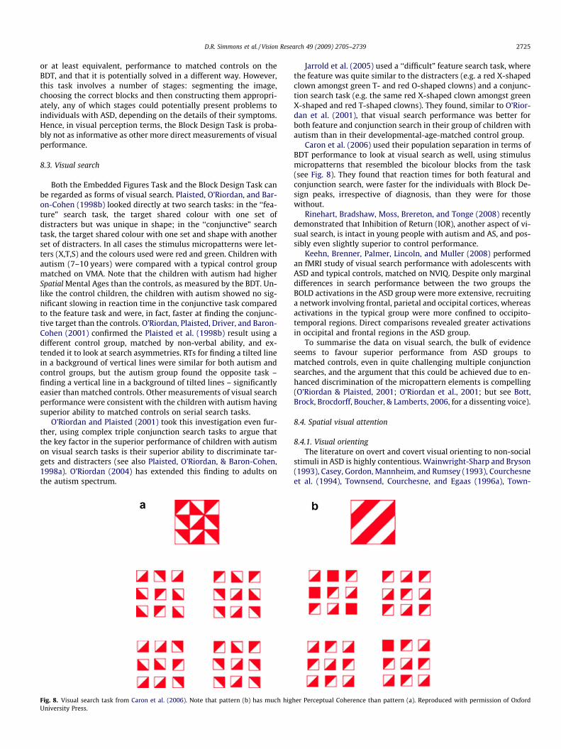
D.R. Simmons et al. / Vision Research 49 (2009) 2705–2739 2725
or at least equivalent, performance to matched controls on theBDT, and that it is potentially solved in a different way. However,this task involves a number of stages: segmenting the image,choosing the correct blocks and then constructing them appropri-ately, any of which stages could potentially present problems toindividuals with ASD, depending on the details of their symptoms.Hence, in visual perception terms, the Block Design Task is proba-bly not as informative as other more direct measurements of visualperformance.
8.3. Visual search
Both the Embedded Figures Task and the Block Design Task canbe regarded as forms of visual search. Plaisted, O’Riordan, and Bar-on-Cohen (1998b) looked directly at two search tasks: in the ‘‘fea-ture” search task, the target shared colour with one set ofdistracters but was unique in shape; in the ‘‘conjunctive” searchtask, the target shared colour with one set and shape with anotherset of distracters. In all cases the stimulus micropatterns were let-ters (X,T,S) and the colours used were red and green. Children withautism (7–10 years) were compared with a typical control groupmatched on VMA. Note that the children with autism had higherSpatial Mental Ages than the controls, as measured by the BDT. Un-like the control children, the children with autism showed no sig-nificant slowing in reaction time in the conjunctive task comparedto the feature task and were, in fact, faster at finding the conjunc-tive target than the controls. O’Riordan, Plaisted, Driver, and Baron-Cohen (2001) confirmed the Plaisted et al. (1998b) result using adifferent control group, matched by non-verbal ability, and ex-tended it to look at search asymmetries. RTs for finding a tilted linein a background of vertical lines were similar for both autism andcontrol groups, but the autism group found the opposite task –finding a vertical line in a background of tilted lines – significantlyeasier than matched controls. Other measurements of visual searchperformance were consistent with the children with autism havingsuperior ability to matched controls on serial search tasks.
O’Riordan and Plaisted (2001) took this investigation even fur-ther, using complex triple conjunction search tasks to argue thatthe key factor in the superior performance of children with autismon visual search tasks is their superior ability to discriminate tar-gets and distracters (see also Plaisted, O’Riordan, & Baron-Cohen,1998a). O’Riordan (2004) has extended this finding to adults onthe autism spectrum.
Fig. 8. Visual search task from Caron et al. (2006). Note that pattern (b) has much higUniversity Press.
Jarrold et al. (2005) used a ‘‘difficult” feature search task, wherethe feature was quite similar to the distracters (e.g. a red X-shapedclown amongst green T- and red O-shaped clowns) and a conjunc-tion search task (e.g. the same red X-shaped clown amongst greenX-shaped and red T-shaped clowns). They found, similar to O’Rior-dan et al. (2001), that visual search performance was better forboth feature and conjunction search in their group of children withautism than in their developmental-age-matched control group.
Caron et al. (2006) used their population separation in terms ofBDT performance to look at visual search as well, using stimulusmicropatterns that resembled the bicolour blocks from the task(see Fig. 8). They found that reaction times for both featural andconjunction search, were faster for the individuals with Block De-sign peaks, irrespective of diagnosis, than they were for thosewithout.
Rinehart, Bradshaw, Moss, Brereton, and Tonge (2008) recentlydemonstrated that Inhibition of Return (IOR), another aspect of vi-sual search, is intact in young people with autism and AS, and pos-sibly even slightly superior to control performance.
Keehn, Brenner, Palmer, Lincoln, and Muller (2008) performedan fMRI study of visual search performance with adolescents withASD and typical controls, matched on NVIQ. Despite only marginaldifferences in search performance between the two groups theBOLD activations in the ASD group were more extensive, recruitinga network involving frontal, parietal and occipital cortices, whereasactivations in the typical group were more confined to occipito-temporal regions. Direct comparisons revealed greater activationsin occipital and frontal regions in the ASD group.
To summarise the data on visual search, the bulk of evidenceseems to favour superior performance from ASD groups tomatched controls, even in quite challenging multiple conjunctionsearches, and the argument that this could be achieved due to en-hanced discrimination of the micropattern elements is compelling(O’Riordan & Plaisted, 2001; O’Riordan et al., 2001; but see Bott,Brock, Brocdorff, Boucher, & Lamberts, 2006, for a dissenting voice).
8.4. Spatial visual attention
8.4.1. Visual orientingThe literature on overt and covert visual orienting to non-social
stimuli in ASD is highly contentious. Wainwright-Sharp and Bryson(1993), Casey, Gordon, Mannheim, and Rumsey (1993), Courchesneet al. (1994), Townsend, Courchesne, and Egaas (1996a), Town-
her Perceptual Coherence than pattern (a). Reproduced with permission of Oxford
2726 D.R. Simmons et al. / Vision Research 49 (2009) 2705–2739
send, Harris, and Courchesne (1996b) and Dawson, Meltzoff, Oster-ling, Rinaldi, and Brown (1998) have argued that visual orienting isimpaired in ASD. Wainwright and Brown (1996) and Harris, Cour-chesne, Townsend, Carper, and Lord (1999) reported idiosyncraticvisual-spatial orienting performance, but Pascualvaca, Fantie,Papageorgiu, and Mirsky (1998), Leekam, Lopez, and Moore(2000), Iarocci and Burack (2004) and Senju et al. (2004) arguedthat ASD populations do not have a deficit in shifting attentionalfocus. Renner, Klinger, and Klinger (2006) found impairments inexogenous (reflexive), but not endogenous (voluntary) orientingperformance between their diagnostic groups.
Van der Geest, Kemner, Camfferman, Verbaten, and van Engel-und (2001) directly measured attentional shifts using eye tracking.They compared the performance of 16 children with HFA, aged 10–11 years, with CA- and IQ-matched controls in the ‘‘gap-and-over-lap” paradigm. This paradigm uses two phases: in the overlapcondition, the fixation point is visible when the target is presented(in another place); in the gap condition, nothing is visible at fixa-tion when the target is presented. Saccadic latencies to the targetare much shorter in the gap condition, and the difference is calledthe ‘‘gap effect”. It is argued that the two conditions involve differ-ent levels of attentional disengagement and hence can be used toanalyse the strength of these components to visual orientingbehaviour (Fischer & Weber, 1993). Van der Geest et al. (2001)found that, although there were no significant differences betweenthe autism group and controls in each condition individually, thegap effect was smaller in the autism group, suggesting that chil-dren with autism have lower levels of attentional engagement.This result was replicated in a similar clinical population by Kawa-kubo, Maekawa, Itoh, Hashimoto, and Iwanami (2004).
Goldstein, Johnson, and Minshew (2001) compared 103 care-fully characterized individuals with ASD to the same number ofmatched controls on a battery of tests of attentional processingand performed a complex factor analysis of the results (Mirsky, An-thony, Duncan, Ahearn, & Kellam, 1991). They argued from theiranalyses that apparent deficits in attention in ASD could be dueto deficits in complex decision making, or in psychomotor abilities.
Greenaway and Plaisted (2005) used a different approach. Theycompared top-down and bottom-up control using a combination ofcueing and visual search tasks employing task-irrelevant distrac-tors. Their ASD group consisted of 31 children, aged 9.5–13.5 years,and a similar number of typical controls, matched on NVIQ (Ra-ven’s matrices). Two types of target were used, which they de-scribe as ‘‘onset” (i.e. a single white symbol) or ‘‘colour” (i.e. ared symbol among white symbols). They found that the perfor-mance of the ASD group, in terms of top-down modulation, wasequivalent for the colour stimuli, but not for the onset stimuli.They suggested that individuals with ASD have difficulty prioritiz-ing dynamic stimuli, and linked this to a magnocellular pathwayprocessing deficit (Greenaway & Plaisted, 2005).
Belmonte (2000) and Belmonte and Yurgelun-Todd (2003) havelooked at the neural basis of covert attention shifting in ASD, usingERP and fMRI analysis, respectively. In both cases unusual activa-tions were found in the ASD group. In particular, Belmonte andYurgelun-Todd (2003) found less frontal activation and moreoccipital activation in the ASD group. Haist, Adamo, Westerfield,Courchesne, and Townsend (2005) also found this, at least for shortISI attention tasks, but also reported hypo-activation of the cere-bellum in either short or long ISI conditions. They suggested thatindividuals with ASD show a profound deficit in automatic spatialattention ability and an abnormal voluntary attention ability.
8.5. Attention to spatial scale
A number of studies have suggested that global grouping pro-cesses are deficient in ASD due to the unusual performance in
the Navon task (Navon, 1977). In this task, a figure, such as a largeletter ‘S’, is built up from smaller figures, such as small letter ‘c’s.Participants are asked to name either the small or the large letter.Typical observers find it easier to name the larger than the smallerletter, and this is reflected in differential performance in the task(the ‘‘global superiority effect”). Using Navon-type stimuli, somestudies have reported that participants with ASD tend to be moresensitive to the local stimulus than controls (Rinehart Bradshaw,Moss, Brereton, & Tonge, 2000, 2001 (autism only, not AS), Gross,2005; Behrmann et al., 2006a), but others have not (Deruelle, Ron-dan, Gepner, & Fagot, 2006; Edgin & Pennington, 2005; Mottron,Burack, Iarocci, Belleville, & Enns, 2003; Mottron, Burack, Stauder,& Robaey, 1999b; Ozonoff, Strayer, McMahon, & Filloux, 1994;Rondan & Deruelle, 2007).
Further evidence for enhanced attention to local visual informa-tion in ASD comes from Jarrold and Russell’s (1997) countingexperiment, where children with autism tended to count eachdot individually rather than rapidly and automatically enumeratethem (see also Gagnon, Mottron, Bherer, & Joanette, 2004; Mottronet al., 1999a). Plaisted et al. (1999) suggested that the finding of lo-cal precedence in children with autism depended on task instruc-tions. Plaisted et al. (1999) used the Navon task and either toldthe children whether the stimulus was at the local or global level(selective attention task) or gave them no instructions (dividedattention task). They found that no global advantage (local prece-dence) was found in the autism group in the divided attention task,but that performance was the same as controls in the selectiveattention task. These results suggested that switching the spatialscale of attention was most problematic for the children with aut-ism, and that the default state might well be set at a finer scalethan that of typical children.
Other suggestions concerning the spatial spread of attention inASD, tested using a variety of tasks, have been that individuals withASD have difficulty inhibiting distractors (an ‘‘inefficient atten-tional lens”: Burack, 1994; contradicted by Ozonoff & Strayer,1997), problems with broadening the spread of attention (Mann& Walker, 2003), a ‘‘local bias” (Behrmann et al., 2006a), a ‘‘piece-meal strategy of processing” (Iarocci, Burack, Shore, Mottron, &Enns, 2006) or have no difficulties relative to controls (Plaisted, Do-bler, Bell, & Davis, 2006).
Rondan and Deruelle (2007), in another variation on the Navon-type task, looked at configurational matches versus local matchesin schematic face and non-face stimuli. Participants were giventhe choice between matching a pattern with the same spatial con-figuration but different constituent shapes or the same shapes in aslightly different configuration. Adults with ASD favoured the localshape match rather than the configuration match, unlike the typi-cal controls, these being the same adults with ASD who had shownequivalent performance to the controls on a Navon-type task. Ron-dan and Deruelle (2007) argue that configurational, rather thanglobal, processing is disrupted in ASD.
In an epic attempt at empirical and theoretical synthesis, Wang,Mottron, Peng, Berthiaume, and Dawson (2007) presented anexhaustive study of different Navon-type tasks with individualson the autism spectrum, varying such factors as response type,exposure time and stimulus size. They interpreted their complexpattern of results as evidence for atypical local-to-global interfer-ence in ASD and local advantages in incongruent conditions, wherethe local and global stimuli are unmatched. They also argued thatthis local bias for visual processing potentially gives individuals onthe autism spectrum a flexibility which is unavailable to typicalindividuals and which may sometimes enhance and sometimesdiminish performance in visual tasks.
A final spanner in the works of this confusing literature comesfrom Rutherford et al. (2007b). They used the ‘‘Useful Field ofView” task, usually used to look at the spatial scope of attention

D.R. Simmons et al. / Vision Research 49 (2009) 2705–2739 2727
in elderly participants and drivers (e.g. Sekuler, Bennett, & Mamel-ak, 2000), to assess the attentional span of adults with ASD (meanage 25) under different amounts of central attentional load. Thistask comprises a central letter-identification task (focused-central)with a peripheral task (focused-peripheral) which requires the par-ticipant to choose the ‘‘spoke” of the ‘‘wheel” in which a flashedsquare was presented in a 4AFC. The final task is a divided atten-tion task in which both central and peripheral stimuli are pre-sented at the same time and two responses are required. Theperformance of the ASD group was broadly comparable to that ofthe controls, but a difference was found in the ‘‘attentional cost”of divided attention. Whereas performance of the ASD group inthe peripheral task was barely affected by the need to performthe central task at the same time, the control group was signifi-cantly worse. Curiously, the ASD group seemed to be more strate-gic than the controls in maintaining their attentional scope at anintermediate level: good enough to do the central task and maxi-mizing performance in the peripheral task. Rutherford et al.(2007b) argue that this result is problematic to explain in termsof current models of visual processing in ASD and also incompati-ble with much of the previous literature on attention in ASD (e.g.Plaisted et al., 1999).
8.6. Social attention
Social attention refers to the tendency for social stimuli – lar-gely faces and people – to attract attention. This is obviously likelyto be an area of difficulty for people with ASD. Some of the materialrelevant to this section has already been covered in the face andattention sections above, so only specific studies on social atten-tion will be mentioned here.
Swettenham et al. (1998) examined videotapes of children en-gaged in a structured play situation. The children were very young(about 20 months), being part of a study into the early diagnosis ofautism (Baron-Cohen et al., 1996). They found that whereas thecontrol groups tended to focus most on people, shifting attentionmainly from objects to people, the autism group tended to shiftfrom objects to other objects. The autism group also spent less timelooking at people and more time looking at objects (see also Swet-tenham et al., 2003; Willemsen-Swinkels, Buitelaar, Wejnen, & vanEngelund, 1998).
Probably the most famous study in this area is that of Klinet al. (2002) who asked 15 men with HFA and typical controlsto view social scenes (extracts from the movie ‘‘Who’s Afraid ofVirginia Woolf”) whilst their eye movements were tracked. Uponcoding, the best predictor of autism was reduced eye-region fixa-tion time. Individuals who fixated more on mouths than objectstended to score higher in social functioning tests, whereas thosewho focused more on objects showed the opposite tendency. Klinet al. (2002) argued that, in natural social scenes, eyes are theleast salient stimulus for individuals with ASD, and mouths,bodies and objects are relatively more salient. See also Jonesand Klin (2008).
Bird, Catmur, Silani, Frith, and Frith (2006) compared atten-tional modulation to house and face stimuli in an fMRI study using16 individuals with ASD and matched controls. Analysis showedthat responses to houses were modulated by attention in bothgroups, but only the controls showed attentional modulation tofaces. An analysis of effective connectivity suggested that in theASD group there was a failure to modulate connectivity betweenextrastriate areas and V1: which could result in a reduced saliencefor social stimuli.
Fletcher-Watson, Leekam, Benson, Frank, and Findlay (2009)presented adults with ASD with a complex social scene alongsideanother non-social scene and measured eye movements during a3-s viewing period. Their measurements indicated a ‘‘superfi-
cially normal” attentional preference for social information inthe ASD group, but they also found some subtle abnormalitieswhich may be significant in real-life scenarios. Riby and Hancock(2008) have reported reduced attention to people and faces instatic pictures of social interactions, in contrast to their controlsample of individuals with Williams syndrome. See also Ribyand Hancock (2009a, 2009b) for supporting data using differentparadigms and stimuli.
The prevailing view on social attention is, then, that individualswith ASD have difficulty with attending to people, particularly theeye regions of faces, in a range of stimulus situations. However,sometimes this difficulty is not readily apparent, especially in con-trolled laboratory tests.
8.7. Visual attention – summary
The literature on visual attention in ASD presents a confusingpicture. There is probably enough evidence to suggest that the pro-cessing of information across spatial scales works differently inASD, but precisely how is too early to call. Individuals with ASDalso seem to have problems with attending to people in naturalanimated scenes, but whether this is due to low-level visual factorsor higher-level motivational factors is still unclear.
9. Oculomotor problems
Goldberg et al. (2002) looked at saccadic functioning in 11 indi-viduals with HFA and typical controls, and using a number of stan-dard paradigms (anti-saccade, memory-guided saccade, predictedsaccade and gap/overlap tasks). There were a number of differ-ences between ASD and control performance on these tasks, whichsuggested significant abnormalities in oculomotor function. Gold-berg et al. (2002) suggested that these abnormalities implicatedbrain regions such as the dorsolateral pre-frontal cortex and thefrontal eye fields, as well as basal ganglia and parietal lobes asbeing affected in ASD.
Takarae, Minshew, Luna, and Sweeney (2004) measured visu-ally guided saccades in 46 individuals with HFA/AS and IQ-matched controls. The ASD group was found to have increasedvariability in saccade accuracy, together with mild saccadic hypo-metria in those without delayed language development. Takaraeet al. (2004) suggested that this pattern of saccadic abnormalitywas consistent with a chronic functional disturbance in thecerebellar vermis or its output in the fastigial nuclei. Later resultssuggested cerebellar or fronto-striatal circuitry atypicalities alongwith extrastriate areas that extract visual motion information orits transfer to sensorimotor areas (Takarae, Minshew, Luna, &Sweeney, 2005a; Takarae, Minshew, & Sweeney, 2005b).
However, despite this evidence in favour of eye movementabnormalities in ASD, Van der Geest, Kemner, Verbaten, andvan Engelund (2003) tested a variety of eye movement parame-ters as well as attentional tasks in a group of children with aut-ism and found that only the attention task showed groupdifferences.
Finally, Mottron et al. (2007) described atypical exploratorybehaviours towards inanimate objects among young children withautism. The most frequent atypical behaviour amongst 15 childrenwith autism aged 33–73 months was ‘‘lateral glancing”, mostly to-wards moving stimuli. This behaviour is described as ‘‘Fixating on atarget with the pupils turned toward an extreme corner of the eyesocket, where the head is turned in the opposite direction”.Mottron et al. (2007) explain this behaviour as an attempt toover-stimulate peripheral vision in order to regulate excessiveamounts of local detailed information.

2728 D.R. Simmons et al. / Vision Research 49 (2009) 2705–2739
10. Change detection
People with ASD often show strong reactions to changes in theenvironment, suggesting that they may detect changes more effi-ciently than typically developing people. However, Fletcher-Wat-son, Leekam, Turner, and Moxon (2006) reported no differencesin performance between adults with autism and typical adults ina change-detection task. Burack et al. (2009) initially also foundno differences in change-detection performance between childrenwith autism and typical children matched in non-verbal MA, butdifferences emerged when the detection failures were related tothe developmental level of the participants. Detection failures de-creased with increasing developmental level for typical children,but remained constant over the same developmental range forchildren with autism, pointing to an atypical developmental trajec-tory for change-detection among children with autism.
11. Neuroimaging studies
This section covers neuroimaging data not already coveredabove. Relevant reviews include DiCicco-Bloom et al. (2006) orcan be found in the collected volume Bauman and Kemper (2004).
11.1. Neuroanatomy
The developmental neuroanatomy of autism has recently beenreviewed by Courchesne et al. (2007). The key feature in earlydevelopment seems to be early brain overgrowth, possibly due toa failure of neural pruning, and consequent abnormalities in neuralpatterning and wiring. This pattern would be extensive local inter-connectivity at the expense of long-range connectivity, whichcould clearly result in disruption of a number of critical neural cir-cuits. See also McAlonan et al. (2005, 2008) and Hallahan et al.(2009) for more detail on current neuranatomical findings.
11.2. Electrophysiology
Atypical ERP and EEG responses to visual stimulation have beenfound in a number of studies on ASD (see reviews by Dawson et al.,2005; Kemner & van Engelund, 2006). These include abnormal spa-tial frequency processing (Boeschoten et al., 2007; Milne et al.,2009), and apparent functional under- and over-connectivity(Murias, Webb, Greenson, & Dawson, 2007).
11.3. Functional magnetic resonance imaging
Villalobos, Mizuno, Dahl, Kemmotsu, and Müller (2005) lookedat functional connectivity along the dorsal stream during visuallyprompted button presses in a group of eight high-functioningadults with ASD and matched controls. They found intact func-tional connectivity in the ASD group between V1 and superior pari-etal areas, suggesting that dorsal stream connectivity was intact.There was, however, reduced functional connectivity between V1and inferior frontal areas, which Villalobos et al. (2005) suggestmay be consistent with mirror-neuron system dysfunction. In con-trast, however, Mizuno, Villalobos, Davies, Dahl, and Muller (2006)found evidence for hyper-connectivity between thalamus andcerebral cortex using BOLD signal cross-correlation. Further func-tional connectivity abnormalities have been found between FFAand amygdala during face processing (Kleinhans et al., 2008).
Hadjikhani et al. (2004a) mapped visual cortical areas in a groupof individuals with HFA, and found that early visual areas were or-ganized as in typical controls, with typical ratios between centraland visual field representations. They argue that any visual pro-
cessing atypicalities in autism must come from higher-level visualareas and be the result of top-down influences.
12. Summary of visual processing studies
A considerable amount of data has been added to the field sincethe reviews of Dakin and Frith (2005) and Behrmann et al. (2006b),particularly using faces as stimuli. However, most of these data arecontradictory, or at least contested, so it is very hard to say any-thing with certainty. The new findings on colour vision difficulties(Franklin et al., 2008, in press; Heaton et al., 2008) are interestingand of theoretical importance, but await further replication withbetter characterized ASD populations. The possibility that a visualperception task might be able to distinguish individuals with aut-ism from those with Asperger Syndrome is also exciting (Spencer &O’Brien, 2006; Tsermentseli et al., 2008), but there are methodo-logical and replication issues with these results as well. Another is-sue for these studies is the likely disappearance of AspergerSyndrome as a distinct diagnostic category in the upcomingDSM-V. The difficulties for ASD participants with complex motionstimuli, which seemed so clear cut a few years ago, are now muchmore contentious, but local superiority effects in tasks such as vi-sual search seem to be holding their own. The literature on faceprocessing and visual attention is still highly contentious. The bal-ance is in favour of an attentional difficulty resulting in unusualeye gaze, which then results in unusual visual experience anddevelopment. The neuroimaging data indicates unusual patternsof brain activations, particularly when eye movements are not con-trolled, and there is a trend of finding higher activations at lowerlevels of visual processing, consistent with the idea of some sortof bottleneck in the processing pathway somewhere between pri-mary visual cortex and higher visual areas. Difficulties with eye re-gions of faces and low spatial frequencies are also fascinating, butmuch work remains to be done. The issue of visual acuity should beresolved following Ashwin et al. (2009), and no work has yet beendone on stereopsis. A really basic characterization of the visualprocessing capabilities of people with ASD would be extremelyuseful, even if all it could do was say with certainty ‘‘nothing iswrong here”, as some have already suggested (e.g. Hadjikhaniet al., 2004a). However, our suspicion is that there are a numberof subtle differences between the visual processing capabilities ofASD and typical populations which remain to be discovered. Theway forward must be for vision scientists, who know how to runrigorous psychophysical experiments, to collaborate with thoseof a more clinical orientation, who know how to characterize clin-ical populations accurately.
13. Theories of visual processing in ASDs
13.1. Cognitive theories
The dominant cognitive theories in ASD research have recentlybeen reviewed by Rajendran and Mitchell (2007). One of the mostinfluential ideas in the past 25 years has been the ‘‘Theory ofMind” (ToM) hypothesis, championed by Simon Baron-Cohenand colleagues at the University of Cambridge (Baron-Cohen,1995; Baron-Cohen, Leslie, & Frith, 1985). The theory focused onexplaining the poor performance of children with ASD on ‘‘falsebelief” tasks, where participants are required to interpret a situa-tion from the point of view of another, and argued that the cen-tral deficit in ASD was the ability (and possibly also motivation)to ‘‘read” others’ minds in social situations. Frith (2003) prefersto call this ability ‘‘mentalizing”. The ToM hypothesis did notreally have any specific predictions about visual processing inASD, other than a difficulty with interpreting facial expressions,

D.R. Simmons et al. / Vision Research 49 (2009) 2705–2739 2729
as demonstrated in the ‘‘Reading the mind in the Eyes” task (Bar-on-Cohen et al., 1997b). Recently this theory has metamorphosedinto the ‘‘Extreme Male Brain” hypothesis (Baron-Cohen, 2002,2003), which argues that ASD is characterized by poor ‘‘empathiz-ing” and high ‘‘systemizing”. Empathizing is related to Theory ofMind in that it involves interpreting the feelings of others, butadds that feelings must not only be interpreted, but also experi-enced by the viewer. Systemizing, on the other hand, is the abilityto analyse rule-based systems and is a trait associated with math-ematical, engineering and scientific ability. In visual processingterms, again, the implications of this theory are not direct, butif people with ASD are poor empathizers and good systemizers,one might expect a deficit in the recognition and interpretationof complex facial expressions and possibly also a skill in visualanalysis of the components of complex images. This latter skill,however, is possibly more associated with an alternative theoret-ical account of ASD: the ‘‘Weak Coherence” account (WC; Frith,1989; Happé & Frith, 2006).
Frith’s (1989) original formulation of weak coherence (origi-nally called ‘‘weak central coherence”) was built on the observa-tion of local processing biases in ASD populations, combinedwith the relative failure to extract the gist or meaning of eventsin everyday life. The account was supported by the original obser-vations in ASD populations of superior disembedding performancein the Embedded Figures Task (Shah & Frith, 1983), superior per-formance in the Block Design Task (Shah & Frith, 1993) and re-duced sensitivity to visual illusions (Happé, 1996). However,Happé and Frith (2006) have now reformulated the account inthree important ways in response to other empirical findings: (1)it is no longer regarded as a ‘‘core deficit in central processing” inASD, but as a more secondary outcome characterized by a localor detail-focused processing style; (2) this processing style canbe overcome in some situations; (3) it is one aspect of a more de-tailed cognitive profile which includes problems with Theory ofMind.
The final dominant cognitive theory of ASD has been the ‘‘Exec-utive Dysfunction” account (Hill, 2004, 2008; Ozonoff et al., 1991;Russell, 1997). This theory focuses on the difficulties that individ-uals with ASD have with tasks involving planning, mental flexibil-ity, generativity, inhibition and multi-tasking (Hill, 2008). Theoriginal motivation behind the theory was to account for thenon-social symptoms, such as repetitive and obsessional behav-iours, which were not well explained by ToM or WC accounts.Again, there are few precise predictions about visual performancethat come from the Executive Dysfunction theories, although diffi-culties with visual attention (especially attentional disengagementand divided attention), scene exploration and eye-movementsmight be expected.
13.2. Neural theories
One approach to understanding the neural basis of ASD is tocompare its signs and symptoms with those of conditions and syn-dromes of known neurological origin. This approach has led to sug-gestions of pathology in a large number of brain areas in ASD,including the cerebellum (Courchesne, 1997), the temporal lobes,especially the STS and the FFA (Zilbovicius et al., 2006), the amyg-dala (Baron-Cohen et al., 2000; Howard et al., 2000) and the frontallobes (Courchesne & Pierce, 2005). Recent attempts at synthesishave attempted to map the triad of impairments which is thoughtto characterize autism onto specific brain regions (Baron-Cohen &Belmonte, 2005; Frith, 2003). This has led to the idea of ‘‘socialbrain” dysfunction in ASD: a network of brain regions which in-cludes the Superior Temporal Sulcus, Superior Temporal Gyrus,Inferior Occipital Gyrus and Fusiform Gyrus in the occipital andtemporal lobes with the Amygdala and Medial Pre-frontal Cortex,
Anterior Cingulate Cortex and Orbitofrontal Cortex (see Fig. 9).These areas seem to safely cover functions such as poor facialexpression recognition, difficulties with interpreting eye gaze,problems with integrating sensory information and executivefunction problems.
What, however, could cause the (unspecified) neuropathologyin these brain regions? Recently, Baron-Cohen and co-workershave suggested that the difficulty lies with abnormalities in thelevels of the hormone testosterone in the mother’s womb, whichcauses neural developmental abnormalities (Baron-Cohen, 2006).
Other ideas at the neural level, in no particular order, includethe absence of ‘‘top-down” neural control (Frith, 2003), malfunc-tioning social reward mechanisms based around the amygdala(Schultz, 2005), neural connectivity abnormalities (Belmonteet al., 2004; Minshew & Williams, 2007; Rippon, Brock, Brown, &Boucher, 2007), abnormal cortical ‘‘minicolumns” (Casanova, Bux-hoeveden, Switala, & Roy, 2002), abnormal neural synchronization(Brock, Brown, Boucher, & Rippon, 2002), imbalance of excitationand inhibition (Rubenstein & Merzenich, 2003) and imbalancedspectrally timed adaptive resonance (Grossberg & Seidman, 2006).
There has also been a flurry of interest in the ‘‘Mirror neuronhypothesis” of autism, which suggests that the symptoms of aut-ism are related to dysfunctions in the so-called mirror-neuron sys-tem. Key papers on this topic, in chronological order, are: Williams,Whiten, Suddendorf, and Perrett (2001), Oberman et al. (2005),Dapretto et al. (2006), Iacaboni and Dapretto (2006), Oberman,Ramachandran, and Pineda (2008), Southgate and Hamilton(2008), Oberman and Ramachandran (2008).
With this bewildering, and ever growing, variety of hypothesesabout the neural basis of autism, it is difficult, and way beyond thescope of this review, to try to map each of them onto specific pre-dictions about visual processing. Obviously some of these theorieshave been developed with particular behavioural results in mind,such as superior disembedding performance and it relation toweak coherence which inspires the top-down control hypothesisof Frith (2003), whereas others have emerged almost entirely fromstudies at the neuroanatomical level (e.g. Casanova et al., 2002).However, there are three key ideas which have inspired much ofthe recent behavioural work on ASD which do need further treat-ment here.
13.2.1. Dorsal stream vulnerabilityThe ‘‘dorsal stream vulnerability” hypothesis (Atkinson & Brad-
dick, 2005; Braddick et al., 2003; Spencer et al., 2000) is based onthe differential performance in form and motion coherence tasksobserved in many neurodevelopmental disorders, including hemi-plegia, Fragile-X syndrome, dyslexia and Williams syndrome, aswell as autism, suggesting that the neural mechanisms supportingglobal motion sensitivity are particularly susceptible to damage,possibly because of the more stringent neural timing requirementsof this pathway (Atkinson & Braddick, 2005). This theory is notreally intended to explain autism, particularly as the vulnerabilityis not thought to be disorder-specific, but rather describes a poten-tial consequence of developmental disability which may be linkedback to underlying neuropathological causes.
We have shown in the section on motion perception in ASDthat there is no strong evidence for a low-level motion/temporalfrequency processing deficit in ASD, that the data on motioncoherence is somewhat equivocal and that biological motion isprobably affected adversely. These data do not therefore make astrong case for a dorsal pathway problem. As for form processing,Spencer and O’Brien (2006) and Tsermentseli et al. (2008) havefound an interesting deficit in autism, but not in Asperger Syn-drome. Whilst we have made clear that there are potential prob-lems with the stimuli in these experiments it is hard to see howthese problems would cause the pattern of results found. How-

2730 D.R. Simmons et al. / Vision Research 49 (2009) 2705–2739
ever, the new data on colour vision anomalies in ASD, the neuralbasis for which must surely involve parvo- and koniocellularpathways and the ventral stream, suggest that it is unlikely thatthe neuropathology in ASD is confined to the dorsal stream, un-less there are some complicated interactive effects between thetwo pathways.
13.2.2. Enhanced perceptual functioning (EPF)Enhanced perceptual functioning theory (Mottron & Burack,
2001; Mottron et al., 2006) has been increasingly influential in re-cent years as the focus on perceptual processing in ASD hasgrown. The theory is similar in many ways to Weak Coherence(Frith, 1989; Happé & Frith, 2006) and also shares much with Pla-isted’s (2001) reduced generalization theory, but its origins aredifferent.
The original theory states that a key aspect of ASD is a height-ened response to sensory stimulation in the neural mechanismsof perception from early in childhood (Mottron & Burack, 2001).This heightened response leads to an attentional focus on low-levelsensory phenomena, at the expense of social interaction, and aconsequent atypical wiring of the brain during development. Mot-tron and Burack (2001) name four areas of neural developmentwhere this manifests itself: growth of neuronal connections, re-dedication of cortical areas, suppression of inhibition and ‘‘func-tional persistence”, meaning the excessive refinement of low-levelprocesses at the expense of higher level processes. The inspirationfor this theory came from Mottron’s and Burack’s work with‘‘savants” on the autism spectrum, particularly those with en-hanced graphical abilities (e.g. Mottron & Belleville, 1993) andhas been supported by empirical data emerging from the Mon-
Fig. 9. The social brain. Reproduced with permission from the
treal-based labs of Mottron, Burack and their co-workers (e.g. Ber-tone et al., 2003, 2005).
In the later update to this model (Mottron et al., 2006), eight‘‘principles of autistic perception” are enumerated, with support-ing evidence:
[1] The default setting of perception in individuals with autismis more locally oriented than that of typical individuals.
[2] Increased gradient of neural complexity is inversely relatedto level of performance in low-level tasks.
[3] Early atypical behaviours have a regulatory function towardperceptual input.
[4] Perceptual primary and associative brain regions are atypi-cally activated during social and non-social tasks.
[5] Higher-order processing is optional in autism and manda-tory in typicals.
[6] Perceptual expertise underlies savant syndrome.[7] Savant syndrome provides a model for subtyping Pervasive
Developmental Disorders.[8] Enhanced functioning of primary perceptual brain regions
may account for the perceptual atypicalities in autism.
This is something of a ‘‘broad brush” theory, but there areplenty of ideas in it for the vision scientist to consider. Given therecency of the publication of this updated theory, it is largely con-sistent with current literature on perception in ASD and there hasnot been much time for it to be put to the test. Mottron et al.(2006) were careful to distinguish the updated EPF theory fromits competitors and pre-cursors. They argue that it differs fromthe Weak (Central) Coherence account in that it is couched interms of superior rather than inferior performance and that it is
Annual Review of Neuroscience (www.annualreviews.org).
D.R. Simmons et al. / Vision Research 49 (2009) 2705–2739 2731
not an optional ‘‘cognitive style” (Happé & Frith, 2006), but amandatory requirement. There is overlap with Plaisted’s (2001)reduced generalization theory but EPF’s scope is more comprehen-sive. Mottron et al. (2006) also borrowed the idea of the impor-tance of informational complexity from Minshew, Goldstein, andSiegel (1997) but mapped it onto the ‘‘negative” symptoms ofASD: the expected asymmetry therefore is between superior per-formance on ‘‘simple” tasks (e.g. grating detection, Bertone et al.,2005) and inferior performance on ‘‘complex” tasks (e.g. second-order motion, Bertone et al., 2003).
One potential problem with the EPF theory reported herethough is the suggestion of reduced ability of ASD populations inchromatic discrimination (Franklin et al., 2008, in press; Heatonet al., 2008). For example, the colour discrimination experimentused by Franklin et al. (in press) is what most vision scientistswould call a low-level task, so why is the perception of colour dif-ference not enhanced? Explaining these data within the restric-tions of EPF theory is therefore a challenge, assuming thatFranklin et al.’s (in press) result is replicable. Another significantchallenge within the context of vision science is defining what is‘‘simple” and what is ‘‘complex”, although that is potentially amore tractable problem (see Bertone & Faubert, 2006).
However, there is another challenge for the EPF model: whatexactly is the neuropathology that underlies it? We have recentlysuggested an explanation for the empirical data on visual process-ing in ASDs which may have an answer (Simmons et al., 2007). Weshall sketch the theory here, although a full version is in prepara-tion (see also Simmons et al., 2008).
13.2.3. Neural noise and autism spectrum disordersOne of the most surprising aspects of the sensory symptoms
of ASD is that individuals often report both hyper- and hypo-sensitivities within the same sense modality (Baranek et al.,2006; Bogdashina, 2003). We were also fascinated with the resultof Bertone et al. (2005) (see Fig. 1), suggesting that children withASD showed a (small) performance enhancement for first-ordercontrast detection in noise, but a performance decrement forsecond-order contrast detection. Another observation was thelarge variability reported in threshold data from ASD populations,relative to both typical and developmentally delayed populations.Whilst this could obviously be due to the inherent diagnosticvariability even within the different shades of the autism spec-trum, we wondered whether there was a more concrete cause.
Adding noise to a fixed signal intuitively involves a decrease insignal/noise ratio and therefore makes detectability worse. How-ever, the phenomenon of stochastic resonance can amplify the sig-nal/noise ratio under appropriate circumstances (i.e. a non-linearsystem with a fixed threshold, Wiesenfeld & Moss, 1995). This idea,a controversial one in vision science, has recently been used suc-cessfully to model contrast discrimination data (Goris, Wagemans,& Wichmann, 2008b; Goris, Zaenen, & Wagemans, 2008a). Thedetectability advantage for first-order luminance-defined gratingsin noise, therefore, could be due to increased internal noise inthe visual system of the ASD participants amplifying the signalslightly via stochastic resonance. The poorer performance with sec-ond-order gratings, on the other hand, comes because extractingthis signal involves combining information across more visual fil-ters, thereby increasing the noise level beyond the point where sto-chastic resonance can help (Schofield & Georgeson, 1999).
Could this explanation apply to other data reported in this re-view? Dakin and Frith (2005) already suggested that increased lev-els of local motion noise could be a factor in explaining the data onmotion coherence in ASD. Our analysis has shown that a likelycause of the discrepancies in these data is an increased level of cor-respondence noise in the ASD populations who have difficulty withseeing the motion, and preliminary data from our laboratory sug-
gest that increased internal noise may be measurable in partici-pants with ASD using equivalent noise analysis, similar to thatused by Dakin, Mareschal, and Bex (2005) (Simmons et al., 2007),and that adding noise to biological motion stimuli can be highlydisruptive to ASD participants (McKay, Mackie, Piggott, Simmons,& Pollick, 2006).
A further aspect, which is highly speculative, of the explanatorypower of an internal noise model for ASD concerns the colour dis-crimination data of Franklin et al. (in press). Human colour visioninvolves cone opponency, and it is well known that subtractingnoisy signals results in a decrease in signal/noise ratio unless thenoise is correlated. It is plausible, therefore, that the benefits of sto-chastic resonance for detection in ASD will only be found for ach-romatic, and not chromatic judgements.
Added noise in the internal representations of faces could makethe identities of new faces harder to learn, subtle facial expressionsharder to distinguish and the eye regions of faces less distinct andsalient. Other visual perceptual data, such as superior performancein the Embedded Figures and Block Design Tasks, are harder to ex-plain, but we have already pointed out that these are relatively‘‘high-level” tasks into which a number of component atypicalitiescould feed. However, a combination of stochastic resonance ampli-fying local differences and masking hiding global differences wouldseem to have considerable explanatory mileage in the context ofthe visual search data at least.
Where could this noise come from? A number of the neuralmodels cited above suggest that there is a proliferation of neuralconnections in the sensory cortex of individuals with ASD (Bel-monte et al., 2004; Casanova et al., 2002; see Baron-Cohen & Bel-monte, 2005; Minshew et al., 2007, for reviews), so neuralcrosstalk within the cortex is one plausible mechanism. However,recent investigations into the genetic basis of ASD have suggestedthat glutamergic and GABA-ergic synapses could be affected (seeGarber, 2007; Persico & Bourgeron, 2006). Mis-firing synapsescould easily result in noisy signals in the visual system.
Whilst it might seem somewhat brazen to add another theoryto the already theory-rich field of autism research, we would arguethat the neural noise model has considerable explanatory powerand is parsimonious, but flexible enough to account for a wide vari-ety of phenomena in the literature on vision and ASD, and maywell have broader implications for understanding the conditionas a whole. The other advantage is that this theory is readily test-able using established vision science paradigms such as equivalentnoise analysis and is physiologically plausible. Other studies thathave suggested that neural noise is involved in ASD include:Rubenstein and Merzenich (2003), Belmonte and Yugelun-Todd(2003), Dakin and Frith (2005), Thornton (2006), Sanchez-Marinand Padina-Marilla (2008), Alcantara (2008), Lugo, Doti, and Fau-bert (2008) and Franklin et al. (in press).
13.3. A final challenge
Arguably the key theoretical battleground in ASD research atthe moment lies between those who favour a ‘‘social orienting”theory of ASD development (e.g. Schultz, 2005) and those who fa-vour a more bottom-up theory (e.g. Mottron et al., 2006). In otherwords, is the information coming in corrupted and therefore caus-ing the problems that manifest themselves as ASD, or is veridicalinput information corrupted higher up in the brain circuits associ-ated with the social brain, the mirror-neuron system, or the exec-utive function system in the frontal lobes? As often, the truthprobably lies in a compromise between bottom-up and top-downmechanisms, as top-down feedback undoubtedly has a role to play.
There are two further questions which emerge from this review.What does ‘‘social” mean? Does it mean anything to do with a hu-man being, or possibly animal, such that faces, voices, bodies etc.

2732 D.R. Simmons et al. / Vision Research 49 (2009) 2705–2739
count as social and things that are not faces, bodies or voices arenon-social? Or is social just another word for complex or unpre-dictable? If so, what is the critical level of complexity/unpredict-ability and how can this be quantified?
The second is more prosaic. What is the best way to engage ASDpopulations in research? One of the curses in this field is the size ofthe error bars, which always seem to be at least twice as large inthe ASD data compared to the controls. Is this just unavoidableor are there effective ways of splitting the ASD population? Wehave seen that diagnostic measures (Asperger vs. autism), or per-formance measures (Block Design peaks, Famous face recognition,Verbal IQ) can be useful here (see also Happé et al., 2006), but obvi-ously this technique increases the problem of participant recruit-ment. These will be further complicated by the imminentupdates to diagnostic criteria planned for DSM-V.
A final thing to remember is that the ultimate goal of our re-search should be to help individuals with ASD to achieve theirown goals more effectively.
14. Conclusion
In the 4 years or so since the publication of the Dakin and Frith’s(2005) wryly titled review ‘‘Vagaries of visual perception inautism” a great deal of data has been produced, and the theoreticallandscape of vision research in ASD has changed somewhat, withthe even greater influence of neuroimaging techniques. However,some of the most interesting developments have come from old-fashioned psychophysics and it is probably the case that we needmore of these before we can progress much further with the brainimaging.
Acknowledgments
We thank all of the generous individuals who have devotedtime to reading and commenting on draft versions of this review,not least the two anonymous referees, one of whom arguably de-serves acknowledgment as a co-author. In particular we thank Ste-ven Dakin, Anna Franklin, Sue Leekam, Iain McClure, DavidMcGonigle, Branka Milivojevic Mits Ota, Bill Phillips, Thusha Rajen-dran and Mary Stewart. Grant support for the authors has beengratefully received from ESRC (‘‘Social Interactions: A CognitiveNeurosciences Approach” (RES-060-25-0010)) (D.S., F.P.), AutismSpeaks (‘‘The processing of biological motion patterns in adultswith high-functioning autism” No. 1428) (D.S., F.P., P.M.) andEPSRC (P.M., A.R.). Studentships from ESRC/MRC (L.M.) and ESRC/CASE (A.R.) and summer research bursaries from the WellcomeTrust and the Nuffield Foundation (E.T.).
References
Abell, F., Happé, F., & Frith, U. (2000). Do triangles play tricks? Attribution of mentalstates to animated shapes in normal and abnormal development. CognitiveDevelopment, 15, 1–16.
Adolphs, R., Sears, L., & Piven, J. (2001). Abnormal processing of social informationfrom faces in autism. Journal of Cognitive Neuroscience, 13(2), 232–240.
Alcantara (2008). Paper presented at the international meeting for autism research,London, 15th–17th May 2008.
American Psychiatric Association. (1994). In Diagnostic and statistical manual ofmental disorders (4th ed.). Washington, DC: American Psychiatric Association.
Altevogt, B. M., Hanson, S. L., & Leshner, A. I. (2008). Autism and the environment:Challenges and opportunities for research. Paediatrics, 121(6), 1225–1229.
Annaz, D., Remington, A., Milne, E., Coleman, M., Campbell, R., Thomas, M., et al.(submitted for publication). Atypical development of motion processingtrajectories in children with autism.
Ashwin, E., Ashwin, C., Rhydderch, D., Howells, J., & Baron-Cohen, S. (2009). Eagle-eyed visual acuity: An experimental investigation of enhanced perception inautism. Biological Psychiatry, 65, 17–21.
Ashwin, C., Baron-Cohen, S., Wheelwright, S., O’Riordan, M., & Bullmore, E. T. (2007).Differential activation of the amygdala and the ‘social brain’ during fearful face-processing in Asperger syndrome. Neuropsychologia, 45, 2–14.
Ashwin, C., Chapman, E., Colle, L., & Baron-Cohen, S. (2006a). Impaired recognitionof negative basic emotions in autism: A test of the amygdala theory. SocialNeuroscience, 1(3–4), 349–363.
Ashwin, C., Wheelwright, S., & Baron-Cohen, S. (2005). Laterality biases to chimericfaces in Asperger syndrome: What is ‘Right’ about faces-processing? Journal ofAutism and Developmental Disorders, 35(2), 183–196.
Ashwin, C., Wheelwright, S., & Baron-Cohen, S. (2006b). Finding a face in the crowd:Testing the anger superiority effect in Asperger syndrome. Brain and Cognition,61, 78–95.
Asperger, H. (1944). Die autistischen Psychopathen im Kindesalter. In U. Frith (Ed.),Autism and Asperger Syndrome Archiv für Psychiatrie und Nervenkrankheiten(pp. 76–136). Cambridge, UK: Cambridge University Press.
Atkinson, J. (2000). In The developing visual brain. Oxford, UK: Oxford University Press.Atkinson, J., & Braddick, O. (2005). Dorsal stream vulnerability and autistic
disorders: The importance of comparative studies of form and motioncoherence in typically developing children and children with developmentaldisorders. Cahiers de Psychologie Cognitive, 23(1–2), 49–58.
Bach, M. (1996). The Freiburg visual acuity test – Automatic measurement of visualacuity. Optometry and Vision Science, 73, 49–53.
Bach, M., & Dakin, S. (in press). Commentary on ‘‘Eagle-eyed visual acuity: Anexperimental investigation of enhanced perception in autism”. BiologicalPsychiatry.
Back, E., Ropar, D., & Mitchell, P. (2007). Do the eyes have it? Inferring mental statesfrom animated faces in autism. Child Development, 78(2), 397–411.
Bailey, A. J., Braeutigam, S., Jousmäki, V., & Swithenby, S. (2005). Abnormalactivation of face processing systems at early and intermediate latency inindividuals with autism spectrum disorder: A magnetoencephalographic study.European Journal of Neuroscience, 21, 2575–2585.
Bailey, A., Le Couteur, A., Gottesman, I., Bolton, P., Simonoff, E., Yuzda, E., et al.(1995). Autism as a strongly genetic disorder – Evidence from a British twinstudy. Psychological Medicine, 25(1), 63–77.
Baker, A. E. Z., Lane, A., Angley, M. T., & Young, R. L. (2008). The relationship betweensensory processing patterns and behavioural responsiveness in autisticdisorder: A pilot study. Journal of Autism and Developmental Disorders, 38(5),867–875.
Banaschewski, T., Ruppert, S., Tannock, R., Albrecht, B., Becker, A., Uebel, H., et al.(2006). Colour perception in ADHD. Journal of Child Psychology and Psychiatry,47, 568–572.
Baranek, G., David, F. J., Poe, M. D., Stone, W. L., & Watson, L. R. (2006). Sensoryexperiences questionnaire: Discriminating sensory features in young childrenwith autism, developmental delays and typical development. Journal of ChildPsychology and Psychiatry, 47(6), 591–601.
Bar-Haim, Y., Shulman, C., Lamy, D., & Reuveni, A. (2006). Attention to eyes andmouth in high-functioning children with autism. Journal of Autism andDevelopmental Disorders, 36(1), 131–137.
Barlow, H. B., & Tripathy, S. P. (1997). Correspondence noise and signal pooling inthe detection of coherent visual motion. Journal of Neuroscience, 17(20),7954–7966.
Baron-Cohen, S. (1995). Mindblindness: An essay on autism and theory of mind.Cambridge, MA: Bradford/MIT Press.
Baron-Cohen, S. (2002). The extreme male brain theory of autism. Trends inCognitive Sciences, 6, 248–254.
Baron-Cohen, S. (2003). In The essential difference. London, UK: Penguin.Baron-Cohen, S. (2006). The hyper-systemising, assortative mating theory of
autism. Progress in Neuro-psychopharmacology and Biological Psychiatry, 30(5),865–872.
Baron-Cohen, S., & Belmonte, M. K. (2005). Autism: A window onto thedevelopment of the social analytic brain. Annual Review of Neuroscience, 28,109–126.
Baron-Cohen, S., Cox, A., Baird, G., Swettenham, J., Drew, A., Nightingale, N., et al.(1996). Psychological markers of autism at 18 months of age in a largepopulation. British Journal of Psychiatry, 168, 158–163.
Baron-Cohen, S., Leslie, A. M., & Frith, U. (1985). Does the autistic child have a‘‘theory of mind”? Cognition, 21, 37–46.
Baron-Cohen, S., Richler, J., Bisarya, D., Gurunuthan, N., & Wheelwright, S. (2003).The systemizing quotient: An investigation of adults with Asperger syndromeor high-functioning autism, and normal sex differences. Proceedings of the RoyalSociety of London, Series B, Biological Sciences, 358, 361–374.
Baron-Cohen, S., Ring, H., Bullmore, E., Wheelwright, S., Ashwin, C., & Williams, S.(2000). The amygdala theory of autism. Neuroscience and Behaviour Reviews, 24,355–364.
Baron-Cohen, S., Ring, H. A., Wheelwright, S., Bullmore, E. T., Brammer, M. J.,Simmons, A., et al. (1999). Social intelligence in the normal and autistic brain:An fMRI study. European Journal of Neuroscience, 11, 1891–1898.
Baron-Cohen, S., Spitz, A., & Cross, P. (1993). Can children with autism recognizesurprise? Cognition and Emotion, 7, 507–516.
Baron-Cohen, S., & Wheelwright, S. (2004). The empathy quotient: An investigationof adults with Asperger syndrome or high functioning autism, and normal sexdifferences. Journal of Autism and Developmental Disorders, 34, 163–175.
Baron-Cohen, S., Wheelwright, S., Hill, J., Raste, Y., & Plumb, I. (2001a). The ‘‘Readingthe mind in the eyes” test revised version: A study with normal adults, andadults with Asperger syndrome or high-functioning autism. Journal of ChildPsychology and Psychiatry, 42(2), 241–251.
Baron-Cohen, S., Wheelwright, S., & Jolliffe, T. (1997). Is there a ‘‘language of theeyes”? Evidence from normal adults and adults with autism or Aspergersyndrome. Visual Cognition, 4, 311–332.

D.R. Simmons et al. / Vision Research 49 (2009) 2705–2739 2733
Baron-Cohen, S., Wheelwright, S., Skinner, R., Martin, J., & Clubley, E. (2001b). Theautism spectrum quotient (AQ): Evidence from Asperger syndrome/highfunctioning autism, males and female, scientists and mathematicians. Journalof Autism and Developmental Disorders, 31, 5–17.
Barton, J. J. S., Cherkasova, M. V., Hefter, R., Cox, T. A., O’Connor, M., & Manoach, D. S.(2004). Are patients with social developmental disorders prosopagnosic?Perceptual heterogeneity in the Asperger and socio-emotional processingdisorders. Brain, 127, 1706–1716.
Barton, J. J. S., Hefter, R., Cherkasova, M. V., & Manoach, D. S. (2007). Investigationsof face expertise in the social developmental disorders. Neurology, 69, 860–870.
Bauman, M. L., & Kemper, T. L. (Eds.), The neurobiology of autism. Baltimore, MD:Johns Hopkins University Press.
Behrmann, M., Avidan, G., Leonard, G. L., Kimchi, R., Luna, B., Humphreys, K., et al.(2006a). Configural processing in autism and its relationship to face processing.Neuropsychologia, 44, 110–129.
Behrmann, M., Thomas, C., & Humphreys, K. (2006b). Seeing it differently: Visualprocessing in autism. Trends in Cognitive Sciences, 10(6), 258–263.
Belmonte, M. (2000). Abnormal attention in autism shown by steady-state visualevoked potentials. Autism, 4(3), 269–285.
Belmonte, M. K., Allen, G., Beckel-Mitchener, A., Boulanger, L. M., Carper, R. A., &Webb, S. J. (2004). Autism and abnormal development of brain connectivity.Journal of Neuroscience, 24, 9228–9231.
Belmonte, M., & Yurgelun-Todd, D. A. (2003). Functional anatomy of impairedselective attention and compensatory processing in autism. Cognitive BrainResearch, 17, 651–664.
Ben-Sasson, A., Cermak, S. A., Orsmond, G. I., Tager-Flusberg, H., Carter, A. S., Kadlec,M. B., et al. (2007). Extreme sensory modulation behaviors in toddlers withautism spectrum disorders. American Journal of Occupational Therapy, 61(5),584–592.
Bertenthal, B. I., & Pinto, J. (1994). Global processing of biological motions.Psychological Science, 5, 221–225.
Bertone, A., & Faubert, J. (2006). Demonstrations of decreased sensitivity to complexmotion information not enough to propose an autism-specific neural etiology.Journal of Autism and Developmental Disorders, 36(1), 55–64.
Bertone, A., Mottron, L., Jelenic, P., & Faubert, J. (2003). Motion perception in autism:A ‘‘complex” issue. Journal of Cognitive Neuroscience, 15(2), 218–225.
Bertone, A., Mottron, L., Jelenic, P., & Faubert, J. (2005). Enhanced and diminishedvisuo-spatial information processing in autism depends on stimuluscomplexity. Brain, 128, 2430–2441.
Bird, G., Catmur, C., Silani, G., Frith, C., & Frith, U. (2006). Attention does notmodulate neural responses to social stimuli in autism spectrum disorders.NeuroImage, 31(4), 1614–1624.
Blake, R., & Shiffrar, M. (2007). Perception of human motion. Annual Review ofPsychology, 58, 47–73.
Blake, R., Turner, L. M., Smoski, M. J., Pozdol, S. L., & Stone, W. L. (2003). Visualrecognition of biological motion is impaired in children with autism.Psychological Science, 14, 151–157.
Boeschoten, M. A., Kenemans, J. L., van Engelund, H., & Kemner, C. (2007). Abnormalspatial frequency processing in high-functioning children with pervasivedevelopmental disorder (PDD). Clinical Neurophysiology, 118(9), 2076–2088.
Bogdashina, O. (2003). In Sensory perceptual issues in autism: Different sensoryexperiences – Different perceptual worlds. London, UK: Jessica Kingsley.
Bölte, S., Holtmann, M., Poustka, F., Scheurich, A., & Schmidt, L. (2007). Gestaltperception and local–global processing in high-functioning autism. Journal ofAutism and Developmental Disorders, 37, 1493–1504.
Bölte, S., Hubl, D., Dierks, T., Holtmann, M., & Poustka, F. (2008). An fMRI study oflocally oriented perception in autism: Altered early visual processing in theblock design test. Journal of Neural Transmission, 115(3), 545–552.
Boraston, Z., Blakemore, S. J., Chilvers, R., & Skuse, D. (2007). Impaired sadnessrecognition is linked to social interaction in autism. Neuropsychologia, 45(7),1501–1510.
Boraston, Z. L., Corden, B., Miles, L. K., Skuse, D. H., & Blakemore, S. J. (2008). Briefreport: Perception of genuine and posed smiles by individuals with autism.Journal of Autism and Developmental Disorders, 38(3), 574–580.
Bott, L., Brock, J., Brocdorff, N., Boucher, J., & Lamberts, K. (2006). Perceptual similarityin autism. Quarterly Journal of Experimental Psychology, 59(7), 1237–1254.
Boucher, J., & Lewis, V. (1992). Unfamiliar face recognition in relatively able autistic-children. Journal of Child Psychology and Psychiatry and Allied Disciplines, 33(5),843–859.
Boucher, J., Lewis, V., & Collis, G. (1998). Familiar face and voice matching andrecognition in children with autism. Journal of Child Psychology and Psychiatryand Allied Disciplines, 39(2), 171–181.
Bourgeron, T. (2008). Genes, synapses and autism spectrum disorders. In D. J.Selkoe, A. Triller, & Y. Christen (Eds.), Research and perspectives in Alzheimer’sdisease (pp. 169–179). Berlin, Germany: Springer-Verlag.
Bowler, D. M., & Thommen, E. (2000). Attribution of mechanical and social causalityto animated displays by children with autism. Autism, 4, 147–171.
Braddick, O., Atkinson, J., & Wattam-Bell, J. (2003). Normal and anomalousdevelopment of visual motion processing: motion coherence and ‘dorsalstream vulnerability’. Neuropsychologia, 41(8), 1769–1784.
Braverman, M., Fein, D., Lucci, D., & Waterhouse, L. (1989). Affect comprehension inchildren with pervasive developmental disorders. Journal of Autism andDevelopmental Disorders, 19, 301–316.
Brian, J. A., & Bryson, S. E. (1996). Disembedding performance and recognitionmemory in autism/PDD. Journal of Child Psychology and Psychiatry, 37(7),865–872.
Brian, J. A., Tipper, S. P., Weaver, B., & Bryson, S. E. (2003). Inhibitory mechanisms inautism spectrum disorders: Typical selective inhibition of location versusfacilitated perceptual processing. Journal of Child Psychology and Psychiatry, 44,552–560.
Brock, J., Brown, C. C., Boucher, J., & Rippon, G. (2002). The temporal binding deficithypothesis of autism. Developmental Psychopathology, 14, 209–214.
Brosnan, M. J., Scott, F. J., Fox, S., & Pye, J. (2004). Gestalt processing in autism:Failure to process perceptual relationships and the implications for contextualunderstanding. Journal of Child Psychology and Psychiatry, 45(3), 459–469.
Brown, C., Gruber, T., Boucher, J., Rippon, G., & Brock, J. (2004). Gammaabnormalities during perception of illusory contour figures in autism. Cortex,41, 364–376.
Burack, J. A. (1994). Selective attention deficits in persons with autism – Preliminaryevidence of an inefficient attentional lens. Journal of Abnormal Psychology,103(3), 535–543.
Burack, J. A., Iarocci, G., Flanagan, T. D., & Bowler, D. M. (2004). On mosaics andmelting pots: Conceptual considerations of comparison and matchingstrategies. Journal of Autism and Developmental Disorders, 34(1), 65–73.
Burack, J. A., Joseph, S., Russo, N., Shore, D. I., Porporino, M., & Enns, J. T. (2009).Change detection in naturalistic pictures among children with autism. Journal ofAutism and Developmental Disorders, 39(3), 471–479.
Campbell, R., Lawrence, K., Mandy, W., Mitra, C., Jeyakuma, L., & Skuse, D. (2006).Meanings in motion and faces: Developmental associations between theprocessing of intention from geometrical animations and gaze detectionaccuracy. Development and Psychopathology, 18, 99–118.
Capps, L., Yirmiya, N., & Sigman, M. D. (1992). Understanding of simple and complexemotions in non-retarded children with autism. Journal of Child Psychology andPsychiatry, 33, 1169–1182.
Caron, M.-J., Mottron, L., Berthiaume, C., & Dawson, M. (2006). Cognitivemechanisms, specificity and neural underpinnings of visuospatial peaks inautism. Brain, 129(7), 1780–1802.
Casanova, M. F., Buxhoeveden, D. P., Switala, A. E., & Roy, E. (2002). Minicolumnarpathology in autism. Neurology, 58, 428–432.
Casey, B. J., Gordon, C. T., Mannheim, G. B., & Rumsey, J. M. (1993). Dysfunctionalattention in autistic savants. Journal of Clinical and ExperimentalNeuropsychology, 15(6), 933–946.
Castelli, F. (2005). Understanding emotions from standardized facial expressions inautism and normal development. Autism, 9(4), 428–449.
Castelli, F., Frith, C., Happé, F., & Frith, U. (2002). Autism, Asperger syndrome andbrain mechanisms for the attribution of mental states to animated shapes.Brain, 125, 1839–1849.
Celani, G., Battachi, M. W., & Arcidiacono, L. (1999). The understanding of theemotional meaning of facial expressions in people with autism. Journal ofAutism and Developmental Disorders, 29, 57–66.
Charles, J. M., Carpenter, L. A., Jenner, W., & Nicholas, J. S. (2008). Recent advances inautism spectrum disorders. International Journal of Psychiatry in Medicine, 38(2),133–140.
Chawarska, K., Klin, A., & Volkmar, F. (2003). Automatic attention cueing througheye movement in 2-year-old children with autism. Child Development, 74(4),1108–1122.
Chawarska, K., & Volkmar, F. (2007). Impairments in monkey and human facerecognition in 2-year-old toddlers with autism spectrum disorder and.Developmental Delay, 10(2), 266–279.
Constantino, J. N., Davis, S. A., Todd, R. D., Schindler, M. K., Gross, M. M., Brophy, S. L.,et al. (2003). Validation of a brief quantitative measure of autistic traits:Comparison of the social responsiveness scale with the autism diagnosticinterview – Revised. Journal of Autism and Developmental Disorders, 33(4),427–433.
Courchesne, E. (1997). Brainstem, cerebellar and limbic neuroanatomicalabnormalities in autism. Current Opinion in Neurobiology, 7(2),269–278.
Courchesne, E., & Pierce, K. (2005). Brain overgrowth in autism during a critical timein development: Implications for frontal pyramidal neuron and interneurondevelopment and connectivity. International Journal of DevelopmentalNeuroscience, 23(2–3), 153–170.
Courchesne, E., Pierce, K., Schumann, C. M., Redcay, E., Buckwalter, J. A., Kennedy, D.P., et al. (2007). Mapping early brain development in autism. Neuron, 56(2),399–413.
Courchesne, E., Townsend, J., Akshoomoff, N. A., Saitoh, O., Yeung-Courchesne, R.,Lincoln, A. J., et al. (1994). Impairment in shifting attention in autistic andcerebellar patients. Behavioral Neuroscience, 108, 1–18.
Critchley, H. D., Daly, E. M., Bullmore, E. T., Williams, S. C. R., Van Amelsvoort, T.,Robertson, D. M., et al. (2000). The functional neuroanatomy of socialbehaviour: Changes in cerebral blood flow when people with autistic disorderprocess facial expressions. Brain, 123, 2203–2212.
Curby, K. M., Schyns, P. G., Gosselin, F., & Gauthier, I. (2003). Differential use ofspatial frequency scales for face recognition in a person with Asperger’sSyndrome. Journal of Vision, 3(9). doi:10.1167/3.9.821 (pp. 821, 821a), http://journalofvision.org/3/9/821/ [abstract].
Cutting, J. E. (1978). Generation of synthetic male and female walkers throughmanipulation of a biomechanical invariant. Perception, 7, 393–405.
Dakin, S., & Frith, U. (2005). Vagaries of visual perception in autism. Neuron, 48,497–507.
Dakin, S. C., Mareschal, I., & Bex, P. J. (2005). Local and global limitations ondirection integration assessed using equivalent noise analysis. Vision Research,45(24), 3027–3049.

2734 D.R. Simmons et al. / Vision Research 49 (2009) 2705–2739
Dalton, K. M., Nacewicz, B. M., Johnstone, T., Schaefer, H. S., Gernsbacher, M. A.,Goldsmith, H. H., et al. (2005). Gaze fixation and the neural circuitry of faceprocessing in autism. Nature Neuroscience, 8(4), 519–526.
Dapretto, M., Davies, M. S., Pfeifer, J. H., Scott, A. A., Sigman, M., Bookheimer, S. Y.,et al. (2006). Understanding emotions in others: Mirror neuron dysfunction inchildren with autism spectrum disorders. Nature Neuroscience, 9(1), 28–30.
Davies, S., Bishop, D., Manstead, A. S. R., & Tantam, D. (1994). Face perception inchildren with autism and Asperger syndrome. Journal of Child Psychiatry andPsychology, 35, 1033–1057.
Dawson, G., Carver, L., Meltzoff, A. N., Panagiotides, H., McPartland, J., & Webb, S. J.(2002a). Neural correlates of face and object recognition in young children withautism spectrum disorder, developmental delay, and typical development. ChildDevelopment, 73(3), 700–717.
Dawson, G., Meltzoff, A. N., Osterling, J., Rinaldi, J., & Brown, E. (1998). Children withautism fail to orient to naturally occurring social stimuli. Journal of Autism andDevelopmental Disorders, 28(6), 479–485.
Dawson, G., Webb, S. J., Carver, L., Panagiotides, H., & McPartland, J. (2004). Youngchildren with autism show atypical brain responses to fearful versus neutralfacial expressions. Developmental Science, 7(3), 340–359.
Dawson, G., Webb, S. J., & McPartland, J. (2005). Understanding the nature of faceprocessing impairment in autism: Insights from behavioral andelectrophysiological studies. Developmental Neuropsychology, 27(3), 404–424.
Dawson, G., Webb, S. J., Schellenberg, G. D., Dager, S., Friedman, S., Aylward, E., et al.(2002b). Defining the broader phenotype of autism: Genetic, brain andbehavioral perspectives. Developmental Psychopathology, 14, 581–611.
De Gelder, B., Vroomen, J., & van der Heide, L. (1991). Face recognition and lip-reading in autism. European Journal of Cognitive Psychology, 3, 69–86.
De Jong, M. C., van Engelund, H., & Kemner, C. (2008). Attentional effects of gazeshifts are influenced by emotion and spatial frequency, but not in autism.Journal of American Academy of Child and Adolescent Psychiatry, 47(4), 443–454.
de Jonge, M. V., Kemner, C., de Haan, E. H., Coppens, J. E., van den Berg, J. T. P., & vanEngelund, H. (2007). Visual information processing in high-functioningindividuals with autism spectrum disorders and their parents.Neuropsychology, 21(1), 65–73.
De Jonge, M. V., Kemner, C., & van Engelund, H. (2006). Superior disembeddingperformance of high-functioning individuals with autism spectrum disordersand their parents: The need for subtle measures. Journal of Autism andDevelopmental Disorders, 36(5), 677–683.
De Wit, T. C. J., Schlooz, W. A. J. M., Hulstijn, W., & van Lier, R. (2007). Visualcompletion and complexity of visual shape in children with pervasivedevelopmental disorder. European Journal of Child and Adolescent Psychiatry,16, 168–177.
Del Viva, M. M., Igliozzi, R., Tancredi, R., & Brizzolara, D. (2006). Spatial and motionintegration in children with autism. Vision Research, 46, 1242–1252.
Dennis, M., Lockyer, L., Lazenby, A. L., Donnelly, R. E., Wilkinson, M., & Schoonheyt,W. (1999). Intelligence patterns among children with high-functioning autism,phenylketonuria and childhood head injury. Journal of Autism andDevelopmental Disorders, 29(1), 5–17.
Deruelle, C., Rondan, C., Gepner, B., & Fagot, J. (2006). Processing of compound visualstimuli by children with autism and Asperger syndrome. International Journal ofPsychology, 41(2), 97–106.
Deruelle, C., Rondan, C., Gepner, B., & Tardif, C. (2004). Spatial frequency and faceprocessing in children with autism and Asperger syndrome. Journal of Autismand Developmental Disorders, 34, 199–210.
DiCicco-Bloom, E., Lord, C., Zwaigenbaum, L., Courchesne, E., Dager, S. R., Schmitz, C.,et al. (2006). The developmental neurobiology of autism spectrum disorder.Journal of Neuroscience, 26(26), 6897–6906.
Diekhof, E. K., Falkai, P., & Gruber, O. (2008). Functional neuroimaging of rewardprocessing and decision-making: A review of aberrant motivational andaffective processing in addiction and mood disorders. Brain Research Reviews,59(1), 164–184.
Dover, C. J., & Le Couteur, A. (2007). How to diagnose autism. Archives of Disease inChildhood, 92(6), 540–545.
Dunn, L. M., Dunn, L. M., & Whetton, C. (1982). In British picture vocabulary scale.Windsor: NFER-NELSON.
Edgin, J. O., & Pennington, B. F. (2005). Spatial cognition in autism spectrumdisorders: Superior, impaired or just intact? Journal of Autism and DevelopmentalDisorders, 35(6), 729–745.
Ehlers, S., Nyden, A., Gillberg, C., Sandberg, A. D., Dahlgren, S. O., Hjelmquist, E., et al.(1997). Asperger syndrome, autism and attention disorders: A comparativestudy of the cognitive profiles of 120 children. Journal of Child Psychology andPsychiatry and Allied Disciplines, 38(2), 207–217.
Ekman, P., & Friesen, W. V. (1975). Unmasking the face: A guide to recognizingemotions from facial cues. Englewood Cliffs, NJ: Prentice Hall.
Elsabbagh, M., Volein, A., Csibra, G., Holmboe, K., Garwood, H., Tucker, L., et al.(2009). Neural correlates of eye gaze processing in the infant broader autismphenotype. Biological Psychiatry, 65, 31–38.
Falter, C. M., Plaisted, K. C., & Davis, G. (2008). Visuo-spatial processing in autism –Testing the predictions of extreme male brain theory. Journal of Autism andDevelopmental Disorders, 38, 507–515.
Farnsworth, D. (1943). The Farnsworth Munsell 100-hue dichotomous tests forcolour vision. Journal of the Optical Society of America, 33, 568–576.
Fecteau, S., Mottron, L., Berthiaume, C., & Burack, J. A. (2003). Developmentalchanges of autistic symptoms. Autism, 7(3), 255–268.
Field, D. J., Hayes, A., & Hess, R. F. (1993). Contour integration by the human visualsystem: Evidence for a local ‘‘association field”. Vision Research, 33, 173–193.
Fischer, B., & Weber, H. (1993). Express saccades and visual attention. Behaviouraland Brain Sciences, 16, 553–610.
Fletcher-Watson, S., Leekam, S. R., Benson, V., Frank, M. C., & Findlay, J. M. (2009).Eye-movements reveal attention to social information in autism spectrumdisorder. Neuropsychologia, 47(1), 248–257.
Fletcher-Watson, S., Leekam, S. R., Turner, M. R., & Moxon, L. (2006). Do people withautistic spectrum disorder show normal selection for attention? Evidence fromchange blindness. British Journal of Psychology, 97, 537–554.
Franklin, A., Sowden, P., Notman, L., Gonzalez-Dixon, M., West, D., Alexander, I.,et al. (in press). Reduced chromatic discrimination in children with autismspectrum disorders. Developmental Science.
Franklin, A., Pilling, M., & Davies, I. R. L. (2005). The nature of infant colourcategorization: Evidence from eye-movements on a target detection task.Journal of Experimental Child Psychology, 91, 227–248.
Franklin, A., Sowden, P., Burley, R., Notman, L., & Alder, E. (2008). Colour perceptionin children with autism. Journal of Autism and Developmental Disorders, 38,1837–1847.
Freitag, C. M., Konrad, C., Häberlein, M., Kleser, C., von Gontard, A., Reith, W., et al.(2008). Perception of biological motion in autism spectrum disorders.Neuropsychologia, 46, 1480–1494.
Frith, U. (1989). Autism: Explaining the enigma (1st ed.). Blackwell Science.Frith, U. (2003). Autism: Explaining the enigma (2nd ed.). Blackwell Science.Gagnon, L., Mottron, L., Bherer, L., & Joanette, Y. (2004). Quantification judgement in
high functioning autism: Superior or different? Journal of Autism andDevelopmental Disorders, 34(6), 679–689.
Garber, K. (2007). Neuroscience – Autism’s cause may reside in abnormalities at thesynapse. Science, 317, 190–191.
Gauthier, I., & Tarr, M. J. (1997). Becoming a ‘‘greeble” expert: Exploringmechanisms for face recognition. Vision Research, 37(2), 1673–1682.
Gauthier, I., Tarr, M. J., Anderson, A. W., Skudlarski, P., & Gore, J. C. (1999). Activationof the middle fusiform ‘face area’ increases with expertise in naming novelobjects. Nature Neuroscience, 2(6), 568–573.
Gepner, B., de Gelder, B., & de Schonen, S. (1996a). Face processing in autistics:Evidence for a generalized deficit? Child Neuropsychology, 2, 123–139.
Gepner, B., Deruelle, C., & Grynfeltt, S. (2001). Motion and emotion: A novelapproach to the study of face processing by young autistic children. Journal ofAutism and Developmental Disorders, 31(1), 37–45.
Gepner, B., & Mestre, D. (2002). Brief report: Postural reactivity to fast visual motiondifferentiates autistic children from children with Asperger syndrome. Journalof Autism and Developmental Disorders, 32, 231–238.
Gepner, B., Mestre, D., Masson, G., & de Schonen, S. (1996b). Postural effects ofmotion vision in young autistic children. NeuroReport, 6, 1211–1214.
Gillberg, C., & Coleman, M. (2000). In The biology of the autistic syndromes (3rd ed.).Oxford: Mackeith Press.
Gioia, J. V., & Brosgole, L. (1988). Visual and auditory affect recognition in singlydiagnosed mentally retarded patients, mentally retarded patients withautism and normal young children. International Journal of Neuroscience,43, 149–163.
Golarai, G., Grill-Spector, K., & Reiss, A. L. (2006). Autism and the development offace processing. Clinical Neuroscience Research, 6, 145–160.
Goldberg, M. C., Lasker, A. G., Zee, D. S., Garth, E., Tien, A., & Landa, R. J. (2002).Deficits in the initiation of eye movements in the absence of a visual target inadolescents with high-functioning autism. Neuropsychologia, 40(12),2039–2049.
Goldberg, M. C., Mostow, A. J., Vecera, S. P., Larson, J. C. G., Mostofsky, S. H.,Mahone, E. M., et al. (2008). Evidence for impairments in using static linedrawings of eye gaze cues to orient visual–spatial attention in children withhigh functioning autism. Journal of Autism and Developmental Disorders, 38(8),1405–1413.
Goldstein, G., Johnson, C. R., & Minshew, N. J. (2001). Attentional processes inautism. Journal of Autism and Developmental Disorders, 31(4), 433–440.
Goris, R. L. T., Zaenen, P., & Wagemans, J. (2008a). Some observations on contrastdetection in noise. Journal of Vision, 8(9):4, 1–15. http://journalofvision.org/8/9/4/. doi:10.1167/8.9.4.
Goris, R. L. T., Wagemans, J., & Wichmann, F. A. (2008). Modelling contrastdiscrimination data suggest both the pedestal effect and stochastic resonance tobe caused by the same mechanism. Journal of Vision, 8(15):17, 1–21. http://journalofvision.org/8/15/17/. doi:10.1167/8.15.17.
Gosselin, F., & Schyns, P. G. (2001). Bubbles: A technique to reveal the use ofinformation in recognition tasks. Vision Research, 41(17), 2261–2271.
Gowen, E., Stanley, J., & Miall, R. C. (2008). Movement interference in autism-spectrum disorder. Neuropsychologia, 46, 1060–1068.
Grandin, T. (1992). An inside view of autism. In E. Schopler & G. B. Mesibov (Eds.),High-functioning individuals with autism (pp. 105–126). New York: PlenumPress.
Grandin, T. (2009). Visual abilities and sensory differences in a person with autism.Biological Psychiatry, 65(1), 15–16.
Grandin, T., & Scariano, M. (1986). Emergence: Labelled autistic Novato. CA: Arena.Greenaway, R., & Plaisted, K. (2005). Top-down attentional modulation in autistic
spectrum disorders is stimulus specific. Psychological Science, 16, 987–994.Grelotti, D. J., Klin, A., Gauthier, I., Skudlarski, P., Cohen, D. J., Gore, J. C., et al. (2005).
FMRI activation of the fusiform gyrus and amygdala to cartoon characters butnot to faces in a boy with autism. Neuropsychologia, 43(3), 373–379.
Grice, S. J., Halit, H., Farroni, T., Baron-Cohen, S., Bolton, P., & Johnson, M. J. (2005).Neural correlates of eye-gaze detection in young children with autism. Cortex,41, 342–353.

D.R. Simmons et al. / Vision Research 49 (2009) 2705–2739 2735
Grice, S. J., Spratling, M. W., Karmiloff-Smith, A., Halit, H., Csibra, G., de Haan, M.,et al. (2001). Disordered visual processing and oscillatory brain activity inautism and Williams syndrome. NeuroReport, 12, 2697–2700.
Gross, T. F. (2002). Perception of human and non-human facial age bydevelopmentally disabled children. Journal of Autism and DevelopmentalDisorders, 32(3), 169–179.
Gross, T. F. (2004). The perception of four basic emotions in human and nonhumanfaces by children with autism and other developmental disabilities. Journal ofAbnormal Child Psychology, 32(5), 469–480.
Gross, T. F. (2005). Global–local precedence in the perception of facial age andemotional expression by children with autism and other developmentaldisabilities. Journal of Autism and Developmental Disorders, 35(6), 773–785.
Gross, T. F. (2008). Recognition of immaturity and emotional expressions in blendedfaces by children with autism and other developmental disabilities. Journal ofAutism and Developmental Disorders, 38(2), 297–311.
Grossberg, S., & Seidman, D. (2006). Neural dynamics of autistic behaviors:Cognitive, emotional and timing substrates. Psychological Review, 113(3),483–525.
Grossman, J. B., Klin, A., Carter, A. S., & Volkmar, F. R. (2000). Verbal bias inrecognition of facial emotions in children with Asperger syndrome. Journal ofChild Psychology and Psychiatry, 41(3), 369–379.
Hadjikhani, N., Chabris, C. F., Joseph, R. M., Clark, J., McGrath, L., Aharon, I., et al.(2004a). Early visual cortex organization in autism: An fMRI study. NeuroReport,15(2), 267–270.
Hadjikhani, N., Joseph, R. M., Snyder, J., Chabris, C. F., Clark, J., Steele, S., et al.(2004b). Activation of the fusiform gyrus when individuals with autismspectrum disorder view faces. NeuroImage, 22, 1141–1150.
Haist, F., Adamo, M., Westerfield, M., Courchesne, E., & Townsend, J. (2005). Thefunctional neuroanatomy of spatial attention in autism spectrum disorder.Developmental Neuropsychology, 27(3), 425–458.
Hall, G. B. C., Szechtman, H., & Nahmias, C. (2003). Enhanced salience and emotionrecognition in autism: A PET study. American Journal of Psychiatry, 160,1439–1441.
Hallahan, B., Daly, E. M., McAlonan, G., Loth, E., Toal, F., O’Brien, F., et al.(2009). Brain morphometry volume in autistic spectrum disorder: Amagnetic resonance imaging study of adults. Psychological Medicine, 39(2),337–346.
Happé, F. G. E. (1994). Wechsler IQ profile and theory of mind in autism: A researchnote. Journal of Child Psychology and Psychiatry, 35(8), 1461–1471.
Happé, F. G. E. (1996). Studying weak central coherence at low levels: Children withautism do not succumb to visual illusions. A research note. Journal of ChildPsychology and Psychiatry, 37, 873–877.
Happé, F., & Frith, U. (2006). The weak coherence account: Detail-focused cognitivestyle in autism spectrum disorders. Journal of Autism and DevelopmentalDisorders, 37(1), 5–25.
Happé, F., Ronald, A., & Plomin, R. (2006). Time to give up on a single explanation forautism. Nature Neuroscience, 9(10), 1218–1220.
Harris, N. S., Courchesne, E., Townsend, J., Carper, R. A., & Lord, C. (1999).Neuroanatomic contributions to slowed orienting of attention in childrenwith autism. Cognitive Brain Research, 8, 61–71.
Hauck, M., Fein, D., Maltby, N., Waterhouse, L., & Feinstein, C. (1998). Memory forfaces in children with autism. Child Neuropsychology, 4(3), 187–198.
Heaton, P., Ludlow, A., & Roberson, D. (2008). When less is more: Poordiscrimination but good colour memory in autism. Research in AutismSpectrum Disorders, 2, 127–156.
Hefter, R. L., Manoach, D. S., & Barton, J. J. S. (2005). Perception of facial expressionand facial identity in subjects with social developmental disorders. Neurology,65, 1620–1625.
Heider, F., & Simmel, M. (1944). An experimental study of apparent behavior.American Journal of Psychology, 57, 243–259.
Hermelin, B., & O’Connor, N. (1970). Psychological experiments with autistic children.Oxford, UK: Pergamon.
Herrington, J. D., Baron-Cohen, S., Wheelwright, S. J., Singh, K. D., Bullmore, E. T.,Brammer, M., et al. (2007). The role of MT+/V5 during biological motionperception in Asperger syndrome: An fMRI study. Research in Autism SpectrumDisorders, 1(1), 14–27.
Hill, E. L. (2004). Evaluating the theory of executive dysfunction in autism.Developmental Review, 24, 189–233.
Hill, E. L. (2008). Executive functioning in autism spectrum disorder: Where it fits inthe causal model. In E. McGregor, M. Nuñez, K. Cebula, & J. C. Gómez (Eds.),Autism: An integrated view from neurocognitive, clinical and intervention research.Oxford, UK: Blackwell.
Hincha-Ownby, M. (2008). History of autism in the DSM: Diagnostic criteria forautism spectrum disorders. http://autismaspergerssyndrome.suite101.com/article.cfm/history_of_autism_in_the_dsm.
Hobson, R. P. (1986a). The autistic child’s appraisal of expressions of emotion.Journal of Child Psychology and Psychiatry, 27, 321–342.
Hobson, R. P. (1986b). The autistic child’s appraisal of expressions of emotions: Afurther study. Journal of Child Psychology and Psychiatry, 27, 671–680.
Hobson, R. P. (1987). The autistic child’s recognition of age- and sex-relatedcharacteristics of people. Journal of Autism and Developmental Disorders, 17,63–79.
Hobson, R. P., Ouston, J., & Lee, A. (1988a). What’s in a face? The case of autism.British Journal of Psychology, 79, 441–453.
Hobson, R. P., Ouston, J., & Lee, A. (1988b). Emotion recognition in autism:Coordinating faces and voices. Psychological Medicine, 18, 911–923.
Hobson, R. P., Ouston, J., & Lee, A. (1989). Naming emotion in faces and voices:Abilities and disabilities in autism and mental retardation. British Journal ofDevelopmental Psychology, 7, 237–250.
Howard, M. A., Cowell, P. E., Boucher, J., Broks, P., Maves, A., Farrant, A., et al. (2000).Convergent neuroanatomical and behavioural evidence of an amygdalahypothesis of autism. NeuroReport, 11, 2931–2935.
Hoy, J. A., Hatton, C., & Hare, D. (2004). Weak central coherence: A cross-domainphenomenon specific to autism? Autism, 8(3), 267–281.
Hubert, B., Wicker, B., Moore, D. G., Monfardini, E., Duverger, H., Da Fonséca, D., et al.(2007). Brief report: Recognition of emotional and non-emotional biologicalmotion in individuals with autistic spectrum disorders. Journal of Autism andDevelopmental Disorders, 37, 1386–1392.
Hubl, D., Bölte, S., Feineis-Matthews, S., Lanfermann, H., Federspiel, A., Strik, W.,et al. (2003). Functional imbalance of visual pathways indicates alternative faceprocessing strategies in autism. Neurology, 61, 1232–1237.
Humphreys, K., Minshew, N., Leonard, G. L., & Behrmann, M. (2007). A fine-grainedanalysis of facial expression processing in high-function adults with autism.Neuropsychologia, 45, 685–695.
Hutt, C., & Ounstead, C. (1966). The biological significance of gaze avoidance withparticular reference to the syndrome of infantile autism. Behavioural Science, 11,346–356.
Iacaboni, M., & Dapretto, M. (2006). The mirror neuron system and theconsequences of its dysfunction. Nature Reviews Neuroscience, 7, 942–951.
Iarocci, G., & Burack, J. A. (2004). Intact covert orienting to peripheral cues amongchildren with autism. Journal of Autism and Developmental Disorders, 34,257–264.
Iarocci, G., Burack, J. A., Shore, D. I., Mottron, L., & Enns, J. T. (2006). Global–localvisual processing in high-functioning children with autism: Structural vs.implicit task biases. Journal of Autism and Developmental Disorders, 36(1),117–129.
Jackson, L. (2002). Freaks, geeks and Asperger syndrome: A user guide to adolescence.London, UK: Jessica Kingsley.
Jackson, J. (2003). Multicoloured mayhem: Parenting the many shades of adolescentsand children with autism, Asperger syndrome and ADHD. London, UK: JessicaKingsley.
Jambaque, I., Mottron, L., Ponsot, G., & Chiron, C. (1998). Autism and visual agnosiain a child with right occipital lobectomy. Journal of Neurology, Neurosurgery andPsychiatry, 65(4), 555–560.
Jarrold, C., & Brock, J. (2004). To match or not to match? Methodological issues inautism-related research. Journal of Autism and Developmental Disorders, 34(1),81–86.
Jarrold, C., Gilchrist, I. D., & Bender, A. (2005). Embedded figures detection in autismand typical development: Preliminary evidence of a double dissociation inrelationships with visual search. Developmental Science, 8(4), 344–351.
Jarrold, C., & Russell, J. (1997). Counting abilities in autism: Possible implicatationsfor central coherence theory. Journal of Autism and Developmental Disorders,27(1), 25–37.
Jarrold, C., & Scott-Samuel, N. E. (2005). Motion perception in autism spectrumdisorder: We’ll move forward once the data become more coherent. Cahiers dePsychologie Cognitive, 23(1–2), 121–131.
Jemel, B., Mottron, L., & Dawson, M. (2006). Impaired face processing in autism: Factor artifact? Journal of Autism and Developmental Disorders, 36(1), 91–106.
Jennings, W. B. (1973). A study of the preference for affective cues in autisticchildren. Unpublished Ph.D. thesis, Memphis State University.
Johansson, G. (1973). Visual perception of biological motion and a model for itsanalysis. Perception and Psychophysics, 14, 201–211.
Johnson, C. R., & Rakison, D. H. (2006). Early categorization of animate/inanimateconcepts in young children with autism. Journal of Developmental and PhysicalDisabilities, 18(2), 73–89.
Jolliffe, T., & Baron-Cohen, S. (1997). Are people with autism and Aspergersyndrome faster than normal on the embedded figures test? Journal of ChildPsychology and Psychiatry and Allied Disciplines, 38(5), 527–534.
Jones, W., & Klin, A. (2008). Altered salience in autism: Developmental insights,consequences and questions. In E. McGregor, M. Núñez, K. Cebula, & J. C. Gómez(Eds.), Autism: An integrated view from neurocognitive, clinical and interventionresearch (pp. 62–82). Oxford, UK: Blackwell.
Jones, R. S. P., Quigney, C., & Huws, J. C. (2003). First-hand accounts of sensoryperceptual experiences in autism: A qualitative analysis. Journal of Intellectualand Developmental Disability, 28(2), 112–121.
Joseph, R. M., & Tanaka, J. (2003). Holistic and part-based face recognition inchildren with autism. Journal of Child Psychology and Psychiatry, 44(4),529–542.
Kaland, N., Mortensen, E. L., & Smith, L. (2007). Disembedding performance inchildren and adolescents with Asperger syndrome or high-functioning autism.Autism, 11(1), 81–92.
Kanner, L. (1943). Autistic disturbances of affective contact. Nervous Child, 2,217–250.
Kanwisher, N., McDermott, J., & Chun, M. (1997). The fusiform face area: A modulein human extrastriate cortex specialized for face perception. Journal ofNeuroscience, 17(11), 4302–4311.
Kaplan, M. (2006). Seeing through new eyes: Changing the lives of children with autism,Asperger syndrome and other developmental disabilities through vision therapy.London: Jessica Kingsley.
Kaplan, M., Rimland, B., & Edelson, S. M. (1999). Strabismus in autism spectrumdisorder. Focus on Autism and other Developmental Disabilities, 14(2),101–105.

2736 D.R. Simmons et al. / Vision Research 49 (2009) 2705–2739
Kätsyri, J., Saalasti, S., Tiipana, K., von Wendt, L., & Sams, M. (2008). Impairedrecognition of facial emotions from low-spatial frequencies in Aspergersyndrome. Neuropsychologia, 46, 1888–1897.
Kawakubo, Y., Maekawa, H., Itoh, K., Hashimoto, O., & Iwanami, A. (2004). Spatialattention in individuals with pervasive developmental disorders using the gapoverlap task. Psychiatry Research, 125(3), 269–275.
Keehn, B., Brenner, L., Palmer, E., Lincoln, A. J., & Muller, R. A. (2008). Functionalbrain organization for visual search in ASD. Journal of the InternationalNeuropsychological Society, 14(6), 990–1003.
Kemner, C., Lamme, V. A. F., Kovacs, I., & van Engelund, H. (2007). Integrity of lateraland feedbackward connections in visual processing in children with pervasivedevelopmental disorder. Neuropsychologia, 45, 1293–1298.
Kemner, C., Schuller, A.-M., & van Engelund, H. (2006). Electrocortical reflections offace and gaze processing in children with pervasive developmental disorder.Journal of Child Psychology and Psychiatry, 47(10), 1063–1072.
Kemner, C., & van Engelund, H. (2006). ERPs and eye movements reflect atypicalvisual perception in pervasive developmental disorder. Journal of Autism andDevelopmental Disorders, 36(1), 45–54.
Kern, J. K., Trivedi, M. H., Garver, C. R., Grannemann, B. D., Andrews, A. A., Savla, J. S.,et al. (2006). The pattern of sensory processing abnormalities in autism. Autism,10(5), 480–494.
Kern, J. K., Trivedi, M. H., Grannemann, B. D., Garver, C. R., Johnson, D. G.,Andrews, A. A., et al. (2007). Sensory correlations in autism. Autism, 11(2),123–134.
Kleinhans, N. M., Richards, T., Sterling, L., Stegbauer, K. C., Mahurin, R., Johnson, L. C.,et al. (2008). Abnormal functional connectivity in autism spectrum disordersduring face processing. Brain, 131, 1000–1012.
Kleinman, J., Marciano, P. L., & Ault, R. L. (2001). Advanced theory of mind in high-functioning adults with autism. Journal of Autism and Developmental Disorders,31(1), 29–36.
Klin, A. (2000). Attributing social meaning to ambiguous visual stimuli in higher-functioning autism and Asperger syndrome: The social attribution task. Journalof Child Psychology and Psychiatry, 41(7), 831–846.
Klin, A., & Jones, W. (2006). Attributing social and physical meaning to ambiguousvisual displays in individuals with higher-functioning autism spectrumdisorders. Brain and Cognition, 61, 40–53.
Klin, A., Jones, W., Schultz, R., Volkmar, F., & Cohen, D. (2002). Visual fixationpatterns during viewing of naturalistic social situations as predictors of socialcompetence in individuals with autism. Archives of General Psychiatry, 59(9),809–816.
Klin, A., Sparrow, S. S., de Bildt, A., Cicchetti, D. V., Cohen, D. J., & Volkmar, F. R.(1999). A normed study of face recognition in autism and related disorders.Journal of Autism and Developmental Disorders, 29(6), 499–508.
Klinger, L. G., & Dawson, G. (2001). Prototype formation in autism. Development andPsychopathology, 13(1), 111–124.
Kogan, C. S., Bertone, A., Cornish, K., Boutet, I., Der Kaloustian, V. M., Andermann, E.,et al. (2004). Intergrative cortical dysfunction and pervasive motion perceptiondeficit in fragile X syndrome. Neurology, 63, 1634–1639.
Kylliäinen, A., Brauetigem, S., Hietanen, J. K., Swithenby, S. J., & Bailey, A. J. (2006).Face- and gaze-sensitive neural responses in children with autism: Amagnetoencephalographic study. European Journal of Neuroscience, 24,2679–2690.
Kylliäinen, A., & Hietanen, J. K. (2004). Attention orienting by another’s gazedirection in children with autism. Journal of Child Psychology and Psychiatry,45(3), 435–444.
Kylliäinen, A., & Hietanen, J. K. (2006). Skin conductance responses to anotherperson’s gaze in children with autism. Journal of Autism and DevelopmentalDisorders, 36(4), 517–525.
Lahaie, A., Mottron, L., Arguin, M., Berthiaume, C., Jemel, B., & Saumier, D. (2006).Face perception in high-functioning autistic adults: Evidence for superiorprocessing of face parts, not for a configural face-processing deficit.Neuropsychology, 20(1), 30–41.
Langdell, T. (1978). Recognition of faces: An approach to the study of autism. Journalof Child Psychology and Psychiatry, 19, 255–268.
Lee, P. S., Foss-Feig, J., Henderson, J. G., Kenworthy, L. E., Gilotty, S., Gaillard, W.D., et al. (2007). Atypical neural substrates of embedded figures taskperformance in children with autism spectrum disorder. NeuroImage, 38(1),184–193.
Leekam, S., Baron-Cohen, S., Perrett, D., Milders, M., & Brown, S. (1997). Eye-direction detection: A dissociation between geometric and joint attention skillsin autism. British Journal of Developmental Psychology, 15, 77–95.
Leekam, S., Hunnisett, E., & Moore, C. (1998). Targets and cues: Gaze-following inchildren with autism. Journal of Child Psychology and Psychiatry and AlliedDisciplines, 39(7), 951–962.
Leekam, S., Libby, S. J., Wing, L., Gould, J., & Taylor, C. (2002). The diagnosticinterview for social and communication disorders: Algorithms for ICD-10childhood autism and Wing and Gould autistic spectrum disorder. Journal ofChild Psychology and Psychiatry, 43(3), 327–342.
Leekam, S., Lopez, B., & Moore, C. (2000). Attention and joint attention in preschoolchildren with autism. Developmental Psychology, 36(2), 261–273.
Leekam, S., Nieto, C., Libby, S. J., Wing, L., & Gould, J. (2007). Describing the sensoryabnormalities of children and adults with autism. Journal of Autism andDevelopmental Disorders, 37(5), 894–910.
Leopold, D. A., O’Toole, A. J., Vetter, T., & Blanz, V. (2001). Prototype-referencedshape encoding revealed by high-level aftereffects. Nature Neuroscience, 4(1),89–94.
Lockyer, L., & Rutter, M. (1970). A five to fifteen year follow up study of infantilepsychosis: IV. Patterns of cognitive ability. British Journal of Social and ClinicalPsychology, 9, 152–163.
Lopez, B., Donnelly, N., Hadwin, J. A., & Leekam, S. R. (2004). Face processing in high-functioning adolescents with autism: Evidence for weak central coherence.Visual Cognition, 11(6), 673–688.
Lord, C., Risi, S., Lambrecht, L., Cook, E. H., Jr., Leventhal, B. L., DiLavore, P. C., et al.(2000). The autism diagnostic observation schedule – generic: A standardmeasure of social and communication deficits associated with the spectrum ofautism. Journal of Autism and Developmental Disorders, 24, 659–685.
Lord, C., Rutter, M., & Le Couteur, A. (1994). The autism diagnostic interview –Revised: A revised version of a diagnostic interview for caregivers of individualswith possible pervasive developmental disorders. Journal of Autism andDevelopmental Disorders, 24, 659–686.
Loveland, K., Tunali-Kotoski, B., Chen, Y., Ortegon, J., Pearson, D., Brelsford, K., et al.(1997). Emotion recognition in autism: Verbal and nonverbal information.Development and Psychopathology, 9, 579–593.
Ludlow, A. K., & Wilkins, A. J. (2009). Case report: Color as a therapeuticintervention. Journal of Autism and Developmental Disorders, 39, 815–818.
Ludlow, A. K., Wilkins, A. J., & Heaton, P. (2006). The effect of coloured overlays onreading ability in children with autism. Journal of Autism and DevelopmentalDisorders, 36(4), 507–516.
Lugo, E., Doti, R., & Faubert, J. (2008). Ubiquitous crossmodal stochastic resonance inhumans: Auditory noise facilitates tactile, visual and proprioceptive sensations.PLoS One, 3(8), e2860.
Manjaly, Z. M., Bruning, N., Neufang, S., Stephan, K. E., Brieber, S., Marshall, J. C., et al.(2007). Neurophysiological correlates of relatively enhanced local visual searchin autistic adolescents. NeuroImage, 35, 283–291.
Mann, T. A., & Walker, P. (2003). Autism and a deficit in broadening the spread ofvisual attention. Journal of Child Psychology and Psychiatry and Allied Disciplines,44(2), 274–284.
Maurer, D., Le Grand, R., & Mondloch, C. J. (2002). The many faces of configuralprocessing. Trends in Cognitive Sciences, 6(6), 255–260.
Mazefsky, C. A., & Oswald, D. P. (2007). Emotion perception in Asperger’s syndromeand high-functioning autism: The importance of diagnostic criteria and cueintensity. Journal of Autism and Developmental Disorders, 37, 1086–1095.
McAlonan, G. M., Cheung, V., Cheung, C., Suckling, J., Lam, G. Y., Tai, K. S., et al.(2005). Mapping the brain in autism: A voxel-based MRI study of volumetricdifferences and inter-correlations in autism. Brain, 128, 268–276.
McAlonan, G. M., Suckling, J., Wong, N., Cheung, V., Lienenkaemper, N., Cheung, C.,et al. (2008). Distinct patterns of gray matter abnormality in high-functioningautism and Asperger’s syndrome. Journal of Child Psychology and Psychiatry,49(12), 1287–1295.
McCleery, J. P., Allman, E., Carver, L. J., & Dobkins, K. R. (2007). Abnormalmagnocellular pathway visual processing in infants at risk for autism.Biological Psychiatry, 62, 1007–1014.
McClure, I., & Le Couteur, A. (2007). Evidence-based approaches to autism spectrumdisorders. Child Care, Health and Development, 33(5), 509–512.
McDonald, H., Rutter, M., Howlin, P., Rios, P., Le Couteur, A., Evered, C., et al. (1989).Recognition and expression of emotional cues by autistic and normal adults.Journal of Child Psychology and Psychiatry, 30, 865–877.
McKay, L., Mackie, J., Piggott, J., Simmons, D. R., & Pollick, F. E. (2006). Biologicalmotion processing in autistic spectrum conditions: Perceptual and socialfactors. Journal of Vision, 6(6). doi:10.1167/6.6.1036 (pp. 1036, 1036a), http://journalofvision.org/6/6/1036/.
McPartland, J., Dawson, G., Webb, S. J., Panagiotides, H., & Carver, L. J. (2004). Event-related brain potentials reveal anomalies in temporal processing of faces inautism spectrum disorder. Journal of Child Psychology and Psychiatry, 45(7),1235–1245.
Melloni, L., Schwiedrzik, C. M., Rodriguez, E., & Singer, W. (2009). (Micro)saccades,corollary activity and cortical oscillations. Trends in Cognitive Sciences, 13,239–245.
Milne, E., & Scope, A. (2008). Are children with autistic spectrum disorderssusceptible to contour illusions? British Journal of Developmental Psychology, 26,91–102.
Milne, E., Scope, A., Pascalis, O., Buckley, D., & Makeig, S. (2009). Independentcomponent analysis reveals atypical electroencephalgographic activity duringvisual perception in individuals with autism. Biological Psychiatry, 65(1),22–30.
Milne, E., Swettenham, J., & Campbell, R. (2005). Motion perception and autisticspectrum disorder: A review. Cahiers de Psychologie Cognitive, 23(1–2), 3–33.
Milne, E., Swettenham, J., Hansen, P., Campbell, R., Jeffries, H., & Plaisted, K. (2002).High motion coherence thresholds in children with autism. Journal of ChildPsychology and Psychiatry, 43(2), 255–263.
Milne, E., White, S., Campbell, R., Swettenham, J., Hansen, P., & Ramus, F. (2006).Motion and form coherence detection in autistic spectrum disorder:Relationship to motor control and 2:4 digit ratio. Journal of Autism andDevelopmental Disorders, 36(2), 225–237.
Minshew, N. J., Goldstein, G., & Siegel, D. J. (1997). Neuropsychologic functioning inautism: Profile of a complex information processing disorder. Journal of theInternational Neuropsychological Society, 3, 303–316.
Minshew, N. J., & Williams, D. L. (2007). The new neurobiology of autism – Cortex,connectivity and neuronal organization. Archives of Neurology, 64(7), 945–950.
Mirsky, A. F., Anthony, B. J., Duncan, C. C., Ahearn, M. B., & Kellam, S. G. (1991).Analysis of the elements of attention: A neuropsychological approach.Neuropsychology Review, 2, 109–145.

D.R. Simmons et al. / Vision Research 49 (2009) 2705–2739 2737
Mitchell, P., Ledgeway, T., & Landry, O. (2005). Abnormal motion perception inautism: Implications for primacy, heterogeneity, diagnosis and further research.Cahiers de Psychologie Cognitive, 23(1–2), 143–152.
Mizuno, A., Villalobos, M. E., Davies, M. M., Dahl, B. C., & Muller, R. A. (2006).Partially enhanced thalamocortical functional connectivity in autism. BrainResearch, 1104, 160–174.
Moore, S. (2004). In George and Sam. London, UK: Viking.Moore, D. G., Hobson, R. P., & Lee, A. (1997). Components of person perception: An
investigation with autistic, non-autistic retarded and typically developingchildren and adolescents. British Journal of Developmental Psychology, 15,401–423.
Morrone, M. C., Burr, D. C., & Vaina, L. (1995). Two stages of visual processing forradial and circular motion. Nature, 376, 507–509.
Mottron, L., & Belleville, S. (1993). A study of perceptual analysis in a high-levelautistic subject with exceptional graphic abilities. Brain and Cognition, 23,279–309.
Mottron, L., Belleville, S., & Menard, E. (1999a). Local bias in autistic subjects asevidenced by graphic tasks: Perceptual hierarchisation or working memorydeficit? Journal of Child Psychology and Psychiatry, 40, 743–755.
Mottron, L., & Burack, J. A. (2001). Enhanced perceptual functioning in thedevelopment of autism. In J. A. Burack, T. Charman, N. Yirmiya, & P. R. Zelazo(Eds.), The Development of Autism: Perspectives from Theory and Research(pp. 131–148). Mahwah, NJ: Lawrence Erlbaum.
Mottron, L., Burack, J. A., Iarocci, G., Belleville, S., & Enns, J. T. (2003). Locally orientedperception with intact global processing among adolescents with high-functioning autism. Journal of Child Psychology and Psychiatry, 44, 904–913.
Mottron, L., Burack, J. A., Stauder, J. E., & Robaey, P. (1999b). Perceptual processingamong high-functioning persons with autism. Journal of Child Psychology andPsychiatry, 40, 203–211.
Mottron, L., Dawson, M., Soulières, I., Hubert, B., & Burack, J. (2006). Enhancedperceptual functioning in autism: An update, and eight principles of autisticperception. Journal of Autism and Developmental Disorders, 36(1), 27–43.
Mottron, L., Mineau, S., Martel, G., Bernier, C. S., Berthiaume, C., Dawson, M., et al.(2007). Lateral glances toward moving stimuli among young children withautism: Early regulations of locally oriented perception? Development andPsychopathology, 19, 23–36.
Murias, M., Webb, S. J., Greenson, J., & Dawson, G. (2007). Resting state corticalconnectivity reflected in EEG coherence in individuals with autism. BiologicalPsychiatry, 62(3), 270–273.
Nader, R., Oberlander, T. F., Chambers, C. T., & Craig, K. D. (2004). Expression of painin children with autism. Clinical Journal of Pain, 20(2), 88–97.
Navon, D. (1977). Forest before the trees: The precedence of global features in visualperception. Cognitive Psychology, 9, 353–383.
Newsome, W. T., & Paré, E. B. (1988). A selective impairment of motion processingfollowing lesions of the middle temporal visual area (MT). Journal ofNeuroscience, 8, 2201–2211.
O’Connor, K., Hamm, J. P., & Kirk, I. J. (2005). The neurophysiological correlates offace processing in adults and children with Asperger syndrome. Brain andCognition, 59(1), 82–95.
O’Connor, K., Hamm, J. P., & Kirk, I. J. (2007). Neurophysiological responses to face,facial regions and objects in adults with Asperger syndrome. InternationalJournal of Psychophysiology, 63(3), 283–293.
O’Neill, M., & Jones, R. S. P. (1997). Sensory-perceptual abnormalities in autism: Acase for more research? Journal of Autism and Developmental Disabilities, 27(3),283–293.
O’Riordan, M. A. (2004). Superior visual search in adults with autism. Autism, 8(3),229–248.
O’Riordan, M., & Plaisted, K. (2001). Enhanced discrimination in autism. TheQuarterly Journal of Experimental Psychology, 54A(4), 961–979.
O’Riordan, M. A., Plaisted, K., Driver, J., & Baron-Cohen, S. (2001). Superior visualsearch in autism. Journal of Experimental Psychology: Human Perception andPerformance, 27(3), 719–730.
Oberman, L. M., Hubbard, E. M., McCleery, J. P., Altschuler, E. L., Ramachandran, V. S.,& Pineda, J. A. (2005). EEG evidence for mirror neuron dysfunction in autismspectrum disorders. Cognitive Brain Research, 24(2), 190–198.
Oberman, L. A., & Ramachandran, V. S. (2008). Preliminary evidence for deficits inmultisensory integration in autism spectrum disorders: The mirror neuronhypothesis. Social Neuroscience, 3(3–4), 348–355.
Oberman, L. M., Ramachandran, V. S., & Pineda, J. A. (2008). Modulation of musuppression in children with autism spectrum disorders in response to familiarand unfamiliar stimuli: The mirror neuron hypothesis. Neuropsychologia, 46(5),1558–1565.
Ogai, M., Matsumoto, H., Suzuki, K., Ozawa, F., Fukuda, R., Uchiyama, I., et al. (2003).FMRI study of recognition of facial expressions in high-functioning autisticpatients. NeuroReport, 14, 559–563.
Okada, T., Sato, W., Murai, T., Kubota, Y., & Toichi, M. (2003). Eye gaze triggersvisuospatial attention shift in individuals with autism. Psychologia, 46,246–254.
Ozonoff, S., Pennington, B. F., & Rogers, S. J. (1990). Are there emotional perceptiondeficits in young autistic children? Journal of Child Psychology and Psychiatry, 31,343–361.
Ozonoff, S., Pennington, B. F., & Rogers, S. J. (1991). Executive function deficits inhigh-functioning autistic individuals: Relationship to theory of mind. Journal ofChild Psychology and Psychiatry, 32(7), 1081–1105.
Ozonoff, S., & Strayer, D. L. (1997). Inhibitory function in nonretarded children withautism. Journal of Autism and Developmental Disorders, 27(1), 59–77.
Ozonoff, S., Strayer, D. L., McMahon, W. M., & Filloux, F. (1994). Executive functionabilities in autism and Tourette syndrome: An information processingapproach. Journal of Child Psychology and Psychiatry, 35, 1015–1032.
Parron, C., Da Fonseca, D., Santos, A., Moore, D. G., Monfardini, E., & Deruelle, C.(2008). Recognition of biological motion in children with autistic spectrumdisorders. Autism, 12(3), 261–264.
Pascualvaca, D. M., Fantie, B. D., Papageorgiu, M., & Mirsky, A. F. (1998). Attentionalcapacities in children with autism: Is there a general deficit in shifting focus?Journal of Autism and Developmental Disorders, 28(6), 467–478.
Pellicano, E., Gibson, L., Maybery, M., Durkin, K., & Badcock, D. R. (2005). Abnormalglobal processing along the dorsal pathway in autism: A possible mechanismfor weak visuospatial coherence? Neuropsychologia, 43, 1044–1053.
Pellicano, E., Jeffery, J., Burr, D., & Rhodes, G. (2007). Abnormal adaptive face-codingmechanisms in children with autism spectrum disorder. Current Biology, 17,1508–1512.
Pelphrey, K. A., Sasson, N. J., Reznick, J. S., Paul, G., Goldman, B. D., & Piven, J. (2002).Visual scanning of faces in autism. Journal of Autism and DevelopmentalDisorders, 32(4), 249–261.
Persico, A. M., & Bourgeron, T. (2006). Searching for ways out of the autism maze:Genetic, epigenetic and environmental clues. Trends in Neurosciences, 29(7),349–358.
Pessoa, L. (2008). On the relationship between emotion and cognition. NatureReviews Neuroscience, 9(2), 148–158.
Pick, A. D. (1965). Improvement of visual and tactile form discrimination. Journal ofExperimental Psychology, 69, 331–339.
Pierce, K., Haist, F., Sedaghat, F., & Courchesne, E. (2004). The brain response topersonally familiar faces in autism: Findings of fusiform activity and beyond.Brain, 127, 2703–2716.
Pierce, K., Muller, R.-A., Ambrose, J., Allen, G., & Courchesne, E. (2001). Faceprocessing occurs outside the fusiform ‘face area’ in autism: Evidence fromfunctional MRI. Brain, 124, 2059–2073.
Piggot, J., Kwon, H., Mobbs, D., Blasey, C., Lotspeich, L., Menon, V., et al. (2004).Emotional attribution in high-functioning individuals with autistic spectrumdisorder: A functional imaging study. Journal of the American Academy of Childand Adolescent Psychiatry, 43(4), 473–480.
Pitchford, N. J., & Mullen, K. T. (2002). Is the acquisition of basic-colour terms inyoung children constrained? Perception, 31(11), 1349–1370.
Plaisted, K. (2001). Reduced generalization in autism: An alternative to weak centralcoherence. In J. A. Burack, T. Charman, N. Yirmiya, & P. R. Zelazo (Eds.), Thedevelopment of autism: Perspectives from theory and research (pp. 149–169).Mahwah, NJ: Lawrence Erlbaum.
Plaisted, K., Dobler, V., Bell, S., & Davis, G. (2006). The microgenesis of globalperception in autism. Journal of Autism and Developmental Disorders, 36(1),107–116.
Plaisted, K., O’Riordan, M., & Baron-Cohen, S. (1998a). Enhanced discriminationof novel, highly familiar stimuli by adults with autism during aperceptual learning task. Journal of Child Psychology and Psychiatry, 39(5),765–775.
Plaisted, K., O’Riordan, M., & Baron-Cohen, S. (1998b). Enhanced visual search for aconjunctive target in autism: A research note. Journal of Child Psychology andPsychiatry, 39(5), 777–783.
Plaisted, K., Swettenham, J., & Rees, L. (1999). Children with autism showlocal precedence in a divided attention task and global precedence in aselective attention task. Journal of Child Psychology and Psychiatry, 40(5),733–742.
Plomin, R., & McGuffin, P. (2003). Psychopathology in the postgenomic era. AnnualReview of Psychology, 54, 205–228.
Puce, A., & Perrett, D. (2003). Electrophysiology and brain imaging of biologicalmotion. Philosophical Transaction of the Royal Society of London Series B: BiologicalSciences, 358, 435–445.
Rajendran, G., & Mitchell, P. (2007). Cognitive theories of autism. DevelopmentalReview, 27(2), 224–260.
Renner, P., Klinger, L. G., & Klinger, M. R. (2006). Exogenous and endogenousattention orienting in autism spectrum disorders. Child Neuropsychology, 12,361–382.
Riby, D. M., Doherty-Sneddon, G., & Bruce, V. (2009). The eyes or the mouth?Feature salience and unfamiliar face processing in Williams syndrome andautism. Quarterly Journal of Experimental Psychology, 62(1), 189–203.
Riby, D. M., & Hancock, P. J. B. (2008). Viewing it differently: Social scene perceptionin Williams syndrome and autism. Neuropsychologia, 46(11), 2855–2860.
Riby, D. M., & Hancock, P. J. B. (2009a). Looking at movies and cartoons: Eye-tracking evidence from Williams syndrome and autism. Journal of IntellectualDisability Research, 53, 169–181.
Riby, D. M., & Hancock, P. J. B. (2009b). Do faces capture the attention of individualswith Williams syndrome or autism? Evidence from tracking eye movements.Journal of Autism and Developmental Disorders, 39(3), 421–431.
Rinehart, N. J., Bradshaw, J. L., Moss, S. A., Brereton, A. V., & Tonge, B. J. (2000).Atypical interference of local detail on global processing in high-functioningautism and Asperger’s disorder. Journal of Child Psychology and Psychiatry, 41(6),769–778.
Rinehart, N. J., Bradshaw, J. L., Moss, S. A., Brereton, A. V., & Tonge, B. J. (2001). Adeficit in shifting attention present in high-functioning autism but notAsperger’s disorder. Autism, 5(1), 67–80.
Rinehart, N. J., Bradshaw, J. L., Moss, S. A., Brereton, A. V., & Tonge, B. J. (2008). Briefreport: Inhibition of return in young people with autism and Asperger’sdisorder. Autism, 12(3), 249–260.
2738 D.R. Simmons et al. / Vision Research 49 (2009) 2705–2739
Ring, H. A., Baron-Cohen, S., Wheelwright, S., Williams, S. R. C., Brammer, M.,Andrew, C., et al. (1999). Cerebral correlates of preserved cognitive skills inautism. Brain, 122, 1305–1315.
Rippon, G., Brock, J., Brown, C., & Boucher, J. (2007). Disordered connectivity in theautistic brain: Challenges for the ‘new psychophysiology’. International Journalof Psychophysiology, 63(2), 164–172.
Robertson, A. E., & Simmons, D. R. (2008). A detailed examination of the sensorysensitivities of children with autism spectrum disorders and otherdevelopmental disabilities. Poster presented at the international meeting forautism research (IMFAR), London, 15th–17th May 2008.
Robertson, A. E., & Simmons, D. R. (2009). Sensory sensitivities and the autismspectrum quotient. Poster presented at the international meeting for autismresearch (IMFAR), Chicago, 7th–9th May 2009.
Rogers, S. J., Hepburn, S., & Wehner, E. (2003). Parent reports of sensory symptomsin toddlers with autism and those with other developmental disorders. Journalof Autism and Developmental Disorders, 33, 631–642.
Rogers, S. J., & Ozonoff, S. (2005). Annotation: What do we know about sensorydysfunction in autism? A critical review of the empirical evidence. Journal ofChild Psychology and Psychiatry, 46(12), 1255–1268.
Rondan, C., & Deruelle, C. (2007). Global and configural visual processing in adultswith autism and Asperger syndrome. Research in Developmental Disabilities, 28,197–206.
Ropar, D., & Mitchell, P. (1999). Are individuals with autism and Asperger’ssyndrome susceptible to visual illusions? Journal of Child Psychology andPsychiatry, 40, 1283–1293.
Ropar, D., & Mitchell, P. (2001). Susceptibility to illusions and performance in visuo-spatial tasks in individuals with autism. Journal of Child Psychology andPsychiatry, 42, 539–549.
Ropar, D., & Mitchell, P. (2002). Shape constancy in autism: The role of priorknowledge and perspective cues. Journal of Child Psychology and Psychiatry andAllied Disciplines, 45(5), 647–653.
Rouse, H., Donnelly, N., Hadwin, J., & Brown, T. (2004). Do children with autismperceive second-order relational features? The case of the Thatcher illusion.Journal of Child Psychology and Psychiatry, 45, 1246–1257.
Royer, F., & Weitzel, K. E. (1994). Effect of perceptual cohesiveness onpattern recoding in the block design test. Perception & Psychophysics, 21,39–46.
Rubenstein, J. L. R., & Merzenich, M. M. (2003). Model of autism: Increased ratio ofexcitation/inhibition in key neural systems. Genes, Brain and Behavior, 2,255–267.
Ruhl, D., Werner, K., & Poustka, F. (1995). The intelligence structure of individualswith autism. Zeitschrift fur Kinder und Jugendpsychiatrie, 23(2), 95–103.
Russell, J. (Ed.) (1997). Autism as an executive disorder. Oxford, UK: Oxford UniversityPress.
Rutherford, M. D., Clements, K. A., & Sekuler, A. (2007a). Differences indiscrimination of eye and mouth displacement in autism spectrum disorders.Vision Research, 47, 2099–2110.
Rutherford, M. D., & Krysko, K. M. (2008). Eye direction, not movement direction,predicts attention shifts in those with autism spectrum disorders. Journal ofAutism and Developmental Disorders, 38, 1958–1965.
Rutherford, M. D., Pennington, B. F., & Rogers, S. J. (2006). The perception of animacyin young children with autism. Journal of Autism and Developmental Disorders,36, 983–992.
Rutherford, M. D., Richards, E. D., Moldes, V., & Sekuler, A. B. (2007b). Evidence of adivided-attention advantage in autism. Cognitive Neuropsychology, 24(5),505–515.
Rutter, M. (2009). Understanding and testing risk mechanisms for mental disorders.Journal of Child Psychology and Psychiatry, 50(1–2), 44–52.
Rutter, M., Bailey, A., & Lord, C. (2003). In Social communication questionnaire. LosAngeles: Western Psychological Services.
Salter, G., Seigal, A., Claxton, M., Lawrence, K., & Skuse, D. (2008). Can autisticchildren read the mind of an animated triangle? Autism, 12(4), 349–371.
Sanchez-Marin, F. J., & Padilla-Medina, J. A. (2008). A psychophysical test of thevisual pathway of children with autism. Journal of Autism and DevelopmentalDisorders, 38, 1270–1277.
Sasson, N. J. (2006). The development of face processing in autism. Journal of Autismand Developmental Disorders, 36(3), 381–392.
Schaefer, A., & Gray, J. R. (2007). A role for the human amygdala in higher cognition.Reviews in the Neurosciences, 18(5), 355–363.
Scharre, J. E., & Creedon, M. P. (1992). Assessment of visual function in autisticchildren. Optometry and Vision Science, 69(6), 433–439.
Schlooz, W. A. J. M., Hulstijn, W., van den Broek, P. J. A., van der Pijll, A. C. A. M.,Gabreëls, F., van der Gaag, R. J., et al. (2006). Fragmented visuospatial processingin children with pervasive developmental disorder. Journal of Autism andDevelopmental Disorders, 36, 1025–1037.
Schofield, A. J., & Georgeson, M. A. (1999). Sensitivity to modulations of luminanceand contrast in visual white noise: Separate mechanisms with similarbehaviour. Vision Research, 39(16), 2697–2716.
Schopler, E., Reichler, R. J., DeVellis, R. F., & Daly, J. (1980). Toward objectiveclassification of childhood autism: Childhood autism rating scale (CARS). Journalof Autism and Developmental Disorders, 10(1), 91–103.
Schultz, R. T. (2005). Developmental deficits in social perception in autism: The roleof the amygdala and fusiform face area. International Journal of DevelopmentalNeuroscience, 23(2–3), 125–141.
Schultz, R. T., Gauthier, I., Kiln, A., Fulbright, R. K., Anderson, A. W., Volkmar, F., et al.(2000). Abnormal ventral temporal cortical activity during face discrimination
among individuals with autism and Asperger syndrome. Archives of GeneralPsychiatry, 57, 331–340.
Sekuler, A. B., Bennett, P. J., & Mamelak, M. (2000). Effects of ageing on the usefulfield of view. Experimental Ageing Research, 26, 103–120.
Sekuler, A. B., & Palmer, S. E. (1992). Perception of partly occluded objects: Amicrogenetic analysis. Journal of Experimental Psychology: General, 24, 613–620.
Senju, A., Hasegawa, T., & Tojo, Y. (2005). Does perceived direct gaze boost detectionin adults and children with and without autism? The stare-in-the-crowd effectrevisited. Visual Cognition, 12, 1474–1496.
Senju, A., Tojo, Y., Dairoku, H., & Hasegawa, T. (2004). Reflexive orienting inresponse to eye gaze and an arrow in children with and without autism. Journalof Child Psychology and Psychiatry, 45(3), 445–458.
Senju, A., Yaguchi, K., Tojo, Y., & Hasegawa, T. (2003). Eye contact does not facilitatedetection in children with autism. Cognition, 89, B43–B51.
Shah, A., & Frith, U. (1983). An islet of ability in autistic children: A research note.Journal of Child Psychology and Psychiatry, 24(4), 613–620.
Shah, A., & Frith, U. (1993). Why do autistic individuals show superior performanceon the block design task? Journal of Child Psychology and Psychiatry, 34(8),1351–1364.
Siegel, D. J., Minshew, N. J., & Goldstein, G. (1996). Wechsler IQ profiles in diagnosisof high-functioning autism. Journal of Autism and Developmental Disorders, 26,389–406.
Sigman, M. D., Kasari, C., Kwon, J., & Yirmiya, N. (1992). Responses to the negativeemotions of others by autistic, mentally retarded and normal children. ChildDevelopment, 63, 796–807.
Simmons, D. R., McKay, L., McAleer, P., Toal, E., Robertson, A., & Pollick, F. E. (2007).Neural noise and autism spectrum disorders. Perception, 36. ECVP abstractsupplement.
Simmons, D. R., Toal, E., McKay, L., Robertson, A. E., McAleer, P., & Pollick, F. W.(2008). The role of chronic neural noise in autism spectrum disorders. Posterpresented at the international meeting for autism research (IMFAR), London, 15th–17th May 2008.
Skuse, D., Warrington, R., Bishop, D., Chowdury, U., Lau, J., Mandy, W., et al. (2004).The developmental, dimensional and diagnostic interview (3di): A novelcomputerized assessment for autism spectrum disorders. Journal of theAmerican Academy of Child and Adolescent Psychiatry, 43(5), 548–558.
Smith, M. L., Cottrell, G. W., Gosselin, F., & Schyns, P. G. (2005). Transmitting anddecoding facial expressions. Psychological Science, 16(3), 184–189.
Southgate, V., & Hamilton, A. F. D. (2008). Unbroken mirrors: Challenging a theoryof autism. Trends in Cognitive Sciences, 12(6), 225–229.
Spek, A. A., Scholte, E. M., & van Berckelaer-Onnes, I. A. (2008). Brief report: The useof the WAIS-III in adults with HFA and Asperger syndrome. Journal of Autism andDevelopmental Disorders, 38(4), 782–787.
Spencer, J. V., & O’Brien, J. M. D. (2006). Visual form-processing deficits in autism.Perception, 35, 1047–1055.
Spencer, J., O’Brien, J., Riggs, K., Braddick, O., Atkinson, J., & Wattam-Bell, J. (2000).Motion processing in autism: Evidence for a dorsal stream deficiency.NeuroReport, 11, 2765–2767.
Spezio, M. L., Adolphs, R., Hurley, R. S. E., & Piven, J. (2007). Analysis of face gaze inautism using ‘‘Bubbles”. Neuropsychologia, 45, 144–151.
Swettenham, J., Baron-Cohen, S., Charman, T., Cox, A., Baird, G., Drew, A., et al.(1998). The frequency and distribution of spontaneous attention shifts betweensocial and non-social stimuli in autistic, typically developing and nonautisticdevelopmentally delayed infants. Journal of Child Psychology and Psychiatry andAllied Disciplines, 39(5), 747–753.
Swettenham, J., Condie, S., Campbell, R., Milne, E., & Coleman, M. (2003). Does theperception of moving eyes trigger reflexive visual orienting in autism?Philosophical Transactions of the Royal Society of London Part B, 358, 325–334.
Takarae, Y., Minshew, N. J., Luna, B., & Sweeney, J. A. (2004). Oculomotorabnormalities parallel cerebellar histopathology in autism. Journal ofNeurology, Neurosurgery and Psychiatry, 75(9), 1259–1361.
Takarae, Y., Minshew, N. J., Luna, B., & Sweeney, J. A. (2005a). Pursuit eye movementdeficits in autism. Brain, 127, 2584–2594.
Takarae, Y., Minshew, N. J., & Sweeney, J. A. (2005b). Visual motion processing inautism: What studies on visuomotor behaviors tell us. Cahiers de PsychologieCognitive, 23(1–2), 205–212.
Tallon-Baudry, C., Bertrand, O., Delpuech, C., & Pernier, J. (1996). Stimulus specificityof phase-locked and non-phase-locked 40 Hz visual responses in humans.Journal of Neuroscience, 15(13), 4240–4249.
Tantam, D., Monagham, L., Nicholson, H., & Stirling, J. (1989). Autistic children’sability to interpret faces: A research note. Journal of Child Psychology andPsychiatry, 30, 623–630.
Teunisse, J. P., & De Gelder, B. (1994). Do autistic children have a generalized faceprocessing deficit? European Journal of Neuroscience, 77, 1–10.
Teunisse, J. P., & De Gelder, B. (2003). Face processing in adolescents with autisticdisorder: The inversion and composite effects. Brain and Cognition, 52(3),285–294.
Thierry, G., Athanasopoulos, P., Wiggett, A., Dering, B., & Kuipers, J. R. (2009).Unconscious effects of language-specific terminology on color perception.Proceedings of the National Academy of Sciences, USA, 106, 4567–4570.
Thompson, P. (1980). Margaret thatcher – A new illusion. Perception, 9, 483–484.Thornton, I. M. (2006). Out of time: A possible link between mirror neurons, autism
and electromagnetic radiation. Medical Hypotheses, 67(2), 378–382.Townsend, J., Courchesne, E., & Egaas, B. (1996a). Slowed orienting of visual–spatial
attention in autism: Specific deficits associated with cerebellar and parietalabnormality. Developmental Psychopathology, 8, 563–584.

D.R. Simmons et al. / Vision Research 49 (2009) 2705–2739 2739
Townsend, J., Harris, N. S., & Courchesne, E. (1996b). Visual attention abnormalitiesin autism: Delayed orienting to location. Journal of the InternationalNeuropsychological Society, 2, 541–550.
Tsermentseli, S., O’Brien, J. M., & Spencer, J. V. (2008). Comparison of form andmotion coherence processing in autistic spectrum disorders and dyslexia.Journal of Autism and Developmental Disorders, 38(7), 1201–1210.
Van der Geest, J. N., Kemner, C., Camfferman, G., Verbaten, M. N., & van Engelund, H.(2001). Eye movements, visual attention and autism: A saccadic reaction timestudy using the gap and overlap paradigm. Biological Psychiatry, 50, 614–619.
Van der Geest, J. N., Kemner, C., Verbaten, M. N., & van Engelund, H. (2002). Gazebehavior of children with pervasive developmental disorder toward humanfaces: A fixation time study. Journal of Child Psychology and Psychiatry, 43,669–778.
Van der Geest, J., Kemner, C., Verbaten, R., & van Engelund, H. (2003). Lookingbehavior in autism: From saccade to face fixation. Consensus in Child Neurology,65, 68.
Vandenbroucke, M. W. G., Scholte, H. S., van Engelund, H., Lamme, V. A. F., &Kemner, C. (2008). Coherent versus component motion perception in autismspectrum disorder. Journal of Autism and Developmental Disorders, 38, 941–949.
Venter, A., Lord, C., & Schopler, E. (1992). A follow-up study of high-functioningautistic children. Journal of Child Psychology and Psychiatry, 33, 489–507.
Villalobos, M. E., Mizuno, A., Dahl, B. C., Kemmotsu, N., & Müller, R.-A. (2005).Reduced functional connectivity between V1 and inferior frontal cortexassociated with visuomotor performance in autism. NeuroImage, 25, 916–925.
Vlamings, P. H. J. M., Stauder, J. E. A., van Son, I. A. M., & Mottron, L. (2005). Atypicalvisual orienting to gaze- and arrow-cues in adults with high functioning autism.Journal of Autism and Developmental Disorders, 35(3), 267–277.
Volkmar, F. R., Sparrow, S. S., Rende, R. C., & Cohen, D. J. (1989). Facial perception inautism. Journal of Child Psychology and Psychiatry, 30, 591–598.
Volkmar, F. R., State, M., & Klin, A. (2009). Autism and autism spectrum disorders:Diagnostic issues for the coming decade. Journal of Child Psychology andPsychiatry, 50(1–2), 108–115.
von Grünau, M., & Anston, C. (1995). The detection of gaze direction: A stare-in-the-crowd effect. Perception, 24, 1297–1313.
Wainwright, J. A., & Brown, S. E. (1996). Visual–spatial orienting in autism. Journal ofAutism and Developmental Disorders, 26(4), 423–438.
Wainwright-Sharp, J. A., & Bryson, S. E. (1993). Visual orienting deficits in high-functioning people with autism. Journal of Autism and Developmental Disorders,23(1), 1–13.
Wallace, S., Coleman, M., Pascalis, O., & Bailey, A. (2006). A study of impaired eye-gaze direction and related face-processing deficits in autism spectrumdisorders. Perception, 35(12), 1651–1664.
Walter, E., Dassonville, P., & Bochsler, T. M. (2009). A specific autistic trait thatmodulates visuospatial illusion susceptibility. Journal of Autism andDevelopmental Disorders, 39, 339–349.
Wang, A. T., Dapretto, M., Hariri, A. R., Sigman, M., & Bookheimer, S. Y. (2004).Neural correlates of facial affect processing in children and adolescents withautism spectrum disorder. Journal of the American Academy of Child andAdolescent Psychiatry, 43(4), 481–490.
Wang, L., Mottron, L., Peng, D., Berthiaume, C., & Dawson, M. (2007). Local bias andlocal-to-global interference without global deficit: A robust finding in autismuner various conditions of attention, exposure time, and visual angle. CognitiveNeuropsychology, 24(5), 550–574.
Warreyn, P., Roeyers, H., Oelbandt, T., & De Groote, I. (2005). What are you lookingat? Joint attention and perspective taking in young children with autismspectrum disorder. Journal of Developmental and Physical Disabilities, 17(1),55–73.
Watling, R. L., Deitz, J., & White, O. (2001). Comparison of sensory profile scores ofyoung children with and without autism spectrum disorders. American Journalof Occupational Therapy, 55, 416–423.
Wattam-Bell, J. (1994). Coherence thresholds for discrimination of motion directionin infants. Vision Research, 34(7), 877–883.
Webb, S. J., Dawson, G., Bernier, R., & Panagiotides (2006). ERP evidence of atypicalface processing in young children with autism. Journal of Autism andDevelopmental Disorders, 36(7), 881–890.
Webster, S., & Potter, D. D. (2008). Brief report: Eye direction detection improveswith development in autism. Journal of Autism and Developmental Disorders, 38,1184–1186.
Wechsler, D. (1974). In Wechsler intelligence scales for children – Revised. New York,NY: The Psychological Corporation.
Wechsler, D. (1981). In Wechsler adults intelligence scales – Revised. New York, NY:The Psychological Corporation.
Weeks, S. J., & Hobson, R. P. (1987). The salience of facial expression for autisticchildren. Journal of Child Psychology and Psychiatry, 28, 137–151.
Welchew, D. E., Ashwin, C., Berkouk, K., Salvador, R., Suckling, J., Baron-Cohen, S.,et al. (2005). Functional disconnectivity of the medial temporal lobe inAsperger’s syndrome. Biological Psychiatry, 57, 991–998.
White, S., Frith, U., Milne, E., Rosen, S., Swettenham, J., & Ramus, F. (2006). A doubledissociation between sensorimotor impairments and reading disability: Acomparison of autistic and dyslexic children. Cognitive Neuropsychology, 23(5),748–761.
Whitehouse, D., & Harris, J. C. (1984). Hyperlexia in infantile autism. Journal ofAutism and Developmental Disorders, 14, 281–289.
Wiesenfeld, K., & Moss, F. (1995). Stochastic resonance and the benefits of noise –From ice ages to crayfish and squids. Nature, 373, 33–36.
Wilkins, A. J. (2003). Reading through colour. Chichester, UK: Wiley.Willemsen-Swinkels, S. H. N., Buitelaar, J. K., Wejnen, F. G., & van Engelund, H.
(1998). Timing of social gaze behavior in children with a pervasivedevelopmental disorder. Journal of Autism and Developmental Disorders, 28(3),199–210.
Williams, D. (1998). In Nobody nowhere: The remarkable autobiography of an autisticgirl. London, UK: Jessica Kingsley.
Williams, D. (1999). In Like colour to the blind. London, UK: Jessica Kingsley.Williams, J. H. G., Whiten, A., Suddendorf, T., & Perrett, D. I. (2001). Imitation,
mirror neurons and autism. Neuroscience and Behavioural Reviews, 25(4),287–295.
Wilson, R., Pascalis, O., & Blades, M. (2007). Familiar face recognition in childrenwith autism: The differential use of inner and outer face parts. Journal of Autismand Developmental Disorders, 37, 314–320.
Wing, L. (1969). The handicaps of autistic children: A comparative study. Journal ofChild Psychology and Psychiatry, 10, 1–40.
Wing, L. (1976). Diagnosis, clinical description and prognosis. In L. Wing (Ed.), Earlychildhood autism (2nd ed.). Oxford, UK: Pergamon Press.
Witkin, H., Oltman, P., Raskin, E., & Karp, S. (1971). In A manual for the embeddedfigures test. Palo Alto, CA: Consulting Psychologists Press Inc..
World Health Organisation (1993). The ICD-10 classification of mental andbehavioural disorders. Diagnostic criteria for research. Geneva: World healthOrganisation.
Wright, B., Clarke, N., Jordan, J., Young, A. W., Clarke, P., Miles, J., et al.(2008). Emotion recognition in faces and the use of visual context inyoung people with high-funcitoning autism spectrum disorders. Autism,12(6), 607–626.
Young, A. W., Hellawell, D., & Hay, D. C. (1987). Configural information in faceperception. Perception, 16, 747–759.
Yuval-Greenberg, S., Tomer, O., Keren, A. S., Nelken, I., & Deouell, L. Y. (2008).Transient induced gamma-bad response in EEG as a manifestation of miniaturesaccades. Neuron, 58, 429–441.
Zilbovicius, M., Meresse, I., Chabane, N., Brunelle, F., Samson, Y., & Boddaert, N.(2006). Autism, the superior temporal sulcus and social perception. Trends inNeurosciences, 29(7), 359–366.