revision laminectomy of the lumbar spine. a ... - tmj.ro · ioan l. branea et al. 39. case reports....
TRANSCRIPT

_____________________________Ioan L. Branea et al 37
CASE REPORTS
abstract
Received for publication: 06 Nov. 2006. Revised: 08 Mar. 2007.
rEZUMat
1 2nd Department of Orthopedics and Trumatology, 2 1st Department of Orthopedics and Traumatology, Victor Babes University of Medicine and Pharmacy, Timisoara, 3 Florida Spine Institute, Clearwater, Florida
Correspondence to:Dr. Ioan Branea, 10 Dr. I. Bulbuca Blvd., 300736 TimisoaraEmail: [email protected]
INtrODUctION
The failure of a lumbar spine procedure is unfortunately not uncommon, even when appropriate surgical indication and techniques have been followed. In most cases though an explanation for failure is identifiable; this consists in faulty patient selection, incomplete/incorrect diagnosis, poor surgical technique and recurrent pathology.1,2
rEVIsION LaMINEctOMY OF tHE LUMbar sPINE.a rEPOrt ON tEN casEs
Ioan L. Branea1, Radu Prejbeanu2, Scott Webb3, Dinu Vermesan2, Dan V. Poenaru1, Horia Vermesan2
An unsuccessful back surgery is a problem affecting a significant part of the surgical spine patients. Revision surgery may become necessary due to the continuation of the natural evolution of a spinal disorder but most often can be seen as a failure of the initial procedure. In most cases there is an identifiable explanation for a failed back surgery, consisting in faulty patient selection, incomplete/incorrect diagnosis, poor surgical technique and recurrent pathology. All ten consecutive revision surgeries presented in this paper aimed to achieve a significantly more extensive decompression than the primary operations. In the majority of cases in our series an insufficient decompression was performed during the initial surgery. Revision after failed back surgery syndrome is a difficult surgery with significant complication and re-intervention rates. Only the failure of a well conducted conservative treatment in the presence of severe or progressive neurological symptoms and in some cases severe back pain of known origin can justify the need for a revision. Revisions should be performed by experienced surgeons and only when appropriate implants for an instrumented fusion are available.Key Words: spine, revision, laminectomy, fusion
Interven]iile chirurgicale e[uate afecteaz\ o bun\ parte dintre pacien]ii operati pe coloana vertebral\ lombar\. Chirurgia de revizie poate deveni necesar\ n urma evolu]iei naturale a afec]iunii coloanei, ns\ cel mai adesea poate fi considerat\ ca un e[ec al interventiei primare. n cele mai multe cazuri se poate identifica o cauz\ a insuccesului opera]iei, constnd ntr-o selec]ie gre[it\ a pacientului chirugical, un diagnostic incorect/incomplet, o tehnic\ chirurgical\ proast\, sau recuren]a patologiei. Toate cele zece interven]ii chirurgicale prezentate au urm\rit realizarea unei decompresii neurale mai extinse comparativ cu opera]iile primare. n majoritatea cazurilor din seria noastr\, n cadrul interven]iei chirurgicale ini]iale a fost efectuat\ o decompresie insuficient\. Revizia unei interven]ii chirurgicale e[uate pe coloana lombar\ este dificil\, cu rate crescute de re-interven]ie [i complica]ii. Decizia reviziei chirurgicale trebuie luat\ doar n cazul insuccesului unui tratament conservator bine condus, n prezen]a simptomelor neurologice severe sau progresive [i n unele cazuri a durerilor lombare joase severe cu punct de origine decelabil. Reviziile trebuie realizate de un chirurg experimentat [i numai atunci cnd sunt disponibile implanturile necesare pentru o fuziune intervertebral\ instrumentat\.Cuvinte cheie: coloan\ vertebral\, laminectomie, fuziune
Due to the higher rate of complication and re-intervention after revision back surgery, the indication should be carefully considered. The surgeon must bear in mind that any revision will likely have poorer results than the initial surgery, and therefore prevention of a failed back surgery is the best patient management.3-5
Depending on the reason for a failed back surgery syndrome the technique of re-intervention varies with decompression techniques with/without instrumented or non-instrumented spinal fusion. Malter et al, in a retrospective study on 6376 patients with different types of lumbar surgery, found a 16% rate of reintervention and fusion for all types of spinal surgery, with no significant difference in reintervention rate between previous fusion or non-fusion operations on lumbar spine.6
In Romania the most common back surgery is probably lumbar discectomy, although no statistics are available. Keskimaki et al in a study on Finnish

_____________________________38 TMJ 2007, Vol. 57, No. 1
population finds that re-intervention rate increases with the regional discectomy rate.3 The overall risk for reintervention after lumbar discectomy varies: 18.9% (Keskimaki et al), 16.3% (Osterman et al).7 Fritsch et al found after reviewing the literature a 10% to 30% rate of poor results after lumbar diskectomies using a standard technique, with a 5% to 18% reintervention rate; revision rate for microdiscectomy ranged between 7% and 15% and from 14% to 33% for percutaneous discectomies, according to the same authors.4 The risk for another back surgery was increased after the first revision surgery with these authors.4,7
Adherences and fibrosis secondary to the previous procedure determine the need for a significantly wider decompression during revision. Age, weight, the presence of deformity or discogenic back pain and the extent of the decompression are all factors that contribute to the decision to perform a fusion on a previously operated segment.1,6,8
caUsEs FOr FaILED bacK sUrGErY
Several factors can compromise the end result after surgery on the lumbar spine. Degenerative changes in the spine are always likely to progress; when fusion is performed, the segment above has a higher chance of becoming symptomatic due to stenosis and/or instability. Also, reherniation after discectomy occur in 5-15% of patients.2
Inappropriate patient selection seems to be in the general opinion of spine specialists the main cause of a failed back surgery. Among the commonest risk factors are psychological disturbances, mainly hypochondria, hysteria, anxiety and depression. The latter two are common among patients suffering from chronic back and leg pain. Also patients involved in
Table 1. Causes for pain in failed back surgery syndrome.
litigation for work related injury that allegedly led to back/leg pain, are likely to have a poor outcome after surgery.1
Misdiagnosis is another reason for a poor result. The variety of potential pain generators intrinsic to the spine, together with a large range of extra spinal pathology that produces back or leg pain commonly lead to wrong diagnosis and wrong surgery. A less than convincing correlation between symptoms, clinical examination and imaging studies greatly increase the chances of failure. The “usual” errors are missing a painful and/or degenerated segment adjacent to the planned fusion, misdiagnosing degenerative spinal stenosis as a herniated disk, failure to address a far out herniated disk, a lateral recess or foraminal stenosis, failure to recognise segmental instability or spondilolysis.
Surgical errors related to poor technique, wrong procedures and failure to achieve the preoperative plan greatly increase the chance for revision. A frequent mistake related to poor technique is addressing the wrong level for discectomy or fusion. This error can be prevented by systematic use of radiology during the surgical procedure.1 Failure to recognize and address multiple level stenosis, avoiding an adequate decompression (no foraminotomy, insufficient posterior and lateral recess decompression, insufficient levels), or creating iatrogenic instability, all lead to persistent or aggravated symptoms. Other common mistakes are related to compression and damage to the neural elements during retraction, screw or cage placement. Failure to create the optimal conditions for intervertebral fusion (sufficient bone graft, adequate fixation, use of intervertebral grafts when needed) will lead to pseudarthrosis, instability and the recurrence of pain. (Table 1)
“”

_____________________________Ioan L. Branea et al 39
casE rEPOrts
In this paper we review 10 consecutive cases of revision lumbar spine surgeries performed during 2004 and 2006 in the 1st and 2nd Clinics for Orthopaedics and Trauma Timisoara. One case was revised for a transitional stenosis above the fusion site, one for recurrent stenosis at the fusion site 30 years after the previous surgery, one case for a symptomatic pseudomeningocele, two cases for re-herniated disks and five cases after discectomy that failed to provide adequate decompression of neural elements. (Table 2)
The majority of patients have previously undergone discectomy in our series, six of them showing significant lumbar stenosis prior to the initial surgery. All patients had at least 6 months of conservative treatment after the previous surgery with failure to reduce the symptoms to an acceptable level. A favourable response even of short duration to epidural steroids was considered a strong argument for surgery.
Three patients suffered iatrogenic dural tears during revision (30%), with direct repair by suture with Nurolon 4-0 in all cases. The sutures were protected using collagen matrix (Duragen®). One
Table 2. Cases with revision lumbar back surgery operated during 2004-2006.
patient had a CSF leak that spontaneously closed at day 4 postoperatively with the constitution of a pseudomeningocele. This was the site of a deep infection that resolved after surgical drainage and antibiotherapy. This is our only case with an infection after revision surgery.
Results after 6 months of follow-up were good in all nine cases, with significant subjective decrease in pain and increase in walking distance. Eight out of nine patients with longer follow-up felt happy with the surgery at one year, with one patient characterising the result as poor.
We will present below a few representative cases regarding etiology, diagnosis and treatment.
Case 1 Patient No. 6 is a 63 years old woman complaining
of severe back pain (7 points on visual analogic scale - VAS) and neurogenic claudication (VAS = 8 for leg pain, walking distance = 50m), 12 months after her primary operation for lumbar stenosis (L4 laminectomy and discectomy, L5 laminotomy) done in our service. The L4-5 space was identified as the probable main pain generator due to MRI appearance and disc collapse. Fusion was extended to S1 to prevent a slip

_____________________________40 TMJ 2007, Vol. 57, No. 1
after extending the L5 laminectomy due to inadequate foraminal decompression. (Fig. 1) Leg pain was greatly decreased at 1 year follow-up (VAS = 2), with moderate amelioration of back pain (VAS = 5).
Figure 1. Patient No. 6. A. Preoperative X-ray; B. Intraoperative image showing fibrous scarring to the dura mater; C,D. Postoperative X-rays - L4-S1 fusion with L4-5 interbody titanium cage.
Case 2 A 57 years old female (patient No. 8) presented
at initial examination with severe bilateral neurogenic claudication (100 m maximum walking distance, VAS = 9) and moderate back pain. Magnetic resonance proved inconclusive, but CT-myelogram showed lateral recess stenosis at L4 and L5 levels. She responded well for short periods of time to epidural injections.
After discussing options with the patient, decompressive surgery at L4-L5 level was scheduled. During surgery a L5 spondylolysis was noticed, making imperative a L5-S1 fusion after removal of the free “gill” fragment. Because L4 laminectomy was also performed, we decided to fuse L4 to S1 levels through posterolateral instrumented fusion. (Fig. 2)
Osteoconductive material (tricalcium-phosphate) was used in conjunction with local and posterior iliac crest bone graft. An incomplete iatrogenic dural tear led to the formation of a pseudomeningocele and a CSF fistula which closed spontaneously at day 8. The patient felt better after surgery, with increased walking distance and decreased leg pain (VAS = 6).
Figure 2. Patient No.8. A,B. MRI image of transitional stenosis above the initial fusion and the residual liquid pouch (arrow); C. Lateral x-ray after primary surgery and D. after revision.
After 10 months the patient returned with aggravated neurogenic claudication, back and buttock pain. Magnetic resonance showed some transitional stenosis on L2 and L3 levels, and a small liquid collection at the site of the old pseudomeningocele. The transitional stenosis was strongly supported and underlined by the CT-myelogram. This also showed that fusion did not occur between the instrumented levels. Surgical decompression of L2 and L3 levels was then performed, together with the extension of instrumentation and fusion to L2. The graft and osteoconductive material from previous surgery was removed and replaced with new cancelous bone graft from both iliac crests. The old pseudomeningocele was removed and the dura inspected for any defects. Postoperative course showed marked improvement in both function and pain status.
Case 3A 61 years old female (patient No. 9) suffered
a burst fracture at L3 inadequately treated by L3 laminotomy and L3-L4 discectomy in another service 18 months prior to our initial evaluation. Clinical exam shows bilateral neurogenic claudication with significant bilateral progressive neurological deficit of L4 and L5 myotomes and severe back pain (10 on VAS). Magnetic resonance showed protrusion of bone fragments from L3 vertebral body and disc material inside the canal,
A B
CD
A B
CD
A B
C D D
_____________________________Ioan L. Branea et al 41
together with scarring from the previous surgery. The intensity of back pain led us to suspect a vertebral body mobile fracture and to consider additional stabilisation measures such as kyphoplasty. An extensive de-compression (L3 total laminectomy) followed by L3 kyphoplasty and instrumented posterolateral L2-L4 fusion was then performed. (Fig. 3) Postoperative evolution was favourable with marked decrease of back and leg pain (8 points for back and 6 for leg pain on VAS). Neurological deficit remained stable. The patient is now able to walk with a stick for more than 2 km and perform her daily duties comfortably.
Figure 3. Patient No.9. A,B. MRI images showing stenosis and epidural scarring after primary surgery; C. Preoperative X-ray - total collapse of L3; D. Postoperative X-ray after revision with L3 kyphoplasty and L2-L4 posterolateral instrumented fusion.
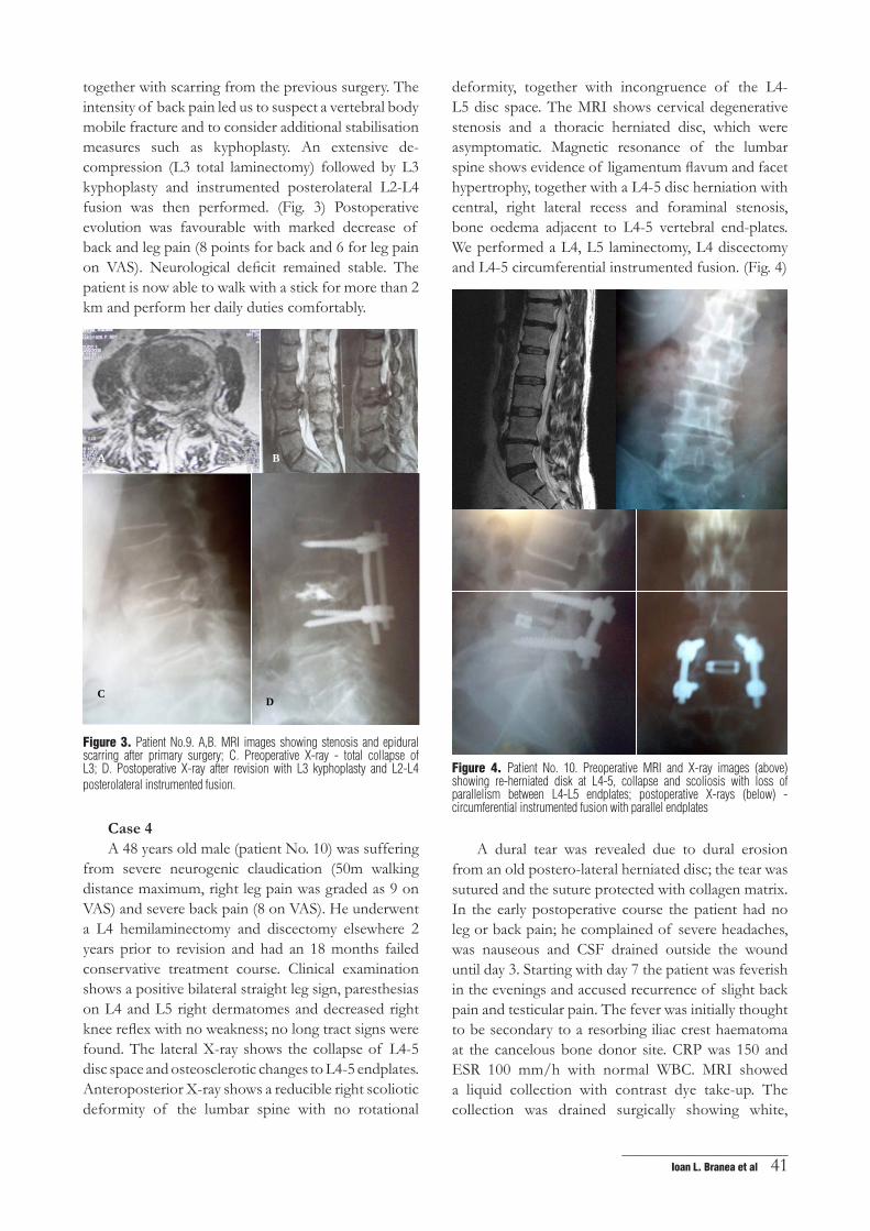
Case 4A 48 years old male (patient No. 10) was suffering
from severe neurogenic claudication (50m walking distance maximum, right leg pain was graded as 9 on VAS) and severe back pain (8 on VAS). He underwent a L4 hemilaminectomy and discectomy elsewhere 2 years prior to revision and had an 18 months failed conservative treatment course. Clinical examination shows a positive bilateral straight leg sign, paresthesias on L4 and L5 right dermatomes and decreased right knee reflex with no weakness; no long tract signs were found. The lateral X-ray shows the collapse of L4-5 disc space and osteosclerotic changes to L4-5 endplates. Anteroposterior X-ray shows a reducible right scoliotic deformity of the lumbar spine with no rotational
A B
CD
A B
CD
A B
CD
A B
CD
deformity, together with incongruence of the L4-L5 disc space. The MRI shows cervical degenerative stenosis and a thoracic herniated disc, which were asymptomatic. Magnetic resonance of the lumbar spine shows evidence of ligamentum flavum and facet hypertrophy, together with a L4-5 disc herniation with central, right lateral recess and foraminal stenosis, bone oedema adjacent to L4-5 vertebral end-plates. We performed a L4, L5 laminectomy, L4 discectomy and L4-5 circumferential instrumented fusion. (Fig. 4)
Figure 4. Patient No. 10. Preoperative MRI and X-ray images (above) showing re-herniated disk at L4-5, collapse and scoliosis with loss of parallelism between L4-L5 endplates; postoperative X-rays (below) - circumferential instrumented fusion with parallel endplates
A dural tear was revealed due to dural erosion from an old postero-lateral herniated disc; the tear was sutured and the suture protected with collagen matrix. In the early postoperative course the patient had no leg or back pain; he complained of severe headaches, was nauseous and CSF drained outside the wound until day 3. Starting with day 7 the patient was feverish in the evenings and accused recurrence of slight back pain and testicular pain. The fever was initially thought to be secondary to a resorbing iliac crest haematoma at the cancelous bone donor site. CRP was 150 and ESR 100 mm/h with normal WBC. MRI showed a liquid collection with contrast dye take-up. The collection was drained surgically showing white,

_____________________________42 TMJ 2007, Vol. 57, No. 1
creamy puss. The responsible micro organism could not be identified. The patient underwent 4 weeks of antibiotherapy with normalisation of CRP and ESR. The back and testicular pain resolved immediately after the collection drainage. He is pain free at 6 months follow-up.
DIscUssIONs
Revising a previous surgery on the lumbar spine provides a series of issues and technical difficulties that need serious consideration regarding surgical indication and preoperative planning. One must take into consideration a longer duration of the procedure compared to the initial surgery, because a more extensive decompression and often a fusion is needed. Blood loss will also be considerable, due to longer duration of the procedure, fibrosis and epidural scarring. The rate of dural tears expected for revision lumbar surgery is high (30% according to Hamill et al), thus adding time and blood loss to the procedure.8
Secondary deformity (scoliosis, spondilolystesis, lumbar kyphosis) is common among patients with failed back surgery due to collapse of disk space after discectomy, and instability due to facet joint arthritis and posterior wall disruption after decompression.
Technical challenges in revision decompression of the lumbar spine are mainly tied to extensive scarring around the dura mater and the nerve roots. This makes decompression very difficult and meticulous. There is often problematic to find cleavage planes and to differentiate structures. In one case of stenosis and re-herniation, the L4-5 disc eroded the dura ventrally and scarred-down to the sacral nerve roots.
The extent of fibrosis and adherences from the previous surgery recommend a significantly wider decompression during revision; this allows a better access to neural structures and better odds of avoiding and if necessary repairing a dural tear. Also, in the majority of cases in our series, an insufficient decompression was performed during the initial surgery. This appears to be the effect of not addressing a lumbar degenerative stenosis or, as the fracture case shows, failure to perform a decompression and instrumented fusion for a burst fracture. In one case of a two level fusion a transitional stenosis quickly developed in the adjacent segments above, a classic complication of lumbar fusion.
If significant back pain is also present, the
previously operated disk space can usually be identified as the main pain generator. Expected findings on the MRI are collapse of the operated disk space, bone oedema under the end-plates and often degenerative spondylolistesis, facet joint and ligamentum flavum hypertrophy generating secondary stenosis at the discectomy level. These factors, together with the need for a wider decompression are reasons to consider instrumented circumferential fusion for revisions of re-herniated disks.
cONcLUsION
Revision after a failed back surgery has significant complication and re-intervention rates. A poor patient selection is likely to affect the outcome of a revision. Only the failure of a well conducted conservative treatment in the presence of severe or progressive neurological symptoms and in some cases severe back pain of known origin can justify the need for a revision. Before any surgery is considered, it is paramount to obtain a detailed history, clinical examination and preoperative imaging studies. Laboratory tests must also rule out an occult infection.
Adherences and fibrosis secondary to the previous procedure determine the need for a wider decompression and often fusion of the segment involved. Revisions should be performed only by experienced surgeons and only when appropriate implants for an instrumented fusion are available.
rEFErENcEs
1. Guyer RD, Patterson M, Ohnmeiss DD. Failed back surgery syndrome: diagnostic evaluation. JAAOS 2006;14(9):534-43.
2. Fritsch E, Heisel J, Rupp S. The failed back surgery syndrome: reasons, intraoperative findings, and long-term results: A report of 182 operative treatments. Spine 1996;21(5):626-33.
3. Keskimaki I, Seitsalo S, Osterman H, et al. Reinterventions after lumbar disc surgery: A population-based study of regional and interspecialty variations. Spine 2000;25(12):1500-8.
4. Jonsson BS. Repeat decompression of lumbar nerve roots: A prospective two-year evaluation. J Bone Joint Surg 1993;75:894-7.
5. Waddell G, Kummel EG, Lotto WN, et al. Failed lumbar disc surgery and repeat surgery following industrial injuries. J Bone Joint Surg 1979;61:201-7.
6. Malter AD, McNeney B, Loeser JD, et al. 5-Year reintervention rates after different types of lumbar spine surgery. Spine 1998;23(7):814-20.
7. Osterman H, Sund R., Seitsalo S, et al. Risk of multiple reinterventions after lumbar discectomy: A population-based study. Spine 2003;28(6): 621-7.
8. Hamill LC, Kowalski JM. Lumbar spinal stenosis: operative and nonoperative treatment. In: Vaccaro, Betz, Zeidmann. Principles of spinal surgery, Mosby 2003, p. 355-63.