rewriting reproduction: perspectives on gene editing...
TRANSCRIPT

AN ANNUAL CELEBRATION OF RESEARCH IN REPRODUCTIVE SCIENCES
ANIMAL REPRODUCTION AND BIOTECHNOLOGY LABORATORY
THE TWELFTH ANNUAL
Rocky Mountain Reproductive Sciences Symposium
2019 CONFERENCE THEME:
Rewriting Reproduction: Perspectives on Gene
Editing & Bioethics MAY 3, 2019
8:30 A.M.– 5:30 P.M.CSU TRANSLATIONAL MEDICINE INSTITUTE
Hosted by Colorado State University’s Animal Reproduction &
Biotechnology Laboratory
arbl.colostate.edu

Program Table of Contents………………………………..…….……..1
About the Symposium……………………….……….………..………..2
Keynote Speakers……………………………………………………………3
Program Venue/Map……………………………………….…….……….5
Program Agenda……………………..………………………..…….……..6Student Moderator Schedule………………………………………….8
Student Platform Session Abstracts………………..……...........9
Poster Session I Abstracts (odd numbers)………….….........18
Poster Session II Abstracts (even numbers)……………….....36
List of Attendees………………………………….……....................53
Acknowledgements…………………….………….…....................57
RMRSS 2019
PROGRAM CONTENTS
1

RMRSS 2019About the Symposium
Symposium Introduction
Now in its twelfth year, the Rocky Mountain Reproductive Sciences Symposium brings together a diverse group of scientists to discuss advances in both human and animal reproduction that deepen our understanding of reproductive physiology. It’s a one-day event focused on student training, not only to significantly improve communication and cross-fertilization of research ideas, but also to share resources and expertise across human and animal models.
Hosted by the Animal Reproduction & Biotechnology Laboratory (ARBL) at CSU’s Lory Student Center, the day's events feature student abstract platform presentations, poster sessions, and keynote lectures by leaders in the field of reproductive physiology. Attendees include post-baccalaureate trainees, faculty, private clinicians, and other research scientists. This year’s event departs slightly from the historical “bench-to-bedside” thematic focus, and will instead focus on a “bench-to-bioethics” perspective. The symposium has led to the establishment of new collaborations between institutes to advance the field of reproductive sciences and is a great platform for student and fellow training.
Keynote Speakers
The basic science keynote lecture will be delivered by Dr. Pablo Ross from UC Davis, who will deliver a talk titled “CRISPRing Livestock Embryos: From Human Organ Generation to Enhancing Agricultural Traits.”
The bioethics keynote lecture will be delivered by Dr. Margaret F. Riley from the University of Virginia School of Law, who will deliver a talk titled “CRISPR Quandaries: Ethical Animal Use and Advanced Technologies.”
2

“CRISPRing Livestock Embryos:
From Human Organ Generation to
Enhancing Agricultural Traits”
Pablo Ross, PhD, DVMAssociate Professor and Vice Chair, UC Davis
Dept. of Animal Science
RMRSS 2019
KEYNOTE SPEAKERS
3
Pablo Juan Ross, PhD, DVM, is an associate professor of reproductive biology in the Department of Animal Science at the University of California Davis. Ross was born and raised in Argentina, where he obtained a degree in veterinary medicine from La Plata National University and a MS degree in animal science from Mar del Plata National University/INTA Balcarce. He then moved to Michigan State University where he obtained his PhD in animal science in 2007 and held a research assistant professor position until he joined the faculty at Davis in 2010.
Ross specializes in animal reproduction with emphasis on gametes, early embryos, and embryonic stem cells. Ross’ studies are aimed at understanding the mechanisms of epigenetic remodeling and transcriptional reprogramming that occur during preimplantation embryo development, somatic cell nuclear transfer, and cellular reprogramming by induced pluripotency.
Ross recently discovered efficient conditions for derivation and culture of livestock embryonic stem cells and proposed their use for in vitro breeding schemes. His laboratory makes extensive use of next generation sequencing approaches and genome editing to determine transcriptome and epigenetic mechanisms operating in livestock embryos and to produce animal models for agriculture and biomedicine. The Ross laboratory has pioneered research in human/pig and human/sheep chimera approaches for human organ generation in animals.

“CRISPR Quandaries: Ethical
Animal Use and Advanced
Technologies”
Margaret F. Riley, JDProfessor, University of Virginia School of Law
RMRSS 2019
KEYNOTE SPEAKERS
4
Margaret F. Riley is a professor at the University of Virginia (UVA) with appointments in the Law School, the School of Medicine (Public Health Sciences), and as an affiliated faculty member of the Batten School of Leadership and Public Policy. She is the director of UVA’s Program in Animal Law. Throughout her career, she has focused on the intersection between ethics and law and regulation particularly as it affects human and animal research and biotechnology.
Riley teaches food and drug law, health law, animal law, bioethics, environmental ethics, regulation of clinical research, and public health law. She has written and presented extensively about health care law, biomedical research, genetics, reproductive technologies, stem cell research, human and animal biotechnology, health disparities, and chronic disease. She has served on National Academies committees including: The Committees on Revisions to the Common Rule for the Protection of Human Subjects, Assessing Toxicological Risks to Human Subjects, Pain Management, Regulatory Strategies to Reduce Prescription Opioid Abuse (consultant to the committee), and Assessment of the Care and Use of Dogs in Biomedical Research funded by or conducted at the U.S. Department of Veterans Affairs. She has advised the Food and Drug Administration and numerous committees of the National Institutes of Health, the National Science Foundation, the Institute of Medicine, and the Virginia Bar.
Riley received her JD from Columbia University and her Bachelor of Arts degree from Duke University.

The Translational Medicine Institute is located at 2350 Gillette Drive, Fort Collins, CO 80523, phone number 970-491-8645. Parking is available on the north side of lot 725 (directly in front of the building) and in lot 740 near the tennis courts on Research Blvd. There is a kiosk in both lots where you can pay for parking at the rate of $1.75 per hour. If you pre-purchased a parking pass when you registered for the symposium, enter your code at the kiosk when you arrive in lot 725 or 740. See map link here and photo below:

8:40 am Opening Remarks – Dr. Thomas Hansen
9:00 am Keynote Lecture: Dr. Pablo Ross“CRISPRing Livestock Embryos: From Human Organ Generation to Enhancing Agricultural Traits”
Trainee Oral Platform Presentations I
10:15 am Development of Immunotolerance in Bovine Fetal Spleens Infected with BVDVHanah M. Georges, Katie J. Knapek, Helle Bielefeldt-Ohmann, Hana Van Campen, and Thomas R. Hansen
10:30 am Single-Cell RNA-sequencing of human embryos reveals critical role of interferon signaling during implantationRachel C. West, Hao Ming, Deirdre M. Logsdon, Rebecca A. Kile, Courtney K. Grimm, Sandeep Rajput, Jiangweng Sun, William Schoolcraft, Rebecca L. Krisher, Zongliang (Carl) Jiang, Ye Yuan
10:45 am Genetic Variants Identified in Cows with an Excess Androgen Ovarian Microenvironment Provides Clues to Women with Polycystic Ovary DiseaseAlexandria P. Snider, Sarah Nafziger, Jeff Bergman, Scott G. Kurz, John S. Davis, Jennifer R. Wood, Jessica L. Petersen, Andrea S. Cupp
11:00 am Effects of Omega-3 Fatty Acids and Hypoxia on Progesterone Biosynthesis and Lipid Droplet Dynamics in the Bovine Corpus LuteumBrian P. Krum, Jessica C. Cedillo, Michele R. Plewes, Terry E. Engle, Jason E. Bruemmer and Patrick D. Burns
11:30 am Lunch
6
RMRSS 2019
PROGRAM AGENDA

12:30 pm Poster Session I- Odd-numbered abstracts
1:15 pm Poster Session II- Even-numbered abstracts
Trainee Oral Platform Presentations II
2:00 pm Progesterone stimulates histone citrullination to increase Insulin Like Growth Factor Binding Protein 1 (IGFBP1) expression in ovine luminal epithelial cells.Young, C., Christensen, A., and Cherrington, B.
2:15 pm Diet affects oocyte metabolic and developmental capacities in maresCatandi G, Obeidat Y, Stokes J, Chicco A, Chen T, Carnevale E
2:30 pm Coordinated changes in fetal metabolism during hypoxemiaAmanda K Jones, Laura D Brown, Paul J Rozance, William W Hay, Jr., Sean W Limesand, Stephanie R Wesolowski
2:45 pm Oleic acid stimulation of amino acid uptake in human trophoblast cells is mediated by phosphatidic acid activation of mTOR signaling Elena Silva, Thomas Jansson, Theresa Powell
3:15 pm Keynote Lecture: Dr. Margaret F. Riley“CRISPR Quandaries: Ethical Animal Use and Advanced Technologies”
4:30 pm Open Discussion and Closing Comments- Dr. Thomas Hansen
5:30 pm Reception at CB & Potts, 1441 E. Horsetooth Rd., Fort Collins 80525, Sponsored by Endolytics, LLC
7
RMRSS 2019
PROGRAM AGENDA

9:00 am Introduction of First Keynote Speaker, Pablo Ross, PhDMichele Plewes, Postdoctoral Research Associate, University of Nebraska-Medical Center
10:00 am Ask First Question, 1st Keynote Speaker, Pablo Ross, Ph.D.10:15 am Rosemary McDonald, Ph.D. Student, University of Colorado
10:00 am Presentation of Plaque, 1st Keynote Speaker, Pablo Ross, Ph.D11:00 am Shelby Springman, Ph.D. Student, University of Nebraska-Lincoln
10:15 am Platform Presentation Session I: Introductions and First Question11:30 am Student 1: Courtney Sutton, University of Wyoming
Student 2: Amelia Tanner, Ph.D. student, Colorado State University Student 3: Coleman Young, University of Wyoming
2:00 pm Platform Presentation Session II: Introductions and First Question3:15 pm Student 1: Hanah Georges, Ph.D. Student, Colorado State
Student 2: Carolina Gonzalez-Berrios, Ph.D. Student, Colorado State Student 3: Brian Krum, Masters Student, UNC
3:15 pm Introduction of 2nd Keynote Speaker, Margaret Riley, JDAdel R. Moawad, Postdoctoral Fellow, Colorado State University
4:15 pm First Question: 2nd Keynote Speaker, Margaret Riley, JD4:30 pm Katie Bidne, Ph.D. Student, University of Nebraska-Lincoln
4:15 pm Presentation of Plaque, 2nd Keynote Speaker, Margaret Riley, JD4:30 pm Alex Snider, Postdoctoral Fellow, University of Nebraska-Lincoln
8
RMRSS 2019
STUDENT MODERATOR SCHEDULE

RMRSS 2019
STUDENT PLATFORM SESSION ABSTRACTS
9

RMRSS 2019Platform Abstracts
1. Development of Immunotolerance in Bovine Fetal Spleens Infected with BVDV
Hanah M. Georges1, Katie J. Knapek1, Helle Bielefeldt-Ohmann2, Hana Van Campen1, and Thomas R. Hansen1.
1Animal Reproduction and Biotechnology Laboratory, Department of Biomedical Sciences, Colorado State University, USA2School of Veterinary Science, The University of Queensland, Gatton Campus, QLD 4343, Australia
Bovine Viral Diarrhea Virus (BVDV) costs the cattle industry billions of dollars in performance loss annually, despite available vaccines. Known as “trojan horses”, persistently infected (PI) animals are generated through transplacental infection prior to day 125 of gestation and asymptomatically shed the virus postnatally. It was hypothesized that PI fetuses have upregulated innate immune responses followed by attenuated adaptive immune responses throughout gestation. This is thought to manifest through exposure to BVDV antigens during fetal lymphocyte maturation, thus resulting in immunotolerance to the virus. To test this hypothesis, naïve heifers were sham inoculated (controls) or infected with a non-cytopathic strain of BVDV on day 75 of gestation. Fetal spleens (n = 4-6) were collected on days 82, 97, 190, and 245 of gestation. RNA, protein, and tissues were assayed by RT-qPCR (.r), western blot (.w), and immunohistochemistry (.i). In PI fetal spleens, during peak viremia on day 97 of gestation, a strong innate immune response (RIGI.r, IRF7.r, ISG15.r.w.i) and an attenuated adaptive response occurred; represented by an unresponsive T-cell response (CD4.r.w, CD8.r) and a decreased B-cell population (CD79.r.w.i). By day 190, 115 days post-infection, the innate immune system returned to control levels, while adaptive response genes were significantly down-regulated (IFI30.r, LMP2.r, LMP7.r, STAT4.r, CD4.r, CD8.r, CD79b.r, CD79a.i). Despite down-regulation of RNA at d190, protein levels in PI samples were not significantly different than controls . It is hypothesized that this difference of RNA and protein is due to flaviviral inhibition of RNA exonuclease XRN1, causing an increase in protein synthesized from accumulated, non-degraded cellular RNA. On day 245 of gestation, 170 days post-infection, gene expression levels were not significantly different between control and PI fetal spleens, possibly due to the suppression of fetal immune responses in preparation for parturition. Collectively, the impaired adaptive response and diminished innate immune response on day 190 of gestation may reflect exhaustion of the immune responses, heralding the development of immunotolerance to BVDV and revealing a potential target for intervention and prevention of persistent infection. USDA AFRI NIFA NNF 2016-38420-25289 and USDA NIFA W3112.
10

RMRSS 2019Platform Abstracts
2. Single-Cell RNA-sequencing of human embryos reveals critical role of interferon signaling during implantation
Rachel C. West1, Hao Ming2, Deirdre M. Logsdon1, Rebecca A. Kile1, Courtney K. Grimm1, Sandeep Rajput1, Jiangweng Sun3, William Schoolcraft1, Rebecca L. Krisher1, Zongliang (Carl) Jiang2, Ye Yuan1
1Colorado Center for Reproductive Medicine, Lone Tree, CO2Louisiana State University, Baton Rouge, LA; 3Old Dominion University, Norfolk, VA;
Implantation is considered the ‘black box’ of human development as there is little insight into the signaling mechanisms that regulate this process. Due to this limited understanding of implantation, implantation failure is a pathology plaguing human fertility. It is estimated that 40-60% of human pregnancies fail, with a large majority of those failures occurring immediately prior to or during implantation. Recently, an extended embryo culture system has been developed that allows human embryos to be cultured up to 14 days post-fertilization. This system allows us, for the first time, to assess the human embryo during implantation and the events that immediately follow. We used this extended culture system to grow human blastocysts in vitro to embryonic days 8, 10 and 12 and assessed the early molecular dynamics that facilitate formation of the primitive placenta. We initially cultured 12 embryos and isolated 139 total cells for single cell RNA-sequencing. These cells were categorized as “small”, inferred to be undifferentiated cytotrophoblast (CTB), “large”, multinucleated primitive syncytium (STB), or “migratory” trophoblast (MTB) based on morphology. When we began to analyze our RNA-sequencing data, we found that interferon signaling related genes begin to appear as early as D8 in cytotrophoblastcells. Gene ontology analysis identified that the Type I and Type II interferon pathways become upregulated from D8 to D12 in a time dependent manner. Additionally, MTB expressed the highest levels of some interferon related genes. We validated these findings using immunofluorescence and 3D-confocal microscopy. The interferon receptors, IFNAR1 and IFNGR1 started to appear as early as D8 as well as the interferon stimulated genes, ISG15 and ISG20. However, ELISAs for interferons detected no levels of interferon production in spent embryo culture medium. This finding was corroborated by the almost nonexistent expression of IFN-α, IFN-β, and IFN-ƴ mRNA in our RNA-seq data. This led us to seek an alternate explanation for the upregulation of ISGs in the absence of interferons. One possible explanation is the expression of human endogenous retroviruses (HERV) in trophoblast cells. The placenta utilizes HERV to support cell synctialization and immune suppression in trophoblast cells. However, as HERV have been shown to modulate the innate immune response in other cell types, we hypothesize that HERV can also trigger ISG production in trophoblast cells. Using our RNA-seq data, we identified 8 individual HERV transcripts in trophoblast cells. Five (ERV3-1, ERVFRD-1, ERVV-1, ERVV-2 andERVW-1) of these 8 transcripts were more abundantly expressed in differentiated cells. Interestingly, the ERVH48 was more abundant in undifferentiated cytotrophoblast cells. ERVH48 encodes for the protein Suppressyn and inhibits cell fusion by competing with ERVW-1 for receptor binding. These results suggest that the constitutive expression of HERV in human embryos could be an important mechanism that initiate trophoblast cell differentiation and placenta formation via IFN signaling. This research was funded by CCRM and approved by WIRB (Study no: 1179872).
11

RMRSS 2019Platform Abstracts
3. Genetic Variants Identified in Cows with an Excess Androgen Ovarian Microenvironment Provides Clues to Women with Polycystic Ovary Disease
Alexandria P. Snider1, Sarah Nafziger1, Jeff Bergman1, Scott G. Kurz1, John S. Davis2, Jennifer R. Wood1, Jessica L. Petersen1, Andrea S. Cupp1
1Department of Animal Science, University of Nebraska–Lincoln; 2Obstetrics and Gynecology, University of Nebraska Medical Center
A naturally-occurring cow model of androgen excess (High Androstenedione; High A4) shares many metabolic and molecular phenotypes of Polycystic Ovary Syndrome (PCOS). High A4 cows have irregular estrous cycles, anovulation, and reduced calving rates but wean calves that are 24 kg heavier. Since producers retain females with heavier offspring to remain in the herd, it is possible High A4 cows have been preferentially selected. Thus, we hypothesize females with a PCOS-like phenotype have genomic variation linked to steroidogenic and metabolic phenotypes comprising the High A4 phenotype. To test this hypothesis, we genotyped 200 individuals with the GeneSeek Genomic Profiler Bovine 150K SNP array. While no loci achieved a genome-wide significance threshold determined by FDR of 0.01, 70 genes within 25 kb of genetic variants associated (raw p<0.01) with the High A4 phenotype were prioritized for investigation. Additionally, approximately 17 million variants were identified in whole-genome sequence of 15 individuals (8 High A4, 7 Control). Annotation with SNPEff found 4,600 of these with predicted high impact. High impact variants were identified in seven of the 70 genes identified in the initial GWAS. We have assayed expression of these, CARNS1, CCR6, GPR31, ALKBH6, CLIP3, SREBP1a and SREBP1c, via ddPCR in ovarian cortex, theca, and granulosa cells, in High A4 compared to controls cows. In High A4 ovarian cortex, SREBP1c and CLIP3 were significantly upregulated compared to controls (p≤0.05). There was a tendency (p≤0.10) for CARNS1 and CCR6 to be downregulated in High A4 granulosa cells. These genes are involved in inflammation or oxidative stress, two processes upregulated in the High A4 phenotype. Understanding genetic variants that predict the High A4 phenotype can provide a potential selection tool for producers. Also, these genetic variants could allow us to better understand metabolic, growth and reproductive trait interactions in human disease such as PCOS.
12

RMRSS 2019Platform Abstracts
4. Effects of Omega-3 Fatty Acids and Hypoxia on Progesterone Biosynthesis and Lipid Droplet Dynamics in the Bovine Corpus Luteum
Brian P. Krum1, Jessica C. Cedillo1, Michele R. Plewes2, Terry E. Engle3, Jason E. Bruemmer3 and Patrick D. Burns1
1School of Biological Sciences, University of Northern Colorado, 2Olson Center for Women's Health, Department of Obstetrics and Gynecology, University of Nebraska Medical Center, 3Department of Animal Sciences, Colorado State University
The corpus luteum (CL) is a transient endocrine gland that forms from the remnants of the ovulatory follicle. Luteal cells of the CL secrete the steroid hormone progesterone which is responsible for both the establishment and maintenance of pregnancy in mammals. In the absence of an embryo, prostaglandin F2a (PGF2a) is secreted from the uterus to regress the CL. The trophoblastic cells of the developing embryo secrete interferon-tau (IFN-t) which prevents PGF2a synthesis and protects the CL from regression. If the IFN-t signal is either delayed or too weak, PGF2a synthesis is not adequately inhibited and the CL regresses, thereby losing a potentially viable pregnancy. Previous studies from our laboratory have shown that dietary supplementation of omega-3 polyunsaturated fatty acids, commonly found in fish oil, reduce luteal cell sensitivity to PGF2a. The mechanism by which these fatty acids protect the CL is largely unknown. The objective of this study was to determine the mechanism behind luteal protectiveness. In experiment 1, cows were supplemented with either fish (n=5) or vegetable (n=4) oil for approximately 60 days. Following the supplementation period, all animals received two intrauterine doses of PGF2a (0.5 mg) on days 10-12 of the estrous cycle at 12 h intervals. Luteal biopsies were obtained at h 0, 18, 24, 36, and 48 of the experimental period. Blood progesterone was measured every 3 h for the first 24 h and every 6 h for the remaining 24 h to assess CL function. Sixty percent of the cows supplemented with fish oil retained a functional CL that did not regress in response to PGF2a infusion (blood progesterone > 1 ng/mL following PGF2a treatment). In fish oil supplemented cows, STARD1, CYP11A1 and LDLR steady-state mRNA decreased from between h 0-18 and then remained unchanged in fish oil supplemented animals regardless of CL function (P < 0.05). However, steady-state mRNA continued to decline in vegetable oil supplemented cows between 18 and 48 h (P < 0.05). Biopsies (0 h) were stained for lipid droplets (LDs). Cows supplemented with fish oil had significantly greater LD volume and number per cell (P < 0.05). In experiment 2, luteal cells were cultured from bovine ovaries to determine the effects of hypoxia on progesterone synthesis and mitochondrial health in fish oil (0.03% v:v) treated cells. Forskolin (10 µM) stimulated progesterone output was significantly reduced in response to hypoxia (5% O2), as compared to the normoxic (20% O2) treatments (P < 0.05). Additionally, fish oil supplementation did not improve progesterone output in a low oxygen environment (P > 0.10). Membrane potential was used to assess mitochondrial health and analyzed using confocal microscopy. There was no significant difference on mitochondrial health in fish oil treated cells as compared to control cells in either a hypoxic or normoxic environment (P > 0.10). Lipid droplet dynamics were determined in response to low oxygen culture. Luteal cells cultured in hypoxia resulted in greater LD number as compared to those in normoxia. Results of this study show that fish oil supplementation is luteal protective in response to PGF2a treatment, however luteal cell responsiveness to hypoxia is inconclusive. This project was supported by National Research Initiative Competitive Grant no. 2013-67015-20966 from the USDA National Institute of Food and Agriculture to PDB.
13

RMRSS 2019Platform Abstracts
5. Progesterone stimulates histone citrullination to increase Insulin Like Growth Factor Binding Protein 1 (IGFBP1) expression in ovine luminal epithelial cells.
Young, C., Christensen, A., and Cherrington, B.
Department of Zoology and Physiology, University of Wyoming 1000 E. University Ave, Laramie, WY, 82071
Peptidylarginine deiminases (PADs) are a family of calcium dependent enzymes that post-translationally convert positively charged arginine amino acids in proteins into neutral citrulline residues. Targets for PAD catalyzed citrullination include arginine residues on histone tails which results in chromatin decondensation and changes in gene expression. PADs were first discovered in the uterus; however, since then no studies have examined their functional significance in this tissue. Our previous work shows that PAD2 and PAD4 are highly expressed in luminal epithelial cells from ovine caruncles and an ovine uterine luminal epithelial (OLE) derived cell line. In OLE cells, inhibiting PAD activity blunts histone citrullination resulting in decreased basal expression of insulin like growth factor binding proteins 1 (IGFBP1). Given that progesterone (P4) is critical to maintain the uterine epithelium during pregnancy and regulates IGFBP1 expression, herein we examined if P4 stimulates PAD catalyzed histone citrullination to regulate IGFBP1 expression in OLE cells. To test this, OLE cells were pretreated for 6 hrwith vehicle or 2 µM BB-CLA, a pan PAD inhibitor, and then treated with 100 nM P4 for 60 minutes. Following 60 minutes of P4 treatment, there is a significant increase in citrullination of histone H3 arginine residues 2, 8, and 17 concurrent with increased IGFBP1 mRNA expression. In contrast, cells pretreated with BB-CLA do not show an increase in IGFBP1 mRNA expression despite P4 treatment. We next examined if citrullinated histones are directly associated with the IGFBP1 gene promoter using an anti-H3Cit2,8,17 antibody for chromatin immunoprecipitation. Stimulation of OLE cells with 100 nM P4 results in significant enrichment of the IGFBP1 gene promoter compared to vehicle treated controls. In conclusion, our work suggests that P4 stimulates PAD catalyzed histone citrullination to regulate IGFBP1 expression, which is important for uterine epithelial cell function during pregnancy.
14

RMRSS 2019Platform Abstracts
6. Diet affects oocyte metabolic and developmental capacities in mares
Catandi G a, Obeidat Y b, Stokes Ja, Chicco A a, Chen T b,c, Carnevale E a
a College of Veterinary Medicine and Biomedical Sciences, Colorado State University (CSU), Fort Collins, CO 80523, USAb Department of Electrical and Computer Engineering, CSUc School of Biomedical Engineering, CSU
Equine maternal aging leads to reduced fertility, in part, by affecting oocyte quality. Mitochondria play a critical role in oocyte developmental competence by supplying energy to oocytes and early embryos through oxidative phosphorylation. The extent that dietary supplements alter equine oocyte viability is unknown. We hypothesized that dietary supplements, including omega-3 (n-3) polyunsaturated fatty acids (FA) and antioxidants, would be beneficial for mitochondrial function and oocyte developmental potential. Mares (13-23 yr, n=10/group) were fed grass/alfalfa hay and one of two equicaloric supplements: (1) GR, mixed grain and corn oil, high in omega-6 (n-6) FA or (2) AC, a complex of additives including flax seed oil (high in n-3 FA), probiotics, and antioxidants (Platinum Performance, Inc., Buellton, CA). Supplements were fed for ≥6 wk before sample collections. Oocytes were collected by transvaginal, ultrasound-guided follicular aspirations of dominant follicles ( 35 mm) during estrus and at 16 2 h after induction of follicular maturation. Recovered oocytes were incubated in medium at 38.2°C in 7% CO2 and air for 26 2 h. Metaphase II oocytes were stripped of cumulus cells and assessed for metabolic function using a novel microchamber with sensors capable of measuring fluctuations in oxygen consumption and pH (indicative of aerobic and anaerobic metabolism). The microchamber was filled with 120 µL of holding medium and overlaid with 120 µL paraffin oil. The baseline current was measured before adding the oocyte through the oil layer. Basal (nonstimulated) oxygen consumption and pH were measured, followed by assessment of maximal noncoupled respiratory capacity obtained by titrations of 1 µM of carbonyl cyanide m-chlorophenyl hydrazone (CCCP) at 8-min intervals. The highest value observed during three CCCP titrations was considered the maximal oxygen consumption. Additional oocytes were injected with sperm, and cleavage and blastocyst development were assessed. Two-tailed Student's t-tests and Fisher’s exact tests were used for analyzing data. Basal, but not maximal, oxygen consumption; ability to switch to anaerobic metabolism; and mitochondrial efficiency were higher (p<0.05) in oocytes for AC than GR, confirming a dietary effect on oocyte metabolism. After sperm injections, the cleavage rate was similar between groups (AC, 11/12, 92% and GR, 12/13, 92%), but blastocyst rates were higher (p<0.05) for oocytes from AC (7/11, 64%) than GR (2/13, 15%). Dietary supplementation of a complex of nutrients with n-3 FA and antioxidants to older mares resulted in improved oocyte metabolic function and embryo development, when compared to other grain-based additives. In conclusion, mare diet altered oocyte function and potential for development into a blastocyst, indicating that maternal diet is capable of affecting the oocytes’ quality and developmental capacity.
15

RMRSS 2019Platform Abstracts
7. Coordinated changes in fetal metabolism during hypoxemia
Amanda K Jones, PhD1, Laura D Brown, MD1, Paul J Rozance, MD1, William W Hay, Jr.1, MD, Sean W Limesand, PhD2, Stephanie R Wesolowski, PhD1.
1University of Colorado School of Medicine, Aurora, CO, USA, 2University of Arizona, Tucson, AZ, USA.
Fetal hypoxemia is associated with pregnancy conditions that cause an early activation of fetal glucose production. However, the independent role of hypoxemia to activate this pathway and the associated coordination of gluconeogenic substrates is not well understood. We hypothesized that fetal hypoxemia would activate fetal glucose production by decreasing umbilical glucose uptake and increasing counter-regulatory hormone concentrations. To test this hypothesis, we induced hypoxemia in pregnant ewes for 9 days with maternal tracheal N2 gas insufflation to reduce maternal and fetal arterial pO2 (HOX = 11) compared with fetuses from ewes receiving intratracheal compressed air (CON = 7). At 0.9 of gestation, fetal metabolic studies were performed to measure hormone and nutrient concentrations, umbilical blood flow, and glucose flux rates. Fetal livers were collected for molecular analysis. Data were analyzed by student’s t-test. Fetal arterial pO2 was 20% lower in HOX (14.6 0.5 mmHg) versus CON (18.3 1.0 mmHg; P < 0.01). Fetal arterial plasma lactate and whole blood pyruvate concentrations were both 2-fold greater in HOX versus CON (P<0.05). Fetal plasma insulin concentrations were lower while cortisol and norepinephrine concentrations were greater in HOX versus CON. Fetal body weight, umbilical blood flow rates, net fetal oxygen and glucose uptake rates, and fetal arterial plasma glucose concentrations were not different between groups. Fetal glucose utilization rates were similar between HOX and CON fetuses and not different from umbilical glucose uptake rates, demonstrating the absence of endogenous glucose production. In liver tissue, mRNA expression of gluconeogenic genes G6PC (P<0.01) and PCK1 (P=0.06) were 6- and 3-fold greater in HOX versus CON fetuses. Fetal lactate uptake rates were similar, yet fetal pyruvate output was 2-fold greater in HOX versus CON (P<0.001). Both mRNA and protein expression for lactate dehydrogenase (LDHA gene; LDH-A protein) were increased 2-fold in HOX vs CON (P<0.05). Further, the ratio of phosphorylated (S293) to total pyruvate dehydrogenase (PDH) protein expression was increased 50% in HOX vs CON livers (P<0.05), supporting decreased pyruvate oxidation. Expression of the oxygen sensing subunit of COXIV (COX42) and antioxidant genes superoxide dismutase (SOD2) and catalase (CAT) were increased in HOX fetal livers (P<0.05). Collectively, these findings support a role for fetal hypoxemia to act with counter-regulatory hormones to potentiate gluconeogenic gene expression in the liver of HOX fetuses. However, in the absence of decreased fetal glucose uptake and plasma glucose concentrations, hypoxemia-induced gluconeogenic gene activation is not sufficient to activate fetal glucose production despite increased gluconeogenic substrate (lactate) availability. Maintained fetal metabolic rate during hypoxemia may reflect the activation of oxygen sensing and antioxidant defense mechanisms and increased utilization of alternate substrates, such as amino acids, by the liver in HOX fetuses.
16

RMRSS 2019Platform Abstracts
8. Oleic acid stimulation of amino acid uptake in human trophoblast cells is mediated by phosphatidic acid activation of mTOR signaling
Elena Silva1, Thomas Jansson1, Theresa Powell2.
1 Department of Obstetrics and Gynecology. 2 Department of Pediatrics. University of Colorado, Anschutz Medical Center.
Placental amino acid (AA) transport is positively associated with fetal growth and regulated by mTORsignaling. Oleic acid (OA) is the most abundant monounsaturated fatty acid in the maternal circulation and has been shown to stimulate AA uptake and the mTOR signaling in primary human trophoblast (PHT) cells. However, the underlying mechanisms are unknown. Phosphatidic acid has been shown to be necessary for the stabilization and regulation of mTOR. We hypothesize that OA regulation of placental AA transport is mediated by phosphatidic acid upstream of mTOR signaling. Methods: PHT cells were isolated from normal term pregnancies and after 60 hrs of culture, PHT cells were treated with OA (100 µM) or BSA for 24 hours. We used RNA silencing of raptor to determine the involvement mTORC1 in OA stimulation of 14C-MeAIB uptake, or System A activity. Scramble (SCR) treatment was used as a control. We also used silencing of 1-acylglycerol-3-phosphate O- acyltransferase- 4 (AGPAT-4), to determine the role of phosphatidic acid synthesis in OA regulation of System A. Data were analyzed by ONE-WAY ANOVA. Results: Silencing of raptor completely prevented the stimulation of MeAIB uptake by OA (P=0.03). Following AGPAT-4 silencing, OA no longer stimulated MeAIB uptake in PHT (P=0.04) or phosphorylation of the mTOR down-stream target rpS6 at ser235 (P=0.04) suggesting that phosphatidic acid synthesis mediates mTOR activation and stimulation of System A activity by OA. Conclusions: We conclude that mTORC1 is required for OA stimulation of AA uptake in the trophoblast and the changes are mediated through phosphatidic acid synthesis. These findings highlight a novel and important role of a specific, highly abundant fatty acid in regulating trophoblast function. Our data suggest that maternal hyperlipidemia, particularly in obesity, may impact the placental AA transport capacity, mediated by fatty acids regulating trophoblast mTOR signaling, and affect fetal growth trajectory.
17

18
RMRSS 2019
POSTER SESSION I ABSTRACTS
RMRSS 2019Poster Session I Abstracts: Odd Numbers
9. The ARID3B-Complex Regulates Genes Governing Placental Development
Asghar Ali, Gina C Nay, Russell V Anthony, Gerrit J Bouma, Quinton A Winger.
Animal Reproduction and Biotechnology Lab, Colorado State University, Fort Collins, CO, USA.
A properly functioning placenta is critical for the health of the growing fetus. Proliferation, invasion and migration of trophoblast cells is necessary for placental development, and functional placental insufficiency results in fetal growth restriction. The pluripotency factors LIN28A and LIN28B regulate gene expression by inhibiting let-7 miRNA, and knockdown of LIN28A or LIN28B results in increased let-7 miRNA in trophoblast cells. Let-7 miRNAs target a number of genes, potentially including the AT-rich Interaction Domains (ARIDs) family of DNA binding proteins. ARID3A is transported to the nucleus by the membrane transporter, importin-9 (IPO9), where it forms a complex with ARID3B and lysine demethylase 4C (KDM4C), called the ARID3B-complex. This complex binds to DNA at sequence specific sites to regulate gene expression by demethylating histones. Genes regulated by this complex have been identified primarily in cancer cell lines and have not been studied in the placenta. Interestingly, several genes regulated by this complex are known to have important functions in placental development, including HMGA1, cMYC, VEGFA and WNT1, and knockout of ARID3A in mice results in embryonic lethality. In this study we used the human first trimester trophoblast cell line, ACH-3P, to investigate gene regulation by the ARID-KDM4C complex. Co-immunoprecipitation confirmed the physical binding of ARID3A, ARID3B and KDM4C in ACH3P cells and chromatin immunoprecipitation (ChIP) confirmed that this complex binds to the promoter region of HMGA1, cMYC, VEGFA and WNT1. CRISPR-Cas9 targeted knockout of ARID3B resulted in significantly decreased HMGA1, cMYC, VEGFA and WNT1 mRNA and protein. These results suggest that, in addition to being direct targets of let-7 miRNA; HMGA1, cMYC, VEGFA and WNT1 are also regulated by let-7 miRNA through the ARID-KDM4C complex in human trophoblast cells. To our knowledge, this is the first study to confirm the presence of the ARID-KDM4C complex in a human trophoblast cell line and that the complex binds to promoter regions of genes known to regulate placental development. In summary these results suggest that the ARID-KDM4C complex regulates gene expression in human trophoblast cells through epigenetic regulation of histone methylation and plays a role in the regulation of important genetic factors necessary for placental development. This project was supported by Agriculture and Food Research Initiative Competitive Grant # 2017-67015-26460 from the USDA National Institute of Food and Agriculture.
19

RMRSS 2019Poster Session I Abstracts: Odd Numbers
11. Evaluating the Effects of Luteinizing Hormone Receptor Expression Levels on Receptor Aggregation and Function
Duaa Althumairy, George Barisas and Deborah Roess
Colorado State University, Fort Collins, CO
Luteinizing hormone receptors (LHR) are G protein-coupled receptors (GPCR) found in female and male reproductive organs where they play a critical role in ovulation and sperm maturation, respectively, as well as maintenance of sex hormone production in both sexes. The role of oligomerization in LHR function is of considerable interest and not well understood. LHR may be constitutively clustered or, alternatively, exist as monomers in the absence of hormone and then undergo some degree of aggregation in the presence of hCG. In this project, we have used polarized homo-transfer fluorescence resonance energy transfer (homo-transfer FRET) to evaluate the aggregation state of LHRs tagged on their C terminus with yellow fluorescence protein (YFP). Cell lines stably expressing <8,000 receptors per cell, 10,000-30,000 receptors per cell, 35,000-80,000 receptors per cell or >85,000 receptors per cell were used. At lower receptor numbers per cell, LHR appeared in membranes as receptor dimers or small oligomers. At higher receptor numbers per cell, LHR appeared in significantly larger clusters. We also measured basal cAMP levels in these cell lines as well as intracellular cAMP levels in response to hCGusing the cAMP sensor, ICUE3, which was transiently expressed in these CHO cell lines. Cells expressing small LHR clusters also had low basal cAMP activity and a robust response to hCG. When LHR were expressed at higher numbers of LHR per cells and LHR were present in larger oligomers in the absence of hormone, basal levels of cAMP per cell were high and cells were comparatively unresponsive to hCG. We also examined the effects of deglycosylated hCG (DG-hCG), a kind gift from Dr. George Bousfield at Wichita State University, on LHR cluster size and intracellular cAMP levels. DG-hCG functions as an hCGantagonist and, in addition to reducing intracellular cAMP, also significantly reduced the cluster size of the LH receptors in concentration-dependent manner. These results suggest that there is a strong relationship between the number of LH receptors expressed per cell, the size of LHR clusters and LHR receptor responsiveness to hCG. Moreover, it suggests that DG-hCG either blocks LHR aggregation or reverses aggregation of large LHR clusters. These results provide to a better understanding of the relationship between LHR clustering and signaling as well as LHR-related diseases that involve mis-oligomerization events. This research was supported by NIH and USDA Animal Health and Disease Program at CSU.
20

RMRSS 2019Poster Session I Abstracts: Odd Numbers
13. Impacts of thermal neutral housing on murine reproduction and metabolism
Katie L. Bidne1, Rong Fan2, Alana L. Rister3, Eric D. Dodds3, Soonkyu Chung2, Jennifer R Wood1
1Department of Animal Science, University of Nebraska – Lincoln2Department of Nutrition and Health Sciences, University of Nebraska – Lincoln3Department of Chemistry, University of Nebraska – Lincoln
When male and female mice fed a western diet are housed at thermal neutral temperatures (30-32º C), the metabolic phenotypes that develop better mimic human obesity than when mice are housed at traditional temperatures (control, 20-23º C). However, there is no information regarding female reproduction in this model. When we housed female C57/BL6J mice fed normal rodent chow at thermal neutral temperature, they had decreased post-plug pregnancy rate (30%) compared to control mice fed normal rodent chow (70%). We hypothesized that increased temperature reduces oocyte quality due to metabolic stress. To test this hypothesis, female C57/BL6J mice were placed in either thermal neutral (n = 8) or traditional housing (n = 9) at weaning and fed normal chow until 8 weeks of age. Ovulation was stimulated and rectal temperatures were taken. After euthanasia ovulated oocytes, body weight, serum, and tissue samples were collected. Thermal neutral housing significantly increased white and brown fat mass compared to control-housed mice. There were no differences in rectal temperature or blood glucose between the thermal neutral and control groups. Liquid chromatography-mass spectrometry of serum showed a tendency for increased corticosterone concentrations (p = 0.07) in the thermal neutral group compared to control mice. Oocytes from both groups were fertilized in vitro and cultured to blastocysts. There were no differences in the number of embryos that developed from the 2-cell to blastocyst stages between experimental groups. Previous studies in the literature demonstrated that exogenous corticosterone administration in mice impaired fertility via reduced implantation without altering oocyte competence or pre-implantation embryo development. In addition, elevated corticosterone downregulates thermogenesis while increasing lipid droplet accumulation in brown adipose tissue. Collectively, these results suggest that thermal neutral housing induces metabolic and thermal stress which decreases female reproduction, likely due to decreased uterine receptivity.
21

RMRSS 2019Poster Session I Abstracts: Odd Numbers
15. Lipopolysaccharide Differentially Affects Pro-Inflammatory Responses in Theca Cells from Androgen Excess compared to Control Beef Cows
KA Bochantin, AP Snider, SA Springman, SG Kurz, JA Keane, S Nafzinger, JW Bergman, RM McFee, AS Cupp, JR Wood
Department of Animal Science, University of Nebraska-Lincoln
A population of cows with increased ovarian androstenedione (High A4; >40ng/mL) concentrations has been identified in the UNL physiology herd. Interestingly, circulating and follicular fluid pro-inflammatory cytokine composition is altered in High A4 cows suggesting chronic inflammation. Lipopolysaccharide (LPS) leak from the gut due to stress-dependent changes in microbial profiles induces expression of pro-inflammatory cytokines via activation of toll-like receptor 4 (TLR4) and the associated myeloid differentiation factor 88 (MyD88). Our hypothesis is that theca cells from High A4 cows, which have similar gene expression profiles as women with PCOS, are chronically exposed to LPS resulting in differential secretion of pro-inflammatory cytokines compared to theca cells from Control cows. To test this hypothesis estrous cycles of High A4 and Control cows were synchronized and stimulated for 3 days with FSH (210 IU) prior to ovariectomy. Theca cells were micro-dissected from follicles >7mm and immediately cultured in untreated medium for 120 minutes or LPS-containing medium (50ng/ml) for 15 to 120 minutes. Western blot analysis showed progressive LPS-dependent increases in MyD88 in theca cells from Control cows. However, MyD88 expression was progressively decreased in theca cells from High A4 cows. Cytokine antibody arrays showed significant increases in pro-inflammatory cytokine concentrations (TNFα, IL-1α, and IL-21) in untreated medium from High A4 compared to Control theca cells. When Control theca cells were treated with LPS, TNF , IL-1 , and IL-21 were significantly increased in a time-dependent manner. However, LPS treatment of High A4 theca cells resulted in significantly decreased levels of these same cytokines. These results suggest insensitivity of High A4 theca cells to LPS-mediated inflammatory response which may be due to chronic in vivo LPS exposure. Previous studies show that LPS increases ovarian steroidogenesis suggesting that chronic exposure of theca cells to LPS may be a mechanism for androgen excess in High A4 cows.
22

RMRSS 2019Poster Session I Abstracts: Odd Numbers
17. Intrauterine Growth Restriction Lowers Amino Acid Uptake and Increases Alanine, Glutamine, and Glycine Production in the Skeletal Muscle of Late Gestation Fetal Lamb Hindlimb
Eileen I. Chang, Elizabeth A. Gilje, Stephanie R. Wesolowski, Paul J. Rozance, Laura D. Brown.
University of Colorado School of Medicine, Aurora, CO, USA.
Intrauterine growth restriction (IUGR) from decreased nutrient and oxygen supply to the fetus reduces fetal skeletal muscle mass, which may contribute to increased risk for metabolic disease in later life. We have shown in a sheep model of placental insufficiency-induced IUGR that skeletal muscle protein accretion rates were decreased in IUGR fetuses when compared to control. Our objective was to identify global metabolic changes in fetal skeletal muscle in response to IUGR. We hypothesized that the IUGR fetal muscle develops adaptations that divert amino acids (AA) away from protein accretion into alternative metabolic pathways that maintain cellular metabolism in muscle and conserve energy for other organs. At 127±1 days gestation (dGA, term=147 dGA), control (CON, n=8) and IUGR (n=10) fetal sheep were catheterized with vascular catheters and an external iliac artery flow probe. At 134±1 dGA, individual AA uptake rates were measured using high performance liquid chromatography (HPLC), and the biceps femoris (BF) muscle was collected. Hydrophilic metabolites were extracted from BF, and metabolomics was performed with ultra-high-performance liquid chromatography tandem mass spectrometry (UHPLC-MS/MS). Metabolites were identified, and statistical and pathway analysis were conducted with MetaboAnalyst 4.0. mRNA was isolated from BF to measure expression of genes in branched chain amino acid (BCAA) catabolism, glycolysis, and the tricarboxylic acid (TCA) cycle using real-time qPCR. A Student's t-test was used to determine significant differences in metabolites between groups (CON and IUGR). Significance was designated at a false discovery rate (FDR) corrected P-value of ≤0.05, and tendency at ≤0.1. The partial least squares discriminant analysis (PLS-DA) revealed a clear separation between CON and IUGR groups in BF. Pathway analysis showed a greater abundance of metabolites enriched within the alanine, aspartate, and glutamate metabolism pathway in IUGR compared to CON. In the IUGR hindlimb vs. CON, the net uptake rates of branched chain amino acids (BCAA) and ammoniagenic AAs (alanine, glutamine, and glycine) were lower by 42-73% and 107-158%, respectively. There were strong correlations between BCAA and ammoniagenic AAs with fetal hindlimbweight. The smallest hindlimb showed a net release of ammoniagenic AAs. The gene expression levels indicated an overall decrease in BCAA catabolism, and glycolysis that favors the conversion of pyruvate to alanine in the IUGR muscle. We conclude that IUGR skeletal muscle has lower uptake of BCAAs that are metabolized within muscle for alanine, glutamine, and glycine production in lieu of protein accretion. These adaptations within AA metabolic pathways in IUGR fetal skeletal muscle provide an experimental basis for potential targets for hormonal, nutrient, and oxygen supplements to increase IUGR muscle growth. This research was supported by NIH HD079404 (LDB), DK088139 (PJR), and DK108910 (SRW), and by the Center for Women’s Health Research at the University of Colorado School of Medicine (LDB).
23
RMRSS 2019Poster Session I Abstracts: Odd Numbers
19. Warburg metabolism is utilized by developmentally competent in vivo produced mouse embryos, but is disrupted by in vitro culture
Benjamin B Goheen, Sandeep K Rajput, Courtney K Grimm, John C Becker, William B Schoolcraft, Rebecca L Krisher
Colorado Center for Reproductive Medicine, Lone Tree, CO
Metabolic processes fueling rapid cell proliferation, as occurs in cancer cells, require glycolytic metabolites upstream of the tri-carboxylic acid (TCA) cycle for synthesis of macromolecules for biomass production and redox regulation. This can be achieved by engaging in aerobic glycolysis, also known as the Warburg Effect. Warburg metabolism has been proposed to occur in preimplantation embryos, and although gene expression data would support that hypothesis there is to date no firm evidence. Our objective was to characterize presence and functional activity of pyruvate kinase M2 isoform (PKM2), pyruvate dehydrogenase (PDH), and lactate dehydrogenase (LDH) proteins in mouse (CF1) blastocysts produced in vivo (IVO) and in vitro (IVC). Replicate (n=3) pools of 30 blastocysts were subjected to western blot analysis for both phosphorylated (p) and total (t) protein; p:t ratio reflects protein activity. Phosphorylation of PKM2 was elevated 2.2-fold (p=0.0034), and PDH was elevated 4.2-fold (n=1) in IVO compared to IVC blastocysts. Phosphorylation inhibits PKM2 activity and redirects glycolytic metabolites away from the TCA cycle, a key component of the Warburg Effect. Phosphorylation of PDH is also inhibitory and acts to slow entry of pyruvate into the TCA cycle. Another hallmark of the Warburg effect is lactate production from pyruvate, catalyzed by LDH. We observed an 11.1-fold increase in total LDHB protein in IVO blastocysts compared to IVC (p<0.0001). These results demonstrate that Warburg metabolism is active in mouse blastocysts produced in vivo. However, culture of mouse embryos in vitro results in metabolic alterations such that Warburg metabolism is no longer utilized. Thus, in vitro produced embryos may aberrantly rely on the TCA cycle and oxidative phosphorylation for energy generation, potentially compromising biomass production and redox control and resulting in the reduced developmental competence that is a well-known hallmark of in vitro embryo production.
24

RMRSS 2019Poster Session I Abstracts: Odd Numbers
21. Toxicity testing of disposable plasticware for ART using human pluripotent stem cell
CK Grimm, RC West, WB Schoolcraft, RL Krisher, Y Yuan
Colorado Center for Reproductive Medicine, Lone Tree, CO
Quality control (QC) testing is an essential part of any human in vitro fertilization (IVF) program due to the potential for plasticware to leach embryotoxic contaminants into culture media. The standard protocol for plasticware QC testing is to use a mouse embryo assay (MEA). Mouse embryos are cultured in media that was exposed to the items being tested, and the rates of cleavage and blastocyst development are compared to a control group and to a pass/fail standard threshold. The objective of this study is to determine whether a human pluripotent stem cell (hPSC) culture system can be used to supplement MEA testing for human IVF QC. We have previously demonstrated (Herrick et al, 2015) that a MEA using embryos from in vitro matured oocytes from outbred mice is highly sensitive to a known contaminant, Triton X-100 (TX100). A significant decrease in day 4 blastocyst development was detected when embryos were exposed to TX100 during in vitro culture at a dilution of 5×10⁻⁶. To determine the sensitivity of hPSC culture to TX100, cells were cultured in StemFlex medium containing TX100 with various dilutions (10-5, 5×10⁻⁶, 10-6 ,10-7) and compared with non-treated control cells. Medium was refreshed daily for each treatment group. After 48h, TX100 at higher concentrations (10-5, 5×10⁻⁶) triggered complete cell death. The number of viable hPSC was significantly decreased when exposed to TX100 at 10-6 (1.1 ± 0.2 ×10⁵ cell/well vs. 5.2 ± 0.2×10⁵ cell/well in control, n=3, p=0.002) and 10-7 (1.9 ±0.2 ×10⁵ cell/well vs. 5.2 ± 0.2×10⁵ cell/well in control, n=3, p=0.003). To compare the hPSC system with known MEA results from IVF QC testing, StemFlex medium was exposed to previously tested plastics for 12h at 37⁰C before hPSC culture. Medium incubated at 37⁰C for 12 h without plastic exposure was used as the control. Medium exposed to plasticware that had passed the MEA yielded a similar number of viable cells compared to control (5.7 ± 0.7 ×10⁵ cell/well vs. 5.2 ± 0.2×10⁵ cell/well in control, n=3, p=0.7). However, exposure to medium exposed to plastics that had failed MEA resulted in less viable cells compared to the control (2.9 ± 0.4 ×10⁵, 1.5 ± 0.4 ×10⁵, and 4.5 ± 0.02 ×10⁵ cell/well for three independent plasticware QC testing vs. 5.2 ± 0.2×10⁵ cell/well in control. n=3, P=0.05, 0.01, and 0.08, respectively). In summary, our hPSC culture system was able to detect the known contaminant, TX100, at a much lower concentration than the MEA, and demonstrated the ability to replicate known MEA results for IVF QC testing for plasticware. Such a system has the potential to provide a sensitive, consistent, cost-effective, and less time-consuming platform for human IVF QC.
25

RMRSS 2019Poster Session I Abstracts: Odd Numbers
23. Correspondence Between Soluble Markers of Endoplasmic Reticulum Stress with In Vitro Fertilization Success in Patients with Diminished Ovarian Reserve.
Elise Bales[1,2], Joan Riley, PhD, Liesl Nel Themaat, Ph.D.[2], Michelle Goldsammler, M.D.[3], Erkan Buyuk, M.D. [3], Emily Jungheim, M.D.[4], Alex J Polotsky, M.D., MS [1,2], Joshua Johnson, Ph.D[1,2]
1 - Division of Reproductive Endocrinology and Infertility 2 - Division of Reproductive Sciences Department of Obstetrics and Gynecology University of Colorado Denver (AMC) Aurora, Colorado 80045; 3 - Department of Obstetrics, Gynecology & Women's Health, Albert Einstein College of Medicine/Montefiore Medical Center 1300 Morris Park Avenue, Block 634 Bronx, NY 10461 4 - Division of Reproductive Endocrinology and Infertility Department of Obstetrics and Gynecology Washington University School of Medicine in St. Louis 4444 Forest Park Avenue St. Louis, Missouri 63108
We hypothesized that in vitro fertilization (IVF) success would negatively correlate with endoplasmic reticulum (ER) stress within the ovarian follicle. In pilot studies, we recognized that two markers of ER stress, C/EBP homologous protein (CHOP) and Glucose Regulated Protein, 78 kD (GRP78; BiP) were detectable in the cell free fraction of follicular fluid collected during IVF retrieval procedures. In this study, we compared FF levels of CHOP and GRP78 between patients that produced greater than 10 eggs (“normal ovarian reserve”; NOR group) with those that produced fewer than 10 eggs (“diminished ovarian reserve”; DOR group), according to IVF cycle pregnancy outcomes. Undiluted FF from the lead follicle was collected during oocyte retrieval procedures at two separate IVF centers (1). Patients were younger than 39 years old with BMI less than 25. Samples were assessed while blinded to demographics, DOR/NOR group, and pregnancy outcomes. CHOP and GRP78 were assayed by ELISA and Western blot. Data were reported as the ratio of CHOP or GRP78 to total FF protein. ER stress marker concentrations were analyzed for associations with positive serum hCG test (as a marker of implantation) and live birth. Data from the first center (“Training” Sample Set I.) were evaluated in full, while samples from the second center (“Test” Sample Set II, n = 12 DOR, NOR samples) was evaluated in terms of the predictive power of levels of either marker for pregnancy outcomes. Western blot controls confirmed the identities of CHOP and GRP78 proteins. Data from Sample Set I showed that DOR FF contained elevated CHOP vs. NOR FF (p = 0.019). GRP78 did not significantly differ between groups. When we stratified CHOP levels by clinical pregnancy outcome, its concentration significantly differed for DOR patients depending upon outcome. CHOP was significantly elevated (p < 0.005) only in DOR patient samples when negative pregnancy outcomes were achieved (e.g., negative hCG test, negative subsequent pregnancy measures). In Sample Set 2, only one of eight patients categorized as DOR, with known outcomes to date, achieved a positive hCG test. That patient’s FF contained the lowest concentration of CHOP of the eight DOR specimens. Elevated FF CHOP in DOR women with poor pregnancy outcomes is a novel finding. These data implicate follicular ER stress both as a mechanistic factor for IVF outcomes and a potential predictive biomarker, as indicated by our use of Training and Test sets of samples from separate clinics.
1. Jungheim, E. S. et al. Associations between free fatty acids, cumulus oocyte complex morphology and ovarian function during in vitro fertilization. Fertil. Steril. 95, 1970–1974 (2011).
26

RMRSS 2019Poster Session I Abstracts: Odd Numbers
25. Early Reduced Growth Rates Predict Delayed or Altered Puberty and May Adversely Affect Reproductive Longevity in Beef Heifers
Jessica Keane1, Sarah Nafziger1, Mohamed A. Abedal-Majed1, Sarah Tenley1, Mariah Hart1, Jeff Bergman1, Scott Kurz1, Jennifer Wood1, Adam Summers2, and Andrea S. Cupp1
1 Department of Animal Science, University of Nebraska-Lincoln, Lincoln, Nebraska2 Department of Animal Science, New Mexico State University, Las Cruces, New Mexico
A population of cows that have excess androstenedione (A4; High A4) in follicular fluid of dominant follicles and secreted from ovarian cortex media (30-fold>controls) has been identified. High A4 cows have similar theca molecular phenotypes to women with polycystic ovary syndrome (PCOS) including: irregular estrous cycles, increased ovarian inflammation, and reduced fertility. Because PCOS is identified at puberty, the manner heifers attain puberty was investigated. Heifers were classified using progesterone (P4) concentrations ≥ 1ng/ml to initiate puberty and continued cyclicity: 1) Typical- 378±2 day of age (DOA) (n=279); 2) Early- 317±4 DOA (n=143); 3) Start-Stop- P4 ≥1ng/ml at 265±4 but discontinued cyclicity (n=91); 4) Non-Cycling heifers- no occurrence of P4 ≥ 1ng/ml during sampling period (n=98). Start-Stop and Non-Cycling heifers also had excess A4 secretion from ovarian cortex cultures similar to High A4 cows. Thus, our hypothesis was that early growth traits may be adversely altered in Non-Cycling and Start-Stop heifers leading to reduced reproductive maturation and performance compared to Early/Typical heifers. Weaning weight (p=0.017) was reduced in Start-Stop heifers and yearling weights (p=0.0074) was reduced in Start-Stop and Non-Cycling compared to Typical/Early heifers. Non-Cycling heifers had the greatest Antral Follicle Counts (p<0.0001) with reduced uterine horn diameter (p=0.0053) compared to Typical (Control, p<0.0001) heifers. There was a lower proportion of Start-Stop and Non-Cycling heifers with a reproductive tract score of 5 compared with Typical/Early heifers, and reduced response to prostaglandin synchronization resulting in fewer calves in the first 21 days of the breeding season in the Non-Cycling group. Interestingly, heifers in the Start-Stop group that do not regain cyclicity have similar growth and reproductive traits as the Non-Cycling heifers. Taken together, reduced growth and maturation observed in the Start-Stop and Non-Cycling heifers is initiated early in development and adversely affects timing of reproductive maturity and longevity in these heifers.
27
RMRSS 2019Poster Session I Abstracts: Odd Numbers
27. NFB Pathway Expression in the OV3121 Granulosa Cell Line
Evelyn Llerena Cari1,2, Jeryl Sandoval3, Elise Bales1, Peter Ka Sam1, Liesl Nel-Themaat2,
Alex J. Polotsky1,2, Clyde Wright3, Joshua Johnson1,2
University of Colorado, Anschutz Medical Campus, Department of Obstetrics and Gynecology, Division of Reproductive Science1 and Division of Reproductive Endocrinology2, Aurora, CO University of Colorado, Anschutz Medical Campus, Department of Pediatrics3, Aurora, CO
Is of our interest to investigate the autocrine or paracrine factors that regulate the activation of primordial ovarian follicles. This is because exhaustion of primordial follicles results in the menopause. Accelerated PFGA can result in early menopause, termed primary ovarian insufficiency (POI) if prior to age 40. POI affects extraordinarily high numbers of women worldwide, 1 in 250 women by age 35, and 1 in 100 by age 40, affecting them with an early loss of fertility and increased health risks including cardiovascular morbidity and mortality. Because the NF B pathway was identified in a human GWAS study as showing an association with the timing of menopause, and we have shown that NF B signaling in primordial follicles impacts the rate of ovarian aging in a mouse model, we are evaluating a murine granulosa cell line, OV3121 cells, to use to dissect NFκB pathway action. The OV3121 cells are maintained in T75 flasks in Advanced DMEM/F12 supplemented with 2% FBS, 2% L-Glutamine, and 1% Penicillin/Streptomycin, refreshed every 3 days. mRNA was isolated from the cell line and from murine ovary, testis, heart, kidney and spleen control tissues. mRNA was reverse transcribed and cDNA was used for semi-quantitative RT-PCR measurement of gene expression. Primers were designed for two categories of genes. The first category was used to validate the cell line as maintaining granulosa cell identity and function. These were: Fshr, Lhr, ER , Amh, Amhr2, and P4r. The second category, NFκB pathway genes, were Tnfα, Tnfr2, Light, Lta, Ltb, Nik, RelB, p100, p105, p65, Creb, and Cbp. The ribosomal 18S gene was used as a housekeeping control. Last, proliferation assays were carried for 24 hours to check if TNFprotein added at different concentrations alters OV3121 proliferation or survival. OV3121 cells expressed ER , Amh, Amhr2, and the P4r. OV3121 cells did not express Lhr or Fshr, suggesting that this cell line is most consistent with granulosa cells from early stages of folliculogenesis (primordial, primary and early secondary follicles). The cells also expressed all NFκB pathway genes assessed, both from canonical (p65, p100/50), and non-canonical (RelB, p105) modes of signaling. Ligand/receptors upstream of NFκBactivation, including Tnfα and Tnfr2, as well as Light, Lta and Ltb were all expressed, as well as co-regulatory Creb, and Cbp. For the proliferation assay, the basal doubling time of OV3121 cells was approximately 24 hours. Tnfα induced accelerated proliferation at 10 ng/ml, an effect that was lost at higher concentrations (20ng/ml and 50ng/ml). Our results support the use of OV3121 cells as a model to probe the action of the NFkB pathway in granulosa cells. In the short term, we will evaluate how NFκB-pathway activating factors, including the above ligand: receptor pairs control the transit of NFκB subunits to the nucleus that then engage in granulosa cell transcriptional control. In the long term, we seek to characterize the transcriptional program governed by NFκB signaling that corresponds to granulosa cell quiescence, growth, and survival.
28

RMRSS 2019Poster Session I Abstracts: Odd Numbers
29. Cell Type Specific Effects of Hyperlipidemia and Hyperinsulinemia, Characteristic of ReprometabolicSyndrome, on Pituitary Function.
Rosemary McDonald, Katherine Kuhn, Andrew Bradford, Nanette Santoro.
University of Colorado Anschutz Medical Campus, Aurora, CO.
Introduction: Obesity has a profound impact on reproductive function, reducing fertility and increasing the risk of pregnancy complications and birth defects. Obesity in women is associated with increased circulating free fatty acids (FFA) and insulin resistance and is characterized by decreased basal and GnRH-stimulated FSH and LH secretion from the pituitary. We have termed this phenotype ‘ReprometabolicSyndrome’. Our lab has previously shown that acute infusions of lipid/insulin into healthy, normal weight, cycling women recapitulates the reprometabolic phenotype observed in women with obesity.
Aim: Our goals in this study were to investigate if lipid/insulin infusion impacted all cell types in the anterior pituitary similarly or whether effects were confined to gonadotropes, which synthesize and secrete FSH and LH. In addition, we sought to confirm that effects on gonadotropin secretion were not attributable to possible hemodilution induced by the infusions.
Methods: Serum samples were used from the same study that measured FSH and LH in 8 normal weight women during two 6-hour visits with either a saline and heparin (control) infusion, or a hyperinsulinemic-euglycemic clamp with Liposyn (Abbott laboratories). Blood was drawn every 10 minutes during each visit, which occurred in a random order in 2 different menstrual cycles. We measured other anterior pituitary hormones, TSH and prolactin (PRL). TSH samples were pooled to measure every 30 minutes, prolactin samples were pooled to measure 0, .5, 3, and 6-hour time points. Free T4 (fT4) was measured at 0, .5, 3, and 6-hour time points. Lastly, serum creatinine was measured hourly in pooled samples.
Results: In contrast to the previously observed decreases in FSH and LH, TSH levels increased in the insulin/lipid-treated women’s samples, with the most dramatic percent change after 160 minutes (28.19% increase), significantly different from TSH levels in the control (saline) infusions (p<.0005), which slightly decreased (-11.37%). Thyroid hormone (fT4), PRL and serum creatinine did not differ between saline or insulin/lipid infusion conditions.
Conclusions: These results indicate that the observed decreases in FSH and LH were not due to hemodilution, as there was no change in serum creatinine between the saline and lipid infusions. Specific anterior pituitary cell types appear to respond differentially to insulin/lipid infusions in normal weight women. The increase in serum TSH may represent a response to altered thyroid function, as TSH is regulated via a negative feedback loop with thyroid hormones. However, fT4 was unchanged, suggesting that the increase in TSH was a pituitary thyrotrope cell response to insulin/lipid. Levels of the lactotrophhormone PRL were not impacted by insulin/lipid, confirming that effects on the pituitary are cell type specific. While levels of TSH, PRL and fT4 remained within a normal clinical range in these normal weight women who were briefly exposed to hyperinsulinemia and hyperlipidemia, these observations imply that the pituitary impact of obesity on the hypothalamic-pituitary-gonadal axis extends beyond reproductive functions, indicating the need for further investigation into the mechanisms underlying the selective modulation of pituitary trophic hormones in response to changes in diet and metabolism.
29

RMRSS 2019Poster Session I Abstracts: Odd Numbers
31. Mouse models of diet-induced-obesity have defective mammary gland development during pregnancy
Jenifer Monks, Jayne Martin-Carli, Elise S. Bales, David J. Orlicky, James L. McManaman.
Division of Reproductive Sciences, Department of Obstetrics & GynecologyUniversity of Colorado Anschutz Medical Campus, Aurora, CO 80045
Being breastfed as an infant protects against disease even in adulthood, with increased duration of breastfeeding conferring increased protection. The American Academy of Pediatrics recommends exclusive breastfeeding for six months, followed by breastfeeding with complimentary foods until 12 months of age. However, the 2018 Breastfeeding Report Card from the Center for Disease Control reports that 83.2 percent start out breastfeeding, 57.6 percent are breastfeeding at 6 months of age, but only 1 in 4 are breastfeeding exclusively, suggesting that low milk supply may be a problem.
We are using mouse models to understand the mechanisms regulating milk secretion. Diet-induced-obesity (DIO) using a high fat diet induces reproductive dysfunction in C57Bl/6J dams. In particular, high fat diet increases the risk of pregnancy failure mid-term, followed by resorption of the fetuses, and adiposity increases the risk of distocia. In addition, consistently, 70% of DIO dams fail to nurse their litters, compared to 20% failure in this strain on a low fat diet. Despite reports that the offspring of these animals are hypotonic, cross-fostering older pups onto the dams did not raise milk secretion. Analysis of the glands at pregnancy day 14, day 1 and 6 postpartum showed glandular tissue which failed to fill the fat pad, but differentiated normally, thus providing less milk to the offspring. Future studies will focus on determining the hormonal mechanisms involved and normalizing glandular development with intervention studies during pregnancy.
This work was supported by: National Institutes of Health: 1R01HD093729‐01A1 to JM and JLM, and 2R01HD45965 and 5P01HD038129 to JLM.
30

RMRSS 2019Poster Session I Abstracts: Odd Numbers
33. Effect of Mitochondrial Dynamin Like GTPase on Steroidogenesis in the Corpus Luteum
Michele R. Plewes1,2, Emilia Przygrodzka1, Pan Zhang1, Xiaoying Hou1, and John S. Davis1,2 1University of Nebraska Medical Center, 2VA Medical Center, Omaha, NE, USA
The corpus luteum (CL) is an endocrine gland that synthesizes/secretes progesterone (P4), which is essential for the establishment and maintenance of pregnancy. Luteinizing hormone (LH) is a luteotropichormone that increases intracellular cAMP, activation of protein kinase A (PKA) and phosphorylation of PKA substrates, ultimately stimulating P4 biosynthesis. Mitochondrial Dynamin Like GTPase (OPA1) is a mitochondrial dynamin-related GTPase that interacts with mitofusion proteins, promoting mitochondrial elongation. OPA1 has several additional functions based posttranslational proteolytic cleaving. Constitutive proteolytic cleaving of OPA1 by OMA1 (Zinc Metallopeptidase) leads to two different isoforms of OPA1: long (L-OPA1) and short (S-OPA1) isoforms. L-OPA1 mediates mitochondrial fusion, while S-OPA1 takes on a soluble form and is involved in modulating mitochondrial energetics. This unique dynamin GTPase also serves as an A-Kinase anchoring protein (AKAP) and studies have demonstrated that OPA1 associates with lipid droplets (LDs), which are abundant in steroidogenic luteal cells. Therefore, understanding the regulation of OPA1 in specialized steroidogenic cells, such as luteal cells is essential. We hypothesize that OPA1 serves as an AKAP for PKA and is required for optimal LH-induced steroidogenesis in bovine luteal cells. The objectives were to determine 1) expression of OPA1 and OMA1 in follicular cells and luteal tissue, 2) influence of knockdown of OPA1 on LH-induce P4 production, 3) the effects of LH on localization of OPA1 with LDs and PKA, and 4) influence of knockdown of OMA1 on LH-induce P4 biosynthesis in luteal cells. Bovine ovaries were obtained from a local abattoir and corpora lutea were digested using collagenase. For all experiments, dispersed luteal cells were enriched for small luteal cells via centrifugal elutriation. P4 secretion was measured by ELISA. Western blotting and confocal microscopy were used to determine the localization of OPA1, PKA and LDs. We demonstrate OPA1 is expressed in the CL but less abundant in follicular cells. Moreover, knockdown of OPA1 inhibited LH-induced P4 synthesis (P<0.05). Furthermore, we demonstrate that OPA1 is present in LD fractions and LH increases colocalization of OPA1 with LDs (P<0.05). Moreover, we show that OPA1 interacts with PKA and LH further increases colocalization of PKA with OPA1 (P<0.05). Lastly, we demonstrate that knockdown of OMA1 result in inhibition of LH-induced P4 synthesis (P<0.05). Taken together, OPA1 may play a novel role in LH-induced P4 steroidogenesis in bovine luteal cells. Supported by the VA, NIH HD092263 & HD087402, USDA 2017-67015-26450, and the Olson Center.
31

RMRSS 2019Poster Session I Abstracts: Odd Numbers
35. Contraceptive vaccination for mares and its effects on cyclicity and estrous behavior
Ashley Reisenauer, Matthew McQuagge, Kristin Klohonatz, Kelli Davis, Pete Graham, Jennifer Gifford, Doug Eckery and Jason Bruemmer
Colorado State University Fort Collins, CO
Overpopulation is an issue for wild horses due to limited forage and decreasing water sources. Sterilizing mares without surgical ovariectomy would be cost effective and safer. There are currently no vaccines that cause permanent sterility in mares. Bone Morphogenetic Protein 15 (BMP-15) and Growth Differentiation Factor 9 (GDF-9) are oocyte-specific proteins involved in every stage of follicular development from primordial activation through ovulation. This study investigated the effects of a combination vaccine consisting of these oocyte-specific growth factors GDF-9 and BMP-15, on mare cyclicity and estrous behavior. We hypothesized that immunization against the combination of these two factors would result in no ovarian cyclicity. Mature, fertile Quarter Horse type mares (n=10/group) each of which had successfully carried a foal within the last 24 months were used. The experiment was conducted from February through September 2018. All mares were vaccinated a total of 5 times starting at week 0, continuing at week 6, week 12, week 18, and week 24. Ten mares received the vaccine consisting of both peptides (GDF-9; SEYFKQFLFPQNEC and BMP-15; QAGSMGSEVLGPSREREGPESNQC) and adjuvant (Seppic MontanideTM Pet Gel A), while control mares received adjuvant alone. All vaccinations were administered IM. Ovarian activity and potential ovulations were recorded by trans-rectal ultrasonography at least once a week and estrous behaviors were evaluated three days a week by interacting individually with a stallion on a tease rail. Follicle diameters were recorded according to measurements and estrous behavior scored on a 5-point scale (0=hostile toward stallion–5=actively seeking stallion with associated behaviors). Jugular blood samples were collected prior to each weekly palpation, and serum was aspirated for further investigation of the progesterone levels. All control mares cycled normally with ovulations associated with estrus at approximately 3-week intervals. None of the 10 treated mares ovulated or grew a follicle larger than 12mm during the experimental period. Mixed estrous behaviors were noted in a few mares throughout the study. Progesterone levels in serum samples confirmed these findings and are associated with the presence and/or lack of detected corpralutea. Future research will focus on the active duration of the vaccination to determine the length of effectiveness. This vaccination could serve as a long-term contraceptive in wild horse herd populations.
Key Words:Contraceptive, Mare, Ovulation
32

RMRSS 2019Poster Session I Abstracts: Odd Numbers
37. Establishment and characterization of day 30 equine chorionic girdle and allantochorion cell linesSaleh Salman, Ali Asghar, Christianne Magee, Quinton Winger, Gerrit Bouma, Jason Bruemmer
Establishing cell lines is a good model for experimental applications to study molecular mechanisms and cell-specific gene expression. Second-generation lentiviral particles were generated using three vector system including transfer vector pLV-hTERT-IRES-hygro and human resistant telomerase reverse transcriptase (hTERT) lentivirus was utilized to establish stable equine embryonic cell lines. Equids have a diffuse epitheliochorial placenta, where the invasive trophoblast is represented by the chorionic girdle (CG) and the noninvasive trophoblast are the allantochorion (AC). Embryonic CG cells are unique to horses compared to other farm animals’ embryos. The CG cells are the predecessor of endometrial cups (EC) that differentiate, proliferate, and invade the endometrium by day 38 of pregnancy, yet morphologically, both have similar characteristics supporting the fetal origin for EC. The CG cells have a crucial role in equine chorionic gonadotropin (eCG) production and maintenance of pregnancy during the first trimester. This study has three objectives: 1) establishing a stable cell line from day 30 CG cells and AC using lentivirus encoding hTERT; 2) Characterization of day 30 CG cells and AC cell morphology and expression of eCG alpha (eCGα) and beta (eCGβ) subunits, major histocompatibility complex class II (MHC II), and Kisspeptin receptor (KISS1R) in CG and AC cells; 3) investigating eCG protein production in vitro from day 30 CG and AC cells. Reverse transcriptase (RT) PCR was used to study gene expression in cells and radioimmunoassay (RIA) was used to investigate protein presence in the media. We established a hygromycin-resistant day 30 CG and AC cell lines that express eCGα, eCGβ, and hTERT and confirmed using RT-PCR yielding the predicted bands. The cell lines were maintained for 16 passages, 10 of which were cultured after the lentiviral infection steps. Also, we characterized CG cells as fast-growing, large, binucleated, and epithelioid, and AC cells as rapid-growing showing smaller, squamous, mononucleate, epithelioid, and elongated fibroblastic cells. The RT-PCR results showed eCGα and eCGβ subunits are expressed by both day 30 CG and AC cells, but MHC II and KISS1R genes were not expressed in either of cells. Moreover, RIA results showed that day 30 CG cells did produce eCG protein in vitro earlier than what previous literature has shown. However, day 30 AC cells did not produce eCG protein in vitro, and both CG and AC cell lines stopped secreting eCG in the media after the lentiviral infection.
33
RMRSS 2019Poster Session I Abstracts: Odd Numbers
39. Follicle Stimulating Hormone Stimulation Restores Ovarian Microenvironment of Beef Heifers with Androgen Excess to Reduce Inflammation
S.A. Springman, S. Nafziger, M. A. Abedel-Majed, A. Snider, K. Bochantin, J. Bergman, R. M. McFee, J Herrick, J.S. Davis, J. R. Wood, and A.S. Cupp
Our laboratory has identified a naturally-occurring cow model with excess androstenedione (A4, High A4) displaying anovulation, and ovarian inflammation. High A4 cows have similar traits as women with polycystic ovary syndrome (PCOS). Because women with PCOS often have cyclicity problems at puberty, we monitored pubertal attainment in heifers and identified 4 pubertal classifications: Typical, Early, Start-Stop and Non-Cycling. Non-Cycling and Start-Stop heifers have irregular or no cyclicity during prepuberty, secrete excess A4 from their ovarian cortex and appear to become High A4 cows. Follicle stimulating hormone (FSH) treatment often induces ovulation in women with PCOS. We hypothesized that low dose, multiple injections of FSH (245 total IU) in Non-Cycling heifers (n=3) would restore the ovarian microenvironment to controls (Typical; n=3). Ovarian cortical pieces were collected at ovariectomy from unstimulated or FSH-stimulated heifers and cultured for 7 days with media collected daily. Non-Cycling heifers had increased A4, progesterone, androsterone, and 11-deoxycorticosterone (P ≤ 0.04) in cortex media compared to Typicals. Cytokines CCL5, decorin, and IL-10 were also greater (P ≤ 0.05) in Non-cycling heifers with a tendency (P ≤ 0.09) for increased IFNβ, IL-1β, IL-17, CCL4, and IL-36Ra. However, FSH-stimulated-Non-Cycling (FSH-NC) heifers tended to have reduced fibrosis, decorin, IFNβ, IL-1β, and CCL5 (P ≤ 0.09) to the level of Typical (Control) heifers. In contrast, TNFα was increased (P = 0.05) in FSH-NC heifers compared to other groups. Taken together, FSH-stimulation may restore the ovarian microenvironment of Non-Cycling heifers and reduce ovarian inflammation allowing for ovulation and regular cyclicity to occur.
34

RMRSS 2019Poster Session I Abstracts: Odd Numbers
41. Effect of High Altitude on Human Placental Amino Acid Transport
O.R. Vaughan1, F. Thompson1, R.A. Lorca1, C.G. Julian2, T.L. Powell1,3, L.G. Moore1 and T. Jansson1
Departments of Ob/Gyn1, Medicine2 and Pediatrics3, University of Colorado Anschutz Medical Campus, Aurora, CO, USA
Introduction: Non-native women who are pregnant at high altitude give birth to smaller babies than at sea level. Placental amino acid transport capacity is positively associated with fetal growth and placental mechanistic target of rapamycin signaling, which activates placental amino acid transport, has been reported to be suppressed at high altitude [1]. Severe environmental hypoxia reduces System A (SysA)-mediated amino acid transport in primary human trophoblast and in the mouse placenta [2, 3]. We hypothesized that microvillous membrane (MVM) SysA and System L (SysL) amino acid transporter activity is lower in placentas of women living at high altitude, compared to low altitude controls.
Methods: Placentas were collected, at term, from healthy pregnant women residing at high altitude (HA, >2500m, Summit County, CO, USA, n=14), low altitude (LA, 1600m, Denver, CO, USA, n=14) or sea level (SL, 200m, San Antonio, TX, USA, n=14), following planned Caesarean-section without labor. Villous tissue was dissected, homogenized on ice and the MVM isolated by differential centrifugation and Mg2+-precipitation. SysA and SysL activity in MVM were determined by Na+-dependent 14C-methylaminoisobutyric acid uptake and BCH-inhibitable 3H-leucine uptake, respectively. Intergroup differences and linear interdependence of variables were assessed by one-way ANOVA with Tukey post-hoc and Pearson correlation, respectively.
Results: Birth weight, but not placenta weight, was 13-15% lower in HA pregnancies (2.88±0.10kg) compared to either LA (3.30±0.07kg, P<0.01) or SL neonates (3.39±0.29kg, P<0.01). Three infants in the HA group were diagnosed with intrauterine growth restriction. Both birth weight (R=-0.53) and placenta weight (R=-0.34) were inversely correlated with maternal altitude of residence (P<0.05). However, MVM Sys A and Sys L activity was similar in HA, LA and SL groups and did not correlate with maternal altitude of residence. The effect of altitude was similar when data were stratified by infant sex.
Discussion: Low birth weights in the neonates of women residing at high altitude are not a consequence of reduced placental amino acid transport capacity. These observations are in general agreement with studies of IUGR babies at low altitude, in which MVM SysA activity is down-regulated only in IUGR babies with significant compromise.
[1] Yung et al (2012) FASEB J 26:1970, [2] Nelson et al (2003) Am J Physiol 284:C310, [3] Higgins et al (2016) J Physiol 594:1341
35

36
RMRSS 2019
POSTER SESSION II ABSTRACTS

RMRSS 2019Poster Session II Abstracts: Even Numbers
10. Regulation of Sex Steroid Receptors in Trophoblast Cells by KDM1A and its Possible Role in Placental Growth in Vivo
Asghar Ali, Erin S. McWhorter, Gina C. Nay, Rachel C West, Quinton A. Winger, Gerrit J Bouma.
Animal Reproduction and Biotechnology Lab, Colorado State University, Fort Collins, CO, USA.
In addition to supporting embryo/fetal development, and as such postnatal health, abnormal placental formation and function leads to a variety of serious disorders in animals. In agricultural animals, placental dysfunction can lead to low birth weights and decreased lean muscle tissue in postnatal life. In humans, compromised placentation is associated with pregnancy disorders including intrauterine growth restriction (IUGR) and gestational diabetes mellitus (GDM) that ultimately can lead to adult onset diseases such as hypertension, diabetes, and coronary heart disease. Although the placenta itself is recognized as an endocrine organ, most notably by its secretion of progesterone, androgens and estrogens, the role or regulation of sex steroid signaling in placental development is less clear. Furthermore, the placenta is a target for steroid hormones. Our previous work has demonstrated that human and sheep trophoblast cells contain androgen receptors (AR), and in a prenatal androgenizationsheep model for polycystic ovarian syndrome, AR and its gene target <i>VEGFA</i> exhibit abnormal placental expression. Furthermore, the histone lysine demethylase KDM1A co-localizes and binds to AR in trophoblast cells and targets the same promoter region of <i>VEGFA</i>. In the current study we use both in vitro and in vivo approaches to determine the function of KDM1A in the placenta. We hypothesize that KDM1A regulates trophoblast differentiation through its interaction with AR, and also is necessary for placental angiogenesis. CRISPR-Cas9 lentiviral gene targeting constructs were generated to target both human and sheep KDM1A sequence. Both AR and ESR1 bind KDM1A in ACH-3P (human trophoblast) cells and knockout of KDM1A in results in significant (P < 0.05) lower amounts of <i>AR</i> and <i>ESR1</i>. Furthermore, KDM1A KO ACH-3P cells have significant reduces levels of <i>VEGFA</i>, suggesting KDM1A possibly is involved with regulating placental angiogenesis. Finally, a pilot experiment was conducted this past winter to demonstrate a role for KDM1A in placental development in vivo, using the sheep as a model. Briefly, day 9 embryos were flushed (n=4) and infected with a Lenti-CRISPRv2 KDM1A target construct to knockout KDM1A specifically in the trophectoderm. Infected embryos were transferred to recipient ewes and embryos were collected a week later at gestational day 16. Preliminary results indicate that KDM1A is necessary for embryo elongation. Collectively these data so far suggest that KDM1A plays a role in AR and ESR1 signaling in trophoblast cells, and is necessary for early placental development. Ultimately these studies will provide a necessary, critical advancement in our understanding on the role of KDM1A and sex steroid hormone signaling in placental development and differentiation, which is vital to our ability to better understand placental disorders that lead to compromised pregnancies in humans and domestic livestock.
Research supported by Colorado State University CVMBS College Research Council, NIH Institutional National Research Service Award Training Grant (T32 NRSA) “Biomedical Research Training for Veterinarians”, and USDA National Need Fellowship.
37

RMRSS 2019Poster Session II Abstracts: Even Numbers
12. Effects of vitrification of bovine oocytes on mitochondrial number and functionHayley Benham1, Rolando Pasquiarello1,2, Jennifer Barfield1, Rebecca Krisher1,2, James Graham1.
Colorado State University, Fort Collins, CO1; Colorado Center for Reproductive Medicine, Lone Tree, CO2.
Vitrification of bovine oocytes is still a challenge due to high lipid content in the ooplasm. The objective of this work was to determine whether CryoTop Method® vitrification of bovine oocytes affects mitochondrial DNA (mtDNA) copy number and membrane potential (MMP), along with gene expression. Bovine oocytes were collected from slaughterhouse ovaries and processed in four groups: fresh GV control, vitrified GV, fresh in vitro matured MII control, and vitrified MII. After warming, vitrified oocytes were immediately processed for analysis. mtDNA copy number in single oocytes was determined by real-time quantitative PCR (RT-qPCR) using an absolute quantification assay (n=20 oocytes/group). Mitochondrial membrane potential (MMP) was determined using MitoTracker® Orange CMTMRosstaining, and imaged using confocal microscopy (n>20 oocytes/group). Expression of oxidative stress response and lipid metabolism related genes (GCLC, GPX1, NRF2, KEAP1, SOD2, PARAα, SREBP1, and CPT2) was analyzed by RT-qPCR in three biological replicates (10 pooled oocytes per replicate). Statistical analysis of data was performed using one-way ANOVA with Tukey’s range test and student’s t test. mtDNA copy number did not differ significantly between fresh and vitrified GV and MII oocytes. Active mitochondria were distributed in the cortical region in all groups of oocytes. Cortical MMP was reduced in MII compared to GV oocytes (p<0.0001), and was increased in vitrified GV oocytes compared to fresh (p=0.018). Expression of GCLC decreased in vitrified GV oocytes, but was unchanged in fresh and vitrified MII oocytes. In summary, vitrification of bovine oocytes did not significantly effect mitochondrial DNA copy number, but did effect MMP in GV stage oocytes. Reduced GCLC expression in vitrified GV oocytes may indicate a compromised antioxidative stress response. While vitrification effects are more evident in immature oocytes, future studies will investigate if anti-oxidant supplementation during vitrificationcould be beneficial for both immature and mature oocytes.
38

RMRSS 2019Poster Session II Abstracts: Even Numbers
14. PLS3 as a Target Gene for the Development of a Transgenic Ovine Model of Idiopathic Juvenile Osteoporosis via CRISPR/Cas9
Katie Bisazza, Russ Anthony, Kimberly Jeckel, Kirk McGilvray, Jeremiah Easley.
Colorado State University, Fort Collins, CO
Juvenile osteoporosis is a rare form of osteoporosis that can be classified as either idiopathic or secondary. While both forms result in multiple fractures in children at a young age, secondary juvenile osteoporosis develops as a result of recognizable factors (i.e. disease, medications, etc.) whereas idiopathic juvenile osteoporosis (IJO) is of unknown origin, but is postulated to be linked to genetics. Specifically, mutations of the gene PLS3 have been identified as a potential cause of an X-linked form of IJO, resulting in patients with a low bone density phenotype. PLS3 is responsible for the expression of plastin-3, an actin-binding protein that is fundamental to natural bone development. Unfortunately, there is no current translational research platform available to examine the pathology of this high morbidity disease, making the discovery of novel treatments challenging. The sheep is an effective translational large animal model of orthopaedic diseases due to their many similarities to humans, including comparable bone structure and osteoblastic/osteoclastic characteristics. Current animal models of low bone density are most commonly induced by diet, corticosteroid administration, and ovariectomy. However, these models do not accurately represent the level of bone loss observed clinically and are not naturally occurring. Due to the shortcomings of current animal models of bone loss, there is a critical need for transgenic large animal models to accurately recapitulate IJO. The emergent use of clustered regularly interspaced short palindromic repeats/CRISPR-associated 9 protein (CRISPR/Cas9) allows for the development of animal models of genetic disease(s) by targeting specific locations on the genome and inducing a double-stranded break in the DNA. This technique has been demonstrated to be successful in producing large animal models with directed mutations, including sheep models of cystic fibrosis and hypophosphatasia. To generate a functional mutation of PLS3 in sheep cells, guide-RNAs (gRNAs) were designed and synthesized to target Exons 4, 8, and 9 on the PLS3 gene using available ovine transcript data from NCBI (XM_012107495.3). These exons were selected using E-CRISP (e-crisp.org) to limit potential off-targeting effects that could lead to unwanted phenotype expression, yet still produce functional changes in plastin-3 expression. Preliminary validation work is currently being conducted to demonstrate functional mutation of the PLS3 gene in vitro. To date, our group has been able to successfully insert gRNA sequences into the lentiviral vector lentiCRISPRv2. Currently, ovine fetal fibroblast (oFF) cells are being stably transfected with our targeting lentivirus and the resultant modified cells will be cultured. Sanger sequencing will be used to determine whether the PLS3 gene has been disrupted and SDS-PAGE Western blot analysis will identify a functional mutation of plastin-3 protein production; wild-type oFF will be used as the positive control. Following successful disruption in vitro, we will use perivitelline injection of one-cell embryos to assess efficiency in vivo. Ultimately we want to use somatic cell nuclear transfer (SCNT) to generate an animal model with the potential to examine preclinical biologics and cellular therapies aimed at helping children affected by IJO. This project was funded by the CSU Office for the Vice President of Research (VPR FY18).
39

RMRSS 2019Poster Session II Abstracts: Even Numbers
16. The effect of aflatoxin B1 treatment on expression of TRPM8 in mouse ovary and testes
Kristina Boss, Courtney Sutton, Kathleen Austin, Kristi Cammack, Rebecca Cockrum, Brenda Alexander.
University of Wyoming, Laramie, WY; South Dakota State University, Rapid City, SD; Virginia Polytechnic Institute and State University, Blacksburg, VA
Aflatoxin, specifically aflatoxin B1 (AFB1), has negative effects on fertility in many species. Although aflatoxins decrease numbers of follicles in the female and sperm in the male reducing fertility rate, the mechanism has not been fully elucidated. In aflatoxin treated male rats, aflatoxin affected steroidogenesis by interfering with the StAR protein. The transient receptor potential melastatin 8 (TRPM8) is a nonselective cation channel highly expressed in the male reproductive tract. It binds testosterone and is responsive to steroid concentrations. Since AFB1 influences steroidogenesis, it was of interest to determine if it also influenced the expression of the TRPM8 channel in the gonads of male and female mice. Female mice were fed 0.1 mg/kg BW of AFB1 (n = 8) or a placebo (n = 7) for approximately three weeks prior to parturition. Male mice were treated intraperitoneal with 50 µg/kg/day of AFB1 (n = 4) or placebo (n = 3) daily for 45 days. Male and female gonads were preserved, paraffine infused, and sectioned at 6 µm. Serial sections were stained for TRPM8 using immunohistochemistry. Images of the stained tissue slices were captured at 200x magnification and analyzed using ImageJ software. Mean gray scale intensity was used to determine staining intensity as a reflection of TRPM8 expression. In females, staining intensity of granulosa and theca cells was measured in sections containing visible oocytes. Staining intensity of seminiferous tubules were determined for the males. Staining intensity was greater (P < 0.001) in granulosa than theca cells. Treatment with AFB1 increased staining intensity for TRPM8 in granulosa (P = 0.04) and theca (P = 0.05) cells compared to control animals. Differences in staining intensity were not detected (P = 0.2) in seminiferous tubules between control and AFB1 treated males. Based on this limited data, it appears AFB1 affects expression of TRPM8 in granulosa and theca cells of mice follicles. TRRM8 channels are evident in sheep ovaries and may suggest a similar influence of AFB1 in livestock species.
40

RMRSS 2019Poster Session II Abstracts: Even Numbers
18. Placental Gene Expression Regulation by Androgen Receptor Signaling
Evans, R., Engelhorn, H., Ali, A., Roselli, C., Winger, QA., Bouma, GJ.
The placenta is crucial for fetal development as without it, no fetus can develop. Even partially compromised functionality can cause defects or death of the fetus. It is known that steroids (testosterone, estrogen) are important in pregnancy, and the placenta regulates the production and delivery of steroid hormones. What is not yet understood is how manipulation of steroids can alter function and expression of genes in the placenta. The placenta is a target for steroid hormones and contains steroid hormone receptors, including androgen receptors (AR). A number of pregnancy disorders including gestational diabetes and polycystic ovarian syndrome are accompanied by abnormal AR signaling and result in an unfavorable environment for pregnancy. We take advantage of a well-established prenatal androgenization model to identify placental genes regulated by AR. To this end, term placental tissues (gestational day 135) were obtained from pregnant ewes that were treated with testosterone propionate (a testosterone agonist; TP) and TP with flutamide (an AR antagonist; TP+F) between gestational days 60 and 90.
We analyzed relative gene expression levels of six genes using real time PCR. Three genes involved in placental cell proliferation and angiogenesis are, AR, vascular endothelial growth factor (VEGF) and estrogen receptor (ESR1). In addition, we included three genes with unknown functions in placental development or function, namely peripheral type benzodiazepine receptor (TSPO1), taste receptor (TAS1R) and cannabinoid receptor (CNR2). Prior studies have suggested a possible link to downstream regulation of CNR by increased testosterone/AR, whereas TSPO1 is involved in cholesterol transport and steroid synthesis. Finally, TAS1R has been linked to glucose tolerance and lipid metabolism, and dysregulation of testosterone can reduce tolerance and decrease lipid metabolism. Preliminary data indicates that all three genes with unknown placental functions CNR2, TSPO1, and TASR1 are present in term sheep placenta. We are currently comparing relative expression of these genes in placentae of TP treated ewes and placentae from TP+F treated, to uncover a possible role for AR signaling in regulating their expression.
Identifying genes regulated through AR will further demonstrate an important role for AR signaling in placental cells and function leading to new insight in pregnancy related disorders associated with abnormal androgen signaling.
41
RMRSS 2019Poster Session II Abstracts: Even Numbers
20. Transcriptome Analysis of Embryo Mortality in Holstein Cows
Carolina L. Gonzalez-Berrios1, Leticia D.P. Sinedino1, Hanah Georges1, Jeanette Bishop1, Hana Van Campen1, Milton G. Thomas2, Thomas R. Hansen1
1Animal Reproduction and Biotechnology Laboratory, Department of Biomedical Sciences, Colorado State University, USA2Department of Animal Sciences, College of Agricultural Sciences, Colorado State University, Fort Collins, CO, USA
Keywords: Early Embryo Mortality, IFNT, Maternal Recognition, Dairy Cattle
The exact mechanism on how failure in the conceptus-endometrial signaling causes embryo mortality (EM) is unknown. The hypothesis of this study was that early EM is associated with adaptive immune responses in the conceptus, and impaired IFNT production and anti-luteolytic action on the endometrium to signal pregnancy recognition. Twenty-two lactating Holstein dairy cows were sorted on day 16 into: non-pregnant (NP; not exposed to semen; n = 7) or pregnant (artificially inseminated; n=15) groups. Based on conceptus morphology, pregnant animals were subdivided into: Normal (N) = translucent and elongated (n = 9) and EM = pink, red, opaque and/or non-elongated (n = 6). RNA from conceptus and endometrial tissue were submitted to RNA-Seq. Raw data were analyzed using DESeq2 package and Benjamini-Hochberg procedure to control for false discovery rate in RStudio and submitted to Ingenuity Pathway Analysis® (IPA). The cut-off values used for IPA were P ≤ 0.05 and fold change >1.5. N conceptuses were longer (P<0.006) and had greater (P < 0.0001) IFNT mRNA raw counts (from RNA-Seqdata) than EM conceptuses. N uterine flushings also had greater (P<0.004), but EM only tended (P<0.07) to have greater IFNT protein concentrations than NP uterine flushings. Similarly, N endometrium had greater (P<0.003), but EM tended (P<0.09) to have greater ISG15 mRNA raw counts than NP endometrium. IPA revealed 1,275 up- and 3,751 downregulated genes in N vs EM conceptuses. The top 2 canonical conceptus pathways (P≤10-13) were: hepatic fibrosis/hepatic stellate cell activation and Th1/2 activation. Top upstream regulators (P≤10-49) mediated apoptosis, adaptive immune and estradiol responses. In N vs NP endometrium, 159 genes were differentially regulated, with the top canonical pathway representing IFN signaling (P≤10-4), as expected. In N vs EM endometrium there were only 15 upregulated genes, most of which (14/15) were associated with estradiol signaling. Thus, it is concluded that EM pregnancies were associated with apoptotic and adaptive immune responses in the conceptus and impaired IFNT production and action on the endometrium, causing upregulation of estradiol-mediated luteolytic responses. USDA-AFRI-NNF 2016-38420-25289 and W3112 USDA-AFRI Western Regional Ruminant Reproduction Project.
42

RMRSS 2019Poster Session II Abstracts: Even Numbers
22. High-Fat Diet Causes Dysregulation of the Ovarian Expression of Endothelin-2 Across the Estrous Cycle
Natalie M. Hohos1, Emily M. Elliot1, Malgorzata E. Skaznik-Wikiel1
1Department of OB/GYN, University of Colorado Anschutz Medical Campus
OBJECTIVE: We have previously shown that high-fat diet (HFD) feeding in female mice leads to abnormal estrous cyclicity, subfertility, and aberrant ovarian expression of key genes important in ovulatory function, regardless of obese phenotype. We found that one of the genes critical to normal ovulation, endothelin-2 (Edn2), is downregulated in animals exposed to HFD. Endothelin-2 ovarian expression increases sharply right before ovulation. However, it is unknown how Edn2 is expressed in the ovary across the estrous cycle and how that expression is impacted by HFD. We aimed to evaluate ovarian Edn2expression throughout the estrous cycle in HFD-exposed mice and compare it with healthy chow-fed controls.
MATERIALS AND METHODS: 5-week-old C57Bl/6J mice were fed a standard chow or 60% HFD for 10 weeks. At 10 weeks lean and fat mass were measured by quantitative MRI. Estrous cyclicity was evaluated daily for the last two weeks of feeding and ovaries were collected from mice in each of the four estrous cycle stages (N = 9 per group per stage).
RESULTS: After 10 weeks of diet, HFD mice weighed more and had a higher percentage of body fat than chow controls (body weight: 28.8 0.7g, 21.1 0.2g respectively, p < 0.0001; percent body fat: 28.8 1.6%, 9.9 0.5% respectively, p < 0.0001). HFD feeding also led to a higher prevalence of abnormal estrous cycles compared to chow feeding (58.3% and 21.6% respectively, p = 0.0018). In the chow controls, Edn2 was expressed as expected with basal expression levels during diestrus and proestrus, increased 11.6-fold during estrus, and decreased back to basal levels during metestrus. In the HFD mice, Edn2 was dysregulated across the entire estrous cycle. Compared to chow controls, Edn2 expression in HFD mice was increased 6.2-fold and 10.9-fold during diestrus and proestrus (p < 0.05). In estrus and metestrus, although not statistically significant, HFD mice had a large decrease in Edn2 expression compared to chow controls (6.25-fold and 4.76-fold respectively). Further, when Edn2 expression was examined across all cycle stages in HFD mice, there was no characteristic peak of Edn2 expression in estrus. Instead the lowest levels of Edn2 were observed in the estrus phase of the estrous cycle in HFD mice.
CONCLUSIONS: Our data suggest that Edn2 is dysregulated across the estrous cycle in HFD- fed mice, which may explain the ovulatory dysfunction and subfertility observed with HFD. Work is currently underway to examine ovarian protein Edn2 levels across the estrous cycle to confirm our gene expression data. We are also investigating the mechanisms behind dysregulated Edn2 expression with HFD feeding. Collectively, this work will allow us to understand how HFD leads to ovulatory dysfunction, which will pave the way to developing strategies to target HFD-induced ovulation defects.
43

RMRSS 2019Poster Session II Abstracts: Even Numbers
24. High Fat Diet Induces Repro-metabolic Syndrome in Normal Weight Women
Kelsey Jones, Katherine Kuhn, Andrew P Bradford, Irene Schauer & Nanette SantoroUniversity of Colorado School of Medicine, Aurora, CO
Introduction: Obesity negatively impacts conception and pregnancy. We have shown that infusion of insulin and free fatty acids (FFAs) in normal weight women (NWW) acutely suppresses LH and FSH secretion and reduces pituitary response to GnRH: the repro-metabolic syndrome of obesity. However, the chronic consequences of high-fat feeding-induced insulin resistance and FFA excess are unknown.
Hypothesis: Eucaloric, high-fat diet (HFD) feeding of eumenorrheic NWW will induce the repro-metabolic phenotype, characterized by insulin resistance, high circulating FFAs, and decreased gonadotropin secretion and pituitary responsiveness.
Methods: Regularly cycling NWW (BMI 18-25), aged 18-38, underwent a baseline cycle of study, followed by one month of a eucaloric HFD. Before and after the diet, Participants underwent an early follicular phase 6-hour frequent (q10’) blood sampling with administration of 75 ng/kg GnRH at hour 4, and a 3-hour, 2-stage hyperinsulinemic euglycemic clamp. LH, FSH and insulin sensitivity were the main outcomes. LH pulsatility was assessed using previously published methods. Serum lipidomic analysis before and after the diet confirmed compliance.
Results: To date, we have studied 5 women pre and post HFD. Mean baseline LH, FSH, and GnRH responsiveness all decreased markedly after the HFD along with LH pulse amplitude. Lipdomic analysis confirmed a shift in circulating FFAs. Insulin sensitivity declined in the majority of women indicated by glucose infusion rate (Table 1). Body weight did not change.
Conclusion: These preliminary findings confirm that chronic administration of a high fat diet (50% kcal from fat) in the absence of weight gain induces the repro-metabolic phenotype.
Figure 1: Table 1:
44
Subject # GIR(mg/kg*min)
BMI
1- Pre 6.20 22.39
1-Post __* 22.5
2- Pre 13.29 21.7
2- Post 12.20 20.5
3- Pre 16.15 19.94
3-Post 15.45 19.48
4- Pre 10.38 18.66
4- Post 6.46 18.94
5- Pre 11.7 24.49
5- Post 11.9 25.0

RMRSS 2019Poster Session II Abstracts: Even Numbers
26. ABSTRACT WITHDRAWN
28. Culture of human 3PN zygotes in physiologically relevant reduced nutrients results in normal peri-implantation development after extended culture
Deirdre M. Logsdon, Ye Yuan, William B. Schoolcraft, Rebecca L. Krisher
Colorado Center for Reproductive Medicine, Lone Tree, CO
Previous studies from our laboratory have shown that mouse embryos thrive in culture medium formulated to provide only what embryos consume in vitro, based upon metabolomic analyses. Our objective was to determine if reduced nutrient (RN) medium could support human embryo development and quality. Human 3PN zygotes were cultured in two media treatment groups (Control n=22, RN n=21) to the blastocyst stage using time-lapse imaging, with patient consent. On E5 and E6, blastocysts were dezonated and placed into extended culture (Control n=5, RN n=9). Surviving embryo outgrowths (control n=4, RN n=5) were fixed at E10, and stained with DAPI and antibodies against F-actin and pluripotency marker Pou5f1 to determine the number of epiblast cells and total cells using 3D confocal microscopy. Although not reaching significance, culture in RN medium resulted in improved embryo cleavage (RN 81%, Control 68%; p=0.34) and E5/E6 blastocyst development (RN E5 24%, E6 43%; Control E5 9%, E6 23%; p=0.20, 0.16) compared to control. Embryos cultured in RN medium tended to cleave to the two-cell stage earlier (RN 10.9hr, Control 18.8hr; p=0.09) and reach the morula stage sooner (RN 78.2hr, Control 100.0h; p=0.08) than control embryos. Although embryos cultured in RN resulted in outgrowths with numerically more epiblast cells (RN 60.25 ± 41, Control 18.25 ± 1) and total cells (RN 177 ± 55, Control 97 ± 41) compared to control, this also did not reach significance likely due to the low number of embryo outgrowths assessed in this preliminary experiment. Extended embryo culture offers insight into developmental and implantation potential and has provided information to suggest that RN medium may support human embryo development to the blastocyst stage and beyond as well as, if not better than, traditional embryo culture medium.
45

RMRSS 2019Poster Session II Abstracts: Even Numbers
30. Unconventional signaling of stallion sperm activation
Matthew McQuagge, Ashley Reisenauer, James Graham, Quinton Winger, Jason Bruemmer
Colorado State University Fort Collins, Colorado
In horses, the cell signaling pathways involved in sperm activation are not fully understood resulting in an inability to perform fertilization in vitro or address certain infertility issues. A recent study demonstrated that 2-arachidonoylglycerol (2AG) is present in human sperm membrane and prevents calcium influx through CatSper channel. The report also identified α/β hydroxylase domain containing protein 2 (ABHD2) which is a progesterone receptor. When progesterone binds to ABHD2, it removes 2AG from the membrane allowing CatSper to open. The resulting influx of calcium activates sperm. Whether this mechanism holds true in stallions is unclear. Our hypothesis was that progesterone causes sperm activation through interaction with ABHD2. The purpose of this study was to 1) determine whether the ABHD2 receptor exists on stallion spermatozoa and 2) demonstrate the role of ABHD2 in sperm activation though interaction with 2AG. Immunohistochemistry (IHC) was employed to identify ABHD2 receptor, briefly stallion spermatozoa were mounted on slides and incubated in primary antibody for ABHD2. Slides were imaged via fluorescent microscopy. Additionally, semen was collected from 3 stallions, one ejaculate per, and evaluated for hyperactivation following incubation in capacitation medium with either 1) an endogenous activator; 3 µM progesterone (P4), 2) a positive pharmacological stimulator not associated with ABHD2; procaine 3) a combination of both stimulants; P4 and procaine and each 3,4 and 5) combined with a known ABHD2 inhibitor methyl arachidonyl flourophosphatnate (MAFP). MAFP is a serine hydroxylase inhibitor and functions to prevent the removal of 2AG caused by exposure of progesterone to ABHD2 and, thus, to limit hyperactivation. The results from IHC imaging noted fluorescence on the heads of sperm indicating presence of ABHD2 receptor. The interaction between 2AG and CatSper was successfully exhibited through computer assisted sperm analysis for hyperactivatedmotility. The hyperactive movement was restricted (p < 0.05) in the presence of methyl arachidonylflourophosphatnate (MAFP) when P4 was present but did not alter procaine induced activity which was administered prior to treatment with either P4 and/or procaine. Results were comparable for all ejaculates. The reduced hyperactivation of sperm when incubated with both progesterone and MAFP illustrates the connection between ABHD2 and CatSper. These results indicate 1) that ABHD2 is present on stallion sperm and 2) that the removal of the endogenous inhibitor 2AG from ABHD2, possibly by progesterone, is a necessary step in the process of stallion sperm activation.
Key Words: ABHD2, Sperm, Progesterone
46
RMRSS 2019Poster Session II Abstracts: Even Numbers
32. Clock Genes in the Placenta of Horses and sheep
Agata M. Parsons, Hayley Templeton, Christian M. Bisiau, Patrick McCue, and Gerrit J. Bouma.
Animal Reproduction & Biotechnology Laboratory, Colorado State University, Fort Collins, CO 80523
In horses and sheep, the circannual rhythm of reproduction is cued primarily by photoperiod changes. Both species are seasonal breeders, although the response to photoperiod is different. The horse is positively affected by increasing duration of ambient light exposure, while sheep is positively affected by decreasing light exposure. This strategy ensures birth of offspring at a time of year that is optimal for their survival. The normal gestation period of ewes is approximately 147 days, ranging from 144 to 152 days; and most mares will foal within 330-345 days after successful breeding. In this way the offspring will be born in spring, when the environmental conditions are more benevolent and better forage is available. Despite the common average gestational length, mares have successfully foaled with gestation lengths outside this range. The pregnancy length differs between mares. For example, studies have shown differences in pregnancy length between those pregnant with male and female fetuses; others between mares which foal at the beginning of the season and those that foal towards the end. The circadian rhythm is driven via endogenous biological clocks entrained by a range of environmental cues such as light:dark cycle. The molecular clock machinery comprises a network of ‘clock’ genes, namely Clock, Arnlt (Bmal1), Per1, Per2, Cry1 and Cry2. Several studies have demonstrated the presence of these genes in peripheral tissues, including in female and male reproductive tracts. Furthermore, studies have shown their presence in human and mice placenta, suggesting a potential contribution of peripheral molecular clocks to the process of parturition. The aim of this preliminary study is to determine if ‘clock genes’ are present in term placenta of the mare and ewe, and second determine if the relative amount of ‘clock’ genes correlates with gestational length of pregnancy in mares. To this end, term placentae from 12 healthy mares age 5 to 15 years old and with normal pregnancy development were collected. Mare placental biopsies were collected immediately after placental expulsion, from 4 different areas, including: uterus body, pregnant horn, non-pregnant horn and cervical star. The tissue was rinsed in buffer solution and dried before processing. Ewe placentomes for this study were collected as part of a different experiment, and included gestational day 135 placentomes from 4 pregnant ewes. For molecular analysis, samples were snap frozen in liquid nitrogen. RNA was isolated using the ‘RNeasy Mini Kit’ (Qiagen) and cDNA was made using iScript™ cDNA Synthesis Kit. RT-PCR was performed to detect the presence of Clock, Arnlt, Per1, Per2, Cry1 and Cry2. Relative changes in gene expression were assessed using real time PCR, comparing different locations within the mare uterus, and changes between male and female. RT-PCR analysis revealed the presence of ‘clock’ genes in both equine and ovine term placenta. Studies are currently ongoing to determine if relative amounts of clock genes vary by sex and/or location with the uterus, as well as localization within the placenta. Ultimately we hope to determine if circadian variation is an important component of normal placental function, and whether or not disruption of this rhythm affects pregnancy length.
47
RMRSS 2019Poster Session II Abstracts: Even Numbers
34. Peri-implantation stage human embryos cease cell proliferation and increase metabolism to prioritize important cellular events for implantation
Sandeep K. Rajput1, Rachel C. West1, Hao Ming2, Deirdre M. Logsdon1, Rebecca A. Kile1, Courtney K. Grimm1, Jiangwen Sun 3, William B. Schoolcraft1, Rebecca L. Krisher1, Zongliang (Carl) Jiang2, Ye Yuan1
1Colorado Center for Reproductive Medicine, Lone Tree, CO2Louisiana State University, Baton Rouge, LA 3Old Dominion University, Norfolk, VA
Human blastocysts have the ability to self-organize in vitro and recapitulate some key events of in vivo development during the implantation window. We performed single cell RNA-seq on trophoblast cells collected on embryonic days 8, 10 and 12 during extended embryo culture and found that the cytotrophoblast underwent dynamic changes in development, from concentrating on cell proliferation on d8, to primitive syncytium formation and hormone production on d10, and finally, focusing on extravillous trophoblast formation and invasion on d12. The objective of this study was to elucidate the relationship between major metabolic signaling pathways and the rapid progression of trophoblast differentiation during the peri-implantation period. Using western blot analysis of single extended culture embryo from these same time points, the ratio of phosphorylated to total protein of AKT, mTOR, ERK1/2, and STAT1 was used to determine the activity of each signaling pathway (n=3). Total cell number of embryos from each time point was determined by DAPI counterstaining followed by 3D confocal imaging. Embryo cell number rapidly increased from d8 to d10, and then remained constant from d10 to d12. All of the proteins examined, except mTOR, steadily increased their phosphorylation state during extended culture, peaking at d12. This demonstrates continued upregulation of PI3K-AKT, ERK, and JAK-STAT signaling pathways during the implantation window in human embryos. Therefore, the observed hiatus in cell proliferation at the later time point of extended culture may be not caused by any detrimental effect of the in vitro environment. Instead, embryos appear to pause cellular proliferation around the time of implantation and concentrate on other cellular events, such as hormone production, cell invasion, and immunomodulation, all of which require active cellular metabolism and are critical for the success of implantation. This research was funded by CCRM and approved by WIRB (Study no: 1179872).
48

RMRSS 2019Poster Session II Abstracts: Even Numbers
36. Transfer of Fresh versus Frozen Embryos for Donor Oocyte Recipients
Cassandra Roeca, MD1, Rachel Johnson, MSc2, Nichole Carlson, PhD2, Alex J Polotsky, MD1
1 University of Colorado School of Medicine, Department of Obstetrics & Gynecology, Division of Reproductive Endocrinology & Infertility2 University of Colorado School of Public Health, Center for Innovative Design and Analysis
Background: Among women undergoing in vitro fertilization treatment with their own eggs (autologous), frozen embryo transfer results in higher live birth rates and an increase in large for gestational age infants. It is uncertain whether frozen embryo transfer results in similar outcomes for women receiving donor oocytes who lack the superovulated uterine environment associated with autologous fresh cycles.
Methods: We performed a cohort study on women who underwent fresh donor cycles reported to the United States National Registry, the Society for Assisted Reproductive Technology Clinic Outcome Reporting System database, for the period 2013-2015. Thawed oocytes were excluded. The primary outcome was a good obstetric outcome, defined as a singleton, live birth at term with an appropriate for gestational age birth weight. Multivariable generalized estimating equation models with a log link function were fitted and adjusted for donor and recipient age, BMI, smoking, parity, prior preterm birth, assisted hatching, single embryo transfer, blastocyst transfer and preimplantation genetic testing.
Results: Data was from 25,387 cycles, in which 14,289 were fresh and 11,098 were frozen embryo transfers. A good obstetric outcome was 27% more likely in fresh embryo transfers compared to frozen cycles (26.3% and 20.9% respectively; risk ratio (RR) 1.27; 95% confidence interval [CI] 1.21-1.35; P<0.001). Similarly, fresh embryo transfer was more likely to result in a clinical pregnancy (65.5% and 49.6%; RR 1.21; 95% CI 1.19 to1.25; P<0.001), live birth (55.7% and 39.5%; RR 1.21; 95% CI 1.18 to 1.26; P<0.001), and singleton birth (39.5% and 33%; RR 1.23; 95% CI 1.18 to 1.28; P=0.014). Frozen transfer resulted in more large for gestational age infants, though this did not reach significance.
Conclusion: In this epidemiologic analysis, fresh embryo transfer was associated with better birth outcomes than frozen embryo transfer among donor oocyte recipients.
49
RMRSS 2019Poster Session II Abstracts: Even Numbers
38. Estrogen Stimulates Peptidylarginine Deiminase Expression in Lactotropes
Bryce Snow, Coleman Young, Stanley DeVore, Amy Navratil, and Brian Cherrington
Department of Zoology and Physiology, University of Wyoming, Laramie, WY
Peptidylarginine deiminase (PAD) enzymes post-translationally convert positively-charged arginine amino acids into neutrally-charged, non-coded citrulline residues. Our previous work found that PAD2 and PAD4 are expressed in anterior pituitary gland lactotrope cells. Lactotropes synthesize and secrete the hormone prolactin which promotes development of the mammary gland and is required for lactation. Physiological increases in 17β-estradiol (E2) during the estrous cycle and pregnancy stimulate prolactin production by lactotropes. The expression of PAD2 and PAD4 is highly E2 dependent in uterine and mammary epithelial cells; however, it is unknown if a similar mechanism occurs in lactotropes. To investigate if E2 stimulates PAD2 and PAD4 within lactotropes, we used the somatolactotrope derived GH3 cell line that recapitulates the expression of PAD2 and PAD4. Following the treatment of GH3 cells with vehicle or 10nM E2 for 1, 6, 12, 24, or 48 hours, there is a significant increase in both PAD2 and PAD4 mRNA and protein at 12, 24, and 48 hours respectively. To test if estrogen receptor alpha (ERα) is required for PAD2 and PAD4 expression, GH3 cells were pre-treated with 100nM ERα inhibitor MPP dihydrochloride for 1 hour and then treated with 10nM E2 for 12 hours. Our results show that the inhibition of ERα blunts PAD2 and PAD4 expression following E2 stimulation. Our results indicate ERα is necessary for PAD2 and PAD4 expression in GH3 cells. To confirm our results in vivo, we ovariectomizedmature female mice then one-week post-surgery they received either vehicle or an intraperitoneal dose of 6µg E2. Following 6, 12, 24, 48, or 72 hours post exogenous E2 treatment, pituitaries were harvested and examined for PAD expression by qPCR. Our results show that exogenous E2 treatment stimulates both PAD2 and PAD4 expression in OVX animals. In conclusion, our work suggests that E2, acting through ERα, stimulates PAD expression in lactotrope cells. This work was supported by NICHD, R21HD090541.
50

RMRSS 2019Poster Session II Abstracts: Even Numbers
40. Sex differences in TRPM8 expression in the ram and wether prostate
C. M. Sutton, K. J. Austin, B. M. Alexander
Department of Animal Sciences, University of Wyoming, Laramie, WY 82071
The Transient Receptor Potential Melastatin 8 (TRPM8) channel is a calcium permeable ion channel that readily binds testosterone. These channels have been found in the prostate and testes and may play a role in fertility of the male. It was hypothesized rams, which have increased circulating testosterone, would have increased expression of TRPM8 compared to wethers with low concentrations of testosterone. The objective of this experiment was to determine sex differences in the expression of TRPM8 channels in the prostate of rams and wethers. Rams (n =6) and wethers (n=6) were exsanguinated; prostate tissue was harvested for western blotting and Real-Time semi-quantitative RT-PCR. Testes tissue from rams and ovarian tissue from ewes were also collected to serve as a reference for other reproductive tissues. Results demonstrate that rams had increased (P=0.01) TRPM8 mRNA expression compared to wethers; however, the protein levels by Western blot did not differ (P > 0.1) between rams and wethers. Results from mRNA and western blots indicate a difference in expression among tissues, with testes having the highest expression, followed by the ovaries, and then the prostate with the lowest relative expression. TRPM8 channels are putative testosterone receptors. Expressed number of channels may be dependent on the testosterone environment and may contribute to the reproductive soundness of the animal. These results indicate that the TRPM8 channels are sensitive to testosterone concentrations and may have a role in ram fertility.
51

RMRSS 2019Poster Session II Abstracts: Even Numbers
42. In-utero LPS exposure affects hepatic glucose metabolism leading to systemic metabolic disturbances in late gestation fetal sheepMiguel Zarate, Eileen Chang, Sarah McKenna, Paul Rozance, Randall Wilkening, Stephanie Wesolowski, Clyde Wright.
University of Colorado School of Medicine, Department of Pediatrics, Section of Neonatology, Aurora, CO.
Metabolic disturbances associated with sepsis predict a poor outcome in the perinatal period. The liver is the primary location for glucose metabolism, and uniquely contributes to the innate immune response following exposure to inflammatory stimuli. However, whether activation of the fetal innate immune system directly affects hepatic glucose metabolism is unknown. We hypothesized that a programmed hepatic innate immune response to an inflammatory stimulus shifts glucose metabolism towards lactate production leading to systemic disturbances in the fetus. Seven late gestation fetal sheep (~120 days gestation; term = 147 days) were chronically catheterized in the hepatic vein and abdominal aorta. We also catheterized the maternal femoral artery and vein. A bolus dose of intraamniotic lipopolysaccharide (IA LPS) was administered (20 mg), and fetal blood samples were taken at baseline, 1 and 5 hrs post-LPS exposure for metabolite, blood gas, endocrine and TNF-α (ELISA) measurements. After experiment, we collected and extracted the mRNA from fetal livers to perform RT-qPCR for innate immune response markers (IL1A, IL1B, TNF, IL6, IL8) and target genes related to glucose metabolism, and compared them to another set of control animals (n=4). Data was analyzed as 1-way ANOVA with time as a factor for blood chemistry values and ELISA, and group (LPS vs CON) as a factor for our RT-qPCR data. Significance was declared at P<0.05. IA LPS produced a significant decrease in fetal pH and bicarbonate, and higher pCO2
and lactate concentrations in both vessels at 5 hrs post exposure compared to baseline. Fetal norepinephrine and cortisol levels increased 5 hrs after IA LPS exposure. At the transcript level, IA LPS-exposed fetuses showed a significant downregulation of GLUT4 and LDHB, and an upregulation of PDK4 and PECPK and innate immune response markers in the liver compared to control. TNF-α cytokine values tended to be higher (P=0.08) in the hepatic vein at 1 hr compared to baseline, and returned to basal values 5 hrs post stimulus. We conclude that an acute IA LPS challenge in the fetal sheep produced an initial increase in TNF-α values in the hepatic vein and significant systemic metabolic and neuroendocrine disturbances later (5 hrs). Likewise, IA LPS induced an early activation of the hepatic innate immune response and produced significant effects on glucose metabolism leading to the accumulation of lactate systemically in the fetus 5 hrs after exposure. Our data suggest that an in-utero inflammatory challenge might induce hepatic glucose metabolic disturbances in the fetus and this event might be preceded by an activation of the hepatic innate immune response. This may represent a paradigm shift in how we approach the prevention of fetal multi-organ damage by linking a sustained pro-inflammatory innate immune response to metabolic disturbances and subsequent abnormal development. This research was supported by NIH HL132941-02S1 (CW and MZ)
Keywords: Fetus, glucose metabolism, liver innate immune response
52
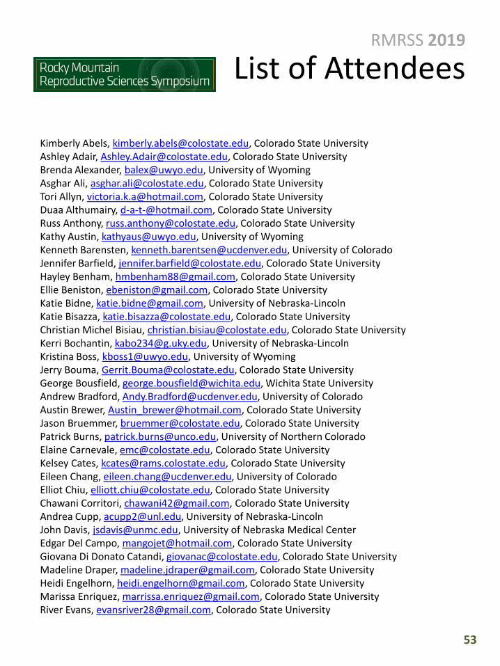
53
RMRSS 2019
List of Attendees
Kimberly Abels, [email protected], Colorado State UniversityAshley Adair, [email protected], Colorado State UniversityBrenda Alexander, [email protected], University of WyomingAsghar Ali, [email protected], Colorado State UniversityTori Allyn, [email protected], Colorado State UniversityDuaa Althumairy, [email protected], Colorado State UniversityRuss Anthony, [email protected], Colorado State UniversityKathy Austin, [email protected], University of WyomingKenneth Barensten, [email protected], University of ColoradoJennifer Barfield, [email protected], Colorado State UniversityHayley Benham, [email protected], Colorado State UniversityEllie Beniston, [email protected], Colorado State UniversityKatie Bidne, [email protected], University of Nebraska-LincolnKatie Bisazza, [email protected], Colorado State University Christian Michel Bisiau, [email protected], Colorado State University Kerri Bochantin, [email protected], University of Nebraska-LincolnKristina Boss, [email protected], University of WyomingJerry Bouma, [email protected], Colorado State UniversityGeorge Bousfield, [email protected], Wichita State University Andrew Bradford, [email protected], University of Colorado Austin Brewer, [email protected], Colorado State UniversityJason Bruemmer, [email protected], Colorado State UniversityPatrick Burns, [email protected], University of Northern Colorado Elaine Carnevale, [email protected], Colorado State UniversityKelsey Cates, [email protected], Colorado State UniversityEileen Chang, [email protected], University of Colorado Elliot Chiu, [email protected], Colorado State UniversityChawani Corritori, [email protected], Colorado State UniversityAndrea Cupp, [email protected], University of Nebraska-LincolnJohn Davis, [email protected], University of Nebraska Medical CenterEdgar Del Campo, [email protected], Colorado State UniversityGiovana Di Donato Catandi, [email protected], Colorado State UniversityMadeline Draper, [email protected], Colorado State UniversityHeidi Engelhorn, [email protected], Colorado State UniversityMarissa Enriquez, [email protected], Colorado State UniversityRiver Evans, [email protected], Colorado State University

54
RMRSS 2019
List of Attendees
Diana Fahrenbruck, [email protected], Colorado State UniversityHugo Fernandes, [email protected], Colorado Center for Reproductive MedicineEmma Fisher, [email protected], Colorado State UniversityShayne Garner, [email protected], Colorado State UniversityHanah Georges, [email protected], Colorado State UniversityCaroline Glezil, [email protected], Colorado State UniversityBen Goheen, [email protected], Colorado Center for Reproductive MedicineCarolina Gonzalez-Berrios, [email protected], Colorado State UniversityJames Graham, [email protected], Colorado State UniversityDorian Green, [email protected], Colorado State UniversityCourtney Grimm, [email protected], Colorado Center for Reproductive MedicineVictoria Handy, [email protected], Colorado State UniversityThomas Hansen, [email protected], Colorado State UniversityHelen Haskin, [email protected], GlycoFormA LtdKimberly Hayes, [email protected], Poudre Valley Health SystemAlex Headlough, [email protected], Colorado State UniversityLisa Herickhoff, [email protected], Membrane Protective TechnologiesJason Herrick, [email protected], Omaha’s Henry Doorly Zoo and AquariumEden Herron, [email protected], Colorado State UniversitySydney Hicks, [email protected], Colorado State UniversityNatalie Hohos, [email protected], University of Colorado Kristy Hood, [email protected], Colorado State UniversityMary Howerton, [email protected], Colorado State UniversityAnna Jacobs, [email protected], Colorado State UniversityJoshua Johnson, [email protected], University of Colorado Amanda Jones, [email protected], University of ColoradoKelsey Jones, [email protected], University of ColoradoJessica Keane, [email protected], University of Nebraska-LincolnShaihla Khan, [email protected], University of WyomingRebecca Kile, [email protected], Colorado Center for Reproductive MedicineWilliam Kinsey, [email protected], Kansas University Medical CenterRebecca Krisher, [email protected], Colorado Center for Reproductive MedicineBrian Krum, [email protected], University of Northern ColoradoKatherine Kuhn, [email protected], University of Colorado Madeleine Lacy, [email protected], Colorado State UniversityEvelyn Llerena Cari, [email protected], University of Colorado

55
RMRSS 2019
List of Attendees
Deirdre Logsdon, [email protected], Colorado Center for Reproductive MedicineMarlena Long, [email protected], Colorado State University Siyu Lu, [email protected], Colorado State University Brenda Martin, [email protected], Colorado State UniversityRosemary McDonald, [email protected], University of ColoradoMatthew McQuagge, [email protected], Colorado State UniversityNatalie Miller, [email protected], Colorado State UniversityAdel Moawad, [email protected], Colorado State UniversityJenifer Monks, [email protected], University of ColoradoLorna Moore, [email protected], University of ColoradoDanielle Moran, [email protected], Colorado State UniversityMona Munshi, [email protected], Colorado State UniversityJessica Myller, [email protected], Colorado State UniversityEmma Parent, [email protected], Colorado State UniversityAgata Maria Parsons Aubone, [email protected], Colorado State UniversityPablo Pinedo, [email protected], Colorado State UniversityMichele Plewes, [email protected], University of Nebraska Medical CenterTheresa Powell, [email protected], University of ColoradoSandeep Rajput, [email protected], Colorado Center for Reproductive MedicineAshley Raup, [email protected], Colorado State UniversityAshley Reisenauer, [email protected], Colorado State UniversityMargaret F. Riley, [email protected], University of VirginaCassandra Roeca, [email protected], University of ColoradoPablo Ross, [email protected], University of California DavisKelly Rouse, [email protected], Colorado State UniversitySaleh Salman, [email protected], Colorado State UniversityKa Sam, [email protected], University of ColoradoMegan Schmad, [email protected], Colorado State UniversityRowan Seabolt, [email protected], Colorado State UniversityGeorge Seidel, [email protected], Colorado State UniversityLaura Severeijns, [email protected], Colorado State UniversityElena Silva, [email protected], University of ColoradoMalgorzata Skaznik-Wikiel, University of ColoradoAlexandria Snider, [email protected], University of Nebraska-LincolnBryce Snow, [email protected], University of WyomingShelby Springman, [email protected], University of Nebraska-LincolnEd Squires, [email protected], Colorado State University

56
RMRSS 2019
List of Attendees
Alexis Susralski, [email protected], Colorado State UniversityCourtney Sutton, [email protected], University of Wyoming Anna Svoboda, [email protected], Colorado State UniversityAurora Talbot, [email protected], Colorado State UniversityAmelia Tanner, [email protected], Colorado State UniversityMilt Thomas, [email protected], Colorado State UniversityLindsey Turner, [email protected], Colorado State UniversityHana Van Campen, [email protected], Colorado State UniversitySue VandeWoude, [email protected], Colorado State UniversityOwen Vaughan, [email protected], University of ColoradoEmma Vickland, [email protected], Colorado State UniversityStephanie Wesolowski, [email protected], University of ColoradoRachel West, [email protected], Colorado Center for Reproductive MedicineRichard Wheeler, [email protected], Colorado State UniversityQuinton Winger, [email protected], Colorado State UniversityColeman Young, [email protected], University of WyomingYe Yuan, [email protected], Colorado Center for Reproductive Medicine\Miguel Zarate, [email protected], University of Colorado

2019 Program Committee
Dr. Thomas (Tod) Hansen, Chair, Colorado State University
Dr. Patrick Burns, Co-Chair, University of Northern Colorado
Dr. Jerry Bouma, Colorado State University
Dr. Jason Bruemmer, Colorado State University
Dr. Andy Bradford, University of Colorado Denver
Dr. Brenda Alexander, University of Wyoming
57
RMRSS 2019ACKNOWLEDGEMENTS
RMRSS, a regional meeting focused on graduate student training, issupported by the Animal Reproduction and BiotechnologyLaboratory, Department of Biomedical Sciences, College ofVeterinary Medicine and Biomedical Sciences, and Vice President forResearch Office at Colorado State University.
The 2019 symposium was also supported in part by Endolytics, CSUVentures, and by a sponsorship from the Society for the Study ofReproduction, which helped maintain our policy of free registrationfor all students.