rhabdomyomas and rhabdomyosarcomas (rms)
TRANSCRIPT

Rhabdomyomas
and Rhabdomyosarcomas (RMS)
David M. Parham, MD
Chief of Anatomic Pathology

Tumors of skeletal muscle:
Rhabdomyomas and rhabdomyosarcomas
2
Embryonal muscle

3

4

5

6

7

8

Rhabdomyoma
• Benign soft tissue counterpart of RMS
• Rare (<2% of soft tissue neoplasms)
• Cardiac and non-cardiac types
– Cardiac more common, associated with tuberous sclerosis
• Non-cardiac types
– Fetal
– Adult
– Genital

Fetal Rhabdomyoma
Clinical Features
• Most patients <3 years of age
• More common in boys
• Sporadic or associated with Gorlin syndrome
• Most common in head and neck
– Post-auricular region is favored site
• Much less common than rhabdomyosarcoma (1:50)

Fetal Rhabdomyoma
Histologic Features
• Parallel bundles of spindle cells and myoid tubules
• Separated by less differentiated cells
• Biphasic pattern
• No significant nuclear pleomorphism
• Low mitotic activity (<1/10 HPFs)
• Intermediate forms may resemble adult rhabdomyosarcomas and
contain smooth and skeletal muscle

12

Fetal Rhabdomyoma
Differential Diagnosis
• Spindle cell rhabdomyosarcoma
• Embryonal rhabdomyosarcoma
• Differential points
– No cambium layer
– RMS has higher mitotic rate (>5/10 HPFs)
– Circumscription: RMS not well circumscribed
– Should be PRE-treatment material for differential dx
13

Fetal Rhabdomyoma
Immunohistochemistry
• Markers of differentiated muscle
– Myoglobin
– Desmin
– Actin (smooth and skeletal muscle
– Not useful in distinction from RMS
• Negative:
– Keratin
– CD68 (useful for rhabdomyoma-like histiocytic tumors, sometimes
desmin-positive)
14

Fetal Rhabdomyoma
Genetics
• Gorlin syndrome mutation: PTCH1
– Negative regulator of hedgehog signaling pathway
– Pathway may also be activated in sporadic, non-syndromic tumors
15

Adult Rhabdomyoma
• Rare benign neoplasm
• Composed of mature, differentiated muscle
• Typically occurs after adolescence

Adult Rhabdomyoma
Clinical Features
• Usually in
– Oral cavity
– Superficial soft tissue of head and neck
– Origin from bronchial musculature?
• Mean age: 50 years
• Rare pediatric cases
• Up to 20% are multifocal
• Local recurrences possible, even years after operation
17

Adult Rhabdomyoma
Pathologic Features
• Gross: Brown lobulated mass
• Micro:
– Large round and polygonal cells
– Uniform cytology
• PAS positive due to glycogen
• Cross-striations may be seen.
18

19

Adult Rhabodyoma
Differential Diagnosis
• Granular cell tumor (S100 positive, desmin negative)
• Hibernomas (S100 positive, desmin negative)
• Crystal storing histiocytosis (CD68 positive, desmin negative,
crystals on EM)
20

Genital Rhabdomyoma
• Benign myogenic tumor of female genital tract
• Possible origin from myogenic stem cells in subepithelial stroma
• Usually in vagina or cervix of reproductive age women
– May be pregnancy-associated
• Male genital lesions occur, but more like fetal rhabdomyoma
• Female lesions resemble adult rhabdomyoma
– No significant pleomorphism
– Focal cross striations

Miscellaneous Benign
Myogenous Tumors
• Neuromuscular choristoma (“benign Triton tumor”)
– A hamartomatous lesion of peripheral nerve
– Muscle within perineurial sheath
– Most in head and neck
– May have associated rhabdomyoma

Benign Triton Tumor
23
Desmin and EMA

Miscellaneous Benign
Myogenous Tumors
• Rhabdomyomatous mesenchymal hamartoma
– Head and neck lesions
– Affects skin of newborn and infant ages
– Polypoid skin tags or mouth lesions containing benign skeletal muscle
• Myotubes, cross-striations
24

Benign rhabdomyomatous hamartoma
25

Focal Myositis
• Solitary mass
• Degenerating and regenerating skeletal muscle
• Infiltrates of macrophages and lymphocytes
• Secondary to focal denervation?
• Relationship to polymyositis?
– Only occasional association

Rhabdomyosarcoma
• Soft tissue tumor with features of embryonic or fetal myogenesis
• Most common soft tissue sarcoma in children
• Can be subclassified into multiple, distinct clinical and genetic
entities
– Embryonal
– Alveolar
– Spindle cell
– Pleomorphic (adult)

Rhabdomyosarcoma
Introduction and Clinical Features: General
• Most present as painless mass
• Rarely have systemic problems, e.g. hypercalcemia
• Symptoms may relate to affected organ
– Proptosis
– Urinary retention
– Bile retention
28

29

• May be affected by genetic syndrome
– P53
– Beckwith-Wiedemann
– Neurofibromatosis 1
– Gorlin
– DICER mutation-associated (pleuropulmonary blastoma)
30

Rhabdomyosarcoma
Pathologic Features: General
• Only some arise in extremity skeletal muscle; most do not
• Histology resembles developing or disordered muscle
– Rhabdomyoblast is key cell
– Undifferentiated “blastoma” cells
– Occasional multinucleated cells
– Differentiated myoblasts (more common after therapy)
31

32
Primitive Differentiated

Embryonal Rhabdomyosarcoma
• A primitive soft tissue tumor with features of embryonic skeletal
muscle

Embryonal Rhabdomyosarcoma
Clinical Features
• Over one half of rhabdomyosarcoma
• Most occur in children <10 years of age
• Rare in adults
• Botryoid type forms polypoid masses in vagina, bladder, mouth, or
bile ducts
34

35

Embryonal Rhabdomyosarcoma
Histologic Features
• Haphazard arrangement with areas of myxoid stroma
• Compact areas
– “Loose and dense” pattern resembling nascent muscle
• Some predominantly loose, others predominantly dense
• Much variation in cytology
– Strap cells, spider cells, racket cells, etc., with bright eosinophilic
cytoplasm
– Undifferentiated, slightly spindled cells with oblong nuclei
• Cambium layer in botryoid variant
36

ERMS
37

ERMS
38

Embryonal Rhabdomyosarcoma
Genetics
• No single specific mutation
• Recurring pattern of chromosome gains and losses
• Profound loss of heterozygosity on chromosome 11p
– Affects Beckwith-Wiedemann locus (11p15.5)
– Associated with imprinting abnormalities
– Loss of maternal allele in syndromic cases

ERMS
40

Embryonal Rhabdomyosarcoma (ERMS)
Differential Diagnosis
• Malignant peripheral nerve sheath tumor (MPNST)
– Triton tumor contains ERMS-like foci
– MPNST may be positive with some muscle markers
• Myofibroblastic tumors
– Inflammatory myofibroblastic tumors (often ALK1 positive, but so is
ERMS)
• Inflammatory or reactive processes
– Eosinophilic cystitis, beware on frozen section

Spindle Cell/Sclerosing Rhabdomyosarcoma
• Spindle cell lesion resembling fibrosarcoma or smooth muscle
neoplasm
• May be hyalinized with abundant collagen (sclerosing RMS)

Spindle Cell/Sclerosing Rhabdomyosarcoma
Clinical Features
• Predilection for paratesticular region in boys
• Head and neck
• Extremities
• Good prognosis in children (different tumor?)
• Bad prognosis in adults
43

Spindle Cell/Sclerosing Rhabdomyosarcoma
Histologic Features
• Spindle cells with elongate borders
• Must comprise >80% of tumor (overlap with ERMS)
• Should have definitive skeletal muscle phenotype
– Some smooth muscle features
– Some areas may be positive for smooth muscle actin
– Isolated, well-differentiated cells with cross striations may be found
• Variable collagen
44
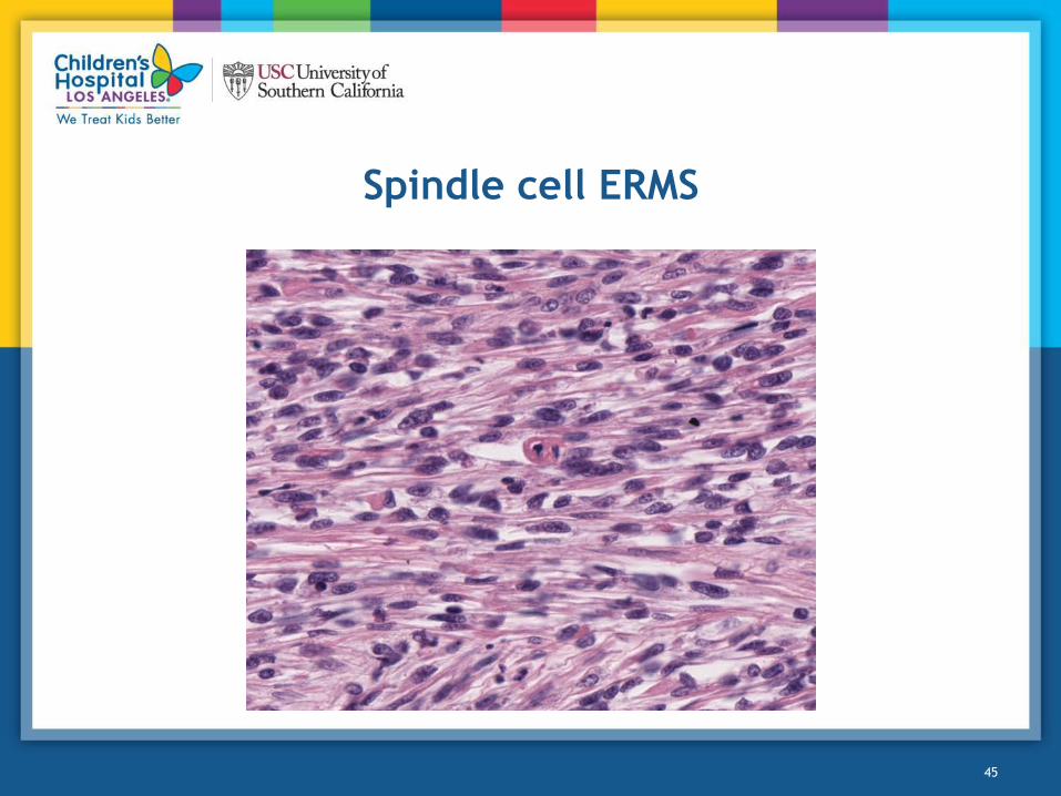
Spindle cell ERMS
45

Sclerosing rhabdomyosarcoma
• Hyalinizing collagen with entrapment of tumor cells defines
sclerosing variant
• Osteoid-like
• Can have a small cell component that resembles an alveolar
pattern
46

Sclerosing RMSA
47

Spindle Cell/Sclerosing Rhabdomyosarcoma
Genetics
• Adult spindle cell RMS:
– 40% have activating mutations in MyoD1 gene (located in chromosome
11p)
• Infantile spindle cell RMS
– May have NCOA2 rearrangements
– Should be negative for PAX gene and FOXO1 rearrangement

Spindle Cell/Sclerosing Rhabdomyosarcoma
Differential Diagnosis
• Leiomyosarcoma (myogenin negative)
• Fibrosarcoma (desmin negative)
• Synovial sarcoma (positive for SS18 rearrangement, negative for
myogenin, typically negative for desmin)
• Spindle cell carcinoma (usually adults)
• Osteosarcoma (vs sclerosing RMS, myogenin-negative)
• Angiosarcoma (CD31-positive)

Alveolar Rhabdomyosarcoma
• A small cell neoplasm
• Patternless sheets of cells or alveolar pattern
• Often (always?) positive for FOXO1 fusion

Alveolar Rhabdomyosarcoma
Clinical Features
• Usually extremity
• Head and neck (sinonasal tumors in particular)
• Parameningeal
• Metastasize to regional lymph nodes
• Predominate in older children, adolescents
– Also occur in younger children, adults
• Incompletely excised lesions recur with drug resistance
• Rare lesions present as leukemia
51

ARMS
52

Alveolar Rhabdomyosarcoma
Pathologic Features
• Alveolar pattern
– Cells lining a hollow space (L. alveolus, cavity)
– Central floating clusters
– Peripheral discohesion or cracking
– Fibrous septa
• Solid pattern
– Patternless sheets
– No apparent alveolar spaces or septa
53

ARMS
54

ARMS, solid variant
55

Alveolar Rhabdomyosarcoma (ARMS)
Genetics
• t(2;13)
– PAX3-FOXO1 fusion
– About 60% of cases
– Overexpressed
• t(1;13)
– PAX7-FOXO1 fusion
– About 20% of cases
– Amplified
• PAX fusion negative
– Are these truly ARMS?

t(2;13)
57

ARMS FISH
58

Fusion vs. histology (Davicioni et al).
59

Alveolar Rhabdomyosarcoma
Differential Diagnosis
• ERMS (fusion negative, with rare exceptions)
• Ewing sarcoma
• Lymphoma
• Neuroblastoma
• Synovial sarcoma
• Other round cell neoplasms

Pleomorphic Rhabdomyosarcoma
• A high grade sarcoma that usually arises in the extremities
• Features similar to other high grade adult sarcomas
• Should have definitive skeletal myogenesis, by definition
– May require immunohistochemistry, i.e. myogenin stain

Pleomorphic Rhabdomyosarcoma
Clinical Features
• Lower extremity lesion most common
• Other sites, abdomen, chest wall, spermatic cord, arm, mouth,
orbit
• Ages: 21-81 years (median 54 years)
• Clinical outcome: 70% die, mean survival 20 months
62

Pleomorphic Rhabdomyosarcoma
Pathologic Features
• Pleomorphic spindle cells and giant cells
• May have “malignant fibrous histiocytoma”-like (storiform) pattern
• Cells with eosinophilic cytoplasm
• Woven, streaming, or indistinct pattern
• Must demonstrate features of skeletal muscle
– Myogenin and MyoD may be negative or focal in some cases
– Desmin should be positive, but is non-specific
– Cytological features should be present
63

Pleomorphic RMS
64

Pleomorphic Rhabdomyosarcoma
Genetics
• Few good studies
• Regions of chromosomal gain and loss
• Regions of gene amplification
• Genetic features of osteosarcoma

Pleomorphic Rhabdomyosarcoma
Differential Diagnosis
• Undifferentiated pleomorphic sarcoma (MFH)
• Dedifferentiated liposarcoma (MDM2 expression, 12p12-15 region
amplification
• Dedifferentiated chondrosarcoma (bone tumor)
• Melanoma (HMB45, other melanocytic markers)
• Spindle cell carcinoma
• Carcinosarcoma

Epithelioid/Rhabdoid Rhabdomyosarcoma
• A newly-described RMS variant with features of carcinoma or
rhabdoid tumor
• Rhabdoid tumor features more typical with children
• Probable variant of ERMS
– Fusion-negative
– Weak or focal myogenin
• Other differential diagnosis features
– INI1 retained
– Melanocyte markers negative (beware of desmin-positive melanoma)

Immunohistochemistry of Rhabdomyosarcoma
• General markers of muscle differentiation
– Muscle actin
– Desmin
• Specific markers of skeletal muscle differentiation
– MyoD (myf3)
• Must be nuclear stain to be positive
– Myogenin (myf4)
– Myoglobin (terminal differentiation)
68

Myogenin
69
ARMS ERMS

Surrogate Markers of Fusion Status in
Rhabdomyosarcoma
• Strong expression
– Myogenin
– NOS-1
– AP2β
– P-cadherin
• Weak expression
– HMGA2
– EGFR
– Fibrillin-2

Prognosis and Clinical Course of Rhabdomyosarcoma
Stage
Stage Sites T T Size Node Status Metastases
1 Favorable site (orbit; head and neck,
excluding parameningeal; and
genitourinary, nonbladder/prostate)
T1 or T2 Any N0, N1 or
NX
M0
2 Unfavorable site (any site not listed
above)
T1 or T2 <5 cm N0 or NX M0
3 Unfavorable site T1 or T2 <5 cm N1 M0
>5 cm N0, N1 or
NX
4 Any T1 or T2 Any N0, N1 or
NX
M1

Prognosis and Clinical Course of Rhabdomyosarcoma
Group
• Group I: Completely resected, non-metastatic
• Group 2: Microscopic residual disease
– non-metastatic
– resected nodal metastatic
• Group 3: Gross residual disease
• Group 4: Distant metastasis
72

Prognosis and Clinical Course of Rhabdomyosarcoma
Prognostic groups
Risk Group Stage/Group Fusion Status
Low Stage 1, Group III (orbit) Negative
Stage 1, Group I-II
Stage 2, Group I-II
Intermediate, subset 1 Stage 1, Group III (non-orbit) Negative
Stage 3, Group I-II
Intermediate, subset 2 Stage 2-3, Group III Negative
Stage 1-3, Group I-III Positive
Intermediate, subset 3 Stage 4, Group IV Negative
High Stage 4, Group IV Positive

Failure free survival
74

Summary
• Rhabdomyoma is a rare benign tumor of adults and children
• Rhabdomyosarcoma is more common, malignant, and usually
affects children
• Diagnosis is improved with immunohistochemistry
• Prognosis depends on stage, group, and PAX fusion status
75