rheumatoid arthritis chris gordon, m.d.. i don't deserve this award, but i have arthritis and i...
TRANSCRIPT
Rheumatoid Arthritis
Chris Gordon, M.D.

• I don't deserve this award, but I have arthritis and I don't deserve that either. – Jack Benny (1894 - 1974)
• History• Mrs. B is a 66 year old Caucasian woman who was treated with Celebrex for joint
pain. Ten days after starting therapy she developed a diffuse, pruritic, erythematous rash and was admitted to the hospital. Dermatology consult and skin biopsy were obtained. Pathology was consistent with a drug eruption. The rash improved with intravenous steriods. On hospital day #3 she complained of dyspnea. A chest x-ray showed a new large left pleural effusion.
• Past Medical History• "Arthritis" since the age of 13; treated only with intermittent NSAIDS and steroids. • Right-sided empyema one year ago treated with prolonged antibiotics and chest tube
drainage. • History of coronary artery disease (CAD), non-insulin dependent diabetes mellitus
(NIDDM), chronic obstructive pulmonary disease (COPD), depression, diverticulitis • Medications
Cardizem, Pepcid, Premarin, Remeron, Darvocet • Social History
She worked as a typist but quit because of arthritis when she was in her 20's. She smoked 1-2 packs of cigarettes a day for 40 years.
• Review of Systems• Diffuse joint pain and stiffness, worse in the morning, lasting all day, denies
joint swelling or warmth. She is able to do all of her activities of daily living. • Denies constitutional symptoms • Mild sicca symptoms, no history of sinusitis or otitis • Denies prior history of rash, photosensitivity, alopecia, oral ulcers,
Raynauds, paresthesias, myalgias, weakness • Physical Examination• The patient was frail, somewhat anxious and confused. • BP 130/60, pulse 70, afebrile. • HEENT - no alopecia, conjunctivae were clear, no nasal or oral ulcers.
Lungs - decreased breath sounds at left base, no crackles or wheezes; surgical scar over right posterior chest. Cardiac and abdominal exams were unremarkable. Neurological exam was nonfocal. The skin showed diffuse erythematous macules over her trunk and extremities.
• Musculoskeletal exam was notable for the following:cervical spine - mildly diminished range of motionshoulders, elbows - full range of motion without synovitis, tenderness, or deformityMCPs - synovial thickening with minimal tenderness, slight ulnar deviation, subluxations of the IPJsPIPs - no synovitis, deformities, or tendernessDIPs - Heberden nodeships, knees, ankles - full range of motion without synovitis, deformities or tendernessfeet - hallux valgus, no synovitis
• Laboratory Studies• Hematocrit 37.5%; WBC 6,800; platelets 674,00; ESR 35;
electrolytes normal; BUN 25; creatinine 0.9; glucose 193; calcium 9.1; albumin 2.7; total protein 5.6; liver function tests normal; uric acid 5.5 urinalysis negative for blood or protein. ABG on room air: pH 7.42 pCO2 35 p02 61, 93% saturation
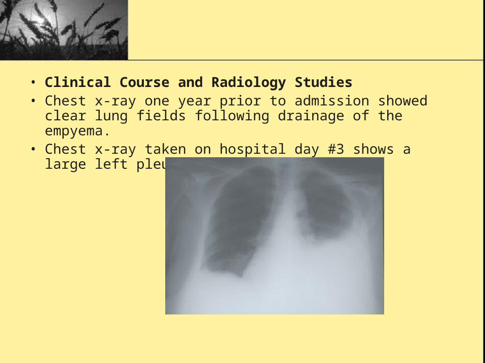
• Clinical Course and Radiology Studies• Chest x-ray one year prior to admission showed clear
lung fields following drainage of the empyema. • Chest x-ray taken on hospital day #3 shows a large left
pleural effusion.
• On hospital day #3 thoracentesis produced 540cc of turbid fluid: WBC 2025, 6% neutrophils, 42% lymphocytes, 52%; reactive mesothelial cells; LDH 9638; glucose 133; total protein 4.6
• On hospital day #4, the left pleural effusion rapidly re-accumulated, requiring a repeat thoracentesis WBC 1346,; LDH 7579; glucose 139; total protein 4.7; pH 7.1
• Pleural fluid showed no malignancy on cytology; a negative AFB and fungal smear; a negative Grams stain and routine culture.
• The patients dyspnea improved after the thoracentesis. She remained clinically stable with minimal joint pain throughout her hospitalization.
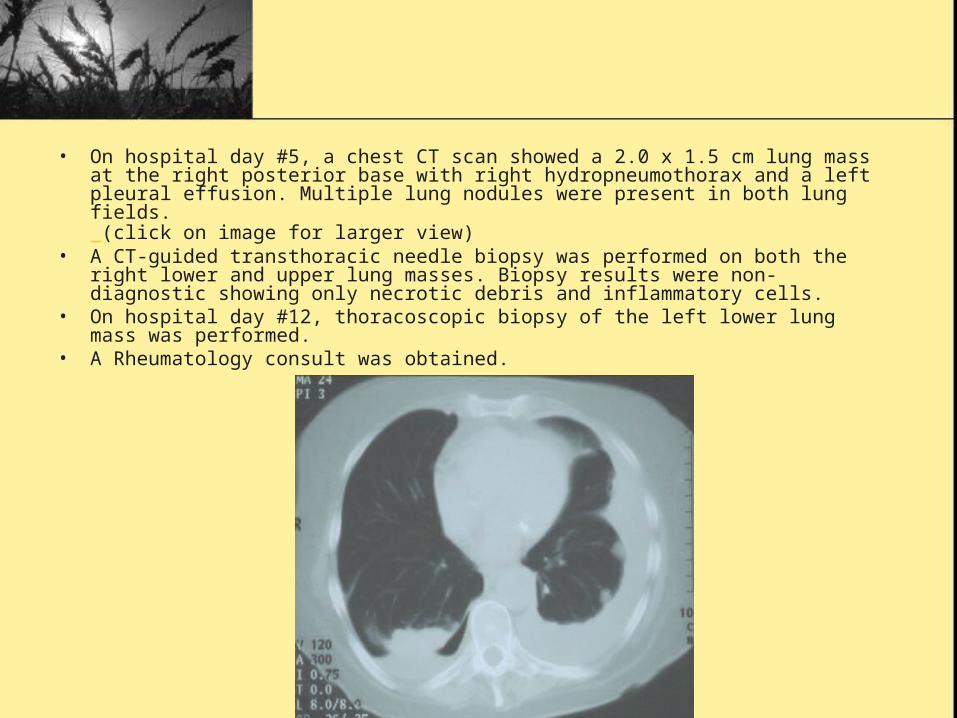
• On hospital day #5, a chest CT scan showed a 2.0 x 1.5 cm lung mass at the right posterior base with right hydropneumothorax and a left pleural effusion. Multiple lung nodules were present in both lung fields. (click on image for larger view)
• A CT-guided transthoracic needle biopsy was performed on both the right lower and upper lung masses. Biopsy results were non-diagnostic showing only necrotic debris and inflammatory cells.
• On hospital day #12, thoracoscopic biopsy of the left lower lung mass was performed.
• A Rheumatology consult was obtained.
• Differential Diagnosis of Exudative Pleural Effusion1.[ ]Infection (parapneumonic, tuberculosis, fungi)2.[ ]Malignancy (bronchogenic, metastatic)3.[ ]Pulmonary Embolism4.[ ]Gastrointestinal disease5.[ ]Rheumatic Conditions (rheumatoid arthritis, systemic lupus erythematosus)
• Differential Diagnosis of Pulmonary Nodules1.[ ]Infection (tuberculosis, fungi, pyogenic abscess)2.[ ]Malignancy (bronchogenic, lymphoproliferative, metastatic)3.[ ]Benign tumors4.[ ]Arteriovenous malformations5.[ ]Rheumatic conditions (rheumatoid arthritis, Wegeners granulomatosis, Churg-Strauss, sarcoidosis, amyloidosis)
• Pathology Results • The gross pathologic examination revealed
multiple small yellowish nodular lesions. Most of the lung showed interstitial fibrosis and chronic inflammation. There were several areas of necrosis of varying size. There were no areas of palisading cells. Special stains for mycotic and acid fast organisms were negative.
• The final pathologic reading described changes most consistent with Wegener's granulomatosis, but the possibility of a rheumatoid nodule could not be excluded.
• Additional Diagnostic Studies
• Rheumatoid factor was highly positive at a titer of 1:1280.
• Anti-neutrophilic antibodies were negative.
• CT scan of the sinuses was normal.
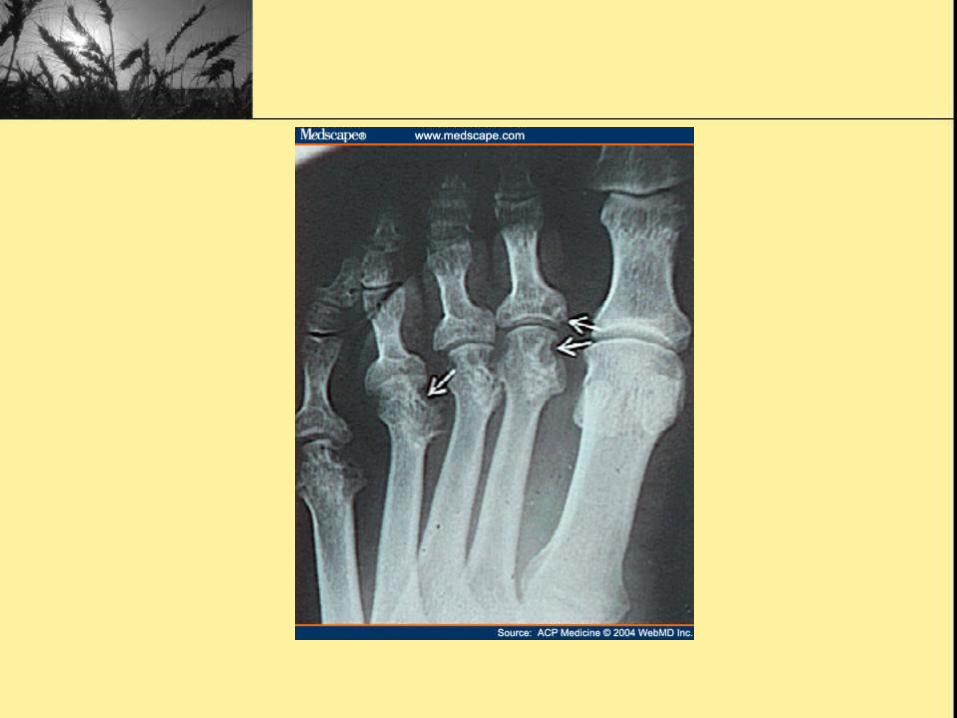
• Hand x-rays showed erosions most consistent with rheumatoid arthritis.
• Case Discussion• Summary:
Mrs. B is a 66 year old smoker with a long history of "arthritis" and an empyema one year ago, who presented with a new exudative pleural effusion and multiple lung nodules. Although the pathology of the lung nodule showed features suggesting Wegeners granulomatosis (necrotizing vasculitis, giant cells, disorganized cellular infiltrate), she lacked clinical evidence to support this diagnosis. The most likely cause of her illness was seropositive, erosive rheumatoid arthritis complicated by pulmonary rheumatoid nodules, bronchopleural fistulae, and recurrent pleural effusions.
• Conclusions • There were several unusual features of this case: the
patient was a woman without nodular disease, the pleural effusions were bilateral with a normal glucose and many mesothelial cells, and the pathology was not classic for a pulmonary rheumatoid nodule. However, she lacked clinical evidence of other diagnoses, and in fact, had a very high titer rheumatoid factor as well as classic findings of erosive RA on hand x-rays. This case illustrates the importance of viewing RA as a systemic disease, not simply a problem of joints
• Most common inflammatory arthritis• Affects 0.8% worldwide• Usually begins between ages 30 and 50• US incidence 25 per 100,000 in men and
54 per 100,000 in women1
• 250,000 hospitalizations and 9 million visits annually2
• 20-30% disabled within 3 years if untreated3
Etiology
• Not fully understood
• Genetic and environmental factors
• 30% concordance rate in monozygotic twins
• 80% of Caucasians with RA express HLA-DR1 or -DR4 subtypes
• Specific arthrogenic peptides to be presented to CD4+ T cells?4
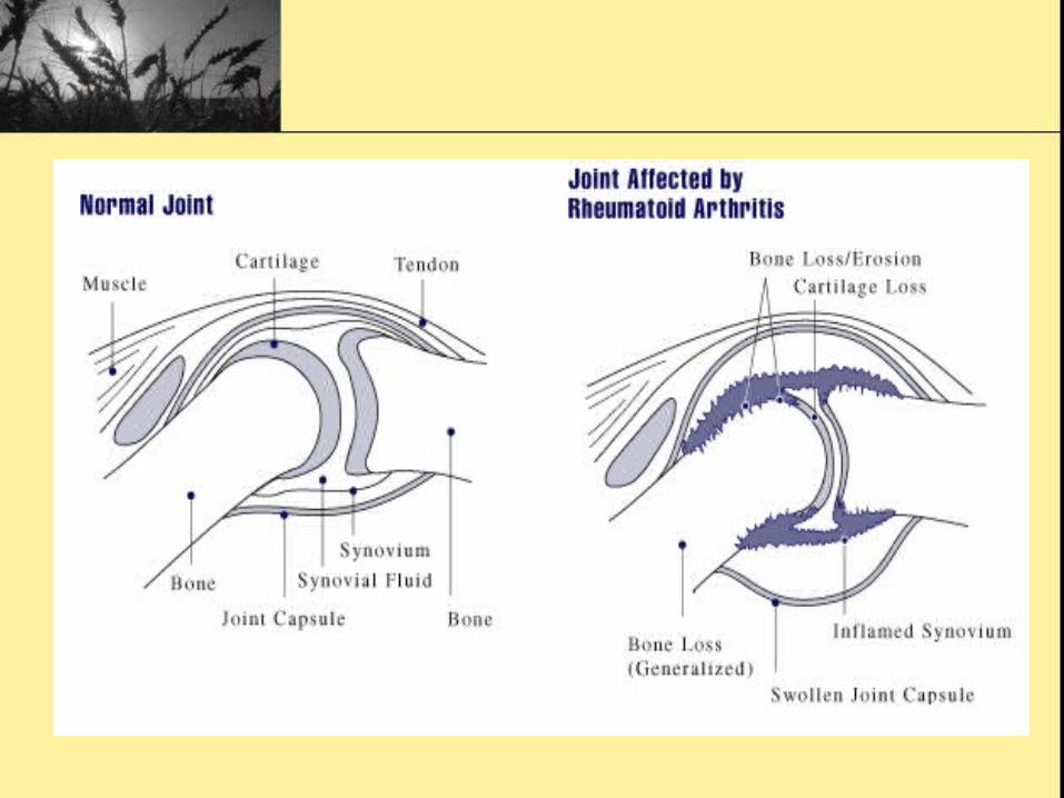
Pathophysiology
• Triggering incident (auto-immune vs infection)• Proliferation of synovial macrophages and
fibroblasts leading to lymphocyte and endothelial cell proliferation
• Small vessel occlusion leading to ischemia, neovascularization, inflammation
• Irregular growth of inflamed tissue (Pannus formation)
• Destruction of cartilage and bone and systemic complications

Risk Factors
Positive6,7
• Female • Smoking• Age• Family history• Silicate exposure• Caffeine ?
Negative8
• High vitamin D intake• Tea consumption• OCP use
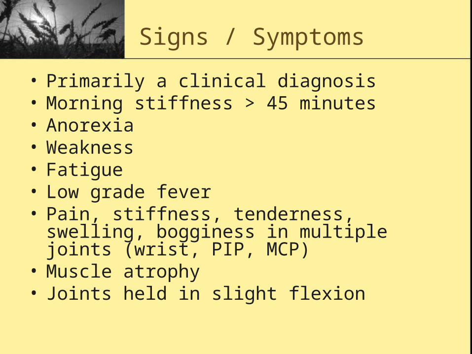
Signs / Symptoms
• Primarily a clinical diagnosis• Morning stiffness > 45 minutes• Anorexia• Weakness• Fatigue• Low grade fever• Pain, stiffness, tenderness, swelling, bogginess
in multiple joints (wrist, PIP, MCP)• Muscle atrophy• Joints held in slight flexion
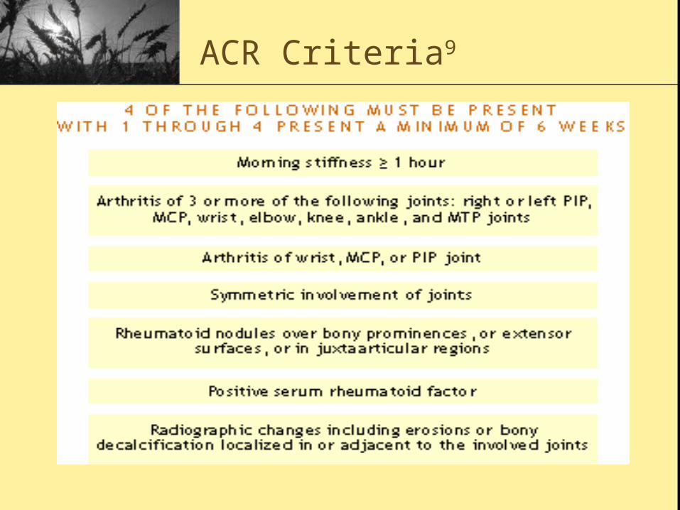
ACR Criteria9
Differential Diagnosis
• Connective tissue diseases• Fibromyalgia• Hemochromatosis• Infectious endocarditis• Polyarticular gout• Polymyalgia rheumatica• Seronegative spondyloarthropathies• Reactive arthritis
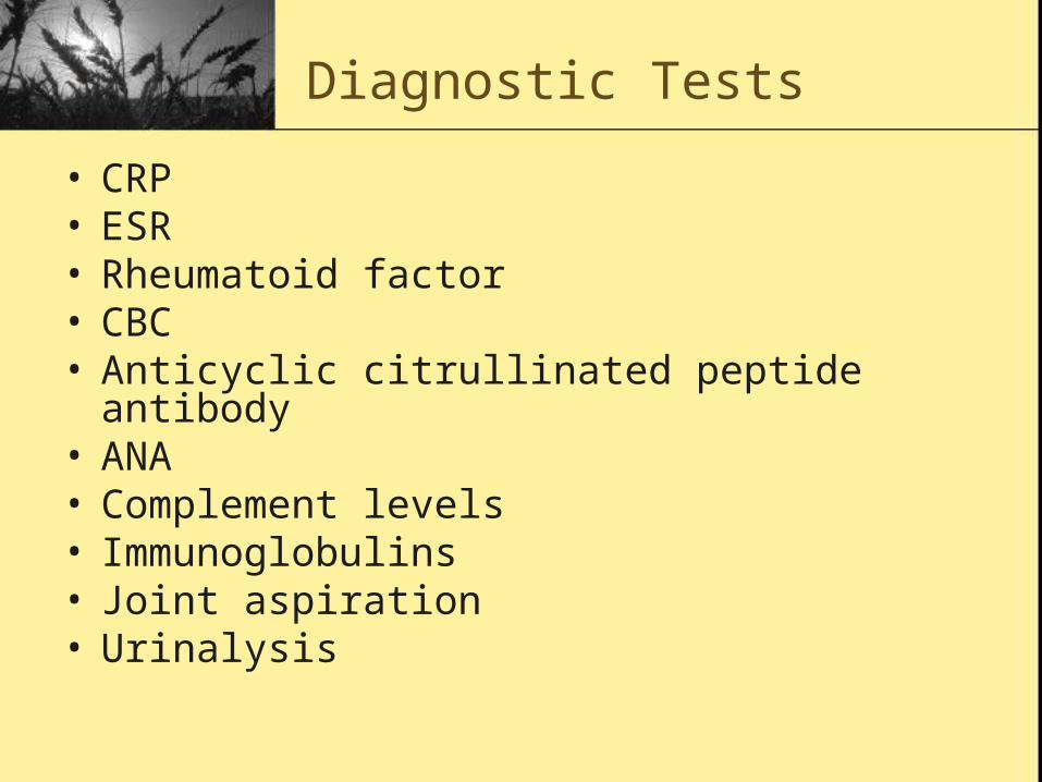
Diagnostic Tests
• CRP• ESR• Rheumatoid factor• CBC• Anticyclic citrullinated peptide antibody• ANA• Complement levels• Immunoglobulins• Joint aspiration• Urinalysis
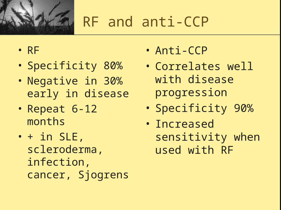
RF and anti-CCP
• RF• Specificity 80%• Negative in 30% early
in disease• Repeat 6-12 months• + in SLE,
scleroderma, infection, cancer, Sjogrens
• Anti-CCP• Correlates well with
disease progression• Specificity 90%• Increased sensitivity
when used with RF
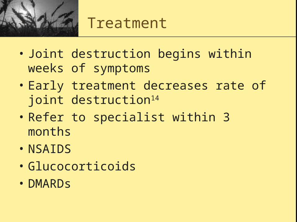
Treatment
• Joint destruction begins within weeks of symptoms
• Early treatment decreases rate of joint destruction14
• Refer to specialist within 3 months
• NSAIDS
• Glucocorticoids
• DMARDs
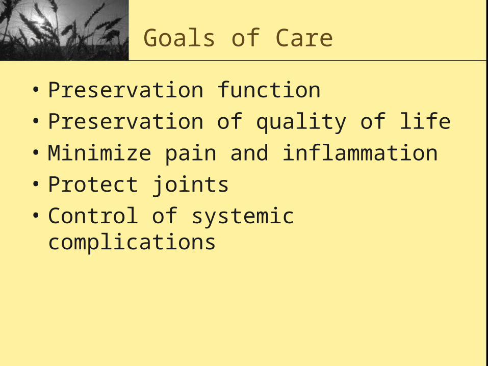
Goals of Care
• Preservation function
• Preservation of quality of life
• Minimize pain and inflammation
• Protect joints
• Control of systemic complications
NSAIDs2
• Used initially for symptom control
• Don’t alter disease progression
• Should not be used alone
• Twice as likely to have NSAID complication then patients with OA
Glucocorticoids
• Highly effective in relieving symptoms• Slow joint damage• Doses as low as Prednisone 10 mg daily
effective• Minimize dose to avoid side effects• Vitamin D and Calcium supplements• Often used as bridging therapy with DMARDs• Joint injection useful if single joint inflamed
DMARDs
• Consider for all patients• Combinations can be more effective than
single drugs• Contraception for women• Comorbidities, experience and severity
guide selection• Sulfasalazine or Hydroxychloroquin
usually initiated• MTX or combination if severe

Algorithm8
Leflunomide
• Arava
• Dose: 10 – 20 mg daily
• Competitive inhibitor of an intracellular enzyme needed for de novo pyrimidine synthesis by activated lymphocytes19
• Slows progression of joint damage
• Prevented new erosions in 80% of patients over two years22
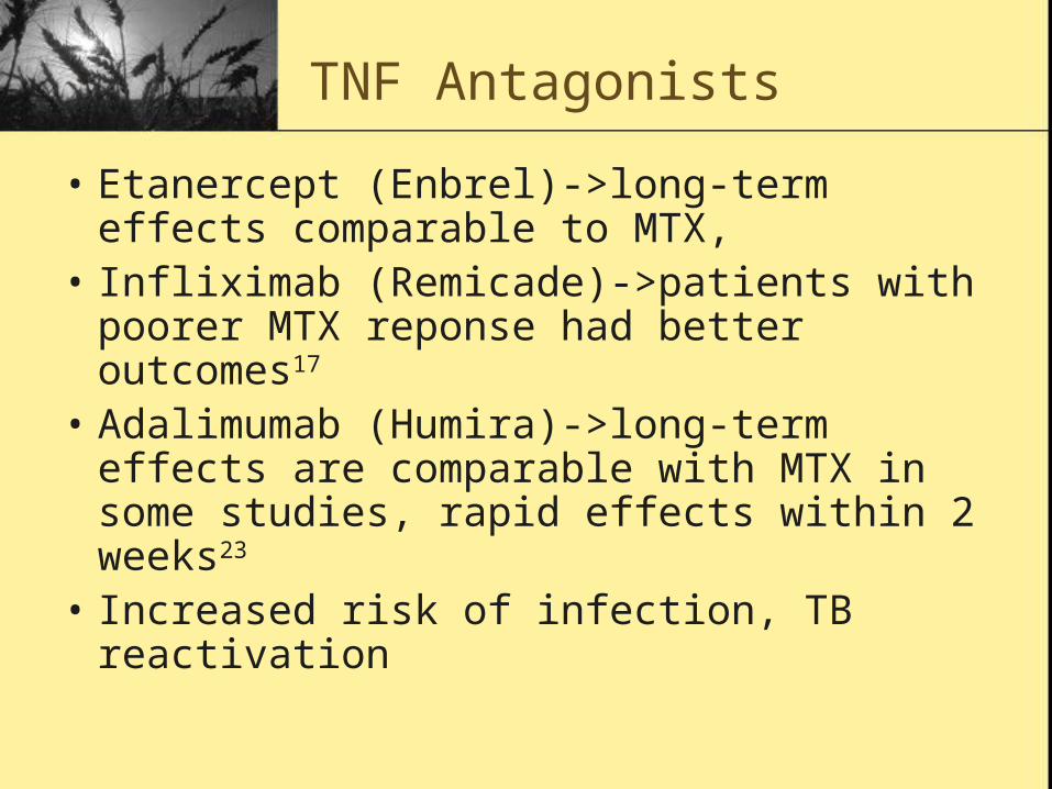
TNF Antagonists
• Etanercept (Enbrel)->long-term effects comparable to MTX,
• Infliximab (Remicade)->patients with poorer MTX reponse had better outcomes17
• Adalimumab (Humira)->long-term effects are comparable with MTX in some studies, rapid effects within 2 weeks23
• Increased risk of infection, TB reactivation

Adjunctive
• + Evidence• Therapeutic fasting• Dietary supplementation
of essential fatty acids• Journaling have shown
benefit26
• Spa therapies27
• Exercise28
• Patient education29
• Multi-disciplinary approach to patient care30
• No evidence• Herbal medications31
• Acupuncture26
• Splinting32
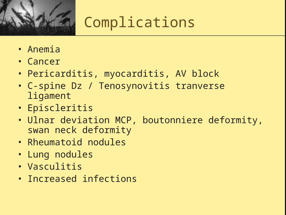
Complications
• Anemia• Cancer• Pericarditis, myocarditis, AV block• C-spine Dz / Tenosynovitis tranverse ligament• Episcleritis• Ulnar deviation MCP, boutonniere deformity, swan neck
deformity• Rheumatoid nodules• Lung nodules • Vasculitis • Increased infections
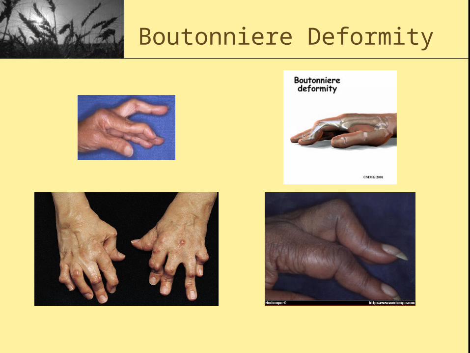
Boutonniere Deformity

Swan Neck Deformity
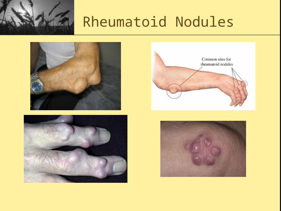
Rheumatoid Nodules

The End!