rigaku biosaxs webinar 093009
TRANSCRIPT
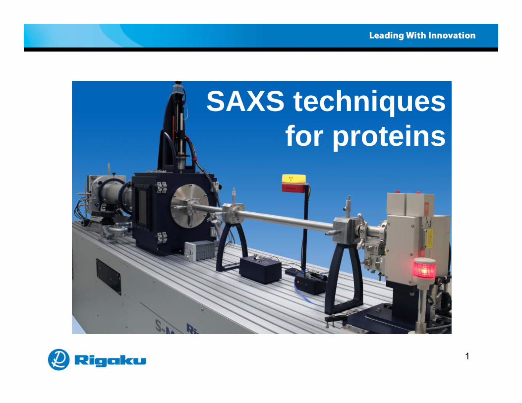
SAXS techniques f ifor proteins
1

SAXS techniques for proteinsSAXS techniques for proteins
Bi SAXS li ti• BioSAXS applications• BioSAXS theoretical overview• Experimental hardware for the home lab• Application examplesApplication examples• References and resources
2

SAXS techniques for proteins
Bi SAXS li ti
SAXS techniques for proteins
• BioSAXS applications• BioSAXS theoretical overview• Experimental hardware for the home lab• Application examplesApplication examples• References and resources
3
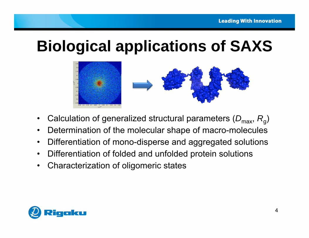
Biological applications of SAXSBiological applications of SAXS
• Calculation of generalized structural parameters (Dmax, Rg)• Determination of the molecular shape of macro-moleculesp• Differentiation of mono-disperse and aggregated solutions• Differentiation of folded and unfolded protein solutions
Characterization of oligomeric states• Characterization of oligomeric states
4

SAXS techniques for proteins
Bi SAXS li ti
SAXS techniques for proteins
• BioSAXS applications• BioSAXS theoretical overview• Experimental hardware for the home lab• Application examplesApplication examples• References and resources
5

Crystallography vs SAXSCrystallography vs. SAXS
Integrated profile
Log(
I)
q [Å-1]
6

Complimentary techniquesComplimentary techniques
Method Crystallography SAXSSamples Single crystals Dilute solutions (1 ~ 100mg/ml)
Advantages High resolution (up to 0.1nm)At i t t i f ti
Analysis in native conditionsAtomic structure information
Limitations Crystal required Low resolution (~1-2nm)Modeling ambiguity
7

Scattering intensityScattering intensity)(*)(∝)( AAI
X-ray diffraction
)(*•)( ∝)( qAqAqI
xy
z∑
1=)}++(2exp{ =)(
N
jjjjjhkl lzkyhxiπfA q
iA d)()(Δ)( ∫
x
rSmall angle x-ray scattering
riqrrρqA d)exp()(Δ =)( ∫V
8

Pair distribution functionPair distribution functionCrystal Solution
Patterson function Pair distribution function
P(r)
9r

Profile conversionProfile conversionSAXS pattern) SAXS pattern
Log(
I)q [Å-1]
Guinier plot
n(I)
Kratky plot
q2I
Pair distributionfunction
P(r
)q2 [Å-2]
ln
q [Å-1] r [Å]P
10
q [Å ] q [Å ] r [Å]

Guinier plotGuinier plot32Rq
3)]0(ln[=)](ln[ GRqIqI
Mono-disperse Aggregated
11
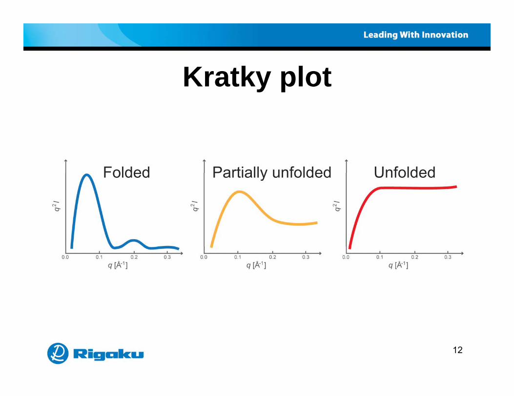
Kratky plotKratky plot
Folded Partially unfolded UnfoldedFolded Partially unfolded Unfolded
12
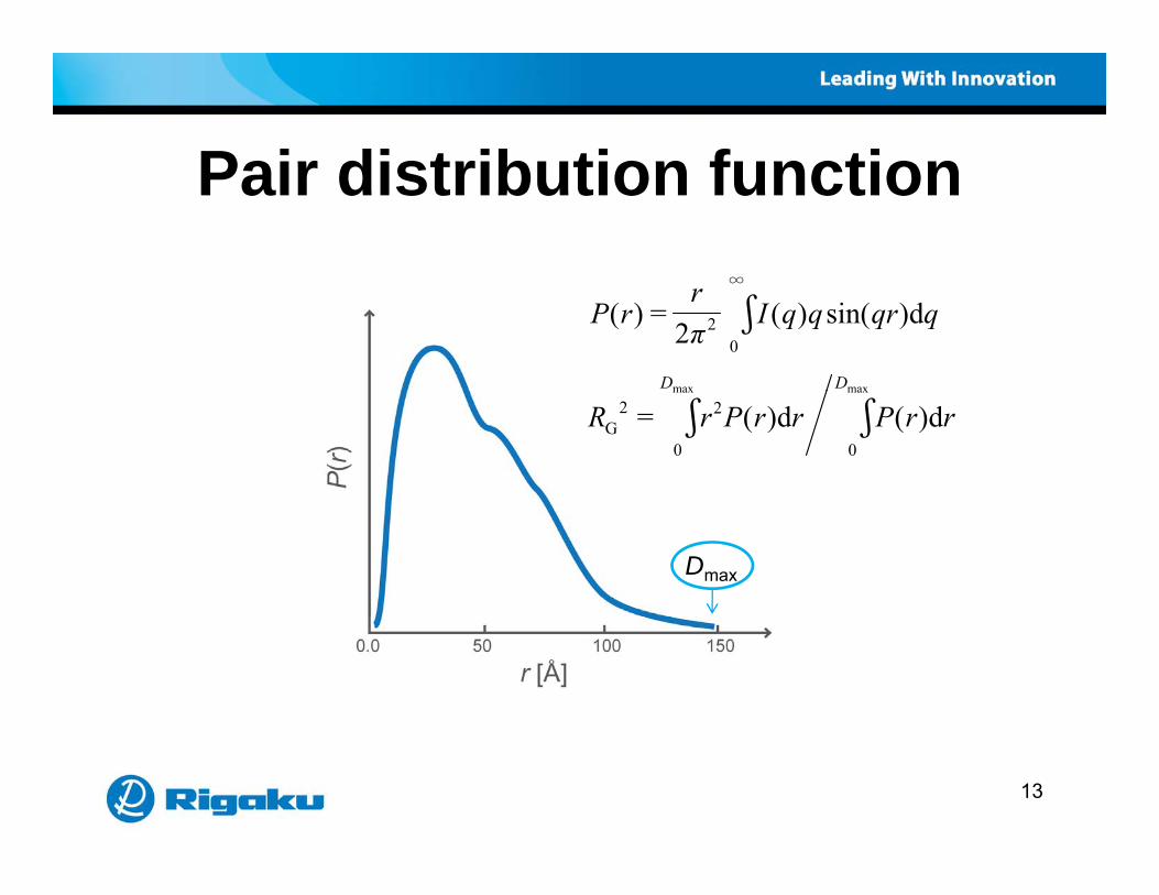
Pair distribution functionPair distribution function∞
qqrqqIπr
rP d)sin()( 2
=)( ∫∞
02
DD maxmax
rrPrrPrR d)(d)( = ∫∫maxmax
0
2
0
2G
Dmax
13

Shapes & scattering patternsShapes & scattering patternsSAXS patterns Pair distribution functionsSAXS patterns Pair distribution functions
14

Refinement of molecular envelopeRefinement of molecular envelope
Squeeze a bean bag Compare Pcal(r) and Pobs(r)
Pcal(r)
Pobs(r)obs( )
15

SAXS techniques for proteins
Bi SAXS li ti
SAXS techniques for proteins
• BioSAXS applications• BioSAXS theoretical overview• Experimental hardware for the home lab• Application examplesApplication examples• References and resources
16
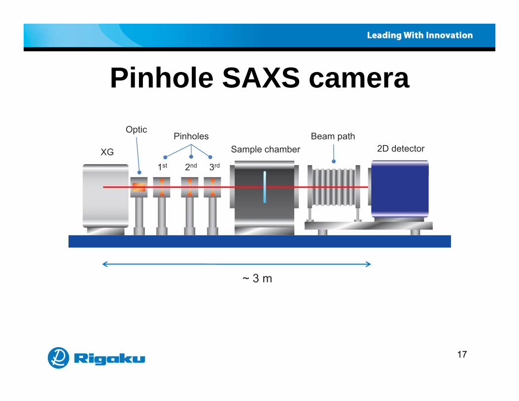
Pinhole SAXS cameraPinhole SAXS camera
XG
Optic
1st 3rd
Pinholes
2nd
Sample chamberBeam path
2D detector
3 m~ 3 m
17

MicroMax 007MicroMax 007
MM007MM007
18

MicroMax 002+MicroMax 002+
MM002+
19

MicroMax 007 / 002+ specsMicroMax 007 / 002+ specsMM007 MM002+MM007 MM002+
Camera length ~ 3 m
S l i 1 5 5 15 lSample size 1.5 x 5 mm, 15 μl
Beam size at sample 0.5 mm
C K fl t l 1 108 2 107Cu Kα flux at sample 1 x 10⁸ cps 2 x 10⁷ cps
2θ minimum 0.1 º
Q minimum 0 006 Å-1Q minimum 0.006 Å 1
Maximum length scale 100 nm
20

SMAX 3000SMAX 3000Dual Chamber SAXS Camera
21

SMAX 3000SMAX 3000Simultaneous WAXS/SAXS
22
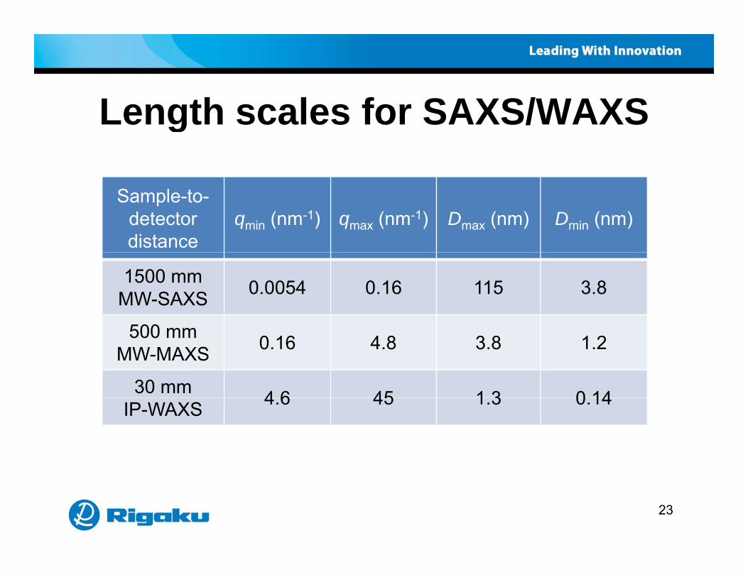
Length scales for SAXS/WAXSLength scales for SAXS/WAXS
Sample-to-detector distance
qmin (nm-1) qmax (nm-1) Dmax (nm) Dmin (nm)
1500 mmMW-SAXS 0.0054 0.16 115 3.8
500500 mmMW-MAXS 0.16 4.8 3.8 1.2
30 mm 4 6 45 1 3 0 14IP-WAXS 4.6 45 1.3 0.14
23

Flow cell sample handlingFlow cell sample handling
SamplePositioning stageCooling water
HeaterX-ray beam
Sample feeder
Linkam high temp unitManual flow cell
24
SAXS techniques for proteins
Bi SAXS li ti
SAXS techniques for proteins
• BioSAXS applications• BioSAXS theoretical overview• Experimental hardware for the home lab• Application examplesApplication examples• References and resources
25
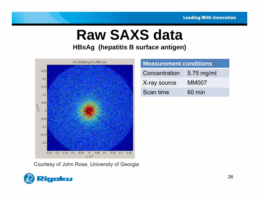
Raw SAXS dataRaw SAXS dataHBsAg (hepatitis B surface antigen)
M t ditiMeasurement conditionsConcentration 5.75 mg/mlX-ray source MM007Scan time 60 min
C t f J h R U i it f G i
26
Courtesy of John Rose, University of Georgia
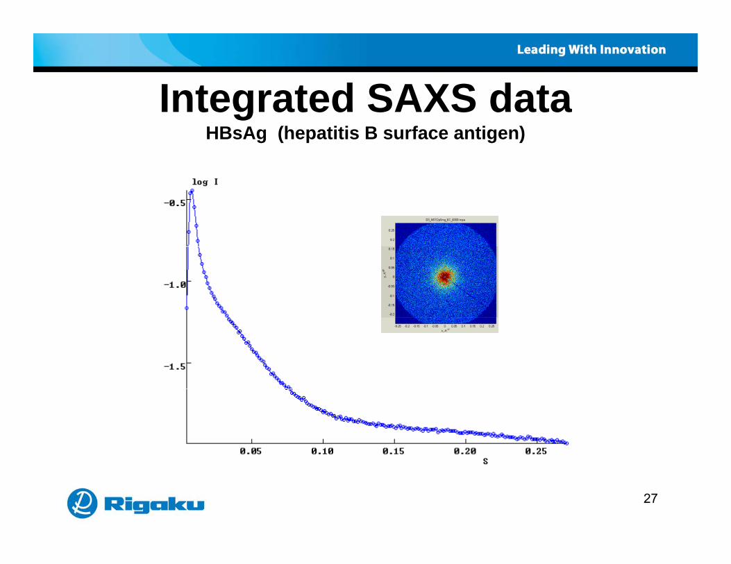
Integrated SAXS dataIntegrated SAXS dataHBsAg (hepatitis B surface antigen)
27

Integrated buffer dataIntegrated buffer dataHBsAg (hepatitis B surface antigen)
28

Background corrected dataBackground corrected dataHBsAg (hepatitis B surface antigen)
29

Guinier plot – determination or RGuinier plot determination or RgHBsAg (hepatitis B surface antigen)
Rg = 132. +/- 1.24
30
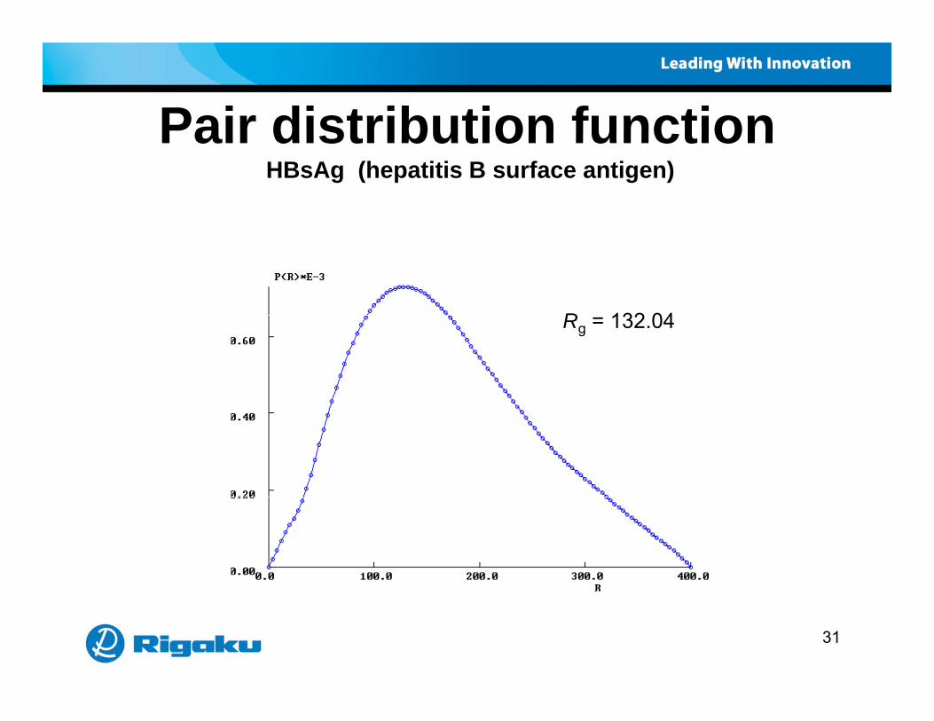
Pair distribution functionPair distribution functionHBsAg (hepatitis B surface antigen)
R 132 04Rg = 132.04
31
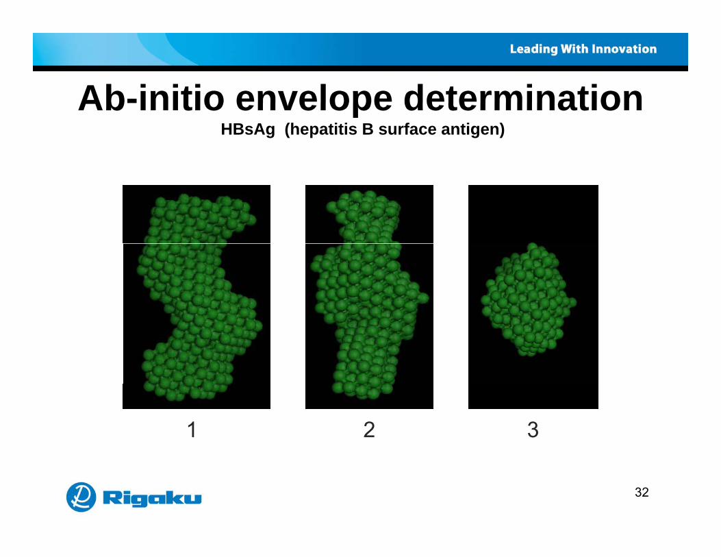
Ab-initio envelope determinationAb-initio envelope determinationHBsAg (hepatitis B surface antigen)
1 2 3
32

Raw SAXS dataRaw SAXS dataHSA (human serum albumin)
M t ditiMeasurement conditionsConcentration 5 mg/mlX-ray source MM007Scan time 90 min
33

Integrated SAXS dataIntegrated SAXS dataHSA (human serum albumin)
34
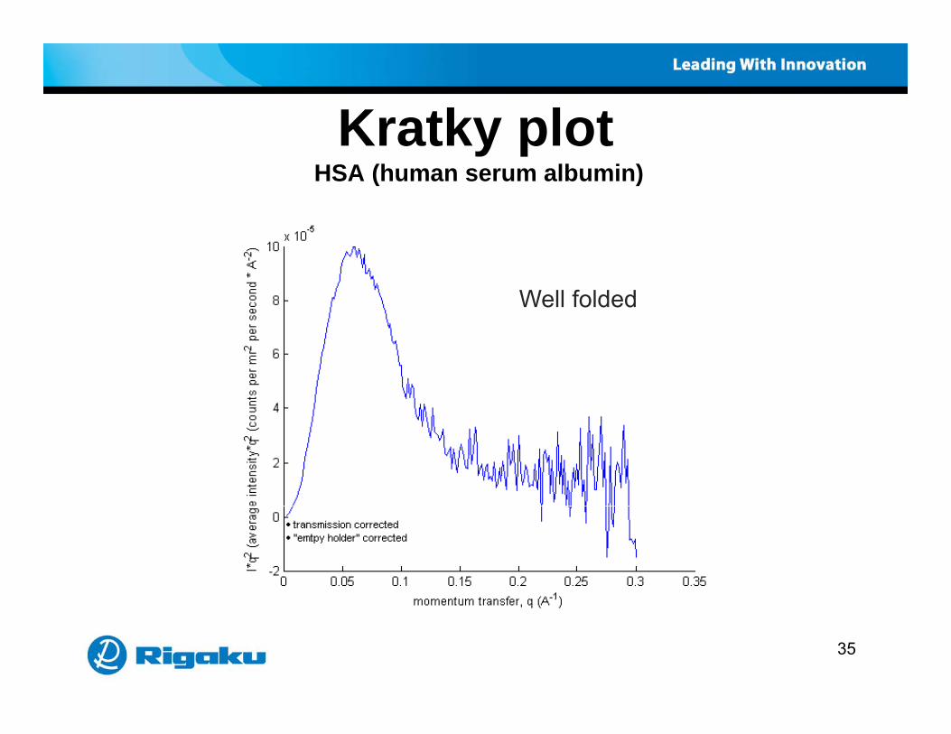
Kratky plotKratky plotHSA (human serum albumin)
Well folded
35

Pair distribution functionPair distribution functionHSA (human serum albumin)
R 29 02Rg = 29.02
36

Crystal structure andymolecular envelope (HSA)
37
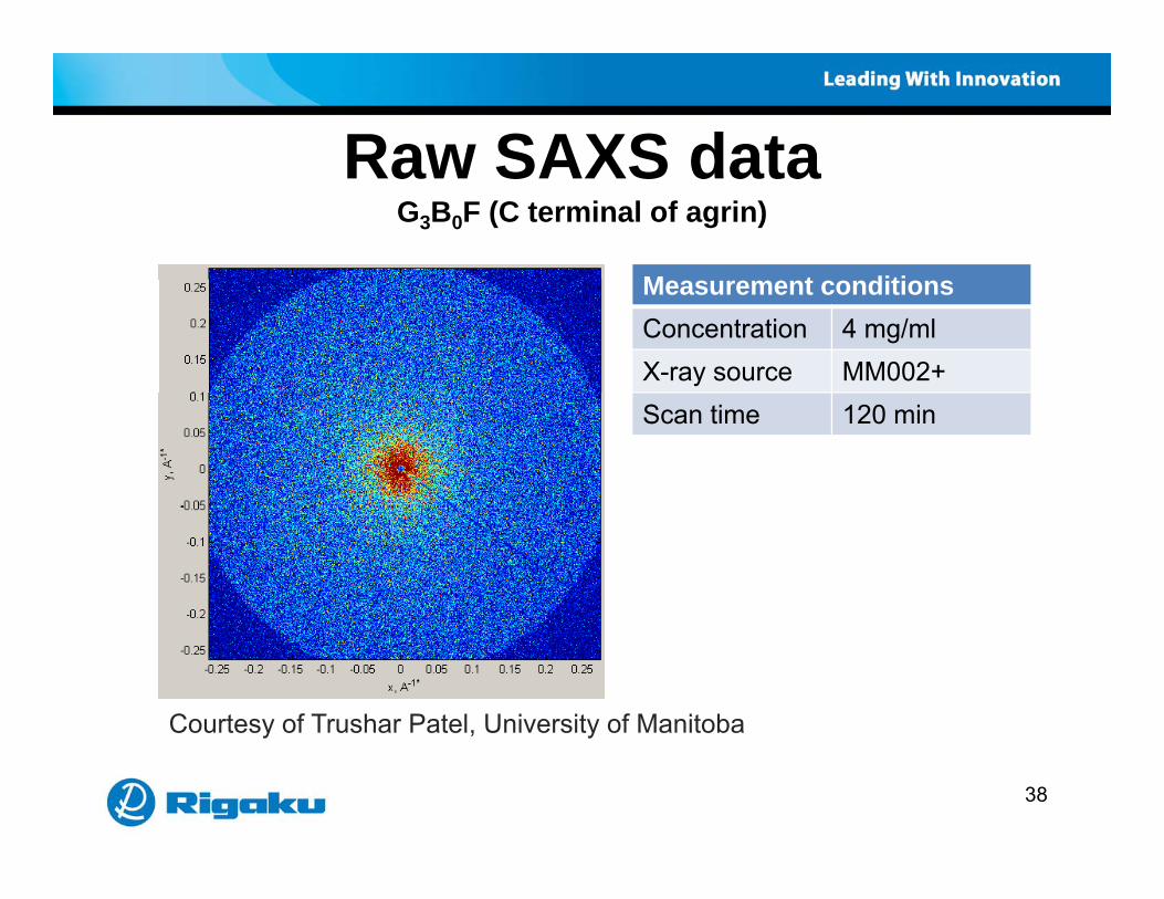
Raw SAXS dataRaw SAXS dataG3B0F (C terminal of agrin)
M t ditiMeasurement conditionsConcentration 4 mg/mlX-ray source MM002+Scan time 120 min
Courtesy of Trushar Patel, University of Manitoba
38
y , y

Integrated SAXS dataIntegrated SAXS dataG3B0F (C terminal of agrin)
39

Pair distribution functionPair distribution functionG3B0F (C terminal of agrin)
Rg = 52.4
Characteristic shape of multi-domain protein
g 5Dmax = 175
40
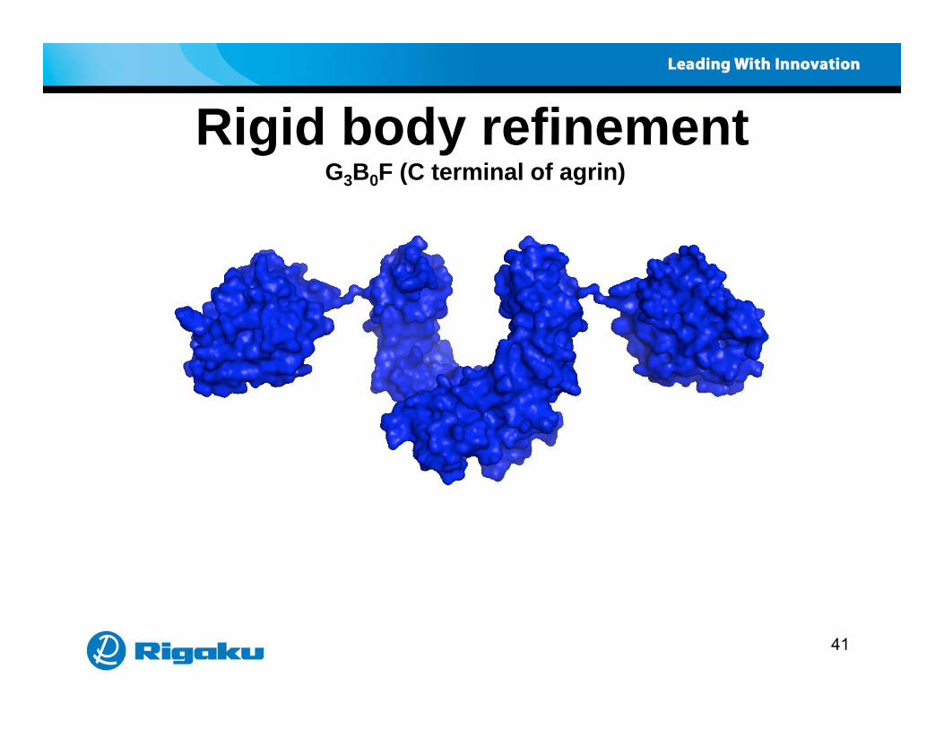
Rigid body refinementRigid body refinementG3B0F (C terminal of agrin)
41

Comparison with synchrotron dataComparison with synchrotron data
Structure envelopes
Courtesy of Thomas Grant, HWI
42

SAXS techniques for proteins
Bi SAXS li ti
SAXS techniques for proteins
• BioSAXS applications• BioSAXS theoretical overview• Experimental hardware for the home lab• Application examplesApplication examples• References and resources
43

Biological Small AngleBiological Small Angle Scattering Group - DESY
• ATSAS 2.2• Software download
htt // bl h b d /E t lI f /http://www.embl-hamburg.de/ExternalInfo/Research/Sax/software.html
44
Review papersReview papers• Robust high throughput solution structural analyses by small• Robust, high-throughput solution structural analyses by small
angle X-ray scattering (SAXS)Greg L Hura et al., Nature Methods, July 20 (2009)
• Small-angle scattering studies of biological macromolecules in solutionDmitri I Svergun et al Rep Prog Phys 66 1735 (2003)Dmitri I Svergun et al., Rep. Prog. Phys. 66 1735 (2003)
• X-ray solution scattering (SAXS) combined with crystallography and computationcrystallography and computationChristopher D Putnam et al., Q. Rev. Biophys. 40, 191 (2007)
45
Reference bookReference book
S ll l tt i f X• Small-angle scattering of X-raysAndre Guinier & Gerard FournetWil (1955) ( t f i t)Wiley (1955) (out-of-print)
ProQuest www.umi.com
46
ConferenceConferenceTh 67th A l Pitt b h Diff ti C f• The 67th Annual Pittsburgh Diffraction ConferenceOctober 29th – 31stUniversity of Georgia Center for Continuing Educationhttp://www.pittdifsoc.org/PDC_2009/
47

Presentation for downloadPresentation for downloadThi t ti i il bl f d l d t• This presentation is available for download at http://www.rigaku.com/protein/webinars.html
48

Thank you !a youw w w . R i g a k u . c o m
49