right andleft heart biplane cine-angiocardiography
TRANSCRIPT
Brit. Heart J., 1964, 26, 755.
RIGHT AND LEFT HEART BIPLANE CINE-ANGIOCARDIOGRAPHY
BY
HAMISH WATSON AND C. PICKARD
From the Departments of Medicine and Diagnostic Radiology in the University of St. Andrews, Queen's College,Dundee, and the Dundee Royal Infirmary
Received November 15, 1963
Technological progress in the design and construction of image intensifiers has been so rapidsince their introduction by Teves (1955) and Feddema (1955) that they are already in daily use formany types of radiological examination. Though much of the pioneer work in this field was angio-cardiographic (Lind, Wegelius, and Lichtenstein, 1954; Lind, Wegelius, and Boesen, 1955;Lind, Rocha, and Wegelius, 1957; Stauffer et al., 1955; Astley, 1955, 1956; Pickard and Watson,1957; Watson et al., 1958), there were certain disadvantages associated with the use of the earlyintensifiers for this type of investigation. The 5-inch field, for example, placed considerablelimitation on their usefulness, and this, coupled with the fact that it was only possible to film in oneplane, meant that the procedure had to be carefully planned not only to decide which part of theheart was to be visualized but also how best to position the patient to ensure that its opacificationwould be seen to the best possible advantage. Despite the difficulties, cine-photography of theintensified image has already revolutionized the visualization of the opacified heart and contributedgreatly not only to our understanding of valvular action but also to our knowledge of the intra-cardiac circulation in congenital heart disease.
The field size of intensifiers has been increased during the past few years and a much largerimage can now be obtained. The definition of detail has been greatly improved and the recentaddition of closed circuit television allows the clearer pictures to be much more easily seen. Theobject of this paper is to present a preliminary communication on our experience of simultaneousbiplane cine-angiocardiography using two 9-inch Philips image intensifiers.
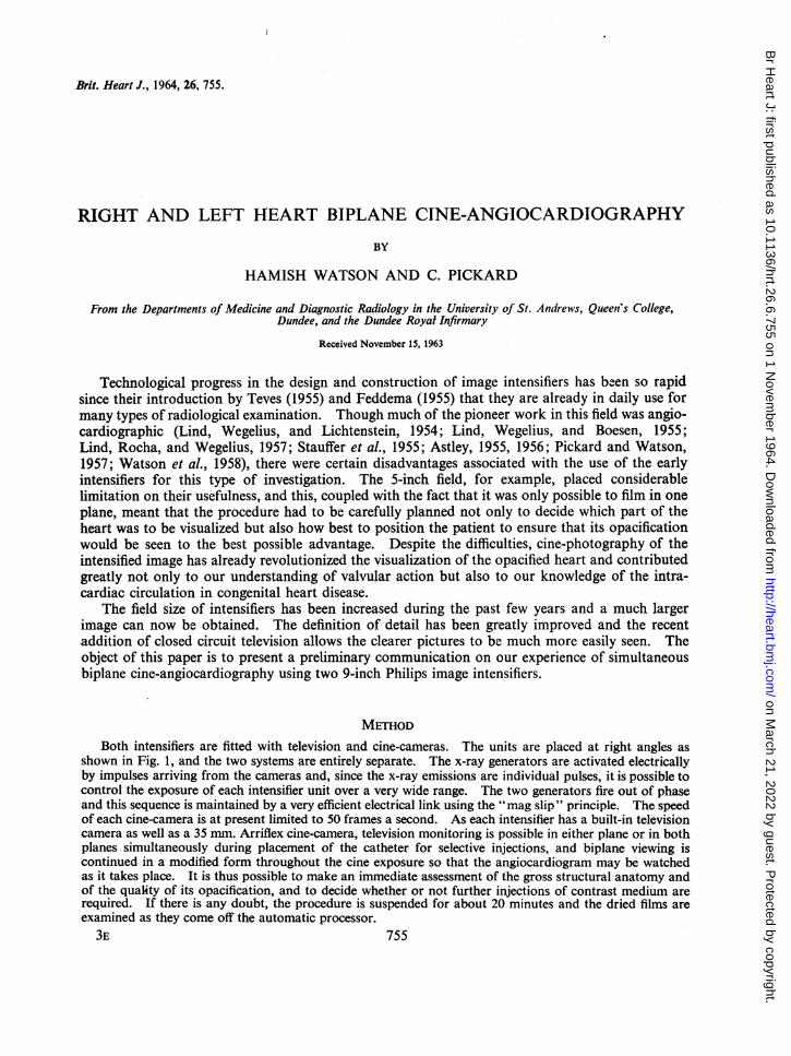
METHODBoth intensifiers are fitted with television and cine-cameras. The units are placed at right angles as
shown in Fig. 1, and the two systems are entirely separate. The x-ray generators are activated electricallyby impulses arriving from the cameras and, since the x-ray emissions are individual pulses, it is possible tocontrol the exposure of each intensifier unit over a very wide range. The two generators fire out of phaseand this sequence is maintained by a very efficient electrical link using the "mag slip" principle. The speedof each cine-camera is at present limited to 50 frames a second. As each intensifier has a built-in televisioncamera as well as a 35 mm. Arriflex cine-camera, television monitoring is possible in either plane or in bothplanes simultaneously during placement of the catheter for selective injections, and biplane viewing iscontinued in a modified form throughout the cine exposure so that the angiocardiogram may be watchedas it takes place. It is thus possible to make an immediate assessment of the gross structural anatomy andof the quality of its opacification, and to decide whether or not further injections of contrast medium arerequired. If there is any doubt, the procedure is suspended for about 20 minutes and the dried films areexamined as they come off the automatic processor.3E753E 755
on March 21, 2022 by guest. P
rotected by copyright.http://heart.bm
j.com/
Br H
eart J: first published as 10.1136/hrt.26.6.755 on 1 Novem
ber 1964. Dow
nloaded from

WATSON AND PICKARD
Except in special circumstances, our currentpractice is to make a selective injection into the
_4~ ...'',rightventricle, watch and film it in two planes, and_then to repeat the process by injecting into the left....... , . . ... ,,.D. o lejatrium.
We have found Triosil a safe and satisfactorypreparation especially suited to babies and smallchildren because of its low viscosity, and use 1-0to 15 ml. per kilogram of body weight for eachinjection. The volume required depends on indi-vidual circumstances, varying with the magnitudeand direction of intracardiac shunts and theseverity of closed chamber stenoses. As a general
1 rule satisfactory opacification of the left heart isobtained by using less contrast medium than isrequired for the right, though this probably reflectsonly the difference between atrial and ventricularinjections. Fig. 2 and 3 show excerpts of postero-
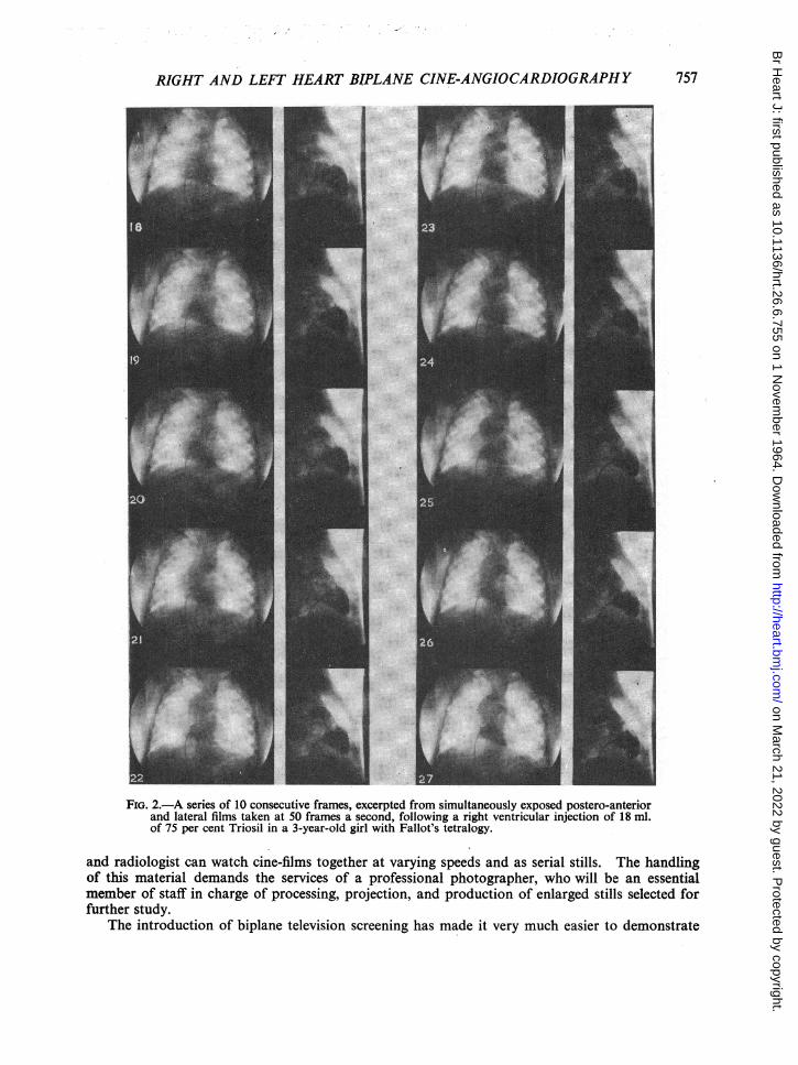
> ! !1 Ianterior and lateral cine-films taken simultaneouslyat 50 frames a second during the investigation ofa 3-year-old girl with Fallot's tetralogy. In Fig. 2,
-.- opacification of both the pulmonary trunk and the* aorta are seen following an injection of 18 ml. of
/4~~ / ,,/ ~~~ // 75 per cent Triosil into the apex of the right yen-i tricle. The severe nature of the outflow tract ob-
struction is revealed in the lateral film where the*~/'_7 infundibulum is seen to be grossly narrowed over
its whole length. It is of interest to note the post-stenotic dilatation of the pulmonary trunk, whichis almost as wide as the dilated overriding aorta.
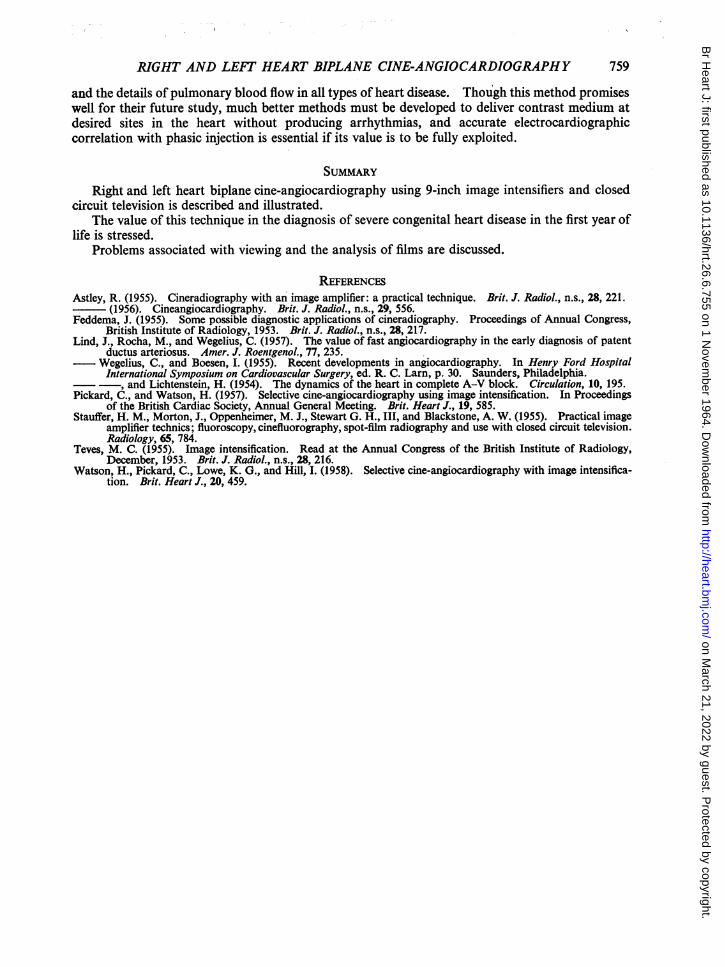
FIG. 1.-The two 9-inch imagge intensifiers, each with Fig. 3 demonstrates the left heart opacificationtelevision and cine-cameras, mounted at right after injection of a further 12 ml. of Triosil into theangles. left atrium which was entered through a valve-patent
foramen ovale. This confirms the presence of a left ventricle and that it gives rise to the dextroposedaorta; in addition it confirms the virtual absence of left-to-right shunting in this case.
DiSCUSSION
This new equipment obviates many of the difficulties that were associated with the use of smallsingle plane image intensifiers. Our early experience with simultaneous biplane cine-angiocardio-graphy of the right and left heart has been encouraging. Following cardiac catheterization thismethod of investigation seldom leaves one short of essential information and we have found itparticularly well suited to children with serious congenital heart defects in the first year of life andin the newborn period. Since an important proportion of current infant mortality results from suchlesions, we have, for some time now, concentrated much of our effort on this age-group, believingthat surgical emphasis will be increasingly directed towards reducing this wastage of infant life.
With present resources a very complete diagnostic investigation can be carried out on suchcases, but much better facilities must be developed for the analysis of cine-angiocardiographicrecords. The problem of viewing is an acute one for there is as yet no easy way of extracting thewealth of information that is available on the cine-films. It is, of course, part of the somewhatdifficult transition from standard radiology with large single films, which everyone is equipped toview, to the new rapidly spreading radiology with image intensifiers, television cameras, and cine-films which no one is properly equipped to view. The time is rapidly approaching when a smallprojection room will be required in all large departments of diagnostic radiology, where cardiologist
756
on March 21, 2022 by guest. P
rotected by copyright.http://heart.bm
j.com/
Br H
eart J: first published as 10.1136/hrt.26.6.755 on 1 Novem
ber 1964. Dow
nloaded from
RIGHT AND LEFT HEART BIPLANE CINE-ANGIOCARDIOGRAPHY
L
I
FIG. 2.-A series of 10 consecutive frames, excerpted from simultaneously exposed postero-anteriorand lateral films taken at 50 frames a second, following a right ventricular injection of 18 ml.of 75 per cent Triosil in a 3-year-old girl with Fallot's tetralogy.
and radiologist can watch cine-films together at varying speeds and as serial stills. The handlingof this material demands the services of a professional photographer, who will be an essentialmember of staff in charge of processing, projection, and production of enlarged stills selected forfurther study.
The introduction of biplane television screening has made it very much easier to demonstrate
757
on March 21, 2022 by guest. P
rotected by copyright.http://heart.bm
j.com/
Br H
eart J: first published as 10.1136/hrt.26.6.755 on 1 Novem
ber 1964. Dow
nloaded from

WATSON AND PICKARD
I.:
'.. . ,!.
FIG. 3.-A series of 10 consecutive frames, excerpted from simultaneously exposed postero-anteriorand lateral films taken at 50 frames a second, following a left atrial injection of a further 12 ml.of 75 per cent Triosil in the same patient as in Fig. 2.
cardiac catheterization and angiocardiography to an audience, and this is a valuable aid to post-graduate teaching. Furthermore, it makes these procedures much less tedious for the manyessential assistants who, in the past, have had to stand about for long hours in the dark seeing littleor nothing of what was actually happening.A great deal remains to be learned about the intracardiac circulation, the movement of valves,
758
.I
on March 21, 2022 by guest. P
rotected by copyright.http://heart.bm
j.com/
Br H
eart J: first published as 10.1136/hrt.26.6.755 on 1 Novem
ber 1964. Dow
nloaded from
RIGHT AND LEFT HEART BIPLANE CINE-ANGIOCARDIOGRAPHY
and the details ofpulmonary blood flow in all types of heart disease. Though this method promiseswell for their future study, much better methods must be developed to deliver contrast medium atdesired sites in the heart without producing arrhythmias, and accurate electrocardiographiccorrelation with phasic injection is essential if its value is to be fully exploited.
SUMMARYRight and left heart biplane cine-angiocardiography using 9-inch image intensifiers and closed
circuit television is described and illustrated.The value of this technique in the diagnosis of severe congenital heart disease in the first year of
life is stressed.Problems associated with viewing and the analysis of films are discussed.
REFERENCESAstley, R. (1955). Cineradiography with an image amplifier: a practical technique. Brit. J. Radiol., n.s., 28, 221.
(1956). Cineangiocardiography. Brit. J. Radiol., n.s., 29, 556.Feddema, J. (1955). Some possible diagnostic applications of cineradiography. Proceedings of Annual Congress,
British Institute of Radiology, 1953. Brit. J. Radiol., n.s., 28, 217.Lind, J., Rocha, M., and Wegelius, C. (1957). The value of fast angiocardiography in the early diagnosis of patent
ductus arteriosus. Amer. J. Roentgenol., 77, 235.- Wegelius, C., and Boesen, I. (1955). Recent developments in angiocardiography. In Henry Ford Hospital
International Symposium on Cardiovascular Surgery, ed. R. C. Larn, p. 30. Saunders, Philadelphia.- , and Lichtenstein, H. (1954). The dynamics of the heart in complete A-V block. Circulation, 10, 195.
Pickard, C., and Watson, H. (1957). Selective cine-angiocardiography using image intensification. In Proceedingsof the British Cardiac Society, Annual General Meeting. Brit. Heart J., 19, 585.
Stauffer, H. M., Morton, J., Oppenheimer, M. J., Stewart G. H., III, and Blackstone, A. W. (1955). Practical imageamplifier technics; fluoroscopy, cinefluorography, spot-film radiography and use with closed circuit television.Radiology, 65, 784.
Teves, M. C. (1955). Image intensification. Read at the Annual Congress of the British Institute of Radiology,December, 1953. Brit. J. Radiol., n.s., 28, 216.
Watson, H., Pickard, C., Lowe, K. G., and Hill, I. (1958). Selective cine-angiocardiography with image intensifica-tion. Brit. Heart J., 20, 459.
759
on March 21, 2022 by guest. P
rotected by copyright.http://heart.bm
j.com/
Br H
eart J: first published as 10.1136/hrt.26.6.755 on 1 Novem
ber 1964. Dow
nloaded from