risk factors of respiratory diseases among neonates in
TRANSCRIPT

IntroductionThe neonatal period is a very critical period in life due to high possibility of acquiring potential life-threatening diseases and the complexity of the adaptive process of the neonate [1]. According to the American Academy of Pediatrics, approximately 10% of neonates need some assistance to begin breathing at birth, with up to 1% requiring extensive resuscitation [2]. Respiratory diseases are the leading cause of early neonatal morbidity and mor-tality, as well as the most frequent indication for both term and preterm neonates admission to the special care nurs-ery [3]. In fact, neonates with respiratory distress have 2–4 times more susceptibility to death than neonates without respiratory distress [4]. Neonatal respiratory distress is rec-ognized by one or more signs of increased work of breath-ing such as tachypnea, chest retractions, nasal flaring and grunting. Normally, the newborn’s respiratory rate is 40
to 60 breaths per minute and tachypnea is defined as a respiratory rate greater than 60 breaths per minute [5]. Certain risk factors increase the susceptibility for neonatal respiratory diseases as prematurity, meconium aspiration, caesarian section delivery, gestational diabetes, maternal chorioamnionitis, and prenatal ultrasonographic findings, such as oligohydramnios and structural lung disorders [6, 7]. Regardless of the cause, if not recognized and managed quickly, respiratory disease can progress to respiratory failure and cardiopulmonary arrest. Decreased gestational age predisposes to respiratory distress and there are three times more chances to develop respiratory distress at 37 weeks of gestation than at 39–40 weeks [5]. The underly-ing causes of neonatal respiratory distress are diverse and does not always lie within the lungs, so, after initial resus-citation and stabilization, it is important to take a detailed history, perform a physical examination, and if needed radiographic and laboratory evaluation to determine a more specific diagnosis and appropriate management [8]. A thorough history may point to risk factors associated with common causes of neonatal respiratory diseases. A detailed physical examination should focus carefully beyond the lungs to identify non-pulmonary causes of neonatal respiratory distress as airway obstruction, car-
Abdel Baseer KA, et al. Risk Factors of Respiratory Diseases Among Neonates in Neonatal Intensive Care Unit of Qena University Hospital, Egypt. Annals of Global Health. 2020; 86(1): 22, 1–9. DOI: https://doi.org/10.5334/aogh.2739
* Department of Pediatrics, Qena Faculty of Medicine, South Valley University, EG
† Pediatrics department, Qena university hospital, South Valley University, EG
Corresponding author: Khaled A. Abdel Baseer, MD, ([email protected])
ORIGINAL RESEARCH
Risk Factors of Respiratory Diseases Among Neonates in Neonatal Intensive Care Unit of Qena University Hospital, EgyptKhaled A. Abdel Baseer*, Mostafa Mohamed† and Eman A. Abd-Elmawgood*
Background: Respiratory diseases in newborns are considered major causes of neonatal morbidity and mortality especially in developing countries. Its causes are diverse and require early detection and man-agement. This study aimed for detection of the prevalence and risk factors of respiratory diseases in addition to outcome among neonates admitted in neonatal intensive care unit.Methods: Our study was a prospective observational study that was undertaken at the neonatal intensive care unit of Qena University Hospital, Egypt from July 2017 to July 2018. Demographic and clinical data of newborns and their mothers were evaluated and tabulated.Results: In this period, 312 neonates were admitted to the neonatal intensive care unit, out of them 145 suffered respiratory diseases giving a prevalence of (46.5%), and (55.9%) were males. The mean neonatal age at admission was 4.33 ± 7.19 days and mean gestational age was 34.49 ± 3.31 weeks. The most com-mon detected respiratory diseases were respiratory distress syndrome (RDS; 49.6%), transient tachypnea of newborn (TTN; 22%), neonatal pneumonia (17.2%) and meconium aspiration syndrome (MAS; 6.21%). Premature rupture of membrane (PROM), maternal diabetes and fetal prematurity had the highest risk factors for respiratory diseases occurrence in neonates. Neonatal mortality rate was 26.2%, mainly due to hyaline membrane disease and pneumonia. Conclusion: Respiratory diseases constitute major part of total admission in neonatal intensive care unit especially RDS, TTN, pneumonia and MAS. Prematurity and maternal diabetes were the most important risk factors associated with respiratory diseases. Respiratory distress syndrome carried the highest risk of mortality and TTN carried the highest survival rate.

Abdel Baseer et al: Risk Factors of Respiratory Diseases Among Neonates in Neonatal Intensive Care Unit of Qena University Hospital, Egypt
Art. 22, page 2 of 9
diovascular and neuromuscular diseases. A tremendous decrease in mortality rate of neonatal respiratory diseases has occurred over the past six decades in developed coun-tries due to renovations in neonatology care that are lack-ing in developing ones [9].
Patients and MethodsThe current study was a prospective observational study that was done at the neonatology care unit, Qena University hospital from July 2017 and July 2018 to evaluate the prevalence of respiratory diseases among neonates in neonatal care unit. The study included both full-term and preterm neonates with respiratory diseases. Demographic, clinical and laboratory data were evaluated. Out of total 312 neonates admitted to neonatal intensive care unit, 145 neonates were diagnosed with respiratory diseases; 81 males, 61 females and 3 undefined sex. Through interviewed questionnaires to parents and physical examination of neonates, we studied socio-demo-graphic characteristics, antenatal history, intrapartum history, history of prematurity, gestational age, postnatal age, age of onset of the respiratory disease and clinical findings of neonates. Maternal data obtained included age of mother, status of mother according to gravidity, par-ity, abortion and live Births. Any obstetric complications like ante-partum hemorrhage (APH), premature rupture of membranes (PROM), preeclampsia, eclampsia or dia-betes were recorded. Presence of fetal distress, presence of meconium, place and mode of delivery and whether pregnancy was single, twin or triple gestation also were recorded. Clinical examination included general, chest, cardiac and neurological examination. Regarding radio-logical investigations, chest x-ray, abdominal ultrasound, CT chest and Echocardiography were done in selected cases when indicated. Regarding laboratory investiga-tions, complete blood count with differential, C-reactive protein and blood culture were done in selected cases when indicated. The study was approved by scientific and ethical committee of Qena Faculty of Medicine, South Valley University. An informed consent was obtained from the parents of the newborn.
Statistical analysisAnalysis of data was evaluated using SPSS version 20. Values that were recorded as mean and standard devia-tion were compared using Student’s t test. Differences in proportions were compared by applying the chi-square test. Finally, the univariate linear regression Pearson cor-relation was performed as indicated. P value <0·05 was considered significant.
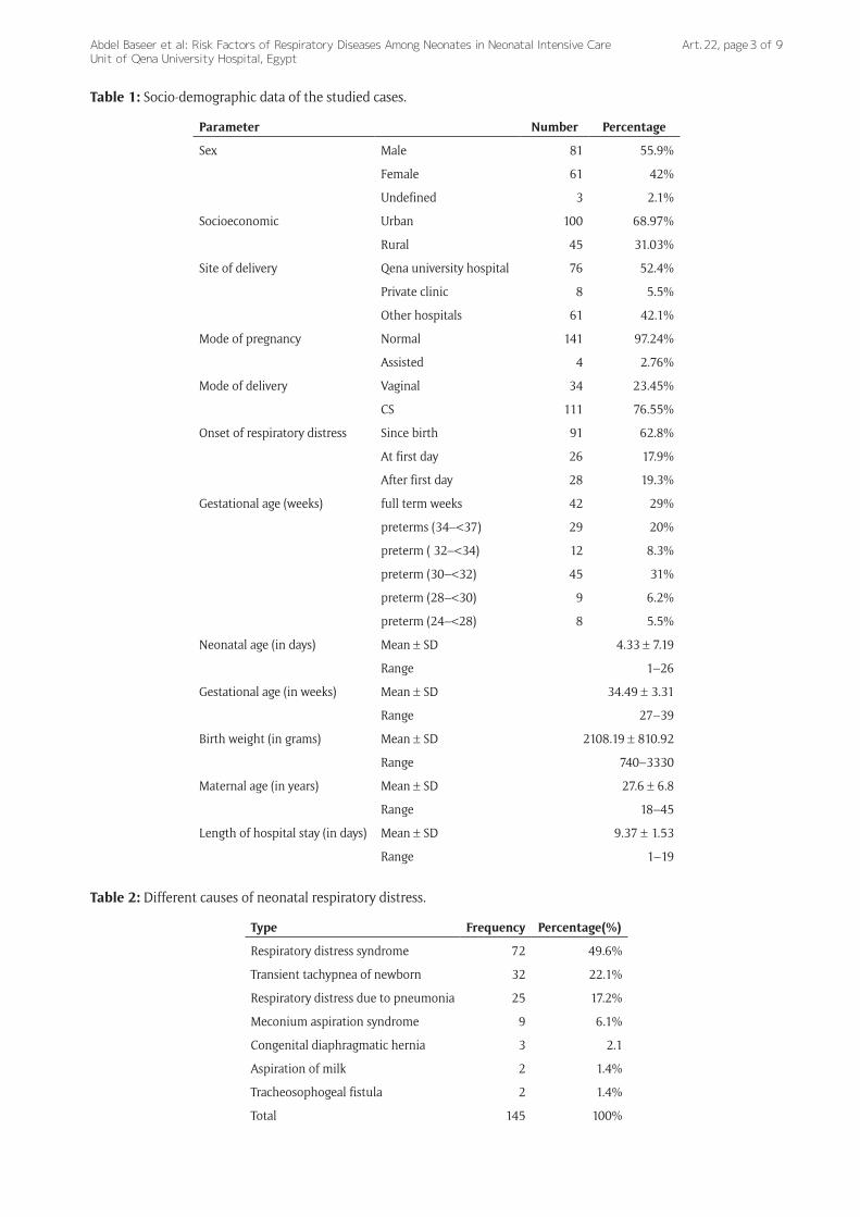
ResultsIn the period of the study, 312 neonates were admitted to our neonatal intensive care unit; of them 145 cases suffered from respiratory diseases giving an incidence of (46.5%) for neonatal respiratory diseases among total admission. Table 1 showed the socio-demographic data of the studied cases. They were 81 (55.9%) males, 61 (42%) females and 3 (2.1%) undefined sex. The mean neonatal age at admission was 4.33 ± 7.19 days, mean
gestational age was 34.49 ± 3.31 weeks, mean maternal age was 27.6 ± 6.8 years and the mean duration of hos-pitalization at our NICU was 9.37 ± 1.59 days. Regarding the site of the delivery, 76 cases (52.4%) of those born at our hospital needed admission at our neonatal intensive care unit due to respiratory problems, 61 newborns were referred to our unit from other hospitals (42.1%) and 8 referred from private clinic (5.5%). One hundred newborns were from urban areas and 45 from rural areas. Most of cases with respiratory distress (97.24%) occurred after normal pregnancy and 4 cases (2.76%) only after assisted pregnancy. One hundred and seven newborns (73.79%) were single, 26 (17.9%) were one of twins, 9 (6.21%) were triplets and 3 newborns were one of quadruple (2.06%). The onset of respiratory distress was since birth in 91 cases (62.8%), at first day in 26 cases (17.9%) and after first day in 28 (19.3%) cases.
Table 2 showed that preterms presented with RDS were 72 cases (49.6%), full-terms presented with transient tachypnea of newborn were 32 cases (22.1%), pneumonia caused respiratory distress in 25 cases (17.2%), meconium aspiration syndrome in 9 cases (6.2%), con-genital diaphragmatic hernia in 3 cases (2.1%), aspiration pneumonia in two cases (1.4%) and tracheosophegeal fistula in two cases (1.4%).
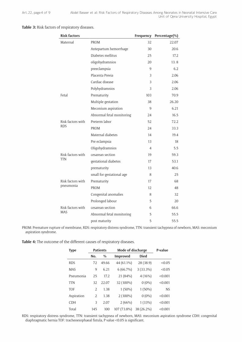
Table 3 showed risk factors of respiratory diseases occurrence. Regarding maternal illness, PROM was a risk factor in 32 cases (22.07%), antepartum hemorrhage in 30 cases (20.6%), maternal diabetes in 25 cases (17.24%), oligohydraminos in 20 cases (13.79%), preeclampsia in 9 cases (6.20%), placenta previa in 3 cases (2.06%) and cardiac diseases in 3 cases (2.06%). Regarding fetal condi-tions, prematurity was a risk factor in 103 cases (70.97%), multiple gestations in 38 cases (26.20%), meconium aspiration in 9 cases (6.21%) and abnormal fetal moni-toring in 24 cases (16.55%). Prematurity had the highest risk for RDS followed by PROM, maternal diabetes and pre-eclampsia. Cesarean section had the highest risk for TTN followed by gestational diabetes and prematurity. Prematurity had the highest risk for pneumonia followed by PROM, congenital anomalies and prolonged labor. Cesarean section had the highest risk for MAS followed by abnormal fetal monitoring and post maturity.
Table 4 showed the outcome in relation to different causes of respiratory diseases. Statistical significant differ-ence was found between outcome and incidence of differ-ent causes of respiratory disorders. Death rate increased significantly as the incidence of RDS, MAS and pneumonia increased. During the period of the study the case fatality rate was 38.9% in newborns with RDS, 33.3% in MAS and 16% in pneumonia. The best outcome was significantly higher with TTN.
Table 5 showed logistic regression analysis of vari-ables affecting the outcome of neonatal respiratory dis-eases. Many variables had a great effect on the outcome with an odds ratio >1 as male gender, RDS, CDH, pneu-monia, apnea, tachypnea >60 c/min, retraction, cyano-sis and irritability. Maternal risk factors that had a great effect on the outcome were diabetes, pre-eclampsia, oligohydramnios and overcrowding condition with odds
Abdel Baseer et al: Risk Factors of Respiratory Diseases Among Neonates in Neonatal Intensive Care Unit of Qena University Hospital, Egypt
Art. 22, page 3 of 9
Table 1: Socio-demographic data of the studied cases.
Parameter Number Percentage
Sex Male 81 55.9%
Female 61 42%
Undefined 3 2.1%
Socioeconomic Urban 100 68.97%
Rural 45 31.03%
Site of delivery Qena university hospital 76 52.4%
Private clinic 8 5.5%
Other hospitals 61 42.1%
Mode of pregnancy Normal 141 97.24%
Assisted 4 2.76%
Mode of delivery Vaginal 34 23.45%
CS 111 76.55%
Onset of respiratory distress Since birth 91 62.8%
At first day 26 17.9%
After first day 28 19.3%
Gestational age (weeks) full term weeks 42 29%
preterms (34–<37) 29 20%
preterm ( 32–<34) 12 8.3%
preterm (30–<32) 45 31%
preterm (28–<30) 9 6.2%
preterm (24–<28) 8 5.5%
Neonatal age (in days) Mean ± SD 4.33 ± 7.19
Range 1–26
Gestational age (in weeks) Mean ± SD 34.49 ± 3.31
Range 27–39
Birth weight (in grams) Mean ± SD 2108.19 ± 810.92
Range 740–3330
Maternal age (in years) Mean ± SD 27.6 ± 6.8
Range 18–45
Length of hospital stay (in days) Mean ± SD 9.37 ± 1.53
Range 1–19
Table 2: Different causes of neonatal respiratory distress.
Type Frequency Percentage(%)
Respiratory distress syndrome 72 49.6%
Transient tachypnea of newborn 32 22.1%
Respiratory distress due to pneumonia 25 17.2%
Meconium aspiration syndrome 9 6.1%
Congenital diaphragmatic hernia 3 2.1
Aspiration of milk 2 1.4%
Tracheosophogeal fistula 2 1.4%
Total 145 100%
Abdel Baseer et al: Risk Factors of Respiratory Diseases Among Neonates in Neonatal Intensive Care Unit of Qena University Hospital, Egypt
Art. 22, page 4 of 9
Table 3: Risk factors of respiratory diseases.
Risk factors Frequency Percentage(%)
Maternal PROM 32 22.07
Antepartum hemorrhage 30 20.6
Diabetes mellitus 25 17.2
oligohydramnios 20 13. 8
preeclampsia 9 6.2
Placenta Previa 3 2.06
Cardiac disease 3 2.06
Polyhydramnios 3 2.06
Fetal Prematurity 103 70.9
Multiple gestation 38 26.20
Meconium aspiration 9 6.21
Abnormal fetal monitoring 24 16.5
Risk factors with RDS
Preterm labor 52 72.2
PROM 24 33.3
Maternal diabetes 14 19.4
Pre eclampsia 13 18
Oligohydramnios 4 5.5
Risk factors with TTN
cesarean section 19 59.3
gestational diabetes 17 53.1
prematurity 13 40.6
small for gestational age 8 25
Risk factors with pneumonia
Prematurity 17 68
PROM 12 48
Congenital anomalies 8 32
Prolonged labour 5 20
Risk factors with MAS
cesarean section 6 66.6
Abnormal fetal monitoring 5 55.5
post maturity 5 55.5
PROM: Premature rupture of membrane, RDS: respiratory distress syndrome, TTN: transient tachypnea of newborn, MAS: meconium aspiration syndrome.
Table 4: The outcome of the different causes of respiratory diseases.
Type Patients Mode of discharge P-value
No. % Improved Died
RDS 72 49.66 44 (61.1%) 28 (38.9) <0.05
MAS 9 6.21 6 (66.7%) 3 (33.3%) <0.05
Pneumonia 25 17.2 21 (84%) 4 (16%) <0.001
TTN 32 22.07 32 (100%) 0 (0%) <0.001
TOF 2 1.38 1 (50%) 1 (50%) NS
Aspiration 2 1.38 2 (100%) 0 (0%) <0.001
CDH 3 2.07 2 (66%) 1 (33%) <0.001
Total 145 100 107 (73.8%) 38 (26.2%) <0.001
RDS: respiratory distress syndrome, TTN: transient tachypnea of newborn, MAS: meconium aspiration syndrome CDH: congenital diaphragmatic hernia TOF: tracheoesophaeal fistula, P value <0.05 is significant.

Abdel Baseer et al: Risk Factors of Respiratory Diseases Among Neonates in Neonatal Intensive Care Unit of Qena University Hospital, Egypt
Art. 22, page 5 of 9
ratio >1. Use of mechanical ventilation, C.S delivery and small for gestational age had an odds ratio >1.
DiscussionRespiratory diseases are one of the most common reasons for newborn admission to the neonatal intensive care unit [5]. Fifteen percent of term and 29% of preterm infants admitted to NICU develop significant respiratory morbidity specially in infants born before 34 weeks of gestation [10]. The etiologies of RD in newborn are large and diverse, including TTN, RDS, MAS, pneumonia and
other miscellaneous causes [6, 7]. As there is paucity of studies regarding the cause of respiratory diseases in our localities, our study was conducted to find out the preva-lence and etiology of respiratory diseases among admitted neonates. The total cases admitted to our NICU during one year study were 312 cases; of them 145 cases were admitted due to respiratory diseases giving an incidence of (46.5%). This was relatively comparable to the inci-dence reported by other studies like Verma et al., (39%) and Tochie et al., (47.5%) [11, 12]. The present study showed that neonatal respiratory diseases were higher in
Table 5: Logistic regression analysis of variables affecting the outcome of neonatal respiratory diseases.
Variables Odds ratio CI 95% P-value
Min. Max.
Sex Male 1.698 0.840 2.956 0.042
Female 0.912 0.904 3.245 0.4
Causes of neonatal respiratory diseases
RDS 1.698 0.840 2.956 0.042
TTN 1.254 0.751 3.717 0.049
MAS 1.478 0.892 2.532 0.064
Pneumonia 1.254 0.751 3.717 0.049
Aspiration of milk 1.075 0.649 4.128 0.066
CDH 0.912 0.904 3.245 0.097
TOF 1.211 0.413 1.716 0.067
Signs of RD Apnea 7.236 2.003 16.536 0.0021
Tachypnea >60 c/min 1.137 0.462 3.375 0.764
Retraction 1.743 0.923 1.978 0.0079
Abdominal distension 0.738 0.677 1.016 0.758
Cyanosis 4.235 1.251 9.33 0.0046
Maternal diseases
Diabetes mellites 1.310 0.444 1.965 0.758
Preeclampsia 1.273 0.581 2.725 0.842
Placenta Previa 0.643 0.734 1.398 0.529
Cardiac diseases 0.834 0.619 2.065 0.758
oligohydramnios 1.056 0.861 1.301 0.523
polyhydramnios 0.856 0.309 1.478 0.107
Antepartum hemorrhage 1.623 0.847 2.981 0.0249
Overcrowding 1.672 0.371 1.213 0.36
Management Mechanical ventilation 29.432 8.654 141.043 0.000001
Oxygenation 1.913 0.916 2.071 0.171
Type of delivery Vaginal 0.758 0.947 1783 0.0945
Caesarean 1.435 0.935 2.051 0.031
Duration of hospitalization 0.862 0.636 1.045 0.0367
Weight for gestational age
SGA 2.076 1.840 4.179 0.0021
LGA 1.276 0.869 1.687 0.0723
AGA 0.934 0.469 1.379 0.648
OR: odd ratio, CI: confidence interval. RDS: respiratory distress syndrome, TTN: transient tachypnea of newborn, MAS: meconium aspiration syndrome, APH: ante-partum hemorrhage, CDH: congenital diaphragmatic hernia TOF: tracheo- esophaeal fistula, SGA: small gestational age, LGA: large gestational age, AGA: appropriate gestational age, RD: respiratory distress, P value <0.05 is significant.

Abdel Baseer et al: Risk Factors of Respiratory Diseases Among Neonates in Neonatal Intensive Care Unit of Qena University Hospital, Egypt
Art. 22, page 6 of 9
males (55.9%) than females (44.1%). This was similar to previous studies [13, 14]. It is well known that lung growth and development start in the prenatal period and lung maturation is more advanced in the female fetus. Oral movement starts between the 16th and 26th weeks of ges-tation, reflecting fetal breathing, and this is considered a critical determinant for the development of the lung [15]. Other fundamental regulators of lung maturation are sex hormones. While testosterone secreted by fetal testes hav-ing mainly inhibitory effects and delays the surge of sur-factant production, oestrogens produced by the placenta have positive effects on both the production of fetal sur-factant and on the alveologenesis during neonatal and pubertal periods [16]. Additionally, our study detected that neonates born by a caesarean section have more inci-dence of respiratory diseases. This was evident by other studies [17–19]. Infants born by caesarean section have a larger residual volume of lung fluid, a smaller residual capacity and consequently secrete less surfactant into the alveolar space while during vaginal delivery, as the chest of the infant is squeezed, part of the fetal lung fluid is removed and the adrenergic stimulation associated with vaginal labor releases surfactant into the airways [20]. Respiratory distress syndrome was detected as the com-monest respiratory disease and earlier workers had found similar observations [10, 11]. RDS in the current study was detected in (49.66%) followed by TTN (22.07%), pneumo-nia in 17.2% and MAS in 6.2%. Parkash et al., reported RDS in (20.8%), pneumonia in (22.5%), MAS in (16.7%) and TTN in (11.7%) [21]. The study of Abou-Faddan and Nafisa revealed that RDS is the most common neonatal respiratory disease (45.8%) followed by pneumonia and TTN [22]. Regarding risk factors, our study showed that the most common maternal factors for occurrence of res-piratory diseases were PROM (22%), antepartum hemorrhage (20.6%), maternal diabetes (17.24%) and oli-gohydraminos (13.8%). Some of serious complications associated with PROM include chorioamnionitis, neonatal sepsis and preterm labor leading to neonatal pulmonary hypoplasia and respiratory distress syndrome [23]. Similar to other studies, the most commonly detected fetal risk factor for respiratory disorders was prematurity (70.9%) [17, 22]. It is well documented that all forms of respiratory morbidity, including TTN, RDS, pneumonia, and pulmo-nary hypertension, affect late-preterm infants at a higher rate than infants of more advanced GA [24]. A study by De Luca et al., revealed that more than ten folds increase in respiratory morbidity in infants of 34 weeks’ GA com-pared with term infants [25]. A retrospective study by Kitsommart et al., revealed significantly worse respiratory outcomes including prevalence of pneumothorax, need for positive pressure therapy, and mechanical ventilation assistance in infants of 34 to 36 weeks’ GA compared with infants of ≥37 weeks’ GA [26]. The current study found that multiple gestation pregnancies was associated with high risk of neonatal respiratory diseases (26.2%). This was in accordance with the study of Ziadeh and Badria which reported that the second twin has a higher risk of respiratory distress compared to single newborns and
added that caesarean section carried out before the onset of labor increase this risk [27]. Multifetal gestation brings a high risk of maternal complications during pregnancy and postnatal complications for babies specially when born prematurely. The risk of preterm delivery for triplet pregnancies is approximately 90% while in twin gesta-tions is more than 50%, compared with only 10% incidence of preterm labor in single infants [28]. Regard-ing RDS, the most common risk factor detected in our study was prematurity (37.7%). By Indian study, researchers found that the incidence rates of RDS ranged from 86% at 24 weeks to less than 1% at 39 weeks and mentioned that RDS should be anticipated in any IDM and preterm deliv-ery [29]. The results of our study demonstrated that PROM was also an important risk factor for neonatal RDS. Intrau-terine infection and chorioamnionitis caused by PROM can cause injury to the fetal lungs and alveolar type II cells directly resulting in decreased synthesis and release of sur-factant. Furthermore, premature birth can result from PROM [30]. Infants of diabetic mothers are more sus-ceptible to RDS occurrence compared to those of non-diabetic mothers of equivalent gestational age as having disturbed pattern of surfactant synthesis in addition to delayed appearance of phosphatidylglycerol [31]. Thirty-eight (26.2%) of the studied 145 newborns admitted with respiratory diseases died and this result was comparable to previous studies [32, 33]. Kumar et al. reported mortality rate of 19% in India with effective surfactant administration and respiratory support while Bajad et al. reported mortality of 22.33% and Abdelrahman et al. reported 36.0% in Sudan [32–34]. Also, the causes of death followed the differences in the causes of respiratory diseases where statistically significant difference was detected between the outcome and the incidence of variable causes of respiratory diseases. Higher mortality rate accompanied higher incidence of RDS, MAS, and pneumonia while higher survival rate with increased inci-dence of TTN. During the period of the study, the case fatality rate was (38.9%) in RDS, (33.3%) in MAS and 16% in pneumonia. In a study by Adebami et al., mortality rate was 46.9% in RDS and 40.0% in MAS while Kumar et al. reported mortality of (57.1%) in RDS, (21.8%) in MAS and (15.6%) in pneumonia [32, 35]. Many variables had a great effect on the outcome of our studied cases as had an odds ratio >1 like male gender, RDS, CDH, pneumonia, apnea, tachypnea >60/min, cyanosis and irritability where apnea had the highest odd ratio of clinical signs. In the study of Panda et al. apnea attack was recognized as a predictor of infant mortality and Sathenahalli et al. described apnea as a sign of poor outcome in neonatal respiratory distress [36, 37]. Furthermore we found that need for mechanical ventilation had the highest odd ratio. The results of the Sabzehei et al. study showed that the outcomes of the NRD had a significant correlation with apnea and respira-tory failure requiring mechanical ventilation [38]. Regard-ing relation between causes of respiratory diseases and their fatalities, our study revealed that RDS had the highest odd ratio followed by MAS in addition to maternal diabetes, preeclampsia, oligohydramnios, living in

Abdel Baseer et al: Risk Factors of Respiratory Diseases Among Neonates in Neonatal Intensive Care Unit of Qena University Hospital, Egypt
Art. 22, page 7 of 9
overcrowding conditions and small gestational age. Logis-tic regression analysis showed that premature SGA neonates were at a higher risk of mortality than prema-ture AGA infants were. These findings were consistent with previous studies that showed premature SGA infants more liable to bronchopulmonary disease and chronic lung disease [39, 40]. Strength of this research include use of a reasonable sample size for study and the situation of the study being a big referral center with a high rate of neonatal admission. Also, being a prospective study avoids bias and shortage of data in retrospective studies that depends on file records. The most important limitation of this study is that it was a single centered study and frequent missed cases that go for local centers.
ConclusionsThis study indicated higher incidence of respiratory dis-eases in neonates admitted at NICU most commonly were RDS, TTN, MAS and pneumonia. Prematurity was the most important risk factor associated with respiratory diseases in addition to presence of antepartum hemorrhage, multiple gestation and maternal diabetes. Respiratory distress syn-drome carried the highest risk of mortality and TTN carried the most survival rate. Although decreasing the incidence through preventive measures is ideal, early recognition and treatment of the common neonatal respiratory diseases will decrease both short- and long-term complications and related mortality of at-risk infants.
AbbreviationsAGA: appropriate gestational ageAPH: ante-partum hemorrhageCDH: congenital diaphragmatic herniaC.S: caesarean sectionLGA: large gestational age MAS: meconium aspiration syndromeNICU: neonatal intensive care unitPIH: Pregnancy induced hypertensionPROM: Premature rupture of membrane RD: respiratory distressRDS: respiratory distress syndrome SGA: small gestational age TOF: tracheoesophaeal fistulaTTN: transient tachypnea of newborn
Declarations• The work described has not been published previously• It is not under consideration for publication elsewhere• Its publication is approved by all authors • Journal policies detailed have been reviewed• Permission for use of copyrighted material from other
sources (including the Internet) has been obtained
Ethics and ConsentThis study was approved by ethical committee of south valley university.
Competing InterestsThe authors have no competing interests to declare.
References 1. Lawn JE, Davidge R, Paul VK, et al. Born
too soon: Care for the preterm baby. Reprod Health. 2013; 10(Suppl 1): S5. DOI: https://doi.org/10.1186/1742-4755-10-S1-S5
2. Barber CA, Wyckoff MH. Use and efficacy of endotracheal versus intravenous epinephrine dur-ing neonatal cardiopulmonary resuscitation in the delivery room. Pediatrics. 2006; 118: 1028–1034. DOI: https://doi.org/10.1542/peds.2006-0416
3. Pramanik AK, Rangaswamy N, Gates T. Neonatal respiratory distress: A practical approach to its diagnosis and management. Pediat Clin N Am. 2015; 62: 453–69. DOI: https://doi.org/10.1016/ j.pcl.2014.11.008
4. Swarnkar K, Swarnkar M. Neonatal respiratory distress in early neonatal period and its outcome. Int J Biomed Adv Res. 2015; 6(9): 643–7.
5. Edwards MO, Kotecha SJ, Kotecha S. Respiratory distress of the term newborn infant. Paediatr Respir Rev. 2013; 14(1): 29–36. DOI: https://doi.org/ 10.1016/j.prrv.2012.02.002
6. Williams O, Hutchings G, Hubinont C, Debauche C, Greenough A. Pulmonary effects of prolonged oligohydramnios following mid-tri-mester rupture of the membranes—antenatal and postnatal management. Neonatology. 2012; 101(2): 83–90. DOI: https://doi.org/10.1159/000329445
7. Jobe AH. Effects of chorioamnionitis on the fetal lung. Clin Perinatol. 2012; 39(3): 441–457. DOI: https://doi.org/10.1016/j.clp.2012.06.010
8. Warren JB, Anderson JM. Newborn respiratory disorders. Pediatr Rev. 2010; 31(12): 487–495 DOI: https://doi.org/10.1542/pir.31-12-487
9. Kamath BD, Macguire ER, McClure EM, Goldenberg RL, Jobe AH. Neonatal mortality from respiratory distress syndrome: Lessons for low-resource countries. Pediatrics. 2011; 127(6): 1139–46. DOI: https://doi.org/10.1542/peds.2010-3212
10. Hibbard JU, Wilkins I, Sun L, Gregory K, Haberman S. Respiratory morbidity in late preterm births. JAMA. 2010; 304(4): 419–25. DOI: https://doi.org/10.1001/jama.2010.1015
11. Verma J, Anand S, Kapoor N, Gedam S, Patel U. Neonatal outcome in newborns admitted in NICU of tertiary care hospital in central India: A 5-year study. Int J Contemp Pediatr. 2018; 5(4). DOI: https://doi.org/10.18203/2349-3291.ijcp20182512
12. Tochie JN, Simeon-Pierre C, Langmia RN, Barla E, Koki-Ndombo P. Neonatal respiratory distress in a reference neonatal unit in Cameroon: an analysis of prevalence, predictors, etiologies and outcomes. Pan Afr Med J. 2016; 24: 152. DOI: https://doi.org/ 10.11604/pamj.2016.24.152.7066
13. Liu J, Yang N, Liu Y. High-risk Factors of Res-piratory Distress Syndrome in Term Neonates: A Retrospective Case-control Study. BALK MED J. 2014; 33(1): 64–68. DOI: https://doi.org/10.5152/balkanmedj.2014.8733

Abdel Baseer et al: Risk Factors of Respiratory Diseases Among Neonates in Neonatal Intensive Care Unit of Qena University Hospital, Egypt
Art. 22, page 8 of 9
14. Niesłuchowska-Hoxha A, Cnota W, Czuba B, et al. A Retrospective Study on the Risk of Respira-tory Distress Syndrome in Singleton Pregnancies with Preterm Premature Rupture of Membranes between 24+0 and 36+6 Weeks, Using Regres-sion Analysis for Various Factors. Biomed Res Int. 2018 Oct 4; 2018: 7162478. DOI: https://doi.org/10.1155/2018/7162478
15. Becklake MR, Kauffmann F. Gender differences in airway behaviour over the human life span. Thorax. 1999; 54: 1119–1138. DOI: https://doi.org/ 10.1136/thx.54.12.1119
16. Seaborn T, Simard M, Provost PR, et al. Sex hormone metabolism in lung development and maturation. Trends Endocrinol Metab. 2010; 21: 729–738. DOI: https://doi.org/10.1016/ j.tem.2010.09.001
17. Baumert M, Fiala M, Walencka Z, Paprotny M, Sypniewska K. Cesarean delivery and respira-tory distress in late preterm and term infants. Cent Eur J Med. 2012; 7(2): 230–234. DOI: https://doi.org/10.2478/s11536-011-0139-5
18. Wax JR, Herson V, Carignan E, Mather J, Ingardia CJ. Contribution of elective delivery to severe respiratory distress at term. Am. J. Perinatol. 2002; 19: 81–6. DOI: https://doi.org/ 10.1055/s-2002-23558
19. Sutton L, Sayer GP, Bajuk B, Richardson V, Berry G, Henderson-Smart DJ. Do very sick neonates born at term have antenatal risks? 2. Infants ventilated primarily for lung disease. Acta Obstet Gynecol Scand. 2001; 80: 917–925. DOI: https://doi.org/ 10.1034/j.1600-0412.2001.801008.x
20. Odom MJ, Snyder JM, Mendlson CR. Adenosine 3’, 5’ monophosphate analogs and beta-adrenergic agonist induce the synthesis of the major surfactant apoprotein in human fetal lung in vitro. Endocrinology. 2002: 121: 1155–63. DOI: https://doi.org/ 10.1210/endo-121-3-1155
21. Parkash A, Haider N, Khoso ZA, Shaikh AS. Frequency, causes and outcome of neonates with respiratory distress admitted to Neonatal Inten-sive Care Unit, National Institute of Child Health, Karachi. J Pak Med Assoc. 2015 Jul; 65(7): 771–5. PMID: 26160089
22. Abou-Faddan H, Abdelaziz NHR. Respiratory Distress and Its Outcome among Neonates Admitted to Neonatal Intensive Care Unit of Assiut University Children Hospital, Egypt. The Egyptian Journal of Community Medicine (EJCM). 2018; 36(2): 69–78. DOI: https://doi.org/10.21608/ejcm.2018. 11052
23. Medina TM, Hill DA. Preterm Premature Rupture of Membranes: Diagnosis and management. Am Fam Physician. 2006; 73: 659–64. PMID:16506709.
24. Khashu M, Narayanan M, Bhargava S, Osiovich H. Perinatal outcomes associated with preterm birth at 33 to 36 weeks’ gestation: A population-based cohort study. Pediatrics. 2009; 123(1): 109–113. DOI: https://doi.org/10.1542/peds.2007-3743
25. De Luca R, Boulvain M, Irion O, Berner M, Pfister RE. Incidence of early neonatal mortality and morbidity after late-preterm and term cesarean delivery. Pediatrics. 2009; 123(6): e1064–71. DOI: https://doi.org/10.1542/peds.2008-2407
26. Kitsommart R, Janes M, Mahajan V, et al. Outcomes of late-preterm infants: a retrospective, single-center, Canadian study. Clin Pediatr (Phila). 2009; 48(8): 844–850. DOI: https://doi.org/ 10.1177/0009922809340432
27. Ziadeh SM, Badria LF. Effect of mode of delivery on neonatal outcome of twins with birthweight under 1500 g. Arch Gynecol Obstet. Nov. 2000; 264(3): 128–130. DOI: https://doi.org/10.1080/01443610050112039
28. Blumenfeld Z, Abdallah W, Sela-Guttmann O, Brook OR. Triplet gestation prevention, risks & management dilemmas. Open Women’s Health J. 2008; 2: 11–21. DOI: https://doi.org/ 10.2174/1874291200802010011
29. Ramesh TK, Suresh PM, Arul SV. Prevalence of Antenatal Steroids Coverage in Preterm Labor and Its Influence on Neonatal Respiratory Morbidity and Mortality in Kanyakumari District. Int J Sci Study. 2017; 5(1): 197–199.
30. Jobe A. Effects of Chorioamnionitis on the Fetal Lung. Clin Perinatol. 2012; Sep; 39(3): 441–457. DOI: https://doi.org/10.1016/j.clp.2012.06.010
31. Kjos SL, Walther FJ, Montoro M, Paul RH, Diaz F, Stabler M. Prevalence and etiology of respiratory distress in infants of diabetic mothers: Predictive value of fetal lung maturation tests. Am J Obstet Gynecol. 1990 Sep; 163(3): 898–903. DOI: https://doi.org/10.1016/0002-9378(90)91092-Q
32. Kumar A, Vishnu Bhat B. Epidemiology of respiratory distress of newborns. Indian J Pediatr. 1996; 63: 93–8. DOI: https://doi.org/10.1007/BF02823875
33. Bajad M, Goyal S, Jain B. Clinical profile of neonates with respiratory distress. Int J Contemp Pediatr. 2016; 3(3): 1009–13. DOI: https://doi.org/10.18203/2349-3291.ijcp20162382
34. Abdelrahman SMK, Hamed SMA, Nasr A. Neonatal respiratory distress in Omdurman Maternity Hospital, Sudan. Sudan J Paediatr. 2014; 14(1): 65–70. PMID:27493392 PMCID: PMC4949919
35. Adebami OJ, Joel-Medewase VI, Agelebe E, et al. Determinants of outcome in newborns with respiratory distress in Osogbo, Nigeria. Int J Res Med Sci. 2017 Apr; 5(4): 1487–1493. DOI: https://doi.org/10.18203/2320-6012.ijrms20171252
36. Panda S, Aradhana A, Mahapatra T. Predictors of Mortality in Neonates with Respiratory Distress in a Tertiary Care Centre. IOSR Journal of Dental and Medical Sciences (IOSR-JDMS). 2016; 15(11): 62–3.
37. Sathenahalli VB, Dwivedi D, Bajaj N, Singh HP. Predictors of poor outcome in neonates with respiratory distress. Int J Contemp Pediatr. 2016; 3: 76–9. DOI: https://doi.org/10.18203/2349-3291.ijcp20160092

Abdel Baseer et al: Risk Factors of Respiratory Diseases Among Neonates in Neonatal Intensive Care Unit of Qena University Hospital, Egypt
Art. 22, page 9 of 9
38. Sabzehei MK, Basiri B, Shokouhi M, Fayyazi A. Causes and Outcomes of Respiratory Distress in Neonates Hospitalized in the Neonatal Intensive Care Unit of Be’sat Hospital in Hamadan, Iran. Int J Pediatr. Dec. 2017; 5(12): 6253–6260.
39. Prasad V, Singh N. Causes of morbidity and mortality in neonates admitted in Government Medical College Haldwaniin Kumaun Region
(Uttrakhand) India. J Pharm Biomed Sci. 2011; 8: 1–4.
40. Eriksson L, Haglund B, Odlind V, Altman M, Ewald U, Kieler H. Perinatal conditions related to growth restriction and inflammation are associated with an increased risk of bronchopulmonary dysplasia. Acta Pediatr. 2015; 104: 259–263. DOI: https://doi.org/10.1111/apa.12888
How to cite this article: Abdel Baseer KA, Mohamed M, Abd-Elmawgood EA. Risk Factors of Respiratory Diseases Among Neonates in Neonatal Intensive Care Unit of Qena University Hospital, Egypt. Annals of Global Health. 2020; 86(1): 22, 1–9. DOI: https://doi.org/10.5334/aogh.2739
Published: 26 February 2020
Copyright: © 2020 The Author(s). This is an open-access article distributed under the terms of the Creative Commons Attribution 4.0 International License (CC-BY 4.0), which permits unrestricted use, distribution, and reproduction in any medium, provided the original author and source are credited. See http://creativecommons.org/licenses/by/4.0/.
Annals of Global Health is a peer-reviewed open access journal published by Ubiquity Press. OPEN ACCESS