rna clamping by vasa assembles a pirna amplifier … · 4laboratoire biologie a` grande echelle,...
TRANSCRIPT

RNA Clamping by Vasa Assemblesa piRNA Amplifier Complexon Transposon TranscriptsJordi Xiol,1,2,7 Pietro Spinelli,1,2 Maike A. Laussmann,1,2,3 David Homolka,1,2 Zhaolin Yang,1,2 Elisa Cora,1,2
Yohann Coute,4 Simon Conn,1,2 Jan Kadlec,1,2 Ravi Sachidanandam,5 Marko Kaksonen,3 Stephen Cusack,1,2
Anne Ephrussi,6 and Ramesh S. Pillai1,2,*1European Molecular Biology Laboratory, Grenoble Outstation2Unit for Virus Host-Cell InteractionsUniversity Grenoble Alpes-EMBL-CNRS, 71 avenue des Martyrs, 38042, France3Cell Biology and Biophysics Unit, EMBL, 69117 Heidelberg, Germany4Laboratoire Biologie a Grande Echelle, IRTSV, CEA, 38054 Grenoble, France5Department of Oncological Sciences, Icahn School of Medicine at Sinai, New York, NY 10029, USA6Developmental Biology Unit, EMBL, 69117 Heidelberg, Germany7Present address: Department of Cell Biology, Harvard Medical School, Boston, MA 02115, USA
*Correspondence: [email protected]://dx.doi.org/10.1016/j.cell.2014.05.018
SUMMARY
Germline-specific Piwi-interacting RNAs (piRNAs)protect animal genomes against transposons andare essential for fertility. piRNAs targeting activetransposons are amplified by the ping-pong cycle,which couples Piwi endonucleolytic slicing of targetRNAs tobiogenesisofnewpiRNAs.Here,wedescribethe identification of a transient Amplifier complex thatmediates biogenesis of secondary piRNAs in insectcells. Amplifier is nucleated by the DEAD box RNAhelicase Vasa and contains the two Piwi proteinsparticipating in the ping-pong loop, the Tudor proteinQin/Kumo and antisense piRNA guides. These com-ponents assemble on the surface of Vasa’s helicasedomain, which functions as an RNA clamp to anchorAmplifier onto transposon transcripts. We show thatATP-dependent RNP remodeling by Vasa facilitatestransfer of 50 sliced piRNA precursors between ping-pong partners, and loss of this activity causes sterilityin Drosophila. Our results reveal the molecular basisfor the small RNA amplification that confers adaptiveimmunity against transposons.
INTRODUCTION
Mobile genomic elements called transposons constitute a large
part of eukaryotic genomes and present a formidable threat to
genome integrity due to their ability to cause mutations or
ectopic recombination events (Kazazian, 2004). Animal gonads
employ the dedicated PIWI clade Argonautes and their associ-
ated 24–30 nucleotide (nt) Piwi-interacting RNAs (piRNAs) to
suppress transposon activity. piRNAs guide Piwi proteins to their
1698 Cell 157, 1698–1711, June 19, 2014 ª2014 Elsevier Inc.
transposon targets, which are identified by sequence comple-
mentarity between the piRNA and the target. Some Piwi pro-
teins function as small RNA-guided endoribonucleases (slicers),
whereas nuclear Piwi complexes may also mediate silencing by
altering chromatin or DNA methylation status of target genomic
loci. The importance of this pathway is highlighted by the fact
that Piwi mutants display infertility due to defects in germline
development, presumably as a result of genome instability
caused by transposon activation (Bagijn et al., 2012; Ghildiyal
and Zamore, 2009; Luteijn and Ketting, 2013).
Guidance information for Piwi proteins is genetically encoded
into a few hundred genomic regions called piRNA clusters (Bren-
necke et al., 2007). These genomic intervals serve as a registry of
all transposons hosted in the genome by containing a high den-
sity of transposon fragment insertions. Clusters are transcribed
into a long single-stranded piRNA precursor (up to 100 kb
long) and are then exported to the cytoplasm by RNA export
factors like Uap56 (Zhang et al., 2012) for maturation into tens
of thousands of piRNAs via a poorly understood primary pro-
cessing pathway (Brennecke et al., 2007). Processing is believed
to take place in cytoplasmic perinuclear granules called nuage
that concentrate most known piRNA processing factors (Lim
and Kai, 2007). By providing germ cells with a complex mixture
of sequences to target all mobile elements present in the
genome, these primary piRNAs act as the genetically encoded
component of an RNA-based innate immune system (Aravin
et al., 2007).
Germcells have an additional andmore elegant piRNAbiogen-
esis pathway where target silencing itself triggers generation
of new piRNAs. Sequence analyses of small RNAs associating
with the Drosophila Piwi proteins (Aubergine and Ago3) formed
the basis for such a proposal, and these ideas are incorporated
into the so-called ping-pong model (Brennecke et al., 2007;
Gunawardane et al., 2007). It is proposed that antisense primary
piRNAs guide Aubergine (Aub) for cytoplasmic endonucleolytic

cleavage (slicing) of target transposonRNAs. One of the resulting
cleavage fragments is then utilized to generate a new secondary
piRNA that associates with Ago3, with the cleavage site marking
its 50 end. Given their origin from transposon transcripts, Ago3-
bound piRNAs are of sense orientation. These, in turn, guide
Ago3 slicer activity to complementary cluster transcripts, with a
cleavage fragment maturing exactly as the same Aub-bound
antisense piRNA that initiated the transposon cleavage. In such
a scenario, Ago3 slicing serves to prioritize production of specific
antisense piRNAs from piRNA cluster transcripts (Li et al., 2009).
The system is built such that abundant transposons will produce
more Ago3-loaded piRNAs, resulting in even greater production
of antisense piRNAs in Aub. Such a feedforward amplification
loop represents the adaptive armof theRNA-based immune sys-
tem that monitors transposon activity and selectively amplifies
thosepiRNAs that target themost active transposons. The versa-
tility of theping-pongcycle is evident from the fact thatmaternally
deposited piRNAs can also serve as inputs, thereby allowing the
embryo to rapidly ramp up immunity against paternally inherited
transposons to avert infertility caused by hybrid dysgenesis
(Brennecke et al., 2008; Grentzinger et al., 2012). It can also pro-
mote emergence of newpiRNA loci by targeting genomic regions
in trans (de Vanssay et al., 2012).
We currently do not understand how such a complex small
RNA amplification pathway works. RNA slicing by Piwi proteins
and that occurring during RNA interference (RNAi) (Meister,
2013) are biochemically indistinguishable. However, RNAi leads
to complete degradation of the target. Germ cells must therefore
possess an undefined machinery that safely transfers the Piwi-
generated slicer cleavage fragments from one ping-pong
Piwi partner to the other for maturation as secondary piRNAs.
Genetic and biochemical analyses have uncovered a number
of factors that are essential for secondary piRNA biogenesis
(Czech et al., 2013; Malone et al., 2009), but how they contribute
to what is likely to be amultistep processing pathway is currently
unclear. RNA helicases are prime candidates for bringing about
dynamic associations and remodeling of ribonucleoprotein
(RNP) complexes during a variety of RNA processing reactions
(Linder and Jankowsky, 2011), and they are among the list of
known piRNA biogenesis factors (Figure S1A available online).
Among these, we focused our attention on the DEAD box RNA
helicase Vasa, as it is the only factor with a conserved role in sec-
ondary biogenesis in both flies and mice (Kuramochi-Miyagawa
et al., 2010; Malone et al., 2009). vasa was originally identified
as a maternal-effect gene that is required for development of
the female germline in Drosophila (Lasko and Ashburner, 1988;
Schupbach and Wieschaus, 1986). Like other members of the
DEAD box family, Vasa is demonstrated to have ATPase, RNA
binding, and RNA unwinding activities (Liang et al., 1994; Sen-
goku et al., 2006), but its molecular role in the piRNA pathway
is not known.
Here, we explored the in vivo function of Vasa using the insect
ovarian cell culture model Bombyx mori BmN4 (Kawaoka et al.,
2009). RNA helicases like Vasa are ATP-gated RNA-binding pro-
teins that engage in dynamic associations. We used a point mu-
tation (DQAD) within the DEAD box of Vasa to prevent release of
ATP hydrolysis products, locking the protein onto its bound RNA
targets and reducing its in vivo dynamics. Surprisingly, this also
trapped a piRNA amplification complex that provides detailed
insight into how endonucleolytic cleavage during transposon
silencing is linked to generation of a new secondary piRNA.
This transiently assembled complex, which we named Amplifier,
includes Vasa, the two ping-pong Piwi partners, and the tudor
protein Qin/Kumo. Biochemical and structural data reveal that
RNA clamping on transposon transcripts by Vasa creates a
binding platform for Amplifier components at the surface of
Vasa’s helicase domain, which is in its ATP-bound, closed
conformation. ATP hydrolysis by Vasa opens the helicase do-
mains, resulting in Amplifier disassembly and safe handover of
sliced 50 processed precursors between Piwi proteins for further
maturation into piRNAs. Genetic experiments in flies underscore
the importance of this ATP-controlled process for fertility. Our
results provide detailed mechanistic insight into how transposon
cleavage fragments are utilized for generating new piRNAs that
preserve genome integrity in the germline.
RESULTS
Comprehensive Transcriptome of the BmN4 Cell LineThe Bombyx mori (silkworm) ovary-derived BmN4 cell line is the
only cell culture model for piRNA research that has an active
ping-pong cycle in operation. These cells express two Piwi pro-
teins—Siwi (Aub ortholog in Bombyx) and Ago3—that associate
with piRNAs bearing sequence signatures consistent with sec-
ondary piRNA biogenesis (Kawaoka et al., 2009). To facilitate
studies with this cell line, we cataloged the entire set of piRNA
pathway factors in BmN4 using de novo assembly of tran-
scriptome sequencing data (Figure S1A). When compared to
Drosophila, several of the factors essential for both primary
and secondary biogenesis (including Vasa) are conserved.
However, notable absences include that of the effector protein
Squash and nuclear factors like Rhino, Cutoff, and Deadlock,
which are needed for piRNA biogenesis from dual-strand
(ping-pong) clusters (Chen et al., 2007; Czech et al., 2013; Klat-
tenhoff et al., 2009; Pane et al., 2007) (Figure S1A). The absence
of the latter is likely due to the euchromatic nature of piRNA clus-
ters in BmN4 (Kawaoka et al., 2013), precluding the need for
these factors, as they serve to accessDrosophila piRNA clusters
that are embedded in constitutive heterochromatin (Brennecke
et al., 2007; Klattenhoff et al., 2009). We also did not find any
evidence for the existence of a nuclear Piwi pathway in these
cells. Overall, these findings reveal that a unique selection of
biogenesis factors fuels the ping-pong cycle in BmN4 cells.
ADEAD BoxMutation Reduces In Vivo Dynamics of VasaOur initial attempt at understanding Bombyx mori Vasa’s role
in the piRNA pathway consisted of characterizing isolated com-
plexes from transfected BmN4 cells. However, these failed to
reveal any interaction with core piRNA pathway components
(see below). DEAD box RNA helicases like Vasa are known to
mediate dynamic associations as part ofmultiprotein assemblies
in vivo (Linder and Jankowsky, 2011), which might be hard to
detect in biochemical experiments due to their transient nature.
This prompted us to examine the dynamics of the proteins
involved in the ping-pong cycle by live-cell imaging experiments
rather than by looking at a steady-state situation.
Cell 157, 1698–1711, June 19, 2014 ª2014 Elsevier Inc. 1699

A
B C D
E
Figure 1. DQAD Mutation in the DEAD Box of Vasa Reduces Its In Vivo Dynamics
(A) BmN4 cells expressing fluorescently tagged Vasa, VasaDQAD, or Ago3 display abundant cytoplasmic granules, and these were tracked by live-cell imaging.
Start and end of tracking is highlighted with a red and blue dot, respectively. Analysis indicates a random, nonlinear movement of these granules, which is
unaffected by the DQAD mutation in Vasa.
(legend continued on next page)
1700 Cell 157, 1698–1711, June 19, 2014 ª2014 Elsevier Inc.

Transiently expressed EGFP-Vasa and mCherry-Ago3 coloc-
alize in numerous perinuclear cytoplasmic granules in BmN4
cells. These are analogous to the electron-dense granules called
nuage observed in Drosophila ovarian nurse cells, which are
credited to be piRNA processing centers (Liang et al., 1994;
Lim and Kai, 2007). Cell tracking experiments now reveal that,
far from being static structures, these display random, nonlinear
movements (Figure 1A and Movie S1). More frequently, granules
meet to fuse briefly before dissociating, whereas occasionally
smaller granules fuse into larger structures. To evaluate the dy-
namics of the proteins within the nuage, we carried out fluores-
cence recovery after photobleaching (FRAP) experiments (Movie
S2). The measured halftime of fluorescence recovery rates for
EGFP-Vasa (5.89 ± 0.44 s) (Figures 1C and 1E) and EGFP-
Ago3 (3.01 ± 0.37 s) (Figures 1D, S1B, and S1C) suggest a rapid
transit of the proteins through the granules. Thus, any interac-
tions mediated by them within the nuage should be short lived.
Because most activities of RNA helicases are linked to their
consumption of ATP (Linder and Jankowsky, 2011), we exam-
ined whether abolishing ATPase activity might interfere with
the dynamics of Vasa. The DEAD box family is characterized
by the presence of a number of sequence motifs within the heli-
case module (Caruthers and McKay, 2002), prominent among
them being the motif II (Asp-Glu-Ala-Asp; D-E-A-D). A single
amino acidmutationwithin thismotif (DEAD/DQAD) (Figure 1B)
is shown to abolish ATPase activity in other family members
(Pause and Sonenberg, 1992). We created a similar mutation
within Vasa and expressed its EGFP-tagged version in BmN4
cells. A first indication of an impact of themutationwas the accu-
mulation of EGFP-VasaDQAD in granules that are slightly larger
than those formed by EGFP-Vasa (Figures 1E, 2F, and 2G).
Nevertheless, tracking experiments did not reveal any impact
of the mutation on the random mobility of the granules (Fig-
ure 1A). Strikingly, FRAP experiments showed that fluorescence
in VasaDQAD granules failed to recover, irrespective of the size of
the granule examined (Figures 1C and 1E). This is likely to be a
consequence of the protein becoming trapped in the granule,
preventing further ingress from the surrounding cytoplasm
(Movies S3–S5). Thus, an emerging picture is one of highly dy-
namic events orchestrated within the nuage, which wemanaged
to dampen as a result of the DQAD mutation within Vasa.
Vasa Is Part of a Transient piRNA Amplifier ComplexThe retention of VasaDQAD in enlarged nuage prompted us to ask
whether the point mutant might be accumulating in certain com-
plexes in vivo. To this end, we isolated HA-Vasa or HA-VasaDQAD
complexes and identified components by mass spectrometry.
The protein arginine methyltransferase 5 (Prmt5 or Capsuleen
in Drosophila) is common to both complexes, and this is
explained by the presence of 20 arginine residues at the N termi-
(B) Sequence alignment of DEAD box helicases showing critical residues for ATP
Vasa used in this study are indicated in the cartoon.
(C) Recovery of fluorescence after photo bleaching (FRAP) of EGFP-Vasa or EGF
(D) Recovery times for EGFP-Ago3.
(E) Time series snapshots (time in seconds, s) focusing on the granules in the cytop
fluorescently labeled proteins. The bleached granule is indicated with a red circle.
the middle panel showing a granule where the laser photobleaches the center o
nus of Bombyx Vasa (Figure 2A). Such arginine residues in
Drosophila Vasa are shown to be targets of Prmt5 for symmetri-
cal dimethyl arginine (sDMA) modifications (Kirino et al., 2010).
Interestingly, HA- and FLAG-VasaDQAD complexes contained
a number of additional factors not present in wild-type Vasa im-
munoprecipitations (Figures 2A and S2A). In particular, the two
most abundant hits among VasaDQAD interactors are the endog-
enous ping-pong Piwi partners Siwi (the Bombyx ortholog of
Aub) and Ago3, which we confirmed by western blot analysis
(Figure 2B). Furthermore, tandem immunopurification of FLAG-
VasaDQAD followed by anti-Siwi immunoprecipitation reveals
that all three proteins are part of the same entity (Figure 2C).
Because the DQAD mutation in Vasa is predicted to prevent
ATP hydrolysis, association with Piwi proteins is likely to be
favored when Vasa is in its ATP-bound state. To confirm this,
we created a mutation in the motif I (GKT/ GNT) of the helicase
domain that is predicted to prevent ATP binding (Figure 1B).
Supporting the requirement for bound ATP, the VasaGNT mutant
did not reveal any association with endogenous Piwi proteins
(Figure 2B). The Tudor domain-containing protein Qin/Kumo, a
factor essential for the ping-pong cycle (Anand and Kai, 2012;
Zhang et al., 2011), was also identified in the VasaDQAD purifica-
tions (Figure 2A). We confirmed this result by western analysis
using rabbit polyclonal antibodies to the endogenous Qin/
Kumo expressed in BmN4 cells (Figures 2D and S2B). Because
all of the validated components are involved in piRNA ampli-
fication, we called this Vasa-Siwi-Ago3-Qin/Kumo complex
Amplifier.
Arginine methylation of Piwi proteins is shown to act as
an affinity enhancer for mediating protein-protein interactions
(Mathioudakis et al., 2012), so we wondered whether Vasa
methylation plays any role in Amplifier assembly. Pointing to
the absence of such a role, whenwemutated all of theN-terminal
arginines (to lysines; R/K) in VasaDQAD, the interaction with
the Amplifier components was still maintained, as examined
by mass spectrometry and western blot analysis (Figures 2A
and 2E). In contrast, recovery of Prmt5 was abolished in both
Bombyx VasaR-K and VasaDQAD(R-K) purifications (Figure 2A).
We propose that the Amplifier complex is assembled on wild-
type Vasa, but its transient nature prevents its detection
(Figure S2C). However, the DQAD mutation in Vasa freezes this
complex by preventing its disassembly, thereby allowing its
identification.
Amplifier Assembly Takes Place within the NuageIn BmN4 cells, Vasa and Ago3 are nuage residents, while the
vast majority of Siwi is present diffused in the cytoplasm (Xiol
et al., 2012). To examine where in the cell they might transiently
associate, cells expressing HA-tagged Vasa proteins were coun-
terstained for endogenous Amplifier components using specific
binding (motif I) and ATP hydrolysis (motif II). Point mutations within Bombyx
P-VasaDQAD granules is indicated as a function of time (in seconds, s).
lasm fromFRAP experiments conductedwith BmN4 cells expressing indicated
Note that VasaDQAD granules are generally larger but show a range of sizes, with
f the granule.
Cell 157, 1698–1711, June 19, 2014 ª2014 Elsevier Inc. 1701

Western
- Vasa
Vasa
DQAD
Vasa
GNT
Siwi
HA
Ago3
- Vasa
Vasa
DQAD
Vasa
GNT
Input IP-HA
HA-tag
A D
E
B
Western- Va
sa
Siwi
HA
Ago3
Vasa
DQAD
Input IP-HA
Vasa(
R-K)Va
sa(R-K)DQAD
- Vasa
Vasa
DQADVa
sa(R-K)
Vasa(
R-K)DQAD
HA-tag
F
C
G
-
-
-300250
100
Input
HA-tag
IP-HA
Vasa
Vasa
DQAD
Vasa
Vasa
DQAD
kDa
Qin/Kumo
HA
HA-Vasa HA-VasaDQAD
HAEn
doge
nous
Ago
3Me
rge
12 μm 12 μm
HA-Vasa HA-VasaDQAD
HAMe
rge
Endo
geno
us Q
in/Ku
mo
12 μm12 μm
Merg
eHA
-Vas
aDQAD
Endo
geno
us S
iwi
24 μm
H
VasaDQAD
FLAG
Siwi
Ago3
FLAG-IPand
Peptide Elution
Siwi-IP
FLAG
Ago3
Siwi
Western
InputFLAG-IP(elution)
Siwi-IPFLAG-VasaDQAD - + - +
1st-IP 2nd-IPTandem Immunopurification of Amplifier complex
FLAG
-Vasa
DQAD
FLAG
-Vasa
HA-Vasa
HA-Vasa
DQAD
Protein Domains HA-Vasa
(R-K)
HA-Vasa
(R-K)
DQAD
Vasa Helicase 73 54 337 270 163 60Capsuleen PRMT 22 9 16 11 - -
Valois WD 15 6 15 8 - -
Siwi PIWI, PAZ, MID 7 49 - 19 - 52Ago3 PIWI, PAZ, MID - 25 - 36 - 29
Qin/Kumo Tudor, E3 ligase - 24 - 13 - 9
Complex accumulating with VasaDQAD (Amplifier components)
Figure 2. A Transient piRNA Amplifier Complex Containing Vasa Is Assembled in the Nuage
(A) Mass spectrometry analyses of indicated tagged protein complexes isolated from transiently transfected BmN4 cells. Note that Prmt5/Capsuleen is common
to both Vasa and VasaDQAD, though the latter has a unique set of factors (see Figure S2A for a complete list), including ping-pong Piwi proteins and Qin/Kumo,
which were validated by western blotting analyses. A ratio of spectral counts in the sample versus beads control is indicated.
(B) Immunoprecipitation and western blot analysis confirming specific association of endogenous Piwi proteins Siwi and Ago3 with HA-VasaDQAD.
(C) Tandem affinity purification showing presence of VasaDQAD and the ping-pong Piwi partners in the same complex.
(D) Western blot analysis showing specific association of endogenous Qin/Kumo with HA-VasaDQAD.
(E) Mutation of 20 N-terminal arginines to lysines (R–K) in VasaDQAD does not affect Amplifier assembly but abolishes recovery of Capsuleen (see A).
(F–H) Accumulation of VasaDQAD in large nuage in BmN4 cells that are also enriched in endogenous Amplifier components like Ago3 (F), Qin/Kumo (G),
and Siwi (H). Note that, in untransfected cells lacking HA-VasaDQAD (white arrow in H), Siwi remains dispersed in the cytoplasm. Scale bars (in mm) are
indicated.
1702 Cell 157, 1698–1711, June 19, 2014 ª2014 Elsevier Inc.

antibodies. We find that, like endogenous Ago3 (Figure 2F), Qin/
Kumo (Figure 2G) also accumulates in cytoplasmic granules
that also contained HA-Vasa, whereas Siwi is spread out in the
cytoplasm (Figure 2H). Interestingly, examination of the endoge-
nous proteins in cells expressing HA-VasaDQAD shows that the
enlarged nuages formed by VasaDQAD also contained a higher
enrichment of Ago3 and Qin/Kumo (Figures 2F, 2G, and S2D).
Furthermore, cytoplasmic Siwi was now concentrated in these
perinuclear granules. The redistribution of Siwi from the cyto-
plasm to the granules is striking, given the almost complete
absence of Siwi granules in untransfected or HA-Vasa-express-
ing cells (Figures 2H, S2E, and S2F). The above experiments
suggest that all of the factors enter the nuage from the surround-
ing cytoplasm to assemble the Amplifier and then exit after
execution of their functions (Figure S2G). Nevertheless, we
cannot rule out the possibility that complex assembly may
already take place in the cytoplasm, but its eventual disassembly
(which is blocked by the DQADmutation in Vasa) definitely takes
place within the nuage.
Amplifier Contains Antisense piRNAs and SenseTransposon TargetsGiven the presence of Piwi proteins in the Amplifier complex and
to get an insight into its function within the nuage, we examined
the presence of small RNAs in VasaDQAD complexes. To this end,
we immunoprecipitated HA-tagged Vasa complexes fromBmN4
cells and revealed the presence of small RNAs by 50 end labeling
and 20% polyacrylamide gel electrophoresis (PAGE). Small
RNAs consistent with the length of piRNAs (�30 nt) are observed
in VasaDQAD purification, but not in Vasa and VasaGNT complexes
(Figure 3A). As previously shown for Piwi proteins (Figures 2A
and 2E), small RNA association with VasaDQAD is also indepen-
dent of Vasa arginine methylation (Figure S3A). Surprisingly,
another smeary RNA species of �12 nt was also abundantly
detected in VasaDQAD complexes (Figures 3A and 3B).
To uncover the identity of the short RNAs, we subjected
them to deep sequencing (Figure 3B). The�30-mer RNA species
(duplicate libraries) have a read-length profile (peak at 27 nt)
similar to that reported for Bombyx piRNAs (Figure S3B). They
also display the characteristic nucleotide biases seen in piRNAs,
such as preference for a uridine at the 50 end (1U-bias, �85%)
and presence of an adenosine at position 10 (A10-bias, �40%)
(Figure S3C). Based on comparison to available Siwi and Ago3
piRNA libraries (Xiol et al., 2012),�75%of the reads in the Ampli-
fier complex can be classified as piRNAs (hereafter referred to
as Amplifier-piRNAs) (Figure 3C). Next, we wished to ascertain
whether these piRNAs represent those bound to Siwi or Ago3,
as this could help in determining the position of Amplifier within
the ping-pong cycle. This task is complicated by the fact that
the bulk of the reads is shared between Siwi and Ago3 libraries
when only sequence identity is considered. However, when their
relative abundance (see Extended Experimental Procedures) in a
particular library is also accounted for, the reads can be sorted
as belonging to either Siwi or Ago3. Based on this calculation,
piRNAs from Siwi accounted for the majority in the Amplifier-
piRNA pool (�75%) (Figure 3C). This is also evident when the
reads are aligned on transposon consensus sequences. Ampli-
fier-piRNA reads have a predominant antisense bias very much
like Siwi piRNAs, whereas those in Ago3map to the sense strand
(Figure 3D-E). Thus, Amplifier contains essentially Siwi-bound
antisense piRNAs (Figure 3F).
Next, we prepared two independent deep-sequencing li-
braries for the abundantly present 12-mer species (median size
of 12 nt) (Figure 3B). Mapping short reads like 12-mers can
give multiple genomic hits, preventing unambiguous annotation.
Therefore, we confined our search to transposon consensus
sequences, allowing only perfect matches. Strikingly, the 12-
mer reads predominantly mapped to the sense strand of the
transposons (Figure 3E). This opposing polarity with Amplifier-
piRNAs (that map to the antisense strand) is maintained over
several Bombyx transposon sequences (Figure 3F). The short
size of the 12-mers and their smeary nature suggests that these
might be in vivo RNA footprints left by Vasa on bound transposon
transcripts after degradation by cellular nucleases. In agreement
with this, a recombinant preparation of VasaDQAD containing only
the helicase core domain generates footprints (�6–10 nt) on a
radioactively labeled RNA in vitro (Figure 3G). We conclude
that the 12-mers are unlikely to be intermediates of the piRNA
biogenesis process but might rather reflect the entire repertoire
of RNA-binding sites of Vasa (Figures S3D–S3F and see discus-
sion in Extended Experimental Procedures). To examine the
presence of piRNA precursors within Vasa-bound transcripts,
we extracted longer RNAs present in Amplifier complexes and
prepared a strand-specific RNAseq library (fragment size of
�200 nt). Comparison to the total polyA+ transcriptome from
BmN4 cells reveals an enrichment of transposon sequences
(Figure 3H). Mapping to transposon consensus reveals these
to be predominantly sense-oriented sequences (Figure 3I).
The ping-pong model predicts that the Piwi protein with
antisense piRNAs (Siwi in BmN4 cells) facilitates biogenesis of
sense-orientedAgo3-boundpiRNAs from transposon transcripts
(Brennecke et al., 2007). We found that Vasa gathers both Siwi
and Ago3 in the nuage to assemble Amplifier, which contains
mainly Siwi-bound antisense piRNAs and their target sense
transposon transcripts. Considered together, the above results
strongly suggest that Amplifier is the biochemical platform for
generation of Ago3-bound secondary piRNAs from sense trans-
poson precursors. Consistent with a role for RNA in complex
formation, retention of both Siwi and Ago3 in the complex is sen-
sitive to RNase treatment. However, Siwi was the most affected
(Figures 3J and S3G), hinting to a requirement for piRNA-medi-
ated base-pairing to the target for its inclusion in the complex.
Amplifier Complex Contains 50 Processed piRNAIntermediatesThere could be two potential roles for Vasa within the Amplifier
complex. Vasa might regulate slicer activity of Siwi so that it oc-
curs only after the two ping-pong partners are brought together.
This should ensure safe transfer of the sliced precursor fragment,
i.e, the secondary piRNA intermediate, from Siwi to Ago3. Alter-
natively, slicing by Siwi may have already taken place, but trans-
fer of the intermediate to Ago3 requires Vasa to complete ATP
hydrolysis.
Because the repeat-rich nature of endogenous piRNAs pre-
vents unambiguous determination of intermediates, we created
an artificial secondary piRNA precursor. We identified 14 highly
Cell 157, 1698–1711, June 19, 2014 ª2014 Elsevier Inc. 1703

A B C
D E F
G
H I J
Figure 3. Amplifier Contains Siwi-Bound Antisense piRNAs and Sense Transposon Transcripts
(A) Association of VasaDQAD with short RNAs in BmN4 cells. The two prominent small RNA species—�30-mer RNAs and�12-mer Vasa footprints—are identified.
(B) Read-length distribution of indicated RNAs.
(C) Overlap of�30-mer RNAswith known piRNA libraries identifies�75%of the reads as piRNAs (Amplifier-piRNAs). Sorting of piRNAs as those associating with
the two Piwi proteins using sequence identity and enrichment filters (see Extended Experimental Procedures). The majority of the Amplifier-piRNAs are those
associated with Siwi.
(D) Mapping of reads from indicated Piwi libraries on a transposon consensus sequence. Note the opposing polarity of Siwi- and Ago3-associated reads.
(E) Mapping of reads on two transposon consensus sequences. Note the opposing polarity of Amplifier-piRNAs and Vasa footprints.
(F) Heatmap showing strand bias of reads fromSiwi, Ago3, Vasa-footprints, and Amplifier-piRNAs on 34Bombyx transposon consensus sequences. Ago3-bound
16-mer refers to byproducts originating from Siwi piRNA precursors and has same strand orientation as Siwi piRNAs (Xiol et al., 2012).
(G) In vitro footprint generated by recombinant core helicase domain of VasaDQAD on a radioactively labeled RNA probe.
(H) Enrichment of transposon sequences within Amplifier (VasaDQAD-IP) complexes when compared to the general PolyA+ transcriptome of BmN4 cells.
(I) The Amplifier-bound (VasaDQAD-IP) transposon reads are mainly mapping to the sense strand of the indicated transposon consensus sequence.
(J) Presence of Piwi proteins in Amplifier (VasaDQAD) complexes is sensitive to RNase treatment. Note that Siwi is more sensitive to the treatment than Ago3.
See also Figure S3G.
1704 Cell 157, 1698–1711, June 19, 2014 ª2014 Elsevier Inc.

A
B
C
D
E
F G
H
Figure 4. Amplifier Contains 50 ProcessedSecondary piRNA Intermediates
(A) Design of the artificial secondary piRNA pre-
cursor. The noncoding green fluorescent protein
(GFP) sequence has perfectly complementary
binding sites for 14 piRNAs abundantly present in
endogenous Siwi complexes in BmN4 cells. De-
tails of three such targeting sites and placement
of reverse-transcription primers used for RACE
experiments (RACE-RT primer) are shown. Shown
below is a density plot (reads per million, rpm) of
Siwi-bound piRNAs complementary to the artifi-
cial precursor. The shaded regions indicate the
Siwi-piRNA-binding sites on the reporter.
(B) Endogenous Piwi proteins (Siwi and Ago3)
were isolated from BmN4 cells expressing the
reporter and presence of reporter-derived se-
quences (sense; shown in red) examined. Re-
porter-derived piRNAs are specifically sorted into
endogenous Ago3 complexes, but not Siwi.
(C) The reporter-derived Ago3-bound piRNAs
have their 50 ends overlapping exactly by 10 nt
with that of Siwi-bound antisense piRNAs, indi-
cating their origin from Siwi slicing of the reporter.
(D) Reporter-dervied piRNAs are truly artificial, as
they are chimeric with 10 nt of Bombyx repeat
sequence (in black) and �16 nt of GFP sequence
(in green).
(E) Experimental setup to assay for presence
of reporter RNA (by RT-PCR) or of piRNA inter-
mediate (by 50-RACE) in Vasa complexes or total
cellular RNAs.
(F) Detection of reporter RNA or piRNA interme-
diate (50-RACE product). Control reactions carried
out without added reverse transcriptase is also
shown.
(G) Sanger sequencing of 50 RACE products,
revealing their 50 ends (marked with an arrowhead)
to be exactly the same as those of mature re-
porter-derived Ago3-bound piRNAs (for example,
in 28 of 28 sequenced clones for first target site).
The sequence highlighted in red is the 50 adaptorused for cloning RACE products. The relevant
portion of the reporter sequence is shown below.
(H) Mapping of Vasa footprints on reporter.
abundant Siwi piRNAs and placed perfectly complementary
binding sites for these (spaced by �50 nt) in the context of a
noncoding (all ATGs removed) green fluorescent protein (GFP)
sequence (Figure 4A) and expressed it in BmN4 cells. We then
monitored the presence of reporter-derived piRNAs in endo-
genous Siwi and Ago3 complexes by deep sequencing (two
independent libraries for each protein) (Figure S4A).
Mapping of reads (perfect match only) to the reporter
sequence indicates the expected presence of antisense piRNAs
in Siwi, and these are complementary to the inserted binding
sequences (Figure 4A). Such antisense reads are also found
in Ago3 libraries (Figure S4B) but with fewer read counts, again
highlighting the difficulty of accurately assigning endogenous
repeat piRNAs to distinct ping-pong partners. Significantly, re-
porter-derived (sense) piRNA sequences are found exclusively
in the Ago3 libraries (Figure 4B). Such reads are nonrandomly
distributed on the sense strand and map at specific sites across
from the complementary Siwi piRNAs. Confirming their origin
from Siwi-guided slicing of the reporter, the 50 ends of sense re-
porter reads in Ago3 display an overlap of 10 nt with that of the
complementary Siwi piRNAs (Figure 4C). The sense reads thus
generated are truly artificial secondary piRNAs, as it is a chimera
between Bombyx repeat sequence (1–10 nt) and the GFP back-
bone (11–26 nt) (Figure 4D). Thus, the reporter is a genuine
precursor for Siwi-guided biogenesis of secondary piRNAs
associating with Ago3. To examine the presence of the reporter
within the Amplifier complex, we coexpressed the reporter and
HA-tagged Vasa proteins in BmN4 cells. After anti-HA purifica-
tions, we examined the presence of the reporter sequence by
RT-PCR analysis (Figure 4E). The reporter was easily detected
in HA-VasaDQAD complexes, but not in HA-Vasa purifications,
again reflecting the dynamic nature of the process (Figure 4F).
Cell 157, 1698–1711, June 19, 2014 ª2014 Elsevier Inc. 1705

Next, we examined the presence of secondary piRNA
processing intermediates within the Amplifier complex. Slicer
cleavage by Argonautes like Siwi is expected to generate two
fragments: one with a 50 monophosphate and another carrying
a 30 hydroxyl (OH) group at the site of cleavage (Meister, 2013).
The former (piRNA intermediate) is utilized to generate a second-
ary piRNA, whereas the latter is discarded. Taking advantage
of the presence of a 50 phosphate on the piRNA intermediate
fragment, we attempted 50 RACE experiments to detect these
(Figures 4A and S4C). This revealed the specific presence of
RACE products only in RNA isolated from the VasaDQAD complex
(Figure 4F). Significantly, Sanger sequencing revealed that 100%
of the 50 RACE products (28 out of 28 sequenced clones for first
target site) contain 50 ends that are generated by Siwi slicer
cleavage and match precisely to that of mature Ago3-bound re-
porter-derived piRNAs (Figure 4G). This strongly suggests that
Amplifier contains piRNA intermediates with an already defined
50 end that are awaiting further 30 processing after loading into
Ago3. Identical results were obtained in RACE experiments
that were carried out for detection of another intermediate (at
target site 4) (Figure 4G) in independent transfection experi-
ments. Interestingly, 50 processed piRNA intermediates (RACE
products) are also detected in total RNA from cells expressing
HA-VasaDQAD, but not from those expressing HA-Vasa (Fig-
ure 4F), even though they are a necessary intermediate for pro-
ducing mature secondary piRNAs found in Ago3 (Figure 4B).
This is likely explained by the fact that, in the latter situation,
piRNA intermediates are transferred to Ago3 and are rapidly pro-
cessed, whereas in the case of cells expressing HA-VasaDQAD,
these are trapped in the Amplifier complex, allowing its efficient
detection even in total cellular RNAs. Finally, mapping of the
Vasa footprints on the reporter sequence reveals peaks that
cluster around the ping-pong sites (Figures 4H and S4D). This
indicates that Vasa physically binds to the secondary piRNA
intermediate for safely transferring it to Ago3 for maturation.
Thus, although target slicing by Siwi has already taken place in
the snapshot of Amplifier that we have frozen, handover of the
cleavage fragment to Ago3 and maturation of the fragment
as a new secondary piRNA fail to occur in the absence of ATP
hydrolysis/disassembly of the complex by Vasa.
The DQAD Mutation in Vasa Prevents Release of ATPHydrolysis ProductsOur cell culture experiments point to an important role for Vasa’s
ATPase cycle in regulating Amplifier assembly, as we were able
to trap this complex only by using a mutation that is predicted
to abolish its ATPase activity. We therefore took an in vitro
approach to gain molecular insight into this process. We pre-
pared recombinant versions of Bombyx Vasa containing only
the helicase core domain (135–564 aa) (Figure S5A). As expected
from studies of other DEAD box helicases (Pause and Sonen-
berg, 1992), the VasaGNT mutant failed to bind ATP, whereas
both Vasa and VasaDQAD showed binding in an UV crosslinking
assay (Figure 5A). We note that VasaDQAD consistently gave
stronger signals in the ATP-binding assays. Vasa and VasaDQAD
also displayed ATP-stimulated binding activity to a poly-U RNA
(Figure S5B). The DQADmutation in eIF4A, a prototypic member
of the DEAD box helicase family, is reported to abolish its
1706 Cell 157, 1698–1711, June 19, 2014 ª2014 Elsevier Inc.
ATPase activity (Pause and Sonenberg, 1992), so we were
surprised to find that VasaDQAD still retained catalytic activity,
as assayed in our low-substrate conditions (Figure 5B). We
conclude that VasaDQAD behaves very much like the wild-type
protein in the in vitro experiments, leaving us with no explanation
for the observed in vivo effects. This prompted us to take a closer
look at the helicase core by X-ray crystallography (Figure S5C
and Extended Experimental Procedures).
We were able to determine the structure of Bombyx VasaDQAD
helicase core domain in complex with a 6 nt RNA and ATP at
2.1 A resolution. The structure of the wild-type version of the
Drosophila ortholog had been previously determined in the pres-
ence of a nonhydrolyzable ATP analog AMPPNP (Sengoku et al.,
2006), likely reflecting the need to freeze the protein in an ATP-
bound state in order to perform crystallographic studies. The
BmVasaDQAD-RNA-ATP structure revealed the two RecA-like do-
mains in a closed conformation, with the ATP-binding site created
in the cleft between them. The characteristically bent RNA con-
tactsboth theRecA-likedomains and isheld in apositionopposite
to the ATP-binding pocket (Figure 5C). The overall architecture
is similar to that found in wild-type Drosophila Vasa (Figure S5D)
(Sengoku et al., 2006), and our second Bombyx VasaDQAD
structure with AMPPNP shows that the catalytic water is correctly
positioned prior to ATP hydrolysis (Figures S5E and S5F).
Surprisingly, in the structure obtained from crystals containing
added ATP, the ligand was hydrolyzed and the products (ADP
and g-phosphate [Pi]) were retained in the protein (Figures 5C
and S5G). ATP hydrolysis products are normally released from
ATPases and are not observable within RNA helicases, as they
promote recycling of the enzyme (Liu et al., 2008). Our structure
now provides the basis for retention of the hydrolysis products
within VasaDQAD. First, the mutation (E339Q) converts the nega-
tively charged side chain of glutamate (E) to a neutral charge in
glutamine (Q), removing a repulsive force that would normally
be used to eject the negatively charged g-phosphate. Second,
the free phosphate is held in place by hydrogen bonding with a
number of residues, with the mutated residue Q339 in VasaDQAD
providing an additional interaction that is not available in the
native context (Sengoku et al., 2006) (Figure 5C). Failure to
release hydrolysis products can prevent turnover of the enzyme
(Liu et al., 2008), perhaps explaining the increased crosslinking
of ATP to the protein (Figure 5A) and reduced ATPase activity
under high substrate concentrations (Figure S5H). Thus, we
conclude that VasaDQAD is a product-release-trap mutant.
ATP-Gated RNA Clamping by Vasa Allows AmplifierAssembly on Its Helicase Core DomainNext, we asked how failure to release ATP hydrolysis products
by Vasa contributes to the observed in vivo effects in BmN4
cells. DEAD box helicases are ATP-gated RNA-binding proteins;
ATP binding promotes the closed conformation of the two RecA-
like domains, and this translates into a tight binding to the RNA,
whereas ATP hydrolysis releases this grip (Linder and Jankow-
sky, 2011). The fact that RNA intermediates of the ping-pong
cycle are detected in Amplifier (Figure 4F) prompted us to look
at the dynamics of RNA binding by Vasa. We used surface plas-
mon resonance (SPR) experiments to monitor the dynamics
of Vasa affinity for single-stranded RNA in the presence of ATP
F
E
B
A
BmVasaDQAD + ADP+Pi + RNA
NTD
CTD
C
DkDa130
70
55
- FL Hel
Del-N
HA-IP
HA
Ago3
Siwi
+ Pi
ATP ATP
ADPRNA
RNA
ADP + Pi
DEAD boxhelicases DQAD
mutation
ADP + Pi
Asp341
Asp338
Gln339
Arg524Arg521
His517
Thr231
ADP
Mg+2
Pi
Ala340 D
QD
A
(Low substrate)
- + - + - + - +Pi
ATP
Cold Poly-URNA
TLC
BSA
Vasa
Vasa
DQAD
Vasa
GNT
Helicase FL (1-601 aa)
HelicaseDel-N (135-601 aa)Helicase
Hel (135-564 aa)
VasaDQAD
0
90
70
50
30
10
Vasa
kon = 55.087 M-1s-1
koff = 4.17 s-1
Resp
onse
Unit
s [RU
]
500 1000 1500 2000 2500Time (s)
0
500 1000 1500 2000 2500
Time (s)
kon = 15.694 M-1s-1
koff = 0.14 s-1
VasaDQAD
0
90
70
50
30
10
Resp
onse
Unit
s [RU
]
0
20µM10µM
2,5µM
5µM
Vasa-ATPcomplex
BSA
Vasa
Vasa
DQAD
Vasa
GNT
- + - + - + - +Cold poly-URNA
ATP-binding(UV crosslinking)
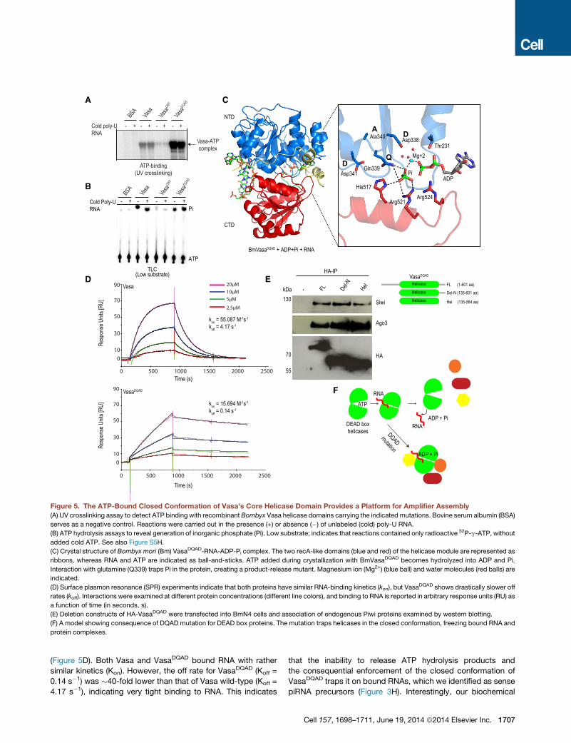
Figure 5. The ATP-Bound Closed Conformation of Vasa’s Core Helicase Domain Provides a Platform for Amplifier Assembly
(A) UV crosslinking assay to detect ATP binding with recombinant Bombyx Vasa helicase domains carrying the indicated mutations. Bovine serum albumin (BSA)
serves as a negative control. Reactions were carried out in the presence (+) or absence (�) of unlabeled (cold) poly-U RNA.
(B) ATP hydrolysis assays to reveal generation of inorganic phosphate (Pi). Low substrate; indicates that reactions contained only radioactive 32P-g-ATP, without
added cold ATP. See also Figure S5H.
(C) Crystal structure of Bombyx mori (Bm) VasaDQAD-RNA-ADP-Pi complex. The two recA-like domains (blue and red) of the helicase module are represented as
ribbons, whereas RNA and ATP are indicated as ball-and-sticks. ATP added during crystallization with BmVasaDQAD becomes hydrolyzed into ADP and Pi.
Interaction with glutamine (Q339) traps Pi in the protein, creating a product-release mutant. Magnesium ion (Mg2+) (blue ball) and water molecules (red balls) are
indicated.
(D) Surface plasmon resonance (SPR) experiments indicate that both proteins have similar RNA-binding kinetics (kon), but VasaDQAD shows drastically slower off
rates (koff). Interactions were examined at different protein concentrations (different line colors), and binding to RNA is reported in arbitrary response units (RU) as
a function of time (in seconds, s).
(E) Deletion constructs of HA-VasaDQAD were transfected into BmN4 cells and association of endogenous Piwi proteins examined by western blotting.
(F) A model showing consequence of DQAD mutation for DEAD box proteins. The mutation traps helicases in the closed conformation, freezing bound RNA and
protein complexes.
(Figure 5D). Both Vasa and VasaDQAD bound RNA with rather
similar kinetics (Kon). However, the off rate for VasaDQAD (Koff =
0.14 s�1) was �40-fold lower than that of Vasa wild-type (Koff =
4.17 s�1), indicating very tight binding to RNA. This indicates
that the inability to release ATP hydrolysis products and
the consequential enforcement of the closed conformation of
VasaDQAD traps it on bound RNAs, which we identified as sense
piRNA precursors (Figure 3H). Interestingly, our biochemical
Cell 157, 1698–1711, June 19, 2014 ª2014 Elsevier Inc. 1707

A B C
Figure 6. ATP Hydrolysis by Vasa Is Essential for Fertility in Drosophila Females
(A) Fluorescence detection of GFP fusion proteins in transgenic fly ovaries in the wild-type genetic background. GFP-DmVasaDQAD accumulates in granules that
are removed from the regular perinuclear ring-like nuage localization, potentially indicating an impact on the in vivo dynamics of the protein.
(B) Eggs laid by the vasmutant fail to hatch, and this is rescued by a GFP-Vas transgene, but not by the GFP-DmVasaDQAD transgene. Number of eggs hatched is
indicated as a percentage of that seen in the wild-type. The partial rescue (�16%) is explained by the fact that the transgenes are expressed from a nonnative
promoter.
(C) Cuticle staining of such hatched embryos shows the segmentation pattern expected of normal development (in wild-type and vas mutants complemented
with GFP-Vas).
experiments (Figures 2A–2E) in BmN4 cells suggest that such
an ATP-bound and RNA-clamped state of Vasa also provides
a binding platform for Amplifier assembly.
Vasa has a rather simple architecture with an N-terminal
arginine-rich region (R-rich domain), followed by the canonical
DEAD box helicase core domain (Figure 1B). To understand
how Vasa canmediate Amplifier assembly, we prepared deletion
constructs and examined their ability to support association with
other factors in vivo. Transiently expressed deletion constructs
were purified by anti-HA immunoprecipitations and probed
with antibodies to detect endogenous Piwi proteins. Deletion
of the R-rich domain (1–134 aa) did not affect association with
Siwi and Ago3 (Figure 5E). This adds to our observation that
N-terminal arginines in Vasa are not required for mediating
complex formation (Figures 2A and 2E). Remarkably, a construct
having only the helicase core domain was capable of associ-
ating with Siwi and Ago3 (Figure 5E), indicating that Amplifier is
assembled on the surface of the helicase core of Vasa. We pro-
pose that the binding interface for Amplifier components is
created only when the helicase domain is in a closed conforma-
tion and, consequently, only when Vasa is bound to its target
RNAs (Figure 5F). Subsequent ATP hydrolysis would promote
release of the RNA and would result in an open conformation
of the helicase domain, disrupting the protein interaction surface
and leading to complex disassembly.
ATP Hydrolysis by Vasa Is Essential for Fertility in theDrosophila Female GermlineImpaired piRNA pathway leads to infertility in all animal models,
including in Drosophila vasa (vas) mutants (Malone et al., 2009).
1708 Cell 157, 1698–1711, June 19, 2014 ª2014 Elsevier Inc.
Because the DQAD mutation within Bombyx Vasa prevents
Amplifier disassembly, we wished to examine whether such a
mutation within Drosophila Vasa might have a functional conse-
quence for fly fertility. We expressed Drosophila Vasa trans-
genes (GFP-Vas or GFP-VasDQAD) in wild-type Drosophila
ovaries (Figure S6A). Whereas GFP-Vas localized as a perinu-
clear ring to the nuage of nurse cells (Lasko and Ashburner,
1988), GFP-VasDQAD protein appeared to form distinct foci that
are removed from the perinuclear location (Figure 6A). These
are reminiscent of enlarged granules observed in BmN4 cells
(Figures 1E, 2F, and 2G). Next, we expressed the transgenes
in the vasPD/vasD1 mutant background (these mutants lay eggs
that fail to hatch) (Lasko and Ashburner, 1988). When GFP-
Vas is expressed in the mutant background, it complements
infertility (Figures 6B, S6A, and S6B). The rescued embryos
showed normal segmentation patterns, very similar to those
hatched from eggs laid by wild-type females (Figure 6C). Signif-
icantly, the GFP-VasaDQAD transgene failed to rescue the infer-
tility phenotype of the mutant (Figures 6B and 6C and Extended
Experimental Procedures). These experiments provide strong
genetic evidence for the physiological effect of preventing disas-
sembly of Vasa RNPs in the Drosophila germline.
DISCUSSION
Secondary piRNA biogenesis (the ping-pong cycle) links post-
transcriptional silencing of transposons by Piwi endonucleases
to piRNA biogenesis by converting one of the cleavage frag-
ments into a new secondary piRNA (Brennecke et al., 2007;
Gunawardane et al., 2007). This poses a unique problem, as

Figure 7. A Model for Vasa’s Role in the
Ping-Pong Cycle
Recognition of transposon transcripts by Siwi-
bound antisense piRNAs is proposed to result in
recruitment of Vasa. The open conformation of
Vasa’s helicase domain is modeled on the eIF4A
structure (PDB: 1FUU). Vasa binds RNA in its ATP-
bound form, taking up a closed conformation of its
RNA helicase domain. This closed conformation
also provides an assembly platform for Amplifier
components (ping-pong Piwi partners and the
Tudor domain protein Qin/Kumo). After Siwi slicing
of the transposon transcript, the cleavage frag-
ment carrying the 50 monophosphate (piRNA in-
termediate) is transferred to Ago3. ATP hydrolysis
in Vasa triggers this exchange and eventual
disassembly of the Amplifier. Subsequently, the
piRNA intermediate is subject to 30 endmaturation
by a putative exonuclease (Trimmer) to mature as
a new secondary piRNA. Loaded Ago3 then enters
the feedforward step of the ping-pong cycle to
generate more of the Siwi-bound piRNAs.
endonucleolytic cleavage of a target RNA by an Argonaute
normally results in complete degradation of the two resulting
cleavage fragments in all cell types. Germ cells must therefore
possess an unknown machinery that safely delivers a cleavage
fragment (piRNA intermediate) from one Piwi protein to another
(ping-pong partners), but the molecular basis for this process
is not known. Among the list of over 30 factors that are genet-
ically linked to the piRNA pathway (Figure S1A), we chose to
investigate Vasa, as it is the only factor that is demonstrated
to have a conserved and exclusive role in secondary piRNA
biogenesis in flies and mice (Kuramochi-Miyagawa et al.,
2010; Malone et al., 2009). Our investigations reveal that
Vasa functions as an RNA clamp to coordinate the assembly
of a piRNA Amplifier complex on transposon transcripts. Within
this protected environment, ATP hydrolysis by Vasa promotes
safe handover of sliced piRNA intermediates between ping-
pong partners.
Our results allow us to propose a step-by-step biochemical
model for the Ping-pong cycle operating in BmN4 cells (Figure 7).
Transposon silencing is initiated by cytoplasmic scanning
of transposon transcripts by antisense primary piRNAs loaded
into Siwi. This is closely followed by deposition of Vasa on the
transcript and by assembly of the Amplifier by the joining of
Qin/Kumo and Ago3 within the nuage. Amplifier assembly is
linked to a conformational change in the core helicase domain
of Vasa as it transitions from an ATP-unbound open state to
the ATP- and RNA-bound closed state, such that the latter
provides an interaction platform for the different components.
Interestingly, Qin is already reported to facilitate heterotypic
ping-pong between Aub (Siwi ortholog in Drosophila) and Ago3
by promoting interactions between them in the fly germline
(Anand and Kai, 2012; Zhang et al., 2011). Siwi initiates the
ping-pong cycle by slicing the target RNA to generate two frag-
ments. Following this, the cleavage fragment carrying themature
50 end of a future secondary piRNA (piRNA intermediate) is trans-
ferred fromSiwi to Ago3.We show that ATP hydrolysis by Vasa is
critical for triggering this exchange and subsequent disassembly
of the complex. We propose that the role of Vasa in assembling
Amplifier is to generate a protective environment in which the
ping-pong partners are brought to physical proximity, such
that the cleaved target is not accessible to nucleases and
is instead loaded onto Ago3 upon release from Siwi. Once ac-
quired by Ago3, themature 30 end of the new piRNA is generated
by an unknown 30–50 exonuclease, tentatively called Trimmer
(Kawaoka et al., 2011). Such sense-oriented secondary piRNAs
then guide Ago3 to participate in the feedforward step of the
ping-pong cycle (Brennecke et al., 2007).
Our results provide, for the first time, a biochemical snapshot
of events that take place during the ping-pong cycle. However,
a number of questions still remain. It is not clear how the closed
conformation of Vasa’s helicase core domain can promote
Amplifier assembly. In this context, it is tempting to draw a par-
allel between Vasa’s role and the function of the helicase domain
of the related DEAD box helicase eIF4AIII in nucleating the for-
mation of the exon junction complex (EJC) on spliced mRNAs
(Le Hir et al., 2000). In its ATP-bound closed conformation, the
eIF4AIII core domain clamps on spliced mRNAs to provide
a binding surface for other EJC components Mago, Y14, and
Barentsz (Andersen et al., 2006; Bono et al., 2006). A similar
situation can be envisaged during Amplifier assembly. Also un-
known is whether any control is exercised over Siwi slicer activity
to prevent futile cleavages that do not result in secondary piRNA
production. Because piRNA intermediate exchange and com-
plex disassembly are linked to ATP hydrolysis by Vasa, thismight
also be subject to regulation. It is only logical that target slicing
by Siwi is followed by ATP hydrolysis by Vasa.We can only spec-
ulate that perhaps allosteric interactions between Piwi proteins
and Vasa might link slicing of the target to ATP hydrolysis in
Vasa. Recent genome-wide genetic screens have identified a
number of factors that are implicated in the germline piRNA
pathway in flies (Czech et al., 2013). An examination of our top
hits in the VasaDQAD proteomics analyses (Figure S2A) did not
reveal any overlaps except for the ping-pong Piwi partners and
Qin/Kumo, which we validated in our study.
Cell 157, 1698–1711, June 19, 2014 ª2014 Elsevier Inc. 1709

The ping-pong model predicts a reciprocal relationship in
which piRNA-guided Siwi facilitates biogenesis of Ago3-bound
piRNAs and vice versa (Brennecke et al., 2007). This means
that there are potentially two distinct complexes mediating
piRNA amplification: one occupying the Siwi-to-Ago3 segment
and another one mediating the Ago3-to-Siwi feedforward step
of the amplification loop. Our data strongly support a role for
Vasa in the former step of the ping-pong cycle, but we cannot
entirely rule out its participation in the latter. It is also possible
that such a role in the feedforward segment might be fulfilled
by other RNA helicases like Spn-E, a factor that is essential for
the ping-pong cycle in flies (Malone et al., 2009), but not in
mice (Shoji et al., 2009), wherein a linear secondary piRNA
biogenesis pathway operates (De Fazio et al., 2011).
The DEAD box family is the largest family of helicases, and
they are found from bacteria to humans, with most of the 26
members in yeast being essential for viability (de la Cruz et al.,
1999). The in vivo RNA targets and RNP complexes of most
DEAD box helicases have not been identified due to their tran-
sient nature. Here, we used a DQAD mutation to trap dynamic
in vivo associations of Vasa. Structures of several DEAD box hel-
icases with bound ATP analogs and RNA are closely related,
indicating that perhaps similar outcomes to those we describe
here can be expected for other family members assembling dy-
namic complexes in various RNA processing pathways.
EXPERIMENTAL PROCEDURES
Clones and Constructs
For expression studies in BmN4 cells, the coding sequence for the full-length
Bombyx mori Vasa (GenBank accession number: NM_001043882.1) or its
deletion versions were cloned into the pBEMBL-HA (Xiol et al., 2012) or
pBEMBL-Myc vectors. Point mutations in the BmVasa helicase domain were
introduced by an overlap PCR strategy: ATP-binding mutant (K230N; GKT/
GNT) or ATP hydrolysis product-trap mutant (E339Q; DEAD/DQAD). The
R/K mutant was created by converting 20 arginine (R) residues to lysines
(K) at the Vasa N terminus (within a region spanning 1–117 aa) using a chem-
ically synthesized DNA fragment. For live-cell imaging studies, the coding
sequence for BmVasa, VasaDQAD, or Ago3 was cloned downstream of an
enhanced green fluorescent protein (EGFP) tag or the red fluorescent protein
variant mCherry in the pBEMBL vector backbone.
Crystallization and Biochemical Procedures
The helicase domain of Bombyx Vasa (135–564 aa) carrying the E339Q
(DEAD/DQAD) mutation was produced in E. coli as a His-tagged fusion.
Purified protein was incubated with a 6-mer (UGACAU) RNA and ATP or
the ATP analog (AMPPNP) at a molar ratio of 1:1.2:1.2 (protein:RNA:ATP/
AMPPNP). A summary of data collection statistics is provided in the Extended
Experimental Procedures as Table S1.
Small RNA Sequencing
RNA libraries were prepared from immunoprecipitated complexes and were
deep sequenced on the Illumina Hi-Seq (50 cycles) or MiSeq platforms
(32 cycles). For long RNAs present in VasaDQAD complexes, a strand-specific
RNA-seq library was prepared and sequenced on an Illumina Hi-Seq platform
(105 cycles). Reads were aligned to the Bombyx genome and were analyzed
using custom pipelines.
Fly Experiments
Drosophila vas mutant stocks were: vasPD (Schupbach and Wieschaus,
1986) and vasD1 (Lehmann and Nusslein-Volhard, 1991). The vas-deficient
background was achieved by the use of the transheterozygotes (vasPD/
vasD1). Expression of GFP-tagged Drosophila melanogaster Vasa transgenes
1710 Cell 157, 1698–1711, June 19, 2014 ª2014 Elsevier Inc.
(Johnstone and Lasko, 2004) in the fly germline was controlled from
the upstream activating sequence (UAS) using GAL4 driven from a nanos
promoter.
ACCESSION NUMBERS
The PDB accession codes for crystal structures are 4D26 and 4D25.
Deep-sequencing data are deposited with Gene Expression Omnibus (GEO)
(GSE57420).
SUPPLEMENTAL INFORMATION
Supplemental Information includes Extended Experimental Procedures, six
figures, five movies, and two tables and can be found with this article online
at http://dx.doi.org/10.1016/j.cell.2014.05.018.
ACKNOWLEDGMENTS
We thank Paul Lasko, Hugo Bellen, and Beat Suter for fly stocks/reagents. We
are grateful to Sanjay Ghosh, Andrea Picco, Enrico Zamattia, Sandra Muller,
and Anna Cyrklaff for help with data analysis and experiments. We thank the
following EMBL core facilities: Advanced Light Microscopy, Genomics, Pro-
tein Expression and Purification, and High-Throughput Crystallization. Crystal-
lographic data collection was at European Synchrotron Radiation Facility
(ESRF), Grenoble, France. This work was supported by grants from European
Union (European Research Council; ‘‘pisilence’’) to R.S.P. We are grateful for
fellowships from the EMBL Interdisciplinary Postdoc Programme (EIPOD) un-
der Marie Curie COFUND Actions (M.A.L), EMBO (D.H), and Region Rhone Alp
(E.C.). This work was supported by the EMBL.
Received: February 25, 2014
Revised: April 28, 2014
Accepted: May 15, 2014
Published: June 5, 2014
REFERENCES
Anand, A., and Kai, T. (2012). The tudor domain protein kumo is required
to assemble the nuage and to generate germline piRNAs in Drosophila.
EMBO J. 31, 870–882.
Andersen, C.B., Ballut, L., Johansen, J.S., Chamieh, H., Nielsen, K.H., Oliveira,
C.L., Pedersen, J.S., Seraphin, B., Le Hir, H., and Andersen, G.R. (2006).
Structure of the exon junction core complex with a trapped DEAD-box
ATPase bound to RNA. Science 313, 1968–1972.
Aravin, A.A., Hannon, G.J., and Brennecke, J. (2007). The Piwi-piRNA pathway
provides an adaptive defense in the transposon arms race. Science 318,
761–764.
Bagijn, M.P., Goldstein, L.D., Sapetschnig, A., Weick, E.M., Bouasker, S.,
Lehrbach, N.J., Simard, M.J., and Miska, E.A. (2012). Function, targets, and
evolution of Caenorhabditis elegans piRNAs. Science 337, 574–578.
Bono, F., Ebert, J., Lorentzen, E., and Conti, E. (2006). The crystal structure
of the exon junction complex reveals how it maintains a stable grip on
mRNA. Cell 126, 713–725.
Brennecke, J., Aravin, A.A., Stark, A., Dus, M., Kellis, M., Sachidanandam, R.,
and Hannon, G.J. (2007). Discrete small RNA-generating loci as master regu-
lators of transposon activity in Drosophila. Cell 128, 1089–1103.
Brennecke, J., Malone, C.D., Aravin, A.A., Sachidanandam, R., Stark, A., and
Hannon, G.J. (2008). An epigenetic role for maternally inherited piRNAs in
transposon silencing. Science 322, 1387–1392.
Caruthers, J.M., and McKay, D.B. (2002). Helicase structure and mechanism.
Curr. Opin. Struct. Biol. 12, 123–133.
Chen, Y., Pane, A., and Schupbach, T. (2007). Cutoff and aubergine mutations
result in retrotransposon upregulation and checkpoint activation in Drosophila.
Curr. Biol. 17, 637–642.

Czech, B., Preall, J.B., McGinn, J., and Hannon, G.J. (2013). A transcriptome-
wideRNAi screen in the Drosophila ovary reveals factors of the germline piRNA
pathway. Mol. Cell 50, 749–761.
De Fazio, S., Bartonicek, N., Di Giacomo, M., Abreu-Goodger, C., Sankar, A.,
Funaya, C., Antony, C., Moreira, P.N., Enright, A.J., and O’Carroll, D. (2011).
The endonuclease activity of Mili fuels piRNA amplification that silences
LINE1 elements. Nature 480, 259–263.
de la Cruz, J., Kressler, D., and Linder, P. (1999). Unwinding RNA in Saccha-
romyces cerevisiae: DEAD-box proteins and related families. Trends Biochem.
Sci. 24, 192–198.
de Vanssay, A., Bouge, A.L., Boivin, A., Hermant, C., Teysset, L., Delmarre, V.,
Antoniewski, C., and Ronsseray, S. (2012). Paramutation in Drosophila linked
to emergence of a piRNA-producing locus. Nature 490, 112–115.
Ghildiyal, M., and Zamore, P.D. (2009). Small silencing RNAs: an expanding
universe. Nat. Rev. Genet. 10, 94–108.
Grentzinger, T., Armenise, C., Brun, C., Mugat, B., Serrano, V., Pelisson, A.,
and Chambeyron, S. (2012). piRNA-mediated transgenerational inheritance
of an acquired trait. Genome Res. 22, 1877–1888.
Gunawardane, L.S., Saito, K., Nishida, K.M., Miyoshi, K., Kawamura, Y.,
Nagami, T., Siomi, H., and Siomi, M.C. (2007). A slicer-mediated mechanism
for repeat-associated siRNA 50 end formation in Drosophila. Science 315,
1587–1590.
Johnstone, O., and Lasko, P. (2004). Interaction with eIF5B is essential for
Vasa function during development. Development 131, 4167–4178.
Kawaoka, S., Hayashi, N., Suzuki, Y., Abe, H., Sugano, S., Tomari, Y.,
Shimada, T., and Katsuma, S. (2009). The Bombyx ovary-derived cell line
endogenously expresses PIWI/PIWI-interacting RNA complexes. RNA 15,
1258–1264.
Kawaoka, S., Izumi, N., Katsuma, S., and Tomari, Y. (2011). 30 end formation
of PIWI-interacting RNAs in vitro. Mol. Cell 43, 1015–1022.
Kawaoka, S., Hara, K., Shoji, K., Kobayashi, M., Shimada, T., Sugano, S.,
Tomari, Y., Suzuki, Y., and Katsuma, S. (2013). The comprehensive epigenome
map of piRNA clusters. Nucleic Acids Res. 41, 1581–1590.
Kazazian, H.H., Jr. (2004). Mobile elements: drivers of genome evolution.
Science 303, 1626–1632.
Kirino, Y., Vourekas, A., Kim, N., de Lima Alves, F., Rappsilber, J., Klein, P.S.,
Jongens, T.A., and Mourelatos, Z. (2010). Arginine methylation of vasa protein
is conserved across phyla. J. Biol. Chem. 285, 8148–8154.
Klattenhoff, C., Xi, H., Li, C., Lee, S., Xu, J., Khurana, J.S., Zhang, F., Schultz,
N., Koppetsch, B.S., Nowosielska, A., et al. (2009). The Drosophila HP1 homo-
log Rhino is required for transposon silencing and piRNA production by dual-
strand clusters. Cell 138, 1137–1149.
Kuramochi-Miyagawa, S., Watanabe, T., Gotoh, K., Takamatsu, K., Chuma,
S., Kojima-Kita, K., Shiromoto, Y., Asada, N., Toyoda, A., Fujiyama, A., et al.
(2010). MVH in piRNA processing and gene silencing of retrotransposons.
Genes Dev. 24, 887–892.
Lasko, P.F., and Ashburner, M. (1988). The product of the Drosophila gene
vasa is very similar to eukaryotic initiation factor-4A. Nature 335, 611–617.
Le Hir, H., Izaurralde, E., Maquat, L.E., and Moore, M.J. (2000). The spli-
ceosome deposits multiple proteins 20-24 nucleotides upstream of mRNA
exon-exon junctions. EMBO J. 19, 6860–6869.
Lehmann, R., and Nusslein-Volhard, C. (1991). The maternal gene nanos has
a central role in posterior pattern formation of the Drosophila embryo. Devel-
opment 112, 679–691.
Li, C., Vagin, V.V., Lee, S., Xu, J., Ma, S., Xi, H., Seitz, H., Horwich, M.D.,
Syrzycka, M., Honda, B.M., et al. (2009). Collapse of germline piRNAs in the
absence of Argonaute3 reveals somatic piRNAs in flies. Cell 137, 509–521.
Liang, L., Diehl-Jones, W., and Lasko, P. (1994). Localization of vasa protein
to the Drosophila pole plasm is independent of its RNA-binding and helicase
activities. Development 120, 1201–1211.
Lim, A.K., and Kai, T. (2007). Unique germ-line organelle, nuage, functions
to repress selfish genetic elements in Drosophila melanogaster. Proc. Natl.
Acad. Sci. USA 104, 6714–6719.
Linder, P., and Jankowsky, E. (2011). From unwinding to clamping - the DEAD
box RNA helicase family. Nat. Rev. Mol. Cell Biol. 12, 505–516.
Liu, F., Putnam, A., and Jankowsky, E. (2008). ATP hydrolysis is required for
DEAD-box protein recycling but not for duplex unwinding. Proc. Natl. Acad.
Sci. USA 105, 20209–20214.
Luteijn, M.J., and Ketting, R.F. (2013). PIWI-interacting RNAs: from generation
to transgenerational epigenetics. Nat. Rev. Genet. 14, 523–534.
Malone, C.D., Brennecke, J., Dus, M., Stark, A., McCombie, W.R., Sachida-
nandam, R., and Hannon, G.J. (2009). Specialized piRNA pathways act in
germline and somatic tissues of the Drosophila ovary. Cell 137, 522–535.
Mathioudakis, N., Palencia, A., Kadlec, J., Round, A., Tripsianes, K., Sattler,
M., Pillai, R.S., and Cusack, S. (2012). The multiple Tudor domain-containing
protein TDRD1 is a molecular scaffold for mouse Piwi proteins and piRNA
biogenesis factors. RNA 18, 2056–2072.
Meister, G. (2013). Argonaute proteins: functional insights and emerging roles.
Nat. Rev. Genet. 14, 447–459.
Pane, A., Wehr, K., and Schupbach, T. (2007). zucchini and squash encode
two putative nucleases required for rasiRNA production in the Drosophila
germline. Dev. Cell 12, 851–862.
Pause, A., and Sonenberg, N. (1992). Mutational analysis of a DEAD box RNA
helicase: the mammalian translation initiation factor eIF-4A. EMBO J. 11,
2643–2654.
Schupbach, T., and Wieschaus, E. (1986). Germline autonomy of maternal-
effect mutations altering the embryonic body pattern of Drosophila. Dev.
Biol. 113, 443–448.
Sengoku, T., Nureki, O., Nakamura, A., Kobayashi, S., and Yokoyama, S.
(2006). Structural basis for RNA unwinding by the DEAD-box protein
Drosophila Vasa. Cell 125, 287–300.
Shoji, M., Tanaka, T., Hosokawa, M., Reuter, M., Stark, A., Kato, Y., Kondoh,
G., Okawa, K., Chujo, T., Suzuki, T., et al. (2009). The TDRD9-MIWI2 complex
is essential for piRNA-mediated retrotransposon silencing in the mouse
male germline. Dev. Cell 17, 775–787.
Xiol, J., Cora, E., Koglgruber, R., Chuma, S., Subramanian, S., Hosokawa, M.,
Reuter, M., Yang, Z., Berninger, P., Palencia, A., et al. (2012). A role for
Fkbp6 and the chaperone machinery in piRNA amplification and transposon
silencing. Mol. Cell 47, 970–979.
Zhang, Z., Xu, J., Koppetsch, B.S., Wang, J., Tipping, C., Ma, S., Weng, Z.,
Theurkauf, W.E., and Zamore, P.D. (2011). Heterotypic piRNA Ping-Pong
requires qin, a protein with both E3 ligase and Tudor domains. Mol. Cell 44,
572–584.
Zhang, F., Wang, J., Xu, J., Zhang, Z., Koppetsch, B.S., Schultz, N., Vreven, T.,
Meignin, C., Davis, I., Zamore, P.D., et al. (2012). UAP56 couples piRNA
clusters to the perinuclear transposon silencing machinery. Cell 151, 871–884.
Cell 157, 1698–1711, June 19, 2014 ª2014 Elsevier Inc. 1711