robert c. fahey, ralph m. buschbacher, and gerald l ... · ot-d grown to stationary phase at 45°...
TRANSCRIPT

J. MOL. EVOL., IN PRESS
THE EVOLUTION OF GLUTATHIONE METABOLISM IN PHOTOTROPHIC MICROORGANISMS
Robert C. Fahey, Ralph M. Buschbacher, and Gerald L. Newton
Department of Chemistry, University of California, San Diego,
La Jolla, California 92093
Correspondence to: Robert C. Fahey
Department of Chemistry
University of California, San Diego
La Jolla CA 92093
Phone (619) 452-2163
— =3
Received ^
3 -._ :" 73
03 f «C
':.-' '•' a•̂ TOCO •<
00
I (KflSA-CE-182,SC2) Z'£E EVG102ICK Gf N88-26015G10JA1HIGME BITAEC11SB.IB FEGTCTBCPHICBICBOCfiGAKISBE (California U E i v . ) 2:9 p
CSC1 06B UaclasG3/51 0116713
https://ntrs.nasa.gov/search.jsp?R=19880016631 2018-07-10T10:33:16+00:00Z
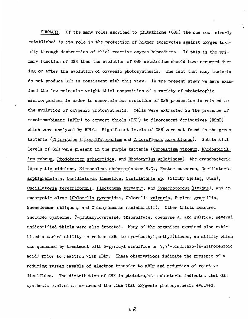
SUMMARY. Of the many roles ascribed to glutathione (GSH) the one most clearly
established is its role in the protection of higher eucaryotes against oxygen toxi-
city through destruction of thiol reactive oxygen biproducts. If this is the pri-
mary function of GSH then the evolution of GSH metabolism should have occurred dur-
ing or after the evolution of oxygenic photosynthesis. The fact that many bacteria
do not produce GSH is consistent with this view. In the present study we have exam-
ined the low molecular weight thiol composition of a variety of phototrophic
microorganisms in order to ascertain how evolution of GSH production is related to
the evolution of oxygenic photosynthesis. Cells were extracted in the presence of
monobromobimane (mBBr) to convert thiols (ESH) to fluorescent derivatives (RSmB)
which were analyzed by HPLC. Significant levels of GSH were not found in the green
bacteria (Chlorobium thiosulfatophilum and Chloroflexus aurantiacus). Substantial
levels of GSH were present in the purple bacteria (Chromatium vinosum, Rhodospiril-
ium rubrum, Rhodobacter sphaeroides, and Rhodocyclus gelatinosa), the cyanobacteria
(Anacystis nidnl^ns, Microcoleus chthonoplastes _S_..G., Nostoc muscorum, Oscil latoria
amphigranulata, Oscillatoria limnetica, Oscillatoria sp. (Stinky Spring, Utah),
Oscillatoria terebriformls, Plectonema boryanum, and Synechococcus lividus), and in
eucaryotic algae (ChloreTi» pyrenoidsa, Chlorella vulgaris, Euglena gracillis,
Scenedesmus obliquus, and Chlamydomonas rheinhardtii). Other thiols measured
included cysteine, X-glutamylcysteine, thiosulfate, coenzyme A, and sulfide; several
unidentified thiols were also detected. Many of the organisms examined also exhi-
bited a marked ability to reduce mBBr to syn- (methyl,methyl)bimane, an ability which
was quenched by treatment with 2-pyridyl disulfide or 5j5'-bisdithio-(2-nitrobenzoic
acid) prior to reaction with mBBr. These observations indicate the presence of a
reducing system capable of electron transfer to mBBr and reduction of reactive
disulfides. The distribution of GSH in phototrophic eubacteria indicates that GSH
synthesis evolved at or around the time that oxygenic photosynthesis evolved.

SUMMARY. Of the many roles ascribed to glutathione (GSH) the one most clearly
established is its role in the protection of higher eucaryotes against oxygen toxi-
city through destruction of thiol reactive oxygen biproducts. If this is the pri-
mary function of GSH then the evolution of GSH metabolism should have occurred dur-
ing or after the evolution of oxygenic photosynthesis. The fact that many bacteria
do not produce GSH is consistent with this viev. In the present study we have exam-
ined the lov molecular weight thiol composition of a variety of phototrophic
microorganisms in order to ascertain how evolution of GSH production is related to
the evolution of oxygenic photosynthesis. Cells were extracted in the presence of
monobromobimane (mBBr) to convert thiols (RSH) to-fluorescent derivatives (RSmB)
which were analyzed by HPLC. Significant levels of GSH were not found in the green
sulfur bacteria (Chlorobium thiosulfatophilum and Chloroflexus aurantiacus). Sub-
stantial levels of GSH were present in the purple bacteria (Chromatium vinosum, Rho-
dospirillum rubrum, Rhodobacter sphaeroides, and Rhodocyclus gelatinosa), the
cyanobacteria (Anacystis nidulans, Microcoleus chthonoplastes S_.Q., Nostoc muscorum,
Oscillatoria amphigranulata, Oscillatoria limnetica, Oscillatoria sp. (Stinky
Spring, Utah), Oscillatoria terebriformis, Plectonema boryanum, and Synechococcus
lividus), and in eucaryotic algae (Chlorella pyrenoidsa, Chlorella vulgaris, Euglena
gracillis, Scenedesmus obliquus, and Chlamydomonas rheinhardtii). Other thiols
measured included cysteine, X-glutamylcysteine, thiosulfate, coenzyme A, and sul-
fide; several unidentified thiols were also detected. Many of the organisms exam-
ined also exhibited a marked ability to reduce mBBr to syn-(methyl,methyl)bimane, an
ability which was quenched by treatment with 2-pyridyl disulfide or 5,5'-bisdithio-
(2-nitrobenzoic acid) prior to reaction with mBBr. These observations indicate the
presence of a reducing system capable of electron transfer to mBBr and reduction of
reactive disulfides. The distribution of GSH in phototrophic eubacteria indicates
that GSH synthesis evolved at or around the time that oxygenic photosynthesis
evolved.
ffi WORDS. Glut«+n
INTRODUCTION
Glutathione has been postulated to play a key role in many biological processes
(Jocelyn, 1972; Kosower and Kosover, 1978; Meister and Anderson, 1983) but its func-
tion in protecting cells against the toxicity of oxygen and other thiol reactive
agents is the role which has been most thoroughly established. Key enzymes involved
in this protection against oxygen toxicity in animals are glutathione peroxidase
(Wendel, 1980), which catalyzes the destruction of hydrogen peroxide and organic
hydroperoxides (Eq l), and the glutathione S-transferases (Chasseaud, 1976; Jakoby
and Habig, 1980), which catalyze the destruction of hydroperoxides and the conjuga-
tion of GSH with epoxides, enones, and other sulfhydryl reactive agents.
2GSH + H202 = GSSG + 2H20 . (l)
Glutathione peroxidase activity has not been found in plants and it is thought (Hal-
liwell, 1981) that ascorbate oxidase fulfills an analogous function with the dehy-
droascorbate formed in the destruction of H-Op (Eq 2) being reduced by GSH back to
ascorbate (Eq 3).
ascorbate + EJ 2̂ = dehydroascorbate + 2H20 (2)
2GSH + dehydroascorbate = GSSG + ascorbate (3)
Glutathione reductase catalyzes the reduction of the GSSG formed in reactions 1 and
3 back to GSH using NADPH as the reductant (Eq 4).
GSSG + RADPH + H* = 2GSH + NADP+ (2)
Under normal conditions the latter reaction maintains the GSH:GSSG ratio at a value
generally in excess of ~50 and insures an environment in which the essential sulfhy-
dryl groups of key enzymes and coenzymes are protected (Kosower and Kosower, 1978).
If the primary function of glutathione is oxygen detoxification, then glu-
tathione metabolism presumably arose at the time oxygen was accumulating as the
consequence of the evolution of oxygenic photosynthesis. Considerable diversifica-
tion must have existed among the bacteria extant on the earth at the time that oxy-

gen became a significant component of the atmosphere and it would be surprising if
the only thiol-based protection system to evolve vas that based upon GSH. The find-
ing that many bacteria, including some strict aerobes, lack GSH but contain other
low molecular weight thiols is consistent with the idea that more than one thiol
protection system has evolved in procaryotes (Fahey et al, 1978). Thiol analysis of
bacteria lacking GSH has suggested that Coenzyme A may be the key compound in the
thiol protecting systems of some aerobic Gram positive bacteria (Fahey and Newton,
1983) whereas /-glutamylcysteine appears to form the basis for such a system in
halobacteria (Newton and Javor, 1985). Glutathione appeared to occur primarily in
the aerobic and facultative Gram negative bacteria but not in anaerobes or in Chro-
mation vinosum based upon results of enzymatic assay for glutathione (Fahey et al,
1978).
In the present study we utilize recently developed methods to analyze the low
molecular weight thiols in a variety of phototrophic microorganisms selected to
represent different stages in the evolution of the metabolism necessary for oxygenic
photosynthesis and different levels of tolerance and utilization of oxygen. We sur-
vey selected representatives of the green bacteria, of all main branches of the evo-
lutionary tree for purple photosynthetic bacteria as derived from l6s rRNA sequence
data (Fox et al, 1980), and of the cyanobacteria, the latter constituting the only
group of prokaryotes capable of oxygenic photosynthesis. The objective was to pin-
point more carefully which groups produce GSH in order to establish a basis for
understanding the origin of glutathione metabolism. A selection of eucaryotic algae
was also examined to further test the generality of occurrence of GSH among
eucaryotes.
EXPERIMENTAL
Organisms. Photoautotrophic growth conditions were used for growth wherever
possible. When heterotrophic growth was used the medium was tested for the pres-
ence of glutathione and media containing yeast extract were found to contain high
levels of glutathione. When the level found was _>. 0-5 uM, the glutathione was
specifically depleted as described previously (Fahey et al, 198M. Cells were
harvested in late log phase by centrifugation and aliquots were used immediately
or stored at -80° until extracted.
The samples of the following organisms were kindly provided by D.E. Carlson,
T. Johnson, and B.B. Buchanan. Chlorobium thiosulfatophilum (Tassajara) was grown
anaerobically and photoautotrophically in the medium described by Buchanan, et al
(1972). Chromatium vinosum D was cultured anaerobically and photoautotrophically
in a CO^-thiosulfate medium described by Arnon et al (1963). Nostoc muscorum 7119
was cultured photoautotrophically as described by Arnon et al (197M. Chlorella
vulgaris was cultured photoautotrophically according to Arnon et al (1955) with
the E7 and Fe-EDTA solutions of Arnon et al (1963), and was grown heterotrophi-
cally in the dark on this medium supplemented with 0.2% glucose. Euglena gracilis
Z was cultured photoautotrophically according to Edmunds (1965) and 0.2% glucose
was added to this medium for dark, heterotrophic growth.
Samples of the following organisms were generously provided by R. Fall.
Plectonema boryanium UTEX 591* was photoautotrophically grown as described by
Stratton et al (1979). Chlorella pyernoidosa UTEX 1230 and Scenedesmus obliquus
UTEX ih^O were cultured photoautotrophically as described by Sorokin and Krauss
(1958).
Chloroflexus aurantiacus ATCC 29362 was cultured anaerobically and pho-
toheterotrophically on ATCC medium 920 (glutathione depleted). Rhodospirillum
rubrum ATCC 11170, via B. Bartsch, was cultured anaerobically and photohetero-
trophically on malate (Ormerod, et al, 196l). Dark heterptrophic grovth on malate
in the presence of air was accomplished using the medium of Ormerod et al (1961).
Anaerobic, photoheterotrophic grovth of Rhodobacter sphaeroides (formerly, Rhodop-
seudomonas sphaeroides) ATCC 550 was carried out on medium S described by Las-
celles (1956), and of Rhodocyclus gelatinosa (formerly, Rhodopseudomonas gela-
tinosa) ATCC 17011 on ATCC medium 112, the media being depleted of glutathione in
both cases. Chlamydomonas reinhardtii 137Cm+ was kindly supplied by S. Howell and
was grown photoautotrophically on the high salt medium (HSM) of Sueoka et al
(1967).
Barbara Javor kindly provided samples of Oscillatoria terebriformis OH-80-
Ot-D grown to stationary phase at 45° in D medium (Castenholz, 1981) buffered with
5 mM Hepps, of Synechococcus lividus OH-53-S harvested in log phase after grown on
DG medium (D medium plus 0.8 g/L glycylglycine buffer) at 4 5 , of Anacystis nidu-
lans 625 cultured in DG medium at 27°, and of Oscillatoria amphigranu.1 .ata WT-RC
harvested in log phase after growth at 1+5 in D medium lacking KNO_ and NaNO_ but
with addition (g/L) of NagHPO^ (0.07), KHgPO^ (0.036), tricine (0.9) and NH^Cl
(0.2).
Yehuda Cohen generously furnished samples of Oscillatoria limnetica (Solar
Lake, Sinai) cultured 7 days at 40° in CHU 11 medium (Cohen, et al, 1975). He
also provided Oscillatoria sp. isolated from Stinky Spring, Utah and grown 7 days
at 1*0° in ASNW III medium (Rippka, et al, 1979). Also provided was Microcoleus
chthonoplastes J3.G_. from Spencer Gulf, South Australia which was cultured 7 days
at kO° in the ASH III medium above but with the NaCl, MgSO]+»7H20, MgCl2, and KC1
at double the given concentrations. Further details are found in Cohen, et al
(1986).
Materials. Glacial acetic acid and (ethylenedinitrilo)tetraacetic acid diso-
dium salt (EDTA) were obtained from Mallincrodt, HPLC grade acetonitrile and
methanol vere from Burdick and Jackson, and 2,2'-dithiodipyridine and 5>5'-
dithiobis(2-nitrobenzoic acid) (DTKB) were from Sigma. Monobromobimane (mBBr) and
K-2-hydroxyethylpiperazine-n'-3-propanesulfonic acid (HEPPS) vere from Calbiochem.
Yeast extract was from Difco and all other culture media components were of
reagent grade or higher purity.
Sample Preparation. The cell extracts were prepared using a minor modifica-
tion of the method of Fahey and Newton (1983). A frozen cell pellet (~200 mg wet
weight) was flushed for 10 min with argon in a 3 ml septum capped vial after which
0.5 ml of argon saturated aqueous 100 mM HEPPS (pH 8.0)" containing 5 mM EDTA and 2
mM mBBr was added and the mixture allowed to stand 5 min. Argon saturated aceton-
itrile (0.5 ml) was then added and the sample incubated at 60°C for 15 min. The
vial was opened, the sample homogenized 1 min with a Tekmar Tissuemizer, glacial
acetic acid added to 1$, and the sample cooled on ice. Protein and cell debris
were removed by centrifugation for 5 min in an Eppendorf Microfuge. The sample
was diluted 1:1 with aqueous 1% acetic acid and recentrifuged prior to HPLC
analysis. The acetonitrile pellets were dried and weighed to obtain the residual
dry weight (~QO% of the total dry weight). For control samples 2,2'-
dithiodipyridine (2 mM) or DTNB (2 mM) was substituted for mBBr during homogeniza-
tion. Just before addition of acetonitrile, mBBr was added to k mM and allowed to
react 5 min at room temperature. Further processing of control samples was as
described above.
HPLC Chromatography. A combination of two HPLC protocols was used to achieve
full resolution of the low molecular weight thiols. The reverse phase protocol
(Method l) described by Fahey and Newton (1986) resolved most of the compounds
listed in Tables 1-3. However, cysteine and thiosulfate, which elute near 6 min,
are poorly separated, and Coenzyme A elutes at 1*5 min as a peak too broad to
detect at low CoA levels. Coenzyme A elutes as a sharp peak at 26 min and is

easily quantitated using a tetrabutylammonium ion pairing system (Method 2, Fahey
and Newton, 1986), and cysteine and thiosulfate vere also well resolved in this
protocol, eluting at 6 and 21 min, respectively. The use of these two different
protocols for analysis of all samples provided reasonable assurance as to the
correctness of the peak assignments.
RESULTS
Extraction of cells was carried out by suspending them in pH 8 buffer con-
taining mBBr and subsequently deproteinizing by adding an equal volume of acetoni-
trile and heating. Since mBBr penetrates cell membranes this approach vas thought
to provide maximal derivatization of thiols prior to their exposure to oxygen.
This conclusion appeared justified by the finding that extraction of most cells
with degassed buffer under argon gave essentially the same result as extraction in
air. When recoveries were tested by adding known mixture containing thiosulfate,
sulfide, cysteine, /-Glu-Cys, GSH, and CoA, all at 1 umole per g, recoveries were
generally acceptable (~80$). However, for Chromatium yinosum recoveries as low as
~50$ were found for some thiols when extraction was conducted in air but higher
recoveries (~80$) where obtained when extraction took place under argon. Thus,
the protocol involving extraction under argon is recommended. Since this pro-
cedure was not used for all samples the present results should be considered to be
semiquantitative. A second sample was extracted with 2-pyridyl disulfide used in
place of mBBr in the extraction buffer, with mBBr being added after the cells were
lysed. Since 2-pyridyl disulfide oxidizes thiols and prevents their labeling by
mBBr, this sample served as a control used to identify nonthiol fluorescent com-
ponents present in the cell or derived from mBBr.
The results obtained are summarized in Tables 1-3. The process used to
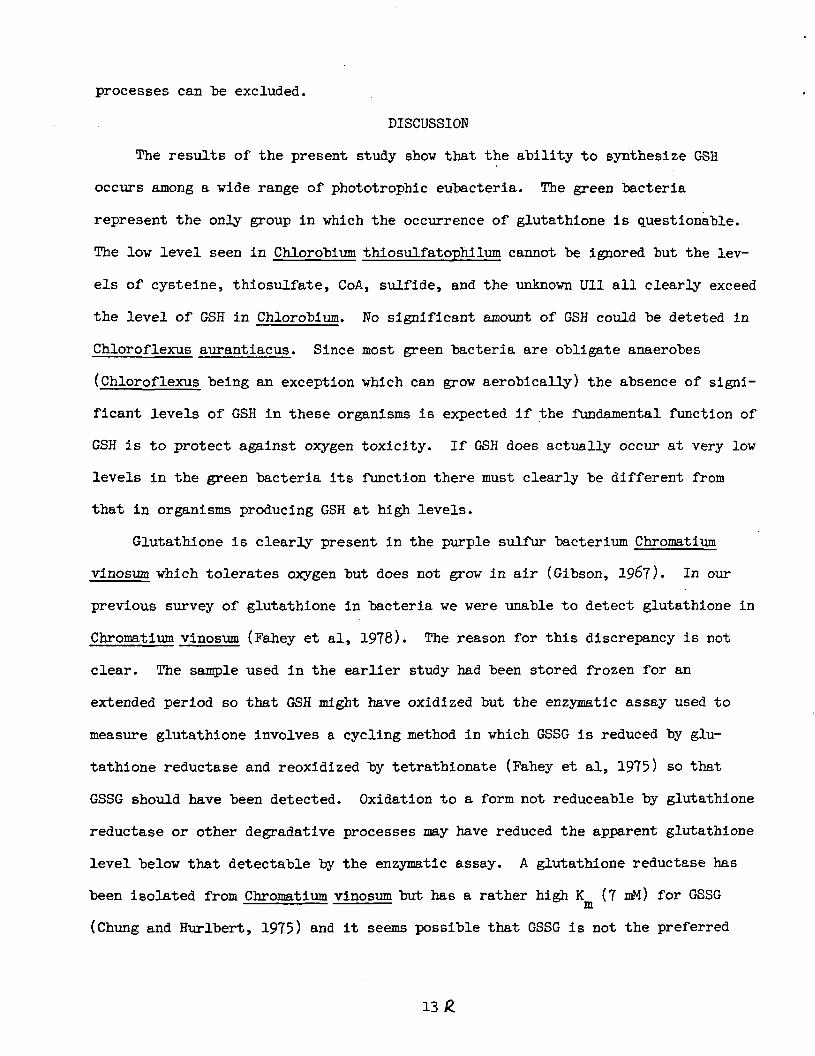
arrive at these results is illustrated for the case of Chromatium vinosum. HPLC
chromatograms obtained using Method 1 are presented in Fig. 1 and by Method 2 in
Fig. 2. Comparison of the chromatogram for the derivatized extract (Fig IB) with
that of the corresponding control sample (Fig. 1A) reveals a number of peaks that
dissapear upon treatment with 2-pyridyl disulfide. The peaks corresponding to
thiosulfate, cysteine, and /-glutamylcysteine were quite small at the amplifica-
tion shown and are not labeled; the position at which they elute is seen in the
10
chromatogram of known thiol derivatives shown in Fig. 1C. At higher gain they
were more evident and the presence of cysteine was verified in the analysis using
Method 2 (Fig. 2A). The peaks for thiosulfate and /-glutamylcysteine are masked
"by other substances in Fig. 2A.
The next major thiol derivative apparent in Fig. IB was that of GSH and its
presence was confirmed as shown in Fig 2A. Following the GSH derivative (Fig. IB)
was a peak that could not be assigned to any of known thiols previously character-
ized (Fahey and Newton, 1983) and was designated Ull4. It eluted most closely to
the derivative of homocysteine using Method 1 but addition of an authentic sample
of the mBBr derivative of homocysteine resulted in a double peak when the sample
was chromatographed using Method 2 (not shown) so that homocysteine was excluded.
Ulk eluted at approximately 8 min under Method 2 (Fig. 2A).
A major peak was observed to elute at 3k min under protocol A (Fig. IB) and
at 16 min under protocol B (Fig. 2A). This did not correspond to any of the
thiols previously characterized and extraction with ethyl acetate removed this
derivative from the mixture. The elution times correspond to those of syn-
(methyl,methyl)bimane, the compound formally resulting from displacement of the
bromide in mBBr by hydride, and addition of authentic syn-(methyl,methyl)bimane to
the sample gave HPLC chromatograms exhibiting no double peaking. The green and
purple sulfur bacteria all produced syn-(methyl,methyl)bimane in large amounts.
The purple nonsulfur bacteria, the cyanobacteria (Table 2), and the phototrophic
eucaryotes (Table 3) studied also produced syn-(methyl,methyl)bimane but in
amounts that varied widely with the specific species. Lesser amounts were
observed for Plectonema boryanum, Euglena gracilis, and light grown Chlorella vul-
gar is than for the other organisms listed. Since syn-(methyl,methyl)bimane was
not produced in the control sample it is evident that the source of the electrons
leading to its formation must be destroyed by 2-pyridyl disulfide.
11
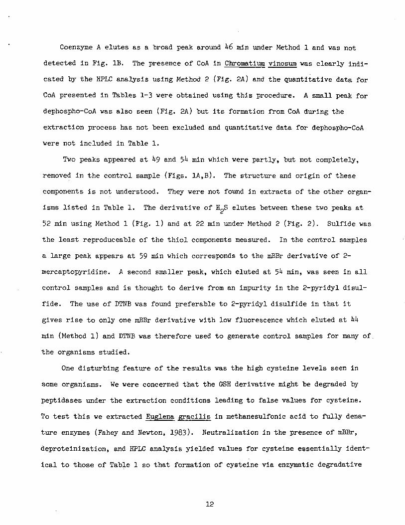
Coenzyme A elutes as a broad peak around h6 min under Method 1 and vas not
detected in Fig. IB. The presence of CoA in Chromatium vinosum vas clearly indi-
cated by the HPLC analysis using Method 2 (Fig. 2A) and the quantitative data for
CoA presented in Tables 1-3 vere obtained using this procedure. A small peak for
dephospho-CoA vas also seen (Fig. 2A) but its formation from CoA during the
extraction process has not been excluded and quantitative data for dephospho-CoA
vere not included in Table 1.
Tvo peaks appeared at k$ and 5^ min vhich vere partly, but not completely,
removed in the control sample (Figs. 1A,B). The structure and origin of these
components is not understood. They vere not found in extracts of the other organ-
isms listed in Table 1. The derivative of HJ3 elutes betveen these tvo peaks at
52 min using Method 1 (Fig. l) and at 22 min under Method 2 (Fig. 2). Sulfide vas
the least reproduceable of the thiol components measured. In the control samples
a large peak appears at 59 min vhich corresponds to the mBBr derivative of 2-
mercaptopyridine. A second smaller peak, vhich eluted at 5^ min, vas seen in all
control samples and is thought to derive from an impurity in the 2-pyridyl disul-
fide. The use of DTNB vas found preferable to 2-pyridyl disulfide in that it-
gives rise to only one mBBr derivative vith lov fluorescence vhich eluted at kk
ttin (Method l) and DTNB vas therefore used to generate control samples for many of
the organisms studied.
One disturbing feature of the results vas the high cysteine levels seen in
some organisms. We vere concerned that the GSH derivative might be degraded by
peptidases under the extraction conditions leading to false values for cysteine.
To test this ve extracted Euglena gracilis in methanesulfonic acid to fully dena-
ture enzymes (Fahey and Nevton, 1983). Neutralization in the presence of mBBr,
deproteinization, and HPLC analysis yielded values for cysteine essentially ident-
ical to those of Table 1 so that formation of cysteine via enzymatic degradative
12
processes can be excluded.
DISCUSSION
The results of the present study show that the ability to synthesize GSH
occurs among a vide range of phototrophic eubacteria. The green "bacteria
represent the only group in which the occurrence of glutathione is questionable.
The low level seen in Chlorobium thiosulfatophilum cannot be ignored but the lev-
els of cysteine, thiosulfate, CoA, sulfide, and the unknown Ull all clearly exceed
the level of GSH in Chlorobium. No significant amount of GSH could be deteted in
Chloroflexus aurantiacus. Since most green bacteria are obligate anaerobes
(Chloroflexus being an exception which can grow aerobically) the absence of signi-
ficant levels of GSH in these organisms is expected if the fundamental function of
GSH is to protect against oxygen toxicity. If GSH does actually occur at very low
levels in the green bacteria its function there must clearly be different from
that in organisms producing GSH at high levels.
Glutathione is clearly present in the purple sulfur bacterium Chromatium
vinosum which tolerates oxygen but does not grow in air (Gibson, 1967). In our
previous survey of glutathione in bacteria we were unable to detect glutathione in
Chromatium vinosum (Fahey et al, 1978). The reason for this discrepancy is not
clear. The sample used in the earlier study had been stored frozen for an
extended period so that GSH might have oxidized but the enzymatic assay used to
measure glutathione involves a cycling method in which GSSG is reduced by glu-
tathione reductase and reoxidized by tetrathionate (Fahey et al, 1975) so that
GSSG should have been detected. Oxidation to a form not reduceable by glutathione
reductase or other degradative processes may have reduced the apparent glutathione
level below that detectable by the enzymatic assay. A glutathione reductase has
been isolated from Chromatium vinosum but has a rather high K (7 mM) for GSSGm
(Chung and Hurlbert, 1975) and it seems possible that GSSG is not the preferred
13
substrate of this enzyme. Since \Jlk and CoA occur at comparable levels to GSH in
Chromatium (Table l) it would be of interest to test the disulfides of these as
alternative or additional sources of substrates for this enzyme. .It is noteworthy
that the glutathione reductase from Chromatium utilizes NADH in preference to
NADPH which characterizes it as substantially different from other glutathione
reductases (Chung and Hurlbert, 1975). Amino acid sequence data show that glu-
tathione reductase and lipoamide dehydrogenase are related (Williams, 1976) and,
based on the wider distribution of lipoamide dehydrogenase than of GSH among pro-
caryotes, it has been suggested that glutathione reductase evolved from lipoamide
dehydrogenase (Fahey, 1977). Since lipoamide dehydrogenase utilizes NADH, the
finding of an NADH-linked glutathione reductase in Chromatium suggests that
Chromatium may typify an early stage in the evolution of glutathione metabolism,
as view which is in accord with the fact that Chromatium is the least oxygen-
adapted organism tested here to produce GSH at substantial levels. Further stu-
dies of GSH metabolism in this organism are therefore likely to be informative.
Quite high levels of GSH were also found in the purple nonsulfur bacteria
(Table l) which is of special significance since this group has oxygen-dependent
metabolism and is closely related to the mitochondria of eucaryotes. More specif-
ically, nucleotide sequencing of 5S ribonosmal RNAs shows that members of the
alpha subgroup of the purple photosynthetic bacteria, which includes Rhodospiril-
lum rubrum and Rhodobacter sphaeroides, are the ones most closely related to wheat
mitochondria (Villanueva, et al, 1985). Thus, the endosymbiotic process (see Mar-
gulls, 1981) giving rise to mitochondria would presumably have incorporated the
ability to produce GSH into eucaryotes. The beta (Rhodocyclus gelatinosa) and
gamma (Chromatium vinosum and IS. coli) subgroups of the purple photosyntheic bac-
teria also have the capacity to synthesize GSH. Glutathione reductase appears to
be widely distributed among these groups, having been purified from Chromatium
Ik
vinosum (Chung and Hurlbert, 1975), Rhodospirilium rubrum (Boll, 1969), and E.
coli (Williams and Arscott, 1971), and assayed in other purple bacteria and rela-
tives (Ondarza, et al, 1983). The purple bacteria were found to have generally
high levels of CoA suggesting that CoA may serve along with GSH in a protective
fashion in the purple bacteria. This possibility is strengthened by the finding
that Rhodospirillum rubrum extracts contain comparable GSSG and CoASSG reductase
activities (Ondarza, et al, 1983).
All of the cyanobacteria studied, including representatives of all four
classes recognized by Cohen, et al (1986) based upon sulfide sensitivity and abil-
ity to carry out anoxygenic photosynthesis, produce high levels of GSH (Table 2).
In contrast to the purple bacteria, CoA levels were quite low in all of the
cyanobacteria and in this regard it is interesting that the cyanobacterium Spiri-
lum maxima was found to exhibit very high GSSG reductase activity but no detecti-
ble CoASSG reductase activity (Ondarza, et al, 1983). The cyanobacteria are
closely related to the eucaryotic chloroplast based upon RNA sequence data (Fox,
et al, 1980) so that the endosymbiotic process giving rise to chloroplasts
represents a second mechanism by which the ability to synthesize GSH could have
been incorporated into phototrophic eucaryotes. Since the purple bacteria and the
cyanobacteria represent the only two groups of bacteria which we have uniformly
found to produce GSH, the endosymbioses leading to mitochondria and chloroplasts
represent the most plausible mechanisms for incorporation of GSH synthesis into
eucaryotes. Entamoeba histolytica represents the only eucaryote clearly demon-
strated not to produce GSH. This, coupled with the fact that this organism lacks
both chloroplasts and mitochondria, supports the view that GSH metabolism evolved
in eucaryotes via the endosymbioses giving rise to mitochondria and chloroplasts
(Fahey, et al, 198M.
All of the eucaryotic phototrophs examined were found to produce GSH (Table
15
3). However, the level vas markedly higher in photoautotrophically grown cells
than in dark, heterotrophically grown cells, an effect not seen in Rhodospirillum
rubrum (Table l). The reason for this variation is not understood.
The results of the present study appear to support the view that GSH syn-
thesis evolved in phototrophic eubacteria before or around the time that oxygenic
photosynthesis evolved. It is therefore tempting to postulate that the initial
function of GSH was to protect against oxygen toxicity but there is little other
evidence to support this idea. Although glutathione reductase is widely found in
these bacteria (see above) relatively little is known about other enzymes of GSH
metabolism. GSH appears to play a protective role in IS. coli, a relative of the
purple bacteria, based upon studies of mutants blocked in GSH synthesis (Apon-
toweil and Berends, 1975; Fuchs and Warner, 1975; Murata and Kimura, 1982), but it
has been reported that GSH peroxidase is absent (Smith and Shrift, 1979) and that
GSH S-transferase is present at only a low level (Lau, et al, I960) in E_. coli.
Since these are key enzymes of oxygen detoxification by GSH in higher organisms,
their apparent absence in ]S. coli raises serious question about the role of GSH in
this organism. Glutathione S-transferase activity was also not found in R_. rubrum
(Morgenstern, et al, 1986) but little else is known concerning the detoxifying
enzymes of GSH metabolism in phototrophic eubacteria. It is possible that GSH
played some entirely different function in early eubacteria and that the oxygen
detoxification function evolved only later. More detailed information on the dis-
tribution of the enzymes which utilize GSH in the purple sulfur and cyanobacteria
is needed to clarify the role of GSH in these organisms.
The only phototrophic bacteria which tolerate oxygen and are known not to
produce GSH are the halobacteria (Newton and Javor, 1985). However, these bac-
teria, members of the archaebacteria (Fox, et al, 1980), produce /-
glutamylcysteine, half the GSH molecule, in large amounts and have a disulfide
16
reductase which maintains it in the reduced state (Newton and Javor, 1985). The
halobacteria do not use photosystems I or II but employ a unique
"bacteriorhodopsin-containing membrane to derive ATP from light. The evolution of
a system based upon /-glutamylcysteine apparently similar to that based upon GSH
in these organisms suggests that there are unique features of the X-
glutamylcysteine moiety which are important in aerobic phototrophic organisms.
Further studies of the role of X-glutamylcysteine in the halobacteria are needed
to clarify it function in these organisms.
The formation of syn-(methyl,methyl)bimane from mBBr by illuminated spinach
chloroplasts and by direct electrochemical reduction has been described by Melis,
et al (1986). They ascribe its formation with chloroplasts to a reduction involv-
ing photosystem II. The present finding that syn-(methyl,methyl)bimane is formed
from mBBr in extracts of the purple bacteria, the cyanobacteria and the eucaryotic
algae is consistent with this view. However, we have observed the formation of
syn-(methyl,methyl)bimane from mBBr in extracts of various nonphotosynthetic bac-
teria including those of Streptococcus mutans and Spirochaeta halophila (G. Newton
and R. Fahey, unpublished) so that the process is not restricted to phototrophic
organisms. The factor or factors responsible for such reduction require further
study.
Finally, we note that the unidentified thiol, Ull, appears to be widely dis-
tributed among phototrophic bacteria and algae so that its structure and function
clearly merits further investigation.
17 £
Acknowledgements. We are indebted to all of those, listed in the Experimen-
tal section, who provided cell samples and thereby made this study feasible. We
thank Barbara Javor for helpful discussions and comments. Support of this work by
the NASA Exobiology Program under Grant NAGW-3^2 in gratefully acknowledged.
18 R
REFERENCES
Apontoweil P, Berends W (1975) Isolation and initial characterization of
glutathione-deficient mutants of Escherichia coli K 12. Biochim
Biophys Acta 399:10-22
Arnon DI, Das VSR, Anderson JD (1963) Metabolism of photosynthetic bacteria
I. Effect of carbon source and hydrogen gas on biosynthetic patterns
of Chromatium. In: Studies on microalgae and photosynthetic bacteria.
Japan Soc Plant Physiol, Univ of Tokyo Press, Tokyo, pp 529-5̂ 5
Arnon DI, Ichioka PS, Vessel G, Fujiwara A, Woolley JT (1955) Molybdenum in
relation to nitrogen metabolism I. Assimilation of nitrate nitrogen by
Scenedesmus. Physiol Plant 8:538-551
Arnon DI, McSwain BD, Tsujimoto HY, Wada K (197*0 Photochemical activity and
components of membrane preparations from blue-green algae. I. Coexistence
of two photosystems in relation to chlorophyll a and removal of phycocyanin.
Biochim Biophys Acta 357:231-21+5
Buchanan BE, Schurmann P, Shanmugam KT (1972) Role of the reductive carboxylie
acid cycle in a photosynthetic bacterium lacking ribulose 1,5-diphosphate
car boxy lase. Bioch Biophys Acta 283:136-lii5
Boll M (1969) Glutathione reductase from Rhodospirillum rubrum. Arch
Mikrobiol 66:37̂ -388
Chasseaud LF (1976) Conjugation with glutathione and mercapturic acid
excretion. In: Arias IM, Jakoby WB (eds) Glutathione: Metabolism and function.
Raven Press, New York, pp IJ-llk
Castenholz RW (1969) Thermophilic blue-green algae and the thermal environment.
Bacteriol Rev 33:̂ 76-50̂
Chung YC, Hurlbert RE (1975) Purification and properties of glutathione
reductase of Chromatium vinosum. J Bacteriol 123:203-211
19
Cohen Y, Jorgensen BB, Revsbech NP, Poplawski R (1986) Adaptation to H^S
of oxygen!c and anoxygenic photosynthesis among cyanobacteria. App Environ
Microbiol 51:398-̂ 07
Cohen Y, Padan E, Shilo M (1975) Facultative anoxygenic photosynthesis in the
cyanobacterium Oscillatoria limnetica. J Bacteriol 123:855-861
Edmunds LN (1965) Studies on synchronously dividing cultures of Euglena
jyacilis Klebs (Strain Z) I. Attainment and characterization of rhythmic
cell division. J Cell and Comp Physiol 66:11*7-158
Fahey RC (1977) Biologically important thiol-disulfide reactions and the role
of cyst(e)ine in proteins: An evolutionary perspective
Fahey RC, Brody S, Mikolajczyk SD (1975) Changes in the glutathione
thiol-disulfide status of Neurospora crassa conidia during germination
and aging. J Bacteriol 121:1̂ -151
Fahey RC, Brown WC, Adams WB, Worsham MB (1978) Occurrence of Glutathione in
Bacteria. J Bacteriol 133:1126-1129
Fahey RC, Newton GL (1983) Occurrence of Low Molecular Weight Thiols in
Biological Systems. In: Larsson A, et al (eds) Functions of glutathione:
physiological, toxicological, and clinical aspects. Raven Press, New
York, pp 151-260
Fahey RC, Newton GL (1986) Determination of low molecular weight thiols using
monobromobimane fluorescent labeling and high-performance liquid chromato-
graphy. Methods Enzymol in press
Fahey RC, Newton GL, Arrick B, Overdank-Bogart T, Aley SB (1981*) Entamoeba
histolytica; A eukaryote without glutathione metabolism. Science 22**:70-72
Fox GE, Stackebrandt E, Hespell RB, Gibson J, Maniloff J, Dyer TA, Wolfe RS,
Balch WE, Tanner RS, Magrum LJ, Zablen LB, Blakemore R, Gupta R, Bonen L,
Lewis BJ, Stahl DA, Luehrsen KR, Chen KN, Woese CR (1980) The phylogeny
20
of prokaryotes. Science 209:457-463.
Fuchs JA, Warner HR (1975) Isolation of an Escherichia coli mutant
deficient in glutathione synthesis. J Bacteriol 1224:l40-l48
Gibson J (196?) Aerobic metabolism of Chromatium sp. strain D.
Arch Mikrobiol 59:104-112
Halliwell B (1981) Chloroplast Metabolism. The structure and function of
chloroplasts in green leaf cells. Clarendon Press, Oxford, Chapt. 8
Jakoby WB, Habig WH (i960) Glutathione transferases. In: Jakoby WB (ed) Enzymatic
basis of detoxification, vol II, Academic Press, New York, pp 63-94
Jocelyn PC (1972) Biochemistry of the SH group. Academic Press, London.
Kosower NS, Kosover EM (1978) The glutathione status of cells. Int Rev Cytol
54:109-160
Lascelles J (1956) The synthesis of porphyrins and bacteriochlorophyll by
cell suspensions of Rhodopseudomonas spheroides. Biochem J 62:78-93
Lau EP, Niswander L, Watson D, Fall RR (i960) Glutathione-S-transferase is
present in a variety of microorganisms. Chemosphere 9:565-569
Margulis L (1981) Symbiosis in cell evolution. W. H. Freeman, San Francisco
Meister A, Anderson ME (1983) Glutathione. Ann Rev Biochem 52:711-760
Melis A, Kosower NS, Crawford NA, Kirowa-Eisner E, Schwarz M, Kosower EM (1986)
Bimanes 26. An electron transfer reaction between photosystem II and
monobromobimane induces static chlorophyll «i quenching in spinach
chloroplasts. Plant Physiol in press
Murata K, Kimura A (1982) Some properties of glutathione biosynthesis-deficient
mutants of Escherichia coli B. J Gen Microbiol 128:1047-1052
Newton GL, Javor B (1985) X-Glutamylcysteine and thiosulfate are the major
low-molecular weight thiols in halobacteria. J Bacteriol l6l:438-441
Ondarza RN, Rendon JL, Ondarza M (1983) Glutathione Reductase in Evolution.
21
J Mol Evol 19:371-375
Ormerod JG, Ormerod KS, Gest H (1961) Light-dependent utilization of organic
compounds and photoproduction of molecular hydrogen by photo synthetic
bacteria; relationships with nitrogen metabolism. Arch Biochem Biophys
Rippka R, Denulles Y, Waterburry JB, Herdman M, Stanier RY (1979) Generic
assignment, strain histories and properties of pure cultures of cyanobacteria.
J Gen Microbiol lll:l-6l
Smith J, Shrift A (1979) Phylogenetic distribution of glutathione peroxidase.
Comp Biochem Physiol 63B:39~M
Sorokin C, Krauss RW (1958) The effects of light intensity on the grovth rates
of green algae. Plant Physiol 33:109-113
Stratton GW, Huber AL, Corke CT (1979) Effect of mercuric ion on the growth,
photosynthesis, and nitrogenase activity of Anabaena inaequalis.
Applied Environ Microbiol 38:537-5̂ 3
Sueoka N, Chiang KS, Kates JR (1967) Deoxyribonucleic acid replication in
meiosis of Chlamydomonas reinhardi. J Mol Biol 25:̂ 7-66
Villanueva E, Luehrsen KR, Gibson J, Delihas N, Fox GE (1985) Phylogenetic
origins of the plant mitochondrion based on a comparative analysis of 5S
ribosomal RNA sequences. J Mol Evol 22:̂ 6-52
Wendel A (i960) Glutathione peroxidase. In: Jakoby VB (ed) Enzymatic basis of
detoxification, vol I. Academic Press, New York, pp 333-353
Williams CH, Arscott LD (1971) 'Glutathione reductase (Escherichia coli).
Meth Enzymol 17B: 503-509
Williams CH (1976) Flavin-containing dehydrogenases. In: Boyer PD (ed) The
enzymes, vol XIII. Academic Press, New York, pp 89-173.
22
CAPTIONS TO FIGURES
Figure 1. HPLC chromatograms obtained using Method 1: (A) Control
sample for Chromatium vinosum. (B) Chromatium vinosum extracted
in the presence of mBBr; (C) Standard mixture. Abbreviations: C - peaks present
in control samples; Pys - peaks derived from 2-pyridyl disulfide reagent;
Uli* - unidentified thiol derivative eluting at Ik min under protocol A; syn-
(CH3,CH3)B - _svn-(methyl,methyl)bimane; /-GLUCYS - /-glutamylcysteine;
R - peaks derived from buffer and reagents;
other abbreviations defined in text.
Figure 2. HPLC chromatograms obtained using Method 2:
(A) Chromatium vinosum extracted in the presence of mBBr; (B) Standard
mixture. Abbreviations: dpCoA - dephosphocoenzyme A; others as in caption
to Fig. 1.
23
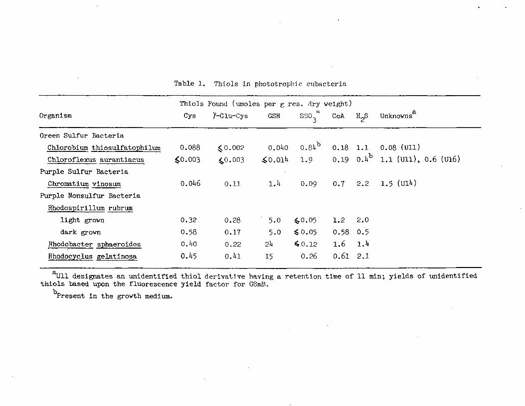
Table 1. Thiols in phototropbic eubacteria
Organism
Thiols Found (umoles per g res. dry weight)
Cys y-Glu-Cys GSH SSO = CoA H2S Unknowns6
Green Sulfur Bacteria
Chlorobium thiosulfatophilum
Chloroflexus aurantiacus
Purple Sulfur Bacteria
Chromatium vinosum
0.088 40.002
40.003 40.003
0.01+6 0.11
O.OUO 0.8U
4̂ 0. OlU 1.9
0.09
b 0.18 l.l 0.08 (uil)0.19 O.Ub l.l (Ull), 0.6 (Ul6)
O.T 2.2 1.5 (UlU)
Purple Nonsulfur Bacteria
Rhodospirillum rubrum
light grown
dark grown
Rhodobacter sphaeroides
Rhodocyclus gelatinosa
0.
0.
0.
0.
32
58
UO
1*5
0
0
0
0
.28
.17
.22
.Ul
5.0
5.0
2k
15
40.
$0.
4o.
0.
05
05
12
26
1.
0.
1.
0.
2
58
661
2
0
1
2
.0
.5
.1*
.1
designates an unidentified thiol derivative having a retention time of 11 min; yields of unidentifiedthiols "based upon the fluorescence yield factor for GSmB.
Present in the growth medium.

Table 2. Thiols in cyanobacteria
Organism
Anacystis nidulans
Nostoc rauscorum
Plectonema boryanura
Synechococcus lividus
Microcoleus chthonoplastes
Oscillatoria amphigranulata
Oscillatoria limnetica
Oscillatoria sp.(Stinky Spring, Utah)
Oscillatoria terebriformis
Thiols
Cys
<0
2
<o
0
<0
0
<0
1°
0
.06
.0
.01
.61*
.01
.17
.01
.02
.17
Found (umoles
y-Glu-Cys
<O.OU
1.6
<0.01
<0.0ll*
0.033
<0.01
0.071
0.031
<0.11
per g
OSH
>6
0.
0.
1.
1.
0.
1*.
3.
6.
79
7
5
0
1*0
9
5
5
res.
0
<0
<0
0
<0
0
<0
<0
<0
dry
.063
.01
.06
.09
.03
.16
.05
.05
.03
weight)
CoA
<0
1°
<0
<0
<0
<0
0
0
<0
.03
.03
.02
.001
.01
.021*
.03
.09
o
HpR Unknowns
3.U
1.7
1.7
1*.6
0.5
2.0
0.06
0.1*
1.5 1.8 (Ull)
designates an unidentified thiol derivatives having a retention time of 11 min.

Table 3. Thiols in algae and chloroplasts
Thiols Found (umoles per g res. dry weight)
Organism Cys 2̂ -Glu-Cys GSH S80 ~ CoA H S
Chlorella pyrenoidsa
light grown 0.15 0.09
dark grown O.U2 <0.05
Chlorella vulgar is
light grown 0.38 0.15
dark grown 0.98 0.10
Euglena gracillis
light grown 6.3 0.3
dark grown 12 10.001
Scenedesmus obliquus <0.003 <0.003
Chlaraydoraonas rheinhardtii 0.28 <0.01
1,2 10.05
1.3 10.05
10 10.02
1.9 0.11
1.8 10.05
0.8 10.05
O.U8 <0.003
0.35 <0.01
0.06
0.22
0.16
0.35
o.ia
0.02
<0.002
0.05T
O.lfc
O.T
1.1*
1.5
0.9
O.U
.£0.01
0.6
a
Unknowns
O.OH (Ull)
O.OU (Ull)
llll designates an unidentified thiol derivatives having a retention time of 11 min.

60MIN

CT>CM