rod outer segment development influences aav …€¦ · · 2017-06-12rod outer segment...
TRANSCRIPT

RESEARCH ARTICLES
Rod Outer Segment Development Influences AAV-MediatedPhotoreceptor Transduction After Subretinal Injection
Lolita Petit,1 Shan Ma,1 Shun-Yun Cheng,1 Guangping Gao,3 and Claudio Punzo1,2,*1Department of Ophthalmology and Gene Therapy Center, 2Department of Neurobiology, and 3Department of Microbiology and Physiological Systems
and Gene Therapy Center, University of Massachusetts Medical School, Worcester, Massachusetts.
Vectors based on the adeno-associated virus (AAV) are currently the preferred tools for delivering genesto photoreceptors (PR) in small and large animals. AAVs have been applied successfully in variousmodels of PR dystrophies. However, unknown barriers still limit AAV’s efficient application in severalforms of severe PR degenerations due to insufficient transgene expression and/or treated cells at thetime of injection. Optimizations of PR gene therapy strategies will likely benefit from the identificationof the cellular factors that influence PR transduction. Interestingly, recent studies have shown that theAAV transduction profile of PRs differs significantly between neonatal and adult mouse retinas aftersubretinal injection. This phenomenon may provide clues to identify host factors that influence theefficiency of AAV-mediated PR transduction. This study demonstrates that rod outer segments arecritical modulators of efficient AAV-mediated rod transduction. During retinal development, rodtransduction correlated temporally and spatially with the differentiation order of PRs when vectorswere introduced subretinally but not when introduced intravitreally. All subretinally injected vectorshad an initial preference to transduce cones in the absence of formed rod outer segments and thendisplayed a preference for rods as the cells matured, independently of the expression cassette or AAVserotype. Consistent with this observation, altered development of rod outer segments was associatedwith a strong reduction of rod transduction and an increase in the percentage of transduced cones by 2-to 2.8-fold. A similar increase of cone transduction was observed in the adult retinal degeneration 1 (rd1)retina compared to wild-type mice. These results suggest that the loss of rod outer segments in diseasedretinas could markedly affect gene transfer efficiency of AAV vectors by limiting the ability of AAVs toinfect dying rods efficiently. This information could be exploited for the development of more efficientAAV-based PR gene delivery procedures.
Keywords: AAV, retina, photoreceptors, cones, rods, gene therapy
INTRODUCTIONRECOMBINANT AAV SEROTYPE 2 (AAV2) is currentlybeing evaluated in Phase 1/2 and 3 gene therapyclinical trials for treating diverse inherited retinaldiseases involving the retinal pigment epithelium(RPE), such as Leber congenital Amaurosis 2,1–9
retinitispigmentosa,10 andchoroideremia.11,12 Thesestudies demonstrate that subretinal delivery of AAVis safe and can provide rescue of vision-guided be-
havior in patients with advanced disease at least forseveral years.13 However, broader application of ret-inal gene therapy will require efficient transduction ofother retinal cells, in particular photoreceptors (PR).
The ability of various AAV serotypes to trans-duce PRs has been evaluated in small14–18 andlarge15,19–26 animals. Among these, pseudotypes-2/5, -2/7, -2/8, -2/9, and -2/rh10 were found to havethe highest transduction efficiencies for PRs after
*Correspondence: Dr. Claudio Punzo, University of Massachusetts Medical School, 368 Plantation Street, Albert Sherman Center, AS6-2041, Worcester, MA 01605-2324.E-mail: [email protected]
ª Lolita Petit et al. 2017; Published by Mary Ann Liebert, Inc. This is an Open Access article distributed under the terms of the CreativeCommons Attribution License, which permits unrestricted use, distribution, and reproduction in any medium, provided the original work isproperly cited.
464 j HUMAN GENE THERAPY, VOLUME 28 NUMBER 6 DOI: 10.1089/hum.2017.020Mary Ann Liebert, Inc.

subretinal delivery.27,28 These vectors have beenused to drive persistent transgene expression suc-cessfully, resulting in improvements of the retinalstructure and/or function in a number of animalmodels of PR dystrophies, in particular in models ofslow PR death.13 Yet, in models of severe PR de-generation, many PR therapies have not been veryeffective at rescuing PR over the long term. This islikely due to the difficulties in treating sufficientcells and/or expressing therapeutic levels of thetransgene in a timely manner. In support of thisidea, AAV8-based vectors have provided higherbenefits than AAV5 in different disease models ofprimary PR dystrophies, such as in the retinal de-generation 10, the Rpgrip1-/- (retinitis pigmentosaGTPase regulator interacting protein 1), theGuy2e-/- (retinal guanylate cyclase 1), the Aipl1-/-
(aryl hydrocarbon interacting protein like 1), andthe Rho-/- (rhodopsin) mice, consistent with thesuperior ability of AAV8 to transduce PRs moreefficiently and thus delivering higher transgenelevels when compared to AAV5 (for review, seePetit et al.13). Moreover, a recent study has shownthat a therapeutic transgene can halt degenera-tion, regardless of the severity of PR loss if acti-vated optimally in all PR cells.29 These resultspoint to the importance of improving PR trans-duction efficiency to maximize clinical impact, andindicate that further development of retinal genetherapy will likely benefit from a better under-standing of the potential host cell factors that re-strict PR transduction in vivo. However, basicaspects of AAV–PR interactions are still unknown,and mechanisms affecting PR transduction aftersubretinal injection remain largely unexplored.20
Interestingly, recent reports have shown thatafter subretinal injection, the AAV transductionprofile of PR cells changes dramatically betweenneonatal and adult mice.30–34 In particular, injec-tion of AAV2/8 at postnatal day (PND) 0 resultsprimarily in cone transduction (*100% of cones,almost no rods),32–34 whereas the vast majority oftransduced cells in adults are rods.14,15,32 Thecurrent study took advantage of this shift in tro-pism to identify host factors critical for AAV-mediated PR transduction. Using 13 different AAVserotypes, cone and rod transduction were specifi-cally assessed after AAV subretinal delivery in thedeveloping retina and in two murine models of rod-cone dystrophies that display either an arrest ofPR outer segment development or a rapid loss ofrod cells. Irrespective of the serotype tested, it wasfound that all vectors preferentially transducedcones when delivered at PND1, whereas delivery atPND21 resulted in a decrease in the percentage of
transduced cones and a dramatic increase in thepercentage of transduced rods. Additional injec-tions at PND5 and PND10 showed that rod trans-duction temporally and spatially correlates withthe development of their inner and outer segments.Furthermore, in mutant mouse models that lackrod outer segments or rod PR cells, a preferentialtransduction of cones was observed. Together,these findings indicate that the access of AAVvectors to PR cells through their segments is a keyfactor for PR transduction upon subretinal deliv-ery. Additionally, the data suggest that maturerods and cones may compete for AAV access. Theseobservations begin to define a new paradigm forAAV–PR interactions upon subretinal delivery,which should impact the development of more ef-ficient AAV-based PR gene delivery procedures byproviding an understanding of host factors re-quired for successful AAV transduction.
MATERIAL AND METHODSPlasmid construction, AAV vector production,and purification
Recombinant AAV2/5-CMV-GFPd, AAV2/5-CMV-H2bGFP, AAV2/5-CMV-Cre, AAV2/5-pQCMV-H2bGFP, and AAV2/5-mCAR-H2bGFP were producedusingthepAAV2-CMV-GFPd,pAAV2-CMV-H2bGFP,pAAV2-CMV-Cre, pAAV2-pQCMV-H2bGFP, orpAAV2-mCAR-H2bGFP plasmids, respectively. ThepAAV2-CMV-GFPd plasmid expresses the destabi-lized GFP cDNA under the control of a human CMVenhancer/promoter, a human b-globin intron and anSV40 polyA signal, flanked by two AAV2 invertedterminal repeat sequences. The pAAV2-CMV-H2bGFP plasmid expresses a histone 2B fused GFP(nuclear GFP). It was constructed by replacing theGFPd cDNA with the H2bGFP sequence derivedfrom the parental pQCMV-H2bGFP plasmid. ThepAAV2-CMV-Cre plasmid expresses a Cre re-combinase. It was constructed by replacing theGFPd cDNA with the Cre sequence derived from theparental pQCMV-H2bGFP-I-Cre plasmid. ThepAAV2-mCAR-H2bGFP plasmid was made by repla-cing the CMV promoter with the mouse cone arrestin(mCAR) promoter from the parental pQCMV-H2bGFP plasmid.35
scAAV1, -2, -3b, -4, -5, -6, -7, -8, -9, rh8, rh10, rh39,and rh43 expressing enhanced GFP (eGFP) underthe control of the CB6 promoter were produced usingthe pAAVsc-CB6-eGFP plasmid, which bears a CB6promoter, a rabbit globin polyA, and engineered ITRsfor scAAV vector. The CB6 sequence was previouslydescribed36 and includes a CMV enhancer/beta-actin(CB) promoter with a CMV IE enhancer.
IMPACT OF RETINAL DEVELOPMENT ON AAV TRANSDUCTION 465

AAV2/5-CMV-GFPd, AAV2/5-CMV-H2bGFP,AAV2/5-CMV-Cre, AAV2/5-pQCMV-H2bGFP, andAAV2/5-mCAR-H2bGFP were produced in thePunzo laboratory by triple transfection of 293 cellsaccording to previously reported methods37 andpurified by two rounds of CsCl2 ultracentrifuga-tion. scAAV vector production was carried out bythe Vector Core of the Horae Gene Therapy Centerof UMASS Medical School (Worcester, MA).38 Viralvector titers were determined simultaneously forall batches by quantitative real-time PCR (qPCR)with primers directed toward SV40pA (forward: 5¢-CGAGTGCTTTATTTGTGAAATTTG-3¢; reverse:5¢-GGGGTTCCTTGTAGTTAATGA-3¢) or eGFP(forward: 5¢-AGCAAAGACCCCAACGAGAA-3¢; re-verse: 5¢-GGCGGCGGTCACGAA-3¢) and expressedas vector genome per milliliter (vg/mL). The finalvector titers were between 8.1 · 1012 and 5.1 · 1013
vg/mL (Table 1).
AnimalsThe CD1, Ai9 Cre reporter mice, and Pde6brd1/rd1
mice were purchased from the Jackson Laboratory.The M-opsin-Cre mice39 (cone-specific Cre line) andthe rhodopsin knockout (Rho-/-) mice40 wereprovided by Yun Z. Le (University of OklahomaHealth Sciences Center) and Janis Lem (TuftsUniversity, Boston), respectively. Ai9+/-_MCre+
mice were generated by crossing MCre+ micewith the Ai9 Cre reporter mice. Pde6brd1/+_Ai9+/-
mice were generated by crossing rd1 mice andAi9 mice. Heterozygotes were mated to producePde6brd1/rd1/+_Ai9+/- mice.
All animals were maintained at UMASS MedicalSchool under a 12 hour/12 hour light/dark cycle withunrestricted access to food and water. Lighting con-ditions were kept constant in all cages, with illumi-nationrangingbetween10and15lux.Allexperimentsinvolving mice were conducted in compliance with theAssociationforResearch inVisionandOphthalmologystatement for the use of animals in ophthalmic andvision research. All procedures were approved by In-stitutional Animal Care and Use Committee ofUMASS Medical School.
Subretinal administration of rAAV vectorsThe same experimenter (L.P.) performed all
subretinal injections of AAV vectors. Subretinalinjection of AAV vectors was performed usinga trans-scleral approach through the posteriorpart of the sclera, as previously described,41 withthe following modifications. Injections were per-formed using thin-wall beveled glass micropipettes(Clunbury Scientific LLC) without sclerotomy. Inmice older than PND1, a small hole was made at
the transition of the cornea and sclera with a 33-gauge needle before the injection in order to releaseintraocular pressure and allow for the formation ofa vector bleb upon subretinal injection. This pro-cedure allowed the vector bleb to occupy 40–60% ofthe retinal surface without injection-related dam-age. Fast green dye was added to the AAV prepa-rations at a final concentration of 0.1% as a tracerto visualize the location of injection and thus en-sure that AAV vectors were injected into the sub-retinal space. Mice received 0.5–0.75 lL of vectorsat PND1, 1–1.5 lL of vectors at PND5–10, and 1.5–2.5 lL of vectors at PND21.
Intravitreal administration of rAAV vectorsIntravitreal injections were performed in PND1
mice, as previously described,41 using the sameglass micropipettes (Clunbury Scientific LLC) in-troduced through the cornea-scleral margin. Fastgreen dye was added to AAV preparations at a finalconcentration of 0.1% as a tracer to visualize thelocation of injection and thus ensure that AAVvectors were injected into the intravitreal cavity.Mice received 0.75–1 lL of vectors.
Quantification of cone and rod transductionIn all cases, the analysis was performed 3 weeks
post AAV injection. The total number of retinasanalyzed in each experimental group is indicatedin Table 1. The efficiency of retinal transductionwas first assessed under an inverted fluorescentmicroscope, and only retinas that displayed nativeGFP fluorescence in >30% of the total retinal sur-face were processed for further analysis. All imageswere acquired with the Leica DM5500 fluorescentmicroscope equipped with a motorized stage fortiling and z-stack image acquisition and deconvo-lution software for confocal-like image quality.
Quantification of cone and rod transduction inselected retinas was performed using three inde-pendent methods. First, for each group of injectedmice, a minimum of two retinas were entirely sec-tioned (20 lm/section) and collected serially on fiveslides, such that each slide contained a represen-tation of the entire eye. A minimum of two slideswere processed for immunohistochemistry usingCy3-labeled peanut agglutinin lectin (PNA; 1:500;Vector Laboratories) and rabbit anti-cone arrestin(1:500; EMD Millipore). Images were taken from 5–10 sections at 40· over the entire transduced areaat different focal plans (z stacks) using epi-fluorescence microscopy (Supplementary Fig. S1;Supplementary Data are available online atwww.liebertpub.com/hum). The acquired epifluo-rescent images were then deconvolved in order to
466 PETIT ET AL.
obtain confocal-like resolution by eliminating theout-of-focus signal that could be misinterpreted ascolocalization (Supplementary Fig. S1a). Conequantification was performed in one selected zstack for each image, based on the colocalizationof the GFP signal with the cone arrestin marker(usually at the level of cell nuclei). Rod quantifi-cation was performed within the same z stack foreach image by manually counting the number oftotal GFP+ PRs and the number of GFP+ cones perimage, and by determining through an automatedcounting algorithm the total number of PR nucleiin the outer nuclear layer (ONL) of the same imageusing the Imaris software package (Supplemen-tary Fig. S1b). Rod transduction was then calcu-lated as an average per eye and per group bydetermining the ratio of the number of total GFP+
PRs – number of GFP+ cones to the total number ofPR nuclei – number of cone cells per image.
Second, for groups of mice injected with AAV-dGFP or AAV-eGFP vectors, cone quantificationwas also performed on retinal flat mounts stainedwith the following lectins/antibodies: Cy3-labeledPNA (1:500; Vector Laboratories), rabbit anti-conearrestin (1:500; EMD Millipore; AB15282), rabbitanti-LM opsin (1:500; EMD Millipore; AB5405),and goat anti-S opsin (1:500; Santa Cruz Bio-technology; SC-14363). For each flat mount, four tosix representative 100 lm2 areas within the trans-duced area (center and periphery) were analyzed.The percentage of transduced cones was determinedas an average per eye and per group by quantifyingthe number of GFP+ cones over the total number ofcones in the same areas. This quantification wasperformed at the level of cone segments, where GFPfluorescence in cones was unambiguously sur-rounded by PNA staining (Supplementary Fig. S1c).
Third, AAV5-CMV-H2bGFP and AAV5-pQCMV-H2bGFP vectors were also evaluated in Ai9+_MCremice, which express CRE-mediated td-Tomato(red) in the entire cone cell bodies (SupplementaryFig. S1d).
Cell linesTwo different Chinese hamster ovary (CHO) cell
lines were used to confirm the specificity of thelectins used in this study. These include the pa-rental cell line Pro5 and sialic-acid deficient cellline Lec2. Both cell lines were kindly provided byMiguel Sena-Esteves (UMASS Medical School).Cells were cultured in Dulbecco’s modified Eagle’smedium (DMEM) supplemented with 10% fetalbovine serum and 1% streptomycin and main-tained at 37�C with 5% CO2. Cells were seeded at105 cells/well in 12-well plates prior to treatment
for 1 h at 37�C with DMEM alone or DMEM sup-plemented with 50 mIU/mL of neuraminidase typeIII from Vibrio cholerae (Sigma–Aldrich; N7785).Cells were then washed three times with DMEM,once with PBS-1· and fixed with 4% PFA for10 min. Cells were subjected to staining for 1 h inPBS-1 · at room temperature (RT) using the fol-lowing lectins: Cy2- or Cy3-labeled PNA (1:500),Cy2-conjugated Maackia amurensis lectin (MAL1;1:500), biotinylated Erythrina cristagalli lectin(ELC; 1:500), and Cy5-conjugated wheat germ ag-glutinin (WGA; 1:500) all from Vector Labora-tories. After three washes with ice-cold PBS-1· toremove unbound lectins, ECL was visualized usingStreptavidin-Cy5 (Molecular Probes). Cells wereimaged using an inverted fluorescence microscope.
Lectin analysisof the inter-photoreceptor matrix
Analysis of the inter-photoreceptor matrix (IPM)was performed on retinal sections, flat mounts, andexplants from Ai9_MCre+ mice euthanized atPND1, PND5, PND10, PND14, and PND21, and onretinal flat mounts from Rho-/- mice euthanized atPND21. Three eyes were used for each experi-mental group. Retinal sections and retinal flatmounts were processed and stained, as previouslydescribed, for the cell culture experiments, withthe following panel of lectins in PBS1X: PNA(1:500; Vector Laboratories), MAL1 (1:500; VectorLaboratories), ELC (1:500; Vector Laboratories),and WGA (1:500; Vector Laboratories). For retinalexplants treated with neuraminidase, retinas weredissected in cold DMEM and incubated in eitherDMEM alone or DMEM with 50 mIU/mL of neur-aminidase type III from V. cholerae (Sigma–Aldrich; N7785) for 1 h at 37�C. Retinal explantswere then washed three times with cold DMEM,once with cold PBS-1 · , and fixed in 4% PFA for30 min. Retinal explants were subjected to lectinstaining for 1 h in PBS-1· at RT. After three washeswith ice-cold PBS-1· to remove unbound lectins,retinal explants were flat mounted and analyzed byfluorescence microscopy (Leica DM5500).
AAV-binding assay and qPCRAAV-binding assay was performed on three bi-
ological samples and repeated two to four timeswith retinal explants from PND1 and PND21 CD1mice. Retinas were dissected in cold DMEM andincubated in either DMEM with or without neur-aminidase for 1 h at 37�C. Retinal explants werethen washed three times with cold DMEM, andpre-chilled at 4�C for 1 h in 500 lL of DMEM. AAVvectors were added at 1 · 1010 vg/well, and explants
IMPACT OF RETINAL DEVELOPMENT ON AAV TRANSDUCTION 467

were incubated at 4�C for 1 h. Retinal explantswere then washed three times with cold DMEMand once with cold PBS-1· to remove unbound AAVparticles. Total DNA was extracted using theDNeasy Blood and Tissue Kit (Qiagen). AAV ge-nome copies were quantified by real-time PCR withthe aforementioned primers for the SV40pA oreGFP sequence. AAV vector genomes (vg) werenormalized to the number of retinal cells, usingprimers complementary to the mouse beta actinDNA (forward: 5¢-ACTGGGACGACATGGAGAAG-3¢; reverse: 5¢-GGGGTGTTGAAGGTCTCAAA-3¢).Samples were run in triplicate. The results wereexpressed as mean AAV vg per genome DNA.
Localization of AAV bindingto retinal photoreceptors
Localization of AAV particles was assessed af-ter subretinal injection of PND1 and PND21Ai9_MCre+ mice with scAAV8 or scAAV9 vectors.Eyes injected with PBS-1· were used as negativecontrols. One hour post injection, animals were eu-thanized and retinas processed for flat mount, aspreviously described.41 Retinas were incubated over-night at 4�C with polyclonal rabbit antibodies raisedagainst intact capsids of AAV8 or AAV9 at 1:500 inblocking solution (kindly provided by G.G.). Sectionswere then incubated with a Cy2-conjugated second-ary anti-rabbit antibody (Jackson ImmunoResearch;1:500) for 2 h at RT. Results were analyzed by fluo-rescence microscopy (Leica DM5500).
Statistical analysisAll data are expressed as mean – standard devi-
ation (SD). All statistical comparisons use Stu-dent’s t-test, with p < 0.05 considered as statisticallysignificant in all comparisons.
RESULTSSubretinal injection of AAV5 at PND1predominantly targets cone photoreceptors
Recent reports showed that subretinal injectionof AAV8 into PND0 mice results primarily in conetransduction, whereas the vast majority of trans-duced cells after subretinal injection in adult miceare rods.32–34 To demonstrate further that PRpostnatal development impacts the pattern of PRtransgene expression, neonatal mice (PND1) weresubretinally injected with 6 · 108–1.2 · 109 vg ofsingle-stranded (ss) AAV2/5-CMV-dGFP (Table 1and Supplementary Fig. S2a). AAV5 was chosenbecause this serotype (i) has been shown to signif-icantly target PR cells in the adult mouse,14,15,18
cat,26 dog,23,42,43 pig,15,22 and nonhuman prima-
te21; (ii) has been used successfully to restore reti-nal function and preserve retinal structure invarious models of PR diseases13; and (iii) is cur-rently planned in upcoming Phase I–II clinicaltrials for the treatment of PR diseases.13 Mice withsuccessful subretinal injections were euthanized 3weeks post injection (wpi), at PND21, after PRdifferentiation and maturation is completed (Fig. 1and Supplementary Fig. S2). As previously de-scribed,30,33,34 subretinal injections at PND1 yiel-ded GFP expression throughout the entire retinalsurface (Fig. 1a and Supplementary Fig. S2a), pre-sumably because the interactions between the RPEand the PR outer segments that could limit thespread of the vector have not yet formed at PND1.Robust transduction of RPE was observed across theretina (Supplementary Fig. S2b). High levels of GFPwere also detected in the ONL, which contains rodand cone PR nuclei (Fig. 1c and SupplementaryFig. S2b), and in the corresponding PR segments(Fig. 1a and c and Supplementary Fig. S2b). Im-munostaining with cone-specific markers (PNA andcone arrestin) revealed that the majority of the GFP+
cells were also positive for the cone-specific markers(Fig. 1a and c and Supplementary Fig. S2b). Hence,as predicted, the transduction of PRs was essentiallyrelegated to cones when considering the percentageof GFP+ cells (cones: 69.1 – 7.4%; rods: 2.7 – 3.1%). L/M and S cones were equally transduced (Supple-mentary Fig. S3).
To rule out an influence of factors such as inhi-bition of CMV promoter activity or dGFP steadystate level on the final transgene expression pat-tern, PR transduction was analyzed in mice in-jected at PND1 with five additional AAV5 vectors.These vectors carry different transgenes and/orubiquitous or PR-specific promoters: AAV5-CMV-H2bGFP, AAV5-CMV-Cre, AAV5-mCAR-H2bGFP,AAV5-pQCMV-H2bGFP, as well as a self-complementary (sc) AAV5-CB6-eGFP (Table 1).Though some construction-dependency was noted,tropism was similar for all vectors examined(Fig. 1b and c and Supplementary Fig. S2b). Hence,the findings suggest that the absence of efficient rodtransduction following subretinal administrationat PND1 is not dependent on factors that altervector genome expression, as well as second-strandsynthesis in the transduced cells, but rather ondifferences in entry of AAV into cones and rods atthe time of delivery. Consistent with that, AAV8vectors carrying different promoters, such as AAV-hRK1-GFP, AAV-mCAR-GFP, or AAV-CMV-GFP,have also been seen to drive transgene expressionpredominantly in cones after subretinal injection inPND0 neonate mice.33,34 Moreover, efficient trans-
468 PETIT ET AL.

fection of rods has been previously obtained afterPND0 electroporation of pCMV-H2bGFP,41 a plas-mid DNA that carries the same expression cassetteas the AAV5-CMV-H2bGFP. Finally, a highernumber of GFP+ rods was observed after adminis-tration of higher doses of AAV5-CMV-H2bGFP(9 · 109 vg; Fig. 1d and Table 1) and AAV5-mCAR-H2bGFP (>1 · 1010 vg)41 at PND1, indicating thatefficient transduction of immature rods is possibleat this age but dependent on the quantity of thevector injected (Fig. 1d and Table 1).
Rod development coincides with increasedrod transduction after subretinal delivery
In mice, cones start to undergo their terminalmitosis to begin their subsequent differentiationbefore rods. Most rods in mouse are born overan extended developmental time period (E12 toPND5) with the peak of rod production aroundbirth and their differentiation into mature rodscontinuing well into the postnatal period.44 To ex-amine further how the PR transduction patterncorrelates with rod PR development, GFP expres-
Table 1. Quantification of cone and rod transduction in mouse retinas 3 weeks after subretinal injectionsof different AAV vectors at different ages
Group Vector Titer (vg/mL) Strain Age Retinas analyzed % Cones (M – SD) % Rods (M – SD)
1 AAV5-CMV-dGFP 1.2 · 1013 CD1 PND1 13 69.1 – 7.4 2.7 – 3.12 PND5 7 56.2 – 18.3 6.0 – 1.23 PND10 6 48.3 – 6.6 18.2 – 8.34 PND21 3 39.9 – 2.2 32.2 – 17.45 rd1 PND21 3 90.5 – 4.4 NA6 Rho-/- PND1 4 59.4 – 2.9 0.09 – 0.0087 PND21 2 87.2 – 7.2 0.9 – 0.2
8 AAV5-CMV-H2bGFP 1.3 · 1013 CD1 PND1 6 65.8 – 9.7 3.2 – 2.89 PND5 3 29.2 – 1.3 2.8 – 0.610 PND10 3 40.5 – 6.6 7.4 – 2.211 PND21 3 41.4 – 13.6 21.3 – 10.9
Ai9/+ PND1 3 69.5 – 7.6 7.3 – 1.212 rd1 PND21 3 84.9 – 2.9 NA13 Rho-/- PND1 2 60.3 – 5.8 1.5 – 0.614 PND21 4 81.2 – 1.6 4.8 – 1.7
15 AAV5-CMV-H2bGFP 5.1 · 1013 CD1 PND1 2 70.3 – 5.2 9.8 – 4.4
16 AAV5-pQCMV-H2bGFP 1.3 · 1013 CD1 PND1 6 67.2 – 4.7 17.9 – 26.6Ai9/+ PND1 2 66.9 – 4.7 4.4 – 2.3
17 Rho-/- PND21 4 71.1 – 6.6 1.2 – 0.3
18 AAV5-mCAR-H2bGFP 1.3 · 1013 CD1 PND1 3 51.1 – 4.4 1.0 – 0.4
19 AAV5-CMV-Cre 1.3 · 1013 Ai9/+ PND1 5 60.6 – 9.2 4.8 – 1.6
20 scAAV1-CB6-eGFP 1.6 · 1013 CD1 PND1 5 50.7 – 10.7 8.9 – 6.7
21 scAAV2-CB6-eGFP 9.8 · 1012 CD1 PND1 3 47.4 – 8.3 3.1 – 1.4
22 scAAV3b-CB6-eGFP CD1 PND1 6 12.3 – 3.6 4.3 – 2.3
23 scAAV4-CB6-eGFP 1.2 · 1013 CD1 PND1 3 0 0
24 scAAV5-CB6-eGFP 1.4 · 1013 CD1 PND1 6 62.5 – 7.8 7.2 – 5.025 PND5 3 31.5 – 9.3 14.8 – 3.626 PND10 4 19.6 – 3.3 19.6 – 3.327 PND21 3 23.7 – 5.7 38.4 – 11.528 rd1 PND21 2 87.2 – 6.2 NA
29 scAAV6-CB6-eGFP 9.8 · 1012 CD1 PND1 3 50.6 – 24.9 6.9 – 3.3
30 scAAV7-CB6-eGFP 1.4 · 1013 CD1 PND1 6 55.1 – 15.3 7.2 – 2.531 PND21 3 25.5 – 5.2 34.9 – 9.932 scAAV8-CB6-eGFP 9.1 · 1012 CD1 PND1 4 57.4 – 11.7 5.1 – 2.033 PND21 8 23.3 – 9.7 46.3 – 8.634 scAAV9-CB6-eGFP 1.0 · 1013 CD1 PND1 5 58.1 – 13.6 6.9 – 1.235 PND21 9 23.5 – 6.7 50.3 – 5.436 rd1 PND21 3 91.6 – 2.8 NA
37 scAAVrh8-CB6-eGFP 8.1 · 1012 CD1 PND1 3 57.3 – 15.2 6.1 – 3.3
38 scAAVrh10-CB6-eGFP 9.7 · 1012 CD1 PND1 7 69.8 – 12.8 6.5 – 2.339 PND21 5 25.3 – 12.4 54.8 – 4.640 scAAVrh39-CB6-eGFP 1.0 · 1013 CD1 PND1 5 61.2 – 12.9 6.4 – 4.7
41 scAAVrh43-CB6-eGFP 8.1 · 1012 CD1 PND1 6 39.8 – 9.9 4.4 – 1.9
AAV, adeno-associated virus; PND, postnatal day; NA, not applicable.
IMPACT OF RETINAL DEVELOPMENT ON AAV TRANSDUCTION 469

Figure 1. Photoreceptor transduction in the neonatal mouse retina by adeno-associated virus serotype 5 (AAV5) vectors. Mice injected on postnatal day 1(PND1) were subretinally injected with similar doses of AAV2/5-CMV-dGFP, AAV2/5-CMV-H2bGFP, AAV2/5-CMV-CRE, AAV2/5-pQCMV-H2bGFP, AAV2/5-mCAR-H2bGFP, or scAAV2/5-CB6-eGFP vectors. (a) Representative retinal flat-mounts and (c, d) cryosections 3 weeks post injection (wpi) labeled with an antibodyraised against anti-cone arrestin (CA) or with peanut agglutinin lectin (PNA; red) and counter stained with DAPI (blue: removed from 60% of panels to visualizered and green staining better) (c,d). AAV-mediated GFP was detected by its native fluorescence (green). AAV-mediated CRE expression was detected in Ai9+
mice with the CRE-mediated expression of td-Tomato (white, native fluorescence). In retinal sections, retinal pigment epithelium (RPE) cells were artificiallyremoved using Photoshop. Asterisks indicate GFP+ or CRE+ (td-Tomato+) cones, while arrowheads indicate GFP+ or CRE+ (td-Tomato+) rods, which are mainlyfound in the inner most rows of the outer nuclear layer (ONL). Quantitative analysis of the percentage of GFP+ or CRE+ photoreceptors in vector-exposed areais shown in (b). Error bars represent standard deviation (SD). Numbers in bars represent the number of retina analyzed. *p < 0.05; **p < 0.01; ***p < 0.001 byStudent’s t-test. (d) After injection of higher dose of AAV2/5-pQCMV-H2bGFP, a higher number of GFP+ rods is observed throughout the ONL (representativesection). Scale bars = 25 lm. INL, inner nuclear layer; IS, inner segments; OS, outer segments; sc, self-complementary; ss, single-strand.
470 PETIT ET AL.

Figure 2. Efficiency of cone and rod photoreceptor transduction with AAV2/5 vectors at different times after birth. AAV2/5-CMV-dGFP, AAV2/5-CMV-H2bGFP,and scAAV2/5-CB6-eGFP were subretinally injected in mice at PND1, PND5, PND10, and PND21. In all cases, analysis was performed 3 wpi. (a) Representativeretinal flat mounts (upper row) and cryosections (middle row) from mice injected with AAV2/5-CMV-dGFP and cryosections (bottom row) from mice injectedwith AAV2/5-CMV-H2bGFP and immunostained for PNA or CA (red) and counterstained with DAPI (blue: removed from 60% of panels to visualize red and greenstaining better). AAV-mediated GFP was detected by its native fluorescence (green). RPE cells were artificially removed using Photoshop. Dotted horizontallines divide the ONL in three sublayers showing that early injections (PND1 and PND5) result in the majority of transduced rods (arrowheads) located in themost inner part of the ONL. Arrowheads indicate GFP+ rods (b). Quantification of cone, rod, and total photoreceptor transduction within the vector-exposedarea after injection of AAV2/5-CMV-dGFP and AAV2/5-CB6-eGFP at the indicated ages. Results are shown as mean – SD. **p < 0.01 and ***p < 0.001 byStudent’s t-test. (c) Average distribution of GFP+ nuclei within the ONL layer after AAV injection at the indicated ages. As shown in (a), the ONL was dividedinto three sublayers: A, outermost part of the ONL; B, middle part of the ONL; C, innermost part of the ONL. The percentage of GFP+ nuclei/total nuclei (DAPI+)was calculated. Scale bars = 25 lm.
IMPACT OF RETINAL DEVELOPMENT ON AAV TRANSDUCTION 471

sion was investigated in mice injected at three ad-ditional ages, corresponding to three differentstages of PR development: PND5 (end of PR birthand early development of PR processes), PND10(intermediate development of PR outer segments),and PND21 (PR development completed; Table 1).Injected retinas were all harvested at 3 wpi.
The histological analyses of three different AAV2/5 vectors (Fig. 2 and Supplementary Figs. S4 and S5)revealed a progressive shift in tropism from cones torods that correlated with the differentiation of theretina. Indeed, after injection of AAV5-CMV-dGFP(Fig. 2a and b and Supplementary Fig. S4), AAV5-CMV-H2bGFP (Fig. 2a), or scAAV5-CB6-eGFP(Supplementary Fig. S5a and Fig. 2b), the percent-age of GFP+ rods increased with the age of the mouseat the time of injection, whereas the percentage ofGFP+ cones decreased. As a result, injections of thethree vectors at PND1 and PND5 directed GFP ex-pression predominantly in cones, while a preferen-tial transduction of rods was observed for injectionsat PND10 and PND21 (Table 1).
Interestingly, the location of GFP+ rod nuclei inthe ONL correlated with the timing of rod genesis(Fig. 2 and Supplementary Figs. S2b, S4, and S5a).After subretinal injection at PND1, the small per-centage of transduced rod nuclei found was pri-marily located in the most inner row of the ONLwhere the first-born rods reside, as previously in-dicated by birth-dating experiments45–47 (Figs. 1and 2a and c, and Supplementary Figs. S2b, S4, andS5a). After vector administration at PND5, whilethe number of transduced rods increased, GFP+
rods were still restricted in their location to the fouror five most inner rows of the ONL (lower third ofONL; Fig. 2a and c and Supplementary Figs. S4 andS5a), consistent with the observation that newbornrods are stacked on the top of the earlier bornones.45 In contrast, injections after PND10 led to awide distribution of GFP+ nuclei across the ONL(Fig. 2a and c, and Supplementary Figs. S4 andS5a). In line with this finding, the transductionpattern of rod and cone PRs followed the central-to-peripheral (data not shown) and ventral-to-dorsalgradient of PR differentiation48 when the vectorwas injected at PND3 (Supplementary Fig. S5b).Hence, the order of differentiation of PR cells isclearly correlated with the cells preferentiallytransduced after subretinal injection of AAV2/5.
Photoreceptor development affects rodtransduction, irrespective of AAV serotypes
To determine whether PR development affectsPR transduction by other AAV serotypes, neonatalmice were subretinally injected with a panel of
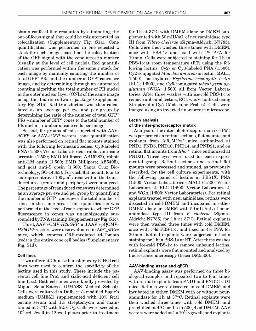
13 scAAV serotypes/variants, including AAV1, -2,-3b, -4, -5, -6, -7, -8, -9, -rh8, -rh10, -rh39, and -rh43,all expressing eGFP under the control of the CB6promoter (Table 1, Figs. 3 and 4, and Supplemen-tary Figs. S6 and S7). As controls, AAV5, -7, -8, -9,and -rh10 were subretinally injected into PND21mice (Table 1 and Supplementary Fig. S6), as thesevectors are known to exhibit excellent tropism foradult rods.14,15,18 Similar to the results observedafter PND1 delivery of AAV5, PND1 injections ofthe all other AAVs resulted in preferential conetransduction when considering the percentage ofGFP+ cells, with the exception of AAV4, which ef-ficiently transduced RPE cells only (Table 1 andFig. 3). On average, AAVrh10 exhibited the mostefficient transduction of cones (although not sta-tistically different from AAV5, -6, -7, -8, -9, -rh8,and -rh39), whereas AAV3b-mediated transgeneexpression was limited to sporadic RPE and PRs(Fig. 3b). Notably, no significant difference in thepercentage of transduced rods was seen betweenall the serotypes (Fig. 3). When considering thepercentage of GPF+ rods, all AAVs led to sparse rodtransduction, with most of the GFP+ rods localizedin the innermost part of the ONL (Fig. 4 and Sup-plementary Figs. S6 and S7). Additionally, thepreferential transduction of rods observed aftersubretinal injection of AAV2/5 vectors at PND21was reproduced after injection of AAV7 (Supple-mentary Fig. S6d and e), -8 (SupplementaryFig. S6a, d, and e), -9 (Supplementary Fig. S6b, d,and e), and -rh10 (Supplementary Fig. S6c–e).Thus, rod PR development is a critical host factorthat influences AAV tropism after subretinal de-livery independently of the serotype and thusprobably independently of the different cell at-tachments factors utilized by each capsid.
Control of AAV transduction by photoreceptordevelopment depends on the injection site
Because a higher number of GFP+ rods was ob-served after administration of higher doses ofAAV5-CMV-H2bGFP (9 · 109 vg; Fig. 1d and Ta-ble 1) and AAV5-mCAR-H2bGFP (>1 · 1010 vg)41 atPND1, the overall absence of efficient rod trans-duction at PND1 may relate to the progressive lossof the episomal vector genomes in rods. AAV vec-tors integrate only at very low frequencies, andthus vector-mediated transgene expression is pre-dicted to diminish over time under conditions ofongoing cellular proliferation. In transduced cones,the number of expressed vector genomes is likely toremain stable over time because cones are alreadypost-mitotic by PND1–PND5.44 In contrast, al-though the vast majority of rod precursors are al-
472 PETIT ET AL.

Figure 3. Predominant transduction of cone photoreceptors after the injection of 12 different AAV serotypes/variants at PND1. (a) Representative images ofretinal flat mounts at 3 wpi of scAAV1, -2, -3b, -4, -6, -7, -8, -9, -rh8, -rh10, -rh39, or –rh43 vectors (all expressing eGFP under the control of the CB6 promoter), asindicated. Labeling with PNA (red) shows expression of eGFP (green) predominantly in cones (asterisks), except for AAV4. (b) Quantification performed onretinal flat mounts and/or cryosections of the percentage of GFP+ cones and GFP+ rods in the vector exposed area (note: actual transduction efficiency ofscAAV4 was 0% for rods and cones). Results are expressed as mean – SD. Numbers in bars represent the number of retinas analyzed. *p < 0.05; **p < 0.01;***p < 0.001 by Student’s t-test. Scale bar = 25 lm.
j 473

ready post-mitotic by PND144 and lineage studiesusing retroviral vectors have revealed that mostrod progenitor cells that persist in the rodent retinaafter birth are terminally dividing,49–51 rod pro-genitors may dilute or lose AAV genomes, resultingin consistent decreased transgene expression inmature rods. Alternatively or concomitantly, theoverall absence of efficient rod transduction atPND1 may relate to the progressive transcriptionalsilencing of AAV vector DNA due to developmentalepigenetic changes. Indeed, histone modificationshave been shown to be important contributors togene regulation in developing rod PRs.52
To investigate these two possibilities, AAV5, -6,-7, -8, and -9 were intravitreally injected in PND1mice. If the factors that determine AAV tropism
during PR development are related to AAV vectorDNA, the final pattern of transgene expressionshould globally be similar between the two routesof administration. However, for all five serotypes, itwas found that rods were the predominant PR cellstransduced when the vectors were injected in-travitreally (e.g., for AAV5: 4.2 – 2.6% of rods vs.0.9 – 0.3% of cones near the injection site; n = 3;Fig. 4 and Supplementary Fig. S7). Notably, rodnuclei expressing detectable levels of GFP wereevenly distributed across the ONL after in-travitreal injection (Fig. 4 and SupplementaryFig. S7), indicating that lack of efficient AAV-mediated transduction of the late-born rods bysubretinal injections at PND1 is not due to cell di-vision or differential silencing of the vector ge-
Figure 4. Comparative analysis of AAV vector transduction in the mouse retina after subretinal or intravitreal injection at PND1. PND1 mice injected eithersubretinally (upper row) or intravitreally (lower row) with scAAV5, -6, -7, -8, or -9 expressing eGFP under the control of the CB6 promoter, as indicated.Histological analysis was performed at 3 wpi on cryosections stained with CA (red) and counterstained with DAPI (blue). AAV-mediated GFP was detected byits native fluorescence (green). Subretinal injections result in a higher number of transduced cones (asterisks) with most of the transduced rods (arrowheads)located in the inner most part of the ONL (see also Figs. 1 and 2), while intravitreal injections result in scattered rods throughout the ONL and fewer transducedcones. Scale bars = 25 lm. GCL, ganglion cell layer.
474 PETIT ET AL.

nomes. The data suggest that factors affecting theefficiency of AAV-mediated transduction of earlyand late-born rods are site specific and likely in-volve the access of AAV particles to the PR cells.Receptors necessary for binding/entry of AAVswhile present in PRs may not yet be accessible onlate-born cells by AAVs from the subretinal space.
Lack of rod outer segments markedlyalters PR tropism of AAV in the adult retina
The timing of rod precursor birth (after E19) istightly linked to the onset of PR segment formationand rhodopsin expression.53 To test whether thegrowth of rod segments and/or the associated ma-trix (Supplementary Fig. S8a and b) plays a role inthe access of AAV particles to PRs, an attempt wasmade to visualize the distribution of AAV particlesafter subretinal injection at PND1 and PND21 us-ing anti-AAV capsid antibodies. However, the largenumber of capsids present in the subretinal spacelimited the analysis (data not shown). Instead,qPCR was first employed to quantify viral bindingon PND1 and PND21 retinas after modification ofthe rod IPM with neuraminidase (SupplementaryFig. S8). Ex vivo retinal explants were used to limitinjection variability related to the surface of theretina exposed to the vector after subretinal deliv-ery at PND1 versus PND21. We found that the rod-associated matrix could specifically interact withAAV particles in vitro (Supplementary Fig. S8d).However, these interactions were not conservedacross AAV5, -7, -8, and -9, indicating that bind-ing of AAV to the rod IPM does not govern thecommon increase in rod transduction observedduring PR development after subretinal delivery.
Next, to determine if the development of a nor-mal rod segment plays a role in the access of AAVparticles to PRs, rod and cone transduction wasquantified in rhodopsin knockout (Rho-/-) mice54
after subretinal injection of AAV2/5-CMV-dGFP,AAV2/5-CMV-H2bGFP, and AAV2/5-pQCMV-H2bGFP vectors at PND1 and PND21 (Table 1 andFig. 5). The Rho-/- mouse is a well-characterizedmodel of rod-cone dystrophy that does not developrod outer segments.40 Importantly, this mousemodel does not display any rod degeneration duringthe first three postnatal weeks. No alterations inthe IPM and outer limiting membrane (OLM)55,56
were detected in Rho-/- mice at PND21 (Supple-mentary Fig. S9). In PND1-injected mice, thetransduction profile was similar between Rho-/-
and wild-type mice, with a preferential transduc-tion of cone PRs (Fig. 5a and c). However, PND21injections resulted in a fairly dramatic difference inthe final expression patterns. On average, the three
AAV vectors tested showed a similarly low trans-duction of rod PRs in Rho-/- retinas, as seen withPND1 injections in healthy retinas (Table 1 andFig. 5a and c). Rod transduction was mainly ob-served at the site of injection near the needle tract(data not shown). GFP+ rods were located in theoutermost part of the ONL (Fig. 5a, arrows) andexpressed qualitatively less GFP than cone PRs(Fig. 5a and b). These observations suggest that thepresence of a sick rod segment can influence theefficiency rod transduction upon subretinal deliv-ery in the adult retina. Furthermore, they suggest apotential correlation between altered rod trans-duction and increased cone transduction.
To delineate further the influence of rods on theefficiency of cone transduction, the rd1 mouse modelwas utilized, in which the vast majority of rods arelost by PND21.57 However, in contrast to the Rho-/-
retinas, the IPM is inevitably altered in this mousemodel due to the loss of rods. AAV5 or AAV9 vectorswere subretinally injected into PND21 rd1 mice andcone transduction was evaluated on retinal flatmounts at 3 wpi (Table 1 and Fig. 5d). All vectorsresulted in dramatically enhanced cone transduc-tion compared to control mice, considering the per-centage of cells transduced (Table 1). The consistentfinding in two mouse models of rod degenerationthat cone transduction increases in the absence ofefficient rod transduction or in the absence of rodssuggest that rods may negatively affect cone trans-duction in the adult mouse retina.
DISCUSSION
Gene therapy targeting PRs holds great poten-tial for the treatment of many forms of retinaldiseases, such as retinitis pigmentosa, Leber con-genital amaurosis, or age-related macular degen-eration.13 A common hallmark of these diseases isthe progressive dysfunction, degeneration, anddeath of PR cells. The first sign of these diseases—visual impairment—is generally associated withearly alterations of the PRs functional structures,the inner and outer segments, while PR cell bodiesare lost later in the process.
This study found that PR development and in-tegrity play a major role in the efficacy of AAV-mediated transduction after subretinal injection. Itconfirmed that rod transduction increases dra-matically during mouse postnatal retinal develop-ment,30–34 irrespective of the AAV serotype (Fig. 1and Supplementary Fig. S6), and showed that rodtransduction correlates with the differentiationorder of PR cells (Fig. 2 and SupplementaryFigs. S4 and S5). This effect was not due to differ-
IMPACT OF RETINAL DEVELOPMENT ON AAV TRANSDUCTION 475

Figure 5. Altered of AAV tropism in Rho-/- and adult rd1 mice. (a–c) PND1 and PND21 Rho-/- mice injected with similar doses of AAV2/5-CMV-H2bGFP,AAV2/5-CMV-dGFP, or AAV2/5-pQCMV-H2bGFP. Three weeks after injection, retinas were labeled with PNA or with an antibody raised against CA (red). AAV-mediated GFP was detected by its native fluorescence (green). Representative images after injection of (a) AAV2/5-CMV-H2bGFP and (b) AAV2/5-CMV-dGFPat age indicated. Asterisks indicate GFP+ cones and arrowheads indicate GFP+ rods. Quantitative analysis of the percentage of GFP+ cones (black bars) androds (white bars) is shown in (c). Error bars represent SD. Numbers in bars represent the number of retina analyzed. **p < 0.01 and ***p < 0.001 by Student’st-test. (d) Retinal flat mounts of rd1 mice injected at PND21with similar doses of AAV2/5-CMV-dGFP, AAV2/5-CMV-H2bGFP, scAAV2/5-CB6-eGFP, or scAAV2/9-CB6-eGFP and stained for CA expression (red) 3 wpi. AAV-mediated GFP was detected by its native fluorescence (green). Percentage of transduced cones foreach vector is indicated in corresponding panel as mean – SD. Scale bars = 25 lm.
476 PETIT ET AL.

ences in promoter activity or second-strand syn-thesis of the viral genome, which in principle couldaccount for the enhanced rod transduction (Fig. 1and Supplementary Fig. S2). Neither was this ef-fect related to the gradual exit of rod progenitorsfrom the cell cycle or to epigenetic/protein inhibi-tion of the vector genomes during development,since both early and late-born cells were efficientlytransduced at PND1 after intravitreal deliveryof AAV vectors (Fig. 4). Thus, while serotype-independent (Figs. 3 and 4, and SupplementaryFigs. S4–S6), the effects of PR development on AAVtransduction were, however, site specific (Fig. 4).
It is possible that restriction to AAV transduc-tion in immature rods occurs after virion inter-nalization due to inefficient viral trafficking.However, intracellular AAV particle traffickinghas been shown to be influenced by the viral capsidsequence,58 which in this case seems not to be rel-evant with regards to the common shift in tropismbetween PND1 and PND21 subretinal injections.Alternatively, the enhanced transduction of rodsduring retinal development could be related to in-creased entry of AAVs into PRs. The OLM, in whichthe zonulae adherents pore size has been estimatedto be between 30 and 36 A,59 may impede the dif-fusion of AAV capsids (250 A) to the ONL, allowingefficient access (and thus entry) of AAV particlesmainly to PRs that have started to developed innersegments that protrude through the OLM (i.e.,cones and early-born rods).33 In support of thismodel,33 the timing of rod precursor birth (afterE19) is known to be tightly linked to the onset of PRsegment formation and rhodopsin expression.53
Concomitantly, the development of rod outer seg-ment may directly or indirectly correlate with in-creased—nonspecific—entry of AAV vectors intoPRs. Indeed, a shift in the distribution of GFP+ rodnuclei was observed when AAV5 vectors were de-livered after PND10, which coincides with theelongation of rod outer segments for the majorityof rods at this age (Fig. 2c). Moreover, rod trans-duction was profoundly inhibited in adult Rho-/-
retinas (Fig. 5) that lack rod outer segment devel-opment but still display rod inner segments thatprotrude through the OLM. Of course, other fac-tors, such as PR stress and degeneration, couldaffect the expression of AAV receptors, as well asinhibit the expression of the transgene in the dis-eased retina. However, there is more rational evi-dence from the observations to suggest that vectoraccess to PR segments is one of the host factorsgoverning PR transduction efficiency upon sub-retinal delivery, in particular since at PND21,Rho-/-retinas had no apparent loss of rod PRs or
alterations of the IPM and OLM integrity (Sup-plementary Fig. S9).
Importantly, the idea that PR outer segmentsmodulate rod transduction in the adult retina doesnot contradict previous reports that demonstratedunequivocal (but transient) structural and/or func-tional rescue of rods by AAV-mediated gene re-placement therapy in two mouse models of retinitispigmentosa that fail to develop rod outer seg-ments; namely the Rho–/–16,60 and the Phrp2–/–61,62
mice, In the Rho-/- mice, therapeutic vectors wereinjected into neonatal mice and mice younger thanPND5—two time points that precede rod outer seg-ment growth in wild-type mice and where it has beenshown that rod transduction is dependent on theviral load injected (Fig. 1d). In the Phrp2-/- mice, ithas been notably shown that there was a significantdifference in the number and quality of outer seg-ment rescue in treated retinas. Depending on theage at which the mice were treated, fewer outersegments developed normally, with the best resultsobtained in younger animals, despite the relativelyslow rate of PR death in that model. Moreover, par-ticular differences in the infectivity of the vectorpreparations and the presumed vector titers mayexist between all these studies, making it difficult tocross-compare them directly. The goal of this studywas to determine host cell factors that influencePR transduction, and oversaturating the systemwith AAV vectors would have been counterproduc-tive in this case. As mentioned, it has previouslybeen shown in wild-type mice that the majority ofimmature rods in addition to inner nuclear layercells can be transduced by subretinal injection atPND1 if a higher dose of vector is used (Fig. 1d).41 Inthis study, the rate of rod transduction after injec-tion at PND21 was limited to 30–55%, while othergroups have reported PR transduction rates of >90%within the vector exposed area.14,17 Thus, the over-all lower transduction in this study can easily ex-plain why in Rho-/- mice such a large difference withPND21 subretinal injections was seen. Another fac-tor that complicates the comparison between ex-periments in the Rho-/- mice and the previouslypublished results by Palfi et al.16,54,60 is that thepresent study used different Rho-/- mice.40 Strainbackground differences between the two strains andthe way both knockout strains were generated couldhave exacerbated the effect of the lower transductionrate used to perform this study. In this regard, it isinteresting to note that the group of Jean Bennetthas also reported difficulties in transducing effi-ciently rods in the same Rho-/- model that was usedin this study63 (Dejneka NS et al., ARVO AnnualMeeting Abstract, 2002).
IMPACT OF RETINAL DEVELOPMENT ON AAV TRANSDUCTION 477

The present findings provide important informa-tion regarding the effects of subretinal gene therapyin animal models of PR dystrophies. First, the resultsindicate that the time of intervention affects the effi-cacy of AAV transduction if gene therapy is appliedbefore the full development of the retina. However, inseveral mouse models of PR dystrophies, the degen-eration of PRs is so fast that most gene therapy at-tempts have been conducted during the first week oflife13 due to the relative slow onset of AAV-mediatedtransgene expression.28 For instance, in the nmf363murine model of PDE6a-deficiency, no apparent PRdegeneration is observed at PND12, but by PND14,30% of PRs are already lost and only one row of PRnuclei remains in the ONL by PND38.64 In thismodel, injection of an AAV8(Y788F)-RHO-mPde6a atPND5 resulted in an initial loss of cells between 1 and2 months of age, followed by a preservation of three tofour rows of PR nuclei in the vector-exposed area, forat least 6 months.65 However, the overall rod func-tional rescue was too low to make a detectable dif-ference by electroretinography.65 Notably, mid-stageintervention at PND21 achieved similar efficacy asPND5 treatment,despite loss of approximately half ofPRs at the time of injection,66 indicating that the ef-ficacy of PND5 treatment may have been limited bythe efficient transduction of rods when compared tothe PND21 intervention. As well, another study re-ported that subretinal injection of AAV2/5-smCBA-mPde6b vector (1 · 1010 genome copies) in rd10 miceat PND4 (before the onset of PR dystrophy) or PND21(after the onset of PR dystrophy) resulted in similar(partial) therapeutic effects.67
Second, the results indicate that if PR integritydetermines the ability of AAV vectors to target thePRs, the spatio-temporal kinetics of retinal degener-ation, the site of injection, and the health status of theretina at the time of treatment are also importantfactors affecting the overall efficacy of transduction.For instance,murine64,68–73 andcanine74–77 modelsofsevere PR dystrophies show significant loss of PRsegments at early stages of the disease. In thesemodels, most preclinical studies have demonstratedimproved PR survival only when gene therapy is ap-plied before or at very early stages of retinal degen-eration.13,23,78–84 Moreover, when gene therapy hasbeen applied at later stages of the disease, the pro-portion of PRs that were not responding to thetreatment (‘‘silent’’ cells) increased within the vector-exposed area.1,23,85 It has been first suggested thatthere is a threshold of accumulated changes afterwhich PR death is inevitable.1 However, recent dataindicate that continued PR loss may instead reflectinsufficient transduction efficiencies.29 An intriguingpossibility is that AAV vectors might not efficiently
access and infect subpopulations of deteriorated PRs.Thus, early alterations of PRs may be associated witha higher heterogeneity of transduction within thevector-exposed area (e.g., 40% of PRs expressing highlevels of transgene and 60% of PRs expressing lowlevels of transgenes rather than 100% of PRs ex-pressing medium levels of transgene). An importantstep in approaching this problem will be to under-stand the longitudinal changes that occur duringphases of PR stress/degeneration at the level ofboth the PRs and AAV vector pharmacology. Futurestudies will also have to be designed to determinewhether a temporal disruption of the OLM86 andhigher doses of vectors may be able to overcome thishurdle. Finally, delineating the relationship betweenPR development, AAV trafficking, and cell receptorusage will be essential toward developing a completeunderstanding of PR transduction upon subretinalinjection.
It did not escape the authors’ attention that theresults support the notion that rods and cones maydirectly or indirectly compete for AAV access.Preclinical studies with several AAV serotypeshave established that subretinal injection in adultsoften results in dominant transduction of rodswithin rod-rich retinas.15,19–22,42,87 For instance, inwild-type mice (cone:rod ratio of 1:30), while fairlyhigh transduction of rods is observed after sub-retinal injection of AAV2/8, cone transduction isrestricted to only 1–12% of cells.15 In pigs, AAV2/8mediates 3.8- to 5- and 1.7-fold higher levels of PRtransduction than AAV2/5 and AAV2/9, respec-tively.15,22 However, all serotypes transduce thesame percentage of cones in the cone-enriched vi-sual streak (cone:rod ratio of 1:3 to 1:5), with up to9.2% of cones readily transduced.15 In dogs, AAV2/5also preferentially targets rods,42 though signifi-cant cone transduction is observed in cone-enrichedareas,23 with the use of high doses of vectors.43
Primate studies with seven different AAV serotypesreported similar findings with weak cone trans-duction in the rod-rich parafoveal region (1–10%),even in the presence of ample rod PR transduction,and with higher levels of cone transduction (20–50%) in the pure-cone fovea.19–21,87
Consistent with the notion that there is a devel-opmental effect in which access by AAV to cone PRsbecomes restricted, a profound decrease in conetransduction was observed in the developing mouseretina (Fig. 2). These observations appear to corre-late with the increase in rod transduction (Fig. 2b).Moreover, cone transduction was significantly en-hanced after subretinal injection of AAV vectors inPND21 Rho-/- (Fig. 5a–c) and rd1 mice (Fig. 5d), inwhich rod transduction was inhibited by the absence
478 PETIT ET AL.

of a healthy rod segments or by the loss of the vastmajority of rod cells at the time of treatment. Al-though comprehensive analysis will be required toelucidate the details of PR transduction upon sub-retinal injection, it should be noted that there is noevidence from the present observations to suggestthat AAV particles use different receptors betweenrods and cones. Indeed,a similar shift in tropism wasobserved for all tested AAV serotypes (Fig. 3 andSupplementary Fig. S6). It is possible that the den-sely stacked rod outer segments form physical bar-riers between the site of delivery and the cones.Concomitantly, cone matrix sheaths located aroundcone inner and outer segments may form a barrierthat selectively reduces access of AAV particles tothe cones. Interestingly, a recent study in the felineretina has shown that unlike mice and nonhumanprimates, cone PRs were more efficiently transducedthan rods.26 The reasons for this difference in cel-lular tropism remain unknown, but the cone matrixsheath of the cat differs significantly from othermammalian species.88 In primates, only AAV9 wasshown to target cones both centrally and peripher-ally efficiently at low doses when directly comparedto five other AAV serotypes.20 It has been hypothe-sized that this property is due to the abundance ofterminal galactose, the cell receptor for AAV9 on thecone PR matrix.20
The notion that rods and cones compete for AAVtransduction emphasizes the need of evaluatingcomponents of AAV vectors planned for humans inmodels that accurately depict physiological char-acteristics of the human retina (i.e., large animalsand all-cone murine retinas).15,20–23,42,43,89,90 In-deed, while in humans macular cones will often bethe primary treatment area, the ability of AAVvectors to transduce the cones and restore their
function is often evaluated in murine models of PRdystrophies that do not display a cone-enrichedarea but a high rod:cone ratio. It also indicates thatloss of rod PRs associated with many forms of re-tinopathies could be exploited to redirect AAVscommonly toward cones.
ACKNOWLEDGMENTS
We are grateful to Christian Mueller (UMASSMedical School) for insightful discussions andcritical reading of the manuscript; to Qin Su(UMASS Medical School) and the Vector Core ofthe Horae Gene Therapy Center for the produc-tion of the scAAV-CB6-eGFP vectors; to SouravChoudhury and Miguel Sena-Esteves (UMASSMedical School) for the gift of the Pro5 and Lec2 celllines; and to Julio Sanmiguel (UMASS MedicalSchool) for the gift of the eGFP qPCR primers. Thiswork was supported by an unrestricted grant from‘‘Information Recherche Retinite Pigmentaire’’ As-sociation (L.P.) and by the US National Institutesof Health (RO1-EY023570, C.P). L.P. also ac-knowledges the following funding: the Fulbright/Fondation Monahan Postdoctoral Fellowship,the Fondation de France ‘‘Young researcher inophthalmology’’ Fellowship, and the AssociationFrancaise contre les Myopathies (AFM-Telethon)Postdoctoral Fellowship.
AUTHOR DISCLOSURE
G.G. is a founder of Voyager Therapeutics, spe-cialized in AAV-based gene therapy for the centralnervous system, and holds equity in the company.G.G. is an inventor of patents with potential roy-alties licensed to Voyager Therapeutics and otherbiopharmaceutical companies. No competing fi-nancial interests exist for the remaining authors.
REFERENCES
1. Cideciyan AV, Jacobson SG, Beltran WA, et al.Human retinal gene therapy for Leber congenitalamaurosis shows advancing retinal degenerationdespite enduring visual improvement. Proc NatlAcad Sci U S A 2013;110:E517–525.
2. Bainbridge JW, Mehat MS, Sundaram V, et al.Long-term effect of gene therapy on Leber’scongenital amaurosis. N Engl J Med 2015;372:1887–1897.
3. Bainbridge JW, Smith AJ, Barker SS, et al. Effectof gene therapy on visual function in Leber’scongenital amaurosis. N Engl J Med 2008;358:2231–2239.
4. Bennett J, Ashtari M, Wellman J, et al. AAV2gene therapy readministration in three adults withcongenital blindness. Sci Transl Med 2012;4:120ra115.
5. Bennett J, Wellman J, Marshall KA, et al. Safetyand durability of effect of contralateral-eye ad-ministration of AAV2 gene therapy in patientswith childhood-onset blindness caused by RPE65mutations: a follow-on Phase 1 trial. Lancet 2016;388:661–672.
6. Maguire AM, Simonelli F, Pierce EA, et al. Safetyand efficacy of gene transfer for Leber’s con-
genital amaurosis. N Engl J Med 2008;358:2240–2248.
7. Hauswirth WW, Aleman TS, Kaushal S, et al.Treatment of leber congenital amaurosis due toRPE65 mutations by ocular subretinal injection ofadeno-associated virus gene vector: short-termresults of a Phase I trial. Hum Gene Ther 2008;19:979–990.
8. Jacobson SG, Cideciyan AV, Roman AJ, et al. Im-provement and decline in vision with gene therapyin childhood blindness. N Engl J Med 2015;372:1920–1926.
IMPACT OF RETINAL DEVELOPMENT ON AAV TRANSDUCTION 479

9. Weleber RG, Pennesi ME, Wilson DJ, et al. Re-sults at 2 years after gene therapy for RPE65-deficient Leber congenital amaurosis and severeearly-childhood-onset retinal dystrophy. Ophthal-mology 2016;123:1606–1620.
10. Ghazi NG, Abboud EB, Nowilaty SR, et al. Treat-ment of retinitis pigmentosa due to MERTK mu-tations by ocular subretinal injection of adeno-associated virus gene vector: results of a Phase Itrial. Hum Genet 2016;135:327–343.
11. Edwards TL, Jolly JK, Groppe M, et al. Visualacuity after retinal gene therapy for choroider-emia. N Engl J Med 2016;374:1996–1998.
12. MacLaren RE, Groppe M, Barnard AR, et al. Ret-inal gene therapy in patients with choroideremia:initial findings from a Phase 1/2 clinical trial.Lancet 2014;383:1129–1137.
13. Petit L, Khanna H, Punzo C. Advances in genetherapy for diseases of the eye. Hum Gene Ther2016;27:563–579.
14. Allocca M, Mussolino C, Garcia-Hoyos M, et al.Novel adeno-associated virus serotypes efficientlytransduce murine photoreceptors. J Virol 2007;81:11372–11380.
15. Manfredi A, Marrocco E, Puppo A, et al. Combinedrod and cone transduction by adeno-associatedvirus 2/8. Hum Gene Ther 2013;24:982–992.
16. Palfi A, Chadderton N, O’Reilly M, et al. Efficientgene delivery to photoreceptors using AAV2/rh10and rescue of the Rho(–/–) mouse. Mol TherMethods Clin Dev 2015;2:15016.
17. Natkunarajah M, Trittibach P, McIntosh J, et al.Assessment of ocular transduction using single-stranded and self-complementary recombinantadeno-associated virus serotype 2/8. Gene Ther2008;15:463–467.
18. Auricchio A, Kobinger G, Anand V, et al. Exchangeof surface proteins impacts on viral vector cellularspecificity and transduction characteristics: theretina as a model. Hum Mol Genet 2001;10:3075–3081.
19. Vandenberghe LH, Bell P, Maguire AM, et al.Dosage thresholds for AAV2 and AAV8 photore-ceptor gene therapy in monkey. Sci Transl Med2011;3:88ra54.
20. Vandenberghe LH, Bell P, Maguire AM, et al.AAV9 targets cone photoreceptors in the nonhu-man primate retina. PLoS One 2013;8:e53463.
21. Boye SE, Alexander JJ, Boye SL, et al. The humanrhodopsin kinase promoter in an AAV5 vectorconfers rod- and cone-specific expression in theprimate retina. Hum Gene Ther 2012;23:1101–1115.
22. Mussolino C, della Corte M, Rossi S, et al. AAV-mediated photoreceptor transduction of the pigcone-enriched retina. Gene Ther 2011;18:637–645.
23. Lheriteau E, Petit L, Weber M, et al. Successfulgene therapy in the RPGRIP1-deficient dog: alarge model of cone-rod dystrophy. Mol Ther2014;22:265–277.
24. Bruewer AR, Mowat FM, Bartoe JT, et al. Eva-luation of lateral spread of transgene expressionfollowing subretinal AAV-mediated gene deliveryin dogs. PLoS One 2013;8:e60218.
25. Stieger K, Colle MA, Dubreil L, et al. Subretinaldelivery of recombinant AAV serotype 8 vector indogs results in gene transfer to neurons in thebrain. Mol Ther 2008;16:916–923.
26. Minella AL, Mowat FM, Willett KL, et al. Differentialtargeting of feline photoreceptors by recombinantadeno-associated viral vectors: implications forpreclinical gene therapy trials. Gene Ther 2014;21:913–920.
27. Surace EM, Auricchio A. Versatility of AAV vectorsfor retinal gene transfer. Vision Res 2008;48:353–359.
28. Vandenberghe LH, Auricchio A. Novel adeno-associated viral vectors for retinal gene therapy.Gene Ther 2012;19:162–168.
29. Koch SF, Tsai YT, Duong JK, et al. Halting pro-gressive neurodegeneration in advanced retinitispigmentosa. J Clin Invest 2015;125:3704–3713.
30. Surace EM, Auricchio A, Reich SJ, et al. Deliveryof adeno-associated virus vectors to the fetalretina: impact of viral capsid proteins on retinalneuronal progenitor transduction. J Virol 2003;77:7957–7963.
31. Pang JJ, Lauramore A, Deng WT, et al. Com-parative analysis of in vivo and in vitro AAV vectortransduction in the neonatal mouse retina: effectsof serotype and site of administration. Vision Res2008;48:377–385.
32. Watanabe S, Sanuki R, Ueno S, et al. Tropisms ofAAV for subretinal delivery to the neonatal mouseretina and its application for in vivo rescue ofdevelopmental photoreceptor disorders. PLoS One2013;8:e54146.
33. Xiong W, Cepko C. Distinct expression patterns ofAAV8 vectors with broadly active promoters fromsubretinal injections of neonatal mouse eyes attwo different ages. Adv Exp Med Biol 2016;854:501–507.
34. Xiong W, MacColl Garfinkel AE, Li Y, et al. NRF2promotes neuronal survival in neurodegenerationand acute nerve damage. J Clin Invest 2015;125:1433–1445.
35. Punzo C, Cepko CL. Ultrasound-guided in uteroinjections allow studies of the development andfunction of the eye. Dev Dyn 2008;237:1034–1042.
36. Rashnonejad A, Chermahini GA, Li S, et al. Large-scale production of adeno-associated viral vectorserotype-9 carrying the human survival motorneuron gene. Mol Biotechnol 2016;58:30–36.
37. Grieger JC, Choi VW, Samulski RJ. Production andcharacterization of adeno-associated viral vectors.Nat Protoc 2006;1:1412–1428.
38. Ayuso E, Mingozzi F, Montane J, et al. High AAVvector purity results in serotype- and tissue-independent enhancement of transduction effi-ciency. Gene Ther 2010;17:503–510.
39. Le YZ, Ash JD, Al-Ubaidi MR, et al. Targetedexpression of Cre recombinase to cone photore-ceptors in transgenic mice. Mol Vis 2004;10:1011–1018.
40. Lem J, Krasnoperova NV, Calvert PD, et al. Mor-phological, physiological, and biochemical chan-ges in rhodopsin knockout mice. Proc Natl AcadSci U S A 1999;96:736–741.
41. Venkatesh A, Ma S, Langellotto F, et al. Retinalgene delivery by rAAV and DNA electroporation.Curr Protoc Microbiol 2013;Chapter 14:Unit14D 14.
42. Beltran WA, Boye SL, Boye SE, et al. rAAV2/5gene-targeting to rods:dose-dependent efficiencyand complications associated with different pro-moters. Gene Ther 2010;17:1162–1174.
43. Komaromy AM, Alexander JJ, Cooper AE, et al.Targeting gene expression to cones with humancone opsin promoters in recombinant AAV. GeneTher 2008;15:1049–1055.
44. Swaroop A, Kim D, Forrest D. Transcriptionalregulation of photoreceptor development andhomeostasis in the mammalian retina. Nat RevNeurosci 2010;11:563–576.
45. Mack AF, Papanikolaou D, Lillo C. Investigation ofthe migration path for new rod photoreceptors inthe adult cichlid fish retina. Exp Neurol 2003;184:90–96.
46. Mack AF, Fernald RD. New rods move beforedifferentiating in adult teleost retina. Dev Biol1995;170:136–141.
47. Henderson RG, Fernald RD. Timing and location ofrhodopsin expression in newly born rod photore-ceptors in the adult teleost retina. Brain Res DevBrain Res 2004;151:193–197.
48. Fei Y. Development of the cone photoreceptormosaic in the mouse retina revealed by fluores-cent cones in transgenic mice. Mol Vis 2003;9:31–42.
49. Cepko C. Intrinsically different retinal progenitorcells produce specific types of progeny. Nat RevNeurosci 2014;15:615–627.
50. Turner DL, Cepko CL. A common progenitor forneurons and glia persists in rat retina late indevelopment. Nature 1987;328:131–136.
51. Hafler BP, Surzenko N, Beier KT, et al. Tran-scription factor Olig2 defines subpopulations ofretinal progenitor cells biased toward specific cellfates. Proc Natl Acad Sci U S A 2012;109:7882–7887.
52. Mo A, Luo C, Davis FP, et al. Epigenomic land-scapes of retinal rods and cones. Elife 2016;5:e11613.
53. Morrow EM, Belliveau MJ, Cepko CL. Two phasesof rod photoreceptor differentiation during ratretinal development. J Neurosci 1998;18:3738–3748.
54. Humphries MM, Rancourt D, Farrar GJ, et al.Retinopathy induced in mice by targeted disrup-tion of the rhodopsin gene. Nat Genet 1997;15:216–219.
480 PETIT ET AL.

55. Jaissle GB, May CA, Reinhard J, et al. Evaluationof the rhodopsin knockout mouse as a model ofpure cone function. Invest Ophthalmol Vis Sci2001;42:506–513.
56. Calame M, Cachafeiro M, Philippe S, et al. Retinaldegeneration progression changes lentiviral vec-tor cell targeting in the retina. PLoS One 2011;6:e23782.
57. Punzo C, Kornacker K, Cepko CL. Stimulation ofthe insulin/mTOR pathway delays cone death in amouse model of retinitis pigmentosa. Nat Neu-rosci 2009;12:44–52.
58. Johnson JS, Li C, DiPrimio N, et al. Mutagenesisof adeno-associated virus type 2 capsid proteinVP1 uncovers new roles for basic amino acids intrafficking and cell-specific transduction. J Virol2010;84:8888–8902.
59. Bunt-Milam AH, Saari JC, Klock IB, et al. Zonulaeadherentes pore size in the external limitingmembrane of the rabbit retina. Invest OphthalmolVis Sci 1985;26:1377–1380.
60. Palfi A, Millington-Ward S, Chadderton N, et al.Adeno-associated virus-mediated rhodopsin re-placement provides therapeutic benefit in micewith a targeted disruption of the rhodopsin gene.Hum Gene Ther 2010;21:311–323.
61. Ali RR, Sarra GM, Stephens C, et al. Restorationof photoreceptor ultrastructure and function inretinal degeneration slow mice by gene therapy.Nat Genet 2000;25:306–310.
62. Sarra GM, Stephens C, de Alwis M, et al. Genereplacement therapy in the retinal degenerationslow (rds) mouse: the effect on retinal degener-ation following partial transduction of the retina.Hum Mol Genet 2001;10:2353–2361.
63. Liang FQ, Dejneka NS, Cohen DR, et al. AAV-mediated delivery of ciliary neurotrophic factorprolongs photoreceptor survival in the rhodopsinknockout mouse. Mol Ther 2001;3:241–248.
64. Sakamoto K, McCluskey M, Wensel TG, et al.New mouse models for recessive retinitis pig-mentosa caused by mutations in the Pde6a gene.Hum Mol Genet 2009;18:178–192.
65. Wert KJ, Davis RJ, Sancho-Pelluz J, et al. Genetherapy provides long-term visual function in apre-clinical model of retinitis pigmentosa. HumMol Genet 2013;22:558–567.
66. Wert KJ, Sancho-Pelluz J, Tsang SH. Mid-stageintervention achieves similar efficacy as conven-tional early-stage treatment using gene therapy ina pre-clinical model of retinitis pigmentosa. HumMol Genet 2014;23:514–523.
67. Yao J, Jia L, Khan N, et al. Caspase inhibitionwith XIAP as an adjunct to AAV vector gene-replacement therapy: improving efficacy andprolonging the treatment window. PLoS One 2012;7:e37197.
68. Blanks JC, Adinolfi AM, Lolley RN. Photoreceptor de-generation and synaptogenesis in retinal-degenerative(rd) mice. J Comp Neurol 1974;156:95–106.
69. LaVail MM, Sidman RL. C57BL-6J mice with in-herited retinal degeneration. Arch Ophthalmol1974;91:394–400.
70. Tansley K. Hereditary degeneration of the mouseretina. Br J Ophthalmol 1951;35:573–582.
71. Gargini C, Terzibasi E, Mazzoni F, et al. Retinalorganization in the retinal degeneration 10 (rd10)mutant mouse: a morphological and ERG study.J Comp Neurol 2007;500:222–238.
72. Won J, Gifford E, Smith RS, et al. RPGRIP1 isessential for normal rod photoreceptor outersegment elaboration and morphogenesis. HumMol Genet 2009;18:4329–4339.
73. Ramamurthy V, Niemi GA, Reh TA, et al. Lebercongenital amaurosis linked to AIPL1: a mousemodel reveals destabilization of cGMP phospho-diesterase. Proc Natl Acad Sci U S A 2004;101:13897–13902.
74. Parry HB. Degenerations of the dog retina. II.Generalized progressive atrophy of hereditary or-igin. Br J Ophthalmol 1953;37:487–502.
75. Aguirre GD, Rubin LF. Rod-cone dysplasia (pro-gressive retinal atrophy) in Irish setters. J Am VetMed Assoc 1975;166:157–164.
76. Tuntivanich N, Pittler SJ, Fischer AJ, et al. Char-acterization of a canine model of autosomal reces-sive retinitis pigmentosa due to a PDE6A mutation.Invest Ophthalmol Vis Sci 2009;50:801–813.
77. Beltran WA, Acland GM, Aguirre GD. Age-dependent disease expression determines re-modeling of the retinal mosaic in carriers of RPGRexon ORF15 mutations. Invest Ophthalmol Vis Sci2009;50:3985–3995.
78. Petit L, Lheriteau E, Weber M, et al. Restorationof vision in the pde6beta-deficient dog, a largeanimal model of rod-cone dystrophy. Mol Ther2012;20:2019–2030.
79. Pang JJ, Boye SL, Kumar A, et al. AAV-mediatedgene therapy for retinal degeneration in the rd10mouse containing a recessive PDEbeta mutation.Invest Ophthalmol Vis Sci 2008;49:4278–4283.
80. Pang JJ, Dai X, Boye SE, et al. Long-term retinalfunction and structure rescue using capsid mutantAAV8 vector in the rd10 mouse, a model of re-
cessive retinitis pigmentosa. Mol Ther 2011;19:234–242.
81. Sun X, Pawlyk B, Xu X, et al. Gene therapy with apromoter targeting both rods and cones rescuesretinal degeneration caused by AIPL1 mutations.Gene Ther 2010;17:117–131.
82. Ku CA, Chiodo VA, Boye SL, et al. Gene therapyusing self-complementary Y733F capsid mutantAAV2/8 restores vision in a model of early onsetLeber congenital amaurosis. Hum Mol Genet 2011;20:4569–4581.
83. Pawlyk BS, Smith AJ, Buch PK, et al. Gene re-placement therapy rescues photoreceptor degen-eration in a murine model of Leber congenitalamaurosis lacking RPGRIP. Invest Ophthalmol VisSci 2005;46:3039–3045.
84. Koch S, Sothilingam V, Garcia Garrido M, et al.Gene therapy restores vision and delays degener-ation in the CNGB1(–/–) mouse model of retinitispigmentosa. Hum Mol Genet 2012;21:4486–4496.
85. Beltran WA, Cideciyan AV, Iwabe S, et al. Suc-cessful arrest of photoreceptor and vision lossexpands the therapeutic window of retinal genetherapy to later stages of disease. Proc Natl AcadSci U S A 2015;112:E5844–5853.
86. West EL, Pearson RA, Tschernutter M, et al.Pharmacological disruption of the outer limitingmembrane leads to increased retinal integrationof transplanted photoreceptor precursors. Exp EyeRes 2008;86:601–611.
87. Bennett J, Maguire AM, Cideciyan AV, et al. Stabletransgene expression in rod photoreceptors afterrecombinant adeno-associated virus-mediatedgene transfer to monkey retina. Proc Natl Acad SciU S A 1999;96:9920–9925.
88. Fariss RN, Anderson DH, Fisher SK. Comparison ofphotoreceptor-specific matrix domains in the catand monkey retinas. Exp Eye Res 1990;51:473–485.
89. Boye SL, Peterson JJ, Choudhury S, et al. Genetherapy fully restores vision to the all-coneNrl(–/–) Gucy2e(–/–) mouse model of Leber con-genital amaurosis-1. Hum Gene Ther 2015;26:575–592.
90. Ye GJ, Budzynski E, Sonnentag P, et al. Cone-specific promoters for gene therapy of achroma-topsia and other retinal diseases. Hum Gene Ther2016;27:72–82.
Received for publication January 24, 2017;
accepted after revision May 16, 2017.
Published online: May 16, 2017.
IMPACT OF RETINAL DEVELOPMENT ON AAV TRANSDUCTION 481

Supplementary Data
Supplementary Figure S1. Quantification of cone and rod transduction. (a and b) For each group of injected mice, quantification of cone and rod transductionwas first performed on retinal sections after immunostaining with cone arrestin (CA; red). Images were acquired using epifluorescence microscopy with decon-volution for confocal-like image quality. Briefly, images were recorded at different focal plans (z-stacks) and 3D deconvolved in order to eliminate out-of-focus signalsfrom separate objects that would have overlapped and could have been misinterpreted as colocalization. (a) Shown is a representative image of a mouse injectedwith AAV2/5-CMV-H2bGFP before and after 3D deconvolution. First panel shows one of the focal planes that are part of the z-stack series that is acquired byepifluorescence. Panels 2–4 show same focal plane after 3D deconvolution of the z-stack series. Panels 3–4 show higher magnification of the boxed area in panel 2,with panel 4 showing only red signal. Cone quantification was performed in one selected plane of the z-stack series for each image, based on the colocalization of theGFP signal (green) with the cone arrestin marker (red; usually at the level of cell nuclei: asterisks mark cones). (b) Shown are images from a mouse injected withAAV2/5-CMV-dGFP exemplifying the images obtained after processing the epifluorescence z-stack series with the Imaris software in order to determine rodtransduction efficiency. Rod quantification was performed within the same focal plane of the deconvolved z-stack series by manually counting the number of totalGFP+ PRs (green: white circles in panel 4) and the number of GFP+ cones (as described above) per image, and by determining through an automated countingalgorithm the total number of PR nuclei in the ONL of the same image using the Imaris software (red dots in panels 1, 3, and 4). Rod transduction was then calculated asan average per eye and per group by determining the ratio of the number of total GFP+ PRs – number of GFP+ cones to the total number of PR nuclei per image (panel 1,red dots). Panel 4 shows higher magnification of the boxed area in panel 3. (c and d) Cone transduction was also quantified using two additional methods. (c) For thegroups of mice injected with the AAV-dGFP or AAV-eGFP vectors, cone quantification was also performed on retinal flat mounts stained with PNA. Images wererecorded at different focal plans (z-stacks), 3D deconvolved, and cone quantification was performed at the level of the cone segments, where GFP fluorescence(green) in cones was unambiguously surrounded by PNA staining (red). Shown is a representative focal plane of the deconvolved z-stack series at the level of thecone segment (panels 1–3) and a 3D reconstruction of the acquired series (panel 4). Images are from a mouse injected with AAV2/5-CMV-dGFP. (d) AAV-H2bGFPvectors were also evaluated in Ai9+_MCre+ mice, which express CRE-mediated td-Tomato (red) throughout the entire cone cell body. Shown is a representative focalplane of a z-stack series after 3D deconvolution from a mouse injected with AAV2/5-CMV-H2bGFP. The last two panels show a higher magnification of the boxed areain the third panel. Asterisks indicate GFP+ cones. INL, inner nuclear layer; GCL, ganglion cell layer; ONL, outer nuclear layer; ss, single-stranded.

Supplementary Figure S2. Subretinal injections of AAV2/5 vectors at PND1. (a) Schematic of the subretinal injection route used in this study andrepresentative fundus photography depicting in vivo GFP fluorescence in adult mouse retinas after subretinal delivery of AAV2/5-CMV-dGFP at PND1.Widespread fluorescence is seen, presumably because the interactions between the RPE and the PR outer segments that could limit the spread of the vectorafter injection have not yet formed at PND1. (b) Histological analysis showing the expression of GFP after subretinal injection of ssAAV5-CMV-dGFP, ssAAV5-CMV-H2bGFP, and scAAV5-CB6-eGFP at PND1. Tissue was harvested 3 wpi. Cryosections were immunostained against CA (red). AAV-mediated GFP wasdetected by its native fluorescence (green). After PND1 subretinal injection, transgene expression is mostly present in the photoreceptors, in particular in thecones. RPE, retinal pigment epithelium; sc, self-complementary.

Supplementary Figure S3. Subretinal injection of AAV5-CMV-dGFP at PND1 directs GFP expression in both LM and S cones. PND1 mice were subretinallyinjected with AAV2/5-CMV-dGFP and sacrificed 3 wpi. Retinal flat mounts were labeled using anti-LM opsin or anti-S opsin immunostaining (blue) to distinguishthe two subtypes of cone photoreceptors. In addition, the total population of cones was labeled using PNA or anti-CA immunostaining (red). (a) Representativeimages and (b) quantification of the percentage of GFP+-LM and GFP+-S cone photoreceptors. The quantification revealed that LM and S cones aretransduced with the same efficacy. Error bars represent SD. Numbers in bars represent the number of retinas analyzed. Scale bar = 25 lm.

Supplementary Figure S4. Rod development coincides with increased rod transduction after subretinal delivery. Histological analysis showing theexpression of dGFP after subretinal injection of ssAAV2/5-CMV-dGFP in the developing mouse retina at PND indicated. Tissue was harvested 3 wpi.Immunostaining against CA (red) shows a high level of GFP+ (green) cones after PND1 injection. A progressive decrease in the number of GFP-expressingcones is observed the later the injection is performed, while the number of GFP-expressing rods increases with later injection time points. RPE cells wereartificially removed using Photoshop. Asterisks indicate GFP+ cones. Arrowheads indicate GFP+ rods. Scale bars = 25 lm.

Supplementary Figure S5. Progressive increase in the number of GFP-expressing rods during retinal development. (a) Histological analysis showing theexpression of eGFP after subretinal injection of scAAV2/5-CB6-eGFP in the developing mouse retina at PND indicated. Tissue was harvested 3 wpi. Im-munostaining against CA (red) shows a high level of GFP+ cones after PND1 injection. A progressive decrease in the number of GFP-expressing cones isobserved the later the injection is performed, while the number of GFP-expressing rods increases with later injection time points. RPE cells were artificiallyremoved using Photoshop. (b) Representative retinal flat mount images of the dorsal and ventral portions of retinas from mice injected at PND3 with AAV5-CMV-dGFP and harvested 3 wpi. Within the dorsal retina, the vast majority of GFP+ cells (green) are positive for the cone-specific marker PNA (red). Incontrast, within the ventral retina, the vast majority of GFP+ cells are rods. Asterisks indicate GFP+ cones. Scale bars = 25 lm.
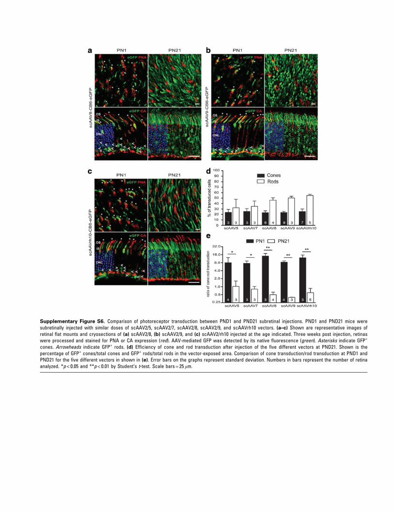
Supplementary Figure S6. Comparison of photoreceptor transduction between PND1 and PND21 subretinal injections. PND1 and PND21 mice weresubretinally injected with similar doses of scAAV2/5, scAAV2/7, scAAV2/8, scAAV2/9, and scAAVrh10 vectors. (a–c) Shown are representative images ofretinal flat mounts and cryosections of (a) scAAV2/8, (b) scAAV2/9, and (c) scAAV2/rh10 injected at the age indicated. Three weeks post injection, retinaswere processed and stained for PNA or CA expression (red). AAV-mediated GFP was detected by its native fluorescence (green). Asterisks indicate GFP+
cones. Arrowheads indicate GFP+ rods. (d) Efficiency of cone and rod transduction after injection of the five different vectors at PND21. Shown is thepercentage of GFP+ cones/total cones and GFP+ rods/total rods in the vector-exposed area. Comparison of cone transduction/rod transduction at PND1 andPND21 for the five different vectors in shown in (e). Error bars on the graphs represent standard deviation. Numbers in bars represent the number of retinaanalyzed. *p < 0.05 and **p < 0.01 by Student’s t-test. Scale bars = 25 lm.
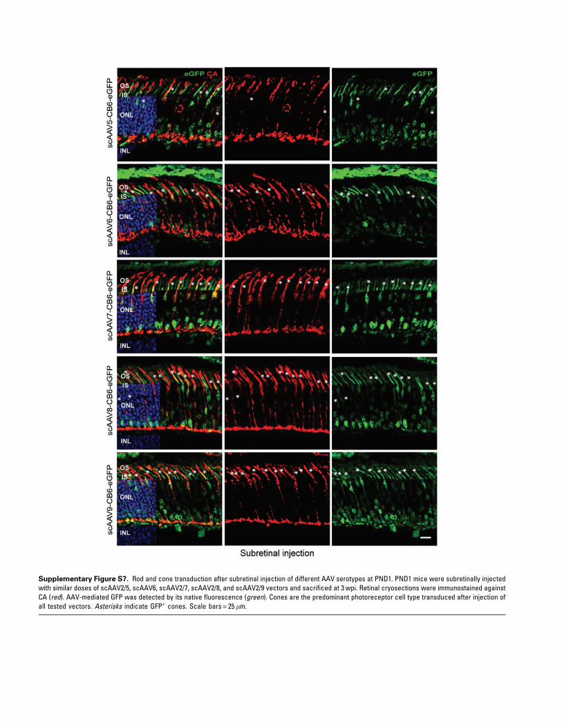
Supplementary Figure S7. Rod and cone transduction after subretinal injection of different AAV serotypes at PND1. PND1 mice were subretinally injectedwith similar doses of scAAV2/5, scAAV6, scAAV2/7, scAAV2/8, and scAAV2/9 vectors and sacrificed at 3 wpi. Retinal cryosections were immunostained againstCA (red). AAV-mediated GFP was detected by its native fluorescence (green). Cones are the predominant photoreceptor cell type transduced after injection ofall tested vectors. Asterisks indicate GFP+ cones. Scale bars = 25 lm.
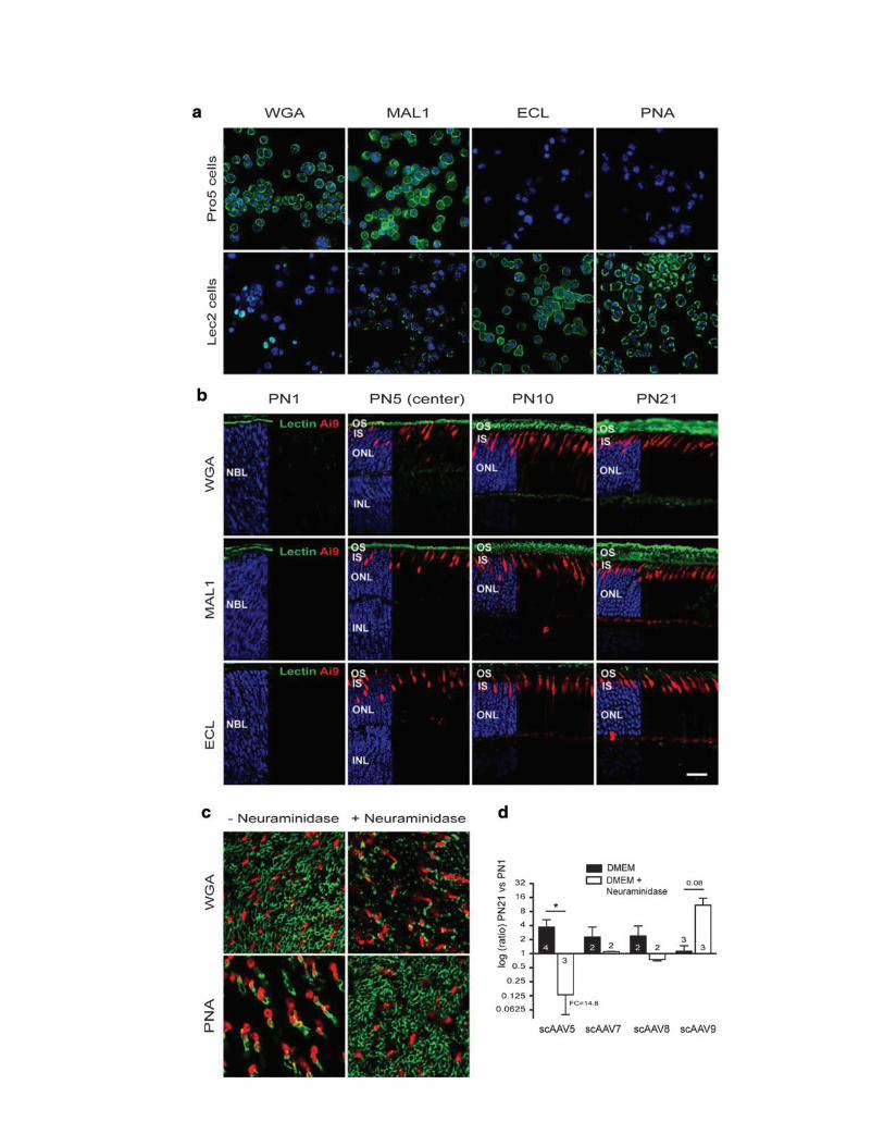

Supplementary Figure S9. Histological analysis of the inter-photoreceptor matrix (IPM) and outer limiting membrane (OLM) integrity in PND21 Rho-/- mice.(a) Retinal flat mounts of PND21 Rho-/- and Rho+/+ mice were labeled with Cy3-PNA (red) and fluorophore-conjugated WGA, MAL1, or ECL (green).Representative images are shown of experiments run in duplicate. Despite absence of rod outer segments, no change of the rod IPM structure is apparent atPND21 in Rho-/- mice. Slight swelling of cone matrix sheaths is observed. (b) Retinal cryosections of PND21 Rho-/- and Rho+/+ mice were labeled with Cy3-Phalloidin (which labels F-actin; red) and counterstained with DAPI (blue). Despite the absence of rod outer segments, no disruption of the OLM integrity isapparent at PND21 in Rho-/- mice. Scale bars = 25 lm.
Supplementary Figure S8. Analysis of IPM formation in the developing mouse retina and its interactions with AAV particles. To address the possibility thatthe IPM influences the transduction of PRs, first the development of the IPM in the mouse retina was examined from PND1 to PND21 using four differentlectins: WGA (a sialic-acid terminal sugar-specific lectin), MAL1 (a Sia[a2,3]-Gal[b1,4]GlcNAc and Gal[b1,4]GlcNAc terminal sugar-specific lectin), ECL (aGal[b1,4]GlcNAc terminal sugar-specific lectin), and PNA (a Gal[b1,4]GlcNac terminal sugar-specific lectin). (a) To verify the specificity of the lectins, Pro5(upper panels) and Lec2 (lower panels) CHO cell lines were stained with fluorophore-conjugated WGA, MAL1, ECL, and PNA lectins (green). DAPI labels thecell nuclei (blue). Lec2 cells lack terminal sialic acids due to a defect in CMV-sialic acid transport and express at their surface glycans with terminal galactosylresidues. As expected, Pro5 cells expressing sialylated glycans demonstrate preferential staining with WGA and MAL1. In contrast, Lec2 cells demonstratepreferential staining by ECL and PNA. (b) Retinal cross sections of PND1, PND5, PND10, and PND21 Ai9_MCre+ (labeling cones by td-Tomato; red) mice werelabeled with fluorophore-conjugated WGA, MAL1 or ECL (green) and counterstained with DAPI (blue). The sections show the increase of IPM duringphotoreceptor development. The different lectins confirm the presence of rod and cone IPM microenvironments with specific glycan compositions. WGA andMAL1 are specifically bound to the rod-associated IPM only. In contrast, only glycoproteins with terminal Gal and GlcNAc residues are detectable in the mousecone matrix sheaths and outer segments, as assessed by specific labeling using PNA and ECL. (c and d) Effect of neuraminidase treatment on AAV binding toex vivo retinal explants. Ex vivo retinal explants were used to limit the variability related to the surface of the retina exposed to the vectors after subretinalinjections at PND1 versus PND21. Retinal explants were pretreated with DMEM alone or DMEM plus neuraminidase and incubated with the same dose ofAAV5, -7, -8, and -9. (c) Loss of terminal Sia residues upon neuraminidase treatment was confirmed using WGA and PNA binding (green) in Ai9+_MCre+ adultretinas that specifically expressed td-Tomato in cones (red). As expected, a reduction in WGA labeling of the rod-associated IPM was observed, as well as theextension of PNA staining to both cone and rod IPM domains. (d) AAV binding on PND21 retinal explants was quantified by qPCR and compared to the bindingobtained on PND1 retinal explants. All experiments were carried out in triplicate. Results are shown as mean – SD. Numbers in bars represent the number ofretina analyzed. AAV particle binding to untreated control retinas was significantly increased from PND1 to PND21 for AAV2/5 (3.72-fold change), AAV2/7 (2.28-fold change), AAV2/8 (2.38-fold change), but not for AAV9. Neuraminidase treatment abrogated the increase of AAV5 binding over time, without affecting AAV7and AAV8 binding. In contrast, hydrolysis of sialic acid residues increased AAV9 binding over time by 10.9-fold. These results are in agreement with previouswork showing that (a2,3) N-linked Sia51–53 and terminal galactose54, 55 may mediate AAV5 and AAV9 transduction, respectively. They suggest that the rod-associated matrix selectively binds AAV5 while masking glycans that facilitate AAV9 binding in vitro. Notably, these differences in AAV binding to the IPM didnot denote a significant different capacity of transducing mouse PRs in vivo at PND21, considering the percentage of cell transduced. *p < 0.05 by Student’st-test. Scale bar = 25 lm. FC, fold-change.
‰