rodents - journal of neuroscience
TRANSCRIPT
Neurobiology of Disease
Recovery from Chronic Demyelination by Thyroid HormoneTherapy: Myelinogenesis Induction and Assessment byDiffusion Tensor Magnetic Resonance Imaging
Laura-Adela Harsan,1,2 Jerome Steibel,1 Anita Zaremba,3 Arnaud Agin,1 Remy Sapin,1 Patrick Poulet,1
Blandine Guignard,1 Nathalie Parizel,1 Daniel Grucker,1 Nelly Boehm,4 Robert H. Miller,3 and M. Said Ghandour1
1UMR 7191, Laboratoire d’Imagerie et de Neurosciences Cognitives, Faculte de Medecine, Universite Louis Pasteur and Centre National de la RechercheScientifique, 67085 Strasbourg, France, 2Medical Physics, Department of Diagnostic Radiology, University Hospital, 79106 Freiburg, Germany, 3CaseWestern Reserve University, Department of Neurosciences, School of Medicine, Cleveland, Ohio 44106, and 4INSERM U666 and Service Central deMicroscopie Electronique, Faculte de Medecine, Universite Louis Pasteur, 67085 Strasbourg, France
The failure of the remyelination processes in multiple sclerosis contributes to the formation of chronic demyelinated plaques that lead tosevere neurological deficits. Long-term cuprizone treatment of C57BL/6 mice resulted in pronounced white matter pathology character-ized by oligodendrocyte depletion, irreversible demyelination and persistent functional deficits after cuprizone withdrawal. The use of acombination of in vivo diffusion tensor magnetic resonance imaging (DT-MRI) and histological analyses allowed for an accurate longi-tudinal assessment of demyelination. Injection of triiodothyronine (T3 ) hormone over a 3 week interval after cuprizone withdrawalprogressively restored the normal DT-MRI phenotype accompanied by an improvement of clinical signs and remyelination. The effectsof T3 were not restricted to the later stages of remyelination but increased the expression of sonic hedgehog and the numbers of Olig2 �
and PSA-NCAM � precursors and proliferative cells. Our findings establish a role for T3 as an inducer of oligodendrocyte progenitor cellsin adult mouse brain following chronic demyelination.
Key words: myelin; oligodendrocytes; demyelination therapy; gliotoxic agent; DT-MRI; myelin repair
IntroductionMultiple sclerosis (MS) is an example of chronic inflammatorydemyelinating disease of the CNS, with a large heterogeneity inclinical course, MRI patterns and responses to therapy (Kornekand Lassmann, 2003). Several causes have been proposed for thefailure of long-term remyelination in MS and for each, therapeu-tic strategies have been developed (Pluchino et al., 2004). A crit-ical step in preclinical studies is, however, the development ofanimal models that can be examined in vivo in acute and chronicphases of the MS pathology to characterize the effects of the ap-plied therapy. In mice, the cuprizone-diet model (Ludwin, 1978;Blakemore, 1984; Matsushima and Morell, 2001) is of particularinterest because it allows the progression of demyelinated lesionsto a chronic state, depending on the duration of cuprizone ad-ministration. Effective spontaneous recovery does not occur in
brains of long-term cuprizone-treated mice and the model allowsthe testing of therapeutic strategies for remyelination.
It is likely that factors playing a role in the normal myelinationprocesses participate in the remyelination of the injured CNS.Particularly, molecules implicated in oligodendrocyte differenti-ation and maturation may act in the generation of positive signalsfor recovery. Thyroid hormones (THs) are necessary for normalaxonal myelination acting at multiple steps during oligodendro-cytes development and myelination, via nuclear hormone recep-tors (Baas et al., 1997; Rodríguez-Pena, 1999; Jones et al., 2003;Sarlieve et al., 2004; Schoonover et al., 2004; Kang et al., 2007),but no information are available about the role of TH in theinduction of oligodendrocyte lineage in chronic demyelination.We therefore, explored the possibility of stimulating endogenousrepair by triiodothyronine (T3) administration in long-termcuprizone-treated mice. Reparative responses were followed invivo by diffusion tensor magnetic resonance imaging (DT-MRI).
Highly sensitive to the water molecule motion, DT-MRI en-ables tissue structure to be probed and imaged on a microscopicscale, providing details of the cytoarchitecture of the neural tissueand identifying changes related to a pathological condition(LeBihan, 2003). In the white matter, the hydrophobic nature ofthe myelin membrane provides barriers for water diffusion andchanges in the permeability of these barriers, which are formedduring normal and pathologic development, generates modifica-tions in DT-MRI derived parameters. For example, examination
Received Sept. 17, 2008; accepted Oct. 27, 2008.This work was supported by grants from the Foundation of European Leukodystrophies Association to M.S.G. and
National Institutes of Health Grant NS 30800 to R.H.M. We thank M. Bury and D. Lam for their valuable technicalassistance. Guinea pig anti-mouse TSH antibody and TSH reference preparation used in the TSH assay were obtainedfrom Dr. A. F. Parlow, National Hormone and Peptide Program, Harbor–University of California, Los Angeles MedicalCenter, Los Angeles, CA.
Correspondence should be addressed to Dr. M. Said Ghandour, UMR 7191, Centre National de la RechercheScientifique/Universite Louis Pasteur, Faculte de Medecine, 11 rue Humann, 67085 Strasbourg, France. E-mail:[email protected].
DOI:10.1523/JNEUROSCI.4453-08.2008Copyright © 2008 Society for Neuroscience 0270-6474/08/2814189-13$15.00/0
The Journal of Neuroscience, December 24, 2008 • 28(52):14189 –14201 • 14189

of directional diffusivity perpendicular(D�) and parallel (D�) to the fiber tractsallows in vivo assessment of mouse braindysmyelination and spontaneous recovery(Song et al., 2003, 2005; Harsan et al.,2006, Mori and Zhang, 2006; Harsan et al.,2007).
While it is clear that T3 contributes tothe differentiation and maturation of oli-godendrocytes (Billon et al., 2001; Schoo-nover et al., 2004) its role in the regulationof the oligodendrocyte lineage in vivo andparticularly in adult brain is unclear. Thepresent findings establish a role for T3 as apotential inducer of oligodendrocyte pre-cursor cells (OPCs) in adult mouse brainin chronic demyelination caused by cupri-zone treatment. Moreover, we provide anaccurate assessment of demyelination andrecovery in vivo, in a longitudinal studythat combined DT-MRI and histologicalanalyses of T3-based therapy. We showclearly that T3 acts as a potent inducer ofoligodendrogenesis through the expres-sion of sonic hedgehog (Shh) and Olig2transcription factors and a promoter ofmyelin regeneration in the chronic demy-elination of adult mouse brain.
Materials and MethodsAnimals and treatmentsThree groups of 8-week-old C57BL/6 femalemice, purchased from Janvier Breeding Centerwere used for DT-MRI exam at different timepoints as presented in the Figure 1. Duplicatesfor each group were kept in the same conditionsof housing and treatment and used for the his-topathological examination.
Group 1. Eight female mice were placedon a diet containing 0.2% cuprizone(bis-cyclohexanone-oxaldihydrazone, Sigma)mixed into milled chow and available ad libi-tum, for 12 weeks (Fig. 1). A cuprizone-free dietfollowed during the next 12 weeks before death.
Group 2. Eight females (Fig. 1, group 2) weresubjected to cuprizone diet as above for 12weeks followed by normal diet until death. Im-mediately after cuprizone-treatment arrest themice received daily IP injections of T3 hormone(0.3 �g/g body weight) during the following 3 weeks. The mice werefurther housed under normal diet for other 9 weeks before death.
Group 3. Eight females (Fig. 1) serving as controls, were bred for 24weeks in the same house conditions, free of any treatment and normallynourished.
DT-MRI exams and the histology were conducted at 0 and 12 weeks ofcuprizone diet (w0 and w12), as well as at 3, 6, and 12 weeks after cupri-zone removal (w12 � 3, w12 � 6, and w12 � 12, respectively). Figure 1presents the treatment groups and the timing of DT-MRI and histologi-cal examinations.
DT-MRI acquisition and processingThe gaseous anesthesia was achieved in mice by using a mixture of 4%isoflurane (Forene) and oxygen for induction during 3 min and 1.5% formaintenance. The mask placed on the animal nose was connected to theinflow line but also to the outflow line, to remove excess isoflurane fromthe magnet bore. The animals were placed in a stereotaxic device toimmobilize the head, with integrated heating facility to maintain the
body temperature at 37°C. An anatomically shaped 1H surface coil forsmall animals (Rapid Biomedical) was placed on the head to serve as areceiver for the magnetic resonance (MR) signal. The system was thenpositioned into a 1H resonator (Rapid Biomedical) for rats and mice,which entered into a 20 cm 4.7 T MR magnet equipped with self-shieldgradient coils from Magnex Scientific. The MS spectrometer is fromS.M.I.S. (now M.R.R.S.). Before the acquisition of images set required forcomputing the diffusion tensor, a fast spin-echo sequence was used toobtain T2-weighted images as sagittal multislices that cover all the brain.The acquisition parameters were as following (for 4 echoes in the echotrain): repetition time (TR) � 3.8 s; spin-echo time (TE) � 40 ms; field ofview (FOV) � 20 � 20 mm 2, with data matrix 256 � 256 (zero-filled to512 � 512) and an average of 4. The slice’s thickness was 0.5 mm.
For diffusion-weighted images (DWI) acquisition, a conventionalspin-echo imaging sequence modified by adding the Stejskal-Tanner dif-fusion gradient pair was used. Brain sagittal slices were acquired over 2.5h for each mouse, with a TR of 1.5 s, TE of 35 ms, time (�) between theapplication of diffusion gradient pulses of 21.7 ms, diffusion gradient
Figure 1. Experimental groups and summary of DT-MRI and histological examinations. A, The longitudinal examination wasconducted starting at w0 time point (week before any treatment) and continuing at w12 (week 12 of cuprizone treatment), w12� 3, w12 � 6, and w12 � 12 (at 3, 6, and 12 weeks after cuprizone removal). B, Results of rotarod test, performed twice a weekin each experimental group. The mice were considered as unaffected when performing minimum 2 min (120 s) on the rotarod. Thefigure shows normal scores for the mice of control group, while the mice subjected to cuprizone diet show clinical signs from thefourth week of regimen, with a relapsing remitting pattern. The mice receiving T3 injections after cuprizone withdrawal (Cupri-zone �T3 group) improve considerably their clinical score during the next 12 weeks of observation. On the contrary, only weakimprovements were quantified in this period for the mice allowed to recover spontaneously, without any stimulation (Cuprizonegroup).
14190 • J. Neurosci., December 24, 2008 • 28(52):14189 –14201 Harsan et al. • Recovery from Demyelination by Thyroid Hormone Therapy

duration (�) of 5.6 ms, ramp time of 400 �s and gradient amplitude ( G)of 0.135 T/m. The slice thickness was 1 mm, FOV 20 � 20 mm 2 and datamatrix 256 � 256 (zero-filled to 512 � 512). Diffusion-sensitizing gra-dients of the same amplitude were applied along six different directionsdefined by the six unit vectors: (gx, gy, gz) � (1, 0, 0); (0, 1, 0); (0, 0, 1); (1,1, 0)/�2; (0, 1, 1)/�2; (1, 0, 1)/�2. The used b factor (Le Bihan et al.,1986) values were 0 and 865 s/mm 2.
Data processing was performed using software written in Matlab.From the diffusion tensor computation for each voxel of the image(Basser et al., 1994) the 3 eigenvalues �1, �2, �3 (�1 � �2 � �3) and theircorresponding eigenvectors (e1, e2, e3) were derived. It is generally as-sumed that in the CNS environment the main diffusivity (�1) representsthe diffusion parallel to the fiber tracts (axial diffusivity, D���1) and themean of the other 2 eigenvalues express the diffusion perpendicular tothe tracts, radial diffusion (D��(�2 � �3)/2). By using these eigenvalues,the mean diffusivity �D� was also calculated and maps were generated.Fractional anisotropy (FA), which is related to the presence of orientedstructures, giving rise to preferred diffusion orientations, was also calcu-lated to yield values from 0 to 1. For an isotropic medium (�1��2��3),FA � 0, while for an anisotropic medium (for example �1���2��3),FA � 1. Generally brain FA maps show the CSF as isotropic medium (lowvalues of FA) and the highly oriented white matter tracts as anisotropicstructures (high FA values). Regions of interest (ROIs) were manuallydefined (Fig. 2) using a mouse brain atlas as reference (Paxinos andFranklin, 2001).
Statistical analysis was performed using ANOVA, followed by Bonfer-roni corrections for multiple testing, to quantify the effects of cuprizonediet and age on DT-MRI indices. The analysis showed a significant effectof the cuprizone intoxication at p � 0.05. The same tests were used toquantify the TH effects for remyelination, as expressed by changes ofDT-MRI parameter values. The results for each ROI were expressed asmean � SD. Difference was considered statistically significant at p �0.05.
Histological analysisImmunohistofluorescence. Mice for histological analysis were killed underpentobarbital deep anesthesia and perfused through the left ventriclewith freshly prepared solution of 4% paraformaldehyde (PFA) in phos-phate buffer (0.1 M, pH 7.5, PBS). Further fixation was achieved by main-taining the brains overnight in the same fixative. The tissues were nextembedded in paraffin wax and 5 �m thick sagittal sections were madeusing the microtome Leica (Leica Instruments).
Double immunolabeling with a rabbit antibody against carbonic an-hydrase II (CA II at 1:200 dilution) and a mouse monoclonal antibodyagainst guinea pig myelin basic protein (MBP at 1:10 dilution) (bothprepared in our laboratory) was performed according to the procedurepreviously described (Harsan et al., 2004). The oligodendrocytes markedby CA II antibody were counted in the total length of corpus callosum(genu, body, and splenium) in the different groups of mice. A minimumof six sagittal sections (at the levels 0.25, 0.50, and 0.75 mm laterally, inboth hemispheres) from each animal (n � 4 for each time point in eachexperimental group) were captured at a 40� magnification using a BX60microscope, equipped with DP70 digital camera (Olympus). The imageswere analyzed and the oligodendrocytes counted using the NIH ImageJsoftware.
To estimate the number of proliferating oligodendrocyte, we per-formed double labeling with a mouse antibody against proliferation cellnuclear antigen (PCNA, 1:100 dilution) (Santa Cruz Biotechnology) andthe rabbit CA II antibody, on paraffin sections. The secondary antibodieswere Alexa Fluor 488-conjugated anti-rabbit IgG (1:200) and Alexa Fluor546-conjugated anti-mouse IgG (1:200) (Invitrogen). We also used adouble immunolabeling procedure to label the proliferating cells usinganti-bromodeoxyuridine (BrdU at 1:50 dilution) antibody after BrdUincorporation. CA II antibody was used for the double labeling as de-scribed in the study by Jalabi et al. (2005). Mice received daily injectionsof 100 �g/g body weight BrdU, during the 4 d previous to perfusion.
The mouse monoclonal anti-polysialic acid (PSA-NCAM, 1:400) aswell as the rabbit polyclonal anti-NG2 chondroitin sulfate proteoglycan
(1:100, Millipore) antibodies were used to detect the migrating cells andthe oligodendrocytes precursors respectively.
A rat anti-mouse CD45 (leukocyte common antigen) at 1:50 dilutionwas also used to detect the reactive gliosis, while the mouse monoclonalglial fibrillary acidic protein antibody (GFAP, 1:300, Dako) was used forthe astrocytosis detection. The sections were examined with BX60 micro-scope equipped with DP70 digital camera (Olympus).
Immunohistochemistry. Paraffin-embedded sections from fixed brainsor cut as frozen sections were used. Sections were subject to antigenretrieval using 10 mM sodium citrate buffer containing 0.05% Tween 20at pH 6.0 for 10 min at 98°C then removed from heat and allowed to coolin buffer at room temperature (RT) for 20 min. Sections were rinsed for10 min in TBS at pH 6.0, followed by serial rinses in 1�PBS, pH 7.4. Allsections were preblocked in PBS containing 5–10% normal goat serumand 0.1– 0.3% Triton X-100 for 1–2 h at RT. They were then incubated inblock containing one of the following primary antibodies at 4°C over-night: Olig-2 at 1:500 (AB9610 Millipore); MBP at 1:500 (Clone SMI-99Covance); Shh at 1:100 (SC-9024 Santa Cruz) and GFAP at 1:500 (Z0334Dako). The next day, sections were rinsed in 1� PBS then incubated inthe appropriate biotinylated secondary for 1 h at RT: goat anti-mouseIgG at 1:200 (55587 ICN) or goat anti-rabbit IgG at 1:250 (AP132 BMillipore). Signal was amplified using an Avidin-Biotin Complex (PK-6100 Vector) for 30 min at RT and visualized by immunoperoxidasereaction using DAB (SK-4100 Vector). Olig2 immunohistochemistry byusing Elite Vecta Stain rabbit ABC kit (Vector Laboratories) was alsocombined with CA II immunohistofluorescence, as described above forvibratomed brain slices. Sections were then dehydrated and mounted inPermount and photographed on a Leica DM5000 B microscope using aDFC-500 camera with Leica Application Software or with BX60 micro-scope equipped with DP70 digital camera (Olympus).
Electron microscopy. At each experimental time point and for eachexperimental group of animals, mice were specially perfused with 2%PFA and 6% glutaraldehyde (Sigma) for electronic microscopy examina-tion of the brain. Sagittal brain sections (100 �m) were prepared andafterward kept overnight in the same fixative. Tissue slices were embed-ded in epoxy resin. Five to ten 1 �m semithin sections from brains werestained with toluidine blue, analyzed under �100 objective and photo-graphs taken. Myelinated axons were counted all over the corpuscallosum.
Sixty-nanometer ultrathin sections were also examined in a transmis-sion electron microscope (Siemens Elmiskop 102) at 60 kV. The calibersof 1500 axons from similar regions of the corpus callosum were measuredin electron micrographs for each examined brain, by using NIH ImageJplugins.
Thyroid hormone assaysFour hundred microliters of blood was collected from each mouse inheparin-treated tubes at the end of cuprizone treatment (w12) and fromnontreated control mice. Plasma samples were obtained after blood cen-trifugation and then stored at �20°C. Total T4, total T3, and thyroidstimulating hormone (TSH) determinations were performed as previ-ously described (Streckfuss et al., 2005) with one modification in the TSHassay: 125I-labeled rat TSH was obtained from BioCode-Hycel(Belgium).
All the experimental procedures were performed in accordance withthe guidelines of Animal Care Committee of Louis Pasteur University,Strasbourg, France.
ResultsTo assess the effects of T3-based therapy, we induced severe whitematter pathology in C57BL/6 mice, by 12 weeks of cuprizone diet.The cuprizone is a well described gliotoxic agent that alters thefunctions of oligodendrocyte mitochondria resulting in celldeath. Importantly, our results showed that long-term cuprizonetreatment does not affect significantly the thyroid hormones lev-els, T3 and T4, nor the TSH in the present model of chronicdemyelination in adult mice (Table 1). The death by apoptosis ofoligodendrocytes results in myelin breakdown and underlies the
Harsan et al. • Recovery from Demyelination by Thyroid Hormone Therapy J. Neurosci., December 24, 2008 • 28(52):14189 –14201 • 14191

progression of the disease toward a chronic state. The experimen-tal program and the established animal groups examined in DT-MRI and histology are presented in Figure 1A (see also Materialsand Methods).
Clinical course of the diseaseDuring the period of cuprizone ingestion mice in groups 1 and 2(Fig. 1B) showed clinical signs of demyelination (loss of bodyweight, seizures, loss of coordinate movements and locomotors
Figure 2. A, B, Representative sagittal brain T2-weighted images of individuals from groups 1 (A) and 2 (B) examined at different time points. At w0, corresponding to the exam prior to thecuprizone ingestion, the myelinated areas, corpus callosum (arrows) and cerebellum showed hypointense signals. Enlarged ventricles corresponding to the hyperintense signal are evident after 12weeks of cuprizone diet in both groups (w12). No significant changes of the T2 images pattern are observed during the w12 to w12 � 12 period in mice from group 1 (A). Partial regain in thehypointensities of corpus callosum and cerebellum are noticed in the group receiving T3 injections (group 2, B) during w12 to w12 � 12 time interval, with spectacular decrease in the ventriclevolumes. The T2-weighted images pattern in control mice is identical with that observed at w0 time point in groups 1 and 2 and is not presented. C, Investigated ROIs overlaid on a sagittal DWI ofa normal mouse brain at w0.
Table 1. T3, T4, and TSH levels measured in blood plasma collected from control and cuprizone-treated mice at the end of intoxication period
T3 ng/ml T4 ng/ml TSH ng/ml
w12 cuprizone-treated and control mice Control Cuprizone Control Cuprizone Control Cuprizone
Mean 0.80 0.79 41.45 35.85 152 190SD 0.17 0.17 9.5 5.85 38.26 55.14No. of animals 10 11 6 6 6 6
t test was not significant for any group.
14192 • J. Neurosci., December 24, 2008 • 28(52):14189 –14201 Harsan et al. • Recovery from Demyelination by Thyroid Hormone Therapy

disabilities assessed on the rotarod) starting at the fourth week(w4) of cuprizone administration, with a relapsing remitting pat-tern and gradually increasing severity till the end of the treat-ment. No significant improvements of the clinical score weredetected in group 1, after returning to the normal diet (Fig. 1B),while the mice that received daily T3 injections at the end of thecuprizone diet showed a gradual significant improvement of clin-ical scores during the w12 to w12 � 12 period (Fig. 1B). Thelocomotor disabilities were assessed by performing the rotarodtest twice a week over a 24 week interval (Fig. 1B).
T2-weighted imagesAcquired T2-weighted images (T2-transverse relaxation timeconstant) revealed dramatic effects of the cuprizone intoxicationon mouse brains at w12 (Fig. 2A,B). Corpora callosa showed ahypointense signal at w0, corresponding to a well myelinatedstructure (because T2 has short values in myelin) before cupri-zone treatment (Fig. 2A,B). This pattern was maintained in allexamined ages of mice receiving normal diet (data not shown).
The first pathologic signs were clearly observed at w12 in T2-weighted images of mice brains receiving cuprizone diet (groups1 and 2). CSF was observed as a hyperintense signal in the en-larged ventricular regions and infiltrating the surrounding struc-
tures (Fig. 2A,B, w12). Simultaneously, the hypointense signal inT2-weighted images was lost in the corpus callosum after 12weeks of cuprizone treatment, corresponding to the myelinbreakdown.
Restoring a normal diet had no effect on the appearance ofbrain T2 images of group 1 (Fig. 2A). The hyperintense signal,denoting major vasogenic edema and inflammation combinedwith an enlargement of the ventricles, was visible at all MRI time-points while corpus callosum and neighboring structures werenot clearly identified.
In contrast, brain images of T3-treated mice of group 2,showed normal ventricle size as early as w12 � 3 and progres-sively, a hypointense signal was regained in the corpora callosaand cerebella (Fig. 2B). However, a quantitative assessment of themyelination state from the T2-weighted images was not possibleand further DT-MRI was performed.
DT-MRI and histopathological observationDWI, as well as the FA maps generated after diffusion tensorcomputation provide good contrast for identifying the brainmacrostructures and for choosing the regions of interest (ROIs)(Fig. 2C). High FA values or high anisotropy are usually found innormal white matter tracts, whereas low FA values are generally
Figure 3. A, B, Fractional anisotropy maps of representative individuals from experimental groups 1 (A) and 2 (B), followed longitudinally at different time points. Normal brain images at w0show high anisotropic values in the well formed corpus callosum (white arrows). FA maps resulted from DT-MRI exams at w12 display very low values of this parameter all along the corpus callosum.No significant recovery of the FA is visible during the 12 weeks after the cuprizone removal in the brain images of mouse belonging to group 1 (A). Gradual increase in the FA is observed in the brainwhite matter of the mouse receiving T3 treatment after returning to normal diet (B, w12 � 3; w12 � 6; w12 � 12). The FA pattern in control mice is identical with that observed at w0 time pointin group 1 and 2 and is not presented. FA values are distributed on a scale from 0 (blue) to 1 (red).
Harsan et al. • Recovery from Demyelination by Thyroid Hormone Therapy J. Neurosci., December 24, 2008 • 28(52):14189 –14201 • 14193
measured in isotropic tissues where water diffuses equally in alldirections (like in CSF or to a lesser degree in gray matter orabnormal white matter tracts).
Cuprizone intoxication has been demonstrated to differen-tially induce demyelination in the mouse brain structures, havingvery significant effects on the corpus callosum (Blakemore, 1984;Komoly, 2005) while other regions are less affected (Tansey et al.,1996). Therefore, two separate areas of corpus callosum, the sple-nium and the genu were selected for analyses, along with otherregions (Fig. 2C). DT-MRI and histological analyses showed thatthe consequence of the cuprizone ingestion was anatomicallyvariable throughout the brain, with statistically significant effectsquantified by DT-MRI only in the corpus callosum and the cer-ebellum. Therefore, these regions were selected for detailed datapresentation.
Cuprizone intoxication effectsDT-MRI was performed on all animals before the onset of cupri-zone treatment, corresponding to w0 time point, the fully my-elinated state. FA maps showed high anisotropy of the white mat-ter, especially in the corpus callosum, as well as in the cerebellar
folia (Fig. 3A,B, w0). The regions that showed the greatest anisot-ropy corresponded to the areas with strong staining intensity ofmyelin on histological sections (Fig. 4). Staining with an anti-myelin basic protein (MBP) antibody revealed well myelinatednormal brains at 8 weeks of age (w0) (Fig. 4A). At this age, manycarbonic anhydase II (CA II)-positive oligodendrocytes were de-tected throughout the brains and predominantly in the whitematter (Fig. 4A). Few cells that expressed proliferation nuclearantigen (PCNA) and low numbers of BrdU� cells were present inthe corpus callosum. Limited number of PSA-NCAM� cells wasobserved and these were restricted to the rostral migratory streamwith no staining detected in the corpus callosum. A small numberof oligodendrocyte precursors NG2� cells were detected.
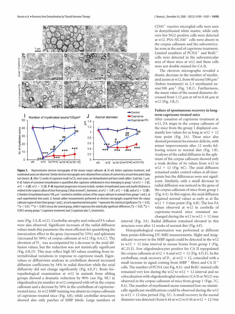
Electron micrographs showed 40 myelinated axons/100�m 2 in the corpus callosum, with a mean axonal diameter of 1.12�m, in all the groups of mice at w0 (Fig. 5 I, J). No significantchanges of DT-MRI and histopathology data were observed incontrol animals from w0 to w12 � 12.
The brain FA maps of all mice subjected to 12 weeks (w12)cuprizone diet (groups 1 and 2) showed abnormally decreasedvalues in the white matter areas, and especially the corpus callo-
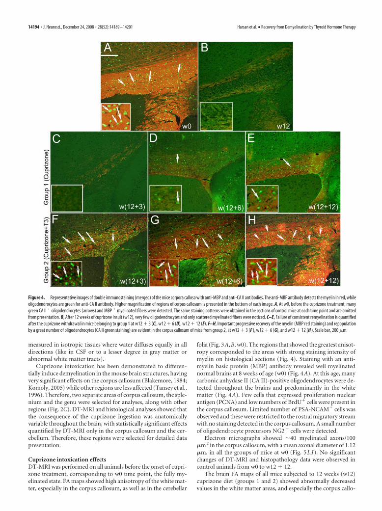
Figure 4. Representative images of double immunostaining (merged) of the mice corpora callosa with anti-MBP and anti-CA II antibodies. The anti-MBP antibody detects the myelin in red, whileoligodendrocytes are green for anti-CA II antibody. Higher magnification of regions of corpus callosum is presented in the bottom of each image. A, At w0, before the cuprizone treatment, manygreen CA II � oligodendrocytes (arrows) and MBP � myelinated fibers were detected. The same staining patterns were obtained in the sections of control mice at each time point and are omittedfrom presentation. B, After 12 weeks of cuprizone insult (w12), very few oligodendrocytes and only scattered myelinated fibers were noticed. C–E, Failure of consistent remyelination is quantifiedafter the cuprizone withdrawal in mice belonging to group 1 at w12 � 3 (C), w12 � 6 (D), w12 � 12 (E). F–H, Important progressive recovery of the myelin (MBP red staining) and repopulationby a great number of oligodendrocytes (CA II green staining) are evident in the corpus callosum of mice from group 2, at w12 � 3 (F ), w12 � 6 (G), and w12 � 12 (H ). Scale bar, 200 �m.
14194 • J. Neurosci., December 24, 2008 • 28(52):14189 –14201 Harsan et al. • Recovery from Demyelination by Thyroid Hormone Therapy
sum (Fig. 3A,B, w12). Cerebellar atrophy and reduced FA valueswere also observed. Significant increases of the radial diffusionvalues made this parameter the most efficient for quantifying theintoxication effect in the genu (increased by 53%) and splenium(increased by 50%) of corpus callosum at w12 (Fig. 6A,C). Theelevation of D� was accompanied by a decrease in the axial dif-fusion values, but the reduction was not statistically significant(Fig. 6B,D). This may reflect high SD values resulting from in-terindividual variations in response to cuprizone insult. Eigen-values or diffusivities analyses in cerebellum showed increaseddiffusion coefficients by 34% in radial direction while the axialdiffusivity did not change significantly (Fig. 6E,F). Brain his-topathological examination at w12 in animals from siblinggroups showed a dramatic reduction by 90% (see Fig. 9K) inoligodendrocyte number at w12 compared with w0 in the corpuscallosum and a decrease by 50% in the cerebellum of cuprizone-treated mice. At w12 MBP staining was absent in corpus callosumof cuprizone-treated mice (Fig. 4B), while cerebellar structuresshowed also only patches of MBP labels. Large numbers of
CD45� reactive microglial cells were seenin demyelinated white matter, while onlyvery few NG2-positive cells were detectedat w12. PSA-NCAM� cells were absent inthe corpus callosum and the subventricu-lar zone at the end of cuprizone treatment.Limited numbers of PCNA� and BrdU�
cells were detected in the subventriculararea of these mice at w12 and these cellswere not double stained for CA II.
The electron micrographs revealed adrastic decrease in the number of myelin-ated axons at w12, from 40 axons/100 �m 2
(before treatment) to 2.3 myelinated ax-ons/100 �m 2 (Fig. 5B, I). Furthermore,the mean value of the axonal diameter de-creased from 1.12 �m at w0 to 0.44 �m atw12 (Fig. 5B, J).
Failure of spontaneous recovery in long-term cuprizone-treated miceAfter cessation of cuprizone treatment atw12, FA maps in the corpus callosum ofthe mice from the group 1 displayed con-stantly low values for as long as w12 � 12time point (Fig. 3A). These mice alsoshowed persistent locomotor deficits, withminor improvements after 12 weeks fol-lowing return to normal diet (Fig. 1B).Analyses of the radial diffusion in the sple-nium of the corpus callosum showed onlya weak decline of its values from w12 tow12 � 12 (Fig. 6C). The axial diffusionremained under control values at all time-points but the differences were not signif-icant. Similarly, only a small decrease inradial diffusion was noticed in the genu ofthe corpus callosum of mice from group 1(Fig. 6A). In this region, the axial diffusionregained normal values as early as at thew12 � 3 time point (Fig. 6B). The low FAvalues observed at w12 in cerebella ofcuprizone-treated mice remained un-changed during the w12 to w12 � 12 time
interval (Fig. 3A). Radial diffusion remained elevated in thisstructure even after 12 weeks of normal diet (Fig. 6E).
Histopathological examination was performed at differenttime points following DT-MRI measurements. Slight and insig-nificant recovery in the MBP signal could be detected in the w12to w12 � 12 time interval in mouse brains from group 1 (Fig.4C,D,E). Few oligodendrocytes positive for CA II repopulatedthe corpus callosum at w12 � 6 and w12 � 12 (Fig. 4D,E). In thecerebellum, weak recovery of D� at w12 � 12, coincided with asmall increase in signal coming from MBP� fibers and CA II�
cells. The number of PCNA (see Fig. 8A)- and BrdU-stained cellsremained very low during the w12 to w12 � 12 interval and nocolocalization with oligodendroglial markers (CA II or NG2) wasobserved in the corpus callosum of mice from group 1 (Figs. 7C,8A). The number of myelinated axons remained low; no statisti-cally significant modifications could be observed during the w12to w12 � 12 time period (Fig. 5I). A small recovery in the axonaldiameter was detected (from 0.44 at w12 to 0.56 at w12 � 12) but
Figure 5. Representative electron micrographs of the mouse corpus callosum. A, At w0, before cuprizone treatment, wellmyelinated axons are observed. Similar electron micrographs were obtained from sections of control mice at each time point (datanot shown). B, After 12 weeks of cuprizone insult (w12), most axons are demyelinated and have small caliber. Scale bar, 1 �m.C–E, Failure of consistent remyelination is quantified after cuprizone withdrawal in mice belonging to group 1 at w12 � 3 (C),w12 � 6 (D), w12 � 12 (E). F–H, Important progressive recovery in both, number of myelinated axons and myelin thickness isevident in the corpora callosa of mice from group 2 (that received T3 hormone), at w12 � 3 (F ), w12 � 6 (G), and w12 � 12 (H ).I, Number of myelinated axons/100 �m 2 counted in semithin sections of the corpus callosum in animals from groups 1 and 2, ateach experimental time point. J, Axonal caliber measurements performed on electron micrographs acquired from the corpuscallosum region of mice from groups 1 and 2, at each experimental time point. * represents the statistical significance (*p � 0.05,**p � 0.01, ***p � 0.001) versus the control group, while # expresses the statistically significant differences (#p � 0.05, ##p �0.001) among groups 1 (cuprizone treatment) and 2 (cuprizone plus T3 treatments).
Harsan et al. • Recovery from Demyelination by Thyroid Hormone Therapy J. Neurosci., December 24, 2008 • 28(52):14189 –14201 • 14195

the increase was not statistically significantdue to large SDs (Fig. 5J).
T3 treatment effects assessed by DT-MRI and histologyInjection of T3 hormone into mice ofgroup 2 induced temporary hyperthyroid-ism, as attested by significantly high T3
level in the blood at w12 � 3 (data notshown). This hyperthyroidism was tran-sient, since at w12 � 6 all mice had normalvalues of circulating T3 in the blood-stream. Administration of T3 however, in-fluences the temporal evolution of DT-MRI derived parameter’s values,calculated in the corpus callosum and cer-ebellum of cuprizone-treated mice. Theabnormally low values displayed in the FAmaps at w12 (Fig. 3B) were progressivelyreplaced by higher coefficients, reachingnormal values in all investigated ROIs atw12 � 12 (Fig. 3B). The most obvious pe-riod of recovery was the w12 to w12 � 6interval in the genu and splenium of thecorpus callosum while the cerebella of T3-treated mice displayed high FA in the mapsby w12 � 3 (Fig. 3B).
The radial diffusion measured from thesplenium of the corpus callosum in the re-covery phase showed a gradual decreasefrom w12 to reach normal values at w12 �6 (Fig. 6A), while the recovery was delayedin the genu of corpus callosum andreached normal values 6 weeks later at w12� 12 (Fig. 6B). The cerebellum showed aquick recovery in D� values by w12 � 3(Fig. 6E). An estimate of the axial diffusionin the corpus callosum and cerebellum ofT3-injected mice revealed normal values ateach time point of the recovery phase (Fig.6B,D,F).
Histological examination at w12 � 3 ofbrain tissue of animals subjected to T3
treatment revealed a great number of CA II� cells repopulatingthe corpus callosum and the cerebellum (Figs. 4, 9K). The num-ber of these cells increased further, at w12 � 6 (Fig. 4G) and w12� 12 (Fig. 4H). At w12 � 3, MBP� staining was already detectedin both splenium and genu of the corpus callosum, and the stain-ing intensity gradually increased with age during the recoveryphase (Fig. 4F,G,H). Reduced MBP labeling was seen in smallareas along the corpus callosum reflecting irregular recovery ofthe oligodendrocyte population and myelin sheath formation.
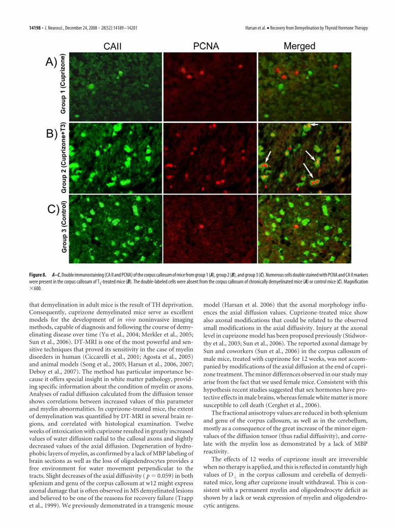
The effect of T3 was not restricted to later stages of the oligo-dendrocyte lineage. Cuprizone intoxication resulted in signifi-cant reduction in the number of Olig2� cells in the splenium ofthe corpus callosum that was reversed by T3 (Fig. 9A–C,J). Anincreased number of PCNA� and BrdU� proliferating cells weredetected in mouse brains of T3-treated mice at w12 � 3 (Fig. 8B).Approximately 60% of the proliferating cells were also doublestained for the CA II marker (Fig. 8B). NG2-positive cells werealso observed at the w12 � 3 and w12 � 6 (Fig. 7C), as were apopulation of PSA-NCAM� cells located all along the corpus
callosum and in the subventricular zone of T3-treated mice (Fig.7B).
These increases in the number of early oligodendrocyte pro-genitor cells in T3-treated animals suggest this treatment modu-lates the expression of cues that influence induction or survival ofearly oligodendrocytes progenitors. A major regulator of earlyOPC development is the morphogen Shh (Orentas and Miller1996; Davies and Miller, 2001) and in T3-treated animals in-creased expression of Shh was detectable in the subventricualrzone (data not shown) and particularly visible in the severelydemyelinated areas as shown in splenium of corpus callosum(Fig. 9D,E,F). The increase in Shh expression was correlated witha gradual increase in the number of Olig2� cells (Fig. 9A–C) afterT3 treatment, predominantly observed in the corpus callosum(Fig. 9J) but also in the subventricular zone (at w12 � 3: 39.4Olig2� cells/field of view in control group, 12.1 Olig2� cells perfield of view in cuprizone-treated group and 28.9 Olig2� cells perfield of view in cuprizone �T3-treated group) and cortex (at w12� 3: 9.15 Olig2� cells/field of view in control group, 4.14 Olig2�
cells/field of view in cuprizone-treated group and 11.1 Olig2�
Figure 6. A–F, Time course of D� (A, C, E) and D� (B, D, F ) in the genu (A, B) and splenium (C, D) of corpus callosum andcerebellum (E, F ) in all experimental groups. * represents the statistical significance (*p � 0.05, **p � 0.001) versus the controlgroup, while # expresses the statistically significant differences (#p � 0.05, ##p � 0.001) among groups 1 (cuprizone treatment)and 2 (cuprizone �T3 treatments).
14196 • J. Neurosci., December 24, 2008 • 28(52):14189 –14201 Harsan et al. • Recovery from Demyelination by Thyroid Hormone Therapy

cells/field of view in cuprizone � T3-treated group). The striatumregion is much less affected. Importantly, the reappearance ofShh� and Olig2�cells under the effect of T3 leads to the normal-ization of MBP expression (Fig. 9G–I). These data suggest that T3
treatment provides an environment that is conducive for the sub-sequent development of OPCs as well as regulating their differ-entiation. In the corpus callosum of cuprizone-treated mice, T3
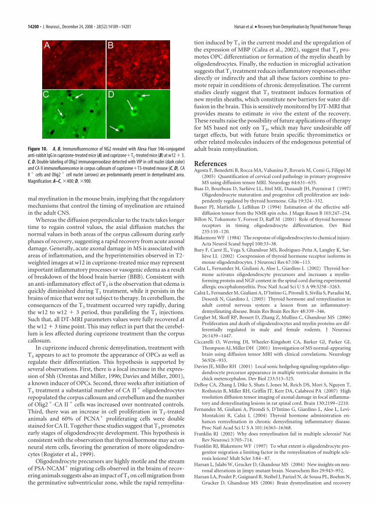
induces NG2� cells (Fig. 10B) and the majority of Olig2� cellsare CA II� (Fig. 10C,D) while the rest are astrocytes (data notshown).
The number of CD45� microglial cells decreased gradually inT3-treated mice at w12 � 3 (Fig. 7A) and throughout the treat-ment period, demonstrating an effect of T3 on the levels of reac-tive microglia.
Ultrastructural analyses revealed a rapid and sustained effectof the T3, with an increase in the number of myelinated axonsduring the recovery phase (Fig. 5F,G,H) as well as by the pro-gressive increase in the axonal caliber that approximated controlvalues by w12 � 12 in the corpus callosum while the cerebellumshowed earlier recovery.
DiscussionRemyelination ultimately fails in the brains of the majority of MSpatients and many studies have tried to elucidate the causes anddesign therapies to stimulate and induce recovery in MS plaques(Franklin, 2002). Particularly, chronic lesions are unresponsive
to various applied treatments in human (Rovaris et al., 2006) andanimal models (Franklin and Blakemore, 1997). The presentstudy provides in vivo evidence that thyroid hormone therapy(T3) is effective in promoting remyelination in a chronically de-myelinated mouse model of MS. The study successfully exploitedthe sensitivity of the in vivo noninvasive DT-MRI technique forlongitudinal following the efficacy of T3-based therapeutic effectsin cuprizone-induced demyelination.
Assessment of cuprizone effects in brainThe cuprizone is a well known gliotoxic agent acting as a selectiveand sensitive copper chelator. Several papers reported that ani-mals treated with various doses of cuprizone develop differentforms of neuropathology, involving in most cases demyelination(Matsushima and Morell, 2001). The exact mechanisms of toxinaction are not well understood. It is known, however, that cupri-zone administration alters the functioning of the mitochondriain the brain oligodendrocytes (Venturini, 1973). Long-term 0.2%cuprizone feeding produces an interesting model of MS that en-compasses the progression of acute demyelinated lesions to achronic state (Mason et al., 2004) that fail to recover after toxinwithdrawal. Both oligodendrocytes and progenitors become pro-gressively depleted in chronically demyelinated lesions (Mason etal., 2004). It is interesting to note that long term cuprizone treat-ment in mice does not affect thyroid hormones (T3 and T4) andthe TSH blood circulating levels which exclude the possibility
Figure 7. A–C, Brain section immunolabeling with anti-CD45 (A), anti-PSA-NCAM (B), and anti-NG2 (C) obtained at w12 � 3 in the corpus callosum of animals from groups 1, 2, and 3. We choseto present the data at w12�3 because they are the most representative for the comparison between mice receiving T3 treatment and remyelinating (group 2) and chronic demyelinated mice (group1). Magnification �400.
Harsan et al. • Recovery from Demyelination by Thyroid Hormone Therapy J. Neurosci., December 24, 2008 • 28(52):14189 –14201 • 14197
that demyelination in adult mice is the result of TH deprivation.Consequently, cuprizone demyelinated mice serve as excellentmodels for the development of in vivo noninvasive imagingmethods, capable of diagnosis and following the course of demy-elinating disease over time (Yu et al., 2004; Merkler et al., 2005;Sun et al., 2006). DT-MRI is one of the most powerful and sen-sitive techniques that proved its sensitivity in the case of myelindisorders in human (Ciccarelli et al., 2001; Agosta et al., 2005)and animal models (Song et al., 2005; Harsan et al., 2006, 2007;Deboy et al., 2007). The method has particular importance be-cause it offers special insight in white matter pathology, provid-ing specific information about the condition of myelin or axons.Analyses of radial diffusion calculated from the diffusion tensorshows correlations between increased values of this parameterand myelin abnormalities. In cuprizone-treated mice, the extentof demyelination was quantified by DT-MRI in several brain re-gions, and correlated with histological examination. Twelveweeks of intoxication with cuprizone resulted in greatly increasedvalues of water diffusion radial to the callosal axons and slightlydecreased values of the axial diffusion. Degeneration of hydro-phobic layers of myelin, as confirmed by a lack of MBP labeling ofbrain sections as well as the loss of oligodendrocytes provides afree environment for water movement perpendicular to thetracts. Slight decreases of the axial diffusivity ( p � 0.059) in bothsplenium and genu of the corpus callosum at w12 might expressaxonal damage that is often observed in MS demyelinated lesionsand believed to be one of the reasons for recovery failure (Trappet al., 1999). We previously demonstrated in a transgenic mouse
model (Harsan et al. 2006) that the axonal morphology influ-ences the axial diffusion values. Cuprizone-treated mice showalso axonal modifications that could be related to the observedsmall modifications in the axial diffusivity. Injury at the axonallevel in cuprizone model has been proposed previously (Stidwor-thy et al., 2003; Sun et al., 2006). The reported axonal damage bySun and coworkers (Sun et al., 2006) in the corpus callosum ofmale mice, treated with cuprizone for 12 weeks, was not accom-panied by modifications of the axial diffusion at the end of cupri-zone treatment. The minor differences observed in our study mayarise from the fact that we used female mice. Consistent with thishypothesis recent studies suggested that sex hormones have pro-tective effects in male brains, whereas female white matter is moresusceptible to cell death (Cerghet et al., 2006).
The fractional anisotropy values are reduced in both spleniumand genu of the corpus callosum, as well as in the cerebellum,mostly as a consequence of the great increase of the minor eigen-values of the diffusion tensor (thus radial diffusivity), and corre-late with the myelin loss as demonstrated by a lack of MBPreactivity.
The effects of 12 weeks of cuprizone insult are irreversiblewhen no therapy is applied, and this is reflected in constantly highvalues of D� in the corpus callosum and cerebella of demyeli-nated mice, long after cuprizone insult withdrawal. This is con-sistent with a permanent myelin and oligodendrocyte deficit asshown by a lack or weak expression of myelin and oligodendro-cytic antigens.
Figure 8. A–C, Double immunostaining (CA II and PCNA) of the corpus callosum of mice from group 1 (A), group 2 (B), and group 3 (C). Numerous cells double stained with PCNA and CA II markerswere present in the corpus callosum of T3-treated mice (B). The double-labeled cells were absent from the corpus callosum of chronically demyelinated mice (A) or control mice (C). Magnification�600.
14198 • J. Neurosci., December 24, 2008 • 28(52):14189 –14201 Harsan et al. • Recovery from Demyelination by Thyroid Hormone Therapy

Remyelination induced by T3-based therapyEffective therapies in demyelination are likely to incorporate fac-tors that play a role in normal myelination development. Thyroidhormones regulate brain development as attested by severe mor-phological, functional and cognitive impairments monitored incongenitally hypothyroid infants (Oppenheimer and Schwartz1997). Particularly, they ensure normal oligodendrocyte matura-tion and myelination (McIntosh et al., 1981; Baas et al., 1997;Rodríguez-Pena 1999; Bury et al., 2002). There are two circulat-ing thyroid hormones: thyroxin (T4) and T3, and their action istransduced by thyroid hormone receptors (TRs), expressed indifferent cell types, including precursors and mature oligoden-drocytes. The effects of T4 hormone on remyelination were re-cently reported in an EAE rat model of immuno-inflammatorydemyelination in which a severe oligodendrocyte population de-pletion is not clear (Calza et al., 2002; Fernandez et al., 2004;
Calza et al., 2005). Because the T4 will be further converted in T3
in the organism, and because the TRs affinity is much higher forT3, we chose to use directly T3 hormone injections as stimulatingtherapy leading to remyelination in chronically demyelinatedmouse brain lesions. Despite that the T3 level increases severalfold over physiological value immediately after T3 administrationinto mice, it becomes normal after 2–3 h.
Moreover, we quantified in vivo by DT-MRI the magnitude ofremyelination at several time points in the recovery phase. Thetime course of radial diffusion in the splenium and genu of thecorpus callosum correlates exactly with remyelination as assessedby MBP expression. The radial diffusion as well as the fractionalanisotropy values normalized earlier in T3 treatment in the sple-nium (w12 � 6) than in the genu (w12 � 12) of the corpuscallosum suggesting local regulation of the remyelination pro-cess. This pattern of myelination recapitulates the pattern of nor-
Figure 9. A–H, Olig2 (A–C), Shh (D–F ), and MBP (G–I ) immunoperoxidase staining in splenium of corpus callosum. In control mice (A) a significant number of Olig2 � cells are present whilethe number was dramatically reduced after cuprizone treatment (B). A massive reappearance of Olig2 � (C) and Shh � cells (F ) was observed under the influence of T3 treatment (C, F ). Shh � cellswere almost absent in control mice (D) and a weak staining of limited number of Shh � cells were observed in cuprizone-treated mice (E). The induction of Olig2 � and Shh � cells was accompaniedby a massive recovery in MBP immunostaining (I ) after a dramatic reduction of MBP immunoreactivity in cuprizone-treated mice (H ) compared with the control (G). Magnification �250. Thenumber of Olig2 cells in splenium of corpus callosum (J ) was counted in 4 – 6 fields per tissue section photographed under microscope using an objective �10 and two sections per animal. Thenumber of CA II � cells/10,000 �m 2 counted in the corpus callosum (K ). The CA II � cells were counted using the Image J software on micrographs taken with the �40 objective along the entirecorpus callosum. Minimum of 5 slides/animal were analyzed (n � 5/group). * represents the statistical significance (*p � 0.05, **p � 0.01, ***p � 0.001) versus the control group, while #expresses the statistically significant differences (###p � 0.001) among groups 1 (cuprizone treatment) and 2 (cuprizone �T3 treatments).
Harsan et al. • Recovery from Demyelination by Thyroid Hormone Therapy J. Neurosci., December 24, 2008 • 28(52):14189 –14201 • 14199
mal myelination in the mouse brain, implying that the regulatorymechanisms that control the timing of myelination are retainedin the adult CNS.
Whereas the diffusion perpendicular to the tracts takes longertime to regain control values, the axial diffusion matches thenormal values in both areas of the corpus callosum during earlyphases of recovery, suggesting a rapid recovery from acute axonaldamage. Generally, acute axonal damage in MS is associated withareas of inflammation, and the hyperintensities observed in T2-weighted images at w12 in cuprizone-treated mice may representimportant inflammatory processes or vasogenic edema as a resultof breakdown of the blood brain barrier (BBB). Consistent withan anti-inflammatory effect of T3 is the observation that edema isquickly diminished during T3 treatment, while it persists in thebrains of mice that were not subject to therapy. In cerebellum, theconsequences of the T3 treatment occurred very rapidly, duringthe w12 to w12 � 3 period, thus paralleling the T3 injections.Such that, all DT-MRI parameters values were fully recovered atthe w12 � 3 time point. This may reflect in part that the cerebel-lum is less affected during cuprizone treatment than the corpuscallosum.
In cuprizone induced chronic demyelination, treatment withT3 appears to act to promote the appearance of OPCs as well asregulate their differentiation. This hypothesis is supported byseveral observations. First, there is a local increase in the expres-sion of Shh (Orentas and Miller, 1996; Davies and Miller, 2001),a known inducer of OPCs. Second, three weeks after initiation ofT3 treatment a substantial number of CA II� oligodendrocytesrepopulated the corpus callosum and cerebellum and the numberof Olig2�-CA II� cells was increased over nontreated controls.Third, there was an increase in cell proliferation in T3-treatedanimals and 60% of PCNA� proliferating cells were doublestained for CA II. Together these studies suggest that T3 promotesearly stages of oligodendrocyte development. This hypothesis isconsistent with the observation that thyroid hormone may act onneural stem cells, favoring the generation of more oligodendro-cytes (Rogister et al., 1999).
Oligodendrocyte precursors are highly motile and the streamof PSA-NCAM� migrating cells observed in the brains of recov-ering animals suggests also an impact of T3 on cell migration fromthe germinative subventricular zone, while the rapid remyelina-
tion induced by T3 in the current model and the upregulation ofthe expression of MBP (Calza et al., 2002), suggest that T3 pro-motes OPC differentiation or formation of the myelin sheath byoligodendrocytes. Finally, the reduction in microglial activationsuggests that T3 treatment reduces inflammatory responses eitherdirectly or indirectly and that all these factors combine to pro-mote repair in conditions of chronic demyelination. The currentstudies clearly suggest that T3 treatment induces formation ofnew myelin sheaths, which constitute new barriers for water dif-fusion in the brain. This is sensitively monitored by DT-MRI thatprovides means to estimate in vivo the extent of the recovery.These results raise the possibility of future applications of therapyfor MS based not only on T3, which may have undesirable offtarget effects, but with future brain specific thyromimetics orother related molecules inducers of the endogenous potential ofadult brain remyelination.
ReferencesAgosta F, Benedetti B, Rocca MA, Valsasina P, Rovaris M, Comi G, Filippi M
(2005) Quantification of cervical cord pathology in primary progressiveMS using diffusion tensor MRI. Neurology 64:631– 635.
Baas D, Bourbeau D, Sarlieve LL, Ittel ME, Dussault JH, Puymirat J (1997)Oligodendrocyte maturation and progenitor cell proliferation are inde-pendently regulated by thyroid hormone. Glia 19:324 –332.
Basser PJ, Mattiello J, LeBihan D (1994) Estimation of the effective self-diffusion tensor from the NMR spin echo. J Magn Reson B 103:247–254.
Billon N, Tokumoto Y, Forrest D, Raff M (2001) Role of thyroid hormonereceptors in timing oligodendrocyte differentiation. Dev Biol235:110 –120.
Blakemore WF (1984) The response of oligodendrocytes to chemical injury.Acta Neurol Scand Suppl 100:33–38.
Bury F, Carre JL, Vega S, Ghandour MS, Rodriguez-Pena A, Langley K, Sar-lieve LL (2002) Coexpression of thyroid hormone receptor isoforms inmouse oligodendrocytes. J Neurosci Res 67:106 –113.
Calza L, Fernandez M, Giuliani A, Aloe L, Giardino L (2002) Thyroid hor-mone activates oligodendrocyte precursors and increases a myelin-forming protein and NGF content in the spinal cord during experimentalallergic encephalomyelitis. Proc Natl Acad Sci U S A 99:3258 –3263.
Calza L, Fernandez M, Giuliani A, D’Intino G, Pirondi S, Sivilia S, Paradisi M,Desordi N, Giardino L (2005) Thyroid hormone and remyelination inadult central nervous system: a lesson from an inflammatory-demyelinating disease. Brain Res Brain Res Rev 48:339 –346.
Cerghet M, Skoff RP, Bessert D, Zhang Z, Mullins C, Ghandour MS (2006)Proliferation and death of oligodendrocytes and myelin proteins are dif-ferentially regulated in male and female rodents. J Neurosci26:1439 –1447.
Ciccarelli O, Werring DJ, Wheeler-Kingshott CA, Barker GJ, Parker GJ,Thompson AJ, Miller DH (2001) Investigation of MS normal-appearingbrain using diffusion tensor MRI with clinical correlations. Neurology56:926 –933.
Davies JE, Miller RH (2001) Local sonic hedgehog signaling regulates oligo-dendrocyte precursor appearance in multiple ventricular domains in thechick metencephalon. Dev Biol 233:513–525.
DeBoy CA, Zhang J, Dike S, Shats I, Jones M, Reich DS, Mori S, Nguyen T,Rothstein B, Miller RH, Griffin JT, Kerr DA, Calabresi PA (2007) Highresolution diffusion tensor imaging of axonal damage in focal inflamma-tory and demyelinating lesions in rat spinal cord. Brain 130:2199 –2210.
Fernandez M, Giuliani A, Pirondi S, D’Intino G, Giardino L, Aloe L, Levi-Montalcini R, Calza L (2004) Thyroid hormone administration en-hances remyelination in chronic demyelinating inflammatory disease.Proc Natl Acad Sci U S A 101:16363–16368.
Franklin RJ (2002) Why does remyelination fail in multiple sclerosis? NatRev Neurosci 3:705–714.
Franklin RJ, Blakemore WF (1997) To what extent is oligodendrocyte pro-genitor migration a limiting factor in the remyelination of multiple scle-rosis lesions? Mult Scler 3:84 – 87.
Harsan L, Jalabi W, Grucker D, Ghandour MS (2004) New insights on neu-ronal alterations in jimpy mutant brain. Neurochem Res 29:943–952.
Harsan LA, Poulet P, Guignard B, Steibel J, Parizel N, de Sousa PL, Boehm N,Grucker D, Ghandour MS (2006) Brain dysmyelination and recovery
Figure 10. A, B, Immunofluorescence of NG2 revealed with Alexa Fluor 546-conjugatedanti-rabbit IgG in cuprizone-treated mice (A) and cuprizone�T3-treated mice (B) at w12 � 3.C, D, Double labeling of Olig2 immunoperoxidase detected with VIP in cell nuclei (dark color)and CA II immunofluorescence in corpus callosum of cuprizone�T3-treated mouse (C, D). CAII � cells and Olig2 � cell nuclei (arrows) are predominantly present in demyelinated area.Magnification: A–C, �400; D, �900.
14200 • J. Neurosci., December 24, 2008 • 28(52):14189 –14201 Harsan et al. • Recovery from Demyelination by Thyroid Hormone Therapy
assessment by noninvasive in vivo diffusion tensor magnetic resonanceimaging. J Neurosci Res 83:392– 402.
Harsan LA, Poulet P, Guignard B, Parizel N, Skoff RP, Ghandour MS (2007)Astrocytic hypertrophy in dysmyelination influences the diffusion anisot-ropy of white matter. J Neurosci Res 85:935–944.
Jalabi W, Boehm N, Grucker D, Ghandour MS (2005) Recovery of myelinafter induction of oligodendrocyte cell death in postnatal brain. J Neuro-sci 25:2885–2894.
Jones SA, Jolson DM, Cuta KK, Mariash CN, Anderson GW (2003) Triiodo-thyronine is a survival factor for developing oligodendrocytes. Mol CellEndocrinol 199:49 – 60.
Kang SM, Cho MS, Seo H, Yoon CJ, Oh SK, Choi YM, Kim DW (2007)Efficient induction of oligodendrocytes from human embryonic stemcells. Stem Cells 25:419 – 424.
Komoly S (2005) Experimental demyelination caused by primary oligoden-drocyte dystrophy. Regional distribution of the lesions in the nervoussystem of mice. Ideggyogy Sz 58:40 – 43.
Kornek B, Lassmann H (2003) Neuropathology of multiple sclerosis-newconcepts. Brain Res Bull 61:321–326.
Le Bihan D (2003) Looking into the functional architecture of the brain withdiffusion MRI. Nat Rev Neurosci 4:469 – 480.
Le Bihan D, Breton E, Lallemand D, Grenier P, Cabanis E, Laval-Jeantet M(1986) MR imaging of intravoxel incoherent motions: application to dif-fusion and perfusion in neurologic disorders. Radiology 161:401– 407.
Ludwin SK (1978) Central nervous system demyelination and remyelina-tion in the mouse: an ultrastructural study of cuprizone toxicity. LabInvest 39:597– 612.
Mason JL, Toews A, Hostettler JD, Morell P, Suzuki K, Goldman JE, Matsus-hima GK (2004) Oligodendrocytes and progenitors become progres-sively depleted within chronically demyelinated lesions. Am J Pathol164:1673–1682.
Matsushima GK, Morell P (2001) The neurotoxicant, cuprizone, as a modelto study demyelination and remyelination in the central nervous system.Brain Pathol 11:107–116.
McIntosh GH, Howard DA, Mano MT, Wellby ML, Hetzel BS (1981) Io-dine deficiency and brain development in the rat. Aust J Biol Sci34:427– 433.
Merkler D, Boretius S, Stadelmann C, Ernsting T, Michaelis T, Frahm J, BruckW (2005) Multicontrast MRI of remyelination in the central nervoussystem. NMR Biomed 18:395– 403.
Mori S, Zhang J (2006) Principles of diffusion tensor imaging and its appli-cations to basic neuroscience research. Neuron 51:527–539.
Oppenheimer JH, Schwartz HL (1997) Molecular basis of thyroidhormone-dependent brain development. Endocr Rev 18:462– 475.
Orentas DM, Miller RH (1996) The origin of spinal cord oligodendrocytesis dependent on local influences from the notochord. Dev Biol 177:43–53.
Paxinos G, Franklin KBJ (2001) The mouse brain in stereotaxic coordinates,Ed 2. New York: Academic.
Pluchino S, Furlan R, Martino G (2004) Cell-based remyelinating therapiesin multiple sclerosis: evidence from experimental studies. Curr OpinNeurol 17:247–255.
Rodríguez-Pena A (1999) Oligodendrocyte development and thyroid hor-mone. J Neurobiol 40:497–512.
Rogister B, Ben-Hur T, Dubois-Dalcq M (1999) From neural stem cells tomyelinating oligodendrocytes. Mol Cell Neurosci 14:287–300.
Rovaris M, Confavreux C, Furlan R, Kappos L, Comi G, Filippi M (2006)Secondary progressive multiple sclerosis: current knowledge and futurechallenges. Lancet Neurol 5:343–354.
Sarlieve LL, Rodríguez-Pena A, Langley K (2004) Expression of thyroid hor-mone receptor isoforms in the oligodendrocyte lineage. Neurochem Res29:903–922.
Schoonover CM, Seibel MM, Jolson DM, Stack MJ, Rahman RJ, Jones SA,Mariash CN, Anderson GW (2004) Thyroid hormone regulates oligo-dendrocyte accumulation in developing rat brain white matter tracts.Endocrinology 145:5013–5020.
Song SK, Sun SW, Ju WK, Lin SJ, Cross AH, Neufeld AH (2003) Diffusiontensor imaging detects and differentiates axon and myelin degenerationin mouse optic nerve after retinal ischemia. Neuroimage 20:1714 –1722.
Song SK, Yoshino J, Le TQ, Lin SJ, Sun SW, Cross AH, Armstrong RC (2005)Demyelination increases radial diffusivity in corpus callosum of mousebrain. Neuroimage 26:132–140.
Stidworthy MF, Genoud S, Suter U, Mantei N, Franklin RJ (2003) Quanti-fying the early stages of remyelination following cuprizone-induced de-myelination. Brain Pathol 13:329 –339.
Streckfuss F, Hamann I, Schomburg L, Michaelis M, Sapin R, Klein MO,Kohrle J, Schweizer U (2005) Hepatic deiodinase activity is dispensablefor the maintenance of normal circulating thyroid hormone levels inmice. Biochem Biophys Res Commun 337:739 –745.
Sun SW, Liang HF, Trinkaus K, Cross AH, Armstrong RC, Song SK (2006)Noninvasive detection of cuprizone induced axonal damage and demy-elination in the mouse corpus callosum. Magn Reson Med 55:302–308.
Tansey FA, Zhang H, Cammer W (1996) Expression of carbonic anhydraseII mRNA and protein in oligodendrocytes during toxic demyelination inthe young adult mouse. Neurochem Res 21:411– 416.
Trapp BD, Ransohoff R, Rudick R (1999) Axonal pathology in multiplesclerosis: relationship to neurologic disability. Curr Opin Neurol12:295–302.
Venturini G (1973) Enzymic activities and sodium, potassium and copperconcentrations in mouse brain and liver after cuprizone treatment in vivo.J Neurochem 21:1147–1151.
Yu O, Steibel J, Mauss Y, Guignard B, Eclancher B, Chambron J, Grucker D(2004) Remyelination assessment by MRI texture analysis in a cuprizonemouse model. Magn Reson Imaging 22:1139 –1144.
Harsan et al. • Recovery from Demyelination by Thyroid Hormone Therapy J. Neurosci., December 24, 2008 • 28(52):14189 –14201 • 14201