role co stimulatory molecules - vhl alliance · phase i clinical trial of anti‐pd‐1 antibody...
TRANSCRIPT

1
Role of Co‐StimulatoryMolecules and Immune Cell
Checkpoints in Cancer Suppression
Gordon Freeman, PhD
Dana-Farber Cancer InstituteHarvard Medical School
DisclosuresIntellectual Property related to the PD-1 / PD-1 Ligand pathway licensed non-exclusively to:
Bristol-Myers-Squibb/MedarexGenentech/RocheMerckMerck-SeronoBoehringer-IngelheimAmplimmune/AstraZenecaNovartis
Intellectual Property related to TIM-3 licensed to Novartis
Consultant: Novartis, BMS, Roche, Lilly, Seattle Genetics, Bethyl Labs
Immunology has offered hope for
curing cancer for 100 years
What is different now?
New Strategy
Blockade of pathways used by tumors to
inhibit anti-tumor immunity
Checkpoint blockade

2
T cells are white blood cells that can kill cancer cells: more is better;
T cell clonal expansion
1000 T cells 18 divisions (6 days) millions of T cells
T cell activation
• There are positive and negative second signals
+-
The PD-1 Pathway Inhibits T Cell Activation
Antigen dose
T c
ell
ac
tiva
tio
n
CD28Positive
Costimulatorysignal
PD-1Negative
Coinhibitorysignal
Costimulation regulates T cell response to antigen dose

3
cloned from a CD3-activated T cell hybridomaundergoing activation-induced cell death (Honjo lab)
• Does not directly activate caspases and cause cell death or apoptosis; not like CD95 (Fas)
• Indirect effect on cell death by reduced cytokines, survival factors (less Bcl-xL, more BIM)
PD-1 = Programmed Death-1 Why have negative signals like PD-1 ?
1. Tune down the immune response after elimination of disease
2. Prevent too strong an immune response damaging tissues
3. Maintain immune tolerance
Identify the target: block PD‐1/PD‐L1 PD-1 or PD-L1 Blockade Stimulates anti-tumor T cell response
CD8+ CTL
IncreasedkillingTCR
MHC
PD-1
PD-L1
Increasedcytokines
antibody drug
IFN-
Tumor cell

4
Cel
l num
ber
Log fluorescence Intensity
MDA-231 SKBR-3 MCF-7 BT474
PD-L1 on Breast cancer cell lines
PD-L1
PD-L1 in Cancer• Expressed on cell surface of ~30% solid tumors and selected hematologic malignancies
• Inhibits anti‐tumor immune responses
Brown = PD-L1 Rodig, Signoretti, McDermott; BWH & DFCI
Kidney tumor Non-small cell lung cancer
CTLA-4 in the lymph node PD-1 in the tumor
Where does checkpoint blockade function?
Why doesn’t directly stimulating the
immune response cure cancer ?

5
Once the tumor gets ahead and expresses PD-L1,Immuno-inhibition is dominant and maintained by a feedback loop
Taube et al:Adaptive resistance
George Murphy, Scott Rodig, Gordon Freeman, BWH & DFCI
PD-1+ T cells at a PD-L1 tumor interface in melanoma
PD-1+ T cellsPD-L1+ melanoma
Labiano et al, Semin Onc 42:378 (2015)
Hypoxia regulates the immune response Hypoxia Response Element in the PD-L1 promoter
Noman et al J Exp Med 211:781 (2014); Barsoum et al., Cancer Res 74:665 (2014) Chen et al, Annals Onc. 27:409 (2016)

6
Hypoxia upregulates PD‐L1 expression
Noman et al J Exp Med 211:781 (2014)
1. ccRCC with bi‐allelic VHL inactivation hadmore PD‐L1 expression compared to thosewith one VHL WT allele.
2. HIF‐2 directly bound to a hypoxia responseelement in the PD‐L1 promoter and was themajor regulator of PD‐L1 expression.
Messai, … Chouaib, European Urology (2016)
VHL loss upregulates PD-L1
Agents in Clinical Trials • Anti‐PD‐1
– Nivolumab (BMS)– Pembrolizumab (Merck)– Pidilizumab (Curetech)– MEDI‐0680 (Medimmune‐AZ)– PDR001 (Novartis)– REGN2810 (Regeneron)
• Anti‐PD‐L1– Atezolimumab (MPDL3280, GNE)– Durvalumab (MEDI‐4736 Medimmune‐AZ)– Avelumab (MSB0010718C EMD Serono)– MDX‐1105 (BMS)
Multiple other agents in development
Phase I clinical trial of anti‐PD‐1 antibody Nivolumab: Kidney Cancer cohort (34 patients)
• Generally tolerable: fatigue, rash, pruritus, diarrhea - Each line follows growth or shrinkage of tumor in one patient- 29% objective responses
6 months
Off TreatmentSurvival
All stopped therapy
Change in tumor
size
Drake ASCO 2013

7
• Well tolerated: This is not chemotherapy or a cell poison!some nausea, no hair loss, no blood count decline.
• Good safety profile
• Most serious adverse events are autoimmune-mediated, like pneumonitis, colitis. Less than 10% of patients
• Physicians will have to learn to manage a different spectrum of adverse events than those seen in chemotherapy
• This can be community hospital medicine: half-hour intravenous drug infusion.
PD-1 Cancer Immunotherapy is different from chemotherapy
Broad anti‐tumor efficacy of anti‐PD‐L1/PD‐1 inhibitors: Overall Response Rates
Modified from D. Chen, BioScience Forum, 2015243 clinical study reports
bladder
liverhead and neckkidney
breast
lung
colonbrain
lymphoma
PD-1 is better than chemo in melanoma
PD-1
Dacarbazinechemo
C Robert et al. N Engl J Med. 2015 Jan 22;372:320-30.
PD-1 antibodies pembrolizumab and nivolumabare now FDA approved for advanced melanoma,
lung cancer, and renal cancer
http://news.bms.com/press-release/rd-news/investigational-pd-
1-immune-checkpoint-inhibitor-nivolumab-receives-us-fda-bre
http://www.roche.com/media/media_releases/med-cor-2014-05-31.htm

8
Better Quality of life: Squamous NSCLC : EQ‐5D Utility Index
Mean Scores Over Time While on Treatment
Lung Cancer Norm (UK-based): 0.67b
Mea
n E
Q-5
D U
tility
Inde
x S
core
97 50 32 32 21 18 13 13 8Nivolumab (n = 97)
88 32 9 5 5 4 4 2 1Docetaxel (n = 89)
0 12 24 30 36 42 48 54 60
1.0
0.9
0.8
0.7
0.6
0.5
0.4
Population Norma
DocetaxelNivolumab
aBharmal M, Thomas J 3rd. Value Health. 2006;9:262–71. bPickard AS, et al. Health Qual Life Outcomes. 2007;5:70.
Higher scores indicate better health status.Only time points that had PRO data available for ≥5 patients in either treatment arm are plotted on the graph.
Week
PD-1
Chemo
• Martin Reck said “suggests prolonged survival occurs with a resumption of normal life”
Better Quality of Life
Checkpoint works equally well in the aged
Meta‐analysis of 6 Phase III PD‐1 and CTLA‐4 trials2,078 younger patients < 65‐70 years1,224 older patients >65‐70 years
Younger: Hazard Ratio, 0.73; P<0.001Older: Hazard Ratio, 0.72; P=0.004
T Funakoshi et al., SITC 2015
90 year old with metastatic melanoma and 4 brain metastases:Treated with PD-1 mab pembrolizumab

9
Predictive biomarkers are essential for getting the right treatment to
the right patient.
Now at least 5 good PD‐L1 IHC mAbs available
extracellular
intracellular
5H1 Chen22C3 Merck - Dako/Quest28-8 BMS - Dako/Quest
SP142 Roche - SpringE1L3N CST9A11 Freeman - CST
A new era in PD-L1 immunohistochemistry 015E1L3N 7G119A11
cHL
DLBCL
NPC
NSCLC
RCC
SP142
Cytoplasmic tail Extracellular domain

10
Callea et al. Cancer Immunol Res 2015;3:1158-1164
Primary Metastasis
20 positive33 negative in primary & met53 cases
9
21% Discordancy between PD‐L1 on Primary and Metastasis in RCC
3
8
• PD-L1 positivity was heterogeneous and almost exclusively detected in high nuclear grade areas (P < 0.001).
• Assessment as a predictive biomarker for PD-1 blockade may require analysis of metastatic lesions.
• Pathologists should select high grade tumor areas for PD-L1 IHC analysis to avoid false negatives.
PD-L1 expression was heterogeneous even within individual RCC lesions
• PD-L1 was almost exclusively detected in high nuclear grade areas (P < 0.001)
Callea et al. Cancer Immunol Res 2015;3:1158-1164
Low grade area High grade area
What does the immune system see
in a tumor to attack ?Tumor cellNormal cell
The immune system recognizes protein coding changes in the tumor cell, called tumor neoantigens.
Tumors have multiple neoantigens that T cells can attack
mutations
neoantigens
Driver mutation

11
Tumor cellNormal cell
Two evolutionary processes in cancer:
1. DNA mutationRare driver mutationsmany passenger mutations
2. Immune evasion: PD-L1, IDO, TGF-b, IL-10, loss of MHC, others
mutations
neoantigens
Driver mutation
Immunotherapy
Moderate percentage but
long-term
High percentage butshort-term
Data from Steve Hodi, JeddWolchok & ECCO
Why the enthusiasm for immunotherapy?
Targeted therapy
Chapman NEJM 2011
5
29-40% ORR19-23%
24-26% ORR
18% ORR
25% ORR
*
Clinical benefit with PD‐1 blockade
18% in TN Bca
Lawrence, et al. Nature 499:214; 2013
Mutation frequencies in protein coding regions from 3,083 tumor–normal pairs
melanomalung
kidney
prostate
bladder
Understanding immunology and genetics has identified groups that respond well to PD‐1/PD‐L1
therapy
• Highly mutated tumors (MSI, defects in DNA repair) : 62%
• Genetically amplified PD‐L1 and PD‐L2 (Hodgkin) : 87%
• With Viral antigens (HPV, Head and neck, Merkel)
• What other cancer types might respond well ??

Slide 42
5 added - please delete it as needeDFCI, 10/2/2013

12
Why did the T cells need PD-1 blockade to attack the tumor ?
The anti-tumor immune response is a years long struggle.
The T cells had tried, failed, and become “exhausted”
PD-1 pathway mediates T cell exhaustion in Chronic Viral Infections
Dan BarberJohn WherryRafi Ahmed2006
PD-1
M
FI
gated on antigen-specific cells
PD-1 is upregulated in both acute and chronic immune responses but stays high in chronic.
PD-1+ cells are “exhausted” and produce less cytokine
Dan Barber, Rafi Ahmed
LCMV infection
Tumor-Infiltrating T cells (TIL) behave like exhausted T cells

13
Human Ovarian Tumor Infiltrating T cells (TIL) express high levels of PD‐1
% PD‐ 1+
Jaikumar DuraiswamyGeorge Coukos
NY‐ESO‐1peptide
TNF‐α
NY‐ESO‐1 peptide
+EH12 αPD‐1 mAb
No peptide
IFN‐γ
PD‐1 blockade of ovarian TILs augments cytokine secretion by tumor antigen‐specific (NY‐ESO‐1‐peptide) CD8 TILs in vitro :TILs are exhausted T cells that respond to PD‐1 blockade
T cell exhaustion is more than PD-1
Exhausted Tumor infiltrating lymphocytes
express multiple
immunoinhibitoryreceptors:
These are druggable targets
for tumor immunotherapy
14
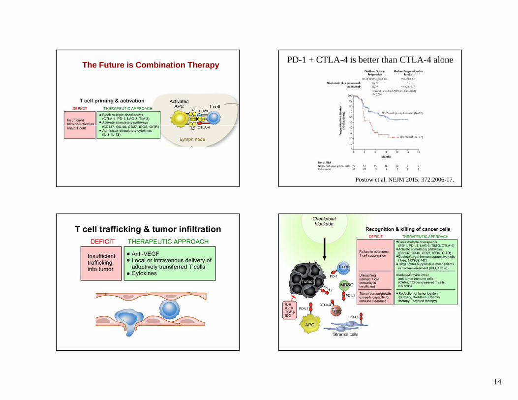
The Future is Combination Therapy PD-1 + CTLA-4 is better than CTLA-4 alone
Postow et al, NEJM 2015; 372:2006-17.

15
Could some chemotherapies synergize with PD‐1 blockade ?
oxaliplatin-cyclophosphamide
The future of cancer therapy decisions• Tumor Immunoevasion Score:
How much PD‐L1, PD‐L2, IDO, Galectin‐1, Galectin‐9, B7‐H3, B7‐H4, VISTA, HHLA2, Arginase, NKG2D‐Ligands ?
Choose best immunotherapy
• Cancer Genome sequencing:Identify which oncogenes are drug targets ?Which mutations are immunogenic ?
Choose best targeted therapy/vaccine

16
To be done• How do we identify who will respond to PD-1
blockade ?
• What are mechanisms of primary failure to respond ?
– Other immunoinhibitors ?
– Failure of immune cells to infiltrate tumor ?
– No good neoantigens ?
• What are mechanisms of secondary failure to respond ?
– Expression of other immunoinhibitory receptors ?
– Loss of MHC ?
It’s a great time to be an oncologist or researcher
• PD-1/PD-L1 works on a wide range of tumors with– moderate percentage of responders
– good safety profile
• PD-1/PD-L1 gives us a foundation to build on
• With this success, human creativity has been unleashed and we’re learning to do better
Dana-Farber Cancer Institute• Yanping Xiao• Kathleen Mahoney• Sanhong Yu• Sarah Klein• Xia Bu• Apoorvi Chaudhri
• Ping Hua• Baogong Zhu• Yahui Hao• Daniel Baumann• Lilly Cai• Ed Greenfield
AcknowledgementsDana-Farber Cancer Institute• David Reardon• Glenn Dranoff• Kwok Wong• Peter Hammerman• Jerry Ritz• Margaret Shipp
Harvard Medical School• Arlene Sharpe• Vijay Kuchroo
Beth Israel Deaconess Medical Center• Vicki Boussiotis• David McDermott• Michael Atkins
Brigham and Women’s Hospital• Sabina Signoretti• Scott Rodig
Emory University• Rafi Ahmed
U of Pennsylvania• Jaikumar Duraiswamy• George Coukos• E. John Wherry
Kyoto University• Tasuku HonjoGenetics Institute
• Clive Wood