role hyaluronic in lubrication - ard is an …ard.bmj.com/content/annrheumdis/33/4/318.full.pdf ·...
TRANSCRIPT
Ann. rheum. Dis. (1974), 33, 318
Role of hyaluronic acid in joint lubrication
DAVID A. SWANN,* ERIC L. RADIN,t MICHAEL NAZIMIEC,*PAUL A. WEISSER,t NANCY CURRAN,* AND GEORGE LEWINNEKtFrom the Departments ofBiological Chemistry and Orthopedics, Harvard Medical School at the Shriners BurnsInstitute* and Children's Hospital Medical Center,t Boston, Mass.
Hyaluronic acid is the macromolecule which endowssynovial fluid with its viscoelastic properties and it isoften assumed these properties are important for thelubrication of the tissue surfaces in diarthrodial joints(Barnett, Davies, and MacConaill, 1961). The rela-tionships, if any, that exist between the chemical andphysical properties ofsynovial fluid and its lubricatingability, however, have not been determined.Recent in vitro studies concerned with the types of
molecules present in synovial fluid which are respon-sible for the lubrication of the joint tissues have indi-cated that hyaluronic acid is a good lubricant for thesynovial membrane (Radin, Paul, Swann, and Schott-staedt, 1971), but showed that this constituent wasnot essential for the lubrication of the articular carti-lage (Radin, Swann, and Weisser, 1970). After thefractionation of synovial fluid by sedimentationequilibrium centrifugation in a cesium chloridedensity gradient, it was shown that the articularcartilage lubricating moiety was present in the proteinfraction. More recent experiments have confirmedthese data and shown that the lubricating moiety iscomplex and contains peptide and glycopeptideconstituents (Swann and Radin, 1972). It thusappears from these in vitro studies that the differenttypes of macromolecular constituents in synovialfluid can function independently in the lubrication ofthe different types of tissues in the joint. However,these constituents in vivo are intimately associatedand the articular lubricating moiety interacts withhyaluronic acid and is retained as a component of theultrafiltrate residue after the ultrafiltration of thesynovial fluid (Swann and Radin, 1972). For thisreason it is important to find out if hyaluronic acidhas any effect on the lubricating ability of the proteinfraction in an articular cartilage system.A well known fact is that the concentration and
intrinsic viscosity of hyaluronic acid are both lowerin synovial fluid from patients with rheumatoidarthritis (Balazs, Watson, Duff, and Roseman, 1967),but it is not clear whether these changes modify thelubricating properties of the fluid. A characteristicfeature of rheumatoid disease is morning stiffness,and a suggestion was made recently that this is causedby a failure in periarticular soft tissue lubrication
(Radin and others, 1971). It is also important, there-fore, to determine whether the changes in the structureof hyaluronic acid and the composition of synovialfluid in patients with rheumatoid arthritis alters itsability to lubricate synovial tissue. The present experi-ments were performed in an attempt to answer someof these questions.
Methods
I PREPARATION OF SAMPLES FORLUBRICATION TESTING IN SYNOVIALMEMBRANE SYSTEM
(a) Human syniovialfluidThis was obtained from patients with classical rheumatoidarthritis by the A.R.A. classification (Hollander andMcCarty, 1972) during joint arthroplasty operations. Thefluids were put on ice immediately and clarified by centri-fugation 90,000 x g for 30 minutes at 4°C. Aliquots werethen taken for analysis and the fluids were used directly totest their lubricating ability.
(b) Hyaluronic acid samplesThe preparations used were obtained from a number ofdifferent tissues. Preparation A was prepared from thecentral and posterior peripheral regions ofthe cow vitreous(Swann and Constable, 1972). After centrifugation at90,000 x g for 60 minutes at 4°C, the hyaluronic acid wasprecipitated with ethanol and the majority of the asso-ciated proteins were denatured by treatment with chloro-form and removed by.ultracentrifugation at 90,000 x g for60 minutes at 4°C. Preparations B, C, D, and E werekindly supplied by Med-Chem Products Inc., Boston,Mass. These samples were obtained from rooster combsby water extraction and were treated with chloroform,repeated precipitation with ethanol and cetylpyridiniumchloride (CPC), and ultracentrifugation to remove de-natured proteins (Swann, 1968a). Preparations F and Gwere obtained from the dissected mucoid layer of roostercombs as described earlier (Swann, 1969a) with the excep-tion that the pronase digestion step was omitted for sampleG. Preparations H and I were obtained from bovine syno-vial fluid by sedimentation equilibrium centrifugation in acesium chloride gradient and digestion with trypsin,respectively, as described below. Solutions containingthese preparations were dialysed against 0-15 mol/l.veronal buffer, pH 7-2 at 4°C before lubrication testing.
Accepted for publication January 21, 1974.Reprint requests to: Dr. David A. Swann, Shriners Burns Institute, 51 Blossom St., Boston, Mass. 02114, U.S.A.
copyright. on 31 A
ugust 2018 by guest. Protected by
http://ard.bmj.com
/A
nn Rheum
Dis: first published as 10.1136/ard.33.4.318 on 1 July 1974. D
ownloaded from

Role ofhyaluronic acid in joint lubrication 319
II PREPARATION OF SAMPLES FORLUBRICATION TESTING IN THE CARTILAGEON CARTILAGE SYSTEM
(a) Bovine synovial fluid not visibly contaminated withblood was obtained from the hind foot joints of adultcattle immediately after slaughter. The fluid from differentjoints was pooled, transported to the laboratory on ice,and clarified by centrifugation at 90,000 x g for 30 minutesat 4°C. The supernatant obtained was used to standardizethe lubrication test systems in vitro and served as a startingmaterial for the preparation of the other synovial fluidfractions.
(b) Ultrafiltration residue (UFR) is the fraction re-tained when clarified bovine synovial fluid is filteredthrough a 0 22,um millipore filter 16 cm in diameter at 4°Cusing vacuum to assist filtration. For lubrication test theUFR was resuspended in 0 15 mol/l. sodium chloride sothat the final volume was equal to the volume of clarifiedsynovial fluid from which the UFR was obtained.
(c) Bovine synovial fluid protein fractionClarified synovial fluid was adjusted to a density of 1 -65 g/ml by the addition of cesium chloride and centrifuged at220,000 x g for 64 hrs at 4°C (Radin, and others, 1970).After centrifugation the base of the tube was pierced andfractions were collected in sequence. The density, hexu-ronic acid, and protein content of each fraction weredetermined and the protein fraction, free of hyaluronicacid, was dialysed against four changes of 0-15 mol/l.veronal buffer pH 7-2 at 4C°.
(d) Bovine synovial fluid hyaluronic acid(1) Fractions sedimenting at densities between 1-60 and1 66 g/ml after centrifugation for 64 hrs at 220,000 x g at4°C in a cesium chloride gradient (Radin and others,1970) were dialysed against distilled water immediately
after collection of the samples, then against I mol/l.sodium chloride at 4°C and finally against two changes of0.15 mol/l.veronal buffer pH 7-2 at 4°C (preparation H,Table I).(2) Clarified synovial fluid adjusted to pH 7-2 with Trisbuffer (final concentration 0.05 mol/l. Tris-HCI, 0001mol/l. calcium chloride) was digested with trypsin (Worth-ington, TRL 1OOS, 27 mg/250 ml synovial fluid) for 24 hrsat 37°C to destroy lubricating ability in the articular carti-lage test system. The hyaluronic acid was then precipitatedby addition of ethanol (3 vol), dissolved in 0-15 mol/l.sodium chloride, treated with chloroform, reprecipitatedwith ethanol, again dissolved withO 15mol/l. sodium chlor-ide, and dialysed against 0- 15 mol/l. veronal buffer pH 7-2at 4°C. The hyaluronic acid was clarified by centrifugationat 90,000 x g for 30 minutes at 4° (preparation I, Table I).Before lubrication testing excess soybean antitrypsin(Worthington, SI, 1 mg/ml) was added both to the testsample and protein fraction, and no residual trypticactivity could be detected after the addition of this in-hibitor.
III SYNOVIAL MEMBRANE LUBRICATIONASSAYThis procedure was carried out by moving a glass platewetted with the test sample beneath a piece of synovialmembrane loaded with a 200 g weight. The drag on thesynovial membrane was measured using a force trans-ducer and was recorded on a strip chart. Three suchmeasurements were made for each test sample and thecoefficient of friction was calculated by dividing the aver-age frictional force recorded by the load (Radin and others,1971).
IV ARTICULAR CARTILAGE LUBRICATIONASSAYThe lubrication tests were carried out using bovine meta-tarsal-phalangeal joints oscillated in a bath of the test
Table I Composition and properties of hyaluronic acid preparations and synovial fluid fractions*
Protein content*
Lowry Amino acidsHexuronic* Intrinsictacid viscosity
Lubricating ability+
Soft tissue Articular cartilage
Hyaluroniic acidsABCDEFGHI
Synovial fluid fractionsSynovial fluidUltrafiltrate residueUltrafiltrate filtrateProtein fraction
9.70360350 111-921 27
1 27§030§0-92§
05
14-22-3169
315148423931302933
0.49§0 45§0-02§
98 0.5
* Data expressed as w/w % of lyophilized sample.t Intrinsic viscosity in ml/g.+ Lubricating ability expressed as % bovine synovial fluid.§ Data expressed as mg/ml fluid sample.
24
Sample
92018502000340040006000920086008000
95005500
00
1001060
105
10090-10700
copyright. on 31 A
ugust 2018 by guest. Protected by
http://ard.bmj.com
/A
nn Rheum
Dis: first published as 10.1136/ard.33.4.318 on 1 July 1974. D
ownloaded from
320 Annals of the Rheumatic Diseases
lubricant at 40 cpm under load of 226 kg as previouslydescribed (Linn, 1967, 1968). The coefficients of frictionobtained with test samples were compared with the valuesfor fresh synovial fluid and buffer which were run as con-trols in the same test series.
V ANALYTICAL DETERMINATIONSProtein was determined using the Folin-Phenol methodof Lowry, Rosebrough, Farr, and Randall (1951) and byamino acid analysis after hydrolysis with 6N hydrochloricacid for 24 hours at 105°C in sealed tubes (Swann, 1968b).Hexuronic acid analyses were carried out using a manualcarbazole reaction in the presence of borate (Balazs,Berntsen, Karossa, and Swann, 1965). Hyaluronic acidconcentrations were obtained by multiplying the hexuronicacid concentrations by a factor of 1-95. Hexosamines weredetermined usinga manual p-dimethylaminobenzaldehydereaction (Swann and Balazs, 1966) and also by ion-ex-change chromatography on an amino acid analyser usinga 15 cm x 0 9 cm column packed with phoenix spherixXX8 resin. Intrinsic viscosity measurements were per-formed at 37°C in a Cannon-Ubbelohde semimicrcdilution viscometer* after dialysis against 0-15 mol/l.sodium chloride pH 7-2 at 4°C. Intrinsic viscosity values ofthe hyaluronic acid samples were determined by extrapol-ating the reduced viscosities to zero concentration. Relativeviscosity measurements were performed at 37°C on 1 ml ofclarified synovial fluid in a semimicro viscometer with aflow time of 109 seconds. Infrared spectra ofthe hyaluronicacid preparations were determined on thin films of thesodium salts with a Perkin Elmer model 237 infraredspectrophotometer.
Results
The major difficulty associated with the measurementof the lubricating ability of test samples was the vari-ability from test series to test series because differentpieces of synovium and different joints were used. Inorder to obtain comparable data, therefore, allcoefficient of friction measurements were related tothe values obtained with 015 mol/l. veronal bufferpH 7-2 and with fresh synovial fluid. The lubricatingadvantage of samples was then calculated using theformula
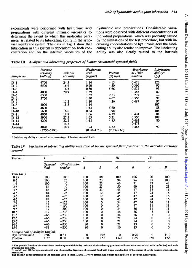
similar to those previously reported (Swann, 1968a).Preparation A contained about 10% protein andpreparations C, D, and E contained less than 0-5 %.Preparation G contained 14 2% as protein. The aminoacid composition of some of these samples used inthe lubrication tests are shown in Table II. The lowestviscosity value was obtained with hyaluronic acidfrom the cow vitreous, which is to be expected fromthe earlier studies (Varga, 1955). Preparations B, C, D,and E were all purified by similar methods, namely,denaturation of proteins by treatment with chloro-form, repeated precipitation with CPC and ethanolfollowed by ultracentrifugation (Swann, 1968a). Thehighest viscosity value was obtained with preparationG from the mucoid layer of rooster combs. Prepara-tion H obtained by fractionation of bovine synovialfluid by cesium chloride density gradient centrifuga-tion also had a high viscosity value (8600 ml/g), but theamino acid content was only 2-3 %. The intrinsicviscosity measurements were performed after dialysisagainst 0 15 mol/l. sodium chloride on samples whichhad not previously been subjected to lyophilization.The viscosity values were calculated after extrapola-tion to zero concentration as shown in Fig. 3. Thechemical analyses were carried out on aliquots of thelyophilized and air dry material after extensivedialyses of the samples against distilled water toremove salts. The sodium and moisture contentswere not determined. As shown in Fig. I the lubricat-ing ability of hyaluronic acid depended on both theintrinsic viscosity of the sample and also its concen-tration in solution. All of the lubricating ability ofsynovial fluid was present in the ultrafiltrate residuefraction.
HUMAN RHEUMATOID SYNOVIAL FLUIDSA total of 14 rheumatoid fluids were obtained insufficient quantity for analysis and lubrication testingin the soft tissue system. The analytical data areshown in Table III. The hyaluronic acid concentrationvaried from 0-8 to 1-7 mg/ml, with a mean value of1 24 mg/ml. There was also more than a twofold
Coefficient of friction (buffer) - coefficient of friction (sample) x 100 = Lubricating advantageCoefficient of friction (buffer)
This value for the sample was then calculated as apercentage of the lubricating advantage obtained withthe fresh synovial fluid used in the same test series toobtain a relative estimate of the sample's lubricatingability.
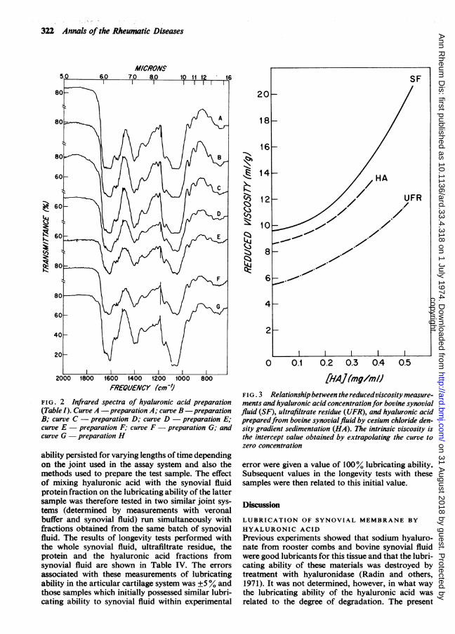
SOFT TISSUE LUBRICATION TESTSThe chemical compositions of the various hyaluronicacid preparations are shown in Table I, and theirlubricating abilities in Fig. 1. The infrared spectra(Fig. 2) are typical of sodium hyaluronate and are* Cannon Instrument Co., State College, Pa., U.S.A.
variation in the concentration of protein. The rangewas 2-53 to 5.66% w/v and the mean value was4 52% w/v. All of the rheumatoid synovial fluidstested had lubricating abilities similar to bovine syno-vial fluid using the synovium membrane assay system.
ARTICULAR CARTILAGE LUBRICATIONThe chemical analysis of the various bovine synovialfluid fractions tested for their lubricating ability inthe articular cartilage system are shown in Table I.The ultrafiltration residue obtained after one filtra-tion was composed of approximately 24% protein.
copyright. on 31 A
ugust 2018 by guest. Protected by
http://ard.bmj.com
/A
nn Rheum
Dis: first published as 10.1136/ard.33.4.318 on 1 July 1974. D
ownloaded from

Role ofhyaluronic acid in joint lubrication 321
The protein fraction obtained after cesium chloridedensity gradient centrifugation contained negligibleamounts of glucuronic acid and was composed of98% protein by amino acid analysis. The hyaluronicacid preparation obtained by tryptic digestion con-tained 16-9% protein by amino acid analysis com-pared to only 2-3% after purification by density gra-dient fractionation and neither of the preparations
possessed any lubricating ability in the articularcartilage system. Both the ultrafiltrate residue fractionand the protein fraction from synovial fluid possessedlubricating ability equivalent to that observed withwhole synovial fluid.When the lubricating ability of the synovial fluid
protein fraction was tested continuously over aperiod of 15 hrs it was observed that the lubricating
Table II Amino acid composition of hyaluronic acid samples, residues/1,000 residues
Ultrafiltrationresidue
LysineHistidineArginineAspartic acidThreonineSerineGlutamic acidProlineGlycineAlanineCystine/2ValineMethionineIsoleucineLeucineTyrosinePhenylalanine
692237868010511060686328917
25803635
Hyaluronic*acid C
46332184511861063717889
54
32481619
Hyaluronicacid G
733455109566210942797521631741873446
* Swann (1968b).
0.5 1.0 1.5 2.0
HYALUIRONIC ACID CONCENTRATION (mg/mI)
FIG. 1 Lubricating ability of hyaluronicacidpreparations (Table I). Data obtainedwith preparation A -0, B - 0, C-A,DuAfEl0rF-t ,Gesu H V;ultrafiltrate residue data v
2.5
HyaluronicacidH
6815169679148201251057520156
32512031
120
;^80-
yZ 60
K 40
>, 20
0
- o o o
v °v v
v~~~~~
v
- OpJ A a
A
0A
* A a A
00
S
A aA
A
0~~~~aU
copyright. on 31 A
ugust 2018 by guest. Protected by
http://ard.bmj.com
/A
nn Rheum
Dis: first published as 10.1136/ard.33.4.318 on 1 July 1974. D
ownloaded from
322 Annals of the Rheumatic Diseases
MICRONS
60:
2000 1800 1600 1400 1200 800
FREQUJENCY (cm')Y
FIG. 2 Infrared spectra hyaluronic acid preparation
(Table I). Curve A -preparation A; curve B -preparation
B; curve C -preparation D; curve D -preparation E;
curve E -preparation F; curve F- preparation G; andlcurve G -preparation H
ability persisted for varying lengths of time depending
on the joint used in the assay system and also the
methods used to prepare the test sample. The effect
of mixing hyaluronic acid with the synovial fluid
protein fraction on the lubricating ability of the latter
sample was therefore tested in two similar joint sys-
tems (determined by measurements with veronal
buffer and synovial fluid) run simultaneously with
fractions obtained from the same batch of synovial
fluid. The results of longevity tests performed with
the whole synovial fluid, ultrafiltrate residue, the
protein and the hyaluronic acid fractions from
synovial fluid are shown in Table IV. The errors
associated with these measurements of lubricating
ability in the articular cartilage system was ±5% and
those samples which initially possessed similar lubri-
cating ability to synovial fluid within experimental
20
18~
16F
41
LIN
I
12
10
81
6
4
21
0 0.1 0.2 0.3 0.4 0.5
[HAJ(mg/m/IFIG. 3 Relationship between the reducedviscosity measure-ments and hyaluronic acid concentrationfor bovine synovialfluid (SF), ultrafiltrate residue (UFR), and hyaluronic acidpreparedfrom bovine synovialfluid by cesium chloride den-sity gradient sedimentation (HA). The intrinsic viscosity isthe intercept value obtained by extrapolating the curve tozero concentration
error were given a value of 100% lubricating ability.Subsequent values in the longevity tests with thesesamples were then related to this initial value.
Discussion
LUBRICATION OF SYNOVIAL MEMBRANE BYHYALURONIC ACID
Previous experiments showed that sodium hyaluro-nate from rooster combs and bovine synovial fluidwere good lubricants for this tissue and that the lubri-cating ability of these materials was destroyed bytreatment with hyaluronidase (Radin and others,1971). It was not determined, however, in what waythe lubricating ability of the hyaluronic acid wasrelated to the degree of degradation. The present
SF
/ HA
- /Z/ UFR
I-- ./
I I I I
copyright. on 31 A
ugust 2018 by guest. Protected by
http://ard.bmj.com
/A
nn Rheum
Dis: first published as 10.1136/ard.33.4.318 on 1 July 1974. D
ownloaded from
Role ofhyaluronic acid in joint lubrication 323
experiments were performed with hyaluronic acidpreparations with different intrinsic viscosities todetermine the extent to which this molecular para-meter is related to its lubricating ability in the syno-vial membrane system. The data in Fig. 1 show thatlubrication in this system is dependent on both con-centration and on the intrinsic viscosities of the
hyaluronic acid preparations. Considerable varia-tions were observed with different concentrations ofindividual preparations, which was probably causedby the limitations of the test procedure, but with in-creasing concentrations of hyaluronic acid the lubri-cating ability also tended to improve. The lubricatingability was also clearly related to the intrinsic
Table m Analysis and lubricating properties of human rheumatoid synovial fluids
Hyaluronicacid(mg/ml)
1-140.900-801-591-671-701-101-141-430-841-061-631-18
Protein(% w/v)4-154 505-66
2 534-624-26
5-603-984.395-214 85
1-24 4 52(0-80-1-70) (2 53-5 66)
* Lubricating ability expressed as a percentage of bovine synovial fluid.
Table IV Variation of lubricating ability with time of bovine synovial fluidfractions in the articular cartilagesystem*
Test no. I II III IV
Synovial Ultrafiltrationfluid residue
Time (hrs)025 1000-5 1001-5 1002 5 843.5 844.5 845-5 846 5 847-5 178-5 179.5 -17
10.5 -1711-5 -6612-5 -6613-5 -6614-5 -6615 5 -83Composition ofsamples (mg/ml)Hyaluronic acid 0-96Protein 1-27
1062500
-25-25-25-25-125-150-200-238-238-238-250-250-250
0-83030
A B A B A B
100 88 100 106 100 100100 23 94 94 87 100100 23 66 71 73 43100 23 50 60 38 21100 23 45 47 38 16100 12 45 47 24 16100 12 45 47 24 16100 0 45 47 24 16100 0 34 47 24 11100 0 34 47 11 11100 0 34 36 11 11100 0 34 36 5 11100 0 34 36 5 0100 0 21 24 0 0100 0 21 24 0 080 0 21 13 0 080 0 10 13 0 0
0 1-051-30 1-58
0 0951-60 1-85
0 1-101-50 1P58
Sample no.
D-1D-2D-3D-4D-5D-6D-7D-8D-9D-10D-1lD-12D-13D-14Average
Intrinsicviscosity(ml/mg)
47006500
400063003750
400046005600405059004200
4870(3750-6500)
Relativeviscosity
24-516-96-5
20-9
15-218-8
16-618-827 522-214-718-7
Absorbanceat 1/100dilution
0-4710-3900-5720 3850-2770 5500-487
0 4940 5500-4620-4750-465
Lubricatingability*( /0)
126959387
9797107881048110880
97
* The protein fraction obtained from bovine synovial fluid by cesium chloride density gradient sedimentation was mixed with buffer (A) and withhyaluronic acid (B).In tests II andM the hyaluronic acid was obtained by digestion ofsynovial fluid with trypsin and in test IV by cesium chloride density gradient sedi-mentation.The protein concentrations in the samples used in tests II and III were determined before the addition of soybean antitrypsin.
copyright. on 31 A
ugust 2018 by guest. Protected by
http://ard.bmj.com
/A
nn Rheum
Dis: first published as 10.1136/ard.33.4.318 on 1 July 1974. D
ownloaded from

324 Annals of the Rheumatic Diseases
viscosity of the samples. The samples with intrinsicviscosity values above 4,000 ml/g possessed similarlubricating abilities to bovine synovial fluid, whereasat most of the concentrations tested preparationsA, B, C, D were inferior lubricants.
It is difficult to interpret these data exactly becausethe intrinsic viscosity value of hyaluronic acid isdependent upon many factors. Earlier experimentshave shown that rooster comb hyaluronic acid con-tained weak linkages that were irreversibly cleaved bytemperatures about 650 (Swann, 1969a) and that athigh pH the intrinsic viscosity was decreased, thoughthe molecular weight remained constant (Swann,1970). Silpananta, Dunstone, and Ogston (1968) alsoshowed that the molecular weight of bovine ultra-filtrate residue hyaluronic acid was unaltered, whereasthe intrinsic viscosity value decreased after fractiona-tion of the polysaccharide and protein constituents.These studies indicate that the polysaccharide chainsof hyaluronic acid can interact with adjacent mole-cules and can assume different conformations insolution. More recent studies using x-ray crystallo-graphy (Atkins and Sheehan, 1972, 1973; Dea, Moor-house, Rees, Arnott, Guss, and Balazs, 1973) haveconfirmed and extended these earlier observationsand shown that the hyaluronic acid polysaccharidechain can occur in the form of a helix as well as otherconformations.
In a previous study it was shown that the mole-cules in solutions of highly purified preparations ofhyaluronic acid had a random coil configuration(Laurent, Ryan, and Pietruszkiewicz, 1960), but themolecules in the connective tissue in vivo may have adifferent structure. In this case the decrease in theintrinsic viscosity of hyaluronic acid that occursduring purification (Swann, 1968a) may be a measureof a change in structure. Under these circumstancesthe superior lubricating ability of those hyaluronicacid preparations with high intrinsic viscosity values(Fig. 1) could be related to one or a number of theabove factors, namely the length, conformation, andstate of aggregation of the polysaccharide chains orthe interaction of the chains with other protein mole-cules. Scher and Hamerman (1972) have obtaineddata which suggest that hyaluronic acid from humansynovial fluid is present as a complex with proteinand the hyaluronic acid preparation prepared frombovine synovial fluid by density gradient fractionationhad a similar protein content to that found by theseworkers (preparation H, Table I). The lubricatingability, however, did not seem to be related to thetotal protein component of the samples, which indi-cates that this function is dependent upon the struc-ture and organization of the polysaccharide chains ofhyaluronic acid. It is not known, however, in whatway the small quantities of protein always found inhyaluronic acid (Swann, 1968b) contribute to thestructure of this substance.
An additional factor is that purified hyaluronicacid occurs in solution as a polydisperse mixture ofmolecules (Laurent and others, 1960; Swann, 1969b).It is thus possible that samples with different intrinsicviscosity values may contain some molecules with asimilar molecular weight and conformation. Thisfact may be responsible for some of the variations inthe lubrication data with individual preparations andthe finding that there was not a clear-cut differencebetween the lubricating abilities of samples withdifferent intrinsic viscosities.One interpretation of the lubrication data which is
compatible with the above data is that hyaluronicacid is able to lubricate synovial tissue when itpossesses a structure which is similar to that whichoccurs in vivo and that if this structure is degradedthen the lubricating ability is decreased. Optimumlubrication would then be achieved when a sufficientnumber of undenatured molecules are present andwhen their function is not impeded by competitionwith other nonlubricating and denatured hyaluronicacid molecules.
LUBRICATION OF SYNOVIAL MEMBRANE BYHUMAN RHEUMATOID SYNOVIAL FLUIDAnalysis of the rheumatoid synovial fluid showedthat the concentration of protein was increased andthat the concentration of hyaluronic acid was de-creased. The ranges for these values were similar tothose reported in the literature. The intrinsic viscosityof the hyaluronic acid in these synovial fluids wasalso somewhat lower than in normals, which againagreed with previous studies (Balazs and others,1967). When the lubricating ability of these synovialfluids was tested in the synovial membrane system,however, it was observed that they possessed similarlubricating properties to whole bovine synovial fluid.Again, a spread of values was observed (80-126%lubricating ability), but the average of all sampleswas 97 %. These data suggest that though there arecertain changes both in concentration and intrinsicviscosity of the hyaluronic acid in rheumatoid syno-vial fluid, these changes are not sufficient to limit theability of the fluid to lubricate the synovial membrane.This conclusion agrees with the data already pre-sented for the various hyaluronic acid preparations.Only those preparations with an intrinsic viscosity ofless than 4,000 ml/g or a concentration less than0 5 mg/ml showed lowered lubricating ability whencompared to whole bovine synovial fluid. The lowesthyaluronic acid concentration in rheumatoid synovialfluids was 0.8 mg/ml and the lowest viscosity was3,750 ml/g, and so it would be expected that all ofthese samples would lubricate synovial fluid in asimilar manner to normal bovine synovial fluid.LUBRICATION OF ARTICULAR CARTILAGEThe data shown in Tables I and IV support the earlierexperiments where it was shown that the moiety
copyright. on 31 A
ugust 2018 by guest. Protected by
http://ard.bmj.com
/A
nn Rheum
Dis: first published as 10.1136/ard.33.4.318 on 1 July 1974. D
ownloaded from
Role ofhyaluronic acid in joint lubrication 325
responsible for articular lubrication was a proteinconstituent (Radin and others, 1970; Swann andRadin, 1972). All the lubricating ability was found tobe present in the protein fraction and the ultrafiltrateresidue in the short-term tests (Table I) and therewas no difference between the lubricating abilities ofthese two samples. When the tests were continued forextended periods of time, however, the synovial fluidand ultrafiltrate residue lost their lubricating abilityand eventually became inferior lubricants whencompared to buffer alone. It is thought that this'gumming up' effect may be caused by the accumula-tion of constituents on the articulating surface whichinhibit the flow of water in and out of the cartilageand thus retard the hydrostatic component of thelubricating mechanism (Radin and Paul, 1972). Alsoas indicated in Table IV the types of results obtainedin the longevity tests varied from test to test and alsodepended on the methods used to prepare testsamples. Thus, with test TI the protein/buffer fractionlubricated continuously for more than 13 hrs, whereasthe same protein fraction when mixed with hyaluronicacid prepared from bovine synovial fluid by digestionwith trypsin gave a lowered initial lubricating abilitywhich decreased gradually with time. In other similarexperiments (test III and IV) using different prepara-tions of protein fraction and hyaluronic acids pre-pared by tryptic digestion (test III) and by densitygradient sedimentation (test IV), the protein/buffersample and the protein/hyaluronic acid sample hadsimilar lubricating abilities in each test. Despite thesevariations, which may have been caused by a greatnumber of factors, it appears that hyaluronic aciddoes not aid in the lubrication of articular cartilage inany way. The fact that the articular lubricant is closelyassociated with the hyaluronic acid in synovial fluidmay not therefore be offunctional significance.An explanation for this finding may be that while
the majority of the hyaluronic acid in joint fluid isproduced by the synovial membrane (Blau, Janis,Hamerman, and Sandson, 1965), the articular lubri-cant or a component of this fraction together withsmall quantities of hyaluronic acid may be derivedfrom the articular cartilage itself and may only occurin synovial fluid as a consequence of natural wear
processes. Recent experiments in vitro have shownthat hyaluronic acid can interact with cartilage proteo-glycans to form aggregates (Hardingham and Muir,1972; Gregory, 1973). Articular lubrication, there-fore, may be an inherent property of the articularcartilage per se and the role of the synovial fluidwould then be to interact with this surface, to act as awash fluid, and to lubricate the adjacent soft tissues.This concept ofjoint function and lubrication is beingtested at the present time.
Summary
Synovial fluid has been shown to be an effective lubri-cant for the synovium membrane if it containshyaluronic acid with an intrinsic viscosity of about4,000 ml/g or higher, and a concentration of greaterthan 0 5 ml/g. Higher concentrations of hyaluronicacid with low viscosity values, however, were stillable, in part, to serve as lubricants. Analysis andlubrication tests performed on synovial fluid frompatients with rheumatoid arthritis showed that thelowest hyaluronic acid concentration was 0.8 mg/mland the lowest intrinsic viscosity observed was 3,750ml/g. All of the fluids, however, were able to lubricatesynovial membrane in a similar manner to normalbovine synovial fluid. It seems unlikely, therefore,that the lubrication of soft tissue in joints would be alimiting function in patients with this disease.The data obtained also confirm the earlier studies
in that they showed the moiety responsible for articu-lar lubrication was present in the protein fraction ofsynovial fluid and further indicate that the lubricatingability of this moiety does not depend upon the pre-sence of hyaluronic acid or the interaction betweenthese two constituents. As an explanation of thesefindings, it is suggested that articular lubrication maybe an inherent property of the articular cartilage andits macromolecular structure per se, and that a com-ponent of the articular lubricating fraction isolatedfrom synovial fluid may occur there as a consequenceof wear processes.
This work was supported by research funds from theNational Institutes of Health, Grant AM 15216 and theShriners Burns Institute.
References
ATKINS, E. D. T., AND SHEEHAN, J. K. (1972) Nature (New Biol.), 235, 253 (Structure for hyaluronic acid)~~, ~~(1973) Science, 179, 562 (Hyaluronates: relation between molecular conformations)BALAZS, E. A., BERNTSEN, K. O., KAROSSA, J., AND SWANN, D. A. (1965) Anal. Biochem., 12, 547 (An automated
method for the determination of hexuronic acids), WATSON, D., DUFF, I. F., AND ROSEMAN, S. (1967) Arthr. and Rheum., 10, 357 (Hyaluronic acid in synovialfluid. 1. Molecular parameters of hyaluronic acid in normal and arthritic human fluids)
BARNETT, C. H., DAVIES, D. V., AND MACCONAILL, M. D. (1961) In 'Synovial Joints-Their Structure andMechanics'. Thomas, Springfield, Illinois
BLAU, S., JANIS, R., HAMERMAN, D., AND SANDSON, J. (1965) Science, 150, 353 (Cellular origin of hyaluronate-protein in the human synovial membrane)
copyright. on 31 A
ugust 2018 by guest. Protected by
http://ard.bmj.com
/A
nn Rheum
Dis: first published as 10.1136/ard.33.4.318 on 1 July 1974. D
ownloaded from

326 Annals of the Rheumatic Diseases
DEA, I. C. M., MOORHOUSE, R., REES, D. A., ARNOTr, S., Guss, J. M., AND BALAZS, E. A. (1973) Ibid., 179, 560(Hyaluronic acid. A novel double helical molecule)
GREGORY, J. D. (1973) Biochem. J., 133, 383 (Multiple aggregation factors in cartilage proteoglycan)HARDINGHAM, T. E., AND MUIR, H. (1972) Biochim. biophys. Acta, 279, 401 (The specific interaction of
hyaluronic acid with cartilage proteoglycans)HOLLANDER, J. L., AND MCCARTY, D. J. (eds.) (1972) In 'Arthritis and Allied Conditions', 8th ed. Lea and
Febinger, Philadelphia; Kimpton, LondonLAURENT, T. C., RYAN, M., AND PIETRUSZKIEWICZ, A. (1960) Biochim. biophys. Acta, 42, 476 (Fractionation of
hyaluronic acid. The polydispersity of hyaluronic acid from the bovine vitreous body)LINN, F. C. (1967) J. Bone Jt. Surg., 49A, 1079 (Lubrication of animal joints. I. The arthrotripsometer)
(1968) J. Biomech., 1, 193 (Lubrication of animal joints. II. The mechanism)LOWRY, 0. H., ROSEBROUGH, N. J., FARR, A. L., AND RANDALL, R. J. (1951) J. biol. Chem., 193, 265 (Protein
measurement with the folin phenol reagent)RADIN, E. L., AND PAUL, I. L. (1972) J. Bone Jt. Surg., 54A, 607 (A consolidated concept of joint lubrication)
-, , SWANN, D. A., AND SCHOTTSTAEDT, E. S. (1971) Ann. rheum. Dis., 30, 322 (Lubrication of synovialmembrane), SWANN, D. A., AND WEISSER, P. A. (1970) Nature, 228, 377 (Separation of a hyaluronate-free lubricatingfraction from synovial fluid)
SCHER, I., AND HAMERMAN, D. (1972) Biochem. J., 126, 1073 (Isolation of human synovial-fluid hyaluronate bydensity-gradient ultracentrifugation and evaluation of its protein content)
SILPANANTA, P., DUNSTONE, J. R., AND OGSTON, A. G. (1968) Ibid., 109, 43 (Fractionation of a hyaluronic acidpreparation in a density gradient. Some properties of the hyaluronic acid)
SWANN, D. A. (1968a) Biochim. biophys. Acta, 156, 17 (Studies on hyaluronic acid. I. The preparation andproperties of rooster comb hyaluronic acid)
- (1968b) Ibid., 160, 96 (Studies on hyaluronic acid. II. The protein component(s) of rooster comb hyaluronicacid)(1969a) Biochem. biophys. Res. Commun., 35, 571 (Hyaluronic acid: structure of the macromolecule in theconnective tissue matrix)
- (1969b) Biochem. J., 114, 819 (Studies on the structure of hyaluronic acid. Characterization of the productformed when hyaluronic acid is treated with ascorbic acid)(1970) 'On the state of hyaluronic acid in a connective tissue matrix,' in 'The Chemistry and MolecularBiology of the Intercellular Matrix', Vol. 2. Glycosaminoglycans and Proteoglycans, ed. E. A. Balazs, p. 743.Academic Press, New York., AND BALAZS, E. A. (1966) Biochim. biophys. Acta, 130, 112 (Determination of the hexosamine content ofmacromolecules with manual and automated techniques using the p-dimethylaminobenzaldehyde reaction), AND CONSTABLE, I. J. (1972) Invest. Ophthal.. 11, 159 (Vitreous structure. I. Distribution of hyaluronate andprotein), AND RADIN, E. L. (1972) J. biol. Chem., 247, 8069 (The molecular basis of articular lubrication. I. Purificationand properties of a lubricating fraction from bovine synovial fluid)
VARGA, L. (1955) Ibid., 217, 651 (Studies on hyaluronic acid prepared from the vitreous body)
copyright. on 31 A
ugust 2018 by guest. Protected by
http://ard.bmj.com
/A
nn Rheum
Dis: first published as 10.1136/ard.33.4.318 on 1 July 1974. D
ownloaded from