role of hydrogen bonding on protein secondary structure …caramboc/protein lesson.pdf · ·...
TRANSCRIPT
Role of Hydrogen Bonding on Protein Secondary Structure Introduction The function and chemical properties of proteins are determined by its three-dimensional
structure. The final �architecture� of the protein begins with the linear assortment of
amino acids (its primary structure) and progresses through three other modifications
(secondary -tertiary to quaternary structures) that create the final shape of the protein.
These modifications are the result of various intermolecular forces that exists as a result
of the protein�s primary structure, but are manifested in the proteins secondary structure.
The secondary structure of proteins is defined as the local conformations of the primary
backbone, which is characterized by regular repeating structures such as α (alpha) helixes
and €β € (beta) sheets. The structural stability of these repeating structures is the result of
hydrogen bonding between the amide proton (the hydrogen atom bonded to the nitrogen
of the peptide bond) and the carbonyl oxygen (the oxygen atom bonded to the carbon
atom in the amide plane).
Αlpha helixes and Beta sheets can function as independent peptides, (alpha keratin,
collagen, and blood vessels). Beta sheets can be found in silk, and beta keratin,
however they function predominantly as structural components of globular proteins.
Globular proteins have varying proportions of helixes, sheets, and other structural
elements known as turns, twists and crossovers. Hydrogen bonding is the primary
stabilizing force in all of these structures. This lesson plan will introduce students to the
chemistry of hydrogen bonding and its affect on the biology of the secondary structure of
proteins.
Outline
I. Chemistry of Hydrogen Bonding
A. Hydrogen Bonding
1. Definition
2. Electronegativity and Bond polarity (Figure 1: Table of Electronegativity)
3. Calculation of electronegativity difference: related to bond polarity
a. N = 3.0: H =2.1: Difference =0.9
b. Ionic bonds, Polar covalent bonds, and non polar covalent bonds
c. Bond dipole (Figure 2: Affects of polar bond)
B. Dipole moment and Bond polarity
1. Affect of Hydrogen Bonding on Boiling point
a. Thermodynamic data: relative strength of the hydrogen bond
b. (Figure 2a: Affect of hydrogen bonds on boiling pt.)
C. The Hydrogen Bond in the polypeptide chain.
1. Notation: Hydrogen Bond donor (D-H) and Hydrogen Bond Acceptor (D-A)
2. (Figure 3: Hydrogen Bonding in the Beta Sheet and
Figure 3a. Hydrogen bonding in the Alpha Helix
II. Secondary Structure of polypeptides
B. Primary structure (reviewed): show a linear organization
C. The Amide Plane and its affect on conformations (Figure 4)
1. Rotations about the α Carbon: φ & ψ dihedral angles of the amide planes
(Figure 4a)
2. Allowed Conformations and Steric Hindrance (Figures 5 and 6)
3. Ramachandran Plot (Figure 7)
4. Ramachandran Conformational angle distribution ( Figure 7a)
D. Helixes: Secondary structure defined
1. p, n and nm notation system (Figure 8)
2. Favorable angles for the alpha helix (Figure 8a)
3. Alpha helices (Figure 9)
E. The Beta Sheets
a. Parallel and anti parallel constructions

Discussion
I. Chemistry of Hydrogen Bonding Hydrogen bonds belong to the class of intermolecular forces that arise as a result of a molecule�s dipolar characteristic. Hydrogen bonds exist between the hydrogen atom in a polar molecule such as (NH) and an unshared pair of electrons in a nearby (highly electronegative atom) such as Nitrogen, Oxygen or Fluorine. The polarity of a covalent bond is the result of the difference in electronegativity between the atoms that are bonded together. Electronegativity is defined as the ability of an atom (in a molecule) to attract shared electrons to itself. Electronegativity values of the elements can be found in figure 1. To determine the relative polarity of a covalent bond, one uses the values in the table of to calculate the difference in relative strength between bonded atoms: A molecule such as NH has an electronegativity difference of O.8; which suggests a polar covalent bond. (Typically values between 0 and 0.5 suggest a non-polar covalent bond, 0.5 to 2.0 a polar covalent bond and 2.0 and above represent ionic compounds). The charge separation in these bonds is such that the electropositive hydrogen atom carries a partial positive charge (σ +) and the more electronegative atom such as Nitrogen carries a partial negative charge (σ −). (See figure 2). A hydrogen bond results when the partially positive hydrogen approaches the lone pair of a Nitrogen, Oxygen or Fluorine atom. Hydrogen bonds exist preferentially between these atoms because of two reasons. The hydrogen atom is small and has but one electron in its electron cloud, when the more electronegative atom distorts the cloud, the nucleus is not shielded by other electrons (as in other atoms), thus the lone pair of the approaching atom can approach the nucleus fairly directly. Secondly, nitrogen, oxygen and fluorine make exceptionally strong hydrogen bonds because of their high electronegativity and relatively small radius. Their small size means that their electron cloud is confined to a small region of space about the atom, this contributes to the strong directionality characteristic of hydrogen bonds. The combination of these factors makes for exceptionally strong interactions between these sets of atoms. The effect of hydrogen bonding can be seen in the graph (Boiling points of group 4,5, 6 and 7). The increased boiling point of H 2O, NH3 and HF is the result of their hydrogen bonds. Although hydrogen bonds energies may seem small on the order of 4 to 25 kJ/mole (Brown & LeMay.p 414), it should be remembered that they normally exist as a system of interconnections: thus they are additive and contribute greatly to the overall stability of systems such as the polypeptide polymer. A typical hydrogen bond is shown in Figure 2: Hydrogen bonding in water and between the Carbonyl atom and the Amide Hydrogen of a polypeptide.) In the figure, the delta positive σ + represents the positive pole and the σ - is the negative pole). Typically in the Polypeptide chain, the hydrogen bonded to the Nitrogen atom is termed (D-H) hydrogen donor and the Carbonyl oxygen is the (D-A); hydrogen acceptor. This notation will be used in the subsequent discussion of hydrogen bonding in the secondary structure of proteins. II. Secondary Structure of Polypeptides: The primary structure of proteins is the linear sequence of amino acid residues. These residues are linked together by amide bonds between the amine and carboxylic acid ends of respective amino acids. The resulting amide bonds are planar and exist on either side of the alpha carbons of the residues: they are referred to as amide planes (see figure 4).
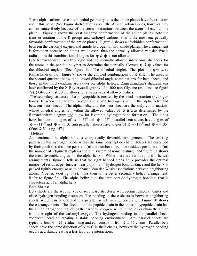
These alpha carbons have a tetrahedral geometry, thus the amide planes have free rotation about this bond (See Figure 4a:Rotations about the Alpha Carbon Bond), however they cannot rotate freely because of the steric interactions between the atoms of each amide plane. Figure 5 shows the least hindered conformation of the amide planes: note the trans orientation of the R groups and carbonyl carbons: this is the most energetically favorable conformation of the amide planes. Figure 6 shows a �forbidden conformation� between the carbonyl oxygen and amide hydrogen of two amide planes. The arrangement is forbidden because the atoms are �closer� than the normally allowed van der Waals radius, thus this combination of angles for φ & ψ is not allowed. G.N Ramachandran used this logic and the normally allowed interatomic distances for the atoms in the peptide polymer to determine the sterically allowed φ & ψ values for the dihedral angles: (See figure 6a: The dihedral angle). The plot of φ vs.ψ: (a Ramachandran plot: figure 7) shows the allowed combinations of φ & ψ . The areas in the second quadrant show the allowed dihedral angle combinations for beta sheets, and those in the third quadrant are values for alpha helixes. Ramachandran�s diagram was later confirmed by the X-Ray crystallography of ~1000 non-Glycene residues: see figure 7a). ( Glycene�s structure allows for a larger area of allowed values.) The secondary structure of a polypeptide is created by the local interaction (hydrogen bonds) between the carbonyl oxygen and amide hydrogens within the alpha helix and between beta sheets. The alpha helix and the beta sheet are the only conformations whose dihedral angles fall within the allowed values of φ & ψ as determined by the Ramachandran diagram and allow for favorable hydrogen bond formation. The alpha helix has torsion angles of φ = -570 and ψ= -470: parallel beta sheets have angles of φ = -1190 and ψ = +113: anti parallel sheets have angles of φ = -1390 and ψ = +1350 (Voet & Voet pg 147.) Helixes As mentioned the alpha helix is energetically favorable arrangement. The twisting pattern creates hydrogen bonds within the same polypeptide chain. Helixes are described by their pitch (p): distance per turn, (n) the number of peptide residues per turn and (m) the number of (figure 8 explains the p, n system of nomenclature), and figure 8a shows the most favorable angles for the alpha helix. While there are various p and n helical arrangements (figure 9 tells us that the right handed alpha helix provides the optimal number of residues per turn, a �nearly optimum� hydrogen bond distance and the helix is packed tightly enough so as to enhance Van der Waals associations between neighboring atoms. (Voet & Voet pg. 149). This then is the better secondary helical arrangement. Refer to figure 3a: The alpha helix: note the intra-peptide hydrogen bonding, that is characteristic of an alpha helix. Beta Sheets: Beta sheets are the second type of secondary structures with optimal dihedral angles and close hydrogen bonding distances. The bonding in these sheets is between neighboring sheets, which can be oriented in a parallel or anti parallel orientation. Figure 10 shows these arrangements. The direction of the peptide chain in the upper polypeptide chain has the amide nitrogen to the left of the carbonyl oxygen, while in the lower chain the amide is to the right of the carbonyl oxygen. The hydrogen bonding in ant parallel sheets �connect� head on creating a stable bonding environment. Anti parallel sheets are typically from 6 � 25 residues long and can consist of from 2 to 15 chains. Parallel beta sheets have the same direction of N to C in their chains, however the hydrogen bonding occurs at a slant, creating a less favorable interactions.
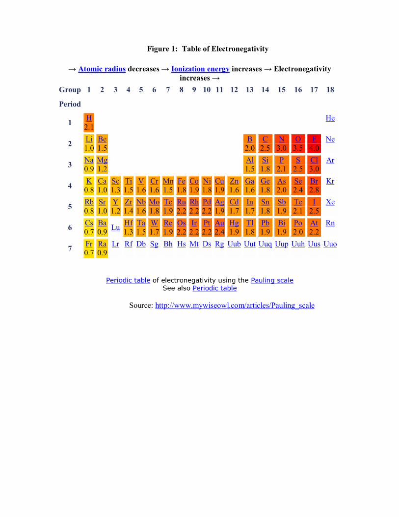
Figure 1: Table of Electronegativity
→ Atomic radius decreases → Ionization energy increases → Electronegativity
increases → Group 1 2 3 4 5 6 7 8 9 10 11 12 13 14 15 16 17 18
Period
1 H 2.1 He
2 Li 1.0
Be 1.5 B
2.0 C
2.5 N
3.0 O
3.5 F
4.0 Ne
3 Na 0.9
Mg 1.2 Al
1.5 Si1.8
P 2.1
S 2.5
Cl 3.0
Ar
4 K 0.8
Ca 1.0
Sc 1.3
Ti 1.5
V 1.6
Cr 1.6
Mn1.5
Fe1.8
Co1.9
Ni1.8
Cu1.9
Zn1.6
Ga1.6
Ge1.8
As 2.0
Se 2.4
Br 2.8
Kr
5 Rb 0.8
Sr 1.0
Y 1.2
Zr 1.4
Nb 1.6
Mo 1.8
Tc1.9
Ru2.2
Rh2.2
Pd2.2
Ag1.9
Cd1.7
In1.7
Sn1.8
Sb 1.9
Te 2.1
I 2.5
Xe
6 Cs 0.7
Ba 0.9 Lu Hf
1.3 Ta 1.5
W 1.7
Re1.9
Os2.2
Ir2.2
Pt2.2
Au2.4
Hg1.9
Tl1.8
Pb1.9
Bi 1.9
Po 2.0
At 2.2
Rn
7 Fr 0.7
Ra 0.9
Lr
Rf
Db
Sg
Bh
Hs
Mt
Ds
Rg
Uub
Uut
Uuq
Uup
Uuh
Uus
Uuo
Periodic table of electronegativity using the Pauling scale
See also Periodic table
Source: http://www.mywiseowl.com/articles/Pauling_scale
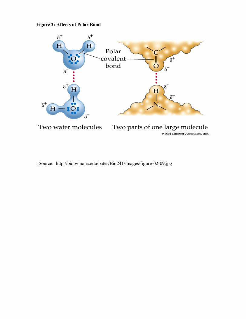
Figure 2: Affects of Polar Bond
. Source: http://bio.winona.edu/bates/Bio241/images/figure-02-09.jpg

Figure 2a: Affect of Hydrogen Bonding on Boling Point of Group 5A, 6A, & 7A Halides Source: http://www.chemguide.co.uk/atoms/bonding/hbond.html
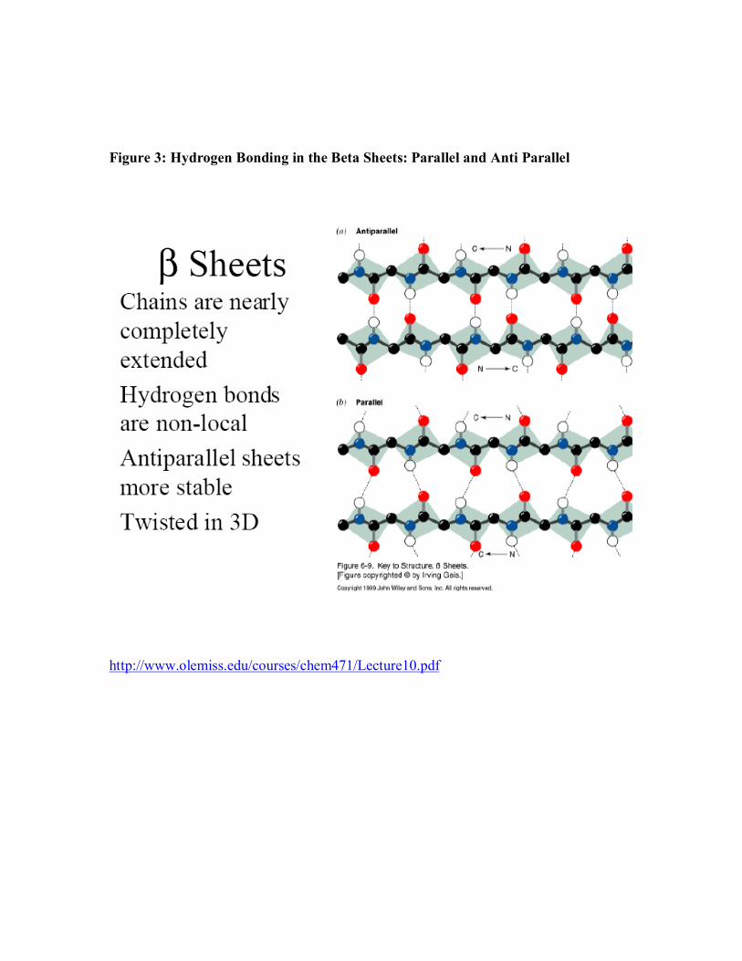
Figure 3: Hydrogen Bonding in the Beta Sheets: Parallel and Anti Parallel
http://www.olemiss.edu/courses/chem471/Lecture10.pdf

Figure 3a. Hydrogen Bonding if the Alpha Helix
Source: http://www.olemiss.edu/courses/chem471/Lecture10.pdf
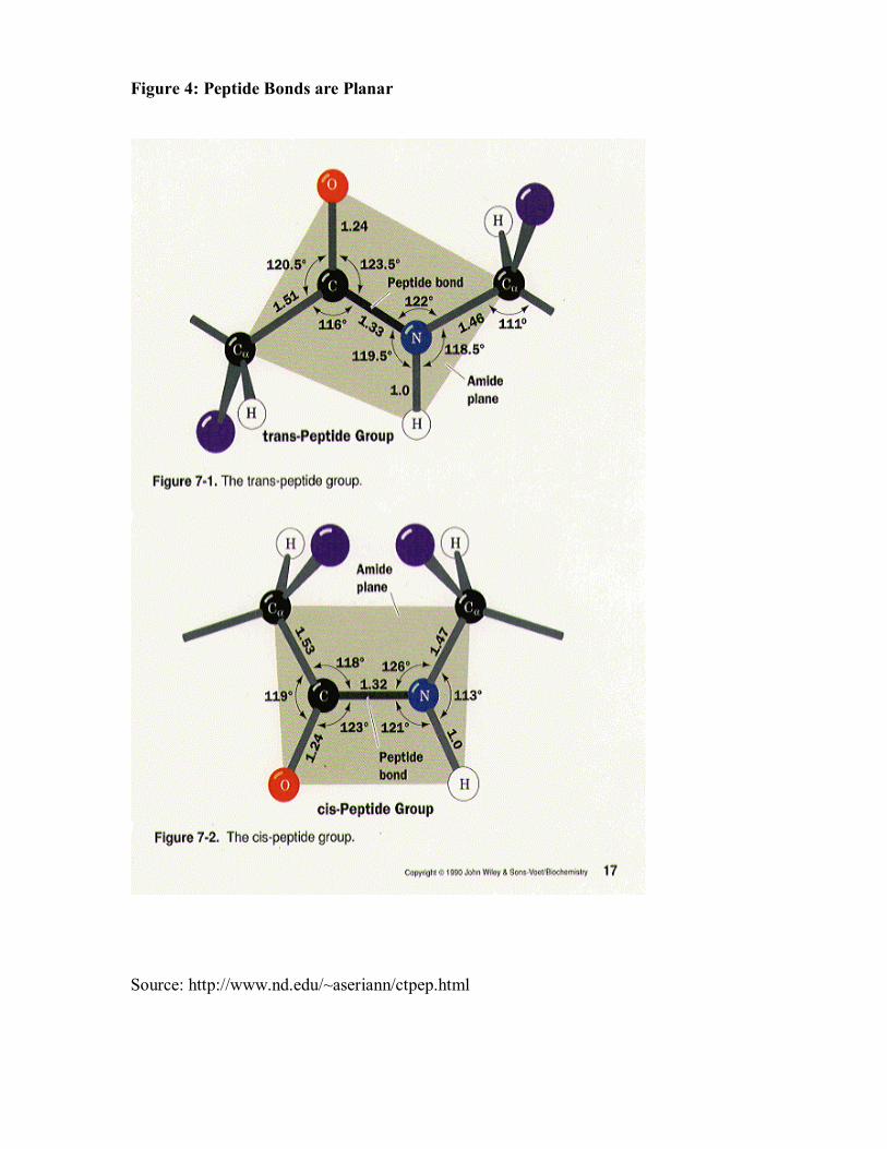
Figure 4: Peptide Bonds are Planar
Source: http://www.nd.edu/~aseriann/ctpep.html

Figure 4a: Rotations about the Alpha Carbon
Source: http://www.nsm.uh.edu/~wwidger/lectures_2000/LC2000_9.pdf
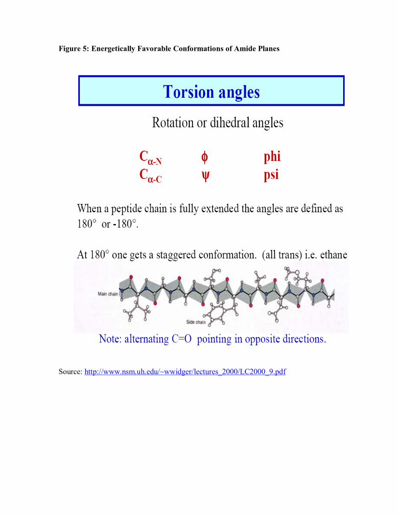
Figure 5: Energetically Favorable Conformations of Amide Planes
Source: http://www.nsm.uh.edu/~wwidger/lectures_2000/LC2000_9.pdf
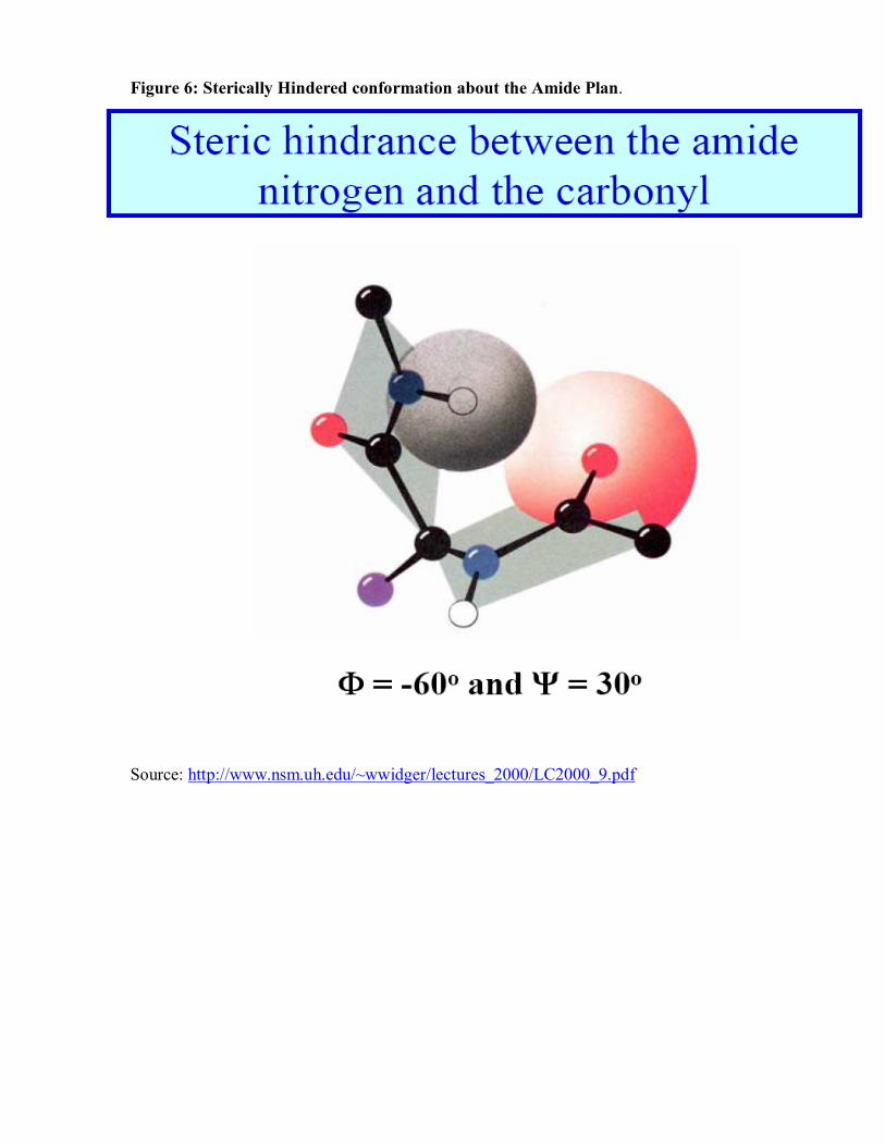
Figure 6: Sterically Hindered conformation about the Amide Plan.
Source: http://www.nsm.uh.edu/~wwidger/lectures_2000/LC2000_9.pdf
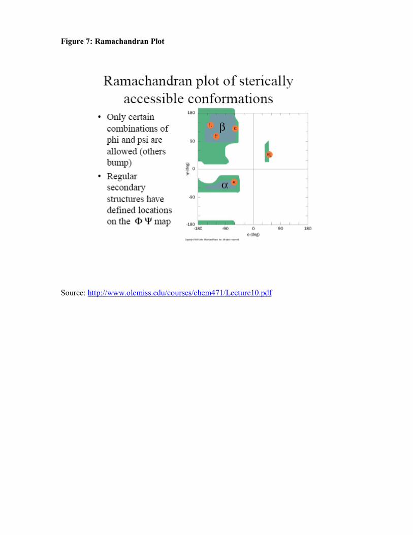
Figure 7: Ramachandran Plot Source: http://www.olemiss.edu/courses/chem471/Lecture10.pdf
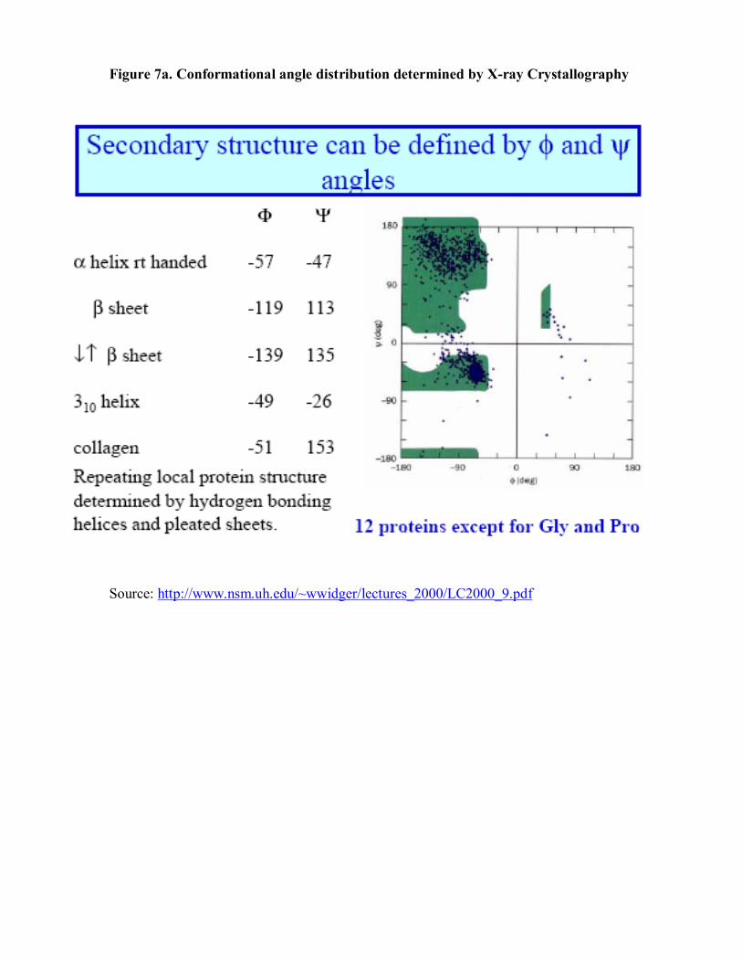
Figure 7a. Conformational angle distribution determined by X-ray Crystallography
Source: http://www.nsm.uh.edu/~wwidger/lectures_2000/LC2000_9.pdf

Figure 8: Nomenclature for Helixes
Source: http://www.nsm.uh.edu/~wwidger/lectures_2000/LC2000_9.pdf
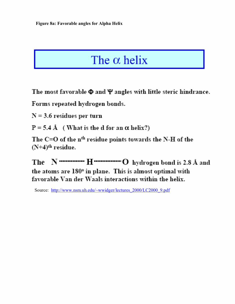
Figure 8a: Favorable angles for Alpha Helix
Source: http://www.nsm.uh.edu/~wwidger/lectures_2000/LC2000_9.pdf
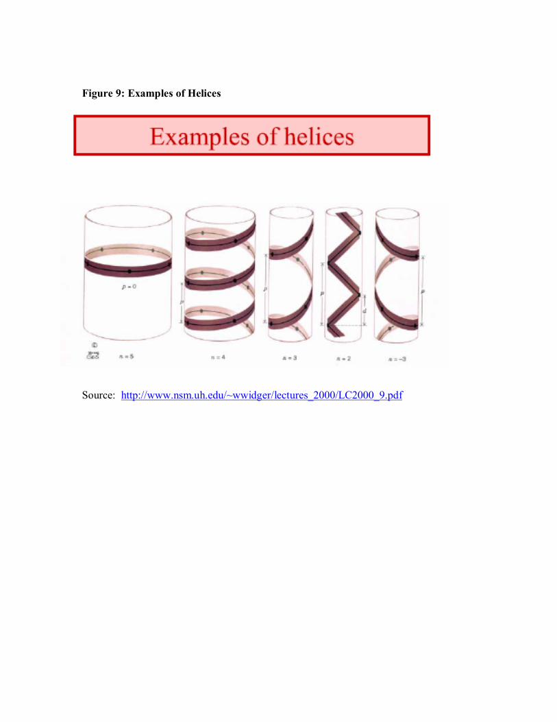
Figure 9: Examples of Helices
Source: http://www.nsm.uh.edu/~wwidger/lectures_2000/LC2000_9.pdf
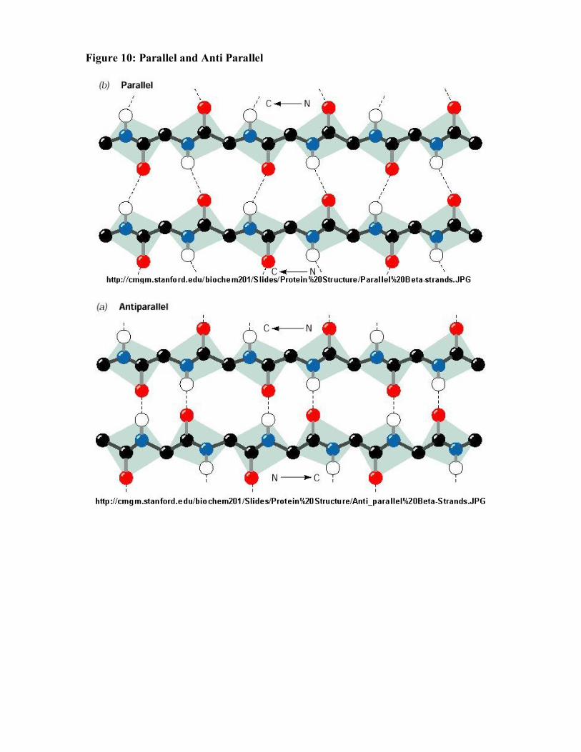
Figure 10: Parallel and Anti Parallel

References: Biochemistry: (1990) Voet, D. & Voet, J. John Wiley and Sons. New York. Chemistry The Central Science: (2003). Brown,T.L., LeMay,H.E., Bursten,B.E., & Burdge,J.R. Pearson Education, Upper Saddle River New Jersey.