role of laea in the regulation of alb1 glip, conidial ... · laea gene was not involved in the...
TRANSCRIPT

EUKARYOTIC CELL, Sept. 2007, p. 1552–1561 Vol. 6, No. 91535-9778/07/$08.00�0 doi:10.1128/EC.00140-07Copyright © 2007, American Society for Microbiology. All Rights Reserved.
Role of laeA in the Regulation of alb1, gliP, Conidial Morphology,and Virulence in Aspergillus fumigatus�
Janyce A. Sugui,1 Julian Pardo,2 Yun C. Chang,1 Arno Mullbacher,6 Kol A. Zarember,3Eva M. Galvez,5 Lauren Brinster,4 Patricia Zerfas,4 John I. Gallin,3
Markus M. Simon,2 and Kyung J. Kwon-Chung1*Laboratory of Clinical Infectious Diseases,1 Laboratory of Host Defense,3 and Office of Research Services,4 National Institutes of
Health, Bethesda, Maryland; Max Planck Institute of Immunology2 and Institut fur Physikalische Chemie, Freiburg Universitat,5
Freiburg, Germany; and John Curtin School of Medical Research, Australian National University, Canberra, Australia6
Received 24 April 2007/Accepted 29 June 2007
The alb1 (pksP) gene has been reported as a virulence factor controlling the pigmentation and morphologyof conidia in Aspergillus fumigatus. A recent report suggested that laeA regulates alb1 expression and conidialmorphology but not pigmentation in the A. fumigatus strain AF293. laeA has also been reported to regulate thesynthesis of secondary metabolites, such as gliotoxin. We compared the role of laeA in the regulation of conidialmorphology and the expression of alb1 and gliP in strains B-5233 and AF293, which differ in colony morphologyand nutritional requirements. Deletion of laeA did not affect conidial morphology or pigmentation in thesestrains, suggesting that laeA is not involved in alb1 regulation during conidial morphogenesis. Deletion of laeA,however, caused down-regulation of alb1 during mycelial growth in a liquid medium. Transcription of gliP,involved in the synthesis of gliotoxin, was drastically reduced in B-5233laeA�, and the gliotoxin level found inthe culture filtrates was 20% of wild-type concentrations. While up-regulation of gliP in AF293 was comparableto that in B-5233, the relative mRNA level in AF293laeA� was about fourfold lower than that in B-5233laeA�.Strain B-5233laeA� caused slower onset of fatal infection in mice relative to that with B-5233. Histopathologyof sections from lungs of infected mice corroborated the survival data. Culture filtrates from B-5233laeA�caused reduced death in thymoma cells and were less inhibitory to a respiratory burst of neutrophils thanculture filtrates from B-5233. Our results suggest that while laeA is not involved in the regulation of alb1function in conidial morphology, it regulates the synthesis of gliotoxin and the virulence of A. fumigatus.
Aspergillus fumigatus, a ubiquitous saprophyte, is the pre-dominant cause of aspergillosis throughout the world. Thisspecies most often causes life-threatening invasive aspergillosis(IA) in individuals with dysfunctional phagocytes or with pro-longed neutropenia resulting from immunosuppressive drugtherapy (9, 19). In previous studies, we have dissected themolecular basis of conidial pigment formation and identified acluster of six genes involved in the biosynthesis of dihy-droxynaphthalene (DHN)-like melanin in A. fumigatus strainB-5233 (34–37). The alb1 (pksP) gene in this cluster encodes apolyketide synthase that catalyzes the first step of DHN-likemelanin synthesis during conidial formation. Conidia of A.fumigatus are bluish green and coarsely echinulated due to theprotrusions on their surfaces as revealed by scanning electronmicroscopy (34). Deletion of the alb1 gene from strain B-5233resulted in two simultaneous changes in conidial morphology:albino instead of bluish green color and a smooth rather thanechinulated surface. Complementation of the alb1� strain withthe wild-type alb1 gene restored the bluish green pigment aswell as the echinulation on the surface (17, 34), indicating thatthe alb1 gene is involved in conidial morphology as well as inconidial pigment synthesis. Furthermore, albino conidia were
found to be more sensitive to hydrogen peroxide, were phago-cytized more readily by neutrophils, and showed higher sus-ceptibility to monocyte-mediated damage than wild-typeconidia. More importantly, mice infected with albino conidiasurvived significantly longer than those infected with wild-typeconidia (12, 17, 30, 34).
It has been reported recently that the function of alb1 isregulated by the LaeA protein (3). LaeA is a nuclear proteinshown to control secondary metabolism in various aspergilli,including A. nidulans, A. terreus, and A. fumigatus. For in-stance, it has been demonstrated that the expression of laeAaffects the synthesis of sterigmatocystin, penicillin, and lo-vastatin. Sequence analysis and experimental data suggestedthat the LaeA protein has methyltransferase activity and ispredicted to function at the level of chromatin remodeling(3, 6, 7, 14). Upon deletion of laeA in strain AF293, theconidia became smooth, a result similar to that reported forthe alb1� strain of B-5233 (34), without affecting the syn-thesis of bluish green pigment (3). This indicated that thepigmentation and the surface protrusions of conidia arecontrolled by separate factors. The laeA deletant strain ofAF293, which we refer to as AF293laeA�, manifested anarray of other features distinct from those of the wild type.These included impaired virulence associated with reducedlevels of gliotoxin in the lungs of animals, higher suscepti-bilities of conidia and hyphae to host phagocytes, and theloss of dark pigment in mycelia submerged in an agar me-dium (3, 6).
* Corresponding author. Mailing address: Molecular MicrobiologySection, Laboratory of Clinical Infectious Diseases, National Instituteof Allergy and Infectious Diseases, NIH, Bethesda, MD 20892. Phone:(301) 496-1602. Fax: (301) 480-3240. E-mail: [email protected].
� Published ahead of print on 13 July 2007.
1552
on February 11, 2019 by guest
http://ec.asm.org/
Dow
nloaded from
Considering the involvement of laeA in the virulence of A.fumigatus and possibly in the regulation of alb1 function inconidial development, we investigated the role of laeA in thepathobiology of A. fumigatus strain B-5233, with special em-phasis on conidial morphology. B-5233 is a highly virulentclinical strain isolated from a leukemic patient who succumbedto IA. This strain was used extensively for characterization ofthe antigenic properties useful for diagnosis of aspergillosis aswell as for studies of host-Aspergillus interactions (10, 26, 27,39) long before it was used to dissect the molecular control ofconidial morphology and pigment synthesis (34–37). Wedeemed it important to study the role of laeA in this well-characterized strain because the genomic strain AF293, unlikeother A. fumigatus strains, fails to grow robustly in definedminimal medium (MM) broth. Furthermore, a study by Bokand colleagues (3) suggests that conidial surface morphologyand conidial pigmentation are controlled by different factors inAF293, while previous reports (17, 34) demonstrated thatthese characteristics are both controlled by the alb1 gene inB-5233. We deleted the laeA gene in strain B-5233, comparedconidial morphology and transcriptional regulation of alb1with those for the AF293laeA� strain, and observed that thelaeA gene was not involved in the regulation of conidial mor-phology in these strains. However, laeA was involved in theexpression of alb1 during the early stages of mycelial growthand showed a reduced amount of transcript at 48 h in bothstrains rather than a larger amount of transcript, as has beenreported previously (3). laeA was also involved in the regula-tion of gliotoxin biosynthesis and the virulence of A. fumigatus.Our findings support the notion that laeA plays an importantrole in the regulation of gliotoxin production and the patho-genicity of A. fumigatus but not in the morphogenesis ofconidia.
MATERIALS AND METHODS
Strains and media. AF293 is a clinical isolate used for the sequencing of theA. fumigatus genome (22). B-5233 is also a clinical strain; it was isolated in a caseof IA from a patient with leukemia (36). A. fumigatus strains were maintained onAspergillus MM (28) or Sabouraud agar slants. Fungal strains were grown eitherin a liquid medium containing MM supplemented with 2 ml/liter of a vitamin mix(2 mg liter�1 p-aminobenzoic acid, 2 mg liter�1 niacin, 2 mg liter�1 pyridoxineHCl, 2 mg liter�1 riboflavin, 2 mg liter�1 thiamine HCl, 2 mg liter�1 choline HCl,0.004 mg liter�1 D-biotin) or in RPMI 1640 medium. RPMI 1640 was used eitheralone or buffered with 25 mM HEPES, pH 7.2 (RPMI-HEPES). The mediumwas supplemented with 10 mM uracil and 10 mM uridine (UU) whenever strainAF293.1 (a pyrG1 mutant) was included in the experiments. Germination ofconidia and the growth rate of mycelia were assayed by culturing the fungus ineither Erlenmeyer flasks or 8-well chambered cover slides (Nalge Nunc Inter-national). For growth in flasks, 1 � 107 conidia were inoculated into 50 ml ofbroth (RPMI-HEPES or MM supplemented with vitamins) and incubated at37°C on a shaker for 1 to 4 days. For growth on chambered cover slides, 3 � 105
conidia were inoculated into 200 �l of medium and incubated at 37°C under 5%CO2 for 24 to 40 h. The wells were then examined using a bright-field micro-scope. Strains AF293, AF293.1, and AF293laeA� (strain 54.2) were kindly pro-vided by N. P. Keller, University of Wisconsin—Madison.
Transformation vectors. (i) Deletion vector. Primers laeA1 (CGACTAGTCCTCGCTCCATCCCAATAGG) and laeA2 (CGTCTAGACGGAGTTGTTTCTTGAGCGG) were used to amplify a 777-bp fragment upstream (5�) of the laeAcoding region, and primers laeA3 (CGAAGCTTACCGAGATTACCCTTGCATG) and laeA4 (CGCTCGAGCGACACACATATCATGACGG) were usedto amplify a 990-bp fragment downstream (3�) of the coding region from thegenomic DNA of strain B-5233. The deletion vector was constructed by cloningthe 777-bp 5� fragment, the 990-bp 3� fragment, and a SacI/XbaI hygromycinresistance cassette from pAN7 into the SacI (blunted)/XhoI site of vector
pDHt-SK (31). This deletion vector was used to transform strain B-5233 viaAgrobacterium tumefaciens-mediated transformation (31).
(ii) Complementation vector. Primers laeA10 (TGGAGCATAACCGAGTCTCC) and laeA11 (GAGGGATATACTGCCGTCCA) were used to amplify a3.5-kb fragment that included 1.6 kb upstream and 0.73 kb downstream of thelaeA coding region. The complementation vector was constructed by cloning the3.5-kb fragment and the phleomycin resistance cassette into the HindIII/EcoRI-digested pDHt-SK vector. The complementation vector was used to transformstrain B-5233laeA� via A. tumefaciens-mediated transformation.
SEM. Conidia from 7-day-old cultures were fixed for 60 min with 4% (vol/vol)glutaraldehyde in phosphate-buffered saline (PBS) (pH 7.4) by adding the fixa-tive to the cultures at a final concentration of 2%. One milliliter of this suspen-sion was filtered through a 0.6-�m-pore-size polycarbonate membrane filter(Sterlitech, Kent, WA), and fixation was continued for an additional 60 min. Thefilters were washed three times with water for a period of 10 min each time,postfixed with 1% osmium tetroxide in water for 60 min, and washed three timesas described above. The filters were serially dehydrated, submitted to criticalpoint drying, mounted on aluminum stubs, and coated with a gold-palladiumalloy. The samples were viewed under a Hitachi S-4700 field emission scanningelectron microscope (SEM) (Hitachi High Technologies America, Inc., Schaum-burg, IL) operated at an accelerating potential of 25 kV.
Quantitative real-time PCR. One milliliter of a conidial suspension (107
conidia) was inoculated into 100 ml of RPMI-HEPES. The cultures were grownat 37°C under 5% CO2 with constant shaking for 24, 48, and 72 h. The RNA wasisolated with Trizol (Invitrogen), purified with an RNeasy kit (QIAGEN), andtreated with Turbo DNase (Ambion) according to the manufacturer’s instruc-tions. Typically 1 �g of the total RNA obtained was used for cDNA synthesis.The RNA was reverse transcribed using a high-capacity cDNA archive kit (Ap-plied Biosystems, Foster City, CA) according to the manufacturer’s instructions.The first-strand cDNA was diluted 1:2, and 5 �l was added to 20 �l of thereal-time PCR (RT-PCR) mixture, consisting of 12.5 �l of TaqMan universalPCR master mix (Roche Molecular Systems, CA), 2.25 �l (each) of 10 mMforward and reverse primers, 0.0625 �l of 10 mM probe, and 2.93 �l of water.The reaction was performed on the ABI Prism 7700 sequence detection system.Total RNA was used as a negative control. The glyceraldehyde-3-phosphatedehydrogenase gene, gpdA (Afu5g01970), was amplified as an endogenous con-trol to standardize the amount of sample added to the reaction mixture. Primerand probe sequences for the following genes are given in parentheses: gpdA(probe, CCCCCATGTTCGTCATGGGTGTC; forward primer, TCTCCGCTCCTTCTGCTGAT; reverse primer, CGGAGGTGTAGGTGGTGTTGT), gliP(probe, CAATCCACCTTGGTCCTGGCCG; forward primer, CCTGAACGCCATGCACAAG; reverse primer, CCAGCCGGCGGTAGAAGT), and alb1(probe, TGCGCAAACGCTTGTCGACCAC; forward primer, GCCATCGTCTCTCTACGCTGAT; reverse primer, CTGGTACTCTGGTTTGTATTTTGTGATC). The difference in the threshold cycle between gpdA in the wild-type andlaeA� strains was less than 1 cycle (data not shown), indicating that gpdAexpression was not affected by deletion of laeA and therefore that gpdA wassuitable as an endogenous control.
Culture filtrate. The culture filtrates used in the EL4 thymoma cell, mouseembryonic fibroblast (MEF), and gliotoxin quantification assays were preparedby inoculating 107 conidia into 100 ml of RPMI 1640 and incubating at 37°Cunder 5% CO2 for 48 h. Culture filtrates were then collected by filtering theculture through a sterile BD Falcon 40-�m-pore-size cell strainer (BD Bio-sciences, Erembodegem, Belgium). Culture filtrates for neutrophil chemilumi-nescence assays were prepared by inoculating 3 � 108 conidia into 100 ml ofRPMI-HEPES and incubating at 37°C under 5% CO2 for 72 h before the culturesupernatant was obtained.
Gliotoxin quantitation. Aspergillus culture filtrates (100 ml), obtained as de-scribed above, were extracted with 100 ml of chloroform, and the organic phasewas recovered, dried, and resuspended in 5 ml of chloroform. High-performanceliquid chromatography (HPLC) analysis was performed as described elsewhere(2). Briefly, 100 �l of chloroform extract was injected in a reverse-phase C18
column (Waters) with a mobile phase (1 ml min�1) of methanol-water (70:30).Gliotoxin was quantified using a standard curve of the pure compound (Sigma,St. Louis, MO).
Neutrophil respiratory burst assay. All human blood samples used in thisstudy were collected after informed consent from healthy subjects (NationalInstitutes of Health protocol 99-CC-0168). Blood was anticoagulated using acidcitrate dextrose, and neutrophils were purified as described previously (42). Thepreparations were 95% pure. Twenty five microliters of RPMI-HEPES alone orcontaining 5 �l of a culture filtrate (5% of the final assay volume) was added tothe wells of a white polypropylene 96-well plate (Whatman UNIPLATE).Twenty five microliters of 106 neutrophils ml�1 in RPMI-HEPES was added to
VOL. 6, 2007 REGULATORY FUNCTION OF THE laeA GENE IN A. FUMIGATUS 1553
on February 11, 2019 by guest
http://ec.asm.org/
Dow
nloaded from
each well and incubated at 37°C under 5% CO2. After 30 min, cells were quicklymixed with prewarmed 2� phorbol 12-myristate 13-acetate (PMA) diluted in aDiogenes chemiluminescent enhancer (National Diagnostics, Atlanta, GA), andrelative light units (RLU) were measured for 0.5 s per well every 90 s in anAnthos Zenyth 3100 luminometer at 37°C with intermittent shaking. Raw dataare presented kinetically as “sum RLU” (equaling the sum of all the measure-ments of RLU over the first 60 min) to facilitate comparison of different PMAdoses and filtrate conditions.
Cell death. EL4 cells (2 � 105) in 1 ml of minimum essential medium sup-plemented with 5% fetal calf serum were incubated for 16 h with 2.5, 5, 10, or20% (vol/vol) culture filtrate. After the cells were washed with PBS, phosphati-dylserine translocation and membrane integrity were monitored using the An-nexin V (AV) detection kit (BD PharMingen, San Diego, CA) and propidiumiodide (PI) staining as described previously (23). Cells positive for AV and PIand cells positive for AV but negative for PI were scored, and the sum of bothis shown as a percentage of the total population. In every experiment, 104 cellswere counted, and specific cell death was calculated by the following formula:[% (AV� PI� � AV� PI� cells)] � [% (AV� PI� � AV� PI� cells) in treatedcells] � [% (AV� PI� � AV� PI� cells) in the untreated control]. The per-centage of the sum of AV� PI� and AV� PI� cells in the control was alwayslower than 15%. Caspase-3 activation was monitored using a fluorescein isothio-cyanate-labeled monoclonal antibody against the active form of the enzyme byfluorescence-activated cell sorting as described elsewhere (23).
Cell detachment. MEFs transformed with simian virus 40 (24, 40) were cul-tured overnight in minimal essential medium supplemented with 10% fetal calfserum and 2-mercaptoethanol (10�5 M) at 37°C under 7% CO2. The mediumwas replaced by various dilutions of Aspergillus culture filtrate (2.5, 5, and 10%),and the MEFs were incubated for an additional 4 h before the cells wereobserved by bright-field microscopy. Photographs were taken using an Axiovert10 microscope (Zeiss) with Axiovision software.
Virulence. 129/Sv mice were immunosuppressed by subcutaneous injection of2 mg of hydrocortisone acetate in 100 �l of PBS–0.1% Tween 20 on days �4, �2,0, 2, and 4. On day zero, mice were inoculated intranasally with 5 � 106 conidiain 20 �l of PBS–0.01% Tween 20. Twelve mice per treatment were used. Mor-bidity and mortality were monitored daily for 21 days, and subsequently thesurviving mice were sacrificed. Control mice, immunosuppressed with hydrocor-tisone and inoculated intranasally with 20 �l of PBS–0.01% Tween 20, survivedduring the observation period without any sign of infection. Histopathologicalsections of lungs were prepared from mice that had been infected with B-5233,B-5233laeA�, or B-5233laeAR and sacrificed at 72 or 96 h after infection. Thetissue sections were fixed, paraffin embedded, sectioned, and stained with hema-toxylin and eosin or Gomori’s methenamine silver.
RESULTS
Growth comparison between B-5233 and AF293. StrainB-5233 grew robustly in MM broth with or without the vitaminmix and formed fungal balls (1 to 2 mm in diameter) within24 h, whereas growth of strain AF293 in MM broth was notvisible until after 4 days. Addition of the vitamin mix to MMbroth allowed strain AF293 to form mycelia that were barelyvisible to the naked eye after 72 h. Since AF293 eventuallyformed mycelia in all the vitamin dropout media, none of thevitamins tested appeared to be essential for the growth ofstrain AF293. The assays on chambered slides showed thatconidia of both B-5233 and AF293 germinated and formed amycelial layer to an equal degree in the rich medium, RPMI-HEPES (Fig. 1A and B). The rates of germination and growthfor the two strains were clearly distinguishable, however, whenthey were grown in MM broth with the vitamin mix. WhileB-5233 conidia germinated and formed a layer of mycelium(Fig. 1C), more than 90% of AF293 conidia remained unger-minated by 40 h (Fig. 1D). AF293 also showed slower growthon a solid agar medium containing MM alone or supplementedwith the vitamin mix (Fig. 1E and data not shown). The severegrowth retardation of AF293 in MM broth and not in a richmedium suggests that the strain is nutritionally deficient com-
pared to B-5233. Furthermore, RPMI 1640 is better suited forgrowth of the genomic sequencing strain of A. fumigatus thanthe widely used Aspergillus MM with vitamins.
Deletion of laeA and regulation of alb1 expression. We andothers have previously observed that alb1 is a virulence factor,that its expression is conidial stage specific, and that it catalyzesthe synthesis of DHN-like melanin conidial color (17, 34). Inaddition to color, the alb1 gene also controls the surface mor-phology of conidia. Since laeA has been reported to controlconidial morphology but not conidial color in AF293, we de-leted laeA in order to investigate its regulatory role in theexpression of alb1 in strain B-5233. laeA was deleted by replac-ing the coding region of the gene with a hygromycin resistancecassette. Southern blot hybridization analysis identified a trans-formant that had undergone homologous gene replacement,resulting in the deletion of laeA (data not shown). The deletantstrain, designated B-5233laeA�, was then complemented withthe wild-type laeA gene, and this reconstituted strain, whichharbored a single copy of the gene, was named B-5233laeAR.A consistent phenotype of the laeA deletant that distinguishedit from wild-type strain B-5233 was the absence of a yellowcompound secreted in MM agar. Due to the absence of thisyellow compound, the colony reverse of strain B-5233laeA�remained white (Fig. 1E, lower left panel), while those ofstrains B-5233 (Fig. 1E, upper left panel) and B-5233laeAR(data not shown) became yellow after 5 days of growth at 25°C.Strain AF293 showed a colony reverse with darkly pigmentedmycelia and without secretion of the yellow compound (Fig.1E, upper right panel). Unlike that of AF293, the colony re-verse of AF293laeA� was white due to the loss of dark mycelialpigment (Fig. 1E, lower right panel). Other than the absence ofthe yellow exudates, deletion of laeA in B-5233 caused nosignificant phenotypic alteration in the size and morphology ofthe colony or in conidial pigmentation except for a slight delayin sporulation in cultures grown on MM agar (Fig. 1F). Fur-thermore, microscopic observation of conidia incubated inchambered cover slides for 8 h in RPMI-HEPES at 37°Cshowed comparable germination and hyphal emergence forB-5233 and B-5233laeA� (data not shown). Although the col-ony morphologies of B-5233 and AF293 were different on solidmedium, the morphologies of their conidia examined under acompound microscope as well as by SEM were indistinguish-able (Fig. 1G and H). Conidia of strains B-5233laeA� andAF293laeA� were morphologically identical to those of theirparental strains, including the degree of protrusions (Fig. 1Iand J). A more extensive search revealed rare smooth conidiadevoid of protrusions for both the wild-type and the laeA�strains at similar frequencies (less than 0.1%), indicating thatthe surface morphology of conidia is unrelated to the laeA gene(Fig. 1G, H, and J insets).
Using Northern blot hybridization, Bok and collaborators(3) monitored the expression of alb1 in cultures grown in liquidMM for 24, 48, and 72 h and observed that the expression ofalb1 at 48 h was significantly higher in strain AF293laeA� thanin strain AF293. To assess the expression levels of alb1 in strainB-5233, we used MM as the growth medium and includedstrains AF293.1 and AF293laeA� as controls. AF293.1 waschosen because this strain was used as the background for thedeletion of laeA (3). Strains AF293.1 and AF293laeA� grewextremely poorly in MM plus UU. Even when the medium was
1554 SUGUI ET AL. EUKARYOT. CELL
on February 11, 2019 by guest
http://ec.asm.org/
Dow
nloaded from

supplemented with a vitamin mix, strains with an AF293 back-ground failed to attain growth comparable to that of B-5233and B-5233laeA� within 48 h. Since it is essential to use amedium that can yield similar growth for the comparison ofgene expression, we used the relatively rich medium RPMI1640, which allowed comparable growth rates for all the strains(Fig. 1A and B and data not shown). The level of alb1 expres-
sion in each strain was analyzed by RT-PCR. At 48 h, strainB-5233laeA� exhibited a down-regulation of alb1 compared tolevels in B-5233 and B-5233laeAR (Fig. 2A). A similar reduc-tion in transcription, instead of an increase, was observed forstrain AF293laeA� compared to AF293.1 (Fig. 2A). At 24 and72 h, similar patterns of down-regulation were observed in thelaeA deletant strains of B-5233 and AF293 (data not shown).
FIG. 1. Phenotype of A. fumigatus strains. (A to D) Two different isolates from A. fumigatus, strains B-5233 and AF293, were grown in liquidmedia (chambered slide) at 37°C for 40 h. (A and B) B-5233 and AF293, respectively, grown on RPMI-HEPES. (C and D) B-5233 and AF293,respectively, grown on MM plus a vitamin mix. Panel D shows a few mycelia with branched hyphae. The majority of conidia remain ungerminated(small dots). (Inset) Nongerminated (white arrow) and germinated (arrow in black) conidia. Original magnifications, �25 for panels A to D and�1,000 for the inset in panel D. (E and F) AF293 and B-5233, and their respective laeA deletants, B-5233laeA� and AF293laeA�, grown on MMagar. (E) Growth at 25°C for 5 days showed a yellow compound secreted by B-5233 but not by B-5233laeA�, AF293, or AF293laeA�. Submergedmycelia of AF293 show dark pigmentation (white arrow) manifested in the colony reverse. (F) Strains were grown at 37°C for 2 days. (G to J) SEMof conidia from B-5233 (G), B-5233laeA� (I), AF293 (H), and AF293laeA� (J). Arrows indicate protrusions on the conidial surface. Scale barsfor the insets in panels G, H, and J, 2.5, 2.8, and 3 �m, respectively.
VOL. 6, 2007 REGULATORY FUNCTION OF THE laeA GENE IN A. FUMIGATUS 1555
on February 11, 2019 by guest
http://ec.asm.org/
Dow
nloaded from

These results indicate that although laeA does not interfere inthe function of alb1 in conidial morphology, it affects the ex-pression of alb1 during mycelial growth in a liquid medium.
Deletion of laeA affects transcription of the gliP gene andgliotoxin synthesis. We also monitored the transcriptional lev-els of the gliP gene, which is involved in the synthesis of glio-toxin, a secondary metabolite with immunosuppressive prop-erties produced by A. fumigatus. The RT-PCR results from48-h cultures showed that the expression level of gliP wassignificantly lower for B-5233laeA� than for B-5233 andB-5233laeAR. An even more accentuated difference was ob-served for strain AF293 (Fig. 2B). The RT-PCR results fromthe 24- and 72-h cultures showed the same pattern as thatobserved at 48 h (data not shown). Since the expression of gliPwas diminished in the laeA deletants of both strains B-5233 andAF293, we investigated the effect of laeA on gliotoxin produc-tion using only strains B-5233, B-5233laeA�, and B-5233laeAR.Culture filtrates of the three strains were subjected to HPLCanalysis to detect the secreted gliotoxin (Fig. 2C). The analysisshowed that strain B-5233laeA� produced approximately 80%less gliotoxin than strains B-5233 and B-5233laeAR.
Effects of culture filtrates on the respiratory burst of neu-trophils. Since deletion of laeA caused a reduction in thesynthesis of gliotoxin, the toxin known to inhibit the respiratoryburst of neutrophils (38, 41), we analyzed the effects of the
culture filtrates from B-5233, B-5233laeA�, and B-5233laeARon superoxide anion production by human neutrophils. In theabsence of culture filtrates, PMA stimulated neutrophil chemi-luminescence in a dose-dependent manner (Fig. 3A), whereaspreincubation of neutrophils with 5% of the filtrate from eitherB-5233 or B-5233laeAR significantly decreased the chemilumi-nescence but not the viability of neutrophils (Fig. 3A and datanot shown). In contrast, the culture filtrate from strainB-5233laeA� had only a minor effect relative to results for theRPMI control. To exclude the possibility that the inhibitoryactivity present in B-5233 was due to a substance that inter-fered with the detection of chemiluminescence, we tested theeffects of B-5233 and B-5233laeA� culture filtrates on chemi-luminescence produced by xanthine oxidase/hypoxanthine, acell-free superoxide-generating enzyme and substrate. Culturefiltrates of the two strains had equivalent but negligible effectson light production (data not shown), suggesting that the in-hibitory effect of the B-5233 culture filtrate was due to directinhibition of the respiratory burst.
Effects of culture filtrates on EL4 thymoma cells and MEFs.Culture filtrates from A. fumigatus are known to cause damageto various types of mammalian cells (1, 13, 21). Therefore, wecompared the effects of culture filtrates from strains B-5233,B-5233laeA�, and B-5233laeAR on proapoptotic processes inEL4 thymoma cells as well as on MEF adherence. EL4 thy-
FIG. 2. Transcriptional levels of alb1 and gliP and gliotoxin quantitation on culture filtrates. (A and B) Quantitative RT-PCR with RNAsisolated from cultures grown for 48 h in RPMI-HEPES. Shown are relative mRNA levels of alb1 (A) and gliP (B). Open bars, wild-type strains;solid bars, gene deletion strains; shaded bars, reconstituted strains. The mRNA levels were normalized to that of gpdA. The assay was repeatedtwice with similar results. (C) HPLC analysis of culture filtrates from strains B-5233, B-5233laeA�, and B-5233laeAR. Gliotoxin concentrations aremeans � standard errors of the means from three independent experiments. Arrows indicate gliotoxin peaks as determined by comparison witha gliotoxin standard (inset in upper right corner). mAU, milli-absorbance units.
1556 SUGUI ET AL. EUKARYOT. CELL
on February 11, 2019 by guest
http://ec.asm.org/
Dow
nloaded from
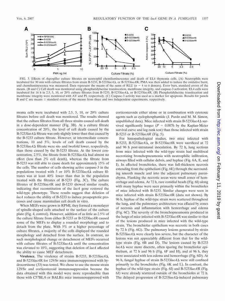
moma cells were incubated with 2.5, 5, 10, or 20% culturefiltrates before cell death was monitored. The results showedthat the culture filtrates from all three strains caused cell deathin a dose-dependent manner (Fig. 3B). At a culture filtrateconcentration of 20%, the level of cell death caused by theB-5233laeA� filtrate was only slightly lower than that caused bythe B-5233 culture filtrate. However, at intermediate concen-trations, 10 and 5%, levels of cell death caused by theB-5233laeA� filtrate were six- and twofold lower, respectively,than those caused by the B-5233 filtrate. At the lowest con-centration, 2.5%, the filtrate from B-5233laeA� had almost noeffect (less than 2% cell death), whereas the filtrate fromB-5233 was still able to cause death for approximately 15% ofthe cells. The number of cells showing active caspase-3 in thepopulations treated with 5 or 10% B-5233laeA� culture fil-trates was at least 40% lower than that in the populationtreated with the filtrates of B-5233 (Fig. 3C). The culturefiltrates of B-5233laeAR and B-5233 showed similar results,indicating that reconstitution of the laeA gene restored thewild-type phenotype. These results suggest that deletion oflaeA reduces the ability of B-5233 to induce proapoptotic pro-cesses and cause mammalian cell death in vitro.
When MEFs were grown in RPMI, they formed a monolayerof spindle-shaped cells attached to the surface of the cultureplate (Fig. 4, control). However, addition of as little as 2.5% ofthe culture filtrate from either B-5233 or B-5233laeAR causedsome of the MEFs to display a rounded morphology and todetach from the plate. With 5% or a higher percentage ofculture filtrates, a majority of the cells displayed the roundedmorphology and detached from the surface. In contrast, nosuch morphological change or detachment of MEFs occurredwith culture filtrates of B-5233laeA� until the concentrationwas elevated to 10%, suggesting that deletion of laeA affectedthe ability to cause MEF detachment.
Virulence. The virulence of strains B-5233, B-5233laeA�,and B-5233laeAR for 129/Sv mice immunosuppressed with hy-drocortisone (33) was tested. We chose to use the mouse strain129/Sv and corticosteroid immunosuppression because thedata obtained with this model were more reproducible thanthose with C57BL/6 or BALB/c mice immunosuppressed with
corticosteroids either alone or in combination with cytotoxicagents such as cyclophosphamide (J. Pardo and M. M. Simon,unpublished data). Mice infected with strain B-5233laeA� sur-vived significantly longer (P � 0.0076 by the Kaplan-Meiersurvival curve and log rank test) than those infected with strainB-5233 or B-5233laeAR (Fig. 5).
For histopathological studies, two mice infected withB-5233, B-5233laeA�, or B-5233laeAR were sacrificed at 72and 96 h post-intranasal inoculation. By 72 h, lung sectionsfrom mice infected with the wild-type strain had multifocalnecrotizing bronchopneumonia with neutrophilic infiltration,airways filled with cellular debris, and hyphae (Fig. 6A, E, andI). In affected bronchioles, there was full-thickness necrosisextending from the epithelium (Fig. 6E) through the surround-ing smooth muscle and into the adjacent pulmonary paren-chyma. Flanking the necrotic areas were small zones of hem-orrhage and edema. At 72 h, rare conidial heads (Fig. 6I) alongwith many hyphae were seen primarily within the bronchiolesof mice infected with B-5233. Similar changes were seen inmice infected with strain B-5233laeAR (data not shown). By96 h, hyphae of the wild-type strain were scattered throughoutthe lung, and the pulmonary architecture was effaced by zonesof necrosis and inflammation with hemorrhage and edema(Fig. 6C). The severity of the bronchopneumonia produced inthe lungs of mice infected with B-5233laeAR was similar to thatof the lesions produced in mice infected with the wild-typestrain. The bronchiolar epithelium was necrotic in both casesby 72 h (Fig. 6G). The pulmonary lesions generated by strainB-5233laeA� were clearly less severe, but the character of thelesions was not appreciably different from that for the wild-type strain (Fig. 6B and D). The lesions caused by B-5233laeA� were more discrete, often sparing the bronchiolar epi-thelium, at 72 h and 96 h (Fig. 6F and H), and at 96 h, theywere associated with less edema and hemorrhage (Fig. 6D). At96 h, fungal hyphae of strain B-5233laeA� were still confinedprimarily to the bronchioles (Fig. 6K and L). In contrast, thehyphae of the wild-type strain (Fig. 6I) and B-5233laeAR (Fig.6J) were already scattered outside of the bronchioles at 72 h.The delayed progression of B-5233laeA�-induced pulmonary
FIG. 3. Effects of Aspergillus culture filtrates on neutrophil chemiluminescence and death of EL4 thymoma cells. (A) Neutrophils wereincubated for 30 min with culture filtrates from strain B-5233, B-5233laeA�, or B-5233laeAR; PMA was then added to induce the oxidative burst,and chemiluminescence was measured. Data represent the means of the sums of RLU (n � 4 to 6 donors). Error bars, standard errors of themeans. (B and C) Cell death was monitored using phosphatidylserine translocation, membrane integrity, and caspase-3 activation. EL4 cells wereincubated for 16 h in 2.5, 5, 10, or 20% culture filtrates from B-5233, B-5233laeA�, or B-5233laeAR. (B) Phosphatidylserine translocation andmembrane integrity were monitored with AV and PI, respectively. (C) Caspase-3 activity was used as a marker for apoptosis. Results for panelsB and C are means � standard errors of the means from three and two independent experiments, respectively.
VOL. 6, 2007 REGULATORY FUNCTION OF THE laeA GENE IN A. FUMIGATUS 1557
on February 11, 2019 by guest
http://ec.asm.org/
Dow
nloaded from

lesions in comparison to that for the wild-type strain was cor-roborated by the survival data.
DISCUSSION
Previously it was reported that deletion of laeA in strainAF293 resulted in conidia with a smooth surface, a phenotype
similar to that observed when alb1 was deleted in strain B-5233(34). In the present study, we show that neither the laeA delet-ant strain of B-5233 nor strain AF293laeA� exhibits alterationsin conidial morphology. Like those of the wild type, the conidiawere bluish green and their surfaces were decorated with pro-trusions. Although a few exceptional conidia (0.1%) exhib-ited smooth surfaces with no clear protrusions, these wereobserved in all strains regardless of the presence or absence ofthe laeA gene. Our study also showed that deletion of laeA inB-5233 as well as in AF293 caused a reduction in the expres-sion of alb1 during mycelial growth, whereas a previous reporthad indicated a pronounced increase in the transcription ofalb1 in the laeA deletant (3). It is possible that this differencereflects an effect of the medium used to grow the cultures.Although alb1 is expressed primarily during the conidiationstage when B-5233 is grown on a solid medium, it has beenshown that the alb1 gene is also expressed in newly germinatedhyphae under certain conditions (18, 43). The function of alb1in conidial pigment synthesis is well characterized, but its func-tion in hyphae is unknown. Whether alb1 participates in thesynthesis of DHN-like melanin or in another secondary me-tabolite pathway during hyphal growth in AF293 is unknown. Itis possible that the lack of dark color in the submerged myce-lium of AF293laeA� is related to the down-regulation of alb1expression in the mycelium.
FIG. 4. A. fumigatus culture filtrates cause MEF detachment. MEFs were incubated for 4 h with different concentrations of the culture filtrates(2.5, 5, and 10%). The majority of the cells incubated in 5% culture filtrates from B-5233 and B-5233laeAR are rounded and detached from theculture plate surface, whereas cells incubated with RPMI only (control) are elongated and attached to the surface. Original magnification, �100.
FIG. 5. Virulence studies. Immunosuppressed 129/Sv mice wereinoculated intranasally with 5 � 106 conidia. Mortality was monitoredfor 21 days. A P value of 0.0076 was found for comparison of survivalbetween B-5233- and B-5233laeA�-infected mice. A Kaplan-Meiersurvival curve with a log rank test was used to compare survival levelsamong the strain groups.
1558 SUGUI ET AL. EUKARYOT. CELL
on February 11, 2019 by guest
http://ec.asm.org/
Dow
nloaded from

The previous study with strain AF293 (3) and the presentstudy with B-5233 showed that laeA plays a role in regulatingthe synthesis of gliotoxin and is also involved in the virulenceof A. fumigatus in a murine model. Comparisons with B-5233showed that B-5233laeA� produces 80% less gliotoxin and isslower in causing fatal infection in 129/Sv mice immunosup-pressed with hydrocortisone. Furthermore, the culture filtrateof the laeA deletant is less cytotoxic to mammalian cells andhas a reduced inhibitory effect on the neutrophil respiratoryburst. Similarly, Bok and collaborators have reported thatstrain AF293laeA� showed significantly reduced virulence rel-ative to AF293 in ICR mice. In addition, gliotoxin was notdetected in the lungs of mice infected with AF293laeA�,whereas it was readily detected in the lungs of mice infected
with AF293 (3). The immunosuppressive and proapoptoticproperties of gliotoxin (11, 20, 29, 32) indicate that this sec-ondary metabolite is an important factor in the pathobiology ofA. fumigatus. Recent studies, however, have suggested thatabrogation of gliotoxin synthesis through deletion of gliP hasno effect on the virulence of A. fumigatus strain AF293 in miceimmunosuppressed with cyclophosphamide and cortisone (4,8, 15). In contrast, we observed that deletion of gliP in strainB-5233 resulted in a mutant that was significantly less virulentthan the wild type in mice immunosuppressed with hydrocor-tisone alone (31b). This disparity is likely due to the differentimmunosuppressive regimens used in the murine models. Al-though, based on our findings, we can hypothesize that glio-toxin is an important virulence determinant of A. fumigatus, it
FIG. 6. Histopathology of mice infected with A. fumigatus strains. (A to H) Hematoxylin-and-eosin-stained sections of lungs from mice infectedintranasally with strain B-5233, B-5233laeA�, or B-5233laeAR. B-5233 (A) produced a more severe bronchopneumonia than B-5233laeA� (B) at72 h. At 96 h, bronchopneumonia caused by strain B-5233laeA� (D) was still significantly less severe than that caused by strain B-5233 (C). Scalebars in panels A to D, 100 �m. At 72 h, bronchioles of mice infected with B-5233 (E) or B-5233laeAR (G) showed a completely necrotic epithelium(arrows). The epithelium of the bronchiolar walls (arrows) in the lungs of mice infected with strain B-5233laeA� was mostly intact at 72 h (F) aswell as at 96 h (H). Scale bars in panels E to H, 10 �m. (I to L) Lung sections stained with Gomori’s methenamine silver to demonstrate the extentof hyphal growth. At 72 h, hyphae of strains B-5233 (I) and B-5233laeAR (J) are seen in the lung. The arrow in panel I points to a rare conidialhead of the wild-type strain within the bronchiole. Hyphae of strain B-5233laeA� are seen within a bronchiole at 72 h (K). At 96 h, hyphae of strainB-5233laeA� were still primarily within the bronchiole (L). Scale bars in panels I to L, 20 �m.
VOL. 6, 2007 REGULATORY FUNCTION OF THE laeA GENE IN A. FUMIGATUS 1559
on February 11, 2019 by guest
http://ec.asm.org/
Dow
nloaded from

is probable that laeA regulates the synthesis of other second-ary metabolites or proteins that may also play a role in thepathobiology of this fungus. In fact, it has recently beenshown that the LaeA protein regulates the expression of atleast 9.5% of the A. fumigatus genome and positively regu-lates the expression of genes involved in the synthesis of 20to 40% of the major classes of secondary metabolites (25).The ability of the LaeA protein to regulate the synthesis ofdifferent secondary metabolites has also been suggested tooccur in Aspergillus nidulans (5).
Histological sections of lungs from mice infected with strainB-5233laeA� or B-5233 supported the survival data. Althoughthe pattern of lung lesions showed no appreciable difference,the progression of the disease was clearly less severe for miceinfected with the laeA� strain. Although B-5233laeA� pro-duces conidial heads as abundantly as the wild-type strain invitro, conidial heads with spores were found only in the bron-chioles of lungs from mice infected with either B-5233 orB-5233laeAR. It is not likely that the conidial heads were partof the inoculum, since the inoculum was filtered through mul-tiple layers of sterile gauze to eliminate hyphae and conidio-phores so that it contained only free conidia. Formation ofconidial heads by aspergilli in vivo has been seen in tissueswhere oxygen tension is high, such as in the sinus or the chroniccavitary lesions of the lung (16). It is not clear why B-5233 andB-5233laeAR, but not B-5233laeA�, contained conidial headswithin the bronchioles. Histological examinations using moremice may be needed to find out whether this phenomenon isrelated to the function of the laeA gene.
The findings presented here indicate that laeA regulates thesynthesis of the secondary metabolite gliotoxin and the expres-sion of the alb1 gene during growth in liquid culture. Our studyshowed that deletion of laeA caused a down-regulation of alb1expression in mycelia of both B-5233 and AF293, whereas theprevious report has indicated a pronounced up-regulation ofalb1 in the laeA deletant (3). It is possible that this differencereflects the media used to grow the cultures. Furthermore,such regulation of alb1 by lae is apparently unrelated to theregulation of conidial morphology as revealed by SEM. Thisstudy confirms that the deletion of laeA reduces virulence in ananimal model, as has been reported for AF293 (3). The re-duced ability of the laeA deletant to cause apoptosis in mam-malian cells and inhibition of the respiratory burst in humanneutrophils may have contributed to the observed reduction invirulence.
ACKNOWLEDGMENTS
We thank N. P. Keller, University of Wisconsin—Madison, for pro-viding the AF293 strains and A. Varma for critical review of themanuscript.
This study was supported by funds from the intramural program ofthe National Institute of Allergy and Infectious Diseases, NationalInstitutes of Health, Bethesda, MD, and by a grant (Technology Trans-fer) from the Max Planck Society, Germany. J. Pardo was supported bythe Alexander von Humboldt Foundation, Germany, and E. M. Galvezwas supported by the Deutsche Forschungsgemeinschaft (DFG),Germany.
REFERENCES
1. Amitani, R., T. Murayama, R. Nawada, W. J. Lee, A. Niimi, K. Suzuki, E.Tanaka, and F. Kuze. 1995. Aspergillus culture filtrates and sputum sols frompatients with pulmonary aspergillosis cause damage to human respiratoryciliated epithelium in vitro. Eur. Respir. J. 8:1681–1687.
2. Belkacemi, L., R. C. Barton, V. Hopwood, and E. G. Evans. 1999. Determi-nation of optimum growth conditions for gliotoxin production by Aspergillusfumigatus and development of a novel method for gliotoxin detection. Med.Mycol. 37:227–233.
3. Bok, J. W., S. A. Balajee, K. A. Marr, D. Andes, K. F. Nielsen, J. C. Frisvad,and N. P. Keller. 2005. LaeA, a regulator of morphogenetic fungal virulencefactors. Eukaryot. Cell 4:1574–1582.
4. Bok, J. W., D. Chung, S. A. Balajee, K. A. Marr, D. Andes, K. F. Nielsen, J. C.Frisvad, K. A. Kirby, and N. P. Keller. 2006. GliZ, a transcriptional regulatorof gliotoxin biosynthesis, contributes to Aspergillus fumigatus virulence. In-fect. Immun. 74:6761–6768.
5. Bok, J. W., D. Hoffmeister, L. A. Maggio-Hall, R. Murillo, J. D. Glasner, andN. P. Keller. 2006. Genomic mining for Aspergillus natural products. Chem.Biol. 13:31–37.
6. Bok, J. W., and N. P. Keller. 2004. LaeA, a regulator of secondary metab-olism in Aspergillus spp. Eukaryot. Cell 3:527–535.
7. Bok, J. W., D. Noordermeer, S. P. Kale, and N. P. Keller. 2006. Secondarymetabolic gene cluster silencing in Aspergillus nidulans. Mol. Microbiol.61:1636–1645.
8. Cramer, R. A., Jr., M. P. Gamcsik, R. M. Brooking, L. K. Najvar, W. R.Kirkpatrick, T. F. Patterson, C. J. Balibar, J. R. Graybill, J. R. Perfect, S. N.Abraham, and W. J. Steinbach. 2006. Disruption of a nonribosomal peptidesynthetase in Aspergillus fumigatus eliminates gliotoxin production. Eukaryot.Cell 5:972–980.
9. Denning, D. W.1998. Invasive aspergillosis. Clin. Infect. Dis. 26:781–805.10. Dupont, B., M. Huber, S. J. Kim, and J. E. Bennett. 1987. Galactomannan
antigenemia and antigenuria in aspergillosis: studies in patients and exper-imentally infected rabbits. J. Infect. Dis. 155:1–11.
11. Eichner, R. D., M. Al Salami, P. R. Wood, and A. Mullbacher. 1986. Theeffect of gliotoxin upon macrophage function. Int. J. Immunopharmacol.8:789–797.
12. Jahn, B., A. Koch, A. Schmidt, G. Wanner, H. Gehringer, S. Bhakdi, andA. A. Brakhage. 1997. Isolation and characterization of a pigmentless-conid-ium mutant of Aspergillus fumigatus with altered conidial surface and re-duced virulence. Infect. Immun. 65:5110–5117.
13. Kamei, K., A. Watanabe, K. Nishimura, and M. Miyaji. 2002. Cytotoxicity ofAspergillus fumigatus culture filtrate against macrophages. Nippon IshinkinGakkai Zasshi 43:37–41.
14. Keller, N., J. Bok, D. Chung, R. M. Perrin, and E. Keats Shwab. 2006. LaeA,a global regulator of Aspergillus toxins. Med. Mycol. 44(Suppl.):83–85.
15. Kupfahl, C., T. Heinekamp, G. Geginat, T. Ruppert, A. Hartl, H. Hof, andA. A. Brakhage. 2006. Deletion of the gliP gene of Aspergillus fumigatusresults in loss of gliotoxin production but has no effect on virulence of thefungus in a low-dose mouse infection model. Mol. Microbiol. 62:292–302.
16. Kwon-Chung, K. J., and J. E. Bennett. 1992. Medical mycology, p. 201–247.Lea & Febiger, Philadelphia, PA.
17. Langfelder, K., B. Jahn, H. Gehringer, A. Schmidt, G. Wanner, and A. A.Brakhage. 1998. Identification of a polyketide synthase gene (pksP) of As-pergillus fumigatus involved in conidial pigment biosynthesis and virulence.Med. Microbiol. Immunol. 187:79–89.
18. Langfelder, K., B. Philippe, B. Jahn, J. P. Latge, and A. A. Brakhage. 2001.Differential expression of the Aspergillus fumigatus pksP gene detected invitro and in vivo with green fluorescent protein. Infect. Immun. 69:6411–6418.
19. Marr, K. A., T. Patterson, and D. Denning. 2002. Aspergillosis. Pathogene-sis, clinical manifestations, and therapy. Infect. Dis. Clin. N. Am. 16:875–894.
20. Mullbacher, A., P. Waring, and R. D. Eichner. 1985. Identification of anagent in cultures of Aspergillus fumigatus displaying anti-phagocytic andimmunomodulating activity in vitro. J. Gen. Microbiol. 131:1251–1258.
21. Murayama, T., R. Amitani, Y. Ikegami, R. Nawada, W. J. Lee, and F. Kuze.1996. Suppressive effects of Aspergillus fumigatus culture filtrates on humanalveolar macrophages and polymorphonuclear leucocytes. Eur. Respir. J.9:293–300.
22. Nierman, W. C., A. Pain, M. J. Anderson, J. R. Wortman, H. S. Kim, J.Arroyo, M. Berriman, K. Abe, D. B. Archer, C. Bermejo, J. Bennett, P.Bowyer, D. Chen, M. Collins, R. Coulsen, R. Davies, P. S. Dyer, M. Farman,N. Fedorova, T. V. Feldblyum, R. Fischer, N. Fosker, A. Fraser, J. L. Garcia,M. J. Garcia, A. Goble, G. H. Goldman, K. Gomi, S. Griffith-Jones, R.Gwilliam, B. Haas, H. Haas, D. Harris, H. Horiuchi, J. Huang, S. Hum-phray, J. Jimenez, N. Keller, H. Khouri, K. Kitamoto, T. Kobayashi, S.Konzack, R. Kulkarni, T. Kumagai, A. Lafon, J. P. Latge, W. Li, A. Lord, C.Lu, W. H. Majoros, G. S. May, B. L. Miller, Y. Mohamoud, M. Molina, M.Monod, I. Mouyna, S. Mulligan, L. Murphy, S. O’Neil, I. Paulsen, M. A.Penalva, M. Pertea, C. Price, B. L. Pritchard, M. A. Quail, E. Rabbinowitsch,N. Rawlins, M. A. Rajandream, U. Reichard, H. Renauld, G. D. Robson, S.Rodriguez de Cordoba, J. M. Rodriguez-Pena, C. M. Ronning, S. Rutter,S. L. Salzberg, M. Sanchez, J. C. Sanchez-Ferrero, D. Saunders, K. Seeger,R. Squares, S. Squares, M. Takeuchi, F. Tekaia, G. Turner, C. R. Vazquez deAldana, J. Weidman, O. White, J. Woodward, J. H. Yu, C. Fraser, J. E.Galagan, K. Asai, M. Machida, N. Hall, B. Barrell, and D. W. Denning. 2005.Genomic sequence of the pathogenic and allergenic filamentous fungusAspergillus fumigatus. Nature 438:1151–1156.
1560 SUGUI ET AL. EUKARYOT. CELL
on February 11, 2019 by guest
http://ec.asm.org/
Dow
nloaded from
23. Pardo, J., A. Bosque, R. Brehm, R. Wallich, J. Naval, A. Mullbacher, A. Anel,and M. M. Simon. 2004. Apoptotic pathways are selectively activated bygranzyme A and/or granzyme B in CTL-mediated target cell lysis. J. CellBiol. 167:457–468.
24. Pardo, J., C. Urban, E. M. Galvez, P. G. Ekert, U. Muller, J. Kwon-Chung,M. Lobigs, A. Mullbacher, R. Wallich, C. Borner, and M. M. Simon. 2006.The mitochondrial protein Bak is pivotal for gliotoxin-induced apoptosis anda critical host factor of Aspergillus fumigatus virulence in mice. J. Cell Biol.174:509–519.
25. Perrin, R. M., N. D. Fedorova, J. W. Bok, R. A. Cramer, J. R. Wortman, H. S.Kim, W. C. Nierman, and N. P. Keller. 2007. Transcriptional regulation ofchemical diversity in Aspergillus fumigatus by LaeA. PLoS Pathog. 3:e50.
26. Rex, J. H., J. E. Bennett, J. I. Gallin, H. L. Malech, E. S. DeCarlo, and D. A.Melnick. 1991. In vivo interferon-gamma therapy augments the in vitroability of chronic granulomatous disease neutrophils to damage Aspergillushyphae. J. Infect. Dis. 163:849–852.
27. Rex, J. H., J. E. Bennett, J. I. Gallin, H. L. Malech, and D. A. Melnick. 1990.Normal and deficient neutrophils can cooperate to damage Aspergillus fu-migatus hyphae. J. Infect. Dis. 162:523–528.
28. Shimizu, K., and N. P. Keller. 2001. Genetic involvement of a cAMP-dependent protein kinase in a G protein signaling pathway regulating mor-phological and chemical transitions in Aspergillus nidulans. Genetics 157:591–600.
29. Stanzani, M., E. Orciuolo, R. Lewis, D. P. Kontoyiannis, S. L. Martins, L. S.St John, and K. V. Komanduri. 2005. Aspergillus fumigatus suppresses thehuman cellular immune response via gliotoxin-mediated apoptosis of mono-cytes. Blood 105:2258–2265.
30. Sugareva, V., A. Hartl, M. Brock, K. Hubner, M. Rohde, T. Heinekamp, andA. A. Brakhage. 2006. Characterisation of the laccase-encoding gene abr2 ofthe dihydroxynaphthalene-like melanin gene cluster of Aspergillus fumigatus.Arch. Microbiol. 186:345–355.
31. Sugui, J. A., Y. C. Chang, and K. J. Kwon-Chung. 2005. Agrobacteriumtumefaciens-mediated transformation of Aspergillus fumigatus: an efficienttool for insertional mutagenesis and targeted gene disruption. Appl. Envi-ron. Microbiol. 71:1798–1802.
31a.Sugui, J. A., J. Pardo, Y. C. Chang, K. A. Zarember, G. Nardone, E. M.Galvez, A. Mullbacher, J. I. Gallin, M. M. Simon, and K. J. Kwon-Chung.2007. Gliotoxin is a virulence factor of Aspergillus fumigatus: gliP deletionattenuates virulence in mice immunosuppressed with hydrocortisone. Eu-karyot. Cell 6:1562–1569.
32. Sutton, P., N. R. Newcombe, P. Waring, and A. Mullbacher. 1994. In vivoimmunosuppressive activity of gliotoxin, a metabolite produced by humanpathogenic fungi. Infect. Immun. 62:1192–1198.
33. Tang, C. M., J. Cohen, T. Krausz, S. Van Noorden, and D. W. Holden. 1993.The alkaline protease of Aspergillus fumigatus is not a virulence determinantin two murine models of invasive pulmonary aspergillosis. Infect. Immun.61:1650–1656.
34. Tsai, H. F., Y. C. Chang, R. G. Washburn, M. H. Wheeler, and K. J.Kwon-Chung. 1998. The developmentally regulated alb1 gene of Aspergillusfumigatus: its role in modulation of conidial morphology and virulence. J.Bacteriol. 180:3031–3038.
35. Tsai, H. F., I. Fujii, A. Watanabe, M. H. Wheeler, Y. C. Chang, Y. Yasuoka,Y. Ebizuka, and K. J. Kwon-Chung. 2001. Pentaketide melanin biosynthesisin Aspergillus fumigatus requires chain-length shortening of a heptaketideprecursor. J. Biol. Chem. 276:29292–29298.
36. Tsai, H. F., R. G. Washburn, Y. C. Chang, and K. J. Kwon-Chung. 1997.Aspergillus fumigatus arp1 modulates conidial pigmentation and complementdeposition. Mol. Microbiol. 26:175–183.
37. Tsai, H. F., M. H. Wheeler, Y. C. Chang, and K. J. Kwon-Chung. 1999. Adevelopmentally regulated gene cluster involved in conidial pigment biosyn-thesis in Aspergillus fumigatus. J. Bacteriol. 181:6469–6477.
38. Tsunawaki, S., L. S. Yoshida, S. Nishida, T. Kobayashi, and T. Shimoyama.2004. Fungal metabolite gliotoxin inhibits assembly of the human respiratoryburst NADPH oxidase. Infect. Immun. 72:3373–3382.
39. Washburn, R. G., J. I. Gallin, and J. E. Bennett. 1987. Oxidative killing ofAspergillus fumigatus proceeds by parallel myeloperoxidase-dependent and-independent pathways. Infect. Immun. 55:2088–2092.
40. Wei, M. C., W. X. Zong, E. H. Cheng, T. Lindsten, V. Panoutsakopoulou,A. J. Ross, K. A. Roth, G. R. MacGregor, C. B. Thompson, and S. J.Korsmeyer. 2001. Proapoptotic BAX and BAK: a requisite gateway to mi-tochondrial dysfunction and death. Science 292:727–730.
41. Yoshida, L. S., S. Abe, and S. Tsunawaki. 2000. Fungal gliotoxin targets theonset of superoxide-generating NADPH oxidase of human neutrophils. Bio-chem. Biophys. Res. Commun. 268:716–723.
42. Zarember, K. A., J. A. Sugui, Y. C. Chang, K. J. Kwon-Chung, and J. I.Gallin. 2007. Human PMN inhibit Aspergillus fumigatus conidial growth bylactoferrin-mediated iron depletion. J. Immunol. 178:6367–6373.
43. Zhang, L., M. Wang, R. Li, and R. Calderone. 2005. Expression of Aspergillusfumigatus virulence-related genes detected in vitro and in vivo with compet-itive RT-PCR. Mycopathologia 160:201–206.
VOL. 6, 2007 REGULATORY FUNCTION OF THE laeA GENE IN A. FUMIGATUS 1561
on February 11, 2019 by guest
http://ec.asm.org/
Dow
nloaded from