role of pkr in the inhibition of proliferation and...
TRANSCRIPT

Research ArticleRole of PKR in the Inhibition of Proliferation and Translationby Polycystin-1
Yan Tang ,1,2 Guang Shi,1 JungWoo Yang,2 Wang Zheng,2 Jingfeng Tang,3
Xing-Zhen Chen ,2 Jianzheng Yang ,1 and ZuochengWang 2
1Department of Oncology, The Second Hospital, Jilin University, Changchun 130041, China2Membrane Protein Disease Research Group, Department of Physiology, Faculty of Medicine and Dentistry, University of Alberta,Edmonton, Canada T6G2H73National “111” Center for Cellular Regulation and Molecular Pharmaceutics, Hubei University of Technology, Wuhan 430086, China
Correspondence should be addressed to Jianzheng Yang; [email protected] andZuochengWang; [email protected]
Received 15 February 2019; Revised 19 May 2019; Accepted 2 June 2019; Published 23 June 2019
Academic Editor: Paul M. Tulkens
Copyright © 2019 Yan Tang et al.This is an open access article distributed under the Creative Commons Attribution License, whichpermits unrestricted use, distribution, and reproduction in any medium, provided the original work is properly cited.
Autosomal dominant polycystic kidney disease (ADPKD) is mainly caused by mutations in the PKD1 (∼85%) or PKD2 (∼15%)gene which, respectively, encode polycystin-1 (PC1) and polycystin-2 (PC2). How PC1 regulates cell proliferation and apoptosis hasbeen studied for decades but the underlying mechanisms remain controversial. Protein kinase RNA-activated (PKR) is activatedby interferons or double-stranded RNAs, inhibits protein translation, and induces cell apoptosis. In a previous study, we foundthat PC1 reduces apoptosis through suppressing the PKR/eIF2𝛼 signaling. Whether and how PKR is involved in PC1-inhibitedproliferation and protein synthesis remains unknown. Here we found that knockdown of PKR abolishes PC1-inhibited proliferationand translation. Because suppressed PKR-eIF2𝛼 signaling/activity by PC1 would stimulate, rather than inhibit, the proliferation andtranslation, we examined the effect of dominant negative PKRmutant K296R that has no kinase activity and found that it enhancesthe inhibition of proliferation and translation by PC1. Thus, our study showed that inhibition of cell proliferation and proteinsynthesis by PC1 is mediated by the total expression but not the kinase activity of PKR, possibly through physical association.
1. Introduction
Autosomal dominant polycystic kidney disease (ADPKD)is one of the most common inherited renal diseases andcharacterized by the development of fluid-filled cysts [1, 2].Up to 95% of the ADPKD cases are caused by mutations inthe PKD1 or PKD2 gene which encodes polycystin-1 (PC1)and polycystin-2 (PC2), respectively. Mutations in PKD1account for ∼85% of ADPKD [3, 4]. PC1 is a 462-kDamembrane protein with 4302 amino acids (aa) in length,eleven transmembrane (TM) domains, a large extracellularN-terminus and a short C-terminus containing domainsinvolved in G-protein activation and interaction with partnerproteins [5–7]. PC1 seems to function as a cell surfacereceptor that mediates mechanosensation of fluid flow ofprimary cilia in renal epithelia and intracellular signalling[8–10].
ADPKD is a disorder characterized by several cellularabnormalities, including cell overproliferation, apoptosis,and dedifferentiation [11], which indicates a high cell turnoverrate. It was reported that cyclic adenosine monophosphate(cAMP) and mitogen-activated protein kinase (MAPK) sig-naling [12], P53, c-JunN-terminal kinase(JNK) induction [13]and activation of cellular Src kinase (c-Src) [14], signal trans-ducers and activators of transcription (STAT) [15], Hippo[16], and 𝛽-catenin/Wnt pathway [17] are connected withoverproliferation or differentiation of renal epithelial cells inADPKD. In cyst-lining epithelial cells of ADPKD patientsand mouse model experiments, the mammalian target ofrapamycin (mTOR) pathway was shown to be activated,which may result from loss of PC1 binding with tuberin,suggesting that PC1 inhibits cell proliferation by downregu-lating mTOR activity through interaction with tuberin [18].Actually, PC1 reduces the cell growth by negatively regulating
HindawiBioMed Research InternationalVolume 2019, Article ID 5320747, 8 pageshttps://doi.org/10.1155/2019/5320747

2 BioMed Research International
mTORanddownstreameffectors ribosomal protein S6 kinasebeta-1(S6K1) and eukaryotic translation initiation factor 4E-binding protein 1 (4EBP1) in a tuberin-dependent manner[18, 19]. mTOR-related translational control pathways havethen been subjects of study with respect to PKD pathogenesis[20–25]. Despite tremendous progress made in the devel-opment of ADPKD pathogenesis over the past years, theunderlying mechanisms are still elusive.
Protein kinaseRNA-activated (PKR) is a serine/threonineprotein kinase that is activated by interferons, double-stranded RNAs (dsRNAs), cytokine, growth factor, and stresssignals [26]. The 551-aa kinase is made up of a C-terminusfor catalysis as well as a N-terminus which contains double-stranded RNA of approximately 70aa residues each; whencombined with dsRNAs, the conformation of PKR changed,and the binding site dimerized to form PKR dimer [10].During virus infection, PKR is activated and blocks viralprotein synthesis through phosphorylation of eukaryotictranslation initiation factor 2 alpha (eIF2𝛼), thus leadingto antiviral defense [27]. PKR can be autophosphorylatedwith the formation of dimer and activated when bindingto dsRNA, and then phosphorylates substrates, includingeIF2𝛼, protein phosphatase 2A (PP2A), and inhibitor ofnuclear factor kappa-B(I𝜅B) kinase (IKK). PKR inhibitstranslation and promotes apoptosis through the substratesand downstream effectors [28, 29]. In addition to PKR, thereare three other eIF2𝛼 kinases, including protein kinase-likeendoplasmic reticulum (ER) kinase (PERK), general controlnonderepressible 2 (GCN2), and heme-regulated inhibitor(HRI) [30]. Phosphorylated eIF2𝛼 (P-eIF2𝛼) blocks transla-tion initiation but activates some selected proteins critical tocell survival, including transcription factor 4(ATF4), growtharrest, and DNA damage gene (GADD34) and C/EBP-homologous protein (CHOP) [31, 32].
Up to now, there has been no literature report on whetheror how PKR mediates the inhibition of proliferation andtranslation caused by PC1. Therefore, the aim of this studywas to investigate the involvement of PKR in PC1-regulatedproliferation and translation.
2. Materials and Methods
2.1. Reagents and Antibodies. Puromycin was a productof Sigma-Aldrich Canada. Phosphorylated PKR (P-PKR,Thr446), P-PKR (pT446), PKR (B-10), and anti-FLAG anti-bodies were purchased from Santa Cruz (Santa Cruz, CA)or Epitomics (Burlingame, CA). eIF2𝛼, P-eIF2𝛼, and mTORantibodies were products of Cell Signaling Technology (NewEngland Biolabs, Pickering, ON). Anti-GFP (B-2) was fromSanta Cruz and anti-GFP (EU4) from Eusera (Edmonton,AB). Mouse monoclonal anti-𝛽-actin (C4, Santa Cruz) anti-bodywas employed as loading controls. Secondary antibodieswere from Santa Cruz or GE Healthcare (Baied’Urfe, QC).
2.2. DNA Constructs, Cell Culture, and Transfection. Plas-mid pcDNA3-GFP-PC1-5TMC(PC1-5TMC, aa 3895-4302)comprising last 5 TMs plus C-terminus was constructedusing Stratagene Quik Change� II XL Site-Directed Muta-genesis Kit (Agilent Technologies Canada Inc., Mississauga,
ON) as described previously [33]. Plasmid eIF2𝛼 was fromSanta Cruz (Santa Cruz, CA). All cDNA sequences of theconstructed plasmids were verified by sequencing. Humanembryonic kidney (HEK293T) or HeLa cells were grown inDulbecco’s modified Eagle’s medium with 10% fetal bovineserum, penicillin-streptomycin, and L-glutamine in an atmo-sphere of 5% CO
2and 37∘C. HEK293T cells with stable
transfection of wild type (WT) mouse PC1 was from onecoauthor Dr. J. Yang and cultured under the above conditionswith 2𝜇g/ml of puromycin [34]. Transient transfection wasperformed on HEK293T or HeLa cells grown to ∼70% con-fluency employing lipofectamine 2000 reagent (Invitrogen).
2.3. Gene Knockdown. Small Interfering RNAs (siRNA) ofPKR (SantaCruz, Cat#sc-36263)was utilized to interferewithHEK293T and HeLa cells according product description.HEK293T or HeLa cells at 50–60% confluency were trans-fected in normal culture medium without antibiotics, sup-plementedwithOpti-MEMmedium (Invitrogen, Burlington,ON) and lipofectamine 2000. 10 pmol of siRNAwas added tothe transfection reagent for 40 hours (hr). The efficiency ofthe siRNA knockdown was assessed by immunoblotting.
2.4. Cell Proliferation Assay. HEK293T or HeLa cells weretransiently transfected with corresponding plasmids such asGFP, GFP-PC1-5TMC, PKR, and PKR siRNA in 100 mmdishes. At 24 hr after transfection, cells were seeded intoeither new 100 mm dishes for further transfection such aseIF2𝛼, PKR, PKR-K296R, and PKR knockdown or a 96-well plate for alarmaBlue (Invitrogen Canada Inc.). Afterincubation for another 16-30 hr, absorbance was measuredusing a microplate reader (Fluoroskan Ascent FL, ThermoLabsystems). The rest of the cells in the 100 mm disheswere collected for immunoblotting at the same time point.HEK293T cells stably expressing WT PC1 were seeded in100 mm dishes overnight and then transfected with PKR-K296R or PKR siRNA using 4𝜇l lipofectamine 2000 reagentin medium lacking serum. 6 hr after transfection, the plateswere replenished with medium containing 10% serum andincubated at 37∘C for an additional 24 hr before measure-ments. The cell proliferation rate (%) was calculated asODtest/ODcontrol×100%.
2.5. 35S Pulse Labelling. HEK293T or HeLa cells were trans-fectedwith plasmids using Lipofectamine 2000 reagent. At 40hr after transfection, equal number of cells was starved for 1hr in the prelabeling medium (L-methionine and L-cysteinefree DMEM with 10% FBS and penicillin/streptomycin,Invitrogen), followed by pulse labeling with 50 𝜇Ci of[35S] methionine/cysteine (EXPRE 35S Protein Labeling Mix,PerkinElmer, Woodbridge, ON) for 10 minutes, as describedpreviously [35, 36]. Cell extracts were used for sodiumdodecyl sulfate–polyacrylamide gel electrophoresis (SDS-PAGE) and autoradiography.
2.6. Coimmunoprecipitation (Co-IP). Experiments were car-ried out according to previously established methods [37].Briefly, HeLa cells (2×10⁷ cells) with plasmid pEGFP-PC1-5TMC or pEGFP transfection were collected for extraction of

BioMed Research International 3
Prol
ifera
tion
(%)
Ctrl PKR si-PKR
Ctrl PKR si-PKR
150
100
50
0
∗∗∗∗
HeLa
Blot
PKR
-actin
(a)
PKR
-actin
Ctrl PKR si-PKR
S35labelling
Blot
HEK293T(b)
Figure 1: Effects of PKR on the proliferation and translation. (a) Effects of PKR on the proliferation of HeLa cells. After being transfected withplasmids PKR, PKR siRNA, or GFP, HeLa cells were plated inmultiple wells of a 96-well plate and grown for 24 hr for cell proliferation assays.Cells from the sample preparations were collected for immunoblotting. Proliferation rate of the control sample was normalized to 100%. PKR,WT PKR; si-PKR, PKR siRNA; Ctrl, GFP. Upper panel, averaged data (N=4, ∗∗p<0.01). Lower panel, effectiveness of transfection and siRNAof PKR assessed by immunoblotting. (b) Effect of PKR on protein synthesis inHEK293Tcells. HEK293T cells transiently transfected with GFP,PKR, or PKR siRNAwere starved for 1 hr followed by pulse labelling for 35S pulse labelling assays followed by SDS-PAGE and immunoblottingassays with the antibody against total PKR. 𝛽-actin served as loading control.
protein and immunoprecipitation at 40 hr after transfection.20mg of the total protein was for immunoblotting and 200mgfor co-IP.
2.7. Statistical Analysis. All data generated were presentedas mean±standard error (SEM). N represents the numberof repeat experiments. Data analyses were measured usingSigmaplot 12.0 software (Systat Software Inc., San Jose, CA).A P-value ⩽0.05 was statistically significant.
3. Results
3.1. Inhibition of Proliferation and Translation by PKR. Wefound that PC1 reduces apoptosis by inhibiting the PKRkinase activity and the phosphorylation of eIF2𝛼 [33]. Herewe tested whether PKR is involved in PC1-inhibited prolifer-ation. In order to clarify the effect of PKR on cell proliferationand translation, we used alarmaBlue to label HeLa cells forcell proliferation assays and performed 35S labelling assaysin HEK293T to evaluate protein translation. We found thatPKR suppresses proliferation and translation whereas PKRknockdown by siRNA does not show stimulation effect(Figures 1(a) and 1(b)), which is in line with previous reports[28, 29] and suggests that the endogenous PKR activity isnot a rate-limiting factor for the proliferation and proteinsynthesis.
3.2. Independency of PC1-Mediated Inhibition of Proliferationfrom eIF2𝛼. We next examined whether PC1 and PKR-eIF2𝛼inhibit cell proliferation or protein translation through thesame pathway. Overexpression of PC1 truncate mutationencoding 5 TMs and C-terminus (PC1-5TMC, aa 3895-4302) inhibited cell proliferation of HeLa cells (Figure 2(a)).HeLa cells overexpressing eIF2𝛼 exhibited much reducedproliferation rates, as expected, and were still inhibitable byPC1-5TMC (Figure 2(a)), indicating that the eIF2𝛼 activityand PC1 inhibit proliferation through two different path-ways. In fact, if inhibition by PC1 were through eIF2𝛼,then because PC1-5TMC reduces the eIF2𝛼 activity [33], aknown proliferation and translation inhibitor, we would see astimulating effect of PC1-5TMC on proliferation, against ourobservation (Figure 2(a)). PC1-5TMC also inhibited proteinsynthesis assessed by 35S labelling, but because overexpressedPKR almost completely stopped 35S labelling, the effectof coexpressed PC1-5TMC on protein synthesis cannot beevaluated (Figure 2(b)).
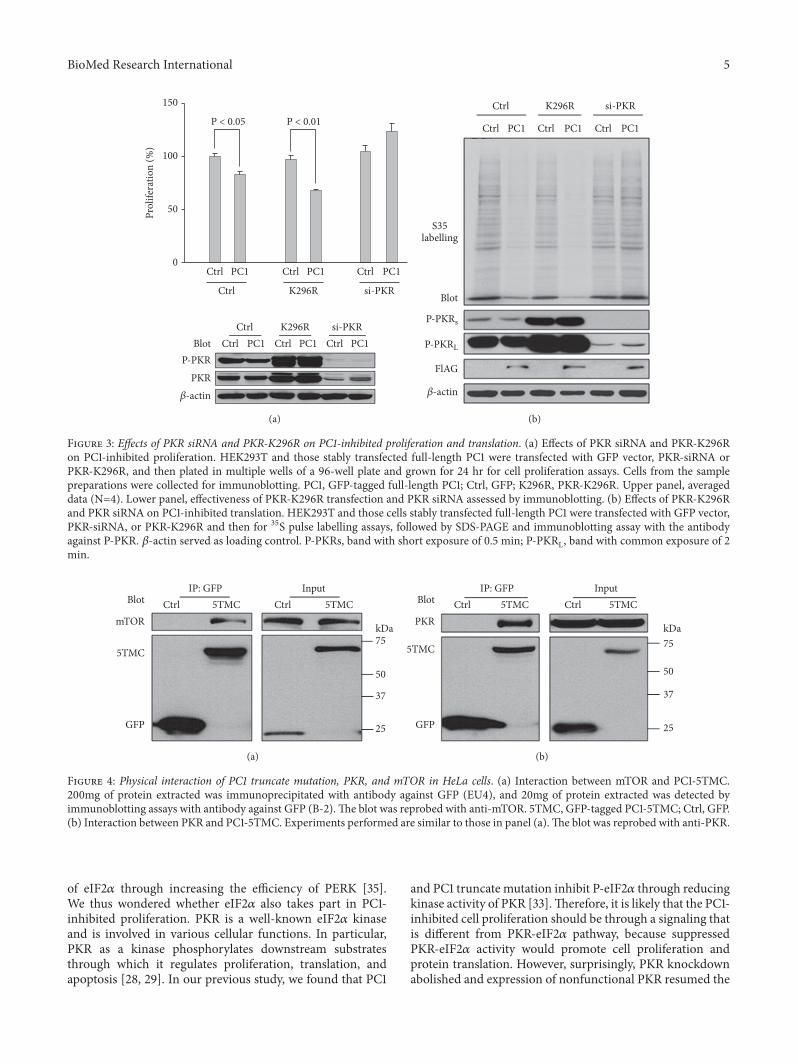
3.3. Dependence of PC1-Inhibited Proliferation and Translationon Total PKR. We further examined the role of PKR inPC1-inhibited proliferation and protein synthesis.When PKRwas knocked down by siRNA, PC1 no longer inhibitedproliferation of HEK cells, indicating the requirement of PKRfor mediating the effect of PC1 (Figure 3(a)). Interestingly,

4 BioMed Research International
Blot
-actin
Ctrl
Ctrl
5TMC
5TMC Ctrl 5TMC
5TMC
Ctrl
Ctrl
Ctrl
Prol
ifera
tion
(%)
150
100
50
0
∗∗
∗
∗∗
eIF2
eIF2
eIF2
P-eIF2
(a)
PKR
P-PKR
-actin
S35labelling
Blot
Ctrl 5TMC Ctrl 5TMC
Ctrl
eIF2
P-eIF2
(b)
Figure 2: Effects of PKR/eIF2𝛼 overexpression on PC1-5TMC-inhibited proliferation and translation in HeLa cells. (a) Effects of eIF2𝛼overexpression on PC1-5TMC-inhibited proliferation. Transfected with GFP or PC1-5TMC, HeLa cells were cotransfected with eIF2𝛼. Theywere then plated in multiple wells of a 96-well plate and grown for 24 hr for cell proliferation assay; cells from the sample preparationswere collected for immunoblotting. 5TMC, GFP-tagged PC1-5TMC; eIF2𝛼, WT eIF2𝛼; Ctrl, GFP. Upper panel, averaged data (N=4,∗p< 0.05,∗∗p< 0.01). Lower panel, effectiveness of transfection of eIF2𝛼 assessed by immunoblotting. (b) Effects of PKR overexpression on PC1-5TMC-inhibited protein synthesis. After transfected with GFP or PC1-5TMC, HeLa cells were cotransfected withWT PKR.They were then used for35S pulse labeling assays followed by SDS-PAGE and immunoblotting assays with the antibodies against P-eIF2𝛼, total eIF2𝛼, and P-PKR.𝛽-actin served as loading control.
expression of PKR-K296R that can retain the autophospho-rylation of PKR but has lost kinase function [38] did not havesignificant effect on proliferation but allowed strong inhibi-tion of proliferation by PC1 (Figure 3(a)). 35S labelling assaysalso showed that PKR knockdown abolishes while expressionof mutant K296R rescues the inhibition of protein synthesisin HEK cells by PC1 (Figure 3(b)). Taken together, out datashowed that PC1-inhibited proliferation and translation aremediated by a pathway that depends on the total PKR but notits kinase activity.
Based on the above results that phosphorylated PKR/eIF2𝛼 exerts an opposite effect on PC1-inhibited prolif-eration/translation and is not involved in PC1-inhibitedproliferation/translation, we deduced that PC1 inhibits cellproliferation/protein translation through the total expressionof but not the kinase activity of PKR.
3.4. Interaction of PC1 with PKR and mTOR. Dependence ofPC1-inhibited proliferation and translation on the total PKRsuggested that PC1 may inhibit proliferation and translation
by physical protein-protein interaction. It is well known thatPC1 reduces cell size by negatively regulating mTOR anddownstream molecules [18, 19]. Furthermore, it was foundthat mTOR and PKR may regulate the expression of PP2Asubunit B56𝛼 independently of their kinase activity [39],while results from our current experiments showed thatsiRNA of B56𝛼 also abolishes PC1-inhibited proliferation andtranslation (data not shown).Therefore, we carried out co-IPexperiments to document the physical interaction of PC1withmTOR and PKR, and found that PC1-5TMC is in the samecomplex with mTOR and PKR in HeLa cells (Figures 4(a)and 4(b)), which is in line with previous reports [33, 40]. Theresults suggested that the PC1-PKR-mTOR association maymediate the effect of PC1 on proliferation and translation.
4. Discussion
Studies have shown that both PC1 and PC2 inhibit cell prolif-eration [19, 35]. Our previous study found that PC2 down-regulates cell proliferation by promoting phosphorylation
BioMed Research International 5
Blot
-actin
Ctrl K296R si-PKR
PKRP-PKR
Ctrl K296R si-PKR
Ctrl PC1
Ctrl PC1 Ctrl PC1 Ctrl PC1
PC1 PC1
P < 0.05 P < 0.01
Ctrl Ctrl
Prol
ifera
tion
(%)
150
100
50
0
(a)
-actin
S35labelling
Blot
Ctrl K296R si-PKR
Ctrl PC1 Ctrl PC1 Ctrl PC1
P-PK2M
P-PK2,
FlAG
(b)
Figure 3: Effects of PKR siRNA and PKR-K296R on PC1-inhibited proliferation and translation. (a) Effects of PKR siRNA and PKR-K296Ron PC1-inhibited proliferation. HEK293T and those stably transfected full-length PC1 were transfected with GFP vector, PKR-siRNA orPKR-K296R, and then plated in multiple wells of a 96-well plate and grown for 24 hr for cell proliferation assays. Cells from the samplepreparations were collected for immunoblotting. PC1, GFP-tagged full-length PC1; Ctrl, GFP; K296R, PKR-K296R. Upper panel, averageddata (N=4). Lower panel, effectiveness of PKR-K296R transfection and PKR siRNA assessed by immunoblotting. (b) Effects of PKR-K296Rand PKR siRNA on PC1-inhibited translation. HEK293T and those cells stably transfected full-length PC1 were transfected with GFP vector,PKR-siRNA, or PKR-K296R and then for 35S pulse labelling assays, followed by SDS-PAGE and immunoblotting assay with the antibodyagainst P-PKR. 𝛽-actin served as loading control. P-PKRs, band with short exposure of 0.5 min; P-PKRL, band with common exposure of 2min.
Blot
mTOR
5TMC
5TMC 5TMC
GFP
IP: GFPCtrl Ctrl
kDa75
50
37
25
Input
(a)
Blot
PKR
5TMC
5TMC 5TMC
GFP
IP: GFPCtrl Ctrl
kDa75
50
37
25
Input
(b)
Figure 4: Physical interaction of PC1 truncate mutation, PKR, and mTOR in HeLa cells. (a) Interaction between mTOR and PC1-5TMC.200mg of protein extracted was immunoprecipitated with antibody against GFP (EU4), and 20mg of protein extracted was detected byimmunoblotting assays with antibody against GFP (B-2).The blot was reprobed with anti-mTOR. 5TMC, GFP-tagged PC1-5TMC; Ctrl, GFP.(b) Interaction between PKR and PC1-5TMC. Experiments performed are similar to those in panel (a).The blot was reprobed with anti-PKR.
of eIF2𝛼 through increasing the efficiency of PERK [35].We thus wondered whether eIF2𝛼 also takes part in PC1-inhibited proliferation. PKR is a well-known eIF2𝛼 kinaseand is involved in various cellular functions. In particular,PKR as a kinase phosphorylates downstream substratesthrough which it regulates proliferation, translation, andapoptosis [28, 29]. In our previous study, we found that PC1
and PC1 truncate mutation inhibit P-eIF2𝛼 through reducingkinase activity of PKR [33].Therefore, it is likely that the PC1-inhibited cell proliferation should be through a signaling thatis different from PKR-eIF2𝛼 pathway, because suppressedPKR-eIF2𝛼 activity would promote cell proliferation andprotein translation. However, surprisingly, PKR knockdownabolished and expression of nonfunctional PKR resumed the

6 BioMed Research International
regulation of proliferation and translation by PC1. Togetherwith the results of physical interaction of PC1-5TMC withPKR and mTOR, our data indicated that total PKR but notits kinase activity mediates the inhibition of proliferation andtranslation by PC1, possibly through physical association.
PC1 reduces cell growth by downregulating mTOR anddownstream effectors in a tuberin-dependent manner [18,19]. Because S6 and 4EBP1 are the well-known substratesof mTOR, while PP2A was shown to regulate translationinitiation through dephosphorylating 4EBP1 and ribosomalprotein S6 kinase beta-1(p70s6k) [40], we consider thatPP2A may act as a major mTOR phosphatase to regu-late downstream effectors. Furthermore, function of PP2Arelies on B56𝛼, an important member of regulatory B sub-unit families [41]. Ruvolo et al. considered that althoughmTOR regulates translational and transcriptional pathwaysby kinase activity, it does not directly regulate B56𝛼 bysuch pathways because a proteasome inhibitor can restoreexpression of the B subunit while PKR can protect B56𝛼by suppressing proteasome-mediated proteolysis [39]. Basedon our present experiment result of B56𝛼 knockdown alsoabolishing PC1-inhibited proliferation and translation (datanot shown), we speculate the possibility that knockdown ofPKR may inactivate PP2A and abolish PC1-dependent inhi-bition of proliferation/translation through promoting B56𝛼proteolysis.
Through its protein-binding domain, PKR can act asan adaptor protein but not its regulatory dsRNA-bindingdomain [42, 43]. It was recently reported that protein-binding function of PKR promotes the proliferation ofpancreatic 𝛽 cells through TNF receptor-associated factor 2(TRAF2)/receptor-interacting protein 1 (RIP1)/nuclear factorkappa-light-chain-enhancer of activated B cells (NF-𝜅B)/c-Myc pathway [44], while cancer cell survival requires mTOR-dependent phosphorylation of 4EBP1 in Myc-dependenttumor, and PP2A-B56𝛼 holoenzyme can negatively regulatec-Myc protein accumulation [45, 46].
Further experiments are needed to elucidate the mecha-nisms of involvement of total PKR,mTOR, PP2A-B56𝛼, Myc,or other proteins in regulation of proliferation/translation byPC1.
5. Conclusions
In summary, our data showed that PKR knockdown bysiRNA abolishes the inhibitory effect of PC1 on prolifer-ation and translation, suggesting the dependence of theinhibition on PKR. Dominant negative PKR mutant K296Rthat retains the auto-phosphorylation ability but has nokinase activity increases the inhibition supported that PC1-inhibited proliferation and translation are mediated by thetotal but not the kinase activity of PKR. PC1-5TMCphysicallyinteracted with PKR andmTOR, which further indicated thatthe PKR-dependent inhibition of proliferation/translation byPC1 may be through physical association. Our study thusunveiled a novel mechanism of PC1-inhibited proliferationand translation that may be important for understandingADPKD pathogenesis.
Data Availability
The data used to support the findings of this study areavailable from the corresponding author upon request.
Conflicts of Interest
The authors declare no conflict of interest.
Authors’ Contributions
Yan Tang, Zuocheng Wang, and Xing-Zhen Chen designedthe experimental strategies, analyzed the data, and wrote themanuscript. Guang Shi, JungWoo Yang, and Wang Zhengperformed the experiments. Jianzheng Yang and JingfengTang aided in the interpretation of experimental data and thestatistical analysis and participated inwriting themanuscript.
Acknowledgments
We thank Dr. E. Meurs (Pasteur Institute, France) for thegifts of PKR-K296R and PKR. This work was supported bya grant from the Natural Sciences and Engineering ResearchCouncil of Canada (to Xing-ZhenChen) and a grant from theNational Natural Science Fund, People’s Republic of China (#81602448, to Jingfeng Tang).
References
[1] P. A. Gabow, “Autosomal dominant polycystic kidney disease,”The New England Journal of Medicine, vol. 329, no. 5, pp. 332–342, 1993.
[2] W. J. Kimberling, S. Kumar, P. A. Gabow, J. B. Kenyon, C.J. Connolly, and S. Somlo, “Autosomal dominant polycystickidney disease: Localization of the second gene to chromosome4q13–q23,” Genomics, vol. 18, no. 3, pp. 467–472, 1993.
[3] D. Ravine, R. G. Walker, R. N. Gibson et al., “Phenotypeand genotype heterogeneity in autosomal dominant polycystickidney disease,” The Lancet, vol. 340, no. 8831, pp. 1330–1333,1992.
[4] H. Happe, E. de Heer, and D. J. M. Peters, “Polycystic kidneydisease: the complexity of planar cell polarity and signalingduring tissue regeneration and cyst formation,” Biochimica etBiophysica Acta (BBA) -Molecular Basis of Disease, vol. 1812, no.10, pp. 1249–1255, 2011.
[5] J. Hughes, C. J. Ward, B. Peral et al., “The polycystic kidneydisease 1 (PKD1) gene encodes a novel protein withmultiple cellrecognition domains,”NatureGenetics, vol. 10, no. 2, pp. 151–160,1995.
[6] S. V. Fedeles, X. Tian, A.-R. Gallagher et al., “A genetic interac-tion network of five genes for human polycystic kidney and liverdiseases defines polycystin-1 as the central determinant of cystformation,” Nature Genetics, vol. 43, no. 7, pp. 639–647, 2011.
[7] R. Sandford, B. Sgotto, S. Aparicio et al., “Comparative analysisof the polycystic kidney disease 1 (PKD1) gene reveals anintegral membrane glycoprotein with multiple evolutionaryconserved domains,” Human Molecular Genetics, vol. 6, no. 9,pp. 1483–1489, 1997.

BioMed Research International 7
[8] S. M. Nauli, F. J. Alenghat, Y. Luo et al., “Polycystins 1 and2 mediate mechanosensation in the primary cilium of kidneycells,” Nature Genetics, vol. 33, no. 2, pp. 129–137, 2003.
[9] W. K. Roberts, A. Hovanessian, R. E. Brown, M. J. Clemens,and I. M. Kerr, “Interferon-mediated protein kinase and low-molecular-weight inhibitor of protein synthesis,” Nature, vol.264, no. 5585, pp. 477–480, 1976.
[10] G.-S. Feng, K. Chong, A. Kumar, and B. R. G. Williams,“Identification of double-stranded RNA-binding domains inthe interferon-induced double-stranded RNA-activated p68kinase,” Proceedings of the National Acadamy of Sciences of theUnited States of America, vol. 89, no. 12, pp. 5447–5451, 1992.
[11] O. Ibraghimov-Beskrovnaya and N. Bukanov, “Polycystic kid-ney diseases: From molecular discoveries to targeted therapeu-tic strategies,” Cellular and Molecular Life Sciences, vol. 65, no.4, pp. 605–619, 2008.
[12] L. Su, L. Liu, Y. Jia et al., “Ganoderma triterpenes retard renalcyst development by downregulating Ras/MAPK signaling andpromoting cell differentiation,”Kidney International, vol. 92, no.6, pp. 1404–1418, 2017.
[13] S. Nishio, M. Hatano, M. Nagata et al., “Pkd1 regulates immor-talized proliferation of renal tubular epithelial cells throughp53 induction and JNK activation,” The Journal of ClinicalInvestigation, vol. 115, no. 4, pp. 910–918, 2005.
[14] J. Elliott, N. N. Zheleznova, and P. D. Wilson, “C-Src inactiva-tion reduces renal epithelial cell-matrix adhesion, proliferation,and cyst formation,” American Journal of Physiology-Cell Physi-ology, vol. 301, no. 2, pp. C522–C529, 2011.
[15] A. Takakura, E. A. Nelson, N. Haque et al., “Pyrimethamineinhibits adult polycystic kidney disease by modulating STATsignaling pathways,” Human Molecular Genetics, vol. 20, no. 21,pp. 4143–4154, 2011.
[16] H. Happe, A. M. van der Wal, W. N. Leonhard et al., “AlteredHippo signalling in polycystic kidney disease,” The Journal ofPathology, vol. 224, no. 1, pp. 133–142, 2011.
[17] M. Lal, X. Song, J. L. Pluznick et al., “Polycystin-1 C-terminal tailassociates with𝛽-catenin and inhibits canonicalWnt signaling,”Human Molecular Genetics, vol. 17, no. 20, pp. 3105–3117, 2008.
[18] J. M. Shillingford, N. S. Murcia, C. H. Larson et al., “ThemTORpathway is regulated by polycystin-1, and its inhibition reversesrenal cystogenesis in polycystic kidney disease,” Proceedings ofthe National Acadamy of Sciences of the United States of America,vol. 103, no. 14, pp. 5466–5471, 2006.
[19] G. Distefano, M. Boca, I. Rowe et al., “Polycystin-1 regulatesextracellular signal-regulated kinase-dependent phosphoryla-tion of tuberin to control cell size through mTOR and itsdownstream effectors S6K and 4EBP1,” Molecular and CellularBiology, vol. 29, no. 9, pp. 2359–2371, 2009.
[20] M. Pema, L. Drusian, M. Chiaravalli et al., “MTORC1-mediatedinhibition of polycystin-1 expression drives renal cyst formationin tuberous sclerosis complex,” Nature Communications, vol. 7,Article ID 10786, 2016.
[21] J. Yamamoto, S. Nishio, F. Hattanda et al., “Branched-chainamino acids enhance cyst development in autosomal dominantpolycystic kidney disease,” Kidney International, vol. 92, no. 2,pp. 377–387, 2017.
[22] K. Aboudehen, S. Farahani, M. Kanchwala et al., “ Long non-coding RNA Hoxb3os is dysregulated in autosomal dominantpolycystic kidney disease and regulates mTOR signaling ,” TheJournal of Biological Chemistry, vol. 293, no. 24, pp. 9388–9398,2018.
[23] H. Ye, X. Wang, M. M. Constans et al., “The regulatory 1𝛼subunit of protein kinase a modulates renal cystogenesis,”American Journal of Physiology-Renal Physiology, vol. 313, no.3, pp. F677–F686, 2017.
[24] S. Qin, M. Taglienti, S. M. Nauli et al., “Failure to ubiquitinatec-Met leads to hyperactivation of mTOR signaling in a mousemodel of autosomal dominant polycystic kidney disease,” TheJournal of Clinical Investigation, vol. 120, no. 10, pp. 3617–3628,2010.
[25] L. de Stephanis, A. Mangolini, M. Servello et al.,“MicroRNA501-5p induces p53 proteasome degradationthrough the activation of the mTOR/MDM2 pathway inADPKD cells,” Journal of Cellular Physiology, vol. 233, no. 9, pp.6911–6924, 2018.
[26] M. B. Garcia-Ortega, G. J. Lopez, G. Jimenez et al., “Clinicaland therapeutic potential of protein kinase PKR in cancer andmetabolism,” Expert Reviews in Molecular Medicine, vol. 19, p.e9, 2017.
[27] Y. He, L. Franchi, and G. Nunez, “The protein kinase PKR iscritical for LPS-induced iNOS production but dispensable forinflammasome activation inmacrophages,” European Journal ofImmunology, vol. 43, no. 5, pp. 1147–1152, 2013.
[28] M. A. Garcıa, J. Gil, I. Ventoso et al., “Impact of protein kinasePKR in cell biology: From antiviral to antiproliferative action,”Microbiology and Molecular Biology Reviews, vol. 70, no. 4, pp.1032–1060, 2006.
[29] P. J. Farrell, K. Balkow, T. Hunt, R. J. Jackson, and H. Trachsel,“Phosphorylation of initiation factor eIF-2 and the control ofreticulocyte protein synthesis,” Cell, vol. 11, no. 1, pp. 187–200,1977.
[30] R. C. Wek, H.-Y. Jiang, and T. G. Anthony, “Coping with stress:eIF2 kinases and translational control,” Biochemical SocietyTransactions, vol. 34, no. 1, pp. 7–11, 2006.
[31] H.-Y. Jiang, S. A. Wek, B. C. McGrath et al., “Activatingtranscription factor 3 is integral to the eukaryotic initiationfactor 2 kinase stress response,”Molecular and Cellular Biology,vol. 24, no. 3, pp. 1365–1377, 2004.
[32] Y. Ma and L. M. Hendershot, “Herp is dually regulated byboth the endoplasmic reticulum stress-specific branch of theunfolded protein response and a branch that is shared withother cellular stress pathways,” The Journal of Biological Chem-istry, vol. 279, no. 14, pp. 13792–13799, 2004.
[33] Y. Tang, Z. Wang, J. Yang et al., “Polycystin-1 inhibits eIF2𝛼phosphorylation and cell apoptosis through a PKR-eIF2𝛼 path-way,” Scientific Reports, vol. 7, no. 1, Article ID 11493, 2017.
[34] Y. Yu, M. H. Ulbrich, M.-H. Li et al., “Structural and molecularbasis of the assembly of the TRPP2/PKD1 complex,” Proceedingsof the National Acadamy of Sciences of the United States ofAmerica, vol. 106, no. 28, pp. 11558–11563, 2009.
[35] G. Liang, J. Yang, Z. Wang, Q. Li, Y. Tang, and X.-Z. Chen,“Polycystin-2 down-regulates cell proliferation via promotingPERK-dependent phosphorylation of eIF2𝛼,”HumanMolecularGenetics, vol. 17, no. 20, pp. 3254–3262, 2008.
[36] H. P. Harding, Y. Zhang, A. Bertolotti, H. Zeng, and D. Ron,“Perk is essential for translational regulation and cell survivalduring the unfolded protein response,” Molecular Cell, vol. 5,no. 5, pp. 897–904, 2000.
[37] Y. Wu, X.-Q. Dai, Q. Li et al., “Kinesin-2 mediates physical andfunctional interactions between polycystin-2 and fibrocystin,”HumanMolecular Genetics, vol. 15, no. 22, pp. 3280–3292, 2006.

8 BioMed Research International
[38] E. F. Meurs, J. Galabru, G. N. Barber, M. G. Katze, and A. G.Hovanessian, “Tumor suppressor function of the interferon-induced double-stranded RNA-activated protein kinase,” Pro-ceedings of the National Acadamy of Sciences of the United Statesof America, vol. 90, no. 1, pp. 232–236, 1993.
[39] V. R. Ruvolo, S. M. Kurinna, K. B. Karanjeet et al., “PKRregulates B56𝛼-mediated BCL2 phosphatase activity in acutelymphoblastic leukemia-derived REH cells,” The Journal ofBiological Chemistry, vol. 283, no. 51, pp. 35474–35485, 2008.
[40] C. S. Bonnet, M. Aldred, C. Von Ruhland, R. Harris, R.Sandford, and J. P. Cheadle, “Defects in cell polarity underlieTSC and ADPKD-associated cystogenesis,” Human MolecularGenetics, vol. 18, no. 12, pp. 2166–2176, 2009.
[41] U. S. Cho andW.Xu, “Crystal structure of a protein phosphatase2A heterotrimeric holoenzyme,” Nature, vol. 445, no. 7123, pp.53–57, 2007.
[42] J. Gil, M. A. Garcıa, P. Gomez-Puertas et al., “TRAF familyproteins link PKR with NF-𝜅B activation,” Molecular andCellular Biology, vol. 24, no. 10, pp. 4502–4512, 2004.
[43] G. Oganesyan, S. K. Saha, B. Guo et al., “Critical role of TRAF3in the toll-like receptor-dependent and -independent antiviralresponse,” Nature, vol. 439, no. 7073, pp. 208–211, 2006.
[44] L. Gao, W. Tang, Z. Ding et al., “Protein-binding function ofRNA-dependent protein kinase promotes proliferation throughTRAF2/RIP1/NF-𝜅B/c-Myc pathway in pancreatic 𝛽 cells,”Molecular Medicine, vol. 21, pp. 154–166, 2015.
[45] M. Pourdehnad, M. L. Truitt, I. N. Siddiqi, G. S. Ducker,K. M. Shokat, and D. Ruggero, “Myc and mTOR convergeon a common node in protein synthesis control that conferssynthetic lethality in Myc-driven cancers,” Proceedings of theNational Academy of Sciences of the United States of America,vol. 110, no. 29, pp. 11988–11993, 2013.
[46] H. K. Arnold and R. C. Sears, “Protein phosphatase 2Aregulatory subunit B56𝛼 associates with c-Myc and negativelyregulates c-Myc accumulation,”Molecular and Cellular Biology,vol. 26, no. 7, pp. 2832–2844, 2006.

Hindawiwww.hindawi.com
International Journal of
Volume 2018
Zoology
Hindawiwww.hindawi.com Volume 2018
Anatomy Research International
PeptidesInternational Journal of
Hindawiwww.hindawi.com Volume 2018
Hindawiwww.hindawi.com Volume 2018
Journal of Parasitology Research
GenomicsInternational Journal of
Hindawiwww.hindawi.com Volume 2018
Hindawi Publishing Corporation http://www.hindawi.com Volume 2013Hindawiwww.hindawi.com
The Scientific World Journal
Volume 2018
Hindawiwww.hindawi.com Volume 2018
BioinformaticsAdvances in
Marine BiologyJournal of
Hindawiwww.hindawi.com Volume 2018
Hindawiwww.hindawi.com Volume 2018
Neuroscience Journal
Hindawiwww.hindawi.com Volume 2018
BioMed Research International
Cell BiologyInternational Journal of
Hindawiwww.hindawi.com Volume 2018
Hindawiwww.hindawi.com Volume 2018
Biochemistry Research International
ArchaeaHindawiwww.hindawi.com Volume 2018
Hindawiwww.hindawi.com Volume 2018
Genetics Research International
Hindawiwww.hindawi.com Volume 2018
Advances in
Virolog y Stem Cells International
Hindawiwww.hindawi.com Volume 2018
Hindawiwww.hindawi.com Volume 2018
Enzyme Research
Hindawiwww.hindawi.com Volume 2018
International Journal of
MicrobiologyHindawiwww.hindawi.com
Nucleic AcidsJournal of
Volume 2018
Submit your manuscripts atwww.hindawi.com